Article

Roles of Lipopolysaccharide Glycosyltransferases in Maintenance of Helicobacter pylori Morphology, Cell Wall Permeability, and Antimicrobial Susceptibilities

1
West China Marshall Research Center for Infectious Diseases, Center of Infectious Diseases, West China Hospital, Sichuan University, Chengdu 610041, China
2
Division of Infectious Diseases, State Key Laboratory of Biotherapy, West China Hospital, Sichuan University, Chengdu 610041, China
3
Helicobacter pylori Research Laboratory, School of Biomedical Sciences, Marshall Centre for Infectious Disease Research and Training, University of Western Australia, Nedlands, WA 6009, Australia
*
Authors to whom correspondence should be addressed.
Int. J. Mol. Sci. 2023, 24(14), 11381; https://doi.org/10.3390/ijms241411381
Submission received: 5 June 2023 / Revised: 1 July 2023 / Accepted: 6 July 2023 / Published: 12 July 2023

Abstract

:
Helicobacter pylori has a unique lipopolysaccharide structure that is essential in maintaining its cell envelope integrity and imbues the bacterium with natural resistance to cationic antimicrobial peptides (CAMPs). Our group has recently elucidated the complete set of LPS glycosyltransferase genes in H. pylori reference strain G27. Here, with a series of eight systematically constructed LPS glycosyltransferase gene mutants (G27ΔHP1578, G27ΔHP1283, G27ΔHP0159, G27ΔHP0479, G27ΔHP0102, G27ΔwecA, G27ΔHP1284 and G27ΔHP1191), we investigated the roles of H. pylori LPS glycosyltransferases in maintaining cell morphology, cell wall permeability, and antimicrobial susceptibilities. We demonstrated that deletion of these LPS glycosyltransferase genes did not interfere with bacterial cell wall permeability, but resulted in significant morphological changes (coccoid, coiled “c”-shape, and irregular shapes) after 48 h growth as compared to the rod-like cell shape of the wild-type strain. Moreover, as compared with the wild-type, none of the LPS mutants had altered susceptibility against clarithromycin, levofloxacin, amoxicillin, tetracycline, and metronidazole. However, the deletion of the conserved LPS glycosyltransferases, especially the O-antigen-initiating enzyme WecA, displayed a dramatic increase in susceptibility to the CAMP polymyxin B and rifampicin. Taken together, our findings suggest that the LPS glycosyltransferases play critical roles in the maintenance of the typical spiral morphology of H. pylori, as well as resistance to CAMPs and rifampicin. The LPS glycosyltransferases could be promising targets for developing novel anti-H. pylori drugs.

1. Introduction

Helicobacter pylori infection is an infectious disease, affecting approximately half of the world’s population [1]. Usually acquired in childhood, H. pylori infection is chronic and persistent, which can lead to the development of peptic ulcers, chronic gastritis, gastric cancer, and mucosa-associated lymphatic tissue (MALT) lymphoma [2]. H. pylori infection is the most important infectious cause of cancer worldwide, accounting for approximately 90% new cases of the gastric cancer globally [3]. Thus, H. pylori eradication has been recommended as the primary strategy for the prevention of gastric cancer development [4,5,6].
As with other Gram-negative bacteria, the surface of H. pylori is composed of an asymmetric outer membrane with phospholipids in the inner leaflet and lipopolysaccharide (LPS) exclusively anchored in the outer leaflet [7,8]. As a major constituent of the outer membrane, LPS plays an essential role in maintaining the cell envelope integrity and forming an effective barrier that is impermeable to many toxic compounds, including antibiotics [9]. H. pylori LPS is composed of three domains: (1) the hydrophobic lipid A anchoring LPS in the outer membrane; (2) the central core oligosaccharide; and (3) the distal O-antigen [8]. H. pylori LPS lipid A is constitutively modified through dephosphorylation and deacylation to confer H. pylori intrinsic resistance to cationic antimicrobial peptides (CAMPs), and the ability to evade Toll-like receptor 4 (TLR-4) recognition [10]. In addition, the molecular mimicry between H. pylori LPS O-antigen and host Lewis blood group antigens also helps camouflage H. pylori from detection by host immune surveillance [11]. Thus, the unique H. pylori LPS structure plays a critical role in the establishment of H. pylori colonization and persistent infection within the host gastric niche.
Our group has recently elucidated the complete LPS structure in H. pylori reference strain G27 (Figure 1) [12]. Unlike the core oligosaccharide of LPS in other Gram-negative bacteria having an inner and outer core, we demonstrated that the H. pylori LPS core oligosaccharide is a short and conserved hexa-saccharide (-Glc-Gal-DD-Hep III-LD-Hep II-LD-Hep I-KDO-). We identified HP1284 as the Hep III glycosyltransferase [12], while the Hep I and Hep II glycosyltransferases have been previously identified as HP0279 and HP1191, respectively [13,14]. H. pylori LPS O-antigen was previously proposed to contain Lewis antigen only [15]. However, through systematic mutagenesis of glycosyltransferase genes in strain G27 combined with LPS structural analysis, we demonstrated that H. pylori LPS O-antigen is a long and linear structure encompassing a trisaccharide (-DD-Hep-Fuc-GlcNAc-) termed as Trio, a glucan, a DD-heptan, and the terminal Lewis antigens. Of note, the Trio, glucan, and heptan were previously assigned as the outer core domain [16]. We also identified HP0102 as the Trio Fuc glycosyltransferase, HP1283 as the heptan glycosyltransferase, and HP1578 as the GlcNAc glycosyltransferase responsible for initiating Lewis antigen synthesis onto the heptan [17]. Our study also enabled the assignment of the GlcNAc in the Trio as the first sugar of the long O-antigen, and the GlcNAc in the Trio is added by the O-antigen initiating enzyme WecA (HP1581) [17]. The DD-Hep in the Trio and the glucan have been previously reported to be transferred by HP0479 [13] and HP0159 [18], respectively.
Considering the identification of the complete set of LPS glycosyltransferase genes in H. pylori strain G27, and in view of the essential roles played by H. pylori LPS in bacterial survival and host-pathogen interactions, the purpose of this study was to characterize the underlying changes in bacterial fitness, cell morphology, cell wall permeability, and antimicrobial susceptibility among a series of systematically constructed LPS glycosyltransferase gene mutants in G27. Our data showed that deletion of these LPS glycosyltransferase genes dose not interfere with bacterial fitness and cell wall permeability, but affects the spiral- and rod-like morphology. Moreover, antibiotic susceptibility testing revealed that deletion of LPS glycosyltransferase genes increases H. pylori sensitivity to polymyxin B and rifampicin. These findings suggest that the LPS glycosyltransferases play critical roles in the maintenance of the typical spiral morphology of H. pylori, as well as resistance to antibiotics.

2. Results

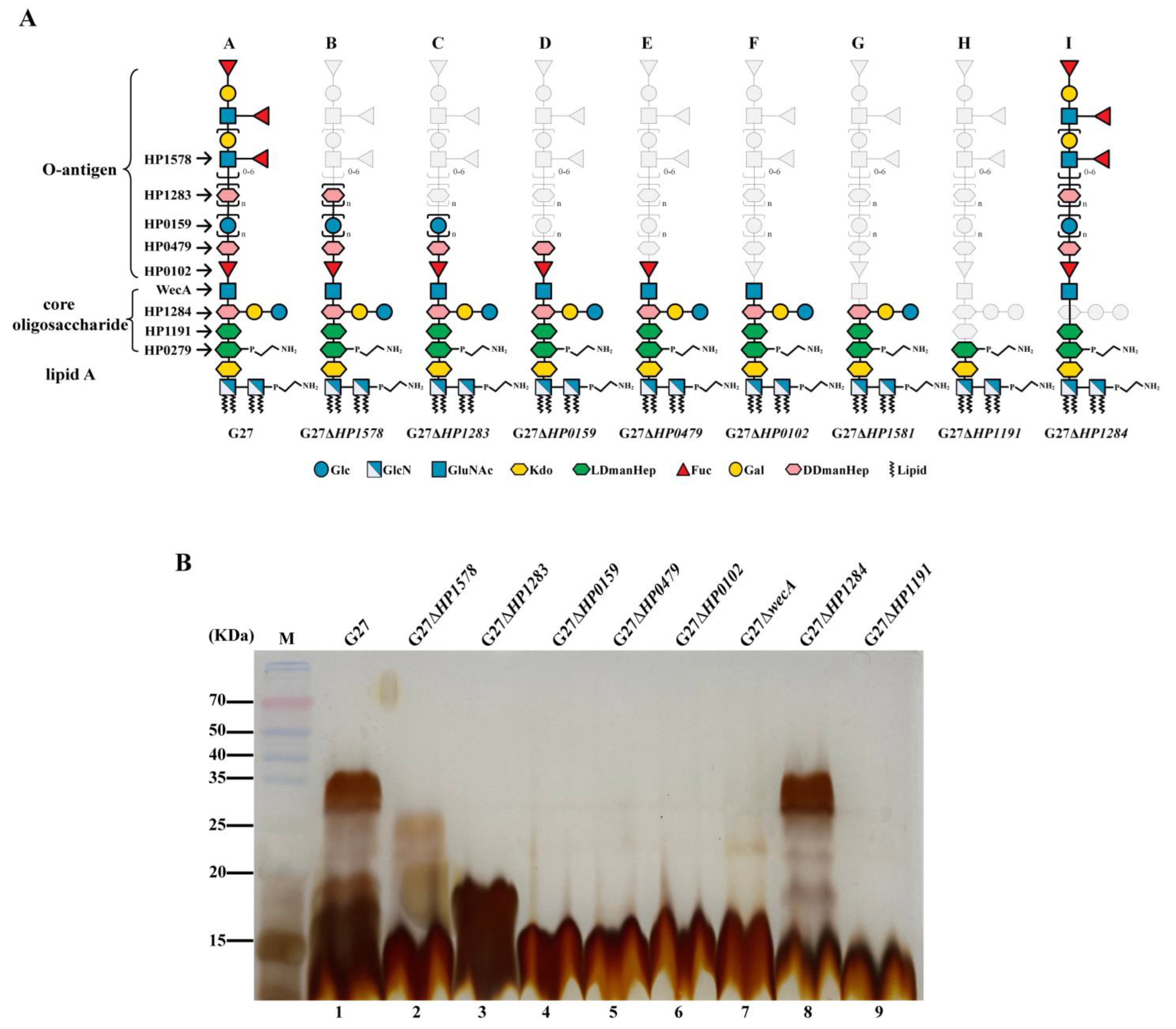
2.1. Deletion of LPS Glycosyltransferase Genes except for HP0279 Leads to Various Degree of H. pylori LPS Truncation, but Does Not Interfere with Bacterial Fitness

A panel of eight LPS glycotransferase gene mutants constructed previously in the same background strain G27 was included in this study (Table 1). Of note, numerous attempts to delete HP0279 gene coding for the Hep I transferase was unsuccessful in our laboratory, which is consistent with a previous study [19], suggesting that HP0279 is essential in H. pylori. The LPS structures of the G27 wild-type strain and the eight isogenic mutants have been previously elucidated and summarized in Figure 1A [17]. Their LPS profiles were compared in this study by silver staining on SDS-PAGE gel (Figure 1B). As expected, apparent LPS truncation was observed in seven of the eight mutants: G27ΔHP1578, lacking the distal Lewis antigen only; G27ΔHP1283, lacking DD-heptan and the Lewis antigen; G27ΔHP0159, lacking glucan, the DD-heptan, and the Lewis antigen; G27ΔHP0479, lacking Hep residue of the Trio, the glucan, the DD-heptan, and the Lewis antigen; G27ΔHP0102, lacking Fuc and Hep residues of the Trio, the glucan, the DD-heptan, and the Lewis antigen; G27ΔwecA, lacking the whole O-antigen; and G27ΔHP1191, lacking Hep II, Hep I residues and the whole O-antigen. As for G27ΔHP1284, its LPS profile was similar to that of the wild-type strain (Figure 1B, Lane 8), as it has been previously demonstrated that the deletion of the Hep III transferase gene HP1284 led to the loss of the Hep III residue and the adjoining Glc-Gal residues only, whereas the O-antigen ligation was not affected, which was attached onto Hep II residue of the incomplete core-oligosaccharide [12,17].
To investigate whether deletion of LPS glycosyltransferase genes or LPS truncation interfere with bacterial fitness, growth curves of the G27 wild-type and the eight LPS mutants were performed in liquid broth, and the optical densities were measured every 6 or 12 h for up to 48 h. Interestingly, all the eight LPS mutants displayed similar or higher growth rate than that of the wild-type strain up to 36 h, suggesting that the deletion of the eight LPS glycosyltransferase genes do not negatively impact bacterial fitness. Of note, G27ΔwecA mutant had the most robust growth among all the stains up to 36 h. From 36 h to 48 h, the optical densities of G27ΔwecA, G27ΔHP0102, G27ΔHP1284, G27ΔHP1283, and G27ΔHP0479 still increased; whereas the G27 wild-type, G27ΔHP1578, and G27ΔHP1191 strains had a marked decrease, suggesting that these three strains had reached the decline phase (Figure 2).

2.2. Deletion of LPS Glycosyltransferase Genes Affects the Spiral and Rod-like Morphology

As LPS is one of the major constituents of the outer membrane, the perturbation of LPS biosynthesis upon the deletion of corresponding glycosyltransferase genes may have a great effect on the cell envelope, thus causing morphological changes to the helical shape of H. pylori.
To investigate the roles played by LPS glycosyltransferase genes on H. pylori morphologies, the G27 wild-type and the eight LPS mutants were visualized by Gram-staining after 24 h and 48 h of culture. All the strains exhibited the characteristic helical and rod-like cell shape after 24 of culture (Figure 3A). However, compared with the wild-type strain, apparent elongated cell shape was observed in G27ΔHP0479, G27ΔwecA, and G27ΔHP1284 mutants. Interestingly, after 48 of culture, the rod-like cell shape was still clearly observed in wild-type, G27ΔHP0159, G27ΔHP0479, and G27ΔHP1191, whereas the cells in G27ΔHP1578 and G27ΔwecA were almost completely coccoid, and a mixture of coccoid, coiled “c”-shape, and irregular shapes were observed in G27ΔHP1283, G27ΔHP0102, and G27ΔHP1284 (Figure 3B).

2.3. Deletion of LPS Glycosyltransferase Genes Affects H. pylori Sensitivity to Polymyxin B

CAMPs are positively charged, which can interact with the negatively charged surface structures (primarily LPS lipid A) of most Gram-negative bacteria, inducing bacterial lysis and death [20]. Thus, CAMPs represent an important component of the host innate immune system. Due to constitutive modification of LPS, H. pylori is naturally resistant to polymyxin B [10], the surrogate of host CAMPs in laboratory settings. Modification or variation in LPS structure has been reported to affect H. pylori resistance to polymyxin B [11,21,22]. However, the roles played by LPS glycosyltransferase genes on H. pylori’s resistance to the CAMP polymyxin B has not been systematically studied.
Using the “gold standard” agar dilution method, MIC of polymyxin B was first determined in G27 wild-type and the eight isogenic LPS mutants. The wild-type G27 had a polymyxin B MIC of 18.67 ± 7.05 μg/mL, and both of the G27ΔHP1578 and G27ΔHP1283 mutants had a MIC of 13.33 ± 2.67 μg/mL, which was comparable to the MIC of the wild-type strain. The polymyxin B MICs in G27ΔHP0159, G27ΔHP479, G27ΔHP0102, HP1284, and G27ΔHP1191 were 2.00 ± 0.00, 1.50 ± 0.50, 6.67 ± 1.33, 3.25 ± 2.37, and 2.33 ± 0.88 μg/mL, respectively, which was a marked decrease in resistance to polymyxin B as compared to the wild-type (Table 2, Figure 4A,B).
Notably, G27ΔwecA had the lowest MIC of 0.96 ± 0.52 μg/mL, suggesting that the wecA gene may play an important role in H. pylori resistance to polymyxin B (Table 2, Figure 4A,B). To confirm that the marked decrease of polymyxin B resistance in the ΔwecA mutant is not strain-specific, we constructed a ΔwecA mutant in another three different H. pylori strains: 26695ΔwecA, J99ΔwecA, and P12ΔwecA. The deletion of wecA in these three strains was confirmed by silver staining showing the loss of the whole O-antigen (Figure S1). The polymyxin B MICs in wild-type 26695, J99, and P12 were 72.00 ± 30.29, 53.33 ± 10.67, and 7.67 ± 4.18, respectively. In comparison, the polymyxin B MICs in their ΔwecA mutants showed a marked 36, 63, and 25-fold decrease to 2.25 ± 0.94, 0.83 ± 0.17, and 0.31 ± 0.09, respectively (Table 2, Figure 4C–G).

2.4. Deletion of LPS Glycosyltransferase Genes Does Not Affect H. pylori Outer Membrane Permeability, but Increases Bacteria Sensitivity to Rifampicin

Given that LPS is the major component of the H. pylori outer membrane, we investigated whether the deletion of LPS glycosyltransferase genes or LPS truncation had any effect on the outer membrane permeability. We measured the influx of fluorescent probe NPN among wild-type and the LPS mutants, and no NPN fluorescence intensity difference was observed (Figure 5), suggesting that the deletion of the LPS glycosyltransferase genes does not affect H. pylori outer membrane permeability.
We also investigated whether the deletion of various LPS glycosyltransferase genes had any effect on H. pylori susceptibility to another six antibiotics. In G27 strain background, the wild-type strain and all the eight LPS mutants had similarly low MICs against clarithromycin, levofloxacin, amoxicillin, tetracycline, and metronidazole, and were all sensitive to these antibiotics. However, G27 wild-type strain was found to be resistant to rifampicin, whereas all eight LPS mutants were found to be sensitive to rifampicin. This was further confirmed in 26695, J99, and P12 strain background: The deletion of the wecA gene in 26695, J99, and P12 strains did not change their susceptibility or MICs against clarithromycin, levofloxacin, amoxicillin, tetracycline, and metronidazole; however, 26695, J99, and P12 wild-type strains were all resistant to rifampicin, whereas all their corresponding ΔwecA mutants became rifampicin-sensitive (Table 2, Figure 6).

3. Discussion

In the present study, using a series of H. pylori LPS mutants, we systematically analyzed the possible roles of LPS glycosyltransferase genes in maintenance of bacterial growth, morphology, cell wall integrity, and susceptibility to antibiotics. We observed that all eight LPS mutants had no defects in growth fitness. However, we observed that the deletion of LPS glycosyltransferase genes affected the spiral and rod-like morphology, especially after 48 h of culture: The cells in G27ΔHP1578 and G27ΔwecA were almost completely coccoid, whereas the rod-like cell shape was still clearly observed in wild-type. Notably, as the high level of strain–strain variation of H. pylori, it would be of interest to repeat the cell morphology aspects of the study in different strain backgrounds to determine whether the effects are conserved across H. pylori or unique to strain G27. Moreover, since the helical shape is essential for H. pylori motility [23], H. pylori motility assay for LPS glycosyltransferase gene mutants needs to be conducted in future studies. Previous studies have shown that multiple proteins including cell-shape-determining proteins (Csd1, Csd2, Csd5 and Csd7) and bactofilin homolog CcmA are required to generate the helical shape of H. pylori cells, influencing the shape and composition of the peptidoglycan sacculus directly or indirectly [23]. The morphology change in the LPS glycosyltransferase gene mutants could be an indirect change to the composition of the peptidoglycan sacculus. As UDP-GlcNAc is the universal substrate utilized by both the LPS pathway and the peptidoglycan pathway [23], it is possible that the perturbation of one pathway could influence the other. For example, as both HP1578 and WecA are the GlcNAc transferases, deletion of HP1578 and wecA genes blocks the use of the intracellular GlcNAc for LPS biosynthesis, which in turn may drive synthesis of peptidoglycan, offsetting the relaxation of peptidoglycan by the multiple endopeptidases. In addition, the cell morphology change upon the deletion of LPS glycosyltransferase genes in H. pylori may be explained by interference of other cytoskeletal elements. For example, it has been reported that the coiled-coil-rich proteins (Ccrp), which have a similar molecular architecture as intermediate filaments, are found to be associated with the helical shape of H. pylori [24,25]. In Caulobacter crescentus, LPS mutation was reported to interfere with the filament-like protein crescentin-mediated cell curvature, causing a disruption of normal cell morphogenesis [26]. It should be noted that the helical cell shape of H. pylori plays an important role in efficient stomach colonization [27,28]. Previous studies reported that HP1284, HP0102, and HP0159 mutants failed to colonize C57BL/6 mice [12,17,29], which could be partly ascribed to the morphological changes induced by the deletion of corresponding LPS glycosyltransferase genes.
The deletion of LPS glycosyltransferase genes including the G27ΔHP1191 mutant (with LPS truncation starting from the Hep II residue) does not affect H. pylori outer membrane permeability. This is in sharp contrast to the markedly increased permeability phenotype observed in the deep rough LPS mutants of other Gram-negative bacteria [30,31], which could be explained by the appearance of phospholipid bilayer patches in the outer membrane due to the decreased incorporation of outer membrane proteins, or the perturbation of the lateral interactions between the neighboring LPS molecules in the presence of defective LPS molecules. The crucial structure for the LPS-protein or lateral LPS–LPS interactions in these Gram-negative bacteria has been proposed to lie in the presence of the negative phosphate groups on the core Hep II residue [32]. For example, in E. coli and P. aeruginosa, the core Hep residues are decorated with negatively charged phosphate groups, which are believed to play an important role in interacting with basic groups of the outer membrane proteins, and in enabling the neighboring LPS molecules to be cross-linked by divalent cations, and thus stabilizing the outer membrane [33,34,35]. However, in H. pylori, the LPS molecules are constitutively modified to have a core oligosaccharide without the presence of negatively charged phosphate groups on Hep II [8]. Thus, the deletion of Hep II glycosyltransferase gene HP1191 does not change the electrostatic interactions between the H. pylori LPS and the outer membrane proteins, or the electrostatic repulsion between the neighboring LPS molecules, which may explain the unaltered outer membrane permeability in H. pylori deep rough LPS mutant (G27ΔHP1191). Of note, Hep I glycosyltransferase gene HP0279 deletion mutant in H. pylori has never been successfully constructed or identified [17], which may suggest that HP0279 is essential, and that the minimal LPS structure required for viability and outer membrane permeability of H. pylori consists of the lipid A, KDO, and the Hep I residue.
H. pylori is naturally resistant to the positively charged polymyxin B, an experimental substitute for CAMPs in laboratory settings [8]. It is well documented that the removal or “masking” of lipid A by phosphate groups is the primary mechanism involved in resistance to CAMPs [36]. The constitutive removal of the lipid A 1’-phosphate group and 4’-phosphate group by LpxE and LpxF, respectively, reduces the negative charge of lipid A, thus rendering H. pylori naturally resistant to polymyxin B [10]. In the present study, we observed that the polymyxin B MICs in the G27ΔHP1578 (lacking Lewis antigen) and G27ΔHP1283 (lacking heptan and Lewis antigen) were comparable to that of the G27 wild-type strain. This is in line with previous reports that the variable heptan and Lewis antigen structures are not essential for establishing colonization in mouse models [8]. Interestingly, we observed that deleting any of the conserved LPS glycosyltransferase genes including HP0159, HP0479, HP0102, wecA, HP1284, and HP1191 led to a marked increase in susceptibility to polymyxin B. Of note, irrespective of parent strain background, deletion of wecA displayed a dramatic decrease in MIC to polymyxin B compared to wild-type. As the outer membrane permeability was unaltered among all the LPS mutants (especially the ΔwecA mutant), the increased susceptibility to polymyxin B might be attributed to an increased net negative charge of the mutants’ lipid A due to inefficient modification. In H. pylori, the lipid A-core is assembled in the cytoplasm, and translocated by MsbA to the periplasmic face, where it is constitutively modified by a five-step enzymatic pathway [8]. It is possible that the truncated lipid A-core in the ΔwecA mutant may not serve as a good substrate for LpxE and LpxF, leading to an inefficient removal of the negatively charged phosphate groups, and resulting into an increased sensitivity to polymyxin B.
In the present study, we showed that none of the eight LPS glycosyltransferase gene mutants had increased susceptibility to the commonly used five anti-H. pylori antibiotics (clarithromycin, levofloxacin, amoxicillin, tetracycline, and metronidazole) as compared to the wild-type strain. This was understandable as the outer membrane permeability was not affected among all the LPS mutants. However, we observed that deleting any of the LPS glycosyltransferase genes rendered H. pylori sensitive to rifampicin. Of note, rifampicin is an effective antibiotic against Gram-positive bacteria, but less effective against Gram-negative bacteria due to the impermeable LPS outer membrane [37]. In this study, we showed that G27, 26695, J99, and P12 wild-type strains were all resistant to rifampicin, whereas deletion of wecA rendered them sensitive to polymyxin B. As all the LPS mutants had unaltered outer membrane permeability, and in combination with the fact that rifampicin carries one positive charge favoring interaction with LPS [38], the increased susceptibility to rifampicin due to the deletion of LPS glycosyltransferase genes might also be attributed to an increased net negative charge of the mutants’ outer membrane.
In conclusion, we have shown that LPS glycosyltransferase genes played an important role in the maintenance of H. pylori’s morphology, and the deletion of these genes resulted in significant morphological changes (coccoid, coiled “c”-shape, and irregular shapes) after 48 h growth as compared to the rod-like cell shape of the wild-type strain. Moreover, we showed that deletion of conserved LPS glycosyltransferase genes does not affect H. pylori outer membrane permeability, but increases bacteria sensitivity to polymyxin B and rifampicin. Of note, irrespective of parent strain background, deletion of wecA displayed a dramatic increase in susceptibility to polymyxin B. Future genetic complementation studies will be performed and the MICs in different complemented strains will be determined. Interfering LPS biosynthesis through the deletion or inhibition of the key LPS glycosyltransferase genes (such as the wecA) does not kill the bacteria but rather prevents the bacteria from forming a typical helical shape and leads to the loss of natural resistance to polymyxin B, which may disable the bacteria establishing successful colonization. Thus, conserved H. pylori LPS glycosyltransferases could be promising targets for developing novel drugs aiming at “disarming” H. pylori.

4. Materials and Methods

4.1. Bacterial Strains, Plasmids, Growth Conditions, and Oligonucleotide Primers

Bacterial strains and plasmids used in this study are summarized in Table 1. The following H. pylori strains were used: G27 wild-type strains and the associated eight LPS truncated strains, 26695, J99, P12 wild-type strains and their ΔwecA mutant strains. H. pylori strains were cultured in Columbia blood agar (CBA) plates supplemented with 5% defibrinated sheep blood and 5% fetal calf serum (FCS) and incubated at 37 °C under microaerobic condition (85% N2: 5% H2: 10% CO2) generated by the Anoxomat Mark-II system (Mart Microbiology B.V., Drachten, The Netherlands). DNA oligonucleotide primers for the identification of wecA deletion in 26695, J99, and P12 are named as WecA-F with sequence (5′- > 3′) of CACGCTATGACCGATATTAAGC and WecA-R with sequence (5′- > 3′) of GCTGTTCTGTTTGAGACAAG.

4.2. Construction of wecA Gene Mutant in H. pylori 26695, J99, and P12

For constructing wecA mutant in H. pylori 26695 (streptomycin resistant), we used a highly efficient Xer-cise gene deletion method [39]. Briefly, 26695 was naturally transformed with previously constructed plasmid pWecA-AB-difH-RC in our lab [17] and cultured in BHI blood agar plates (brain heart infusion, 5% defibrinated sheep blood and 5% FCS) supplemented with 10 μg/mL chloramphenicol. Single colonies were sub-cultured in BHI blood agar plates with 10 μg/mL streptomycin to generate clean deletion of wecA in 26695. Similar to the construction of wecA in 26695, plasmid pWecA-AB-difH-RC was used to transform strain J99 and P12 (both streptomycin sensitive) to generate replacement knockout of wecA with difH-rpsl-cat-difH cassette.

4.3. H. pylori LPS Microextraction for Silver Staining

H. pylori LPS was micro-extracted as previously performed [12]. Briefly, bacterial cells with an amount of OD600 = 3 were harvested from CBA plates and suspended in LPS lysis buffer that composed of 2% SDS, 4% β-mercaptoethanol, 0.1% bromophenol blue, 10% glycerol, 1 M Tris-HCl (pH 6.8). Samples were heated at 100 °C for 10 min and then cooled. Thereafter, the samples were added with 5 μL proteinase K (20 mg/mL) and incubated in 55 °C of water overnight. The obtained LPS samples were run in 15% SDS-PAGE gels. After the gels underwent staining through three steps—oxidation, silver dyeing and color development—the LPSs were visualized thoroughly for structural analysis.

4.4. Morphology Analysis of H. pylori Strains by Gram-Staining

One drop of saline was added on a clean glass slide. Bacteria colonies grown on Columbia blood agar plates supplemented with 5% defibrinated sheep blood and 5% fetal calf serum (FCS) and incubated at 37 °C under microaerobic conditions (85% N2: 5% H2: 10% CO2) for 24 h or 48 h were transferred with a plastic loop onto the saline drop and spread evenly. The bacterial solution was air dried and stained with Gram’s crystal violet for 1 min, and then rinsed with tap water. Subsequently, the bacteria were stained with iodine solution for 1–3 min and rinsed with tap water. The excess water was gently removed with a tissue paper. Thereafter, the bacteria were counter-stained with Carbol Fuchsin for 1 min and rinsed with tap water, and the excess water was gently removed with a tissue paper. Finally, the stained bacteria were mounted with coverslip by paraffin and visualized using an Olympus microscope (Olympus Corporation, Tokyo, Japan). Digital images were taken using a camera connected with the microscope via a microscope adapter. Microscopy field with uniform bacteria was randomly selected to take images for each strain. Three independent replicate experiments were conducted.

4.5. Growth Analysis of H. pylori Strains

The growth properties of the H. pylori LPS mutants was determined in brucella broth supplemented with 10% FCS (BB10). Individual 5 mL cultures of BB10 were inoculated with G27 wild-type or LPS truncated strains to give a starting concentration of bacterial solution as OD600 = 0.1. Cultures were incubated at 37 °C with 100 rpm shaking under microaerobic condition. The optical densities of the cultures were measured every 6 h and/or 12 h for up to 48 h.

4.6. Outer Membrane Permeability Assay

Fluorescent probe N-Phenyl-1-naphthylamine (NPN) (Sigma-Aldrich, St. Louis, MO, USA) was used to examine the outer membrane integrity of H. pylori wild-type and its LPS mutant strains, as described previously [40]. Briefly, H. pylori strains were harvested from CBA plates in logarithmic growth stage, and then washed for three times in HEPES (pH 7.0, 5 mM). The bacterial pellets were suspended with HEPES and OD600 value of obtained bacterial suspension was adjusted to 0.4. Afterwards, the dye NPN was added to the bacterial suspension, achieving a final concentration of 10 μM. The NPN-bacteria mixture was incubated at 37 °C for 30 min, away from light. Finally, the mixture was transferred to a 96-well plate with black walls at 200 μL per well, following fluorescence measurement on a Varioskan Flash-Full Wavelength Microplate Reader (Thermo Scientific, Waltham, MA, USA) with the excitation wavelength at 350 nm and an emission wavelength of 420 nm.

4.7. Antimicrobial Susceptibility Testing

The minimum inhibitory concentrations (MICs) of polymyxin B and six clinically used antibiotics including clarithromycin, metronidazole, levofloxacin, amoxicillin, tetracycline, and rifampicin to H. pylori wild-type and LPS mutant strains were determined by the standard agar dilution method. H. pylori strain NCTC 11637 was included as quality control. A range of antibiotic concentrations was prepared by twofold serial dilutions of antimicrobial (ranging from 0.125 to 128 μg/mL) incorporated into BHI blood agar plates. The inoculum was delivered onto the surface of the agar plates with antibiotics by a Multipoint Inoculator (Denley, UK) to obtain approximately 1 × 10⁵ CFU per spot. After static incubation for 3–5 days at 37 °C under microaerobic conditions, the MIC was defined as the lowest concentration of drug inhibiting visible H. pylori growth. According to the recommendation of European Committee on Antimicrobial Susceptibility Testing (EUCAST), version 10.0, 2020, resistance to clarithromycin, metronidazole, levofloxacin, amoxicillin, tetracycline, and rifampicin was defined as MIC > 0.5 mg/L, >8 mg/L, >1 mg/L, >0.125 mg/L, >1 mg/L and >4 mg/L, respectively [41].

4.8. Statistical Analysis

The software Graphpad Prism version 9.1.2 (SPSS Inc., Chicago, IL, USA) was used for the statistical analyses in this study. The significance of different values from different H. pylori strains was determined by Student’s t-test, with a p value of less than 0.05.

Supplementary Materials

The following supporting information can be downloaded at: https://www.mdpi.com/article/10.3390/ijms241411381/s1.

Author Contributions

Conceptualization, M.B., H.T. and H.L.; Formal analysis, X.T., T.Y., Y.S., X.S., H.T. and H.L.; Funding acquisition, B.J.M., H.T. and H.L.; Investigation, X.T., T.Y., M.B. and H.L.; Methodology, X.T., T.Y., Y.S. and X.S.; Project administration, B.J.M., H.T. and H.L.; Supervision, M.B., B.J.M. and H.T.; Writing—original draft, X.T.; Writing—review & editing, H.T. and H.L. All authors have read and agreed to the published version of the manuscript.

Funding

This study was supported by the National Natural Science Foundation of China (grant 82072248), the International Cooperation Excellence Initiative Grant, West China Hospital, Sichuan University (grant 139220012), and the Fund for the transformation of science and technological achievements, West China Hospital, Sichuan University (grant CGZH19005).

Institutional Review Board Statement

Not applicable.

Informed Consent Statement

Not applicable.

Data Availability Statement

The data that support the findings of this study are available in the relevant figures/tables of this article.

Conflicts of Interest

The authors declare no conflict of interest.

References

  1. Ren, S.; Cai, P.; Liu, Y.; Wang, T.; Zhang, Y.; Li, Q.; Gu, Y.; Wei, L.; Yan, C.; Jin, G. Prevalence of Helicobacter pylori infection in China: A systematic review and meta-analysis. J. Gastroenterol. Hepatol. 2021, 37, 464–470. [Google Scholar] [CrossRef] [PubMed]
  2. Backert, S.; Neddermann, M.; Maubach, G.; Naumann, M. Pathogenesis of Helicobacter pylori infection. Helicobacter 2016, 21 (Suppl. S1), 19–25. [Google Scholar] [CrossRef] [PubMed]
  3. de Martel, C.; Georges, D.; Bray, F.; Ferlay, J.; Clifford, G.M. Global burden of cancer attributable to infections in 2018: A worldwide incidence analysis. Lancet Glob. Health 2020, 8, e180–e190. [Google Scholar] [CrossRef] [Green Version]
  4. Chey, W.D.; Leontiadis, G.I.; Howden, C.W.; Moss, S.F. ACG Clinical Guideline: Treatment of Helicobacter pylori Infection. Am. J. Gastroenterol. 2017, 112, 212–239, Erratum in Am. J. Gastroenterol. 2018, 113, 1102. [Google Scholar] [CrossRef]
  5. Malfertheiner, P.; Megraud, F.; Rokkas, T.; Gisbert, J.P.; Liou, J.-M.; Schulz, C.; Gasbarrini, A.; Hunt, R.H.; Leja, M.; O’Morain, C.; et al. Management of Helicobacter pylori infection: The Maastricht VI/Florence consensus report. Gut 2022, 71, 1724–1762. [Google Scholar] [CrossRef] [PubMed]
  6. El-Serag, H.B.; Kao, J.Y.; Kanwal, F.; Gilger, M.; LoVecchio, F.; Moss, S.F.; Crowe, S.; Elfant, A.; Haas, T.; Hapke, R.J.; et al. Houston Consensus Conference on Testing for Helicobacter pylori Infection in the United States. Clin. Gastroenterol. Hepatol. 2018, 16, 992–1002.e6. [Google Scholar] [CrossRef] [Green Version]
  7. Silva, L.M.; Correia, V.G.; Moreira, A.S.; Domingues, M.R.M.; Ferreira, R.M.; Figueiredo, C.; Azevedo, N.F.; Marcos-Pinto, R.; Carneiro, F.; Magalhães, A.; et al. Helicobacter pylori lipopolysaccharide structural domains and their recognition by immune proteins revealed with carbohydrate microarrays. Carbohydr. Polym. 2021, 253, 117350. [Google Scholar] [CrossRef]
  8. Li, H.; Liao, T.; Debowski, A.W.; Tang, H.; Nilsson, H.O.; Stubbs, K.A.; Marshall, B.J.; Benghezal, M. Lipopolysaccharide Structure and Biosynthesis in Helicobacter pylori. Helicobacter 2016, 21, 445–461. [Google Scholar] [CrossRef] [Green Version]
  9. Simpson, B.W.; Trent, M.S. Pushing the envelope: LPS modifications and their consequences. Nat. Rev. Genet. 2019, 17, 403–416. [Google Scholar] [CrossRef]
  10. Cullen, T.W.; Giles, D.K.; Wolf, L.N.; Ecobichon, C.; Boneca, I.G.; Trent, M.S. Helicobacter pylori versus the Host: Remodeling of the Bacterial Outer Membrane Is Required for Survival in the Gastric Mucosa. PLoS Pathog. 2011, 7, e1002454. [Google Scholar] [CrossRef] [Green Version]
  11. Sijmons, D.; Guy, A.J.; Walduck, A.K.; Ramsland, P.A. Helicobacter pylori and the Role of Lipopolysaccharide Variation in Innate Immune Evasion. Front. Immunol. 2022, 13, 868225. [Google Scholar] [CrossRef] [PubMed]
  12. Li, H.; Yang, T.; Liao, T.; Debowski, A.W.; Nilsson, H.-O.; Fulurija, A.; Haslam, S.M.; Mulloy, B.; Dell, A.; Stubbs, K.A.; et al. The redefinition of Helicobacter pylori lipopolysaccharide O-antigen and core-oligosaccharide domains. PLoS Pathog. 2017, 13, e1006280. [Google Scholar] [CrossRef] [PubMed] [Green Version]
  13. Hiratsuka, K.; Logan, S.M.; Conlan, J.W.; Chandan, V.; Aubry, A.; Smirnova, N.; Ulrichsen, H.; Chan, K.H.; Griffith, D.W.; Harrison, B.A.; et al. Identification of a D-glycero-D-manno-heptosyltransferase gene from Helicobacter pylori. J. Bacteriol. 2005, 187, 5156–5165. [Google Scholar] [CrossRef] [PubMed] [Green Version]
  14. Chandan, V.; Logan, S.M.; Harrison, B.A.; Vinogradov, E.; Aubry, A.; Stupak, J.; Li, J.; Altman, E. Characterization of a waaF mutant of Helicobacter pylori strain 26695 provides evidence that an extended lipopolysaccharide structure has a limited role in the invasion of gastric cancer cells. Biochem. Cell Biol. 2007, 85, 582–590. [Google Scholar] [CrossRef]
  15. Altman, E.; Chandan, V.; Li, J.; Vinogradov, E. Lipopolysaccharide structures of Helicobacter pylori wild-type strain 26695 and 26695 HP0826: Kan mutant devoid of the O-chain polysaccharide component. Carbohydr. Res. 2011, 346, 2437–2444. [Google Scholar] [CrossRef]
  16. Altman, E.; Chandan, V.; Li, J.; Vinogradov, E. Lipopolysaccharide structure of Helicobacter pylori serogroup O:3. Carbohydr. Res. 2013, 378, 139–143. [Google Scholar] [CrossRef]
  17. Li, H.; Marceau, M.; Yang, T.; Liao, T.; Tang, X.; Hu, R.; Xie, Y.; Tang, H.; Tay, A.; Shi, Y.; et al. East-Asian Helicobacter pylori strains synthesize heptan-deficient lipopolysaccharide. PLoS Genet. 2019, 15, e1008497. [Google Scholar] [CrossRef] [Green Version]
  18. Moran, A.P.; Shiberu, B.; Ferris, J.A.; Knirel, Y.A.; Senchenkova, S.N.; Perepelov, A.V.; Jansson, P.E.; Goldberg, J.B. Role of Helicobacter pylori rfaJ genes (HP0159 and HP1416) in lipopolysaccharide synthesis. FEMS Microbiol. Lett. 2004, 241, 57–65. [Google Scholar] [CrossRef] [Green Version]
  19. Mobley, H.L.T.; Mendz, G.L.; Hazell, S.L. (Eds.) Helicobacter Pylori: Physiology and Genetics; ASM Press: Washington, DC, USA, 2001. [Google Scholar]
  20. Brown, K.L.; Hancock, R.E. Cationic host defense (antimicrobial) peptides. Curr. Opin. Immunol. 2006, 18, 24–30. [Google Scholar] [CrossRef]
  21. Pernitzsch, S.R.; Alzheimer, M.; Bremer, B.U.; Robbe-Saule, M.; De Reuse, H.; Sharma, C.M. Small RNA mediated gradual control of lipopolysaccharide biosynthesis affects antibiotic resistance in Helicobacter pylori. Nat. Commun. 2021, 12, 4433. [Google Scholar] [CrossRef]
  22. Matsuura, M. Structural Modifications of Bacterial Lipopolysaccharide that Facilitate Gram-Negative Bacteria Evasion of Host Innate Immunity. Front. Immunol. 2013, 4, 109. [Google Scholar] [CrossRef] [PubMed] [Green Version]
  23. Holtrup, S.; Greger, M.; Mayer, B.; Specht, M.; Waidner, B. Insights Into the Helical Shape Complex of Helicobacter pylori. Front. Microbiol. 2022, 13, 929194. [Google Scholar] [CrossRef] [PubMed]
  24. Waidner, B.; Specht, M.; Dempwolff, F.; Haeberer, K.; Schaetzle, S.; Speth, V.; Kist, M.; Graumann, P.L. A Novel System of Cytoskeletal Elements in the Human Pathogen Helicobacter pylori. PLoS Pathog. 2009, 5, e1000669. [Google Scholar] [CrossRef]
  25. Specht, M.; Schätzle, S.; Graumann, P.L.; Waidner, B. Helicobacter pyloriPossesses Four Coiled-Coil-Rich Proteins That Form Extended Filamentous Structures and Control Cell Shape and Motility. J. Bacteriol. 2011, 193, 4523–4530. [Google Scholar] [CrossRef] [PubMed] [Green Version]
  26. Cabeen, M.T.; Murolo, M.A.; Briegel, A.; Bui, N.K.; Vollmer, W.; Ausmees, N.; Jensen, G.J.; Jacobs-Wagner, C. Mutations in the Lipopolysaccharide Biosynthesis Pathway Interfere with Crescentin-Mediated Cell Curvature in Caulobacter crescentus. J. Bacteriol. 2010, 192, 3368–3378. [Google Scholar] [CrossRef] [PubMed] [Green Version]
  27. Sycuro, L.K.; Pincus, Z.; Gutierrez, K.D.; Biboy, J.; Stern, C.A.; Vollmer, W.; Salama, N.R. Peptidoglycan crosslinking relaxation promotes Helicobacter pylori’s helical shape and stomach colonization. Cell 2010, 141, 822–833. [Google Scholar] [CrossRef] [Green Version]
  28. Salama, N.R. Cell morphology as a virulence determinant: Lessons from Helicobacter pylori. Curr. Opin. Microbiol. 2020, 54, 11–17. [Google Scholar] [CrossRef]
  29. Altman, E.; Chandan, V.; Larocque, S.; Aubry, A.; Logan, S.M.; Vinogradov, E.; Li, J. Effect of the HP0159 ORF mutation on the lipopolysaccharide structure and colonizing ability of Helicobacter pylori. FEMS Immunol. Med Microbiol. 2008, 53, 204–213. [Google Scholar] [CrossRef] [Green Version]
  30. Raetz, C.R.; Whitfield, C. Lipopolysaccharide endotoxins. Annu. Rev. Biochem. 2002, 71, 635–700. [Google Scholar] [CrossRef] [PubMed] [Green Version]
  31. Meng, J.; Xu, J.; Huang, C.; Chen, J. Rcs Phosphorelay Responses to Truncated Lipopolysaccharide-Induced Cell Envelope Stress in Yersinia enterocolitica. Molecules 2020, 25, 5718. [Google Scholar] [CrossRef] [PubMed]
  32. Nikaido, H. Molecular basis of bacterial outer membrane permeability revisited. Microbiol. Mol. Biol. Rev. 2003, 67, 593–656. [Google Scholar] [CrossRef] [PubMed] [Green Version]
  33. Yethon, J.A.; Vinogradov, E.; Perry, M.B.; Whitfield, C. Mutation of the Lipopolysaccharide Core Glycosyltransferase Encoded by waaG Destabilizes the Outer Membrane of Escherichia coli by Interfering with Core Phosphorylation. J. Bacteriol. 2000, 182, 5620–5623. [Google Scholar] [CrossRef] [Green Version]
  34. Fernández, L.; Álvarez-Ortega, C.; Wiegand, I.; Olivares, J.; Kocíncová, D.; Lam, J.S.; Martínez, J.L.; Hancock, R.E.W. Characterization of the Polymyxin B Resistome of Pseudomonas aeruginosa. Antimicrob. Agents Chemother. 2013, 57, 110–119. [Google Scholar] [CrossRef] [Green Version]
  35. Lugtenberg, B.; Van, A.l.p.h.e.n.L. Molecular architecture and functioning of the outer membrane of Escherichia coli and other gram-negative bacteria. Biochim. Biophys. Acta 1983, 737, 51–115. [Google Scholar] [CrossRef] [PubMed]
  36. Trent, M.S.; Stead, C.M.; Tran, A.X.; Hankins, J.V. Diversity of endotoxin and its impact on pathogenesis. J. Endotoxin Res. 2006, 12, 205–223. [Google Scholar] [CrossRef]
  37. Chaudhary, N.; Aggarwal, B.; Saini, V.; Yavvari, P.S.; Sharma, P.; Srivastava, A.; Bajaj, A. Polyaspartate-derived synthetic antimicrobial polymer enhances the activity of rifampicin against multidrug-resistant Pseudomonas aeruginosa infections. Biomater. Sci. 2022, 10, 5158–5171. [Google Scholar] [CrossRef] [PubMed]
  38. Cetuk, H.; Anishkin, A.; Scott, A.J.; Rempe, S.B.; Ernst, R.K.; Sukharev, S. Partitioning of Seven Different Classes of Antibiotics into LPS Monolayers Supports Three Different Permeation Mechanisms through the Outer Bacterial Membrane. Langmuir ACS J. Surf. Colloids 2021, 37, 1372–1385. [Google Scholar] [CrossRef]
  39. Debowski, A.W.; Gauntlett, J.C.; Li, H.; Liao, T.; Sehnal, M.; Nilsson, H.-O.; Marshall, B.J.; Benghezal, M. Xer-cise inHelicobacter pylori: One-step Transformation for the Construction of Markerless Gene Deletions. Helicobacter 2012, 17, 435–443. [Google Scholar] [CrossRef]
  40. Luo, G.; Zhang, J.; Wang, H.; Sun, Y.; Cheng, B.; Xu, Z.; Zhang, Y.; Li, H.; Lu, W.; Nemeth, E.; et al. Human defensin-inspired discovery of peptidomimetic antibiotics. Proc. Natl. Acad. Sci. USA 2022, 119, e2117283119. [Google Scholar] [CrossRef]
  41. EUCAST Breakpoint Tables for Interpretation of MICs and Zone Diameters, Version 10.0. 2020. Available online: https://www.eucast.org/ (accessed on 1 March 2020).
Figure 1. LPS structural characterization of H. pylori LPS mutants. (A) Chemical structure model of wild-type G27 and its associated LPS mutants. The gene was named according to standard H. pylori strain 26695. The gray represents the truncated portion of LPS. Gal, galactose; Glc, glucose; Fuc, fucose; GlcNAc, nitroacetyl glucosamine; DDmanHep, D-glycerol-D-mannoheptanose; LDmanHep, L-glycerol-D-mannoheptanose; GlcN, glucosamine; Kdo, 3-deoxy-D-mannose-octanoic acid. (B) LPS structural characterization of wild-type G27 and its associated LPS mutants detected by silver staining. M, molecular weight marker.
Ijms 24 11381 g001
Figure 2. Deletion of H. pylori LPS glycosyltransferase genes does not interfere with bacterial fitness. Growth curve analysis of wild-type G27 and LPS mutants through measuring the optical density at 600 nm (OD600) wavelength of the cultures every 6 h and/or 12 h for up to 48 h. Each strain was grown in triplicate, and data from two independent experiments are represented as mean with standard error of the mean (±SEM).
Ijms 24 11381 g002
Figure 3. H. pylori cell morphology is perturbed by deletion of LPS glycosyltransferase genes. Gram-staining depicting morphological patterns of H. pylori G27 wild-type and LPS mutants after 24 h (A) and 48 h (B) growth. Images were taken using light microscope (1000×).
Ijms 24 11381 g003
Figure 4. Deletion of LPS glycosyltransferase genes leads to reduction of H. pylori resistance to polymyxin B. Resistance patterns of H. pylori G27 (A), 26695 (C), J99 (D), P12 (E) and their associated LPS mutants to polymyxin B were profiled by agar dilution assay. The strains were grown in brucella broth infusion (BHI) blood agar plates supplemented with various concentrations of polymyxin B. The minimum inhibitory concentration (MIC), outlined in red, was defined as the lowest concentration of antibiotics at which bacteria grew was obviously inhibited. (B) MICs of wild-type G27 and its LPS mutants to polymyxin B obtained by three independent experiments. (F,G) MICs of wild-type 26695, J99 and P12 and their ΔwecA mutants to polymyxin B obtained by three independent experiments. All values were shown as mean with standard error of the mean (± SEM). *, p < 0.05, **, p < 0.01; Student’s t-test, two-tailed.
Ijms 24 11381 g004
Figure 5. Deletion of LPS glycosyltransferase genes has no effect on the outer membrane permeability of H. pylori. Outer membrane permeability of H. pylori was measured by the influx of fluorescence probe N-Phenyl-1-naphthylamine. Fluorescent intensity is positively correlated with the permeability of the outer membrane. Data from three independent experiments are presented as mean ± standard errors of the mean (SEM). ns, not significant. Student’s t-test, two-tailed.
Ijms 24 11381 g005
Figure 6. Deletion of LPS glycosyltransferase genes increases H. pylori susceptibility to rifampicin but not to other clinically used antibiotics. Minimum inhibitory concentrations (MICs) of wild-type H. pylori strains and their associated LPS mutants to rifampicin (A), clarithromycin (B), levofloxacin (C), amoxicillin (D), tetracycline (E), and metronidazole (F) tested by agar dilution assay. All values were indicated as mean with standard errors of the mean (±SEM) from three independent experiments. *, p < 0.05. ns, not significant. Student’s t-test, two-tailed.
Ijms 24 11381 g006
Table 1. Bacterial strains and plasmids used in this study.
Strain or Plasmid NameDescriptionLPS BiosynthesisReference or Source
H. pylori Strains
G27Wild-type strain, streptomycin resistantFull-length LPS[17]
G27ΔHP1578Clean deletion of HPG27_1515 in G27Deletion of Lewis antigen[17]
G27ΔHP1283Clean deletion of HPG27_1235 in G27Deletion of Lewis antigen and heptan[17]
G27ΔHP0159Clean deletion of HPG27_146 in G27Deletion of Lewis antigen, DD-heptan, and glucan[17]
G27ΔHP0479Clean deletion of HPG27_437 in G27Deletion of Lewis antigen, DD-heptan, glucan, and LD-heptose of Trio[17]
G27ΔHP0102Clean deletion of HPG27_94 in G27Deletion of Lewis antigen, DD-heptan, glucan, Trio LD-heptose and fucose[17]
G27ΔwecAClean deletion of HPG27_1518 in G27Deletion of the entire O-antigen[17]
G27ΔHP1284Clean deletion of HPG27_1236 in G27Deletion of side-chain and heptose III in the core oligosaccharide[17]
G27ΔHP1191Clean deletion of HPG27_1136 in G27Deletion of the entire O-antigen, and side-chain, heptose III and heptose II in the core oligosaccharide[17]
26695Wild-type strain, streptomycin resistantFull-length LPSStored in our lab
26695ΔwecAClean deletion of HP1581 in 26695Deletion of the entire O-antigenThis work
J99Wild-type strainFull-length LPSStored in our lab
J99ΔwecADeletion of wecA in J99 by replacing it with difH-rpsl-cat-difHDeletion of the entire O-antigenThis work
P12Wild-type strainFull-length LPSStored in our lab
P12ΔwecADeletion of wecA in P12 by replacing it with difH-rpsl-cat-difHDeletion of the entire O-antigenThis work
Plasmids
pWecA-AB-difH-RCpGEM®-T Easy vector containing sequences flanking HPG27_1518, with difH-rpsL-cat-difH inserted between the flanking sequences [17]
Table 2. Minimum inhibitory concentrations (MICs) of H. pylori wild-type and LPS glycosyltransferase genes mutant strains against antibiotics.
StrainsPolymyxin B (μg/mL)Rifampicin (μg/mL)Clarithromycin (μg/mL)Levofloxacin (μg/mL)Amoxicillin (μg/mL)Tetracycline (μg/mL)Metronidazole (μg/mL)
G2718.67 ± 7.056.67 ± 1.330.03 ± 0.010.23 ± 0.020.04 ± 0.010.27 ± 0.053.30 ± 0.33
G27ΔHP157813.33 ± 2.671.83 ± 0.160.03 ± 0.010.18 ± 0.040.04 ± 0.010.27 ± 0.053.30 ± 0.33
G27ΔHP128313.33 ± 2.671.42 ± 0.360.03 ± 0.010.21 ± 0.020.03 ± 0.010.27 ± 0.053.30 ± 0.33
G27ΔHP01592.00 ± 0.002.50 ± 0.760.02 ± 0.010.19 ± 0.040.04 ± 0.010.23 ± 0.023.00 ± 0.57
G27ΔHP04791.50 ± 0.500.83 ± 0.080.02 ± 0.010.17 ± 0.020.03 ± 0.000.27 ± 0.053.00 ± 0.57
G27ΔHP01026.67 ± 1.330.66 ± 0.080.03 ± 0.010.17 ± 0.020.04 ± 0.010.27 ± 0.053.33 ± 0.33
G27ΔwecA0.96 ± 0.520.42 ± 0.040.02 ± 0.010.21 ± 0.020.03 ± 0.000.27 ± 0.053.33 ± 0.66
G27ΔHP12843.25 ± 2.370.58 ± 0.080.02 ± 0.010.19 ± 0.040.04 ± 0.010.27 ± 0.053.17 ± 0.83
G27ΔHP11912.33 ± 0.880.58 ± 0.080.03 ± 0.010.17 ± 0.020.04 ± 0.010.27 ± 0.052.50 ± 0.76
2669572.00 ± 30.295.33 ± 1.330.08 ± 0.020.33 ± 0.080.08 ± 0.020.21 ± 0.0485.3 ± 21.3
26695ΔwecA2.25 ± 0.941.08 ± 0.510.08 ± 0.020.31 ± 0.090.08 ± 0.020.21 ± 0.0485.3 ± 21.3
J9953.33 ± 10.675.33 ± 1.330.02 ± 0.010.09 ± 0.030.03 ± 0.010.10 ± 0.040.67 ± 0.17
J99ΔwecA0.83 ± 0.172.33 ± 0.880.02 ± 0.010.09 ± 0.020.03 ± 0.010.09 ± 0.020.63 ± 0.19
P127.67 ± 4.186.00 ± 1.150.03 ± 0.010.23 ± 0.020.02 ± 0.000.21 ± 0.020.83 ± 0.17
P12ΔwecA0.31 ± 0.092.16 ± 0.930.02 ± 0.010.21 ± 0.040.02 ± 0.000.23 ± 0.020.83 ± 0.17
Disclaimer/Publisher’s Note: The statements, opinions and data contained in all publications are solely those of the individual author(s) and contributor(s) and not of MDPI and/or the editor(s). MDPI and/or the editor(s) disclaim responsibility for any injury to people or property resulting from any ideas, methods, instructions or products referred to in the content.

