Article

Aflatoxin B1 Increases Soluble Epoxide Hydrolase in the Brain and Induces Neuroinflammation and Dopaminergic Neurotoxicity

1
Department of Entomology and Nematology, and UC Davis Comprehensive Cancer Center, University of California, Davis, CA 95616, USA
2
Department of Pharmacology, University of California Davis, Davis, CA 95616, USA
*
Author to whom correspondence should be addressed.
Int. J. Mol. Sci. 2023, 24(12), 9938; https://doi.org/10.3390/ijms24129938
Submission received: 25 May 2023 / Revised: 6 June 2023 / Accepted: 7 June 2023 / Published: 9 June 2023

Abstract

:
Parkinson’s disease (PD) is an increasingly common neurodegenerative movement disorder with contributing factors that are still largely unexplored and currently no effective intervention strategy. Epidemiological and pre-clinical studies support the close association between environmental toxicant exposure and PD incidence. Aflatoxin B1 (AFB1), a hazardous mycotoxin commonly present in food and environment, is alarmingly high in many areas of the world. Previous evidence suggests that chronic exposure to AFB1 leads to neurological disorders as well as cancer. However, whether and how aflatoxin B1 contributes to the pathogenesis of PD is poorly understood. Here, oral exposure to AFB1 is shown to induce neuroinflammation, trigger the α-synuclein pathology, and cause dopaminergic neurotoxicity. This was accompanied by the increased expression and enzymatic activity of soluble epoxide hydrolase (sEH) in the mouse brain. Importantly, genetic deletion or pharmacological inhibition of sEH alleviated the AFB1-induced neuroinflammation by reducing microglia activation and suppressing pro-inflammatory factors in the brain. Furthermore, blocking the action of sEH attenuated dopaminergic neuron dysfunction caused by AFB1 in vivo and in vitro. Together, our findings suggest a contributing role of AFB1 to PD etiology and highlight sEH as a potential pharmacological target for alleviating PD-related neuronal disorders caused by AFB1 exposure.

1. Introduction

Parkinson’s disease (PD) is a neurodegenerative disorder affecting millions of people with rising global prevalence [1,2]. Common features of PD are the appearance of α-synuclein-containing Lewy bodies, loss of dopaminergic neurons, and deficiency of dopamine, which leads to both motor and non-motor symptoms [3]. The etiology of PD remains unclear, while accumulating evidence supports that environmental risk factors act as major contributors to PD [4,5]. Indeed, prolonged exposure to environmental pollutants, including various chemical and biological toxins, has been shown to cause PD not only by triggering pro-inflammatory responses and oxidative stress, but also by promoting Lewy body formation and neuronal dysfunction in the brain [6,7]. It is possible that there are multiple contributing factors to sensitivity to Parkinson’s disease. There currently are no effective therapies that slow down or stop the progression of PD [1]. Given the serious individual and financial burdens caused by the increasing incidence of PD, there is an urgent need to better understand the neurotoxic effect of environmental risk factors and establish target-based strategies for preventing and attenuating the course of PD.
Aflatoxin B1 (AFB1), a mycotoxin produced from Aspergillus flavus and Aspergillus parasiticus, is commonly found in a wide range of food commodities, including maize, peanuts, wheat, and peppers [8]. It is estimated that 4.5 billion people are chronically exposed to AFB1 via contaminated food worldwide [9]. In addition to its genotoxic, mutagenic, and immunotoxic effects, emerging evidence supports the accumulation of AFB1 and subsequent neurotoxicity in the brain [10,11]. Notably, AFB1 was detected in brain samples of children who were exposed to contaminated food in Nigeria [12]. Animal studies showed that exposure to AFB1 disrupts cognitive function, impairs spatial memory, and promotes anxious depressive behaviors in mice [13,14]. Prenatal exposure to AFB1 in rats leads to deficits in both motor coordination and learning ability in offspring, suggesting that AFB1 affects neuronal development and function [15]. In addition, AFB1 triggers cytotoxicity, induces pro-inflammatory reactions, and elicits oxidative damage in neurons, microglial, and astrocytes in vitro [16,17,18]. However, the mechanism by which AFB1 induces neuroinflammation and affects the function of dopaminergic neurons in vivo remains to be elucidated.
Oxylipins, the lipid mediators that are derived from polyunsaturated fatty acid (PUFA) metabolism, play a vital role in controlling inflammation and the pathophysiology of neurological diseases [19,20,21]. Soluble epoxide hydrolase (sEH) is a crucial oxylipin-metabolizing enzyme, which converts the inflammation-resolving epoxy fatty acids into pro-inflammatory diols [22]. Recent evidence showed that sEH is actively involved in the pathogenesis of a series of neurological disorders, including Alzheimer’s disease, depression, and autism [23,24,25]. In particular, sEH contributes to the pathogenesis of PD induced by environmental toxins. Indeed, in the 1-methyl-4-phenyl-1,2,3,6-tetrahydropyridine (MPTP)-induced mouse model of PD, genetic ablation, or pharmacological inhibition of sEH, attenuated dopaminergic neuron loss, reduced oxidative stress, and improved motor performance in the MPTP-treated mice [26,27]. Inhibition of sEH also alleviated paraquat- or rotenone-induced neuroinflammation and dopaminergic neurotoxicity [26,28]. However, whether sEH is involved in the pathogenesis of AFB1-induced neurotoxicity is still unknown.
To this end, the effect of AFB1 on neuroinflammation and dopaminergic neurotoxicity was investigated in a C57BL/6J mouse model. Using sEH genetic knockout mice (Ephx2−/− mice) and a potent sEH inhibitor, EC5026, we tested the functional role of sEH in AFB1-associated pro-inflammatory response and neuronal dysfunction.

2. Results

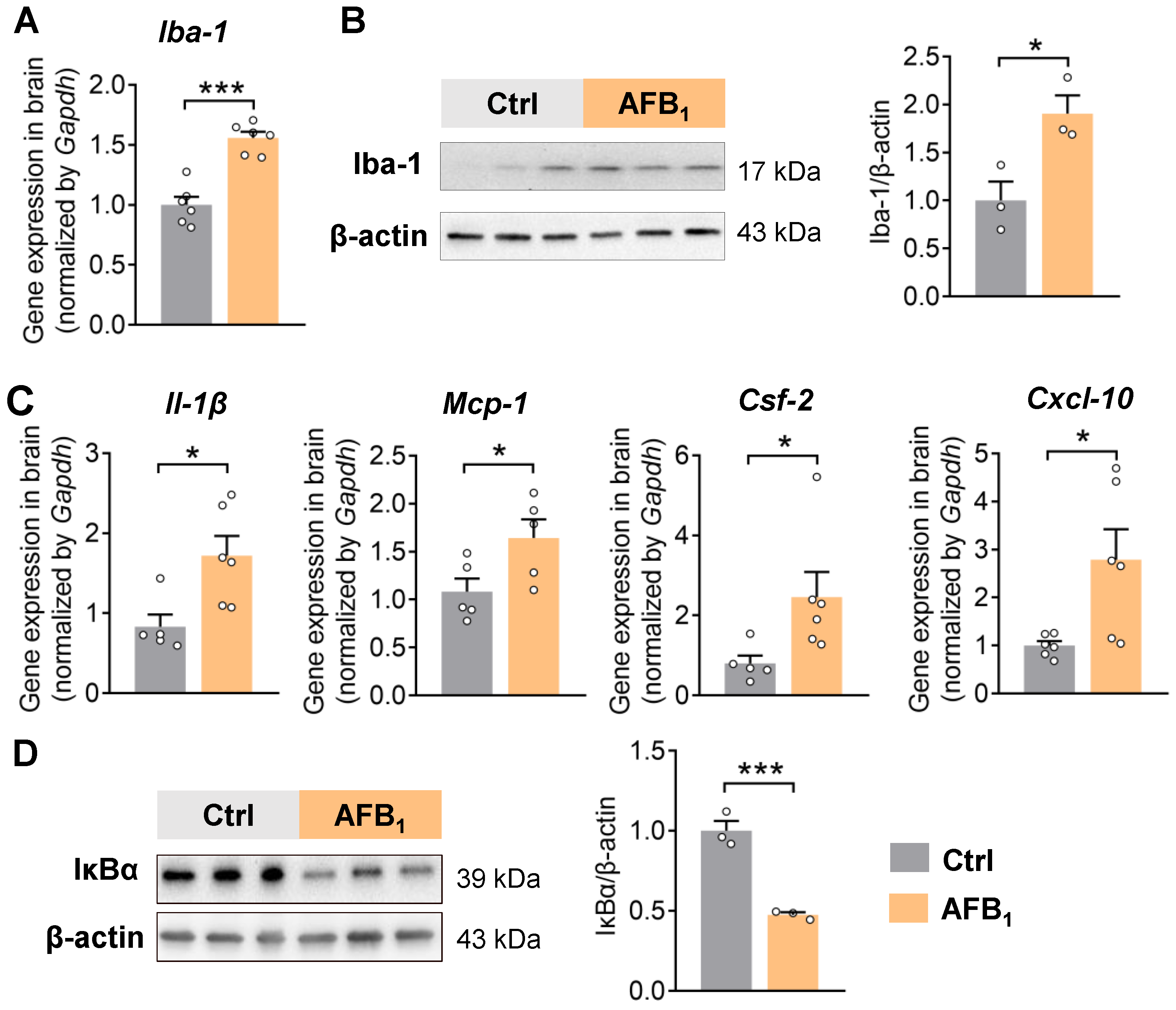
2.1. AFB1 Increases Neuroinflammation

We orally exposed 8-week-old male C57BL/6J mice to AFB1 (1500 ppb) in drinking water for 21 days. The AFB1 exposure dose was selected as comparable to the contamination levels in food and feed materials that people could be exposed to [29,30,31,32]. Microglia are resident immune cells in the brain and are crucial for immune surveillance and regulating inflammation [33]. AFB1 exposure increased the gene expression and protein levels of Iba-1, indicating microglial cell activation in the brain (Figure 1A,B). In line with microglia activation, the qRT-PCR analysis also showed increased expression of pro-inflammatory cytokines Il-1β, Mcp-1, Csf-2, and Cxcl-10 in the brain (Figure 1C). Furthermore, the level of the inhibitor of NF-κB alpha (IκBα) was decreased after 21 days of AFB1 exposure, suggesting the activation of the NF-κB pathway in the brain (Figure 1D). Together, these results support the premise that AFB1 induces microglia activation and inflammation in the brain.

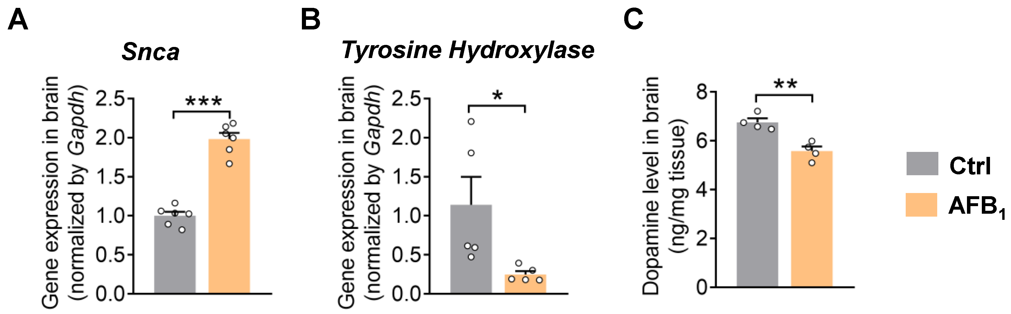
2.2. AFB1 Stimulates α-Synuclein Upregulation and Causes Dopaminergic Neurotoxicity

The accumulation of α-synuclein and dopaminergic neuron dysfunction are two hallmarks and driving factors in PD pathology [3]. The qRT-PCR analysis showed that AFB1 increased the expression of α-synuclein in the brain, indicating the involvement of AFB1 in α-synuclein pathology (Figure 2A). Moreover, AFB1 exposure suppressed the expression of the dopaminergic neuron marker tyrosine hydroxylase (TH) and decreased the level of dopamine in the brain, suggesting dopaminergic neuron dysfunction (Figure 2B,C). Consistent with these in vivo findings, treatment with AFB1 dose-dependently reduced the cell viability of N27 rat dopaminergic neuronal cells in vitro (Figure S1). Together, these results support that AFB1 leads to dopaminergic neurotoxicity.

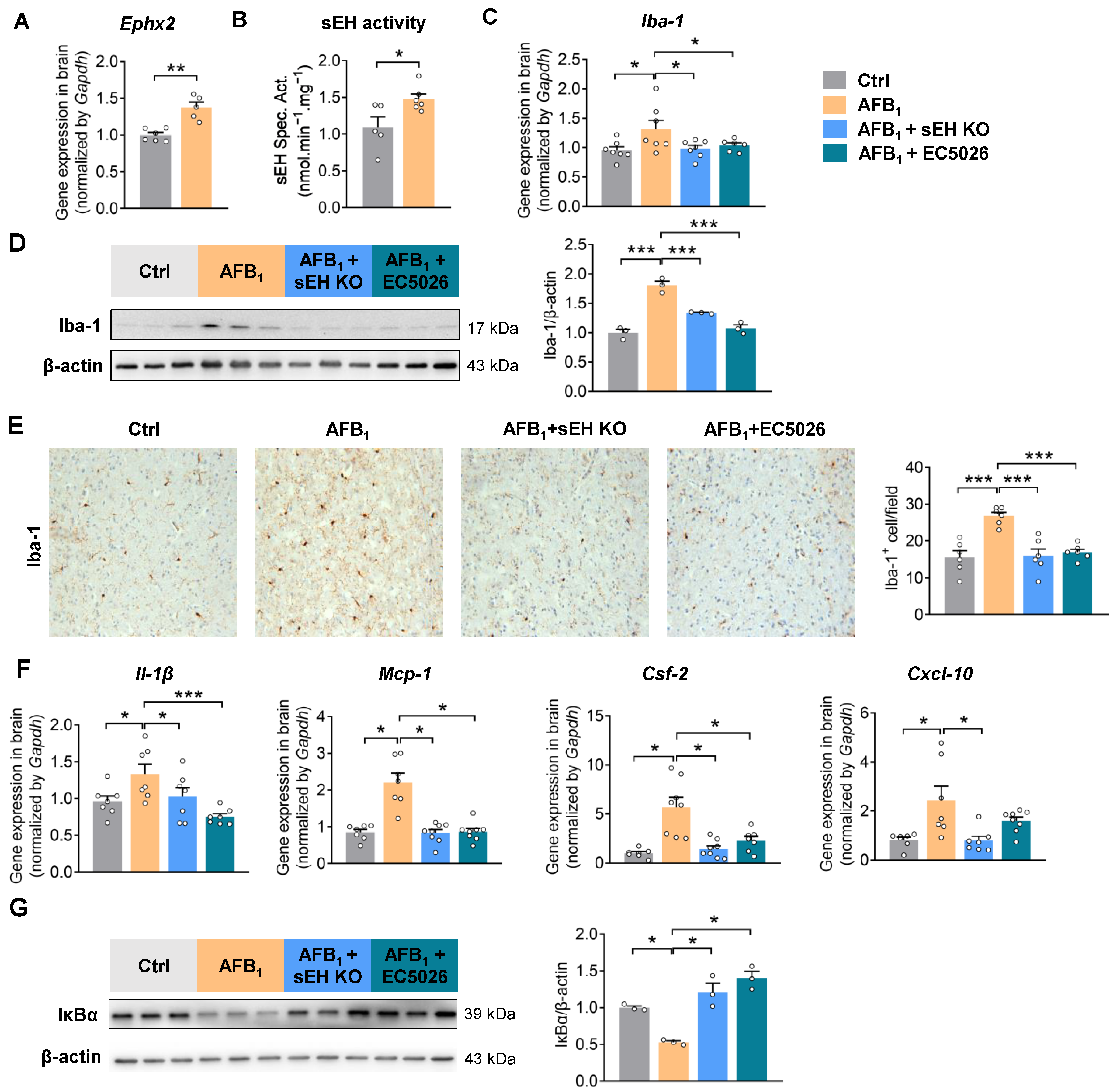
2.3. AFB1 Induces Neuroinflammation in an sEH-Dependent Manner

sEH contributes to environmental toxin-induced parkinsonism in mice [26]. Here, AFB1 exposure increased the gene expression and enzymatic activity of sEH in the brain, suggesting its potential involvement in AFB1 toxicity (Figure 3A,B). To determine the functional role of sEH in AFB1-induced neuroinflammation, 8-week-old male wild-type (WT) or sEH genetic knockout (Ephx2−/−) mice were treated with AFB1 for 21 days. Moreover, the small-molecule transition state inhibitor EC5026 was applied as a pharmacological intervention approach to block the activity of sEH in mice. sEH deficiency or inhibition attenuated AFB1-induced neuroinflammation by suppressing the accumulation of Iba-1+ microglia cells, decreasing the levels of pro-inflammatory cytokines (Il-1β, Mcp-1, Csf-2, and Cxcl-10), and suppressing the activation of the NF-κB pathway in the brain (Figure 3C–G). Together, these results support the involvement of sEH in mediating the pro-inflammatory effects of AFB1 in the brain.

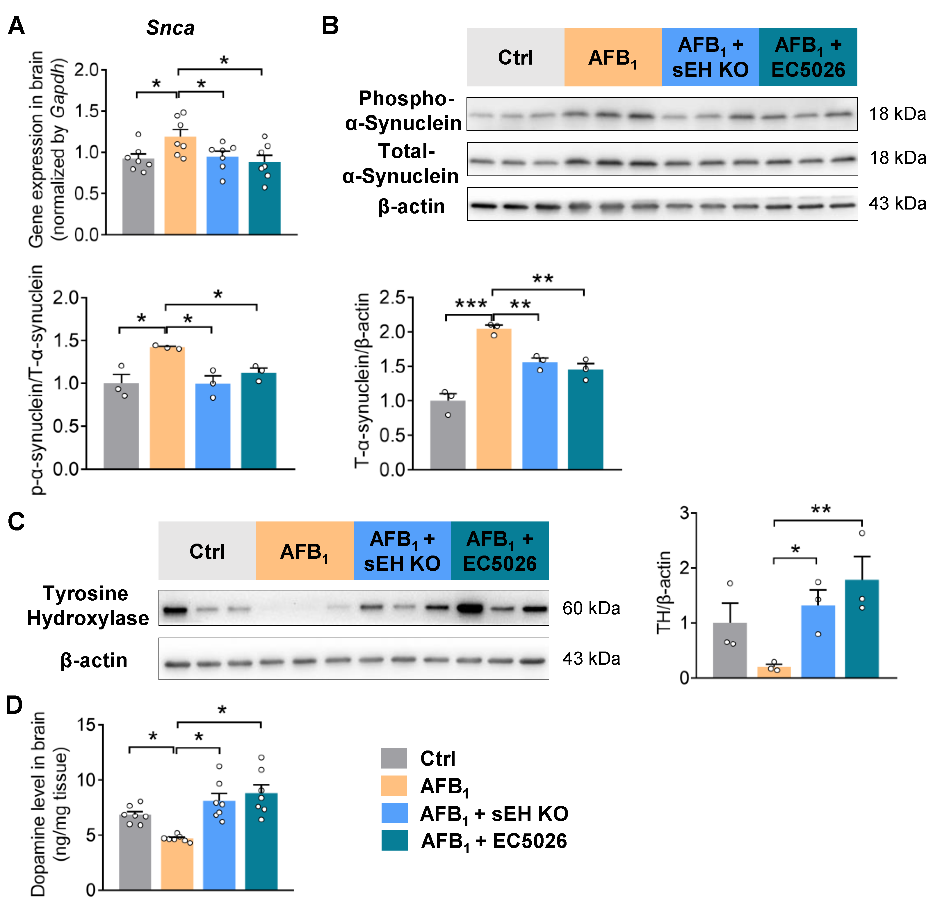
2.4. sEH Deficiency or Inhibition Alleviates AFB1-Induced α-Synuclein Pathology and Dopaminergic Neurotoxicity

Having demonstrated a functional role of sEH in modulating AFB1-associated neuroinflammation, we next tested whether sEH is also involved in mediating the AFB1-induced α-synuclein pathology and dopaminergic neurotoxicity in the brain. Genetic knockout or pharmacological inhibition of sEH reduced the AFB1-induced α-synuclein upregulation in the brain (Figure 4A,B). Phosphorylation of α-synuclein, which controls protein aggregation and Lewy body formation [34,35], was also decreased in Ephx2−/− or EC5026-treated mice (Figure 4B). Moreover, sEH deficiency or inhibition prevented the loss of the dopaminergic neuron marker TH and attenuated the reduction in dopamine in the brain under AFB1 exposure, pointing to the alleviated dopaminergic neuron damage after sEH inhibition (Figure 4C,D). In N27 rat dopaminergic neuronal cells, treatment with EC5026 suppressed the AFB1-induced oxidative marker Inos, as well as pro-inflammatory cytokines Tnf-α and Cxcl-10. (Figure S2). Together, these results support the vital role of sEH in modulating the α-synuclein pathology and dopaminergic neurotoxicity caused by AFB1 in the brain.

3. Discussion

The incidence and prevalence of PD have risen rapidly worldwide [1,2]. Epidemiological and animal studies reported a higher risk of onset and progression of PD due to environmental toxicant exposure [4,5]. However, whether AFB1, a widespread and dangerous foodborne mycotoxin, is involved in the pathogenesis of PD is poorly understood. Here, the results suggest that exposure to AFB1 could contribute to PD by stimulating neuroinflammation, triggering α-synuclein pathology, and impairing the function of dopaminergic neurons in vivo. Moreover, both the expression and enzymatic activity of sEH are increased in the brains of AFB1-exposed mice. Blocking of sEH, genetically or pharmacologically, attenuates the AFB1-induced neuroinflammation, α-synuclein accumulation, and dopaminergic neurotoxicity in mice. This is consistent with earlier evidence that an increase in sEH expression or activity is both a marker and cause of inflammation in the brain. Altogether, these results support that AFB1 can induce neuroinflammation and PD-like pathology through a mechanism that involves the increased level of sEH.
Neuroinflammation has been implicated in the pathogenesis of a variety of neurodegenerative diseases, including PD [36]. Here in mice, AFB1 exposure induces microglia activation and leads to activation of the NF-κB pathway, and up-regulation of the expression of pro-inflammatory cytokines, notably Il-1β, Mcp-1, Csf-2, and Cxcl-10, in the brains of AFB1-exposed mice. In line with these findings, previous studies showed that AFB1 stimulated the production of pro-inflammatory cytokines and induced the activation of NF-κB in human or mouse microglia cell lines [16,17]. Promisingly, sEH deficiency or inhibition suppressed the AFB1-induced Iba-1+ microglia accumulation and pro-inflammatory cytokine up-regulation in the brain, indicating an important role for sEH in mediating neuroinflammation following AFB1 exposure. These results agree with previous reports showing that blocking the action of sEH attenuated neuroinflammation by decreasing the abundance of Iba-1+ microglia and levels of pro-inflammatory regulators in the brains of 5xFAD- or LPS-treated mice [37]. In vitro, the treatment with an sEH inhibitor reduced the expression of pro-inflammatory cytokines in LPS-treated microglia or rotenone-treated N27 dopaminergic cells [28,37]. Together, the oral intake of AFB1 increased neuroinflammation via increased sEH, which could further contribute to neurological changes toward PD in the brain.
Dopaminergic neuron damage is a core manifestation of PD [38]. Here, AFB1 suppressed the viability of N27 dopaminergic neurons in vitro and caused dopaminergic neuron dysfunction in mice. Similar effects have been also described in models of dopaminergic dysfunction in rats or C. elegans where AFB1 causes dopaminergic neurodegeneration and suppresses the level of dopamine in the brain [39,40], suggesting that AFB1 causes dopaminergic neurotoxicity in multiple animal models. Exposure to AFB1 also causes nerve fiber depletion [41]. In this study, inhibiting the function of sEH attenuated AFB1-induced dopaminergic neurotoxicity by restoring the expression of dopaminergic neuron marker TH and the level of dopamine in the brain. In agreement with these findings, previous studies showed that sEH inhibition reduced MPTP- or paraquat-induced TH+ dopaminergic neuron loss [26]. Moreover, treatment with an sEH inhibitor restored dopamine levels in MPTP-treated mice [42] or rotenone-treated Drosophila melanogaster [28], suggesting that sEH contributes to environmental toxin-induced dopaminergic neurotoxicity. A limitation of the current study is it may not reflect the sex difference in AFB1-caused dopaminergic neurotoxicity. Further studies are needed to better characterize the pro-PD effects of AFB1 on both male and female mice. Another limitation of this study is that we only analyzed dopaminergic neuronal damage in frontal brain regions, mainly the striatum; however, whether AFB1 also causes toxicity to dopaminergic neurons in the substantia nigra (SN) is still unclear. Further studies are needed to determine dopaminergic neuronal dysfunction and inflammation and to evaluate the functional role of sEH in mediating neuroinflammation and neurotoxicity in the SN under AFB1 exposure.
In addition to its direct toxic effect on dopaminergic neurons, AFB1 also increased the level and phosphorylation of α-synuclein in the mouse brain. It is well-known that α-synuclein acts as the primary structural component of Lewy-bodies and contributes to progressive neuronal damage during PD development [43,44]. In particular, Ser129 phosphorylation of α-synuclein has been shown to control the aggregation process and has been linked to neurotoxic effects in the pathogenesis of PD [34,35]. Moreover, α-synuclein stimulates the inflammation response in microglia, which in turn boosts α-synuclein pathology and dopaminergic neuronal death [45,46]. A recent study also highlights that pathogenic gut microbes contribute to PD development by inducing α-synuclein aggregation [47]. Thus, targeting synuclein, especially its phosphorylation, is an attractive approach for preventing Lewy body formation and PD development. Here, inhibiting the action of sEH helps to attenuate AFB1-stimulated α-synuclein upregulation and phosphorylation in the brain. Similarly, treatment with an sEH inhibitor has been shown to reduce phosphorylated α-synuclein in the brains of MPTP-treated mice [42]. Further studies are needed to determine whether AFB1 has a direct interaction with a-synuclein, whether it affects aggregation/fibril formation in vitro and in vivo, and how sEH exactly is involved in such aggregation processes.
In the current study, both the expression and enzymatic activity of sEH were found to be increased in the brains of AFB1-exposed mice. Additionally, sEH has been reported to be upregulated in the brains of MPTP- or glyphosate-treated mice [26,42,48], suggesting the broad involvement of sEH in neurological disorders induced by environmental factors. In humans, the level of sEH is increased in the brains of Lewy body dementia patients [42]. sEH mainly exhibits its pro-inflammatory role by converting epoxy fatty acids, especially epoxyeicosatrienoic acids (EETs), into responding diols. Indeed, previous studies showed that 14,15-EET help to restore the MPTP-disrupted dopaminergic neurons and improve rotarod performance in mice [26]. In addition, the treatment of 14,15-EET protects the dopaminergic neuronal N27 from oxidative damage [49]. Given the neuron protective effects of 14,15-EET, further studies investigating the functions of 14,15-EET in AFB1 neurotoxicity models are warranted to further consolidate the molecular mechanism by which the inhibition of sEH by stabilizing 14,15-EET achieves its neuroprotective effects.
To facilitate translation to humans, AFB1-exposed mice were treated with the sEH inhibitor EC5026. We found that the treatment of EC5026 effectively attenuated neuroinflammation and dopaminergic neurotoxicity. In N27 dopaminergic cells, the treatment of EC5026 blocked the AFB1-upregulated pro-inflammatory factors Inos, Tnf-α and Cxcl-10 in vitro. These findings are in agreement with previous studies showing that EC5026 reduces LPS-induced neuroinflammation in mice [37] and alleviated neuropathic pain in a chronic constriction injury rat model [50]. All these findings are of translational relevance, since EC5026 is currently in clinical trials for neuropathic pain [50]. Further studies are needed to explore the efficacy and mechanism of EC5026 in treating AFB1-caused neurological and behavioral disorders in classic PD models.
In conclusion, AFB1 oral exposure promotes PD pathogenesis by enhancing neuroinflammation and disrupting the dopaminergic neuron function in mice. More importantly, sEH acts as a critical cellular regulator in mediating AFB1-induced neurotoxicity. This study exemplifies one approach for using clinically developed synthetic or natural sEH inhibitors to reduce the toxicity induced by chronic AFB1 exposure in humans. Further preclinical and clinical studies include the following: the efficacy, drug dose optimization, brain penetration, neuro pharmacokinetics, and safety characteristics of sEH inhibitors in the brain for alleviating neurotoxicity in multiple models of AFB1 exposure. In addition, this study suggests that future studies should also investigate the effects of targeting sEH in alleviating other foodborne or environmental toxins-associated neuroinflammation and dopaminergic neurotoxicity.

4. Materials and Methods

4.1. Animal Study

All animal experiments were performed in accordance with the protocol approved by the Institutional Animal Care and Use Committee (IACUC, protocol # 21628) of the University of California-Davis. All the mice were maintained on a standard chow diet ad libitum and great care was taken to ensure the welfare of the included animals.

4.1.1. Animal Experiment 1: Effects of AFB1 Exposure on Neuroinflammation and Neurotoxicity in Mice

C57BL/6J male mice (8-week-old) were purchased from Charles River and randomly assigned to two groups (AFB1 or vehicle-treated group, n = 6 mice per group). AFB1 (Sigma-Aldrich, St. Louis, MO, USA, catalog # A6636) was firstly dissolved in dimethyl sulfoxide (DMSO, Thermo Fisher Scientific, Hampton, NH, USA) and then added into sterile drinking water at a final concentration of 1500 ppb (final concentration of DMSO is 0.1%). The AFB1 drinking water was freshly prepared every other day and put in aluminum foil-wrapped bottles to reduce the photochemical degradation of AFB1 and minimize degradation-caused dose changes. After 21 days, the mice were euthanized using isoflurane (5%, Dechra Pharmaceuticals, Northwich, UK) inhalation followed by cervical dislocation. The frontal brain regions (striatum and an associated portion of the cerebral cortex) were dissected for further analysis.

4.1.2. Animal Experiment 2: Effects of sEH Deficiency or Inhibition on AFB1-Induced Neuroinflammation and Neurotoxicity in Mice

C57BL/6J male WT mice or Ephx2−/− mice (8-week-old, n = 8 mice per group, maintained at the University of California, Davis) were treated with AFB1 (1500 ppb) or the vehicle (drinking water containing 0.15% DMSO). To determine the effect of pharmacological inhibition of sEH, another group of C57BL/6J male mice WT mice (8-week-old) were given AFB1 (1500 ppb) together with the potent sEH inhibitor EC5026 (10 mg/L) in the vehicle (drinking water containing 0.15% DMSO). EC5026 was synthesized as previously described [50]. The vehicle, AFB1 only, and AFB1 plus EC5026 drinking water were freshly prepared every other day and put in aluminum foil-wrapped bottles. After 21 days, the mice were euthanized using isoflurane (5%) inhalation followed by cervical dislocation. The frontal brain regions (striatum and an associated portion of the cerebral cortex) were dissected for further analysis.

4.2. Total RNA Isolation and Quantitative Polymerase Chain Reaction (qPCR) Analysis

The frontal brain regions were dissected and ground after being frozen in liquid nitrogen. The TRIzol reagent (Ambion, Austin, TX, USA) was used to isolate the total RNA according to the manufacturer’s instructions. A NanoDrop Spectrophotometer (Thermo Fisher Scientific, Waltham, MA, USA) was used to measure the quality and quantity of the extracted RNA. A High-Capacity cDNA Reverse Transcription kit (Applied Biosystems, Waltham, MA, USA) was used to reverse transcribe RNA into cDNA. An Mic qPCR Cycler (Bio Molecular Systems, Upper Coomera, Australia) was used to perform qPCR. The results of target genes were normalized to glyceraldehyde-3-phosphate dehydrogenase (Gapdh) and the final gene expressions were calculated using the 2−ΔΔCt method. All the murine primers used in this study were obtained from Thermo Fisher Scientific. The information on these primers is listed in Table S1.

4.3. Protein Extraction and Immunoblotting

Protein extraction and immunoblotting were performed as described [51]. Proteins from the frontal brain regions were extracted with RIPA lysis buffer (Boston BioProducts, Milford, MA, USA) with a protease inhibitor cocktail (Boston BioProducts). A BCA protein assay kit (Thermo Fisher Scientific) was used to determine the protein concentrations. The tissue samples (15 μg) were resolved on SDS-PAGE gels (Bio-Rad Laboratories, Hercules, CA, USA) and transferred onto nitrocellulose membranes (Bio-Rad Laboratories). The membrane was blocked in 5% milk buffer, incubated with primary antibodies against Iba-1 (Cell Signaling, Danvers, MA, USA, catalog # 17198), IκBα (Cell Signaling, catalog # 4814), phospho-α-synuclein (Ser129) (Cell Signaling, catalog # 23706), α-synuclein (Cell Signaling, catalog # 4179), and tyrosine hydroxylase (EMD Millipore, Burlington, MA, USA, catalog # AB152) in 5% BSA solution at 4 °C overnight. The membranes were incubated with HRP-linked anti-rabbit, or anti-mouse antibodies (Cell Signaling, catalog #7074, #7076). Clarity Western Enhanced Chemiluminescence (ECL) Substrates (Bio-Rad Laboratories) were used for imaging. The chemiluminescence was developed by Clarity Western Enhanced Chemiluminescence (ECL) Substrates (Bio-Rad Laboratories) and detected using Bio-Rad ChemiDoc™ Imaging Systems. Quantification of immunoblotting was performed using Image J software (Version 1.53v). β-actin (Sigma-Aldrich, catalog # A2228) was used as a loading control and for normalization.

4.4. Tissue Staining

The frontal brain tissues were fixed in 4% paraformaldehyde overnight at 4 °C, transferred to 30% sucrose solution for dehydration, and sliced into 12 μm sections using the cryostat (Leica Biosystems, Wetzlar, Germany). Antigen retrieval was performed by heating the sections in 0.01 M citrate buffer (pH 6.0) to 95 °C for 5 min. Immunohistochemistry staining was conducted using the HRP/DAB (ABC) Detection IHC kit (Abcam, Cambridge, UK) according to the manufacturer’s instructions. The anti-Iba-1 (Cell Signaling, catalog # 17198) was used to probe the target protein in the tissue section. The number of Iba-1+ cells per field was counted using Image J software (Version 1.53v).

4.5. Enzyme-Linked Immunosorbent Assay (ELISA) Analysis of Dopamine

Proteins from the frontal brain regions were extracted using PBS with sonication on ice and kept in −80 °C. Protein concentrations were determined using the BCA protein assay kit. The level of dopamine in brain lysis was determined using the mouse dopamine ELISA kit (MyBiosource, San Diego, CA, USA, catalog # MBS162171) according to the manufacturer’s instructions. The dopamine level from each sample was normalized using the protein concentration of the corresponding sample and was expressed in a unit of ng/mg of tissue protein.

4.6. sEH Activity Measurement

The frontal brain regions were lysed to measure the sEH activity. sEH activity was measured as previously described [52]. Briefly, to 100 µL of tissue suspension, 1 µL of a 5 mM solution of [3H]-trans-diphenyl-propene oxide (t-DPPO) in DMSO was added ([S]final = 50 µM; 10,000 cpm) and was incubated at 37 °C for 30 min. The reaction was quenched by the addition of 60 µL of methanol and 200 µL of iso-octane. The enzymatic activity was determined by measuring the quantity of radioactive diol formed in the aqueous phase using a scintillation counter (TriCarb 2810 TR, Perkin Elmer, Shelton, CT, USA). The enzymatic activity sample was normalized using the corresponding protein concentration.

4.7. Cell Assays

The N27 rat dopaminergic neural cells were obtained from EMD Millipore (catalog # SCC048). N27 cells were cultured in RPMI 1640 (Corning, New York, NY, USA, catalog # 10040CVa) supplemented with 10% fetal bovine serum. All cells were maintained in an atmosphere of 5% CO2 and at 37 °C.
For the MTT assay, N27 cells were seeded in 96-well plates at a density of 30,000 cells/well and incubated for 24 h. Cells were then treated with AFB1 (0.1, 1, 10 μM) or the vehicle control (DMSO, 0.1%) for 24 h. After that, cells were washed with PBS, 0.5 mg/mL MTT (catalog # M6494, Thermo Fisher Scientific) in fresh RPMI 1640 was added to each well and then incubated for 2 h at 37 °C. After the supernatant was removed, 100 mL of DMSO was added to each well. The difference in absorbance at 570 nm was measured on a microplate reader (Molecular Devices). The results were expressed as percentages of the control (%).
For quantitative PCR analysis, N27 cells were seeded in 12-well plates at a density of 300,000 cells/well and incubated for 24 h. Cells were then treated with AFB1 (10 μM) with or without EC5026 (0.5 μM) for 24 h. Total RNA extraction, reverse transcription, quantitative PCR, and data analysis were performed as described above. The information on rat primers is listed in Table S1.

4.8. Statistical Analysis

All data are expressed as the mean ± standard error of the mean (SEM). The Shapiro–Wilk test was used to verify the normality of data and Levene’s mean test was used to assess the equal variance of data before the statistical analysis. Statistical comparison of the two groups was performed using either Student’s t-test or the Wilcoxon–Mann–Whitney test (when the normality test failed). Statistical comparison of three groups was determined using one-way ANOVA followed by Tukey’s or Fisher’s post hoc test, or using the Kruskal–Wallis test on ranks (when the normality test failed), followed by the appropriate post hoc test. All the data analyses were performed by using SigmaPlot software (Version 11, Systat Software, Inc., Chicago, IL, USA). p values less than 0.05 were reported as statistically significant.

Supplementary Materials

The supporting information can be downloaded at: https://www.mdpi.com/article/10.3390/ijms24129938/s1.

Author Contributions

Conceptualization: B.D.H. and W.W.; funding acquisition: B.D.H. and H.W.; investigation: W.W., Y.W., K.M.W., R.D.L. and C.M.; resources: S.H.H.; supervision: B.D.H.; writing—original draft: W.W.; writing—review and editing: B.D.H. and H.W. All authors have read and agreed to the published version of the manuscript.

Funding

This research was supported by the NIH/NIEHS RIVER award R35 ES030443 (BDH), NIH/NIEHS Superfund Research Program P42 ES004699 (BDH), NIH/NINDS R01 NS100294 (HW) and NIH/NINDS U54 NS127758-01 (HW and BDH). This work was partially supported by a donation in memory of Dr. David Fork.

Institutional Review Board Statement

The animal study protocol was approved by the Institutional Animal Care and Use Committee of the University of California-Davis (protocol # 21628).

Informed Consent Statement

Not applicable.

Data Availability Statement

All data are available in the main text or the Supplementary Materials. Additional raw data are available on request.

Acknowledgments

We thank Aldrin V Gomes at the Department of Neurobiology, Physiology, and Behavior at UC Davis for helping with the immunoblotting analysis. Brain tissue imaging was conducted using the CAMI core facility at the UC Davis Center for Health and Environment.

Conflicts of Interest

K.M.W. and S.H.H. are part-time employees of and B.D.H. is the cofounder and CSO of EicOsis LLC. All other authors declare that they have no competing interest.

References

  1. Bloem, B.R.; Okun, M.S.; Klein, C. Parkinson’s disease. Lancet 2021, 397, 2284–2303. [Google Scholar] [CrossRef]
  2. GBD 2016 Parkinson’s Disease Collaborators. Global, regional, and national burden of Parkinson’s disease, 1990–2016: A systematic analysis for the Global Burden of Disease Study 2016. Lancet Neurol. 2018, 17, 939–953. [Google Scholar] [CrossRef] [Green Version]
  3. Poewe, W.; Seppi, K.; Tanner, C.M.; Halliday, G.M.; Brundin, P.; Volkmann, J.; Schrag, A.E.; Lang, A.E. Parkinson disease. Nat. Rev. Dis. Prim. 2017, 3, 17013. [Google Scholar] [CrossRef]
  4. Chen, H.; Ritz, B. The Search for Environmental Causes of Parkinson’s Disease: Moving Forward. J. Park. Dis. 2018, 8, S9–S17. [Google Scholar] [CrossRef] [Green Version]
  5. Goldman, S.M. Environmental toxins and Parkinson’s disease. Annu. Rev. Pharmacol. Toxicol. 2014, 54, 141–164. [Google Scholar] [CrossRef]
  6. Anderson, F.L.; Coffey, M.M.; Berwin, B.L.; Havrda, M.C. Inflammasomes: An Emerging Mechanism Translating Environmental Toxicant Exposure Into Neuroinflammation in Parkinson’s Disease. Toxicol. Sci. 2018, 166, 3–15. [Google Scholar] [CrossRef] [Green Version]
  7. Chin-Chan, M.; Navarro-Yepes, J.; Quintanilla-Vega, B. Environmental pollutants as risk factors for neurodegenerative disorders: Alzheimer and Parkinson diseases. Front. Cell. Neurosci. 2015, 9, 124. [Google Scholar] [CrossRef] [Green Version]
  8. EFSA Panel on Contaminants in the Food Chain (CONTAM); Schrenk, D.; Bignami, M.; Bodin, L.; Chipman, J.K.; Del Mazo, J.; Grasl-Kraupp, B.; Hogstrand, C.; Hoogenboom, L.R.; Leblanc, J.C.; et al. Risk assessment of aflatoxins in food. EFSA J. 2020, 18, e06040. [Google Scholar] [CrossRef]
  9. Williams, J.H.; Phillips, T.D.; Jolly, P.E.; Stiles, J.K.; Jolly, C.M.; Aggarwal, D. Human aflatoxicosis in developing countries: A review of toxicology, exposure, potential health consequences, and interventions. Am. J. Clin. Nutr. 2004, 80, 1106–1122. [Google Scholar] [CrossRef] [Green Version]
  10. Benkerroum, N. Chronic and Acute Toxicities of Aflatoxins: Mechanisms of Action. Int. J. Environ. Res. Public Health 2020, 17, 423. [Google Scholar] [CrossRef] [Green Version]
  11. Pei, X.; Zhang, W.; Jiang, H.; Liu, D.; Liu, X.; Li, L.; Li, C.; Xiao, X.; Tang, S.; Li, D. Food-Origin Mycotoxin-Induced Neurotoxicity: Intend to Break the Rules of Neuroglia Cells. Oxidative Med. Cell. Longev. 2021, 2021, 9967334. [Google Scholar] [CrossRef]
  12. Oyelami, O.A.; Maxwell, S.M.; Adelusola, K.A.; Aladekoma, T.A.; Oyelese, A.O. Aflatoxins in the autopsy brain tissue of children in Nigeria. Mycopathologia 1995, 132, 35–38. [Google Scholar] [CrossRef]
  13. Linardaki, Z.I.; Lamari, F.N.; Margarity, M. Saffron (Crocus sativus L.) Tea Intake Prevents Learning/Memory Defects and Neurobiochemical Alterations Induced by Aflatoxin B1 Exposure in Adult Mice. Neurochem. Res. 2017, 42, 2743–2754. [Google Scholar] [CrossRef]
  14. Gugliandolo, E.; Peritore, A.F.; D’Amico, R.; Licata, P.; Crupi, R. Evaluation of Neuroprotective Effects of Quercetin against Aflatoxin B1-Intoxicated Mice. Animals 2020, 10, 898. [Google Scholar] [CrossRef]
  15. Kihara, T.; Matsuo, T.; Sakamoto, M.; Yasuda, Y.; Yamamoto, Y.; Tanimura, T. Effects of prenatal aflatoxin B1 exposure on behaviors of rat offspring. Toxicol. Sci. 2000, 53, 392–399. [Google Scholar] [CrossRef] [Green Version]
  16. Mehrzad, J.; Malvandi, A.M.; Alipour, M.; Hosseinkhani, S. Environmentally relevant level of aflatoxin B(1) elicits toxic pro-inflammatory response in murine CNS-derived cells. Toxicol. Lett. 2017, 279, 96–106. [Google Scholar] [CrossRef]
  17. Mehrzad, J.; Hosseinkhani, S.; Malvandi, A.M. Human Microglial Cells Undergo Proapoptotic Induction and Inflammatory Activation upon in vitro Exposure to a Naturally Occurring Level of Aflatoxin B1. Neuroimmunomodulation 2018, 25, 176–183. [Google Scholar] [CrossRef]
  18. Huang, B.; Chen, Q.; Wang, L.; Gao, X.; Zhu, W.; Mu, P.; Deng, Y. Aflatoxin B1 Induces Neurotoxicity through Reactive Oxygen Species Generation, DNA Damage, Apoptosis, and S-Phase Cell Cycle Arrest. Int. J. Mol. Sci. 2020, 21, 6517. [Google Scholar] [CrossRef]
  19. Piomelli, D.; Sasso, O. Peripheral gating of pain signals by endogenous lipid mediators. Nat. Neurosci. 2014, 17, 164–174. [Google Scholar] [CrossRef] [Green Version]
  20. Anand, S.; Ansari, M.A.; Sukrutha, S.K.; Alomary, M.N.; Khan, A.A.; Elderdery, A.Y. Resolvins Lipid Mediators: Potential Therapeutic Targets in Alzheimer and Parkinson Disease. Neuroscience 2022, 507, 139–148. [Google Scholar] [CrossRef]
  21. Shen, W.; Jiang, L.; Zhao, J.; Wang, H.; Hu, M.; Chen, L.; Chen, Y. Bioactive lipids and their metabolism: New therapeutic opportunities for Parkinson’s disease. Eur. J. Neurosci. 2022, 55, 846–872. [Google Scholar] [CrossRef]
  22. Warner, J.; Hardesty, J.; Zirnheld, K.; McClain, C.; Warner, D.; Kirpich, I. Soluble Epoxide Hydrolase Inhibition in Liver Diseases: A Review of Current Research and Knowledge Gaps. Biology 2020, 9, 124. [Google Scholar] [CrossRef]
  23. Hashimoto, K. Role of Soluble Epoxide Hydrolase in Metabolism of PUFAs in Psychiatric and Neurological Disorders. Front. Pharmacol. 2019, 10, 36. [Google Scholar] [CrossRef] [Green Version]
  24. Borsini, A. The role of soluble epoxide hydrolase and its inhibitors in depression. Brain Behav. Immun. Health 2021, 16, 100325. [Google Scholar] [CrossRef]
  25. Ren, Q. Soluble Epoxide Hydrolase Inhibitor: A Novel Potential Therapeutic or Prophylactic Drug for Psychiatric Disorders. Front. Pharmacol. 2019, 10, 420. [Google Scholar] [CrossRef]
  26. Qin, X.; Wu, Q.; Lin, L.; Sun, A.; Liu, S.; Li, X.; Cao, X.; Gao, T.; Luo, P.; Zhu, X.; et al. Soluble Epoxide Hydrolase Deficiency or Inhibition Attenuates MPTP-Induced Parkinsonism. Mol. Neurobiol. 2015, 52, 187–195. [Google Scholar] [CrossRef]
  27. Huang, H.J.; Wang, Y.T.; Lin, H.C.; Lee, Y.H.; Lin, A.M. Soluble Epoxide Hydrolase Inhibition Attenuates MPTP-Induced Neurotoxicity in the Nigrostriatal Dopaminergic System: Involvement of alpha-Synuclein Aggregation and ER Stress. Mol. Neurobiol. 2018, 55, 138–144. [Google Scholar] [CrossRef]
  28. Lakkappa, N.; Krishnamurthy, P.T.; Pandareesh, M.D.; Hammock, B.D.; Hwang, S.H. Soluble epoxide hydrolase inhibitor, APAU, protects dopaminergic neurons against rotenone induced neurotoxicity: Implications for Parkinson’s disease. Neurotoxicology 2019, 70, 135–145. [Google Scholar] [CrossRef]
  29. Arrus, K.; Blank, G.; Clear, R.; Holley, R.A.; Abramson, D. Microbiological and aflatoxin evaluation of Brazil nut pods and the effects of unit processing operations. J. Food Prot. 2005, 68, 1060–1065. [Google Scholar] [CrossRef]
  30. Lewis, L.; Onsongo, M.; Njapau, H.; Schurz-Rogers, H.; Luber, G.; Kieszak, S.; Nyamongo, J.; Backer, L.; Dahiye, A.M.; Misore, A.; et al. Aflatoxin contamination of commercial maize products during an outbreak of acute aflatoxicosis in eastern and central Kenya. Environ. Health Perspect. 2005, 113, 1763–1767. [Google Scholar] [CrossRef]
  31. Johansson, A.S.; Whitaker, T.B.; Hagler, W.M., Jr.; Bowman, D.T.; Slate, A.B.; Payne, G. Predicting aflatoxin and fumonisin in shelled corn lots using poor-quality grade components. J. AOAC Int. 2006, 89, 433–440. [Google Scholar] [CrossRef] [Green Version]
  32. Desai, S.; Ghewande, M.P.; Nagaraj, G.; Narayan, P.; Chauhan, S.; Singh, H. Screening for resistance to Aspergillus flavus and aflatoxin production in groundnut. Mycotoxin Res. 1991, 7, 79–84. [Google Scholar] [CrossRef]
  33. Kim, Y.S.; Joh, T.H. Microglia, major player in the brain inflammation: Their roles in the pathogenesis of Parkinson’s disease. Exp. Mol. Med. 2006, 38, 333–347. [Google Scholar] [CrossRef] [PubMed] [Green Version]
  34. Chen, L.; Feany, M.B. Alpha-synuclein phosphorylation controls neurotoxicity and inclusion formation in a Drosophila model of Parkinson disease. Nat. Neurosci. 2005, 8, 657–663. [Google Scholar] [CrossRef] [PubMed]
  35. Xu, Y.; Deng, Y.; Qing, H. The phosphorylation of alpha-synuclein: Development and implication for the mechanism and therapy of the Parkinson’s disease. J. Neurochem. 2015, 135, 4–18. [Google Scholar] [CrossRef] [PubMed]
  36. Glass, C.K.; Saijo, K.; Winner, B.; Marchetto, M.C.; Gage, F.H. Mechanisms underlying inflammation in neurodegeneration. Cell 2010, 140, 918–934. [Google Scholar] [CrossRef] [Green Version]
  37. Ghosh, A.; Comerota, M.M.; Wan, D.; Chen, F.; Propson, N.E.; Hwang, S.H.; Hammock, B.D.; Zheng, H. An epoxide hydrolase inhibitor reduces neuroinflammation in a mouse model of Alzheimer’s disease. Sci. Transl. Med. 2020, 12, eabb1206. [Google Scholar] [CrossRef]
  38. Michel, P.P.; Hirsch, E.C.; Hunot, S. Understanding Dopaminergic Cell Death Pathways in Parkinson Disease. Neuron 2016, 90, 675–691. [Google Scholar] [CrossRef] [Green Version]
  39. Gonzalez-Hunt, C.P.; Leung, M.C.; Bodhicharla, R.K.; McKeever, M.G.; Arrant, A.E.; Margillo, K.M.; Ryde, I.T.; Cyr, D.D.; Kosmaczewski, S.G.; Hammarlund, M.; et al. Exposure to mitochondrial genotoxins and dopaminergic neurodegeneration in Caenorhabditis elegans. PLoS ONE 2014, 9, e114459. [Google Scholar] [CrossRef] [Green Version]
  40. Coulombe, R.A., Jr.; Sharma, R.P. Effect of repeated dietary exposure of aflatoxin B1 on brain biogenic amines and metabolites in the rat. Toxicol. Appl. Pharmacol. 1985, 80, 496–501. [Google Scholar] [CrossRef]
  41. Wangikar, P.B.; Dwivedi, P.; Sharma, A.K.; Sinha, N. Effect in rats of simultaneous prenatal exposure to ochratoxin A and aflatoxin B1. II. Histopathological features of teratological anomalies induced in fetuses. Birth Defects Res. B Dev. Reprod. Toxicol. 2004, 71, 352–358. [Google Scholar] [CrossRef] [PubMed]
  42. Ren, Q.; Ma, M.; Yang, J.; Nonaka, R.; Yamaguchi, A.; Ishikawa, K.I.; Kobayashi, K.; Murayama, S.; Hwang, S.H.; Saiki, S.; et al. Soluble epoxide hydrolase plays a key role in the pathogenesis of Parkinson’s disease. Proc. Natl. Acad. Sci. USA 2018, 115, E5815–E5823. [Google Scholar] [CrossRef] [Green Version]
  43. Lashuel, H.A.; Overk, C.R.; Oueslati, A.; Masliah, E. The many faces of alpha-synuclein: From structure and toxicity to therapeutic target. Nat. Rev. Neurosci. 2013, 14, 38–48. [Google Scholar] [CrossRef] [PubMed] [Green Version]
  44. Stefanis, L. alpha-Synuclein in Parkinson’s disease. Cold Spring Harb. Perspect. Med. 2012, 2, a009399. [Google Scholar] [CrossRef] [PubMed] [Green Version]
  45. Daniele, S.G.; Beraud, D.; Davenport, C.; Cheng, K.; Yin, H.; Maguire-Zeiss, K.A. Activation of MyD88-dependent TLR1/2 signaling by misfolded alpha-synuclein, a protein linked to neurodegenerative disorders. Sci. Signal 2015, 8, ra45. [Google Scholar] [CrossRef] [PubMed] [Green Version]
  46. Gordon, R.; Albornoz, E.A.; Christie, D.C.; Langley, M.R.; Kumar, V.; Mantovani, S.; Robertson, A.A.B.; Butler, M.S.; Rowe, D.B.; O’Neill, L.A.; et al. Inflammasome inhibition prevents alpha-synuclein pathology and dopaminergic neurodegeneration in mice. Sci. Transl. Med. 2018, 10, eaah4066. [Google Scholar] [CrossRef] [Green Version]
  47. Huynh, V.A.; Takala, T.M.; Murros, K.E.; Diwedi, B.; Saris, P.E.J. Desulfovibrio bacteria enhance alpha-synuclein aggregation in a Caenorhabditis elegans model of Parkinson’s disease. Front. Cell Infect. Microbiol. 2023, 13, 1181315. [Google Scholar] [CrossRef]
  48. Pu, Y.; Yang, J.; Chang, L.; Qu, Y.; Wang, S.; Zhang, K.; Xiong, Z.; Zhang, J.; Tan, Y.; Wang, X.; et al. Maternal glyphosate exposure causes autism-like behaviors in offspring through increased expression of soluble epoxide hydrolase. Proc. Natl. Acad. Sci. USA 2020, 117, 11753–11759. [Google Scholar] [CrossRef]
  49. Terashvili, M.; Sarkar, P.; Nostrand, M.V.; Falck, J.R.; Harder, D.R. The protective effect of astrocyte-derived 14,15-epoxyeicosatrienoic acid on hydrogen peroxide-induced cell injury in astrocyte-dopaminergic neuronal cell line co-culture. Neuroscience 2012, 223, 68–76. [Google Scholar] [CrossRef] [Green Version]
  50. Hammock, B.D.; McReynolds, C.B.; Wagner, K.; Buckpitt, A.; Cortes-Puch, I.; Croston, G.; Lee, K.S.S.; Yang, J.; Schmidt, W.K.; Hwang, S.H. Movement to the Clinic of Soluble Epoxide Hydrolase Inhibitor EC5026 as an Analgesic for Neuropathic Pain and for Use as a Nonaddictive Opioid Alternative. J. Med. Chem. 2021, 64, 1856–1872. [Google Scholar] [CrossRef]
  51. Gilda, J.E.; Ghosh, R.; Cheah, J.X.; West, T.M.; Bodine, S.C.; Gomes, A.V. Western Blotting Inaccuracies with Unverified Antibodies: Need for a Western Blotting Minimal Reporting Standard (WBMRS). PLoS ONE 2015, 10, e0135392. [Google Scholar] [CrossRef] [PubMed]
  52. Borhan, B.; Mebrahtu, T.; Nazarian, S.; Kurth, M.J.; Hammock, B.D. Improved radiolabeled substrates for soluble epoxide hydrolase. Anal. Biochem. 1995, 231, 188–200. [Google Scholar] [CrossRef] [PubMed]
Figure 1. AFB1 increases neuroinflammation. (A) Gene expression of microglia marker Iba-1 in the brain. (B) Immunoblotting analysis of Iba-1 in the brain (n = 3 mice per group). (C) Gene expression of pro-inflammatory cytokines Il-1β, Mcp-1, Csf-2, and Cxcl-10 in the brain. (D) Immunoblotting analysis of IκBα in the brain (n = 3 mice per group). The results are expressed as mean ± SEM. n = 5–6 mice per group. The statistical significance of the two groups was determined using Student’s t-test or Wilcoxon–Mann–Whitney test. * p < 0.05, *** p < 0.001.
Ijms 24 09938 g001
Figure 2. AFB1 stimulates α-synuclein upregulation and causes dopaminergic neuron dysfunction. (A) Gene expression of Snca in the brain. (B) Gene expression of dopaminergic neuron marker tyrosine hydroxylase in the brain. (C) Level of dopamine in the brain. The results are expressed as mean ± SEM. n = 4–6 mice per group. The statistical significance of the two groups was determined using Student’s t-test or Wilcoxon–Mann–Whitney test. * p < 0.05, ** p < 0.01, *** p < 0.001.
Ijms 24 09938 g002
Figure 3. sEH deficiency or inhibition attenuates the AFB1-induced neuroinflammation. (A) Gene expression of Ephx2 in the brain. (B) Enzymatic activity of sEH in the brain. (C) Gene expression of microglia marker Iba-1 in the brain. (D) Immunoblotting analysis of Iba-1 in the brain. (E) Immunohistochemical staining and quantification of Iba-1+ (magnification 200×) in the brain. (F) Gene expression of pro-inflammatory cytokines Il-1β, Mcp-1, Csf-2, and Cxcl-10 in the brain. (G) Immunoblotting analysis of IκBα in the brain (n = 3 mice per group). The results are expressed as mean ± SEM. n = 7–8 mice per group. Statistical significance was determined using one-way ANOVA or Kruskal–Wallis test on ranks. * p < 0.05, ** p < 0.01, *** p < 0.001.
Ijms 24 09938 g003
Figure 4. sEH deficiency or inhibition alleviates the AFB1-stimulated α-synuclein upregulation and dopaminergic neuron dysfunction. (A) Gene expression of Snca in the brain. (B) Immunoblotting analysis of total and phosphorylation of α-synuclein in the brain (n = 3 mice per group). (C) Immunoblotting analysis of dopaminergic neuron marker tyrosine hydroxylase in the brain (n = 3 mice per group). (D) Level of dopamine in the brain. The results are expressed as mean ± SEM. n = 7–8 mice per group. Statistical significance was determined using one-way ANOVA or Kruskal–Wallis test on ranks. * p < 0.05, ** p < 0.01, *** p < 0.001.
Ijms 24 09938 g004
Disclaimer/Publisher’s Note: The statements, opinions and data contained in all publications are solely those of the individual author(s) and contributor(s) and not of MDPI and/or the editor(s). MDPI and/or the editor(s) disclaim responsibility for any injury to people or property resulting from any ideas, methods, instructions or products referred to in the content.

