Article

Analysis of the Ischemia-Modified Albumin as a Potential Biomarker for Cardiovascular Damage in Obstructive Sleep Apnea Patients with Acute Coronary Syndrome

by
Pilar Resano-Barrio
1,2,†,
Enrique Alfaro
3,4,†,
Esther Solano-Pérez
1,
Carlota Coso
1,
Carolina Cubillos-Zapata
3,4,
Elena Díaz-García
3,4,
Sofía Romero-Peralta
1,2,
Jose Luis Izquierdo-Alonso
1,2,
Ferran Barbé
3,5,
Francisco García-Rio
3,4,6,7,
Manuel Sánchez-de-la-Torre
3,8,9,‡,
Olga Mediano
1,2,3,*,‡ and
on behalf of the Spanish Sleep Network
§
1
Sleep Unit, Pneumology Department, Hospital Universitario de Guadalajara, 19002 Guadalajara, Spain
2
Medicine Department, Universidad de Alcalá, 28805 Madrid, Spain
3
Centro de Investigación Biomédica en Red de Enfermedades Respiratorias (CIBERES), 28029 Madrid, Spain
4
Respiratory Diseases Group, Respiratory Service, Hospital Universitario La Paz, IdiPAZ, 28046 Madrid, Spain
5
Translation Research in Respiratory Medicine, Hospital Universitari Arnau de Vilanova-Santa Maria, IRBLleida, 25198 Lleida, Spain
6
Pneumology Department, Hospital Universitario La Paz, IdiPAZ, 28046 Madrid, Spain
7
Faculty of Medicine, Universidad Autónoma de Madrid, 28049 Madrid, Spain
8
Precision Medicine Group in Chronic Diseases, Respiratory Department, Hospital Universitari Arnau de Vilanova-Santa María, IRBLleida, 25198 Lleida, Spain
9
Department of Nursing and Physiotherapy, Faculty of Nursing and Physiotherapy, Universidad de Lleida, IRBLleida, 25002 Lleida, Spain
*
Author to whom correspondence should be addressed.
Co-first authors.
Co-senior authors.
§
Contributors: M. Esther Viejo-Ayuso, Laura Silgado-Martínez, Leticia Álvarez-Balado, Rosa Mediano San Andrés, Irene Cano-Pumarega, Jorge Abad, Joaquín Duran-Cantolla, Amaia Urrutia, María José Masdeu, Estrella Ordax-Carbajo, Juan Fernando Masa, Mónica De la Peña, Mercè Mayos, Ramón Coloma, Josep María Montserrat and Eusebi Chiner.
Int. J. Mol. Sci. 2023, 24(10), 9019; https://doi.org/10.3390/ijms24109019
Submission received: 20 April 2023 / Revised: 9 May 2023 / Accepted: 18 May 2023 / Published: 19 May 2023
(This article belongs to the Special Issue Sleep Apnea and Intermittent Hypoxia 4.0)

Abstract

:
Obstructive sleep apnea (OSA) has been identified as a cardiovascular (CV) risk factor. The potential of OSA promoting the synthesis of CV biomarkers in acute coronary syndrome (ACS) is unknown. Ischemia-modified albumin (IMA) has been identified as a specific CV biomarker. The aim of this study was to evaluate the role of IMA as a potential biomarker for determining the impact of OSA in ACS patients. A total of 925 patients (15.5% women, age: 59 years, body mass index: 28.8 kg/m2) from the ISAACC study (NCT01335087) were included. During hospitalization for ACS, a sleep study for OSA diagnosis was performed and blood samples extraction for IMA determination were obtained. IMA values were significantly higher in severe OSA (median (IQR), 33.7 (17.2–60.3) U/L) and moderate (32.8 (16.9–58.8) U/L) than in mild/no OSA (27.7 (11.8–48.6) U/L) (p = 0.002). IMA levels were very weakly related to apnea–hypopnea index (AHI) as well as hospital and intensive care unit stay, although they only maintained a significant relationship with days of hospital stay after adjusting for sex, age and BMI (ß = 0.410, p = 0.013). The results of the present study would suggest a potentially weaker role of OSA in the synthesis of the CV risk biomarker IMA in patients with ACS than in primary prevention.

1. Introduction

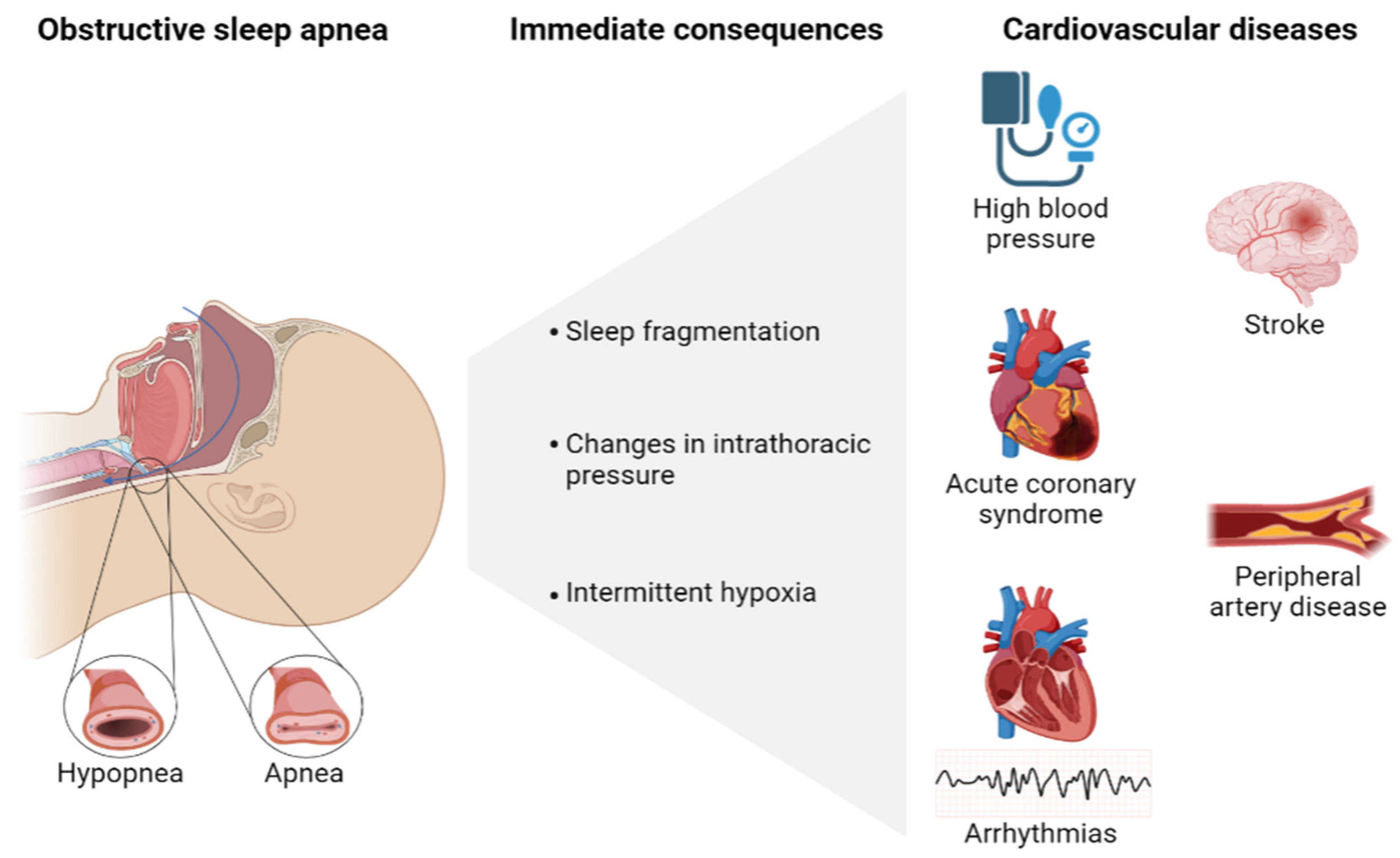
1.1. Role of Obstructive Sleep Apnea in the Development of Cardiovascular Disease

Obstructive sleep apnea (OSA) is a sleep-related breathing disorder defined as episodes of recurrent upper-airway obstructions during sleep that causes intermittent hypoxia, changes in intrathoracic pressure and sleep fragmentation. The prevalence of OSA has been increasing over time and has been estimated that it might affect 1 billion people worldwide [1]. OSA severity is measured by the number of respiratory events per hour (apnea–hypopnea index (AHI)) considering moderate–severe OSA when the AHI is ≥15 events/h. The most effective treatment for OSA is the continuous positive airway pressure (CPAP) that restores the AHI, improves symptoms and the quality of life of OSA patients [2].
Based on the evidence, OSA can be considered a condition that increases cardiovascular (CV) risk and mortality, including: high blood pressure (HBP), acute coronary syndrome (ACS), arrhythmias, heart failure, stroke and peripheral artery disease (Figure 1). In fact, the prevalence of OSA is higher in patients with CV disease (up to 49%) [3]. The strongest evidence of this relationship is in HBP [4,5] and has been established to have a higher prevalence of OSA (up to 85%) in patients with resistant HBP, with OSA being the most frequent cause of secondary HBP [6]. Additionally, OSA independently increases the risk of coronary artery disease, coronary events [7,8] and has been shown as a possible risk factor for suffering a nocturnal ischemic episode [9]. OSA has also been implicated in an increased risk of CV events after coronary revascularization [10] and with a significant increase in ACS recurrence compared to mild or no OSA [11,12,13].
Whether CPAP therapy prevents major CV events is uncertain. Well-designed randomized controlled studies analyzing the effect of CPAP treatment on CV consequences have not been able to demonstrate the effect of CPAP in CV events prevention [14,15,16,17]. Nevertheless, different studies suggested a potential heterogeneous deleterious role of OSA that would be dependent of some characteristics such as good compliance treatment [14,15], the patient endotype (patients without a previous CV event) [14,18] or the type of the CV event (specifically cerebrovascular) [16] were associated with a better response to treatment. This special response in a certain patient profile suggests that these individuals could be more susceptible to the negative effects of OSA, increasing the risk of developing a new CV event and/or improving the treatment response.
The mechanisms by which some groups of OSA patients would have an increased risk of CV events recurrence and the mechanism implicated are still unknown. It is mandatory to search for specific CV biomarkers for a better understanding of the disease and move toward a personalized medicine in OSA.

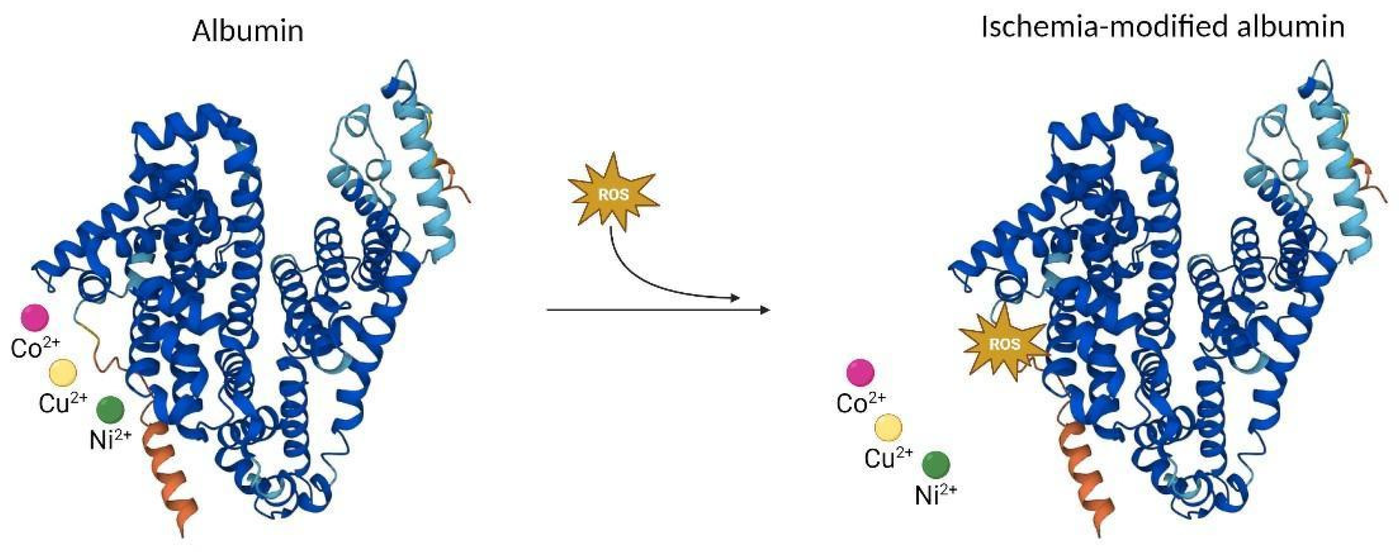
1.2. Obstructive Sleep Apnea Biomarkers

The measurements obtained in the sleep study establish the severity through the AHI, but this index does not always correlate with the patient’s symptoms and is not predictive of the morbidities associated with OSA. From a pathophysiological point of view, episodes of upper airway obstruction involve a decrease in oxygen saturation (SaO2), with intermittent hypoxia followed by reoxygenation that causes sympathetic activation, endothelial dysfunction, hypercoagulability, oxidative stress, inflammation, and metabolic dysregulation [19,20]. Chronic inflammation, as well as oxidative stress, measured through blood biomarkers, play a very important role in diseases associated with OSA, such as CV diseases. In patients with OSA, an increase in blood biomarkers has been described as inflammation biomarkers (C-reactive protein (CRP) and tumor necrosis alpha factor (TNF-alpha)) [21,22], inflammatory cytokines (interleukin 6 (IL-6), interleukin 8 (IL-8)) [21], endothelial dysfunction markers (nitric oxide) [23], hypercoagulability disorders (D-dimer, tissue factor, thrombin and antithrombin levels) [24] and oxidative stress markers (reactive oxygen species (ROS)) [25].
However, none of these biomarkers have been shown to be useful in the identification of patients with OSA, the determination of its severity, or to have the ability to predict the prognosis or response of treatment to date. For this reason, investigations into new biomarkers, with true clinical utility, are required. Recently, a new CV biomarker, ischemia-modified albumin (IMA) has been postulated as a better predictor of disease implications than classical biomarkers [26].
Albumin is the protein with the highest plasma concentration, and among its functions is the transport of metal ions, with a high affinity for copper, cobalt and nickel in its amino terminal region (N-terminal). When there is ischemia, a change in the N-terminal occurs that reduces this binding capacity, with ROS probably being the causative agent of this change, giving rise to IMA [27,28] (Figure 2). This change allows us to determine IMA concentration using the albumin cobalt binding test, which identifies colored complexes from unbound cobalt ions [29].
IMA is a new blood biomarker accepted by the Food and Drug Administration (FDA, USA) for the diagnosis of ACS in adults [30,31] as a direct measurement of oxidative stress, an intermediate mechanism involved in the development of arteriosclerosis and CV disease. IMA has been considered a biomarker for the increased risk of CV disease [32,33,34]. Kazanis et al. found that IMA levels were associated with an increased risk of coronary disease (OR = 1.23, 1.16–1.31, p < 0.001) and established that the best cut-off point of the IMA for this condition was 101.5 kU/L (sensitivity and specificity of 87.7% and a negative predictive value of 83.3%) [35].
IMA has been postulated to increase the diagnostic sensitivity of conventional cardiac biomarkers. Sinha et al. determined the levels of IMA (in addition to myoglobin, creatine kinase isoform 2 (CK-MB), troponin I (TnI)) in patients with suspected ischemia [36]. The combination of myoglobin, CK-MB and TnI provided a diagnostic sensitivity of 57%, whereas, when they were evaluated in combination with IMA sensitivity, increased to 97% with a negative predictive value of 92%. Gurumurthy et al. detected higher IMA serum concentrations in ACS patients compared to healthy subjects, with a positive predictive value of 96%, a specificity of 80% and a sensitivity of 70%, suggesting a potential role of this protein as a biomarker of ACS [37]. Mehta et al. found higher levels of IMA and cardiac troponin T (hs-cTnT) in patients with ACS compared to controls, indicating that it may be useful for the risk stratification of ACS and a better accuracy in the early diagnosis [38]. Finally, Shin et al. found that patients with unstable angina had greater serum IMA values than patients with non-ischemic chest pain admitted to the emergency department with symptoms suggestive of ACS. The sensitivity of serum IMA was estimated to be 0.74 and the specificity, 0.40 [39].
It could be plausible that the repeated desaturation–resaturation events produced by OSA create a state of ischemia, which influences the albumin N-terminal region. Oxidative stress is one of the demonstrated intermediate mechanisms involved in OSA and is likely responsible for CV damage caused by the illness, but the implicated mechanism has not been completely elucidated to this day [40].
To date, there are only a few studies that correlate OSA and IMA levels (Table 1), having described high levels of it in patients with OSA that could normalize after treatment with CPAP [41,42,43,44,45]. Yang LX et al. [41] were the first researchers investigating the relationship between IMA and OSA. The authors reported that elevated IMA levels significantly associated with OSA and positively correlated with the AHI (r = 0.462, p = 0.008). The author determined that the optimal diagnostic cut-off for the estimation of OSA was 54.00 U/L, and the levels had a significant response after CPAP treatment. These results were later confirmed by other authors [42,43,45] who found higher IMA levels in patients with OSA when compared with control, and positively correlated with OSA severity (AHI), but also with body mass index (BMI), being higher in obese patients with OSA relative to non-obese patients. However, Ozben et al. did not find a significant relation between IMA and OSA [40]. In all of these studies, patients with CV risk or disease were excluded. Uygur F et al. [43] also evaluated the impact of CPAP on circulating IMA concentrations in patients with OSA. They found significantly higher serum IMA concentrations in the OSA group than in the control group at baseline, and after three months of CPAP treatment, OSA patients had significantly lower serum IMA concentrations (0.555 ± 0.062 absorbance units (ABSU) to 0.431 ± 0.063 ABSU, p < 0.001). This response to CPAP treatment was confirmed by other authors in different studies [44,46].
In the evaluation of IMA levels in OSA patients with CV consequences, carotid intima media thickness has been evaluated, being significantly higher in the OSA group and positively correlated with the AHI and oxygen desaturation index (ODI). These results reflect that IMA, as an oxidative stress biomarker, is related to an increased risk of atherosclerosis production [47].
Taking into account all this evidence, the aim of the present study was to evaluate the role of IMA as a potential biomarker for determining the impact of OSA in ACS patients. We tried to identify other factors associated with ACS severity implicated in this association.

2. Results

2.1. Patient Characteristics

A total of 925 patients with OSA and ACS, from the ISAACC study (Impact of sleep apnea syndrome in the evolution of acute coronary syndrome. Effect of intervention with CPAP) (NCT01335087) were included. For the whole sample, the mean age of the population was 59 ± 10 years with a mean BMI of 28.8 ± 4.6 kg/m2 and 15.5% were female. Excessive daytime sleepiness (EDS) measured by the Epworth sleepiness scale (ESS) was 5 ± 3. According to the sleep study, patients were divided into three groups based on OSA severity (AHI): non-OSA/mild OSA (27.6%), moderate OSA (32.4%) and severe OSA (40.0%). The mean AHI registered was 28.50 ± 21.61 events/h (non-OSA/mild OSA: 6.11 ± 3.97; moderate OSA: 21.38 ± 4.50; severe OSA: 49.70 ± 17.42). Nocturnal SaO2 parameters were reduced, including mean SaO2 (92.2 ± 7.5%) and lowest SaO2 (82.4 ± 10.0%), related to OSA severity (Table 2). Moreover, similar comorbidities were present in the three groups except for HBP, which was more prevalent in severe patients and all groups received similar pharmacological treatment, except for calcium antagonist and beta blockers. The patients’ characteristics, pharmacological treatment and sleep study parameters of the included participants specified by OSA severity are presented in Table 2. Blood sample variables are shown in detail in Table A1.

2.2. Cardiovascular Parameters Related to the Severity of Acute Coronary Syndrome

The analysis of the parameters related to the severity of the ACS among groups showed similar results with a mean number of coronary vessels affected as determined by a catheterism procedure of 1.75 ± 0.92 with a mean degree of obstruction of 87.10 ± 20.17%. Related to the treatment procedures needed by the patients in relation to the ACS, a mean of 85.1% of the patients required a stent implantation with a mean number of stents of 1.41 ± 1.14. A mean of 4.7% of the sample required a by-pass surgery procedure as treatment. The mean intensive care unit (ICU) stay was 2.21 ± 1.76 days. However, differences between groups were found in the left ventricular ejection fraction (LVEF), with a mean result of 54.94 ± 10.11% (non-OSA/mild OSA: 57.51 ± 9.36; moderate OSA: 53.21 ± 10.81; severe OSA: 54.59 ± 9.70; p < 0.001) and hospital stay (non-OSA/mild OSA: 5.98 ± 2.87; moderate OSA: 7.01 ± 5.17; severe OSA: 6.66 ± 4.29; p = 0.017). The parameters related to the severity of ACS of the included participants specified by OSA severity are presented in Table 3.

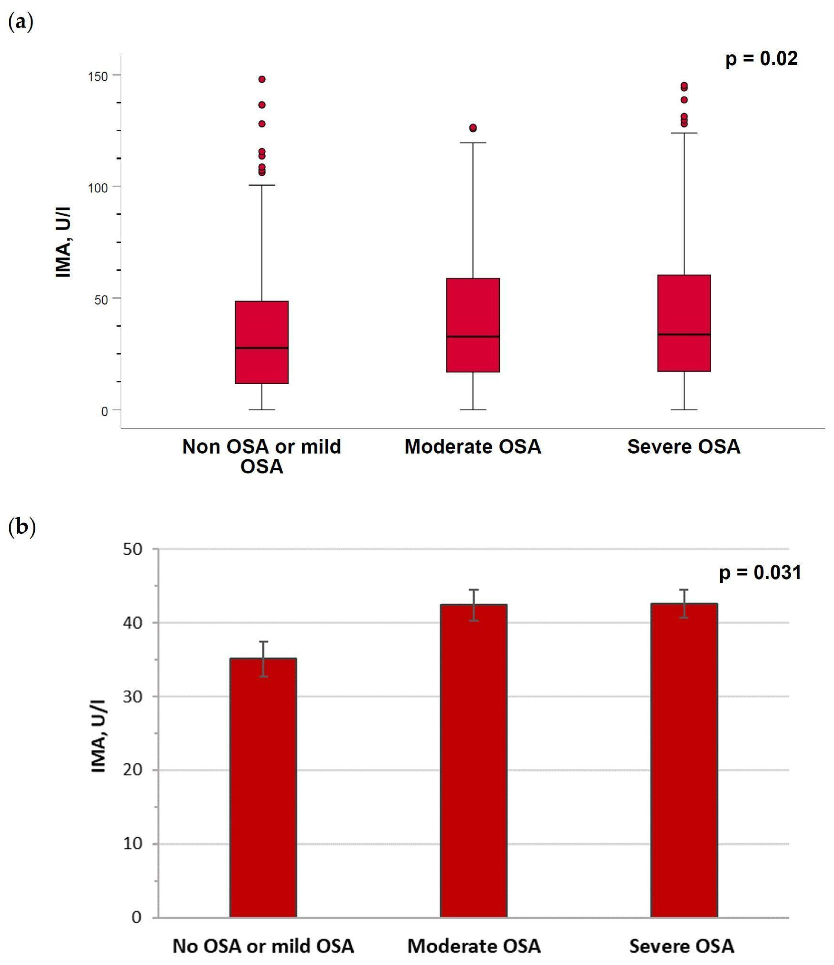
2.3. Serum Levels of Ischemia-Modified Albumin Are Increased in Patients with Moderate–Severe Obstructive Sleep Apnea

IMA levels were higher in patients with severe (median (IQR), 33.7 (17.2–60.3) U/L) or moderate (32.8 (16.9–58.8) U/L) OSA compared with non-OSA/mild OSA (27.7 (11.8–48.6) U/L) (p = 0.002) (Figure 3a) in the unadjusted analysis. When the analysis of the IMA values was performed, adjusted by gender, age, BMI, neck circumference and waist/hip ratio, the levels remained higher in severe (adjusted mean ± SEM, 42.6 ± 1.9 U/L) or moderate (42.4 ± 2.1 U/L) OSA patients versus non-OSA/mild OSA patients (35.1 ± 2.4 U/L) (p = 0.031) (Figure 3b).

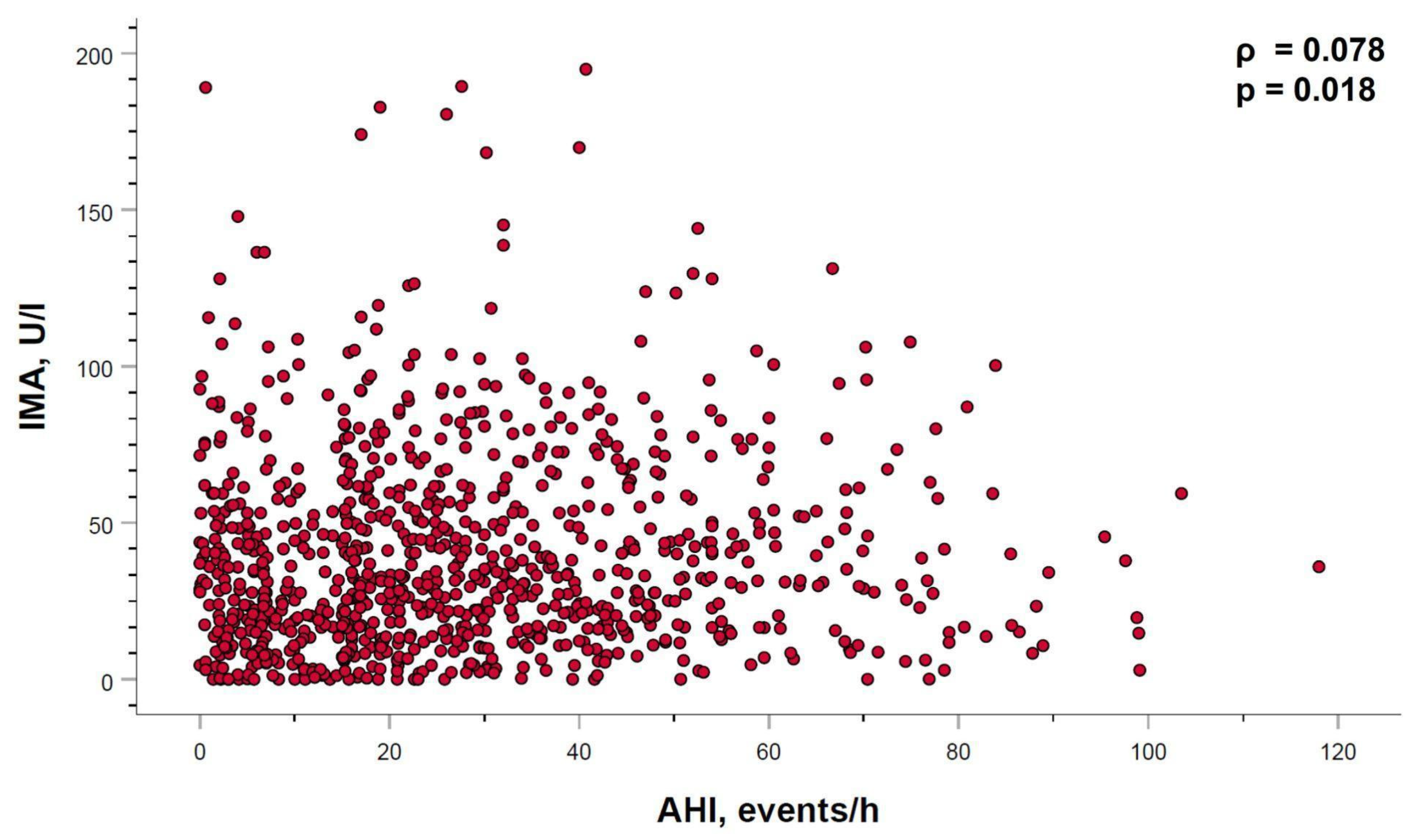
2.4. Relation between Ischemia-Modified Albumin Levels and Sleep Parameters

In the overall study subjects, serum IMA levels showed a very weak but statistically significant positive correlation with the AHI (ρ = 0.078, p < 0.018) (Figure 4). However, when the AHI–IMA relationship was adjusted for age, sex and BMI, statistical significance disappeared (adjusted regression coefficient (ß): 0.066; 95% CI: −0.038 to 0.171; p = 0.213). No other relationship between IMA levels and the remaining evaluated sleep parameters were identified.

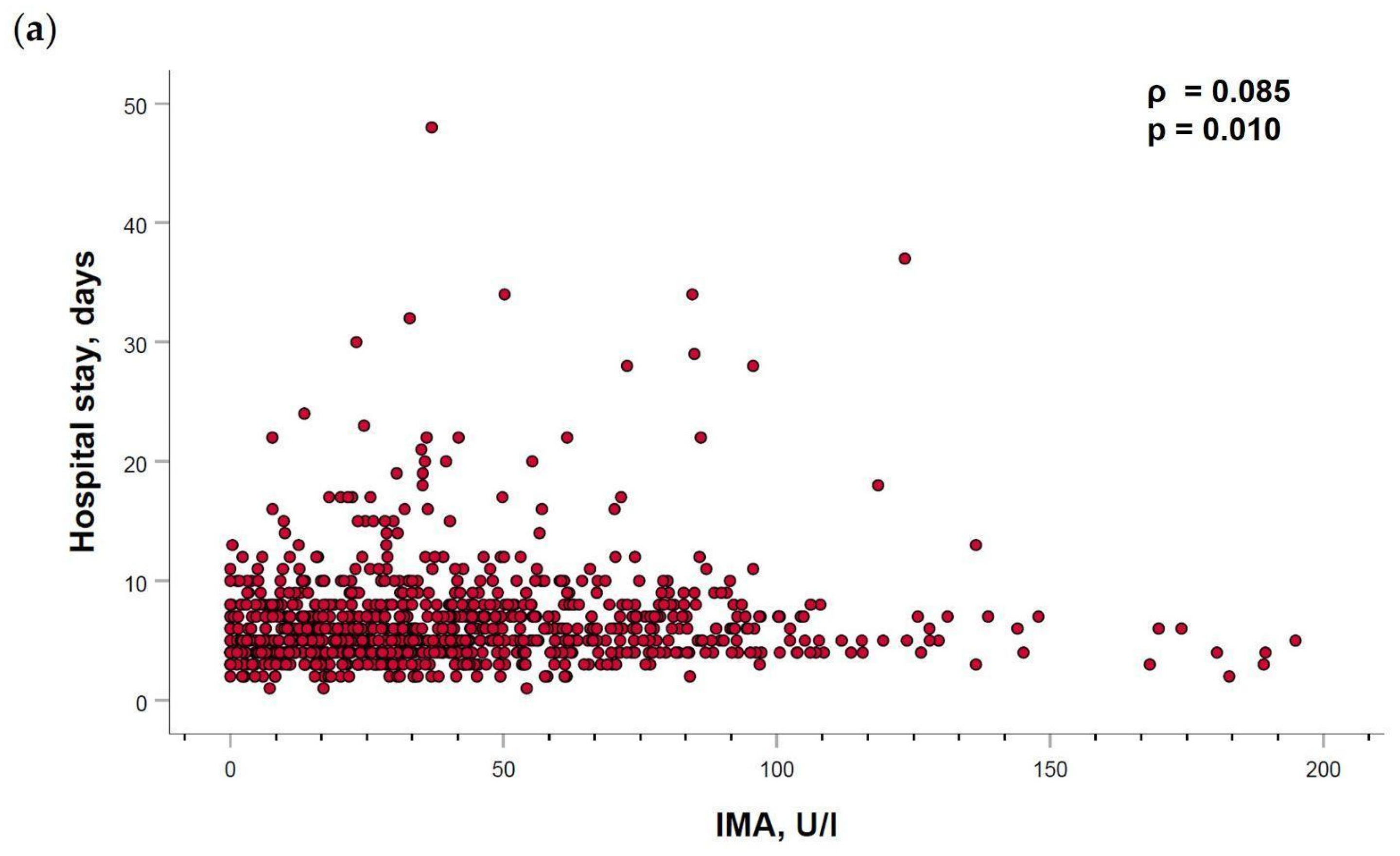
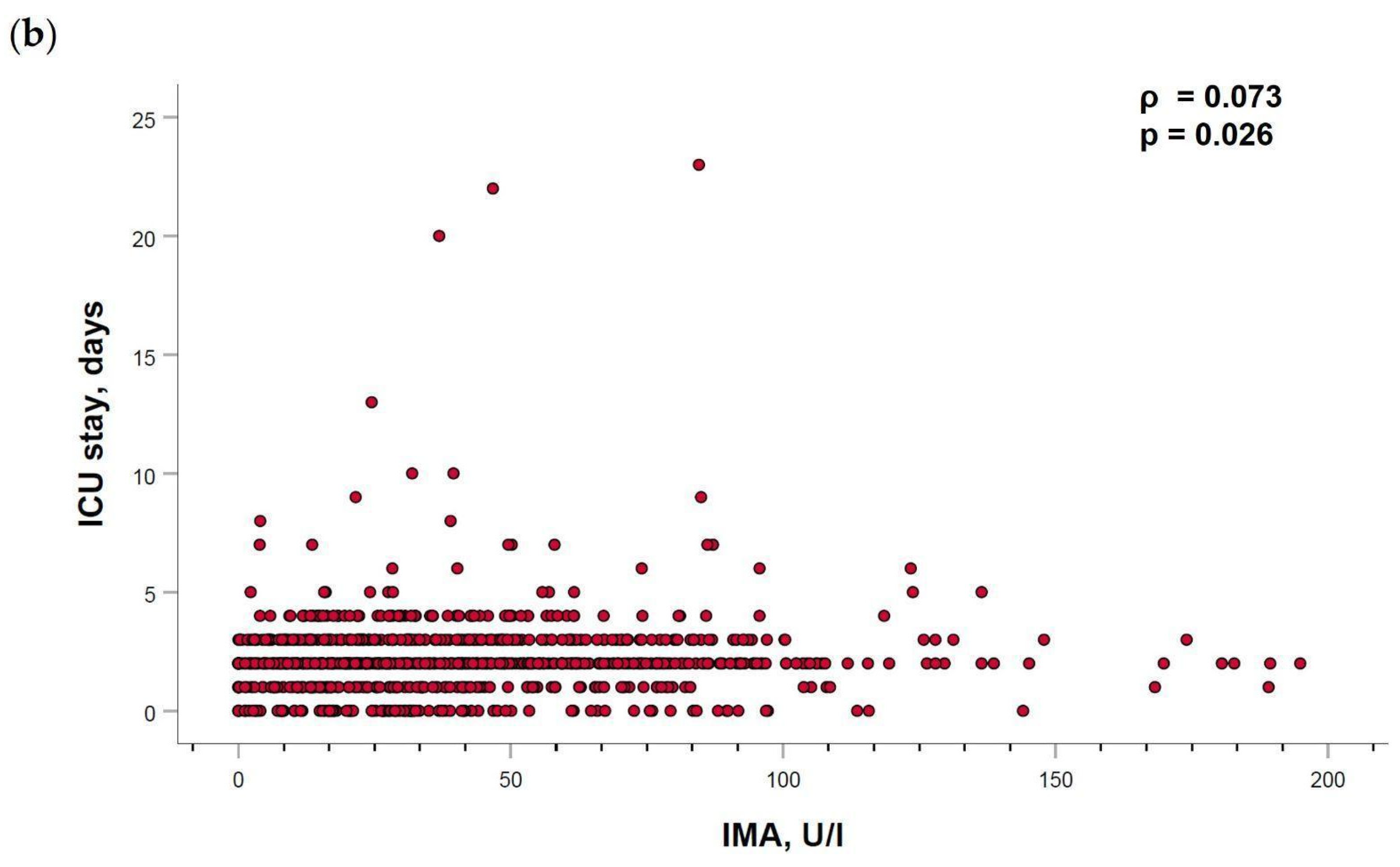
2.5. Ischemia-Modified Albumin Levels Correlation with Cardiovascular Parameters

IMA showed a weak positive significant correlation with the days of hospital stay (ρ = 0.085, p = 0.01) and the days needed in the ICU (ρ = 0.073, p = 0.026) (Figure A1). Interestingly, the relationship between IMA levels and days of hospital stay was maintained after adjusting for age, sex and BMI (adjusted regression coefficient (ß): 0.410; 95% CI: 0.001 to 0.907; p = 0.013). However, IMA values were not correlated with the rest of severity CV parameters such as LVEF, number of coronary vessels affected, degree of coronary obstruction, type of treatment procedures needed (stent implantation or by-pass surgery), history of previous ACS nor the Killip scale.

3. Discussion

The results of the present study report, for the first time, the potential role of OSA promoting the synthesis of IMA biomarkers in patients with an ACS. This result indicates that in patients admitted with an ACS, OSA is weakly associated with increased levels of IMA, suggesting a smaller potential implication of OSA as a CV risk factor in this type of patients.
Only a few of the previous studies showed results in OSA patients, with smaller samples, and excluding patients with CV disease. Düger et al. [26], in a retrospective study in 86 OSA patients, showed that serum IMA levels were significantly higher in patients than controls (p = 0.008). The level increased significantly with OSA severity (r = 0.50, p < 0.001), with results that were consistent with previous studies [41,42,43]. However, some other studies did not find different levels of IMA between OSA patients and controls [40]. These previous existing studies excluded patients with CV risk and/or disease as it is a very frequent condition in OSA patients. To the best of our knowledge, the impact of OSA severity in IMA levels of patients with ACS has not been previously analyzed, and our results establish for the first time this association.
Importantly, the identified association of IMA levels with OSA severity groups in the present study remained significant when adjusted for confounding factors such as age, sex, BMI, neck circumference and waist/hip ratio. These results would indicate the potential relationship between OSA severity and this CV risk biomarker and point to a possible pathophysiological pathway in relation to OSA/CV risk. OSA has been related with an increased CV morbidity and mortality, with oxidative stress as one of the most important mechanisms proposed. The AHI reflects repetitive episodes of hypoxia and reoxygenation experienced during sleep, resulting in the increased production of ROS, responsible for N-terminal albumin modification. These results indicate that IMA may act as a biomarker for oxidative stress in patients with ACS and concomitant OSA.
It is important to highlight that the previous analysis from the ISAACC cohort did not demonstrate a deleterious effect of OSA in CV recurrence. Nevertheless, specific endotypes of patients with OSA and ACS would be vulnerable to OSA increasing the risk of CV recurrence [18]. These results manifest a potential heterogeneously deleterious effect of OSA that would be dependent on the patient’s profile [48]. Moreover, a previous analysis from the ISAACC study evaluated the influence of OSA on the severity of ACS and a short-term prognosis [49], concluding that OSA is associated with an increased ACS severity and length of stay in the ICU. The result of IMA evaluation of the present study would suggest a possible deleterious effect of OSA in the expression of this biomarker of CV disease. Additionally, these results could have an important interpretation in the negative results of RCT, exploring the effect of CPAP in the prevention of CV events or mortality. If OSA has weaker deleterious CV effects in ACS patients, this could explain the negative results. In the ISAACC [17] and SAVE [16] studies, all the participants had previous CV illness. This hypothesis could suggest a possible positive effect of OSA treatment as a primary prevention tool in CV risk prevention.
Another important aspect is the lack of correlation between the IMA and the respiratory sleep parameters. Currently, it has been suggested that the classical parameters that define OSA severity would be insufficient for the identification of associated CV morby-mortality. A new and promising parameter, the hypoxic burden, has recently been described, with a better association to CV risk than AHI in OSA patients [50]. This parameter explores not only the number and frequency of events/desaturation, but also the depth and length of the desaturation. These results would encourage exploring the value of these new variables in the severity and consequences of OSA. Other authors suggest investigating different pathophysiological pathways (dysbiosis, alterations in the autonomic nervous system) responsible for increasing CV risk in these patients [51].
Some study limitations need to be commented on. Firstly, in relation to the timing of the blood sample extraction, it has been suggested by some authors [52] that IMA could have a dynamic ischemic damage in OSA with regard to anoxia produced during the night. This hypothesis has not yet been confirmed, but could be relevant in IMA determination in OSA patients. If there exists a dynamic change in IMA concentrations in OSA patients, this point could help determine the results. However, IMA increases within minutes after the onset of ischemia until 6–12 h [53]. Blood samples in our study were drawn early in the morning, so were properly timed to yield positive results. Secondly, since the results of the present study have been performed in a population of patients with ACS, the present results cannot be generalized to other profiles of patients with OSA. Thirdly, in the ISAACC study, patients with EDS were excluded. Nevertheless, the number of patients excluded for this cause were relatively low and the sample is representative of the phenotype of patients with OSA and ACS. Fourthly, women are underrepresented in this sample, with a predominance of male subjects. Finally, the sleep study for the OSA diagnosis was respiratory polygraphy, which could underestimate OSA severity. However, the effect of OSA in IMA values is most likely determined more by the desaturation than the arousability, and this underestimation is probably slight.
The strengths of the study comprise the large number of samples collected in a well-designed randomized clinical trial in an OSA and ACS multicenter study from a large number of patients. Additionally, all participating centers performed the same blood process methodology and the sleep study was performed with the same model of polygraph. All the variables were collected systematically.

4. Materials and Methods

4.1. Participants

This is an ancillary blood sample analysis of the ISAACC study: Impact of sleep apnea syndrome in the evolution of acute coronary syndrome. Effect of intervention with CPAP. ISAACC is a multicenter, open-label, parallel, prospective, randomized and controlled trial (registration number: NCT01335087) of OSA with ACS [17]. The 925 patients were hospitalized for ACS at the time of the inclusion of the study in 15 different coronary units or cardiology wards around Spain. They were patients without EDS with an ESS < 10. The inclusion and exclusion criteria and details of the ISAACC study’s method has been previously reported in a previous publication [54].

4.2. Procedures

4.2.1. General Procedures

The demographic (age and sex) and anthropometric characteristics (BMI, neck and waist/hip ratio), medical history of chronic disease and usual pharmacological treatment were recorded systematically by a sleep specialist before the sleep study. The degree of EDS was calculated using validated questionnaires to this aim (ESS) [55]. Seated BP was determined from two replicate measurements obtained at least 1 min apart.

4.2.2. Sleep Study Methodology

For OSA diagnosis, all patients underwent a respiratory polygraphy (Embletta, ResMed, Bella Vista, NSW, Australia) in the first 24–72 h after admission, with a register of nocturnal respiratory parameters. Airflow and snoring were measured using a nasal transducer; thoracic and abdominal movements using inductive plethysmography. Blood oxygen saturation and heart rates were measured by digital pulse oximetry and position was recorded by actigraphy. Respiratory events were manually scored, and the criteria for OSA diagnosis and severity were established according to the national guidelines [56]. OSA severity was defined by AHI: mild ≥ 5 events/h and <15/h; moderate ≥ 15 events/h and <30; and severe ≥ 30 events/h.

4.2.3. Blood Sample Determination

For the current post hoc study, to identify the IMA values, a blood sample test was performed the morning after the sleep study. Overnight fasting venous blood samples were obtained from all participants. EDTA (ethylene diamine tetra-acetic acid) anticoagulant tubes were centrifuged (1500× g for 10 min at 4 °C) to separate the serum fraction. All specimens were immediately aliquoted, frozen and stored in a dedicated −80 °C freezer until used in the assay. Serum IMA was measured using the human IMA ELISA (enzyme-linked immunosorbent assay) (CSB-E09594h; Cusabio, Houston, TX, USA). Measurements for serum samples were performed in duplicate. The detection limits of the assays were 3.12 IU/mL, while intra-assay and inter-assay variations were below 20%.
All patients underwent fasting blood extraction tubes without anticoagulant, EDTA and citrate. Measures determined were as follows: hemogram, basic biochemical profile (glucose, triglycerides, total cholesterol, LDL cholesterol, HDL cholesterol, uric acid, creatinine, AST, ALT).

4.2.4. Cardiovascular Parameters

Parameters related to ACS severity were reported systematically. Echocardiographic evaluation and Killip classification were routinely performed during patient admission. Other parameters, such as the existence of a previous ACS, parameters derived from the coronarography (number of arteries affected, level of coronary obstruction) and the date related with the procedure needed (stent or surgery and/or number of stents needed) were registered. The hospital stay and the days spent in the intensive care unit were also noted.

4.3. Statistical Analysis

Data are expressed as mean ± standard deviation (SD), median (IQR) or absolute numbers and percentages, according to the type and distribution of the variables. Normality was explored using the Shapiro–Wilk and skewness–kurtosis tests. Differences between groups were analyzed using the chi-squared test or Fisher’s exact test (categorical variables: using the second when the expected values in any of the cells of a contingency table were below 5 or below 10 when there was only one degree of freedom), ANOVA (normal variables) and the Kruskal–Wallis test (ordinary or non-normal metric variables). For between-group comparisons, IMA levels were adjusted by gender, age and body mass index using general linear models. The relationship between IMA levels with sleep parameters and clinical or biochemical variables were evaluated using Spearman’s correlation. All tests were two-tailed and a statistical significance level of 0.05 was retained. Analyses were performed using the Statistical Package for the Social Sciences (version 26.0; SPSS, Chicago, IL, USA).

5. Conclusions

IMA emerges as a biomarker that would contribute to identify a smaller deleterious CV effect of OSA when ACS is present. This could be related with a possible pathophysiological pathway in relation to oxidative stress produced by OSA, responsible for the albumin modification. In addition, said results could suggest a possible different beneficial effect of OSA treatment between primary and secondary CV risk prevention. New studies should deepen the identification of useful biomarkers for the characterization of CV damage associated with ACS–OSA patients. The identification of new variables related to OSA severity on a better correlation with CV risk could improve the identification of these types of biomarkers.

Author Contributions

Conceptualization, F.G.-R. and O.M.; methodology, F.B., O.M., P.R.-B. and C.C.-Z.; validation, E.A., C.C.-Z., E.D.-G., M.S.-d.-l.-T. and O.M.; formal analysis, F.G.-R.; investigation, P.R.-B., E.A., E.S.-P., C.C., C.C.-Z., E.D.-G., S.R.-P., J.L.I.-A., F.B., F.G.-R., M.S.-d.-l.-T., O.M. and SSN; resources, F.B., M.S.-d.-l.-T. and O.M.; data curation, E.S.-P., C.C. and C.C.-Z.; writing—original draft preparation, P.R.-B., E.S.-P., C.C., F.G.-R., M.S.-d.-l.-T. and O.M.; writing—review and editing, P.R.-B., E.S.-P., C.C., M.S.-d.-l.-T., F.G.-R. and O.M.; visualization, O.M.; supervision, F.G.-R. and O.M.; funding acquisition, F.B., M.S.-d.-l.-T. and O.M. All authors have read and agreed to the published version of the manuscript.

Funding

This research was supported by funds received by the ISCIII (PI18/00565 and PI22/01653, PI21/00337) co-funded by the European Union—FEDERco “Una manera de hacer Europa”, the Spanish Respiratory Society (Sociedad Española de Neumología y Cirugía Torácica- SEPAR (1282/22) and NEUMOMADRID (Sociedad Madrileña de Neumología y Cirugía Torácica). M.S.-T. received financial support from a “Ramón y Cajal” grant (RYC2019-027831-I) from the “Ministerio de Ciencia e Innovación—Agencia Estatal de Investigación” co-funded by the European Social Fund (ESF) “Investing in your future”.

Institutional Review Board Statement

The study was conducted in accordance with the Declaration of Helsinki and approved by the Ethics and Clinical Trials Committee of the Hospital Universitario de Guadalajara.

Informed Consent Statement

Informed consent was obtained from all subjects involved in the study.

Data Availability Statement

All data generated or analyzed during this study are included in this published article.

Acknowledgments

We greatly appreciate the patients included in the ISAACC study, the clinical and research teams implicated in the studies and the Spanish Sleep Network.

Conflicts of Interest

F.B. received a research grant from ResMed (an Australian company that develops products related to sleep apnea), the Health Research Fund, the Spanish Ministry of Health, the Spanish Respiratory Society, the Catalonian Cardiology Society, Esteve-Teijin (Spain), Oxigen Salud (Spain) and ALLER to develop the ISAACC trial. ResMed partly funded the ISAACC study but did not participate in nor was involved in decisions regarding study development or the present manuscript. All other authors declare no competing interest.

Abbreviations

ABSUabsorbance units
ACSacute coronary syndrome
AHIapnea hypopnea index
ALTalanine aminotransferase
APTTactivated partial thromboplastin clotting time
ARAIIangiotensin II receptor antagonists
ASTaspartate aminotransferase
ACEIangiotensin convertase enzyme inhibitors
BMIbody mass index
BPblood pressure
CKcreatine kinase
CK-MBcreatine kinase isoform 2
CPAPcontinuous positive airway pressure
CRPC-reactive protein
CVcardiovascular
EDSexcessive daytime sleepiness
EDTAethylene diamine tetra-acetic acid
ELISAenzyme-linked immunosorbent assay
ESSEpworth sleepiness scale
FDAFood and Drug Administration
HBPhigh blood pressure
HDLhigh density lipoprotein
Hs-cTnThigh sensitive cardiac troponin T
ICUintensive care unit
IL-6interleukin 6
IL-8interleukin 8
IQRinterquartile range
IMAischemia-modified albumin
LDLlow density lipoprotein
LVEFleft ventricular ejection fraction
Min SaO2minimum nocturnal oxygen saturation
mDBPmedian diastolic blood pressure
mSBPmedian systolic blood pressure
N-terminalamino terminal region
NYHANew York Heart Association
OSAobstructive sleep apnea
ODIoxygen desaturation index
SaO2oxygen saturation
SDstandard deviation
SEMstandard error of the mean
ROSreactive oxygen species

Appendix A

Table A1. Blood pressure and blood sample parameters in different groups.
ParametersNo/Mild OSAModerate OSASevere OSAp-Value
mSBP, mm Hg121.93 ± 17.26122.85 ± 16.41124.73 ± 18.360.122
mDBP, mm Hg72.01 ± 11.3171.76 ± 10.5572.72 ± 10.600.491
Heart rate, bpm72.25 ± 12.0872.27 ± 12.5372.64 ± 13.340.907
Hematocrit, %42.40 ± 5.6041.91 ± 4.4342.27 ± 4.270.431
Hemoglobin, g/L144.92 ± 16.34142.20 ± 15.83143.22 ± 15.720.133
Erythrocytes, ×1012/L4.72 ± 0.554.67 ± 0.534.70 ± 0.520.568
Leukocytes, ×103/µL10.11 ± 3.1710.12 ± 2.919.97 ± 2.780.774
Platelets, ×103/µL221.72 ± 61.15215.69 ± 55.21217.52 ± 63.120.489
Glucose, mg/dL120.80 ± 50.32125.05 ± 51.69131.06 ± 54.390.052
Creatinine, mg/dL1.13 ± 3.951.84 ± 8.492.96 ± 26.660.426
Uric acid, mg/dL5.59 ± 2.545.96 ± 1.566.46 ± 5.160.047
Cholesterol, mg/dL175.62 ± 41.75178.57 ± 42.20179.65 ± 43.870.523
HDL, mg/dL41.66 ± 11.7441.07 ± 11.2240.79 ± 18.130.780
LDL, mg/dL106.94 ± 37.60107.74 ± 34.90110.36 ± 35.790.487
Triglycerides, mg/dL138.47 ± 79.56149.04 ± 81.31164.74 ± 117.050.005 *
AST, U/L60.83 ± 64.0578.17 ± 93.0768.89 ± 86.430.082
ALT, U/L35.73 ± 29.2543.65 ± 130.8638.74 ± 30.950.502
Data are mean ± standard deviation according to their distribution. * Significant p-values (p < 0.05). OSA: Obstructive sleep apnea; mSBP: mean systolic blood pressure; mDBP: mean diastolic blood pressure; bpm: beats per minute; HDL: high density lipoprotein; LDL: low density lipoprotein; AST: aspartate aminotransferase; ALT: alanine aminotransferase; CK: creatine kinase.

Appendix B

Figure A1. IMA levels correlation with hospitalization. Relationship of serum levels of IMA with (a) hospital and (b) intensive care unit (ICU) stay in patients with OSA and acute coronary syndrome. Spearman’s correlation coefficients (ρ) and p-values are provided.
Ijms 24 09019 g0a1aIjms 24 09019 g0a1b

References

  1. Benjafield, A.V.; Ayas, N.T.; Eastwood, P.R.; Heinzer, R.; Ip, M.S.M.; Morrell, M.J.; Nunez, C.M.; Patel, S.R.; Penzel, T.; Pépin, J.L.; et al. Estimation of the global prevalence and burden of obstructive sleep apnoea: A literature-based analysis. Lancet Respir. Med. 2019, 7, 687–698. [Google Scholar] [CrossRef]
  2. Mediano, O.; González-Mangado, N.; Montserrat, J.M.; Alonso-Álvarez, M.L.; Almendros, I.; Alonso-Fernández, A.; Barbé, F.; Borsini, E.; Caballero-Eraso, C.; Cano-Pumarega, I.; et al. International Consensus Document on Obstructive Sleep Apnea. Arch. Bronconeumol. 2022, 58, 52–68. [Google Scholar] [CrossRef]
  3. Suen, C.; Wong, J.; Ryan, C.M.; Goh, S.; Got, T.; Chaudhry, R.; Lee, D.S.; Chung, F. Prevalence of Undiagnosed Obstructive Sleep Apnea among Patients Hospitalized for Cardiovascular Disease and Associated In-Hospital Outcomes: A Scoping Review. J. Clin. Med. 2020, 9, 989. [Google Scholar] [CrossRef] [PubMed]
  4. Shahar, E.; Whitney, C.W.; Redline, S.; Lee, E.T.; Newman, A.B.; Nieto, F.J.; O’Connor, G.T.; Boland, L.L.; Schwartz, J.E.; Samet, J.M. Sleep-disordered breathing and cardiovascular disease: Cross-sectional results of the Sleep Heart Health Study. Am. J. Respir. Crit. Care Med. 2001, 163, 19–25. [Google Scholar] [CrossRef] [PubMed]
  5. Peppard, P.E.; Young, T.; Palta, M.; Skatrud, J. Prospective study of the association between sleep-disordered breathing and hypertension. N. Engl. J. Med. 2000, 342, 1378–1384. [Google Scholar] [CrossRef] [PubMed]
  6. Khan, A.; Patel, N.K.; O’Hearn, D.J.; Khan, S. Resistant hypertension and obstructive sleep apnea. Int. J. Hypertens. 2013, 2013, 193010. [Google Scholar] [CrossRef] [PubMed]
  7. Gottlieb, D.J.; Yenokyan, G.; Newman, A.B.; O’Connor, G.T.; Punjabi, N.M.; Quan, S.F.; Redline, S.; Resnick, H.E.; Tong, E.K.; Diener-West, M.; et al. Prospective study of obstructive sleep apnea and incident coronary heart disease and heart failure: The sleep heart health study. Circulation 2010, 122, 352–360. [Google Scholar] [CrossRef]
  8. Peker, Y.; Carlson, J.; Hedner, J. Increased incidence of coronary artery disease in sleep apnoea: A long-term follow-up. Eur. Respir. J. 2006, 28, 596–602. [Google Scholar] [CrossRef]
  9. Kuniyoshi, F.H.; Garcia-Touchard, A.; Gami, A.S.; Romero-Corral, A.; Van der Walt, C.; Pusalavidyasagar, S.; Kara, T.; Caples, S.M.; Pressman, G.S.; Vasquez, E.C.; et al. Day-night variation of acute myocardial infarction in obstructive sleep apnea. J. Am. Coll. Cardiol. 2008, 52, 343–346. [Google Scholar] [CrossRef]
  10. Lee, C.H.; Sethi, R.; Li, R.; Ho, H.H.; Hein, T.; Jim, M.H.; Loo, G.; Koo, C.Y.; Gao, X.F.; Chandra, S.; et al. Obstructive Sleep Apnea and Cardiovascular Events after Percutaneous Coronary Intervention. Circulation 2016, 133, 2008–2017. [Google Scholar] [CrossRef]
  11. Nakashima, H.; Kurobe, M.; Minami, K.; Furudono, S.; Uchida, Y.; Amenomori, K.; Nunohiro, T.; Takeshita, S.; Maemura, K. Effects of moderate-to-severe obstructive sleep apnea on the clinical manifestations of plaque vulnerability and the progression of coronary atherosclerosis in patients with acute coronary syndrome. Eur. Heart J. Acute Cardiovasc. Care 2015, 4, 75–84. [Google Scholar] [CrossRef]
  12. Qu, H.; Guo, M.; Zhang, Y.; Shi, D.Z. Obstructive sleep apnea increases the risk of cardiac events after percutaneous coronary intervention: A meta-analysis of prospective cohort studies. Sleep Breath. 2018, 22, 33–40. [Google Scholar] [CrossRef]
  13. Jia, S.; Zhou, Y.J.; Yu, Y.; Wu, S.J.; Sun, Y.; Wang, Z.J.; Liu, X.L.; King, B.E.; Zhao, Y.X.; Shi, D.M.; et al. Obstructive sleep apnea is associated with severity and long-term prognosis of acute coronary syndrome. J. Geriatr. Cardiol. 2018, 15, 146–152. [Google Scholar] [CrossRef]
  14. Barbé, F.; Durán-Cantolla, J.; Sánchez-de-la-Torre, M.; Martínez-Alonso, M.; Carmona, C.; Barceló, A.; Chiner, E.; Masa, J.F.; Gonzalez, M.; Marín, J.M.; et al. Effect of continuous positive airway pressure on the incidence of hypertension and cardiovascular events in nonsleepy patients with obstructive sleep apnea: A randomized controlled trial. JAMA 2012, 307, 2161–2168. [Google Scholar] [CrossRef]
  15. Peker, Y.; Glantz, H.; Eulenburg, C.; Wegscheider, K.; Herlitz, J.; Thunström, E. Effect of Positive Airway Pressure on Cardiovascular Outcomes in Coronary Artery Disease Patients with Nonsleepy Obstructive Sleep Apnea. The RICCADSA Randomized Controlled Trial. Am. J. Respir. Crit. Care Med. 2016, 194, 613–620. [Google Scholar] [CrossRef]
  16. McEvoy, R.D.; Antic, N.A.; Heeley, E.; Luo, Y.; Ou, Q.; Zhang, X.; Mediano, O.; Chen, R.; Drager, L.F.; Liu, Z. CPAP for Prevention of Cardiovascular Events in Obstructive Sleep Apnea. N. Engl. J. Med. 2016, 375, 919–931. [Google Scholar] [CrossRef]
  17. Sánchez-de-la-Torre, M.; Sánchez-de-la-Torre, A.; Bertran, S.; Abad, J.; Duran-Cantolla, J.; Cabriada, V.; Mediano, O.; Masdeu, M.J.; Alonso, M.L.; Masa, J.F.; et al. Effect of obstructive sleep apnoea and its treatment with continuous positive airway pressure on the prevalence of cardiovascular events in patients with acute coronary syndrome (ISAACC study): A randomised controlled trial. Lancet Respir. Med. 2020, 8, 359–367. [Google Scholar] [CrossRef]
  18. Zapater, A.; Sánchez-de-la-Torre, M.; Benítez, I.D.; Targa, A.; Bertran, S.; Torres, G.; Aldomà, A.; De Batlle, J.; Abad, J.; Duran-Cantolla, J.; et al. The Effect of Sleep Apnea on Cardiovascular Events in Different Acute Coronary Syndrome Phenotypes. Am. J. Respir. Crit. Care Med. 2020, 202, 1698–1706. [Google Scholar] [CrossRef]
  19. Sánchez-de-la-Torre, M.; Campos-Rodriguez, F.; Barbé, F. Obstructive sleep apnoea and cardiovascular disease. Lancet Respir Med. 2013, 1, 61–72. [Google Scholar] [CrossRef]
  20. Resano-Barrio, M.P.; Arroyo-Espliguero, R.; Viana-Llamas, M.C.; Mediano, O. Obstructive Sleep Apnoea Syndrome: Continuous Positive Airway Pressure Therapy for Prevention of Cardiovascular Risk. Eur. Cardiol. 2020, 29, e65. [Google Scholar] [CrossRef]
  21. Canto Gde, L.; Pacheco-Pereira, C.; Aydinoz, S.; Major, P.W.; Flores-Mir, C.; Gozal, D. Biomarkers associated with obstructive sleep apnea: A scoping review. Sleep Med. Rev. 2015, 23, 28–45. [Google Scholar] [CrossRef] [PubMed]
  22. Li, K.; Wei, P.; Qin, Y.; Wei, Y. Is C-reactive protein a marker of obstructive sleep apnea?: A meta-analysis. Medicine 2017, 96, e6850. [Google Scholar] [CrossRef] [PubMed]
  23. Jurado-Gámez, B.; Fernandez-Marin, M.C.; Gómez-Chaparro, J.L.; Muñoz-Cabrera, L.; Lopez-Barea, J.; Perez-Jimenez, F.; Lopez-Miranda, J. Relationship of oxidative stress and endothelial dysfunction in sleep apnoea. Eur. Respir. J. 2010, 37, 873–879. [Google Scholar] [CrossRef] [PubMed]
  24. Wu, M.F.; Chen, Y.H.; Chen, H.C.; Huang, W.C. Interactions among Obstructive Sleep Apnea Syndrome Severity, Sex, and Obesity on Circulatory Inflammatory Biomarkers in Patients with Suspected Obstructive Sleep Apnea Syndrome: A Retrospective, Cross-Sectional Study. Int. J. Environ. Res. Public Health 2020, 17, 4701. [Google Scholar] [CrossRef] [PubMed]
  25. Chen, C.Y.; Chen, C.L.; Yu, C.C.; Chen, T.T.; Tseng, S.T.; Ho, C.H. Association of inflammation and oxidative stress with obstructive sleep apnea in ischemic stroke patients. Sleep Med. 2015, 16, 113–118. [Google Scholar] [CrossRef]
  26. Düger, M.; Seyhan, E.C.; Günlüoğlu, M.Z.; Bolatkale, M.; Özgül, M.A.; Turan, D.; Uğur, E.; Ülfer, G. Does ischemia-modified albumin level predict severity of obstructive sleep apnea? Sleep Breath. 2021, 25, 65–73. [Google Scholar] [CrossRef]
  27. Mothes, E.; Faller, P. Evidence that the principal CoII-binding site in human serum albumin is not at the N-terminus: Implication on the albumin cobalt binding test for detecting myocardial ischemia. Biochemistry 2007, 46, 2267–2274. [Google Scholar] [CrossRef]
  28. Shevtsova, A.; Gordiienko, I.; Tkachenko, V.; Ushakova, G. Ischemia-Modified Albumin: Origins and Clinical Implications. Dis. Markers 2021, 2021, 9945424. [Google Scholar] [CrossRef]
  29. Bhagavan, N.V.; Lai, E.M.; Rios, P.A.; Yang, J.; Ortega-Lopez, A.M.; Shinoda, H.; Honda, S.A.; Rios, C.N.; Sugiyama, C.E.; Ha, C.E. Evaluation of human serum albumin cobalt binding assay for the assessment of myocardial ischemia and myocardial infarction. Clin. Chem. 2003, 49, 581–585. [Google Scholar] [CrossRef]
  30. Bar-Or, D.; Lau, E.; Winkler, J.V. A novel assay for cobalt-albumin binding and its potential as a marker for myocardial ischemia-a preliminary report. J. Emerg. Med. 2000, 19, 311–315. [Google Scholar] [CrossRef]
  31. Gaze, D.C. Ischemia modified albumin: A novel biomarker for the detection of cardiac ischemia. Drug Metab. Pharmacokinet. 2009, 24, 333–341. [Google Scholar] [CrossRef]
  32. Talwalkar, S.S.; Bon-Homme, M.; Miller, J.J.; Elin, R.J. Ischemia modified albumin, a marker of acute ischemic events: A pilot study. Ann. Clin. Lab. Sci. 2008, 38, 132–137. [Google Scholar] [PubMed]
  33. Zhong, Y.; Wang, N.; Xu, H.; Hou, X.; Xu, P.; Zhou, Z. Ischemia-modified albumin in stable coronary atherosclerotic heart disease: Clinical diagnosis and risk stratification. Coron. Artery Dis. 2012, 23, 538–541. [Google Scholar] [CrossRef]
  34. Van Belle, E.; Dallongeville, J.; Vicaut, E.; Degrandsart, A.; Baulac, C.; Montalescot, G. Ischemia-modified albumin levels predict long-term outcome in patients with acute myocardial infarction. The French Nationwide OPERA study. Am. Heart J. 2010, 159, 570–576. [Google Scholar] [CrossRef]
  35. Kazanis, K.; Dalamaga, M.; Nounopoulos, C.; Manolis, A.S.; Sakellaris, N.; Jullien, G.; Dionyssiou-Asteriou, A. Ischemia modified albumin, high-sensitivity c-reactive protein and natriuretic peptide in patients with coronary atherosclerosis. Clin. Chim. Acta 2009, 408, 65–69. [Google Scholar] [CrossRef]
  36. Sinha, M.K.; Roy, D.; Gaze, D.C.; Collinson, P.O.; Kaski, J.C. Role of “Ischemia modified albumin”, a new biochemical marker of myocardial ischaemia, in the early diagnosis of acute coronary syndromes. Emerg. Med. J. 2004, 21, 29–34. [Google Scholar] [CrossRef]
  37. Gurumurthy, P.; Borra, S.K.; Yeruva, R.K.; Victor, D.; Babu, S.; Cherian, K.M. Estimation of Ischemia Modified Albumin (IMA) Levels in Patients with Acute Coronary Syndrome. Indian J. Clin. Biochem. 2014, 29, 367–371. [Google Scholar] [CrossRef]
  38. Mehta, M.D.; Marwah, S.A.; Ghosh, S.; Shah, H.N.; Trivedi, A.P.; Haridas, N. A synergistic role of ischemia modified albumin and high-sensitivity troponin T in the early diagnosis of acute coronary syndrome. J. Family Med. Prim. Care 2015, 4, 570–575. [Google Scholar] [CrossRef]
  39. Shin, H.; Kim, J.G.; Jang, B.H.; Lim, T.H.; Kim, W.; Cho, Y.; Choi, K.S.; Na, M.K.; Ahn, C.; Lee, J. Diagnostic Accuracy of Ischemia-Modified Albumin for Acute Coronary Syndrome: A Systematic Review and Meta-Analysis. Medicina 2022, 58, 614. [Google Scholar] [CrossRef]
  40. Ozben, S.; Huseyinoglu, N.; Hanikoglu, F.; Guvenc, T.S.; Yildirim, B.Z.; Cort, A.; Ozdem, S.; Ozben, T. Advanced oxidation protein products and ischaemia-modified albumin in obstructive sleep apnea. Eur. J. Clin. Invest. 2014, 44, 1045–1052. [Google Scholar] [CrossRef]
  41. Yang, L.X.; Ma, S.G.; Liu, H.; Xu, W. Influence of obstructive sleep apnea on serum butyrylcholinesterase activity and ischemia-modified albumin levels. Clinics 2013, 68, 968–973. [Google Scholar] [CrossRef] [PubMed]
  42. Dogan, D.; Ocal, N.; Aydogan, M.; Tasci, C.; Arslan, Y.; Tapan, S.; Yetkin, S.; Bilgic, H. Assessment of the role of serum ischemia-modified albumin in obstructive sleep apnea in comparison with interleukin-6. Postgrad. Med. 2016, 128, 603–608. [Google Scholar] [CrossRef] [PubMed]
  43. Uygur, F.; Tanriverdi, H.; Can, M.; Ornek, T.; Erboy, F.; Altinsoy, B.; Atalay, F.; Damar, M.; Kokturk, F.; Tor, M. The impact of obstructive sleep Apnoea and nasal continuous positive airway pressure on circulating ischaemia-modified albumin concentrations. Mediators Inflamm. 2016, 2016, 8907314. [Google Scholar] [CrossRef] [PubMed]
  44. Xu, Q.; Du, J.; Ling, X.; Lu, Y. Evaluation of MIh Scoring System in Diagnosis of Obstructive Sleep Apnea Syndrome. Med. Sci. Monit. 2017, 23, 4715–4722. [Google Scholar] [CrossRef]
  45. Sunnetcioglu, A.; Asker, S.; Alp, H.H.; Gunbatar, H. Increased Asymmetric Dimethylarginine and Ischemia-Modified Albumin Levels in Obstructive Sleep Apnea. Respir. Care 2016, 61, 1038–1043. [Google Scholar] [CrossRef]
  46. Varikasuvu, S.R.; Dutt, N.; Sahu, D. Obstructive sleep apnea and the effect of CPAP treatment on ischemia-modified albumin levels: A multi effect size meta-analysis with diagnostic test accuracy. Sleep Breath. 2019, 23, 179–191. [Google Scholar] [CrossRef]
  47. Karamanli, H.; Kiyici, A.; Arik, B.; Efe, D.; Akgedik, R. Influence of obstructive sleep apnea on ischemia-modified albumin levels and carotid intima-media thickness. J. Investig. Med. 2016, 64, 1035–1041. [Google Scholar] [CrossRef]
  48. Jung, E.; Ryu, H.H.; Ro, Y.S.; Cha, K.C.; Shin, S.D.; Hwang, S.O. Interactions between Sleep Apnea and Coronary Artery Disease on the Incidence of Sudden Cardiac Arrest: A Multi-Center Case-Control Study. Yonsei Med. J. 2023, 64, 48–53. [Google Scholar] [CrossRef]
  49. Barbé, F.; Sánchez-de-la-Torre, A.; Abad, J.; Durán-Cantolla, J.; Mediano, O.; Amilibia, J.; Masdeu, M.J.; Florés, M.; Barceló, A.; De la Peña, M.; et al. Effect of obstructive sleep apnoea on severity and short-term prognosis of acute coronary syndrome. Eur. Respir. J. 2015, 45, 419–427. [Google Scholar] [CrossRef]
  50. Azarbarzin, A.; Sands, S.A.; Stone, K.L.; Taranto-Montemurro, L.; Messineo, L.; Terrill, P.I.; Ancoli-Israel, S.; Ensrud, K.; Purcell, S.; White, D.P.; et al. The hypoxic burden of sleep apnoea predicts cardiovascular disease-related mortality: The Osteoporotic Fractures in Men Study and the Sleep Heart Health Study. Eur. Heart J. 2019, 40, 1149–1157. [Google Scholar] [CrossRef]
  51. Bock, J.M.; Vungarala, S.; Karim, S.; Somers, V.K. Obstructive Sleep Apnea as a Cardiovascular Risk Factor-Beyond CPAP. Can. J. Cardiol. 2021, 37, 756–765. [Google Scholar] [CrossRef]
  52. Zhong, Y.; Wang, N.; Xu, H. Is there a dynamic change in ischemia-modified albumin in patients with obstructive sleep apnea, which often leads to ischemic diseases? Clinics 2013, 68, 1474. [Google Scholar] [CrossRef]
  53. Lippi, G.; Montagnana, M.; Salvagno, G.L.; Guidi, G.C. Potential value for new diagnostic markers in the early recognition of acute coronary syndromes. CJEM 2006, 8, 27–31. [Google Scholar] [CrossRef]
  54. Esquinas, C.; Sánchez-de-la Torre, M.; Aldomá, A.; Florés, M.; Martínez, M.; Barceló, A.; Barbé, F. Rationale and methodology of the impact of continuous positive airway pressure on patients with ACS and nonsleepy OSA: The ISAACC Trial. Clin. Cardiol. 2013, 36, 495–501. [Google Scholar] [CrossRef]
  55. Johns, M.W. A new method for measuring daytime sleepiness: The Epworth sleepiness scale. Sleep 1991, 14, 540–545. [Google Scholar] [CrossRef]
  56. Consenso Nacional Sobre el Síndrome de Apneas-Hipopneas del Sueño. Spanish National Consensus in Sleep Apnea-Hypopnea Syndrome (SAHS). Arch. Bronconeumol. 2005, 41, 7–9. [Google Scholar]
Figure 1. Consequences and cardiovascular diseases implicated in obstructive sleep apnea (OSA). Created with BioRender.
Ijms 24 09019 g001
Figure 2. Structure of human ischemia-modified albumin (IMA). Albumin binds transport metal ions such as copper, cobalt and nickel in its N-terminal region. During an ischemic episode, an increase in the oxidative stress species (ROS) occurs, modifying this region and giving rise to the IMA, which reduces the binding capacity of the ions. Co: cobalt; Cu: copper; Ni: nickel; ROS: reactive oxygen species.
Ijms 24 09019 g002
Figure 3. Serum levels of IMA in the study groups. (a) Box-and-whisker plots depicting the distribution of serum levels of IMA according to the obstructive sleep apnea (OSA) severity groups corresponding to unadjusted analysis. Data are presented as median (IQR), maximum and minimum values, and overall comparisons were performed using the Kruskal–Wallis test. (b) IMA levels when the analysis was adjusted by gender, age, body mass index, neck circumference and waist/hip ratio in the three study groups. Columns correspond to the adjusted mean and the error bars represent the standard error of the mean of between-group comparisons by ANOVA.
Ijms 24 09019 g003
Figure 4. Relationship between serum levels of IMA and apnea–hypopnea index (AHI). Spearman’s correlation coefficient (ρ) and p-value sare shown.
Ijms 24 09019 g004
Table 1. Main characteristics and results of studies, including relationship between IMA levels and OSA.
Author (Year)Number of Participants (OSA/Controls)Age (Years) and Sex (% Males) (OSA/Controls)Diagnostic Criteria for OSA SeverityIMA Values (OSA/Controls)Main Results
Yang LX et al. (2013) [41]30/3240/40
69/70%
AHI ≥ 5 events/h65.10 ± 9.84/
47.50 ± 9.16 U/L
IMA levels were significantly higher in the OSA group compared with the control group. The CPAP treatment decreased the IMA levels.
There was a positive correlation between IMA values and AHI (r = 0.462, p = 0.008) at baseline.
Ozben S et al. (2014) [40]59/2551/49
56/64%
AHI ≥ 5 events/h30.30 ± 29.02/
31.50 ± 48.43
Plasma IMA levels were not statistically different between the OSA patients and controls.
Dogan D et al. (2016) [42]39/1241/40
100/100%
AHI ≥ 5 events/h0.36 ± 0.04/
0.89 ± 0.14 U/mL
IMA levels were significantly higher in the OSA group compared with the control group (r = 0.600, p < 0.05)
Mean AHI and IMA values were higher in obese + OSA group.
Uygur F et al. (2016) [43]97/3049/52
70/71%
AHI ≥ 5 events/h associated with symptoms or AHI ≥ 15 events/h 0.518 ± 0.09/
0.415 ± 0.07 ABSU
OSA is associated with elevated concentrations of IMA, which can be reversed by effective CPAP treatment. IMA concentrations correlated significantly with the AHI (r = 0.471, p < 0.001).
Sunnetcioglu A et al. (2016) [45] Mild/moderate OSA
Severe OSA
No OSA
29
28
22
46/76%
47/75%
36/82%
AHI ≥ 5 < 30/h
AHI ≥ 30/h
AHI < 5/h
1.98 ± 1.43 U/L
2.28 ± 1.00 U/L
0.84 ± 0.49 U/L
IMA levels were higher in the OSA group compared with controls.
Karamanli H et al. (2016) [47]61/2451/49
62/63%
1.23 ± 0.10/1.09 ± 0.16 ABSUSerum IMA levels were significantly higher in individuals with OSA compared with the control group. IMA levels were positively correlated with the AHI (r = 0.32, p = 0.004) at baseline.
Xu et al. (2017) [44]33/3052/49
70/70%
AHI ≥ 5 events/h associated with symptoms or AHI ≥ 15 events/h 0.58 ± 0.11/0.43 ± 0.09 ABSUThe levels of IMA were significantly higher in patients with OSA than in the control group. These values can be reversed by CPAP treatment.
A significant correlation was noted between IMA and AHI (r = 0.614; p < 0.001).
Varikasuvu S.R et al. (2019) [46]
Meta-analysis
411/216 AHI ≥ 5 events/h associated with symptoms or AHI ≥ 15 events/h OSA patients showed a significantly increased IMA level compared with the control group. These IMA values decreased with CPAP treatment
Meta-analysis of correlations showed significant associations of IMA with AHI (r = 0.448, p < 0.001).
Düger M et al. (2021) [26] 86/8345/43
63/59%
AHI ≥ 5 events/h0.46 ± 0.1/0.31 ± 0.09 ABSUIMA levels were significantly higher in patients with OSA than controls.
The serum IMA levels increased significantly as OSA severity increased and was positively correlated with the AHI (r = 0.41, p < 0.001).
Abbreviations: OSA: obstructive sleep apnea; IMA: ischemia-modified albumin; AHI: apnea–hypopnea index; CPAP: continuous positive airway pressure. ABSU: absorbance units.
Table 2. General characteristics, pharmacological treatment and sleep study parameters of the included participants specified by OSA severity.
ParametersNon-OSA/Mild OSA
(n = 255)
Moderate OSA
(n = 300)
Severe OSA
(n = 370)
Total
(n = 925)
p-Value
General characteristics
Women38 (14.9%)56 (18.7%)49 (13.2%)143 (15.5%)0.149
Age, years58 ± 1259 ± 1060 ± 1059 ± 100.199
BMI, kg/m227.0 ± 3.928.4 ± 4.030.5 ± 4.828.8 ± 4.6<0.001 *
Neck circumference, cm40 ± 440 ± 441 ± 341 ± 4<0.001 *
Waist/hip ratio0.98 ± 0.070.98 ± 0.100.99 ± 0.070.99 ± 0.080.048 *
Sleep parameters
ESS5 ± 35 ± 25 ± 25 ± 3<0.001 *
Study time, min414.40 ± 58.45406.78 ± 51.24408.14 ± 56.42409.42 ± 55.400.232
AHI, events/h6.11 ± 3.9721.38 ± 4.5049.70 ± 17.4228.50 ± 21.61<0.001 *
ODI 4%, events/h9.06 ± 15.3419.99 ± 14.0346.76 ± 39.8327.65 ± 31.96<0.001 *
Mean SaO2, %92.55 ± 8.4292.29 ± 7.6691.92 ± 6.7292.22 ± 7.520.586
Lowest SaO2, %85.05 ± 10.6182.93 ± 8.5580.14 ± 9.3482.40 ± 9.66<0.001 *
Medical history
HBP107 (42.0%)160 (53.3%)217 (58.6%)484 (52.3%)<0.001 *
Diabetes55 (21.6%)73 (24.3%)91 (24.6%)219 (23.7%)0.647
Dyslipidemia134 (52.5%)179 (59.7%)213 (57.6%)526 (56.9%)0.226
CV disease56 (22.0%)59 (19.7%)71 (19.2%)186 (20.1%)0.679
Cerebrovascular disease9 (3.5%)8 (2.7%)10 (2.7%)27 (2.9%)0.793
Respiratory disease17 (6.7%)12 (4.0%)25 (6.8%)54 (5.8%)0.255
Neurological disease18 (7.1%)14 (4.7%)13 (3.5%)45 (4.9%)0.126
Medication
ARAII32 (12.6%)45 (15.0%)69 (18.6%)148 (15.8%)0.113
ACEI56 (22.0%)60 (20.0%)82 (22.2%)198 (21.4%)0.764
Hypolipidemic96 (37.6%)117 (39.0%)140 (37.8%)353 (38.2%)0.935
Oral antidiabetic47 (18.4%)53 (17.7%)75 (20.3%)175 (18.9%)0.675
Insulin13 (5.1%)16 (5.3%)24 (6.5%)53 (5.7%)0.716
Antiplatelet agents56 (22.0%)76 (25.3%)79 (21.4%)211 (22.8%)0.441
Anticoagulants13 (5.1%)12 (4.0%)17 (4.6%)42 (4.5%)0.824
Beta blockers59 (23.1%)69 (23.0%)58 (15.7%)186 (20.1%)0.023 *
Calcium antagonists19 (7.5%)39 (13.0%)60 (16.2%)118 (12.8%)0.005 *
Diuretics36 (14.1%)49 (16.3%)76 (20.5%)161 (17.4%)0.096
Antacids65 (25.5%)74 (24.7%)96 (25.9%)235 (25.4%)0.930
Data are n (%) or mean ± standard deviation. Comparisons between groups by ANOVA or chi-squared tests referring to the global distribution of each variable in the three study groups. * Significant p-values (p < 0.05). OSA: Obstructive sleep apnea; BMI: body mass index; ESS: Epworth sleepiness scale; AHI: apnea–hypopnea index (number of events/h); ODI: oxygen desaturation index; mean SaO2: mean nocturnal oxygen saturation; SaO2: oxygen saturation; HBP: high blood pressure; CV: cardiovascular; ARAII: angiotensin II receptor antagonists; ACEI: angiotensin convertase enzyme inhibitors.
Table 3. Cardiovascular scales and parameters related to the severity of acute coronary syndrome of the included participants specified by OSA severity.
ParametersNon-OSA/Mild OSA
(n = 255)
Moderate OSA
(n = 300)
Severe OSA
(n = 370)
Total
(n = 925)
p-Value
ACS parameters
LVEF, % 57.51 ± 9.3653.21 ± 10.8154.59 ± 9.7054.94 ± 10.11<0.001 *
Number of coronary vessels, mean1.66 ± 0.911.77 ± 0.871.79 ± 0.951.75 ± 0.920.201
Vessel obstruction, %86.78 ± 20.9086.48 ± 18.4187.77 ± 20.8087.10 ± 20.170.753
Stent, % of patients215 (84.3%)261 (87.0%)311 (84.1%)787 (85.1%)0.523
Number of stents, mean1.35 ± 1.121.40 ± 1.081.45 ± 1.211.41 ± 1.140.563
Surgery, % of patients6 (2.4%)16 (5.4%)21 (5.8%)43 (4.7%)0.111
Hospital stay, days5.98 ± 2.877.01 ± 5.176.66 ± 4.296.59 ± 4.290.017 *
ICU stay, days2.06 ± 1.322.36 ± 2.002.19 ± 1.812.21 ± 1.760.127
First episode ACS215 (84.6%)255 (85.0%)320 (86.5%)790 (85.5%)0.779
Complications, % of patients18 (7.1%)38 (12.7%)41 (11.1%)97 (10.5%)0.088
NYHA scale
n1901301794990.417
0179 (94.2%)125 (96.2%)164 (91.6%)468 (93.8%)
19 (4.7%)4 (3.1%)12 (6.7%)25 (5.0%)
22 (1.1%)1 (0.8%)1 (0.6%)4 (0.8%)
30 (0.0%)0 (0.0%)2 (1.1%)2 (0.4%)
Killip
n2422873548830.095
I226 (93.4%)262 (91.3%)312 (88.1%)800 (90.6%)
II15 (6.2%)24 (8.4%)34 (9.6%)73 (8.3%)
III0 (0.0%)0 (0.0%)5 (1.4%)5 (0.6%)
IV1 (0.4%)1 (0.3%)3 (0.8%)5 (0.6%)
Data are n (%) or mean ± standard deviation. Comparisons between groups by ANOVA or chi-squared tests referring to the global distribution of each variable in the three study groups. * Significant p-values (p < 0.05). OSA: Obstructive sleep apnea; ACS: acute coronary syndrome; LVEF: left ventricular ejection fraction; ICU: intensive care unit; NYHA: New York Heart Association.
Disclaimer/Publisher’s Note: The statements, opinions and data contained in all publications are solely those of the individual author(s) and contributor(s) and not of MDPI and/or the editor(s). MDPI and/or the editor(s) disclaim responsibility for any injury to people or property resulting from any ideas, methods, instructions or products referred to in the content.

