Article

Transcriptome Profiling of Circulating Tumor Cells to Predict Clinical Outcomes in Metastatic Castration-Resistant Prostate Cancer

1
Department of Urology, Radboud Institute for Molecular Life Sciences, Radboud University Medical Center, 6525 GA Nijmegen, The Netherlands
2
Department of Medical Oncology, Radboud University Medical Center, 6525 GA Nijmegen, The Netherlands
3
ANGLE Biosciences Inc., Toronto, ON M9W 1B3, Canada
*
Author to whom correspondence should be addressed.
Int. J. Mol. Sci. 2023, 24(10), 9002; https://doi.org/10.3390/ijms24109002
Submission received: 19 April 2023 / Revised: 4 May 2023 / Accepted: 16 May 2023 / Published: 19 May 2023
(This article belongs to the Special Issue Liquid Biopsy in Cancers)

Abstract

:
The clinical utility of circulating tumor cells (CTC) as a non-invasive multipurpose biomarker is broadly recognized. The earliest methods for enriching CTCs from whole blood rely on antibody-based positive selection. The prognostic utility of CTC enumeration using positive selection with the FDA-approved CellSearchTM system has been demonstrated in numerous studies. The capture of cells with specific protein phenotypes does not fully represent cancer heterogeneity and therefore does not realize the prognostic potential of CTC liquid biopsies. To avoid this selection bias, CTC enrichment based on size and deformability may provide better fidelity, i.e., facilitate the characterization of CTCs with any phenotype. In this study, the recently FDA-approved Parsortix® technology was used to enrich CTCs from prostate cancer (PCa) patients for transcriptome analysis using HyCEADTM technology. A tailored PCa gene panel allowed us to stratify metastatic castration-resistant prostate cancer (mCRPC) patients with clinical outcomes. In addition, our findings suggest that targeted CTC transcriptome profiling may be predictive of therapy response.

Graphical Abstract

1. Introduction

The discovery of oncogenes in the 1970s led to the development of targeted therapies and initiated new hope for curative cancer treatments [1]. Solid tumor genotyping has revealed novel druggable oncogenic alterations that contribute to therapy resistance in patients with mCRPC. This has led to the development of novel treatment modalities such as poly ADP-ribose polymerase inhibitors (PARPi) for tumors with multiple loss-of-function alterations in DNA-repair genes [2]. Unfortunately, response rates have been highly variable and unpredictable [3].
Continued reliance on solid tissue biopsies to identify druggable alterations may be limiting progress. Conventional biopsies do not capture clonal heterogeneity and are limited by their invasive nature and the inaccessibility of certain lesions [4]. Inadequate insight into dynamic spatiotemporal alterations in PCa tumors has restricted the overall clinical benefit of novel therapies. Furthermore, the detection limit of traditional diagnostic imaging techniques is currently about one billion cells; below this threshold, malignant lesions are undetectable [5]. Together, these lesions can present a significant tumor burden, yield a high degree of heterogeneity, and carry underlying therapy resistance-associated genetic aberrations [6].
Though CellSearch was introduced in 2005, CTC enumeration has not been implemented as a prognostic, or an early response biomarker in daily clinical PCa management [7]. Recent developments in molecular profiling and cell sorting technologies have increased sensitivity and decreased costs. This could make CTC genotyping viable for precision diagnostics in the near future [8,9]. The collection of tumor material via liquid biopsies allows oncologists to deliver precision medicine and stratify therapy response by the detection of biomarkers, such as androgen receptor splice variant 7 (ARv7), in CTCs [10,11]. With longitudinal sampling, CTC transcriptomes present temporally accurate tumor phenotypes with clinically actionable potential [12]. While biomarker discovery is a growing field in clinical research, therapeutic innovations have outpaced diagnostic capabilities in oncology. As a result, patient stratification for personalized therapeutics remains an unmet clinical need.
In the present work, we used label-free CTC enrichment coupled with CTC transcriptome profiling to characterize multiple oncogenic pathways in mCRPC patients starting a new line of therapy. The Parsortix PR1 microfluidic system, which utilizes the same technology as the FDA-approved PC1 system, was used to enrich CTCs from whole blood using physical properties such as a larger size and reduced deformability compared to leukocytes [13]. This approach enables the capture of epithelial and mesenchymal CTCs in both single and aggregate forms (CTC clusters) [13]. The resulting tumor-enriched samples contain CTCs in a background of 200–800 leukocytes per mL of processed blood [13]. CTC-enriched samples were analyzed using a patented hybridization technology known as HyCEAD [14,15]. This multiplex gene expression assay provides the required sensitivity and specificity to enable the detection of a single CTC target in a background of other circulating epithelial cells and leukocytes [15]. HyCEAD has previously been used to predict malignancy in women with pelvic masses [16].
The primary aim of this study was to test the feasibility of label-free CTC enrichment and targeted transcriptome profiling in mCRPC to prognosticate patients. A secondary aim was identifying transcriptome profiles predictive of therapy response in CTC-enriched mCRPC samples.

2. Results

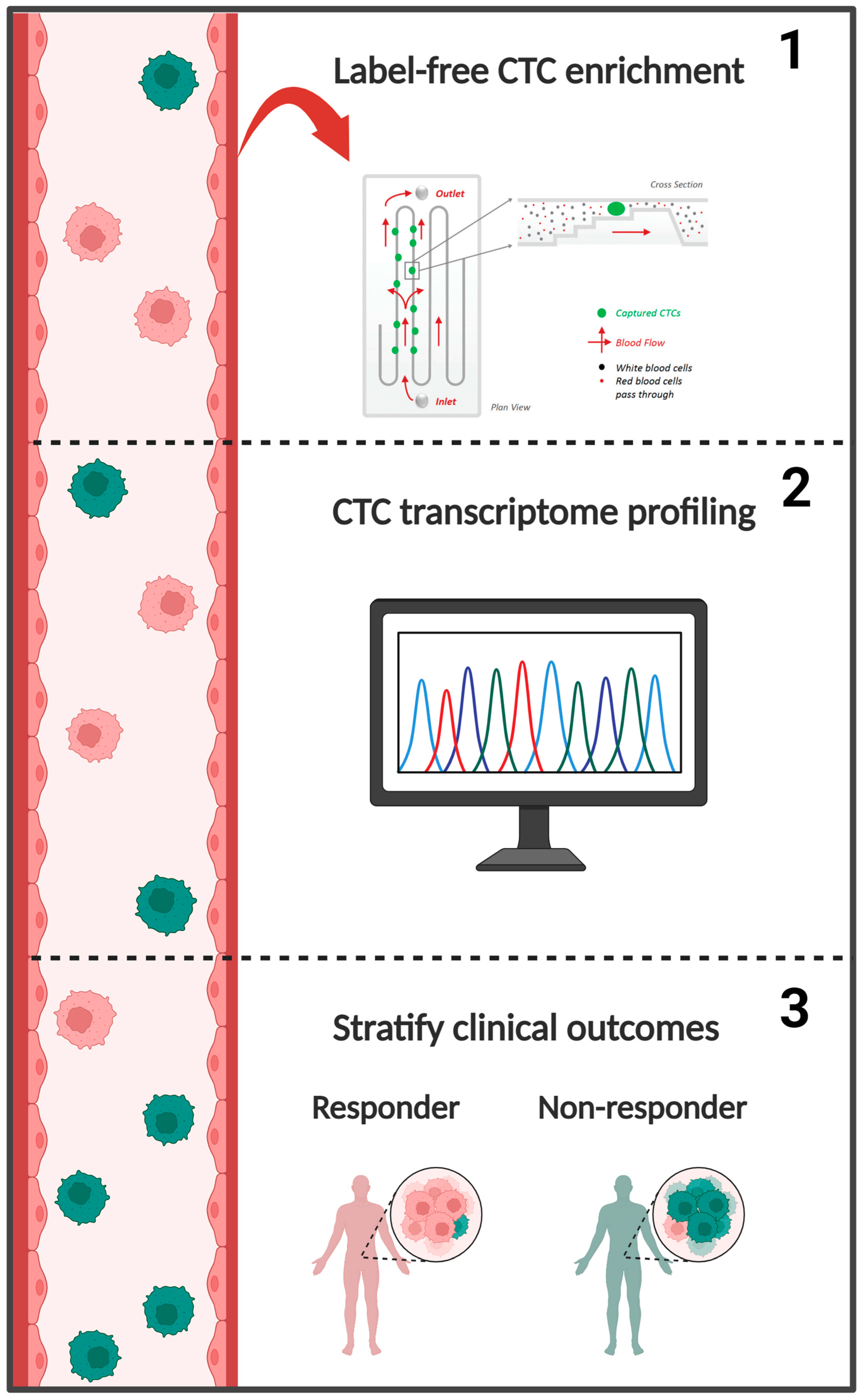
2.1. CTC Transcriptome Gene Panel

The CTC transcriptome panel consists of genes with high expression in PCa and low expression in leukocytes. Genes were selected as described in Section 4.1. In total, 64 genes were included; each gene and its relevance to PCa are shown in Table S1.

2.2. Clinical Follow-Up

In total, 40 patients provided baseline CTC samples (i.e., before starting a new line of therapy). Patients received various therapies: androgen receptor inhibitors (ARSI) (abiraterone or enzalutamide; n = 22), chemotherapy (docetaxel or cabazitaxel; n = 9), immunotherapy (Ipilimumab and Nivolumab; n = 2), radioligand therapy (PSMA-Lu177 or Radium-223; n = 2), PARPi (Olaparib; n = 2), and two were actively surveilled until signs of disease progression (i.e., they did not receive systemic therapy). The clinical trajectories, time of inclusion (CTC collection), and lines of therapy are shown in Figure 1. In one case, two CTC samples were collected from a patient receiving PARPi, ‘RAD30’ at baseline followed by ‘RAD41’ 14 days later (Figure S1). ‘RAD41’ was excluded from the primary analysis as it was not collected at baseline. Baseline alkaline phosphatase and PSA levels, age, de novo metastasis, years since CRPC diagnosis, type of therapy received following CTC collection, and 1-year survival per therapy group are shown in Table 1.

2.3. Genomic Alterations and CTC Transcriptomes

Next-generation DNA sequencing of solid tissue biopsies was done successfully in 38/40 patients. AR alterations were detected in 11 patients (29%): nine amplifications, and three mutations. In one patient, AR was amplified and mutated. TP53 alterations were found in 10 patients (26%): nine mutations, and one shallow deletion. PTEN alterations were identified in seven patients (18%): four mutations, and three shallow deletions. In nine patients (24%), there were alterations in DNA damage repair (DDR) genes: BRCA2, MSH2, RAD51B, CHEK2, and FANCL. BRCA2 was most frequently altered with three mutations and two shallow deletions. Mutations in PPP2R2A, CDK12, MSH2, RAD51B, CHEK2, RB1, PIK3CA, AKT1, SPOP, and FANCL were present at lower frequencies (2.6–5%) (Figure S2).
Due to overlap in the DNA sequencing and CTC transcriptome panels, AR and BRCA2 genomic alterations in solid tissue biopsies were associated with expression in CTCs. In eight of nine patients with AR amplifications, the expression of AR and/or associated signaling genes (KLK3, ARv7, and FOXA1) was increased in matched CTC samples, suggesting high concordance between solid tissue and liquid CTC biopsies [17]. In the patient with both AR amplification and mutation, expression of AR, KLK3, ARv7, and/or FOXA1 were not increased in CTCs (Figure S3A). Surprisingly, in two patients with BRCA2 shallow deletions, BRCA2 and associated DDR genes (BRCA1, FANCA, RRM2, and TOP2A) were expressed in matched CTCs in one case (Figure S3B). This suggests the loss of BRCA2 may not always be concordant between solid tissue and CTCs biopsies [18].

2.4. Prognostic Value of CTC Profiling

Hierarchical clustering of patients irrespective of treatment modality (referred to as the ‘therapy-agnostic cohort’) with the complete gene panel yielded two transcriptionally distinct groups (Figure S4A). Patients with high expression of AR signaling genes (ARv7, DLX1, HOXB13, and KLK3), DDR genes (BRCA1, BRCA2, FANCA, and TOP2A), and oncogenes (ERG and GRHL2) had reduced progression-free survival (PFS) (hazard ratio (HR) = 1.99, p = 0.076), and significantly reduced overall survival (OS) (HR = 5.1, p = 0.007) (Figure S4B,C). Genes with limited prognostic value were excluded from subsequent clustering to improve patient prognostication.
The relative prognostic value of genes from the complete gene panel was determined via multivariate analysis on PFS. Genes were ranked using HR (Figure S5). Notably, the epithelial marker, EPCAM, was not a high-ranking prognosticator in this cohort, yet CTCs expressing EPCAM are known to be prognostically relevant in various malignancies [7,19]. Instead, genes involved in AR signaling (KLK3, HOXB13, and GRHL2), and metastasis (MYO6 and TRPM8) were more informative [20,21]. Prognostic candidates were cross-referenced with relevant literature and went through several iterations of clustering/survival analyses to form a tailored panel consisting of the following genes: AR, ARv7, FOLH1 (aka PSMA), KLK2, KLK3, and TMPRSS2 (hereafter referred to as the ‘agnostic gene panel’) [22].
CTC profiling using the agnostic gene panel produced two transcriptionally distinct groups, ‘Group 1’ and Group 2’ (Figure 2A). There were no significant differences in PTPRC levels between these groups (Figure S6). At baseline, ‘Group 1’ patients (n = 16) had lower mean leukocyte counts (6.7 vs. 9.2 × 10⁹/L), and higher mean levels of alkaline phosphatase (334.5 vs. 103.6 U/L) and PSA (215.8 vs. 40.8 µg/L) compared to ‘Group 2’ patients (n = 24) (Table S2). Risk for progression was significantly increased for patients in ‘Group 1’ (HR = 4.28, p < 0.001) (Figure 2B). In a univariate model, the ‘Group 1’ profile was the best prognosticator for PFS (HR = 4.28, p < 0.001) compared to PSA (HR = 1.64, p = 0.214), ARv7 (HR = 1.26, p = 0.551), and age (HR = 0.99, p = 0.971) (Figure 2D). The ‘Group 1’ profile remained the strongest prognosticator for PFS in a multivariate model (HR = 3.83, p = 0.002) containing age (HR = 1.00, p = 0.850) and PSA (HR = 1.00, p = 0.045) (Figure S7C). ‘Group 1’ patients also had a significantly increased risk for overall mortality (HR = 10.78, p < 0.001), where 63% of patients in ‘Group 1’ were deceased versus 8% of those in ‘Group 2’ at last follow-up (Figure 2C and Figure S7A). In a univariate model, the ‘Group 1’ profile was a better prognosticator for OS (HR = 10.78, p = 0.002) than age (HR = 2.44, p = 0.146), ARv7 expression (HR = 2.33, p = 0.170), and PSA (HR = 1.79, p = 0.321) (Figure 2E and Figure S7B). Likewise, the ‘Group 1’ profile was highly prognostic for OS (HR = 9.99, p = 0.005) in a multivariate model containing age (HR = 1.04, p = 0.199) and PSA (HR = 1.00, p = 0.214) (Figure S7D).

2.5. CTC Profiles Predict Therapy Response

To explore whether targeted CTC transcriptome profiling has predictive value regarding therapy responses, patients were assessed based on their subsequent therapy.
Genes with the potential to predict an ARSI response were identified via a multivariate analysis on PFS using the subset of ARSI patients (n = 22) and the complete gene panel. Genes were ranked using HR (Figure S8). Amongst the top-ranking genes, most were involved in AR signaling such as GRHL2, KLK3, HOXB13, FOXA1, and AGR2. Several iterations of clustering and survival analyses produced a gene panel (aka the ‘ARSI gene panel’) with improved prognostic value over the complete gene panel (Figure 3A–C and Figure S9A–C).
Hierarchical clustering using the ARSI gene panel produced two transcriptionally distinct groups referred to as ‘ARSI 1’ and ‘ARSI 2’ (Figure 3A). There were no significant differences in PTPRC levels between these groups (Figure S10). At baseline, ‘ARSI 1’ patients (n = 6) had lower mean leukocyte counts (7 vs. 10.3 × 10⁹/L), and higher mean levels of alkaline phosphatase (255.3 vs. 99.9 U/L) and PSA (130.1 vs. 39.8 µg/L) compared to ‘ARSI 2’ patients (n = 16) (Table S3). Risk for progression was significantly increased for patients in ‘ARSI 1’ (HR = 13.05, p < 0.001) (Figure 3B). Likewise, ‘ARSI 1’ patients had an increased risk for overall mortality (HR = 21.56, p < 0.001), where 88% of ‘ARSI 1’ patients were deceased versus 6% of ‘ARSI 2’ patients at last follow-up (Figure 3C and Figure S11A). In a univariate model, the ‘ARSI 1’ profile was the best prognosticator for PFS (HR = 13.05, p = 0.002) when compared to ARv7 expression (HR = 1.76, p = 0.282), PSA (HR = 1.35, p = 0.566), and age (HR = 0.78, p = 0.636) (Figure 3D). This was validated in a multivariate model containing age and PSA, where the ‘ARSI 1’ profile carried a significantly increased risk for progression (HR = 11.59, p = 0.006) (Figure S11C). Interestingly, the prognostic value of ARv7 expression on OS was higher in the ARSI cohort than the therapy-agnostic cohort (HR = 6.90 vs. 2.33) (Figure 2E, Figure 3E and Figure S11B). This was not the case for PFS (Figure 2D and Figure 3D). In a univariate model, ‘ARSI 1’ was the strongest prognosticator for OS (HR = 21.56, p = 0.005) when compared to ARv7 expression (HR = 6.90, p = 0.080), PSA (HR = 2.53, p = 0.291), and age (HR = 1.76, p = 0.518) (Figure 3E). This was validated in a multivariate model including age and PSA, where the ‘ARSI 1’ profile was the strongest prognosticator for OS (HR = 27.36, p = 0.005) (Figure S11D).
To identify genes with predictive potential for chemotherapy response, the complete gene panel was analyzed in a multivariate model on PFS using the subset of chemotherapy patients (n = 9). Genes were ranked using HR (Figure S12). AKR1C3, DLX1, HOXC6, and MYO6 were among the top-ranked genes. Kaplan–Meier curves on PFS and OS for ARv7 expression, age, PSA, and the above-mentioned genes can be seen in Figure 4 and Figure 5. Expression of AKR1C3, DLX1, HOXC6, and MYO6 were equally strong prognosticators for PFS (HR = 10.97, p = 0.011) and ranked above age (HR = 0.8, p = 0.806), ARv7 (HR = 2.45, p = 0.320), and PSA (HR = 0.91, p = 0.923) (Figure 4A–G). Interestingly, the prognostic value of ARv7 expression on OS ranked above all other considered variables in this cohort (HR = 6.82, p = 0.081) (Figure 5A–G). Due to the small sample size and the accompanied statistical limitations, no further analysis was done in this cohort.

2.6. Longitudinal CTC Profiling—A Case Discussion

Divergent from the main protocol, one patient was sampled twice: a baseline sample, ‘RAD30’, and a follow-up sample, ‘RAD41’, 14 days after starting PARPi therapy. Both samples had comparable PTPRC levels and relatively similar CTC transcriptome profiles, with some notable exceptions. A clear upregulation of SOX2, TRPM8, NAALADL2, ARv7, DLX1, TUSC3, GHR, FOXA1, FOLH1, and HOXC6 along with a downregulation of WNT5A expression can be seen in ‘RAD41’ when compared to baseline. A smaller, but noticeable, increase in EPCAM expression indicates the epithelial cell fraction in ‘RAD41’ may be higher than in ‘RAD30’ (Figure S1). Many of the upregulated genes are associated with AR signaling, suggesting an increase in AR pathway activity in ‘RAD41’, either via changes in gene expression or CTC load.

3. Discussion

The present work highlights the prognostic utility of CTC transcriptome profiling in advanced PCa. Based on transcriptome analysis of tumor tissue samples, 64 genes were selected for targeted CTC transcriptome profiling. From this gene panel, AR signaling genes were amongst the strongest prognosticators, yielding two transcriptionally distinct groups with differing clinical trajectories. A recent publication from Sperger et al. corroborates these findings [22]. While the present study focused on advanced PCa, the Sperger study included a significant portion of castration-sensitive prostate cancer (CSPC) patients (28%). As highlighted by the considerable overlap between the Sperger-gene panel and our therapy-agnostic gene panel, AR signaling continues to drive both CSPC and mCRPC progression. In recent years, ARv7 has been a primary focus for biomarker research in PCa [11,23]. Its ability to drive AR signaling in the absence of a ligand would suggest ARv7 to be prognostically useful in mCRPC [23]. However, we found CTC ARv7 expression to be an insignificant independent prognosticator for survival in the therapy-agnostic cohort, likely due to the presence of alternative resistance mechanisms [24].
In patients receiving ARSI therapy, CTC ARv7 expression did have prognostic value in terms of OS but not PFS. Not surprisingly, AR signaling genes remained strong prognosticators for survival in the ARSI cohort. Stratifying therapy response in patients receiving ARSIs using a tailored gene panel targeting AR pathway activity significantly improved the prognostic value of CTC transcriptome profiling (Figure 3A and Figure S9A). Scoring AR pathway activity has been successfully used to stratify response to hormone therapy in hormone-sensitive AR-positive salivary duct carcinoma (SDC) [25]. Interestingly, SRD5A1 expression was a stronger prognosticator than AR pathway activity in hormone-sensitive SDC [25]. SRD5A1 increases intracellular dihydrotestosterone (DHT) indicating canonical (androgen-dependent) AR pathway activity [26]. Unlike other AR-associated genes, SRD5A1 expression was unable to stratify the ARSI response in the present study, underscoring the prevalence of non-canonical AR signaling in these patients (Figure S8) [27]. AR/ARv7 also lacked prognostic utility as independent biomarkers for PFS in the ARSI cohort. The expression of AR/ARv7 alone is not informative of AR nuclear translocation, which is a prerequisite for genomic signaling [28]. Instead, the ability of AR/ARv7 to prognosticate PFS relied on the co-expression of AR co-factors and their transcriptional target genes. These findings highlight the importance of using a multifaceted approach when profiling AR pathway activity for stratifying ARSI response in mCRPC.
CTC transcriptome profiling in patients receiving chemotherapy yields a surprisingly different set of prognostic genes when compared to those receiving ARSIs (Figures S8 and S12). AKR1C3, DLX1, HOXC6, and MYO6 were the strongest prognosticators for survival in this cohort.
DLX1 and HOXC6 are primary biomarkers in the SelectMDx liquid biopsy test for the diagnosis of high-risk localized PCa [29]. HOXC6 expression is upregulated in localized, advanced, and metastatic PCa, promoting proliferation [30]. As an androgen-independent AR co-factor, HOXC6 can drive noncanonical AR signaling under castrate conditions and theoretically promote DLX1 expression in ERG-negative mCRPC [30,31,32]. This could explain their consistent prognostic utility in both CTC and urinary liquid biopsy studies [33]. Combined, HOXC6 and DLX1 could act synergistically with regards to driving progression in mCRPC patients receiving chemotherapy, since upregulation of DLX1 promotes metastasis in mice with advanced PCa [32].
AKR1C3 is an androgenic enzyme that is highly expressed in mCRPC. Through intracellular biosynthesis of DHT, AKR1C3 can drive canonical AR signaling in the absence of testosterone [34]. Surprisingly, AKR1C3 is also highly expressed in AR-negative mCRPC, pointing to an AR-independent oncogenic function [34]. One such moonlighting function was found in esophageal adenocarcinoma where AKR1C3 regulates AKT phosphorylation, leading to chemotherapy resistance [35].
MYO6 encodes an actin motor protein with a key function in cell motility [36]. MYO6 is upregulated in PCa and promotes the proliferation of CRPC cells [37]. MYO6 may be of importance for metastasis, as the knockdown of MYO6 mRNA in PCa cell lines impairs cellular migration [38]. In this light, CTC MYO6 expression could be reflective of metastatic potential. We found MYO6 to be uniquely prognostic in patients receiving chemotherapy, a cohort with a high metastatic burden as reflected by their alkaline phosphatase levels (Table S4).
As we were unable to determine CTC counts, it is difficult to elucidate its impact on CTC transcriptome profiles. However, EPCAM, the cell adhesion marker for prognostic CTC-enumeration in the CellSearch platform, was not a strong prognosticator in this study (Figures S5, S8 and S12) [7]. Thus, CTC transcriptome profiling likely carries added benefit above enumeration.
We found AR pathway activity to be a prognosticator across treatment modalities, underscoring its broad scope of oncogenic functions in mCRPC. Recent studies have shown synthetic lethality between AR and PARP inhibition, implying a functional role of AR in DDR [39,40]. PARP inhibition reduces genomic stability and inhibits AR nuclear translocation in PCa [39,40,41]. However, we found the expression of several AR signaling genes upregulated in CTCs of a patient receiving PARPi when compared to baseline (Figure S1). This patient harbored a BRCA2 mutation, which has been associated with increased Src signaling [42]. Phosphorylation of AR by Src kinase promotes AR nuclear translocation constituting a potential mechanism for PARPi resistance [42]. Monitoring CTC AR pathway activity could serve as an early response biomarker for patients with BRCA2 mutations receiving PARPi. Further research is needed to substantiate this claim.
So far, this discussion has focused on the clinical utility of CTC transcriptome profiling in mCRPC. However, CTC survival, invasiveness, and ability to resist vascular stressors are unlikely to be mCRPC-specific [43,44]. CTC clusters are associated with worse clinical outcomes in several cancers due to their resilience to vascular stressors and high metastatic potential [44]. The formation of CTC clusters is highly dependent on the expression of the basal cell marker, KRT14, in ovarian and breast cancer [44,45]. Basal PCa cells are known for their aggressive phenotype [46]. The cell membrane channel, TRPM8, promotes tumor cell invasiveness in several cancers [47]. MYO6 upregulation drives metastasis and progression in breast, gastric, and prostate cancer [36,37,48]. The prognostic value of KRT14, TRPM8, and MYO6 in the present study suggests they may be indicative of CTC metastatic potential (Figures S5 and S12).
CTC transcriptomes offer temporal indications for therapy resistance and metastatic potential. Label-free CTC enrichment coupled with tailored transcriptome profiling is a promising avenue for the stratification of therapy response in mCRPC. Our approach did not permit the quantification of CTC counts. Hence, the inability to establish a correlation between CTC burden and transcriptome profiles is a limitation in this study.

4. Materials and Methods

4.1. Gene Panel Development

An in-house microarray transcriptome dataset containing PCa, and leukocyte samples was used to select a panel of 64 genes [33] (Table S1). The panel consists of genes that are highly expressed in localized PCa and mCRPC, with limited or no expression in peripheral blood cells, and/or have known roles in PCa biology. A leukocyte-specific gene, PTPRC, was included to assess the leukocyte background signal. PTPRC expression was excluded from survival analyses. The 64-gene panel will be referred to as the ‘complete gene panel’ hereafter. Mock CTC samples containing representative fractions of PCa cells and leukocytes (i.e., 10–1000 LNCaP cells spiked in 8 mL whole blood of young and healthy volunteers) were used to test the feasibility of our approach (ATCC, Manassas, VA, USA). Tumor-specificity of the complete gene panel was tested using healthy volunteer blood from age-matched males, young adult males, and age-agnostic females.

4.2. Patient Recruitment

Patients were recruited from a single-center observational study at Radboudumc, Nijmegen, the Netherlands (PROMPT, NCT04746300). Eligible patients were men with confirmed mCRPC. Patients were either therapy naïve or had received a maximum of one line of systemic therapy in the castrate setting. Treatment with up to 6 cycles of docetaxel or ARSI in castrate-sensitive disease was allowed. All patients provided written consent for their participation in the PROMPT study and the biobanking of blood and urine samples for pre-defined biomarker studies.

4.3. Healthy Donor Recruitment

EDTA blood samples from healthy volunteers were collected at the Sanquin blood bank in Nijmegen, Netherlands. In total, 29 donors participated, including 9 age-matched males (≥50 years), 10 young adult males (<50 years), and 10 females. Healthy donor demographics are shown in the supplementary tables (Table S5).

4.4. Next-Generation DNA Sequencing

Molecular profiling of tumor material via next-generation DNA sequencing using the TruSightOncology 500 (TSO500; Illumina, San Diego, CA, USA) panel was done for all PROMPT participants (Illumina, San Diego, CA, USA). Formalin-fixed paraffin-embedded prostate or metastatic tissue biopsy material was used for sequencing, preferably obtained in castrate state. A virtual PCa-specific diagnostic panel was applied, limiting the analysis to 44 genes with prognostic and/or druggable potential in PCa (Table S6).

4.5. Presentation of Genomic Alterations

Genomic alterations from solid tissue biopsies and matched transcriptomic alterations from liquid CTC biopsies were presented in an oncoprint using the online OncoPrinter tool from cBioPortal [49,50].

4.6. Sample Processing

Blood was drawn into 8 mL EDTA vacutainers following inclusion and processed within 48 h. When necessary, whole blood was stored at 4 °C until processing. Blood was passed through a 6.5 µm filtration cassette at 50 mbar using the Parsortix PR1 system, according to the manufacturer’s instructions (ANGLE plc, Guildford, UK). After filtration, a pulsating backflush using phosphate-buffered saline (PBS) was passed through the cassette to harvest trapped cells in HyCEAD lysis buffer per the manufacturer’s protocol. Lysates were stored at −80 °C. All samples were shipped on dry ice to ANGLE Biosciences, Inc. laboratories in Toronto, ON, Canada for transcriptome profiling.

4.7. Transcriptome Profiling

Lysed samples were analyzed using sense-strand HyCEAD probes and Ziplex® Flow-Thru Chip® (ANGLE plc, Guildford, UK) [14,15,51]. Raw expression values were floored to 1 and log2 transformed before analysis. Transcriptome profiles were generated using unsupervised hierarchical clustering as described in Section 4.9.

4.8. Clinical Follow-Up and Data Collection

Patient inclusion started in September 2020 and ended in January 2021. Baseline clinical parameters and genomic alterations in metastatic tissue or prostate biopsies were recorded at the time of CTC collection. Clinical and genomic data were prospectively collected, and clinical outcomes were updated following inclusion until May 2022 or death.

4.9. Statistical Analysis

Clinical trajectories were visualized in a swimmer plot created in Rstudio using the following packages: Tidyverse and swimplot (Rstudio version 4.2.2) [52].
Unsupervised hierarchical clustering of log2 transformed expression values was done using Rstudio and the following packages: Tidyverse, pheatmap, pvclust, RColorBrewer, and Grid [52]. Clustering of CTC transcriptome profiles was done using Euclidean distance and complete clustering. Multiscale bootstrap resampling was done to determine the statistical significance for each cluster using 104 bootstrap replications. The prognostic value of CTC gene expression, serum PSA levels, and age were determined via survival analyses using the median as a cut-off value.
Univariate and multivariate survival analysis was done in RStudio using the following packages: Tidyverse, survivalAnalysis, ggpubr, and ggstatsplot [52,53].

5. Conclusions

Label-free CTC enrichment provides a noninvasive and non-biased opportunity for transcriptome profiling of tumor material. CTC transcriptome profiles can stratify mCRPC patient survival and therapy response. Conventional methods for detecting therapy resistance and disease progression in mCRPC are limited. Tailored transcriptome profiling of liquid CTC biopsies is a promising avenue for the development of novel prognostic and predictive clinical tests.

Supplementary Materials

The following supporting information can be downloaded at: https://www.mdpi.com/article/10.3390/ijms24109002/s1.

Author Contributions

Conceptualization, L.G., P.S., N.M. and J.A.S.; Methodology, L.G., P.S., N.M. and J.A.S.; Formal analysis, L.G. and D.E.; Data curation, L.G., D.E., P.S., K.S. and L.E.; Writing—original draft preparation, L.G.; Writing—review and editing, L.G., G.W.V., I.K., N.M., J.A.S., D.E. and P.S.; Supervision, G.W.V., N.M. and J.A.S.; Funding, J.A.S. All authors have read and agreed to the published version of the manuscript.

Funding

This project was partially funded by MDxHealth (Nijmegen, The Netherlands).

Institutional Review Board Statement

The study was conducted in accordance with the principles of the Declaration of Helsinki and classified by the medical ethical committee of “Commissie Mensgebonden Onderzoek” (CMO) region Arnhem-Nijmegen. No ethics approval was needed for this study. All patients provided written informed consent.

Informed Consent Statement

Informed consent was obtained from all subjects involved in the study.

Data Availability Statement

Data available on request due to privacy restrictions. The data presented in this study are available on request from the corresponding author.

Acknowledgments

All patients that participated in the PROMPT trial and their families. Pinki Nandi for technical assistance with CTC transcriptome analysis.

Conflicts of Interest

At the time of this study Englert, Smith, Seto, and Estafanos were employed by ANGLE Biosciences Inc., Toronto, ON, Canada.

References

  1. Bister, K. Discovery of Oncogenes: The Advent of Molecular Cancer Research. Proc. Natl. Acad. Sci. USA 2015, 112, 15259–15260. [Google Scholar] [CrossRef] [PubMed]
  2. Peyraud, F.; Italiano, A. Combined Parp Inhibition and Immune Checkpoint Therapy in Solid Tumors. Cancers 2020, 12, 1502. [Google Scholar] [CrossRef] [PubMed]
  3. Teyssonneau, D.; Margot, H.; Cabart, M.; Anonnay, M.; Sargos, P.; Vuong, N.S.; Soubeyran, I.; Sevenet, N.; Roubaud, G. Prostate Cancer and PARP Inhibitors: Progress and Challenges. J. Hematol. Oncol. 2021, 14, 51. [Google Scholar] [CrossRef] [PubMed]
  4. Gilson, P.; Merlin, J.L.; Harlé, A. Deciphering Tumour Heterogeneity: From Tissue to Liquid Biopsy. Cancers 2022, 14, 1384. [Google Scholar] [CrossRef]
  5. Frangioni, J.V. New Technologies for Human Cancer Imaging. J. Clin. Oncol. 2008, 26, 4012–4021. [Google Scholar] [CrossRef]
  6. Braun, S.; Vogl, F.D.; Naume, B.; Janni, W.; Osborne, M.P.; Coombes, R.C.; Schlimok, G.; Diel, I.J.; Gerber, B.; Gebauer, G.; et al. A Pooled Analysis of Bone Marrow Micrometastasis in Breast Cancer. N. Engl. J. Med. 2005, 353, 793–802. [Google Scholar] [CrossRef]
  7. Riethdorf, S.; O’Flaherty, L.; Hille, C.; Pantel, K. Clinical Applications of the CellSearch Platform in Cancer Patients. Adv. Drug Deliv. Rev. 2018, 125, 102–121. [Google Scholar] [CrossRef]
  8. Lambros, M.B.; Seed, G.; Sumanasuriya, S.; Gil, V.; Crespo, M.; Fontes, M.; Chandler, R.; Mehra, N.; Fowler, G.; Ebbs, B.; et al. Single-Cell Analyses of Prostate Cancer Liquid Biopsies Acquired by Apheresis. Clin. Cancer Res. 2018, 24, 5635–5644. [Google Scholar] [CrossRef]
  9. Rushton, A.J.; Nteliopoulos, G.; Shaw, J.A.; Coombes, R.C. A Review of Circulating Tumour Cell Enrichment Technologies. Cancers 2021, 13, 970. [Google Scholar] [CrossRef]
  10. Casanova-Salas, I.; Athie, A.; Boutros, P.C.; Del Re, M.; Miyamoto, D.T.; Pienta, K.J.; Posadas, E.M.; Sowalsky, A.G.; Stenzl, A.; Wyatt, A.W.; et al. Quantitative and Qualitative Analysis of Blood-Based Liquid Biopsies to Inform Clinical Decision-Making in Prostate Cancer. Eur. Urol. 2021, 79, 762–771. [Google Scholar] [CrossRef]
  11. Li, H.; Zhang, Y.; Li, D.; Ma, X.; Xu, K.; Ding, B.; Li, H.; Wang, Z.; Ouyang, W.; Long, G.; et al. Androgen Receptor Splice Variant 7 Predicts Shorter Response in Patients with Metastatic Hormone-Sensitive Prostate Cancer Receiving Androgen Deprivation Therapy. Eur. Urol. 2021, 79, 879–886. [Google Scholar] [CrossRef] [PubMed]
  12. Pereira-Veiga, T.; González-Conde, M.; León-Mateos, L.; Piñeiro-Cid, R.; Abuín, C.; Muinelo-Romay, L.; Martínez-Fernández, M.; Brea Iglesias, J.; García González, J.; Anido, U.; et al. Longitudinal CTCs Gene Expression Analysis on Metastatic Castration-Resistant Prostate Cancer Patients Treated with Docetaxel Reveals New Potential Prognosis Markers. Clin. Exp. Metastasis 2021, 38, 239–251. [Google Scholar] [CrossRef]
  13. Miller, M.C.; Robinson, P.S.; Wagner, C.; O’Shannessy, D.J. The ParsortixTM Cell Separation System—A Versatile Liquid Biopsy Platform. Cytom. Part A 2018, 93, 1234–1239. [Google Scholar] [CrossRef]
  14. Englert, D.F.; Seto, K.K.Y. Solid Phase Nucleic Acid Target Capture and Replication Using Strand Displacing Polymerases. U.S. Patent 11,060,131, 13 July 2021. [Google Scholar]
  15. Englert, D.; Kolesnikova, M.; Zaman, N.; Biosciences, A.; Hustler, A.; Wishart, G.; O’shannessy, D.J. Multiplex Gene Expression Using the HyCEAD Assay in CTCs Isolated with the ParsortixTM System. Cancer Res. 2019, 79, 1375. [Google Scholar] [CrossRef]
  16. Moore, R.G.; Khazan, N.; Coulter, M.A.; Singh, R.; Miller, M.C.; Sivagnanalingam, U.; Dubeshter, B.; Angel, C.; Liu, C.; Seto, K.; et al. Malignancy Assessment Using Gene Identification in Captured Cells Algorithm for the Prediction of Malignancy in Women with a Pelvic Mass. Obstet. Gynecol. 2022, 140, 631–642. [Google Scholar] [CrossRef] [PubMed]
  17. Podolak, J.; Eilers, K.; Newby, T.; Slottke, R.; Tucker, E.; Olson, S.B.; Lue, H.-W.; Youngren, J.; Aggarwal, R.; Small, E.J.; et al. Androgen Receptor Amplification Is Concordant between Circulating Tumor Cells and Biopsies from Men Undergoing Treatment for Metastatic Castration Resistant Prostate Cancer. Oncotarget 2017, 8, 71447. [Google Scholar] [CrossRef] [PubMed]
  18. Barnett, E.; Schonhoft, J.; Schultz, N.D.; Lee, J.; Zaidi, S.; Abida, W.; Carmichael, T.; Dago, A.E.; Solit, D.B.; Wenstrup, R.; et al. Prevalence and Tissue Concordance of BRCA2 Copy Number Loss Evaluated by Single-Cell, Shallow Whole Genome Sequencing of Circulating Tumor Cells (CTCs) in Castration-Resistant Prostate Cancer (CRPC). J. Clin. Oncol. 2020, 38, 5531. [Google Scholar] [CrossRef]
  19. Franken, A.; Kraemer, A.; Sicking, A.; Watolla, M.; Rivandi, M.; Yang, L.; Warfsmann, J.; Polzer, B.M.; Friedl, T.W.P.; Meier-Stiegen, F.; et al. Comparative Analysis of EpCAM High-Expressing and Low-Expressing Circulating Tumour Cells with Regard to Their Clonal Relationship and Clinical Value. Br. J. Cancer 2023, 128, 1742–1752. [Google Scholar] [CrossRef]
  20. Di Donato, M.; Ostacolo, C.; Giovannelli, P.; Di Sarno, V.; Monterrey, I.M.G.; Campiglia, P.; Migliaccio, A.; Bertamino, A.; Castoria, G. Therapeutic Potential of TRPM8 Antagonists in Prostate Cancer. Sci. Rep. 2021, 11, 23232. [Google Scholar] [CrossRef]
  21. Ouderkirk, J.L.; Krendel, M. Non-Muscle Myosins in Tumor Progression, Cancer Cell Invasion, and Metastasis. Cytoskeleton 2014, 71, 447–463. [Google Scholar] [CrossRef]
  22. Sperger, J.M.; Emamekhoo, H.; McKay, R.R.; Stahlfeld, C.N.; Singh, A.; Chen, X.E.; Kwak, L.; Gilsdorf, C.S.; Wolfe, S.K.; Wei, X.X.; et al. Prospective Evaluation of Clinical Outcomes Using a Multiplex Liquid Biopsy Targeting Diverse Resistance Mechanisms in Metastatic Prostate Cancer. JCO 2021, 39, 2926–2937. [Google Scholar] [CrossRef]
  23. Wang, Z.; Shen, H.; Liang, Z.; Mao, Y.; Wang, C.; Xie, L. The Characteristics of Androgen Receptor Splice Variant 7 in the Treatment of Hormonal Sensitive Prostate Cancer: A Systematic Review and Meta-Analysis. Cancer Cell Int. 2020, 20, 149. [Google Scholar] [CrossRef]
  24. Dahiya, V.; Bagchi, G. Non-Canonical Androgen Signaling Pathways and Implications in Prostate Cancer. Biochim. Biophys. Acta Mol. Cell Res. 2022, 1869, 119357. [Google Scholar] [CrossRef]
  25. Lassche, G.; Tada, Y.; van Herpen, C.M.L.; Jonker, M.A.; Nagao, T.; Saotome, T.; Hirai, H.; Saigusa, N.; Takahashi, H.; Ojiri, H.; et al. Predictive and Prognostic Biomarker Identification in a Large Cohort of Androgen Receptor-Positive Salivary Duct Carcinoma Patients Scheduled for Combined Androgen Blockade. Cancers 2021, 13, 3527. [Google Scholar] [CrossRef] [PubMed]
  26. Enna, S.J.; Bylund, D.B. Dihydrotestosterone. In xPharm: The Comprehensive Pharmacology Reference; Elsevier: Amsterdam, The Netherlands, 2007; pp. 1–2. [Google Scholar] [CrossRef]
  27. He, Y.; Wei, T.; Ye, Z.; Orme, J.J.; Lin, D.; Sheng, H.; Fazli, L.; Jeffrey Karnes, R.; Jimenez, R.; Wang, L.; et al. A Noncanonical AR Addiction Drives Enzalutamide Resistance in Prostate Cancer. Nat. Commun. 2021, 12, 1521. [Google Scholar] [CrossRef] [PubMed]
  28. Tien, A.H.; Sadar, M.D. Keys to Unlock Androgen Receptor Translocation. J. Biol. Chem. 2019, 294, 8711–8712. [Google Scholar] [CrossRef] [PubMed]
  29. Matuszczak, M.; Schalken, J.A.; Salagierski, M. Prostate Cancer Liquid Biopsy Biomarkers’ Clinical Utility in Diagnosis and Prognosis. Cancers 2021, 13, 3373. [Google Scholar] [CrossRef] [PubMed]
  30. Hamid, A.R.A.H.; Hoogland, A.M.; Smit, F.; Jannink, S.; Van Rijt-Van De Westerlo, C.; Jansen, C.F.J.; Van Leenders, G.J.L.H.; Verhaegh, G.W.; Schalken, J.A. The Role of HOXC6 in Prostate Cancer Development. Prostate 2015, 75, 1868–1876. [Google Scholar] [CrossRef]
  31. Luo, Z.; Farnham, P.J. Genome-Wide Analysis of HOXC4 and HOXC6 Regulated Genes and Binding Sites in Prostate Cancer Cells. PLoS ONE 2020, 15, e0228590. [Google Scholar] [CrossRef]
  32. Goel, S.; Bhatia, V.; Kundu, S.; Biswas, T.; Carskadon, S.; Gupta, N.; Asim, M.; Morrissey, C.; Palanisamy, N.; Ateeq, B. Transcriptional Network Involving ERG and AR Orchestrates Distal-Less Homeobox-1 Mediated Prostate Cancer Progression. Nat. Commun. 2021, 12, 5325. [Google Scholar] [CrossRef]
  33. Leyten, G.H.J.M.; Hessels, D.; Smit, F.P.; Jannink, S.A.; De Jong, H.; Melchers, W.J.G.; Cornel, E.B.; De Reijke, T.M.; Vergunst, H.; Kil, P.; et al. Identification of a Candidate Gene Panel for the Early Diagnosis of Prostate Cancer. Clin. Cancer Res. 2015, 21, 3061–3070. [Google Scholar] [CrossRef] [PubMed]
  34. Wang, B.; Gu, Y.; Hui, K.; Huang, J.; Xu, S.; Wu, S.; Li, L.; Fan, J.; Wang, X.; Hsieh, J.T.; et al. AKR1C3, a Crucial Androgenic Enzyme in Prostate Cancer, Promotes Epithelial-Mesenchymal Transition and Metastasis through Activating ERK Signaling. Urol. Oncol. Semin. Orig. Investig. 2018, 36, 472.e11–472.e20. [Google Scholar] [CrossRef] [PubMed]
  35. Zhou, C.; Wang, Z.; Li, J.; Wu, X.; Fan, N.; Li, D.; Liu, F.; Plum, P.S.; Hoppe, S.; Hillmer, A.M.; et al. Aldo-Keto Reductase 1c3 Mediates Chemotherapy Resistance in Esophageal Adenocarcinoma via Ros Detoxification. Cancers 2021, 13, 2403. [Google Scholar] [CrossRef] [PubMed]
  36. Majewski, Ł.; Sobczak, M.; Wasik, A.; Skowronek, K.; Rędowicz, M.J. Myosin VI in PC12 Cells Plays Important Roles in Cell Migration and Proliferation but Not in Catecholamine Secretion. J. Muscle Res. Cell Motil. 2011, 32, 291–302. [Google Scholar] [CrossRef]
  37. Wang, D.; Zhu, L.; Liao, M.; Zeng, T.; Zhuo, W.; Yang, S.; Wu, W. MYO6 Knockdown Inhibits the Growth and Induces the Apoptosis of Prostate Cancer Cells by Decreasing the Phosphorylation of ERK1/2 and PRAS40. Oncol. Rep. 2016, 36, 1285–1292. [Google Scholar] [CrossRef]
  38. Dunn, T.A.; Chen, S.; Faith, D.A.; Hicks, J.L.; Platz, E.A.; Chen, Y.; Ewing, C.M.; Sauvageot, J.; Isaacs, W.B.; De Marzo, A.M.; et al. A Novel Role of Myosin VI in Human Prostate Cancer. Am. J. Pathol. 2006, 169, 1843–1854. [Google Scholar] [CrossRef]
  39. Asim, M.; Tarish, F.; Zecchini, H.I.; Sanjiv, K.; Gelali, E.; Massie, C.E.; Baridi, A.; Warren, A.Y.; Zhao, W.; Ogris, C.; et al. Synthetic Lethality between Androgen Receptor Signalling and the PARP Pathway in Prostate Cancer. Nat. Commun. 2017, 8, 374. [Google Scholar] [CrossRef]
  40. Nizialek, E.; Haffner, M.; Bhamidipati, A.; Yegnasubramanian, S. The Effect of PARP Inhibition on Androgen Receptor Localization and Activity in Castration Resistant Prostate Cancer. J. Clin. Oncol. 2022, 40, e17037. [Google Scholar] [CrossRef]
  41. Congregado, B.; Rivero, I.; Osmán, I.; Sáez, C.; López, R.M. PARP Inhibitors: A New Horizon for Patients with Prostate Cancer. Biomedicines 2022, 10, 1416. [Google Scholar] [CrossRef]
  42. Ehsani, M.; David, F.O.; Baniahmad, A. Androgen Receptor-Dependent Mechanisms Mediating Drug Resistance in Prostate Cancer. Cancers 2021, 13, 1534. [Google Scholar] [CrossRef]
  43. Au, S.H.; Edd, J.; Haber, D.A.; Maheswaran, S.; Stott, S.L.; Toner, M. Clusters of Circulating Tumor Cells: A Biophysical and Technological Perspective. Curr. Opin. Biomed. Eng. 2017, 3, 13–19. [Google Scholar] [CrossRef]
  44. Castro-Giner, F.; Aceto, N. Tracking Cancer Progression: From Circulating Tumor Cells to Metastasis. Genome Med. 2020, 12, 31. [Google Scholar] [CrossRef]
  45. Bilandzic, M.; Rainczuk, A.; Green, E.; Fairweather, N.; Jobling, T.W.; Plebanski, M.; Stephens, A.N. Keratin-14 (KRT14) Positive Leader Cells Mediate Mesothelial Clearance and Invasion by Ovarian Cancer Cells. Cancers 2019, 11, 1228. [Google Scholar] [CrossRef]
  46. Simper, N.B.; Jones, C.L.; MacLennan, G.T.; Montironi, R.; Williamson, S.R.; Osunkoya, A.O.; Wang, M.; Zhang, S.; Grignon, D.J.; Eble, J.N.; et al. Basal Cell Carcinoma of the Prostate Is an Aggressive Tumor with Frequent Loss of PTEN Expression and Overexpression of EGFR. Hum. Pathol. 2015, 46, 805–812. [Google Scholar] [CrossRef] [PubMed]
  47. Fels, B.; Bulk, E.; Pethő, Z.; Schwab, A. The Role of TRP Channels in the Metastatic Cascade. Pharmaceuticals 2018, 11, 48. [Google Scholar] [CrossRef] [PubMed]
  48. Zhan, X.J.; Wang, R.; Kuang, X.R.; Zhou, J.Y.; Hu, X.L. Elevated Expression of Myosin VI Contributes to Breast Cancer Progression via MAPK/ERK Signaling Pathway. Cell. Signal. 2023, 106, 110633. [Google Scholar] [CrossRef] [PubMed]
  49. Cerami, E.; Gao, J.; Dogrusoz, U.; Gross, B.E.; Sumer, S.O.; Aksoy, B.A.; Jacobsen, A.; Byrne, C.J.; Heuer, M.L.; Larsson, E.; et al. The CBio Cancer Genomics Portal: An Open Platform for Exploring Multidimensional Cancer Genomics Data. Cancer Discov. 2012, 2, 401–404. [Google Scholar] [CrossRef] [PubMed]
  50. Gao, J.; Aksoy, B.A.; Dogrusoz, U.; Dresdner, G.; Gross, B.; Sumer, S.O.; Sun, Y.; Jacobsen, A.; Sinha, R.; Larsson, E.; et al. Integrative Analysis of Complex Cancer Genomics and Clinical Profiles Using the CBioPortal. Sci. Signal. 2013, 6, pl1. [Google Scholar] [CrossRef]
  51. Quinn, M.C.J.; Wilson, D.J.; Young, F.; Dempsey, A.A.; Arcand, S.L.; Birch, A.H.; Wojnarowicz, P.M.; Provencher, D.; Mes-Masson, A.M.; Englert, D.; et al. The Chemiluminescence Based Ziplex® Automated Workstation Focus Array Reproduces Ovarian Cancer Affymetrix GeneChip® Expression Profiles. J. Transl. Med. 2009, 7, 55. [Google Scholar] [CrossRef]
  52. Wickham, H.; Averick, M.; Bryan, J.; Chang, W.; McGowan, L.; François, R.; Grolemund, G.; Hayes, A.; Henry, L.; Hester, J.; et al. Welcome to the Tidyverse. J. Open Source Softw. 2019, 4, 1686. [Google Scholar] [CrossRef]
  53. Patil, I. Visualizations with Statistical Details: The “ggstatsplot” Approach. J. Open Source Softw. 2021, 6, 3167. [Google Scholar] [CrossRef]
Figure 1. The clinical trajectories of study participants. Patient IDs were anonymized and labeled RAD (Radboudumc) accompanied by a chronological number. Event legend: indicating whether patients received prior therapy for castrate-sensitive disease (Up-front Therapy), when CTCs were collected (CTC collection), whether therapy continued (ongoing), or if a patient had become deceased (death) at the last follow-up. Therapy legend: indicating lines of monotherapy or combination therapy.
Ijms 24 09002 g001
Figure 2. Hierarchical clustering of the therapy-agnostic cohort using the agnostic gene panel (A). Cluster p-values are p = 0.06 for ‘Group 1’ and p = 0.07 for ‘Group 2’. PFS (B) and OS (C) of ‘Group 1’ and ‘Group 2’ patients visualized in Kaplan–Meier plots. Independent prognostic values of age, ARv7 expression, ‘Group 1’, and PSA on PFS (D). Independent prognostic values of age, ‘Group 1’, and PSA on PFS (E). Expression annotation: expression is shown in log2 values, from 0 (dark blue) to 14 (deep red). Heatmap annotation: PTPRC, leukocyte background signal in each sample; AR alteration, Amp:Mut, AR amplification and mutation, Amp, AR amplification, Mut, AR mutation, No, no alteration; Survival, Censored, alive at last follow-up, Deceased, deceased at last follow-up. Survival statistics: HR, hazard ratio; CI, 95% confidence interval; p, p-value.
Ijms 24 09002 g002
Figure 3. Hierarchical clustering of the ARSI cohort using the ARSI gene panel (A). Cluster p-values are p = 0.12 for ‘ARSI 1’ and p = 0.14 for ‘ARSI 2’. PFS (B) and OS (C) of ‘ARSI 1’ and ‘ARSI 2’ patients visualized in Kaplan–Meier plots. Independent prognostic values of age, PSA, ARv7 expression, and ‘ARSI 1’ on PFS (D). Independent prognostic values of age, PSA, ARv7 expression, and ‘ARSI 1’ on OS (E). Expression annotation: expression is shown in log2 values, from 0 (dark blue) to 14 (deep red). Heatmap annotations: PTPRC, leukocyte background signal in each sample; AR alteration, Amp, AR amplification, Mut, AR mutation, No, no alteration; Survival, Censored, alive at last follow-up (white), Deceased, deceased at last follow-up survey (green). Survival statistics: HR, hazard ratio; CI, 95% confidence interval; p, p-value.
Ijms 24 09002 g003
Figure 4. Kaplan–Meier plots for PFS in the chemotherapy cohort for age (A), AKR1C3 levels (B), ARv7 levels (C), DLX1 levels (D), HOXC6 levels (E), MYO6 levels (F), and PSA (G). Survival statistics: HR, hazard ratio; CI, 95% confidence interval; p, p-value.
Ijms 24 09002 g004
Figure 5. Kaplan–Meier plots for OS in the chemotherapy cohort for age (A), AKR1C3 levels (B), ARv7 levels (C), DLX1 levels (D), HOXC6 levels (E), MYO6 levels (F), and PSA (G). Survival statistics: HR, hazard ratio; CI, 95% confidence interval; p, p-value.
Ijms 24 09002 g005
Table 1. Patient characteristics.
Number of patients40
Age, median (range)65 (45–85)
Years since CRPC, the median (range)2 (0.8–5.5)
PSA (μg/L) at inclusion, median (range)31.5 (1.2–1600)
De novo metastatic diseases, Num. (%)
  Yes29 (72.5%)
  No11 (27.5%)
Alkaline phosphatase (U/L), median (range)106 (54–1866)
Type of therapy following inclusion, Num. (%)
  ARSI22 (54%)
  Chemotherapy9 (22%)
  Immunotherapy2 (5%)
  Radioligand therapy2 (5%)
  PARPi therapy2 (5%)
  Active surveillance3 (7%)
1-year survival per therapy, percentage (95% CI)
  Therapy-agnostic82% (72–95%)
  ARSI86% (73–100%)
  Chemotherapy 89% (71–100%)
  Immunotherapy 50% (13–100%)
  Radioligand 50% (13–100%)
  PARPi 100%
  Active surveillance 33% (6.7–100%)
Number of study participants, participants’ age, years since CRPC diagnosis, median PSA level at the time of CTC collection, de novo metastasis at the time of PCa diagnosis, median alkaline phosphatase level at the time of CTC collection, type of therapy received following CTC collection, 1-year survival for each therapy group following CTC collection.
Disclaimer/Publisher’s Note: The statements, opinions and data contained in all publications are solely those of the individual author(s) and contributor(s) and not of MDPI and/or the editor(s). MDPI and/or the editor(s) disclaim responsibility for any injury to people or property resulting from any ideas, methods, instructions or products referred to in the content.

