Review

Natural Products as the Potential to Improve Alzheimer’s and Parkinson’s Disease

1
College of Korea Medicine, Woosuk University, Jeonju-si 54986, Republic of Korea
2
Da Capo Co., Ltd., Jeonju-si 54986, Republic of Korea
3
Department of Oriental Health Management, Kyung Hee Cyber University, Seoul 02447, Republic of Korea
*
Authors to whom correspondence should be addressed.
These authors contributed equally to this work.
Int. J. Mol. Sci. 2023, 24(10), 8827; https://doi.org/10.3390/ijms24108827
Submission received: 13 April 2023 / Revised: 8 May 2023 / Accepted: 12 May 2023 / Published: 16 May 2023

Abstract

:
Alzheimer’s disease and Parkinson’s disease are the two most common neurodegenerative diseases in the world, and their incidence rates are increasing as our society ages. This creates a significant social and economic burden. Although the exact cause and treatment methods for these diseases are not yet known, research suggests that Alzheimer’s disease is caused by amyloid precursor protein, while α-synuclein acts as a causative agent in Parkinson’s disease. The accumulation of abnormal proteins such as these can lead to symptoms such as loss of protein homeostasis, mitochondrial dysfunction, and neuroinflammation, which ultimately result in the death of nerve cells and the progression of neurodegenerative diseases. The medications currently available for these diseases only delay their progression and have many adverse effects, which has led to increased interest in developing natural products with fewer adverse effects. In this study, we selected specific keywords and thesis content to investigate natural products that are effective in treating Alzheimer’s and Parkinson’s diseases. We reviewed 16 papers on natural products and found that they showed promising mechanisms of action such as antioxidant, anti-inflammatory, and mitochondrial function improvement. Other natural products with similar properties could also be considered potential treatments for neurodegenerative diseases, and they can be consumed as part of a healthy diet rather than as medicine.

1. Introduction

Neurodegenerative diseases occur when nerve cells in the brain and spinal cord deteriorate, leading to abnormal functioning and eventual cell death. This can be triggered by various types of stress and inflammatory responses. Examples of neurodegenerative diseases include Alzheimer’s disease, Parkinson’s disease, amyotrophic lateral sclerosis, and Huntington’s disease, which are often caused by apoptosis [1]. Alzheimer’s disease is the most common neurodegenerative disease globally, affecting around 24 million people worldwide, with a sharply increasing incidence in those over 65 years of age [2]. Frontotemporal dementia, on the other hand, occurs mainly in people under 65 years of age, with an average onset age of 56 years and an incidence rate of approximately 17 cases per 100,000 people [3]. Parkinson’s disease is the second most common neurodegenerative disease after Alzheimer’s disease, with an incidence rate of 1–2 per 1000 people and affecting 1% of the population over 60 years of age [4]. Multiple sclerosis is the most common demyelinating disease in high-income countries, with a prevalence of over 100 per 100,000 in North America and Europe, compared to 2 per 100,000 in East Asia and Africa [5]. Huntington’s disease has varying prevalence rates globally, ranging from about 6 per 100,000 in North America to 0.42 per 100,000 in Asian countries [6]. Given the significant social and economic burden of Alzheimer’s and Parkinson’s diseases, which affect many patients, we will focus our investigation on these two diseases.
As research progresses, the exact cause of Alzheimer’s and Parkinson’s diseases remains unknown. However, there is a growing body of evidence that suggests that the disease is caused by a complex interplay of environmental and genetic factors [7,8]. Alzheimer’s disease is thought to be caused by neuronal cell death due to the formation of β-amyloid plaques or neurofibrillary tangles caused by the hyperphosphorylation of tau in neurons in the hippocampus of the brain. This is considered to be the primary cause [9]. On the other hand, Parkinson’s disease is associated with Lewy bodies formed by abnormal aggregation of α-synuclein in neurons in the substantia nigra of the brain, resulting in neuronal cell death [10]. In addition to the production of incorrect proteins, various causes such as neuroinflammation caused by external stress, oxidative damage, and mitochondrial dysfunction have also been reported [11]. Alzheimer’s disease is characterized by neurological symptoms such as decreased memory, slurred speech, and cognitive function [7]. Meanwhile, Parkinson’s disease is associated with non-motor neurological abnormalities such as olfactory dysfunction, cognitive impairment, psychosis, and sleep disturbance, as well as motor abnormalities such as tremor, rigidity, and gait disturbance due to abnormalities in the autonomic nervous system [8].
Despite ongoing research into the causes and treatments of Alzheimer’s and Parkinson’s diseases, there is currently no medication available that can fully cure them. While there are some medications that can alleviate or slow down the progression of these diseases by modulating certain neural mechanisms, they often have unwanted adverse effects as they affect other signal transduction pathways [12]. Therefore, there is a need to develop new medications that can overcome these limitations and adverse effects. To achieve this, researchers are turning to natural resources to find new and effective medications with improved safety profiles. In this study, we aim to investigate the potential of plants and plant-derived extracts for improving neurodegenerative diseases.

2. Neurodegenerative Diseases

Neurodegenerative diseases result from a complex interplay between environmental and genetic factors. Protein abnormalities are a significant factor in both diseases, and the ability to regulate protein function is critical. As individuals age, protein homeostasis gradually deteriorates, and the prevalence of neurodegenerative diseases increases. In addition to age-related environmental factors, genetic mutations can also disrupt normal protein production. Molecular changes observed in patients with neurodegenerative diseases include protein homeostasis loss, mitochondrial dysfunction, cellular aging, oxidative stress, and inflammation, which contribute to the disease’s pathology [13]. The objective of this study is to investigate the molecular mechanisms underlying neurodegenerative diseases.

2.1. Main Cause of Alzheimer’s and Parkinson’s Disease

Alzheimer’s disease and Parkinson’s disease are primarily caused by proteins, namely, β-amyloid in Alzheimer’s disease and α-synuclein in Parkinson’s disease [14]. Alzheimer’s disease is associated with various factors, including APP, PSEN1, PSEN2, and APOE. Gamma-secretase, which is encoded by PSEN1 and PSEN2, produces β-amyloid from amyloid precursor protein (APP), a membrane protein that helps in neuronal cell growth and repair. In the non-amyloid pathway, α-secretase and gamma-secretase cleave the amyloid precursor protein, but in the amyloid pathway, β-secretase and gamma-secretase cleave the amyloid precursor protein, producing β-amyloid. The accumulation of β-amyloid as multimers forms amyloid plaques, which disrupt signal transduction and cause neuronal cell death (Figure 1) [15]. Another factor is the tau protein, which helps microfiber proteins stay anchored inside nerve cells. When phosphorylated, tau protein aggregates and forms neurofibrillary tangles, disrupting normal nerve cell function and leading to apoptosis [16]. These changes lead to brain atrophy, affecting memory and learning [17]. The APOE gene is another major factor in Alzheimer’s disease. The APOE gene has three alleles, epsilon 2, epsilon 3, and epsilon 4, with varying risks of Alzheimer’s disease. Heterozygosity of APOE epsilon 4 increases the risk of dementia by 3–4 times compared to homozygosity of APOE epsilon 3, while homozygosity of APOE epsilon 4 increases the incidence of dementia by 12–15 times. APOE also binds with amyloid plaques, worsening the condition [18].
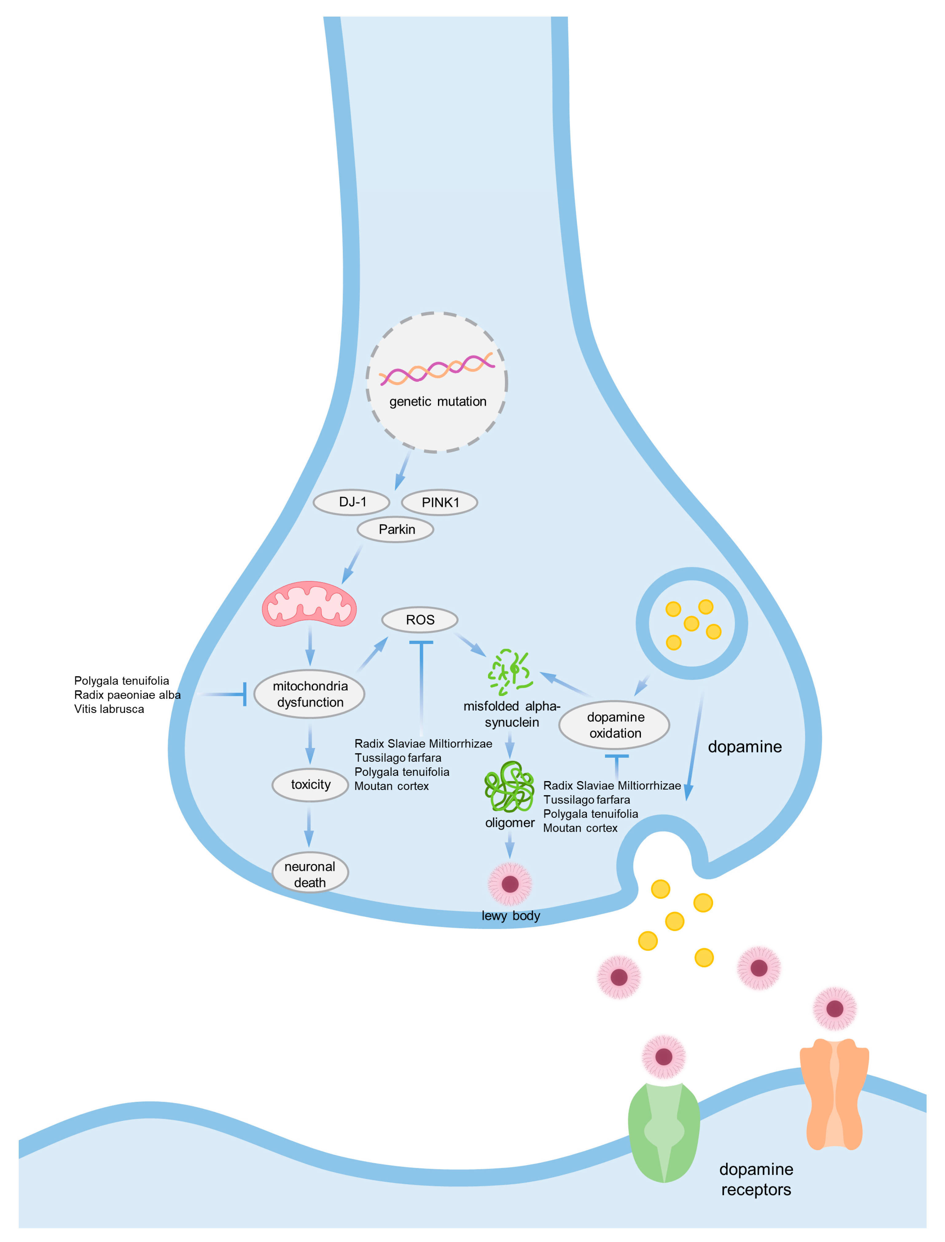
α-synuclein is the main causative agent of Parkinson’s disease. It is a protein encoded by the SNCA gene, and the monomer of this protein does not cause any significant problems. However, mutations in the SNCA gene can cause misfolding of α-synuclein monomers, which can lead to accumulation and aggregation, ultimately forming Lewy bodies. These Lewy bodies are neurotoxic to neurons, causing apoptosis or interfering with the transmission of signaling substances between neurons. Misfolded α-synuclein can also be produced by other factors such as mutations in mitochondrial-related proteins DJ-1, PINK1, and Parkin, which can cause mitochondrial dysfunction and generate reactive oxygen species (ROS). ROS can further produce misfolded α-synuclein or accumulate excessive toxicity in neurons, leading to cell death. Additionally, oxidation of dopamine produced and secreted by nerve cells can also cause α-synuclein aggregation, which can contribute to Parkinson’s disease (Figure 2) [19].
To eliminate toxic proteins, nerve cells typically rely on either autophagy or the ubiquitin proteasome system. If a protein is produced excessively or erroneously, it may be secreted out of the cell. In such cases, microglia or astrocytes perform phagocytosis or endocytosis to bring the secreted proteins back into the cells, where they are then removed through proteolysis. However, there is a limit to the cells’ ability to break down abnormal proteins or maintain normal protein levels, and when neuronal cells fail to maintain protein homeostasis, they eventually undergo apoptosis. This suggests that the brain gradually becomes ill over time [20,21,22].

2.2. Mitochondrial Dysfunction

Mitochondria are responsible for producing energy by using the electron transport chain to generate ATP through oxidative phosphorylation. However, one hallmark of neurodegenerative diseases is the dysfunction of mitochondria in neurons [11]. Mitochondria are vital for supplying ATP to cells through oxidative phosphorylation (OXPHOS), synthesizing biologically essential molecules, and catalyzing various redox reactions. Inefficient OXPHOS can result in mitochondrial dysfunction by producing reactive oxygen species (ROS). While low concentrations of ROS are crucial for normal cell signaling, prolonged or excessive exposure to ROS can lead to oxidative damage to macromolecules such as DNA, lipids, and proteins, ultimately causing cell death [23]. Moreover, it is believed that decreased oxidative phosphorylation can trigger apoptosis by inducing bioenergy depletion in neurons. Although a direct link between respiratory defects and neurons has yet to be established, mitochondrial respiratory deficits have been identified in several neurodegenerative diseases such as Alzheimer’s and Parkinson’s [24].

2.3. Inflammation

Neuroinflammation is an inflammatory response that specifically affects the brain and spinal cord. It can be triggered by factors such as disease, injury, infection, and stress, much like any other type of inflammation. Microglia and astrocytes are key regulators in neuroinflammation [25]. Recent studies have suggested a strong connection between neurodegenerative diseases and neuroinflammation. While acute inflammation of the brain functions as a defense mechanism against infection and injury, chronic inflammation like that seen in Alzheimer’s disease impairs the anti-inflammatory response. Chronic inflammation is caused by cytokines secreted by immune cells [26]. In patients with Alzheimer’s disease, sustained inflammatory responses are accompanied by the presence of β-amyloid plaques. This triggers the production of nitric oxide (NO) and reactive oxygen species (ROS) and promotes neuronal cell death by producing inflammatory cytokines, chemokines, and prostaglandins [27]. Microglia and astrocytes can detect β-amyloid by activating various sensor proteins such as TLR and NLR and then activate an inflammatory response through NF-kB. Recent research has also shown that β-amyloid can activate the NLRP3 protein, which is part of the NLR sensor of microglia, resulting in the production of IL-1b, an inflammatory cytokine [28].
A persistent immune response in the brain occurs not only in Alzheimer’s, but also in Parkinson’s. α-synuclein is a protein found at the synaptic terminal of nerve cells, but when α-synuclein aggregates to form Lewy bodies, it can activate immune cells. Microglia can phagocytose and degrade extracellular aggregated α-synuclein, and can produce inflammatory cytokines or ROS through NF-kB activation [29]. It has been reported that the immune function of microglia activated by α-synuclein is regulated by CD4+ T regulatory cells again [30].
Targeting the activation of immune cells by abnormal proteins such as β-amyloid or α-synuclein and the resulting neuroinflammatory response can be a potential strategy for treating neurodegenerative diseases such as Alzheimer’s or Parkinson’s [31,32].

3. Current Medications for Alzheimer’s and Parkinson’s Disease

As the population ages, the number of individuals with dementia worldwide is increasing, particularly among those over 60 years old. However, the multifactorial causes of Alzheimer’s and Parkinson’s disease are not fully understood [33,34]. In order to develop effective therapeutic agents for these neurodegenerative diseases, it is crucial to understand their molecular and biochemical pathogenesis. Several medications that modulate various molecular and biochemical mechanisms have been developed in the past [12]. Table 1 presents the medications that are currently used for the treatment of AD or PD.

3.1. Donepezil

Donepezil is a medication that works by inhibiting the enzyme acetylcholinesterase, which breaks down the neurotransmitter acetylcholine. By preventing the breakdown of acetylcholine, donepezil enhances cholinergic neurotransmission in the brain, thereby improving cognitive function in patients with Alzheimer’s disease [35]. Although donepezil does not halt the progression of the disease, it can improve symptoms such as memory loss, confusion, and problems with thinking and reasoning. Donepezil is approved by the FDA for use in mild-to-severe Alzheimer’s disease [36]. However, common adverse effects of donepezil include nausea, diarrhea, fatigue, dizziness, and insomnia.

3.2. Galantamine

Galantamine is an alkaloid that can be found in several plants, including daffodil bulbs, and is currently synthesized and provided as a medication. Similar to donepezil, galantamine functions as an acetylcholinesterase inhibitor and an allosteric modulator of nicotinic cholinergic neurotransmitter receptors [37]. Studies have demonstrated the efficacy of galantamine in treating cognitive symptoms in patients with mild-to-moderate Alzheimer’s disease. However, it is also associated with several major adverse effects such as convulsions, nausea, stomach cramps, vomiting, irregular breathing, confusion, and muscle weakness [38].

3.3. Rivastigmine

Rivastigmine is approved for the treatment of both Alzheimer’s and Parkinson’s disease [39]. Although the exact mechanism of action of rivastigmine is unclear, it is believed to work by inhibiting both acetylcholinesterase and butyryl cholinesterase. However, rivastigmine has major adverse effects such as abdominal pain, weight loss, poor gastrointestinal function, loss of appetite, and nausea. Overdosing can cause various symptoms such as irregular breathing, chest pain, and an irregular heartbeat [40].

3.4. Memantine

Memantine is a medication that functions as an antagonist of the N-methyl-D-aspartate (NMDA) receptor, and is prescribed to patients with moderate-to-severe Alzheimer’s disease. It works by preventing overactivation of glutamine receptors and slowing neurotoxicity by blocking NMDA receptors downstream of glutamate receptors. Some common adverse effects of memantine include pain, headache, fatigue, increased blood pressure, vomiting, drowsiness, coughing, and difficulty breathing [41].

3.5. Levodopa

Levodopa is a medication used to supplement dopamine deficiency in patients with Parkinson’s disease by acting as a precursor of dopamine. Once it passes through the blood–brain barrier, it is decarboxylated to dopamine by aromatic amino acid decarboxylase (AADC) and released from the presynaptic terminal of the striatum to compensate for the dopamine deficiency. However, dopamine has the potential to accelerate the disease by causing mitochondrial or lysosomal dysfunction and increasing α-synuclein oligomer concentrations, as suggested by some studies [42]. The long-term administration of levodopa has exercise-related adverse effects such as fluctuations, dyskinesias, and dystonia, and the non-motor adverse effects include autonomic dysfunction, mood control disorder, and cognitive decline [43].

3.6. Catechol O Methyltransferase Inhibitor (COMT)

COMT is an enzyme that catalyzes the methylation of catechol substrates using S-adenosyl-1-methionine as a co-factor. Various substances such as catechol, catecholamine, catecholestrogens, and ascorbic acid can serve as substrates for COMT [44]. The primary function of COMT is to eliminate catechols. In patients with Parkinson’s disease receiving levodopa/aromatic amino acid decarboxylase (AADC) inhibitors, levodopa is methylated to 3-O-methyldopa by COMT, which limits the availability of levodopa in the brain. COMT inhibitors prevent O-methylation of 3-O-methyldopa, a levodopa metabolite, and enhance the conversion of levodopa to dopamine in the brain [45]. However, COMT inhibitors also have adverse effects, such as movement disorders, confusion, hallucinations, urine discoloration, and diarrhea, due to the increased availability of levodopa [46].

3.7. Monoamine Oxidase-B Inhibitor

Monoamine oxidase-B (MAO-B) is a mitochondrial enzyme that is expressed widely in various tissues, including the stomach, liver, and nervous tissue. This enzyme is essential for the detoxification of amines by catalyzing the oxidative deamination of various monoamines and metabolizing the released neurotransmitters. The aldehydes produced during this process are metabolized by aldehyde dehydrogenase and aldehyde reductase to form glycols and carboxylic acids. However, the production of aldehydes with H2O2 suggests that the products of MAO action may be toxic to cells [47]. MAO inhibitors are medications currently being used to treat Parkinson’s disease by inhibiting the MAO enzyme. By improving the movement disorders caused by Parkinson’s disease, MAO inhibitors can help alleviate the symptoms of this condition. However, adverse effects such as nausea, dizziness, constipation, confusion, and hallucinations may occur with their use [48].

3.8. Dopamine Agonist

Dopamine agonists work by mimicking the effects of dopamine and directly activating dopamine receptors to improve the symptoms of Parkinson’s disease. There are two types of dopamine agonists: those derived from the fungus ergot and non-ergot agonists, both of which target dopamine D receptors [49]. Initially used as an add-on treatment for levodopa-induced dyskinesia [50], dopamine agonists are now also used to extend levodopa therapy and reduce the occurrence of motor complications [51]. However, ergot-derived dopamine agonists have adverse effects such as nausea, vomiting, orthostatic hypotension, hallucinations, and delusions, as well as adverse effects related to levodopa use [52].
Table 1. Medications for neurodegenerative diseases.
Target
Disease
Medication NameMechanismAdverse EffectRef.
Alzheimer’s diseaseDonepezilAcetylcholinesterase inhibitorNausea, vomiting, diarrhea, dizziness, trouble sleeping[35,36]
GalantamineAcetylcholinesterase inhibitor and allosteric modulator on nicotinic acetylcholine receptorsNausea, stomach cramps, vomiting, irregular breathing, confusion, muscle weakness[37,38]
RivastigmineAcetylcholinesterase inhibitor and butyrylcholinesterase inhibitorAbdominal pain, weight loss, diarrhea, loss of appetite, nausea, irregular breathing, chest pain, irregular heartbeat[39,40]
MemantineNMDA receptor agonistPain, headache, fatigue, increased blood pressure, vomiting, drowsiness, cough, shortness of breath[41]
Parkinson’s diseaseLevodopaSupplement of dopamineFluctuations, dyskinesias, dystonias, autonomic dysfunction, mood control disorders, cognitive decline[42,43]
Catechol O methyltransferase inhibitorProlongation of levodopa actionLevodopa-related adverse effects, confusion, hallucinations, urine discoloration, diarrhea[44,45,46]
Monoamine oxidase-B inhibitorPreventation of dopamine breakdownNausea, dizziness, constipation, confusion, hallucinations[47,48]
Dopamine agonistInducement of dopamine-like effectsNausea, vomiting, orthostatic hypotension, hallucinations, delusions[49,50,51,52]

4. Natural Materials—Compounds Derived from Natural Products

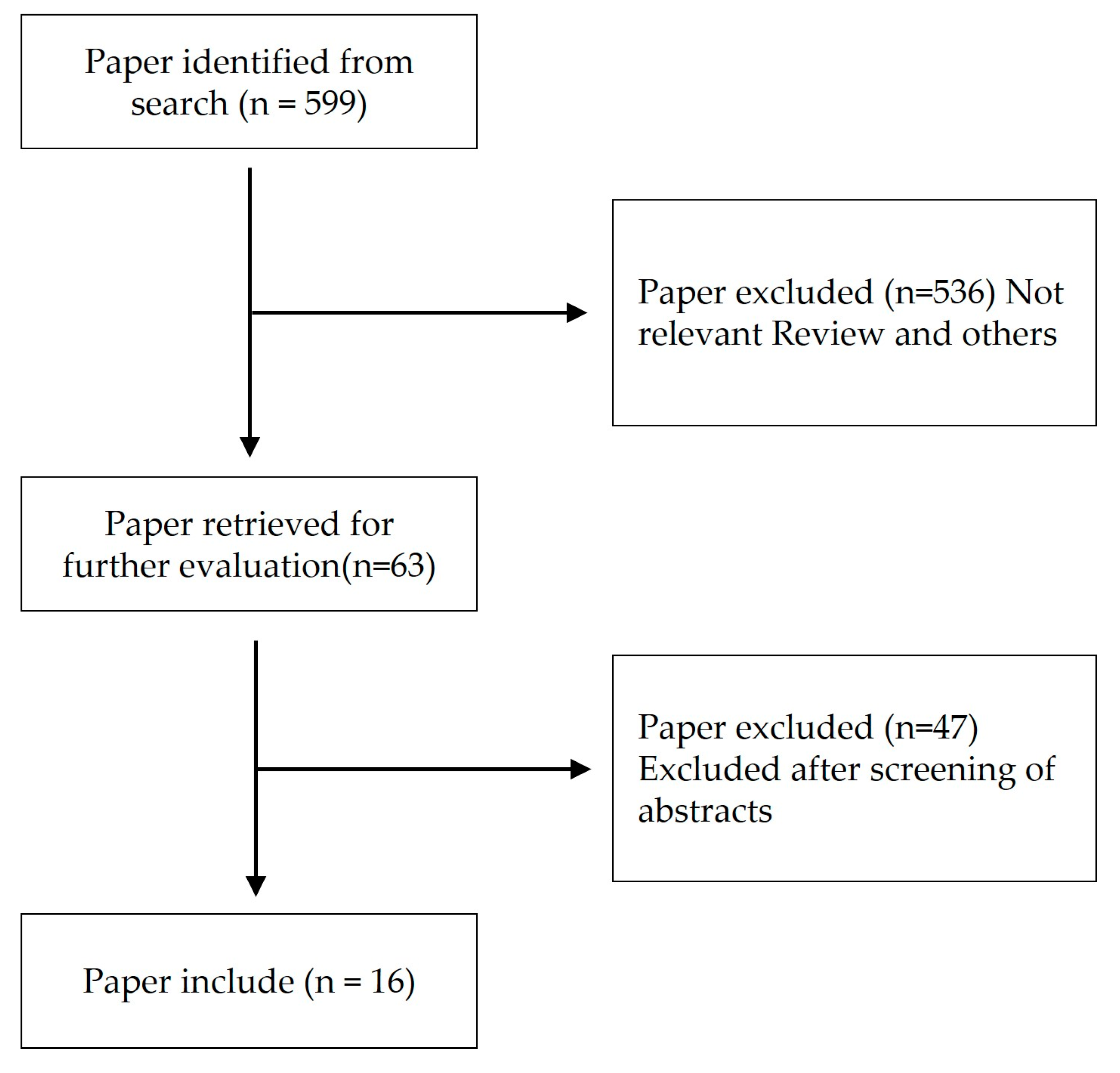
A search was conducted on the National Center for Biotechnology Information (NCBI) database to identify natural products or compounds derived from natural products that could be used as models for Alzheimer’s disease and Parkinson’s disease. The search keywords used were a combination of “Alzheimer’s disease” [MeSH Terms], “Parkinson’s disease” [MeSH Terms], and “Natural products” [MeSH Terms]. The search was not restricted by time, and papers that specified the mechanisms of action through both in vitro and in vivo studies were selected. The search results are presented in Figure 3 and Table 2.
The following documents were excluded based on the following criteria:
A.
Papers that did not contain the specified keywords.
B.
Review articles that covered multiple diseases.
C.
Case reports, clinical trial studies, and literature review studies.
D.
Abstracts and dissertations that were not relevant to the study.

4.1. Reynoutria multiflora Moldenke

Reynoutria multiflora Moldenke is a traditional medicinal herb in East Asia with antioxidant, anti-aging, and anti-inflammatory effects attributed to its various components. It also has a neuroprotective effect by reducing oxidative stress induced by H2O2 or glutamate. Studies have shown that extracts of Reynoutria multiflora Moldenke can prevent apoptosis by restoring FGF2 and BDNF expression in MPP+-induced neurotoxicity in SH-SY5Y cells. In mice models of Parkinson’s disease induced with MPTP, the oral administration of Reynoutria multiflora Moldenke extract activated the FGF2-Akt and BDNF-TrKB signaling pathways and restored dopaminergic neurons in the substantia nigra and corpus striatum [53].

4.2. Achillea fragrantissima Sch.Bip.

Achillea fragrantissima Sch.Bip. is a medicinal herb that has been traditionally used for the treatment of respiratory diseases, fever, obesity, and headaches. It has been found to inhibit the phosphorylation of stress-activated protein kinase/c-Jun N-terminal kinase (SAPK/JNK), extracellular signal-regulated kinase (ERK 1/2), and mitogen-activated protein kinase kinase (MEK1) as well as the transcription factor cyclic AMP response element-binding protein (CREB). This inhibition results in decreased accumulation of reactive oxygen species (ROS), reduced oxidative stress, and inhibition of cell death [54].

4.3. Theobroma cacao L.

Theobroma cacao L. a widely used raw material for chocolate, has been found to have antioxidant effects due to the presence of flavonoids such as flavan-3ols, epicatechin, and catechin. These flavonoids have been shown to inhibit free radical scavenging activity. Studies have demonstrated that cocoa extract can activate the BDNF/TrKB signaling pathway and exhibit cell protective effects in rat Pheocromocitoma PC12 cells treated with β-amyloid plaque or oligomers [55].

4.4. Salvia miltiorrhiza Bunge

Salvia miltiorrhiza Bunge has been reported to exhibit antioxidant and neuroprotective effects against neurotoxicity. In 6-hydroxydopamine-treated PC12 cells, it has been shown to increase Akt phosphorylation and activate the Nrf2 signaling pathway, leading to the upregulation of heme oxygenase-1 (HO-1), an antioxidant enzyme, thereby inhibiting ROS generation. In a zebrafish model of 6-hydroxydopamine-induced neurotoxicity, it demonstrated a neuroprotective effect by reducing neuronal cell death [56].

4.5. Asparagus racemosus Willd.

Asparagus racemosus Willd. is known for its antioxidant and immunomodulatory properties. In Swiss albino mice injected with kainic acid, it was found to inhibit excitotoxicity and reduce oxidative stress in hippocampal and striatal neurons. Asparagus racemosus Willd. also lowered lipid peroxidation and protein carbonyl content induced by kainic acid, and improved the activity of glutathione peroxidase (GPx) and glutathione (GSH) [57].

4.6. Opuntia ficus-indica (L.) Mill.

Opuntia ficus-indica (L.) Mill. are commonly found in the Mediterranean region and have been reported to exhibit antioxidant and anti-inflammatory properties. They promote the synthesis of heat shock proteins and prevent liver damage. Studies have shown that when administered to Saccharomyces cerevisiae with Aβ42(E22G) mutation, viability is improved. In addition, administration to Drosophila with human α-syn A53T mutation increases the survival rate. These plants also inhibit the fibrillogenesis of Aβ42 and α-syn proteins and prevent the disruption of lipid membrane integrity caused by the accumulation of oligomeric aggregates [58].

4.7. Gardenia jasminoides J.Ellis

Gardenia jasminoides J.Ellis, a plant commonly used in medicine and food in East Asia, has been found to have anti-inflammatory and antioxidant properties. In a study using the extract from this plant on APP/PS1 transgenic mice, it activated the phosphatidylinositide 3-kinase/AKT signaling pathway and demonstrated anti-neuroinflammatory effects by regulating the production of inflammatory proteins and cytokines [59].

4.8. Vitis labrusca L.

Vitis labrusca L. is commonly consumed as wine and has been shown to have beneficial effects on cardiovascular disease, cancer, and aging-related neurological diseases. In a Parkinson’s disease model using rats with 6-OHDA, the daily consumption of Vitis labrusca L. compound, procyanidin, led to reduced oxidative stress and improved mitochondria dysfunction, and PC12 cells treated with 6-OHDA showed neuroprotective effects via the activation of the PI3K/Akt signaling pathway [60].

4.9. Paullinia cupana Kunth

Paullinia cupana Kunth, also known as guarana, is a widely used traditional medicine and a common ingredient in energy drinks and foods due to its high caffeine content, a psychoactive pseudoalkaloid. It also contains polyphenols that provide various benefits. Studies have shown that it has antioxidant, antibacterial, and antigenotoxic effects. In Caenorhabditis elegans induced with AD, the administration of Paullinia cupana Kunth extract increases proteasome activity in muscles, prevents polyglutamine protein aggregation, inhibits cell death, prolongs cell lifespan, and delays paralysis. Additionally, it modulates antioxidant activity and proteostasis by reducing intracellular ROS and autophagosome accumulation, and increasing SOD-3 and HSP-16.2 expressions [61].

4.10. Tussilago farfara L.

Tussilago farfara L. has been found to exhibit anti-inflammatory properties. When PC12 cells are stimulated with H2O2 or 6-OHDA and treated with Tussilago farfara L. extracts, the Nrf2 pathway is activated, and the antioxidant HO-1 protects neurons from oxidative stress. In addition, in mice models with Parkinson’s disease induced by 6-OHDA injections, it improves motor function and prevents dopaminergic neuronal damage [62].

4.11. Panax ginseng C.A.Mey.

Panax ginseng C.A.Mey., an important medicinal material in East Asia, is known for its anti-inflammatory and antioxidant effects, as well as its potential to lower blood sugar and cholesterol and improve obesity. In PC12 cells treated with corticosterone, treatment with Panax ginseng C.A.Mey. extract increased cell viability and decreased apoptosis. Additionally, it reduced ROS generation and restored mitochondrial functions, including mitochondrial permeability transition pores and mitochondrial membrane potential. The expression of heat shock protein 90 and histone deacetylase 6, which are related to the glucocorticoid receptor, was increased, while the expression of endoplasmic reticulum stress-related proteins was decreased, thereby restoring endoplasmic reticulum function [63].

4.12. Polygala tenuifolia Willd.

Polygala tenuifolia Willd. is a medicinal herb traditionally used in East Asia for its potential to improve memory and cognitive function. It is known for its neuroprotective, neuroregenerative, antioxidant, and anti-aging effects. In SH-SY5Y cells treated with 6-OHDA, treatment with Polygala tenuifolia Willd. extract increased cell viability and decreased cell death. It protected the mitochondrial membrane potential and increased the expression of glutathione and superoxide dismutase, while reducing caspase-3 expression and increasing tyrosine hydroxylase expression to protect neurons [64].

4.13. Alpinia oxyphylla Miq.

Alpinia oxyphylla Miq. is a medicinal plant traditionally used to enhance memory and learning abilities affected by degenerative brain diseases. It has been found to protect neurons by reducing the expression of amyloid precursor protein, Aβ1-40, and Aβ1-42. Treatment with Alpinia oxyphylla Miq. extract activated Nrf2 through the Akt/GSK3b signaling pathway and inhibited oxidative stress in N2a/APP cells. In addition, administration of the extract to The Senescence Accelerated Mouse-Prone 8 mice showed the potential to delay cognitive function damage, enhance muscle strength, and restore motor ability [65].

4.14. Paeonia suffruticosa Andrews

Paeonia suffruticosa Andrews, a traditional medicine used for thousands of years in East Asia, has been reported to have anti-inflammatory, antioxidant, and anti-allergic effects. It inhibits inflammation, NO synthesis, and COX-2 expression in LPS-treated microglia. Furthermore, it inhibits ROS generation in oxidatively damaged cortical neurons treated with 6-OHDA, thereby reducing oxidative stress and increasing cell viability. This is achieved by increasing superoxide dismutase activity and anti-apoptotic protein expression [66].

4.15. Paeonia lactiflora Pall.

Paeonia lactiflora Pall., a traditional medicine used to treat cerebral ischemia, epilepsy, and degenerative brain disease, has shown potential neuroprotective effects. In glutamate-induced neurotoxicity in PC12 cells, treatment with Paeonia lactiflora Pall. extract increased cell viability by regulating the expression of Bcl-2 and Bax, which are proteins related to apoptosis. Furthermore, it also had a positive effect on mitochondrial function by regulating the mitochondrial membrane potential, which is often decreased in glutamate-induced neurotoxicity [67].

4.16. Cynanchum otophyllum C.K.Schneid.

Cynanchum otophyllum C.K.Schneid. has various uses, including medicine, cosmetics, and food, and has been traditionally used to treat inflammatory diseases such as epilepsy and rheumatism. Administering its extract to 3xTG Alzheimer’s model mice improved their learning and memory, reduced β-amyloid and tau aggregates, and inhibited microgliosis and astrocytosis. The extract showed a neuroprotective effect by activating the PPARα and TFEB signaling pathways, as well as activating the autophagy and lysosomal pathways [68].

5. Conclusions and Discussion

Neurodegenerative diseases are characterized by the death of nerve cells in the central nervous system, including the brain and spinal cord, and their incidence increases with age. Alzheimer’s disease and Parkinson’s disease are the two most common neurodegenerative diseases, but their exact causes and treatment methods are still being studied. In Alzheimer’s disease, β-amyloid has been a major focus of research, while in Parkinson’s disease, α-synuclein has been extensively studied. These pathogenic proteins disrupt the normal function of nerve cells, leading to inflammation, oxidative stress, and mitochondrial dysfunction. While efforts have been made to develop medications to treat these diseases, currently available medications only suppress symptoms and have many adverse effects, highlighting the need for new, more effective treatments. To investigate potential natural substances as the basis for new medications, a search was conducted using keywords related to Alzheimer’s disease, Parkinson’s disease, and natural products in the US National Center for Biotechnology Information. Sixteen papers were selected after screening, including ten related to antioxidants, four related to mitochondrial function, and others related to anti-inflammatory agents, self-destruction, and autophagy.
There are many differences between current drugs and natural products, as the research methods vary depending on when the drugs were developed or studied. The Alzheimer’s and Parkinson’s disease treatments listed in Table 1 are FDA-approved drugs, with donepezil approved in 1996, galantamine in 2001, rivastigmine in 2000, memantine in 2003, levodopa in 1975, tolcapone in 1998, entacapone in 1999, opicapone in 2020, pramipexole and ropinirole in 1997, and apomorphine hydrochloride in 2004. Most of these drugs were developed in the 1990s and 2000s and help ensure the proper transmission of neurotransmitters between nerve cells. However, current drugs have adverse effects and only delay the progression of symptoms, so there is a need for the development of new drugs [69,70,71,72,73,74,75,76,77,78,79].
Currently, there are ongoing clinical trials for drugs that aim to treat AD or PD with new mechanisms of action different from previously FDA-approved drugs. Examples of such drugs include mGluR5 silent allosteric modulator BMS-984923, c-Abl inhibitor IkT-148009, microbe lactobacillus plantarum PS128, LRRK2 inhibitor BIIB122, and sigma-2 receptor antagonist CT1812. Papers about these drugs can be found on NCBI, and clinicaltrial.org provides information about ongoing clinical trials. On the other hand, natural compounds are being studied with a different approach, aiming to activate neuronal cell recovery via Akt and BDNF signaling pathways, protect neuronal cells from ROS via Nrf2/HO-1 signaling pathways, or improve AD or PD via mitochondrial function recovery. However, natural compounds require more research to ensure their safety before undergoing clinical trials or being approved as drugs [80,81,82,83,84].
This study summarizes the neuroprotective effects of many natural products. While the keywords were restricted to Alzheimer’s disease and Parkinson’s disease, the mechanisms by which natural substances such as anti-inflammatory, antioxidant, and autophagy exhibit inhibitory effects are not specific to these diseases. This suggests that the efficacy of natural products that have been widely used to treat other diseases or to protect the body may also be applied to neurodegenerative diseases. Therefore, other natural products with similar efficacy also have potential applications. As natural products can be consumed as food in daily life, guidelines for daily intake should be established for more effective treatments.

Funding

This study was supported by National Research Foundation of Korea (NRF) (NRF-2016R1C1B2016274, 2021R1A2C1012846 and 2021R1I1A1A01059706) funded by the Korean government (Ministry of Science, ICT and Future Planning), and Woosuk University. G.Y. is supported by Intramural Research Program of WOOSUK University. S.W.K. was supported by the grants, Health Fellowship Foundation for 2021.

Institutional Review Board Statement

Not applicable.

Informed Consent Statement

Not applicable.

Data Availability Statement

Not applicable.

Conflicts of Interest

The authors declare no conflict of interest.

References

  1. Gorman, A.M. Neuronal cell death in neurodegenerative diseases: Recurring themes around protein handling. J. Cell. Mol. Med. 2008, 12, 2263–2280. [Google Scholar] [CrossRef]
  2. Erkkinen, M.G.; Kim, M.O.; Geschwind, M.D. Clinical Neurology and Epidemiology of the Major Neurodegenerative Diseases. Cold Spring Harb. Perspect. Biol. 2018, 10, a033118. [Google Scholar] [CrossRef]
  3. Onyike, C.U.; Diehl-Schmid, J. The epidemiology of frontotemporal dementia. Int. Rev. Psychiatry 2013, 25, 130–137. [Google Scholar] [CrossRef]
  4. Tysnes, O.B.; Storstein, A. Epidemiology of Parkinson’s disease. J. Neural Transm. 2017, 124, 901–905. [Google Scholar] [CrossRef]
  5. Leray, E.; Moreau, T.; Fromont, A.; Edan, G. Epidemiology of multiple sclerosis. Rev. Neurol. 2016, 172, 3–13. [Google Scholar] [CrossRef]
  6. Rawlins, M.D.; Wexler, N.S.; Wexler, A.R.; Tabrizi, S.J.; Douglas, I.; Evans, S.J.; Smeeth, L. The Prevalence of Huntington’s Disease. Neuroepidemiology 2016, 46, 144–153. [Google Scholar] [CrossRef]
  7. Knopman, D.S.; Amieva, H.; Petersen, R.C.; Chételat, G.; Holtzman, D.M.; Hyman, B.T.; Nixon, R.A.; Jones, D.T. Alzheimer disease. Nat. Rev. Dis. Prim. 2021, 7, 33. [Google Scholar] [CrossRef]
  8. Bloem, B.R.; Okun, M.S.; Klein, C. Parkinson’s disease. Lancet 2021, 397, 2284–2303. [Google Scholar] [CrossRef]
  9. Leng, F.; Edison, P. Neuroinflammation and microglial activation in Alzheimer disease: Where do we go from here? Nat. Rev. Neurol. 2021, 17, 157–172. [Google Scholar] [CrossRef]
  10. Aarsland, D.; Batzu, L.; Halliday, G.M.; Geurtsen, G.J.; Ballard, C.; Ray Chaudhuri, K.; Weintraub, D. Parkinson disease-associated cognitive impairment. Nat. Rev. Dis. Prim. 2021, 7, 47. [Google Scholar] [CrossRef]
  11. Onyango, I.G.; Khan, S.M.; Bennett, J.P., Jr. Mitochondria in the pathophysiology of Alzheimer’s and Parkinson’s diseases. Front. Biosci. 2017, 22, 854–872. [Google Scholar] [CrossRef]
  12. Esposito, E.; Cuzzocrea, S. New therapeutic strategy for Parkinson’s and Alzheimer’s disease. Curr. Med. Chem. 2010, 17, 2764–2774. [Google Scholar] [CrossRef]
  13. Hou, Y.; Dan, X.; Babbar, M.; Wei, Y.; Hasselbalch, S.G.; Croteau, D.L.; Bohr, V.A. Ageing as a risk factor for neurodegenerative disease. Nat. Rev. Neurol. 2019, 15, 565–581. [Google Scholar] [CrossRef]
  14. Angelova, P.R.; Abramov, A.Y. Alpha-synuclein and beta-amyloid—different targets, same players: Calcium, free radicals and mitochondria in the mechanism of neurodegeneration. Biochem. Biophys. Res. Commun. 2017, 483, 1110–1115. [Google Scholar] [CrossRef]
  15. Murphy, M.P.; LeVine, H., 3rd. Alzheimer’s disease and the amyloid-beta peptide. J. Alzheimer’s Dis. JAD 2010, 19, 311–323. [Google Scholar] [CrossRef]
  16. Medeiros, R.; Baglietto-Vargas, D.; LaFerla, F.M. The role of tau in Alzheimer’s disease and related disorders. CNS Neurosci. Ther. 2011, 17, 514–524. [Google Scholar] [CrossRef]
  17. Penke, B.; Bogár, F.; Fülöp, L. β-Amyloid and the Pathomechanisms of Alzheimer’s Disease: A Comprehensive View. Molecules 2017, 22, 1692. [Google Scholar] [CrossRef]
  18. van der Lee, S.J.; Wolters, F.J.; Ikram, M.K.; Hofman, A.; Ikram, M.A.; Amin, N.; van Duijn, C.M. The effect of APOE and other common genetic variants on the onset of Alzheimer’s disease and dementia: A community-based cohort study. Lancet. Neurol. 2018, 17, 434–444. [Google Scholar] [CrossRef]
  19. Wong, Y.C.; Krainc, D. α-synuclein toxicity in neurodegeneration: Mechanism and therapeutic strategies. Nat. Med. 2017, 23, 1–13. [Google Scholar] [CrossRef]
  20. Fujikake, N.; Shin, M.; Shimizu, S. Association Between Autophagy and Neurodegenerative Diseases. Front. Neurosci. 2018, 12, 255. [Google Scholar] [CrossRef]
  21. Zheng, Q.; Huang, T.; Zhang, L.; Zhou, Y.; Luo, H.; Xu, H.; Wang, X. Dysregulation of Ubiquitin-Proteasome System in Neurodegenerative Diseases. Front. Aging Neurosci. 2016, 8, 303. [Google Scholar] [CrossRef]
  22. Galloway, D.A.; Phillips, A.E.M.; Owen, D.R.J.; Moore, C.S. Phagocytosis in the Brain: Homeostasis and Disease. Front. Immunol. 2019, 10, 790. [Google Scholar] [CrossRef]
  23. Singh, A.; Kukreti, R.; Saso, L.; Kukreti, S. Oxidative Stress: A Key Modulator in Neurodegenerative Diseases. Molecules 2019, 24, 1583. [Google Scholar] [CrossRef]
  24. Area-Gomez, E.; Guardia-Laguarta, C.; Schon, E.A.; Przedborski, S. Mitochondria, OxPhos, and neurodegeneration: Cells are not just running out of gas. J. Clin. Investig. 2019, 129, 34–45. [Google Scholar] [CrossRef]
  25. DiSabato, D.J.; Quan, N.; Godbout, J.P. Neuroinflammation: The devil is in the details. J. Neurochem. 2016, 139 (Suppl. S2), 136–153. [Google Scholar] [CrossRef]
  26. Kinney, J.W.; Bemiller, S.M.; Murtishaw, A.S.; Leisgang, A.M.; Salazar, A.M.; Lamb, B.T. Inflammation as a central mechanism in Alzheimer’s disease. Alzheimer’s Dement. 2018, 4, 575–590. [Google Scholar] [CrossRef]
  27. Kitazawa, M.; Yamasaki, T.R.; LaFerla, F.M. Microglia as a potential bridge between the amyloid beta-peptide and tau. Ann. N. Y. Acad. Sci. 2004, 1035, 85–103. [Google Scholar] [CrossRef]
  28. Glass, C.K.; Saijo, K.; Winner, B.; Marchetto, M.C.; Gage, F.H. Mechanisms underlying inflammation in neurodegeneration. Cell 2010, 140, 918–934. [Google Scholar] [CrossRef]
  29. Allen Reish, H.E.; Standaert, D.G. Role of α-synuclein in inducing innate and adaptive immunity in Parkinson disease. J. Park. Dis. 2015, 5, 1–19. [Google Scholar] [CrossRef]
  30. Beers, D.R.; Henkel, J.S.; Zhao, W.; Wang, J.; Appel, S.H. CD4+ T cells support glial neuroprotection, slow disease progression, and modify glial morphology in an animal model of inherited ALS. Proc. Natl. Acad. Sci. USA 2008, 105, 15558–15563. [Google Scholar] [CrossRef]
  31. Rostami, J.; Mothes, T.; Kolahdouzan, M.; Eriksson, O.; Moslem, M.; Bergström, J.; Ingelsson, M.; O’Callaghan, P.; Healy, L.M.; Falk, A.; et al. Crosstalk between astrocytes and microglia results in increased degradation of α-synuclein and amyloid-β aggregates. J. Neuroinflamm. 2021, 18, 124. [Google Scholar] [CrossRef] [PubMed]
  32. Marsh, S.E.; Blurton-Jones, M. Examining the mechanisms that link β-amyloid and α-synuclein pathologies. Alzheimer’s Res. Ther. 2012, 4, 11. [Google Scholar] [CrossRef] [PubMed]
  33. Xia, X.; Jiang, Q.; McDermott, J.; Han, J.J. Aging and Alzheimer’s disease: Comparison and associations from molecular to system level. Aging Cell 2018, 17, e12802. [Google Scholar] [CrossRef] [PubMed]
  34. Reeve, A.; Simcox, E.; Turnbull, D. Ageing and Parkinson’s disease: Why is advancing age the biggest risk factor? Ageing Res. Rev. 2014, 14, 19–30. [Google Scholar] [CrossRef]
  35. Birks, J.S.; Harvey, R.J. Donepezil for dementia due to Alzheimer’s disease. Cochrane Database Syst. Rev. 2018, 6, Cd001190. [Google Scholar] [CrossRef]
  36. Kumar, A.; Gupta, V.; Sharma, S. Donepezil; StatPearls: Treasure Island, FL, USA, 2022. [Google Scholar]
  37. Olin, J.; Schneider, L. Galantamine for Alzheimer’s disease. Cochrane Database Syst. Rev. 2002, 1, Cd001747. [Google Scholar] [CrossRef]
  38. Mehta, M.; Adem, A.; Sabbagh, M. New acetylcholinesterase inhibitors for Alzheimer’s disease. Int. J. Alzheimer’s Dis. 2012, 2012, 728983. [Google Scholar] [CrossRef]
  39. Inglis, F. The tolerability and safety of cholinesterase inhibitors in the treatment of dementia. Int. J. Clin. Pract. Suppl. 2002, 127, 45–63. [Google Scholar]
  40. Birks, J.S.; Grimley Evans, J. Rivastigmine for Alzheimer’s disease. Cochrane Database Syst. Rev. 2015, 10, CD001191. [Google Scholar] [CrossRef]
  41. Jiang, J.; Jiang, H. Efficacy and adverse effects of memantine treatment for Alzheimer’s disease from randomized controlled trials. Neurol. Sci. Off. J. Ital. Neurol. Soc. Ital. Soc. Clin. Neurophysiol. 2015, 36, 1633–1641. [Google Scholar] [CrossRef]
  42. de Bie, R.M.A.; Clarke, C.E.; Espay, A.J.; Fox, S.H.; Lang, A.E. Initiation of pharmacological therapy in Parkinson’s disease: When, why, and how. Lancet. Neurol. 2020, 19, 452–461. [Google Scholar] [CrossRef]
  43. Thanvi, B.R.; Lo, T.C. Long term motor complications of levodopa: Clinical features, mechanisms, and management strategies. Postgrad. Med. J. 2004, 80, 452–458. [Google Scholar] [CrossRef]
  44. Zhu, B.T.; Patel, U.K.; Cai, M.X.; Conney, A.H. O-Methylation of tea polyphenols catalyzed by human placental cytosolic catechol-O-methyltransferase. Drug Metab. Dispos. Biol. Fate Chem. 2000, 28, 1024–1030. [Google Scholar]
  45. Bonifácio, M.J.; Palma, P.N.; Almeida, L.; Soares-da-Silva, P. Catechol-O-methyltransferase and its inhibitors in Parkinson’s disease. CNS Drug Rev. 2007, 13, 352–379. [Google Scholar] [CrossRef]
  46. Kaakkola, S. Problems with the present inhibitors and a relevance of new and improved COMT inhibitors in Parkinson’s disease. Int. Rev. Neurobiol. 2010, 95, 207–225. [Google Scholar] [CrossRef]
  47. Jenner, P. Oxidative stress in Parkinson’s disease. Ann. Neurol. 2003, 53 (Suppl. S3), S26–S36, discussion S36–S38. [Google Scholar] [CrossRef]
  48. Finberg, J.P.; Rabey, J.M. Inhibitors of MAO-A and MAO-B in Psychiatry and Neurology. Front. Pharmacol. 2016, 7, 340. [Google Scholar] [CrossRef]
  49. Reichmann, H.; Bilsing, A.; Ehret, R.; Greulich, W.; Schulz, J.B.; Schwartz, A.; Rascol, O. Ergoline and non-ergoline derivatives in the treatment of Parkinson’s disease. J. Neurol. 2006, 253 (Suppl. S4), iv36–iv38. [Google Scholar] [CrossRef]
  50. Oertel, W.H.; Quinn, N.P. Parkinson’s disease: Drug therapy. Bailliere’s Clin. Neurol. 1997, 6, 89–108. [Google Scholar]
  51. Rascol, O.; Brooks, D.J.; Korczyn, A.D.; De Deyn, P.P.; Clarke, C.E.; Lang, A.E. A five-year study of the incidence of dyskinesia in patients with early Parkinson’s disease who were treated with ropinirole or levodopa. N. Engl. J. Med. 2000, 342, 1484–1491. [Google Scholar] [CrossRef]
  52. Brooks, D.J. Dopamine agonists: Their role in the treatment of Parkinson’s disease. J. Neurol. Neurosurg. Psychiatry 2000, 68, 685–689. [Google Scholar] [CrossRef] [PubMed]
  53. Yu, Y.; Lang, X.-Y.; Li, X.-X.; Gu, R.-Z.; Liu, Q.-S.; Lan, R.; Qin, X.-Y. 2,3,5,4′-Tetrahydroxystilbene-2-O-β-d-glucoside attenuates MPP+/MPTP-induced neurotoxicity in vitro and in vivo by restoring the BDNF-TrkB and FGF2-Akt signaling axis and inhibition of apoptosis. Food Funct. 2019, 10, 6009–6019. [Google Scholar] [CrossRef] [PubMed]
  54. Elmann, A.; Telerman, A.; Mordechay, S.; Erlank, H.; Rindner, M.; Ofir, R.; Kashman, Y. 3,5,4′-Trihydroxy-6,7,3′-trimethoxyflavone protects astrocytes against oxidative stress via interference with cell signaling and by reducing the levels of intracellular reactive oxygen species. Neurochem. Int. 2014, 78, 67–75. [Google Scholar] [CrossRef] [PubMed]
  55. Cimini, A.; Gentile, R.; D’Angelo, B.; Benedetti, E.; Cristiano, L.; Avantaggiati, M.L.; Giordano, A.; Ferri, C.; Desideri, G. Cocoa powder triggers neuroprotective and preventive effects in a human Alzheimer’s disease model by modulating BDNF signaling pathway. J. Cell. Biochem. 2013, 114, 2209–2220. [Google Scholar] [CrossRef]
  56. Chong, C.-M.; Zhou, Z.-Y.; Razmovski-Naumovski, V.; Cui, G.-Z.; Zhang, L.-Q.; Sa, F.; Hoi, P.-M.; Chan, K.; Lee, S.M.-Y. Danshensu protects against 6-hydroxydopamine-induced damage of PC12 cells in vitro and dopaminergic neurons in zebrafish. Neurosci. Lett. 2013, 543, 121–125. [Google Scholar] [CrossRef]
  57. Parihar, M.; Hemnani, T. Experimental excitotoxicity provokes oxidative damage in mice brain and attenuation by extract of Asparagus racemosus. J. Neural Transm. 2004, 111, 1–12. [Google Scholar] [CrossRef]
  58. Briffa, M.; Ghio, S.; Neuner, J.; Gauci, A.J.; Cacciottolo, R.; Marchal, C.; Caruana, M.; Cullin, C.; Vassallo, N.; Cauchi, R.J. Extracts from two ubiquitous Mediterranean plants ameliorate cellular and animal models of neurodegenerative proteinopathies. Neurosci. Lett. 2017, 638, 12–20. [Google Scholar] [CrossRef]
  59. Zang, C.; Liu, H.; Shang, J.; Yang, H.; Wang, L.; Sheng, C.; Zhang, Z.; Bao, X.; Yu, Y.; Yao, X. Gardenia jasminoides J. Ellis extract GJ-4 alleviated cognitive deficits of APP/PS1 transgenic mice. Phytomedicine 2021, 93, 153780. [Google Scholar] [CrossRef]
  60. Zhang, Y.; Huang, N.; Chen, M.; Jin, H.; Nie, J.; Shi, J.; Jin, F. Procyanidin protects against 6-hydroxydopamine-induced dopaminergic neuron damage via the regulation of the PI3K/Akt signalling pathway. Biomed. Pharmacother. 2019, 114, 108789. [Google Scholar] [CrossRef]
  61. Boasquívis, P.F.; Silva, G.M.M.; Paiva, F.A.; Cavalcanti, R.M.; Nunez, C.V.; de Paula Oliveira, R. Guarana (Paullinia cupana) extract protects Caenorhabditis elegans models for Alzheimer disease and Huntington disease through activation of antioxidant and protein degradation pathways. Oxidative Med. Cell. Longev. 2018, 2018, 9241308. [Google Scholar] [CrossRef]
  62. Lee, J.; Song, K.; Huh, E.; Oh, M.S.; Kim, Y.S. Neuroprotection against 6-OHDA toxicity in PC12 cells and mice through the Nrf2 pathway by a sesquiterpenoid from Tussilago farfara. Redox Biol. 2018, 18, 6–15. [Google Scholar] [CrossRef]
  63. Jiang, Y.; Li, Z.; Liu, Y.; Liu, X.; Chang, Q.; Liao, Y.; Pan, R. Neuroprotective effect of water extract of Panax ginseng on corticosterone-induced apoptosis in PC12 cells and its underlying molecule mechanisms. J. Ethnopharmacol. 2015, 159, 102–112. [Google Scholar] [CrossRef]
  64. Liang, Z.; Shi, F.; Wang, Y.; Lu, L.; Zhang, Z.; Wang, X.; Wang, X. Neuroprotective effects of tenuigenin in a SH-SY5Y cell model with 6-OHDA-induced injury. Neurosci. Lett. 2011, 497, 104–109. [Google Scholar] [CrossRef] [PubMed]
  65. Bian, Y.; Chen, Y.; Wang, X.; Cui, G.; Ung, C.O.L.; Lu, J.-H.; Cong, W.; Tang, B.; Lee, S.M.-Y. Oxyphylla A ameliorates cognitive deficits and alleviates neuropathology via the Akt-GSK3β and Nrf2-Keap1-HO-1 pathways in vitro and in vivo murine models of Alzheimer’s disease. J. Adv. Res. 2021, 34, 1–12. [Google Scholar] [CrossRef] [PubMed]
  66. Tseng, Y.-T.; Hsu, Y.-Y.; Shih, Y.-T.; Lo, Y.-C. Paeonol attenuates microglia-mediated inflammation and oxidative stress–induced neurotoxicity in rat primary microglia and cortical neurons. Shock 2012, 37, 312–318. [Google Scholar] [CrossRef] [PubMed]
  67. Sun, R.; Wang, K.; Wu, D.; Li, X.; Ou, Y. Protective effect of paeoniflorin against glutamate-induced neurotoxicity in PC12 cells via Bcl-2/Bax signal pathway. Folia Neuropathol. 2012, 50, 270–276. [Google Scholar] [CrossRef]
  68. Iyaswamy, A.; Krishnamoorthi, S.K.; Zhang, H.; Sreenivasmurthy, S.G.; Zhu, Z.; Liu, J.; Su, C.-F.; Guan, X.-J.; Wang, Z.-Y.; Cheung, K.-H. Qingyangshen mitigates amyloid-β and Tau aggregate defects involving PPARα-TFEB activation in transgenic mice of Alzheimer’s disease. Phytomedicine 2021, 91, 153648. [Google Scholar] [CrossRef]
  69. Gomolin, I.H.; Smith, C.; Jeitner, T.M. Donepezil dosing strategies: Pharmacokinetic considerations. J. Am. Med. Dir. Assoc. 2011, 12, 606–608. [Google Scholar] [CrossRef]
  70. Metz, C.N.; Pavlov, V.A. Treating disorders across the lifespan by modulating cholinergic signaling with galantamine. J. Neurochem. 2021, 158, 1359–1380. [Google Scholar] [CrossRef]
  71. Haake, A.; Nguyen, K.; Friedman, L.; Chakkamparambil, B.; Grossberg, G.T. An update on the utility and safety of cholinesterase inhibitors for the treatment of Alzheimer’s disease. Expert Opin. Drug Saf. 2020, 19, 147–157. [Google Scholar] [CrossRef]
  72. MartInez-Coria, H.; Arrieta-Cruz, I.; Cruz, M.E.; López-Valdés, H.E. Physiopathology of ischemic stroke and its modulation using memantine: Evidence from preclinical stroke. Neural Regen. Res. 2021, 16, 433–439. [Google Scholar] [CrossRef]
  73. Ahlskog, J.E. Common Myths and Misconceptions That Sidetrack Parkinson Disease Treatment, to the Detriment of Patients. Mayo Clin. Proc. 2020, 95, 2225–2234. [Google Scholar] [CrossRef]
  74. Artusi, C.A.; Sarro, L.; Imbalzano, G.; Fabbri, M.; Lopiano, L. Safety and efficacy of tolcapone in Parkinson’s disease: Systematic review. Eur. J. Clin. Pharmacol. 2021, 77, 817–829. [Google Scholar] [CrossRef]
  75. Rome, B.N.; Egilman, A.C.; Patel, N.G.; Kesselheim, A.S. Using Multiple Authorized Generics to Maintain High Prices: The Example of Entacapone. Value Health 2023, 26, 370–377. [Google Scholar] [CrossRef]
  76. Craft, B.M.; Baker, D.E.; Levien, T.L. Opicapone: Once-Daily COMT Inhibitor for the Treatment of Wearing Off in Parkinson’s Disease. Sr. Care Pharm. 2022, 37, 55–61. [Google Scholar] [CrossRef]
  77. Niu, Z.-X.; Wang, Y.-T.; Zhang, S.-N.; Li, Y.; Chen, X.-B.; Wang, S.-Q.; Liu, H.-M. Application and synthesis of thiazole ring in clinically approved drugs. Eur. J. Med. Chem. 2023, 250, 115172. [Google Scholar] [CrossRef]
  78. Wilson, S.M.; Wurst, M.G.; Whatley, M.F.; Daniels, R.N. Classics in Chemical Neuroscience: Pramipexole. ACS Chem. Neurosci. 2020, 11, 2506–2512. [Google Scholar] [CrossRef]
  79. Thach, A.; Kirson, N.; Zichlin, M.L.; Dieye, I.; Pappert, E.; Williams, G.R. Cost-Effectiveness of Apomorphine Sublingual Film as an “On-Demand” Treatment for “OFF” Episodes in Patients with Parkinson’s Disease. J. Health Econ. Outcomes Res. 2021, 8, 82–92. [Google Scholar] [CrossRef]
  80. Haas, L.T.; Salazar, S.V.; Smith, L.M.; Zhao, H.R.; Cox, T.O.; Herber, C.S.; Degnan, A.P.; Balakrishnan, A.; Macor, J.E.; Albright, C.F.; et al. Silent Allosteric Modulation of mGluR5 Maintains Glutamate Signaling while Rescuing Alzheimer’s Mouse Phenotypes. Cell Rep. 2017, 20, 76–88. [Google Scholar] [CrossRef]
  81. Karuppagounder, S.S.; Wang, H.; Kelly, T.; Rush, R.; Nguyen, R.; Bisen, S.; Yamashita, Y.; Sloan, N.; Dang, B.; Sigmon, A.; et al. The c-Abl inhibitor IkT-148009 suppresses neurodegeneration in mouse models of heritable and sporadic Parkinson’s disease. Sci. Transl. Med. 2023, 15, eabp9352. [Google Scholar] [CrossRef]
  82. Chen, C.M.; Wu, C.C.; Huang, C.L.; Chang, M.Y.; Cheng, S.H.; Lin, C.T.; Tsai, Y.C. Lactobacillus plantarum PS128 Promotes Intestinal Motility, Mucin Production, and Serotonin Signaling in Mice. Probiotics Antimicrob. Proteins 2022, 14, 535–545. [Google Scholar] [CrossRef] [PubMed]
  83. Jennings, D.; Huntwork-Rodriguez, S.; Vissers, M.; Daryani, V.M.; Diaz, D.; Goo, M.S.; Chen, J.J.; Maciuca, R.; Fraser, K.; Mabrouk, O.S.; et al. LRRK2 Inhibition by BIIB122 in Healthy Participants and Patients with Parkinson’s Disease. Mov. Disord. Off. J. Mov. Disord. Soc. 2023, 38, 386–398. [Google Scholar] [CrossRef] [PubMed]
  84. Rishton, G.M.; Look, G.C.; Ni, Z.-J.; Zhang, J.; Wang, Y.; Huang, Y.; Wu, X.; Izzo, N.J.; LaBarbera, K.M.; Limegrover, C.S.; et al. Discovery of Investigational Drug CT1812, an Antagonist of the Sigma-2 Receptor Complex for Alzheimer’s Disease. ACS Med. Chem. Lett. 2021, 12, 1389–1395. [Google Scholar] [CrossRef] [PubMed]
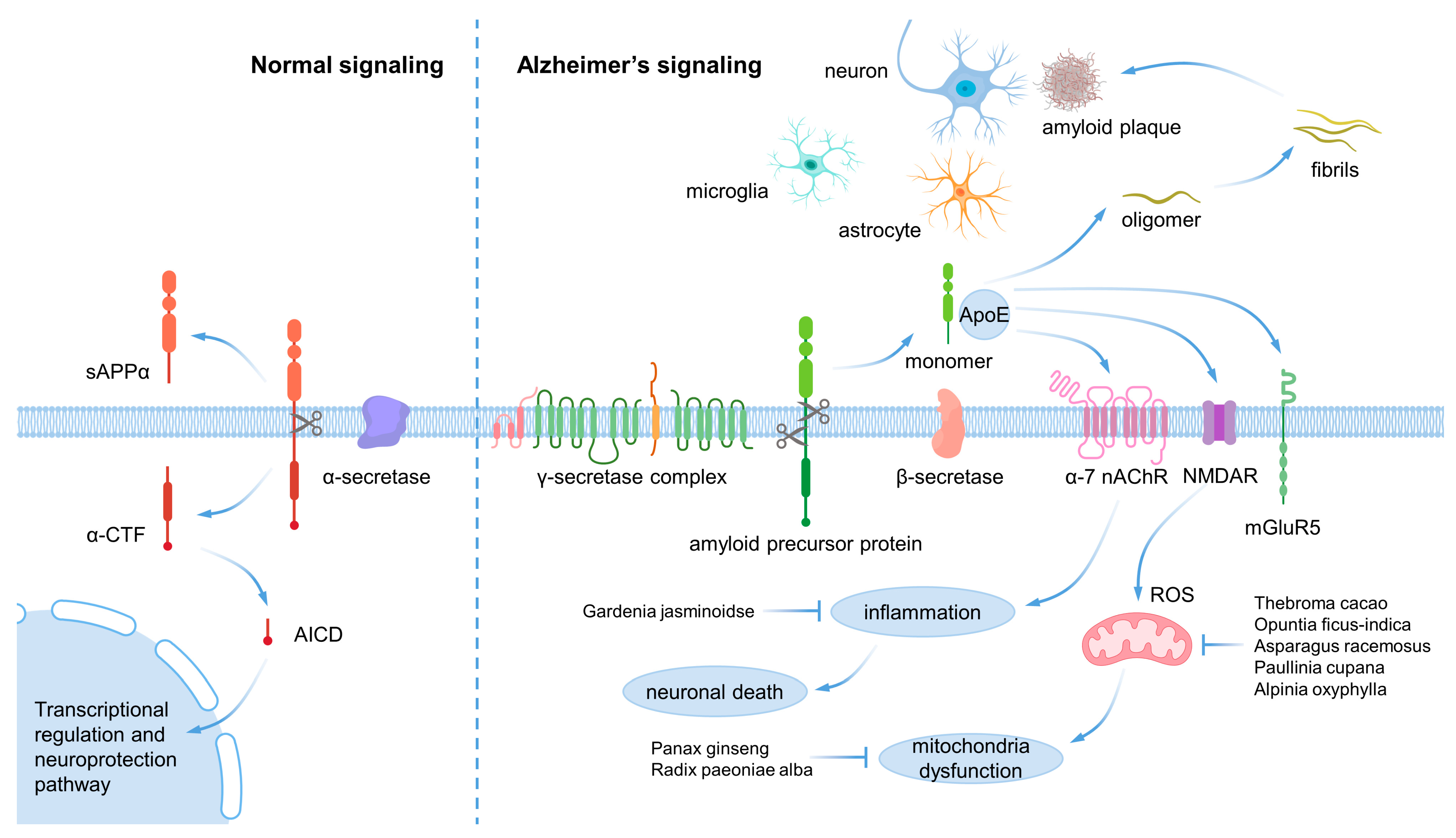
Figure 1. β-amyloid pathology in Alzheimer’s disease.
Ijms 24 08827 g001
Figure 2. α-synuclein pathology in Parkinsons’s disease.
Ijms 24 08827 g002
Figure 3. Flow chart of the study’s selection process.
Ijms 24 08827 g003
Table 2. Effect of natural products for neurodegenerative diseases.
Natural ProductsPart of the Natural
Products
ModelDoseEffectsRef.
Reynoutria multiflora MoldenkePurified compoundMesencephalic dopamine neurons and SH-SY5Y cell100, 200 μM (in vitro)
20 mg/kg (in vivo)
Anti-oxidative, anti-aging, and anti-inflammatory effects
Restoration of the expression of FGF2 and BDNF, which inhibits apoptosis.
Activation of the FGF2-Akt and BDNF-TrkB signaling pathways in the substantia nigra and corpus striatum, leading to the recovery of dopaminergic neurons.
[53]
Achillea fragrantissima Sch.Bip.-H2O2-treated astrocytes and neuronUp to 34.7 μM (in vitro)The inhibition of phosphorylation of stress-activated protein kinase/c-Jun N-terminal kinase (ERK 1/2), mitogen-activated protein kinase kinase (MEK1), kinase (SAPK/JNK), and the transcription factor cyclic AMP response element-binding protein (CREB) was observed.[54]
Theobroma cacao L.Commercial cocoa powderβ-amyloid-treated SH-SY5YMixture of 30 μg/mL of epicatechin, 10 μg/mL catechin and 170 μg total polyphenols (in vitro)Antioxidant, activating the BDNF survival pathway.[55]
Salvia miltiorrhiza Bunge-6-OHDA-treated PC12 and zebrafish100, 200, 400 μMActivated the nuclear translocation of Nrf2 to increase heme oxygenase-1 (HO-1), conferring protection against ROS.
Induced the phosphorylation of Akt.
[56]
Asparagus racemosus Willd.RootIntra-hippocampal and intra-striatal administration of kainic acid18 mg/kgReduction of membranal lipid peroxidation and protein carbonyl following improvement in GPx activity and GSH contents.[57]
Opuntia ficus-indica (L.) Mill.Fruit skinAD fly model with brain-specific expression of Aβ42 and PD fly model based on transgenic expression of the human α-syn A53T mutant1 mg/mL (in yeast)
0.06% (in drosophila)
100, 400, 800, 2000 μg/mL (in vitro)
Inhibition of the fibrillogenesis of both Aβ42 and α-syn
Accumulation of remodeled oligomeric aggregates that are less effective at disrupting lipid membrane integrity.
[58]
Gardenia jasminoides J.EllisFruitAPP/PS1 transgenic mice10, 20, 50 mg/kgSuppressed neuroinflammatory responses in the brain through regulating phosphatidylinositide 3-kinase/AKT (PI3K/AKT) signaling pathway activation, expression of inflammatory proteins and release of inflammatory cytokines.[59]
Vitis labrusca L.Purified compound6-OHDA-treated PC12 and rats12.5, 25, 50 μM (in vitro)
60 mg/kg (in vivo)
Neuroprotection against 6-OHDA-induced neurotoxicity.
Reduction oxidative stress and improvement in mitochondrial dysfunction.
Activation of the PI3K/Akt signaling pathway.
[60]
Paullinia cupana Kunth-Aβ42-induced ad model of Caenorhabditis elegans10, 50 mg/mLAntioxidant activity and modulation of proteostasis.
Intracellular ROS and the accumulation of autophagosomes reduction.
Increased the expression of SOD-3 and HSP-16.2.
[61]
Tussilago farfara L.Buds6-OHDA-treated PC12 and mice1.25, 2.5, 5, 10 μM (in vitro)
5 mg/kg (in vivo)
Activating the Nrf2/HO-1 signaling pathway.[62]
Panax ginseng C.A.Mey.RootPC12 cells were treated with 250 μmol/L corticosterone6.25, 12.5, 25, 50, 100, 200 μg/mLNeuroprotection against corticosterone-induced damage in PC12 cells, and the intervening of HDAC6 and HSP90 of the GR-related function proteins, and subsequent restoration of ER and mitochondria functions.[63]
Polygala tenuifolia Willd.-6-OHDA-treated SH-SY5Y12.5, 25, 50, 100 μMAntioxidative effects, maintenance of mitochondrial function, and regulation of caspase-3 and tyrosine hydroxylase expression and activity.[64]
Alpinia oxyphylla Miq.Purified compoundN2a/APP cells and SAMP8 mice12, 25, 50, 100, 200, 400 μM (in vitro)
10, 20 mg/kg (in vivo)
Antioxidative effect through the Akt-GSK3b and Nrf2-Keap1-HO-1 pathways.[65]
Paeonia × suffruticosa AndrewsPurified compound6-OHDA-treated cortical neurons0.75, 1, 1.5 μMDecreased reactive oxygen species production.
Increased cell viability, superoxide dismutase activity, and the anti-apoptotic protein expression.
[66]
Paeonia lactiflora Pall.-Glutamate-treated PC12 cell0.1, 1, 10 μMNeuroprotective effect on glutamate-induced apoptosis in PC12 cells by regulating the mitochondrial membrane potential and Bcl-2/Bax signal pathway.[67]
Cynanchum otophyllum C.K.Schneid. 3XTg AD mice6.5, 12.5, 25 μg/mL (in vitro)
25, 50, 100 mg/kg (in vivo)
Activation of PPARα-TFEB pathway.[68]
Disclaimer/Publisher’s Note: The statements, opinions and data contained in all publications are solely those of the individual author(s) and contributor(s) and not of MDPI and/or the editor(s). MDPI and/or the editor(s) disclaim responsibility for any injury to people or property resulting from any ideas, methods, instructions or products referred to in the content.