Review

Modulation of Muscarinic Signalling in the Central Nervous System by Steroid Hormones and Neurosteroids

1
Institute of Organic Chemistry and Biochemistry, Czech Academy of Sciences, Flemingovo Náměstí 2, Prague 6, 166 10 Prague, Czech Republic
2
Institute of Physiology, Czech Academy of Sciences, Vídeňská 1083, 142 20 Prague, Czech Republic
*
Authors to whom correspondence should be addressed.
Int. J. Mol. Sci. 2023, 24(1), 507; https://doi.org/10.3390/ijms24010507
Submission received: 28 November 2022 / Revised: 16 December 2022 / Accepted: 19 December 2022 / Published: 28 December 2022
(This article belongs to the Special Issue Steroids and Lipophilic Hormones, and Their Actions 3.0)

Abstract

:
Muscarinic acetylcholine receptors expressed in the central nervous system mediate various functions, including cognition, memory, or reward. Therefore, muscarinic receptors represent potential pharmacological targets for various diseases and conditions, such as Alzheimer’s disease, schizophrenia, addiction, epilepsy, or depression. Muscarinic receptors are allosterically modulated by neurosteroids and steroid hormones at physiologically relevant concentrations. In this review, we focus on the modulation of muscarinic receptors by neurosteroids and steroid hormones in the context of diseases and disorders of the central nervous system. Further, we propose the potential use of neuroactive steroids in the development of pharmacotherapeutics for these diseases and conditions.

Graphical Abstract

1. Introduction

Besides their well-known genomic effects, neurosteroids (NSs) produced by neurons and glia modulate neuronal activity by binding to various membrane proteins. Their neuromodulatory effects vary depending on location and time. Among well-studied receptors modulated by neurosteroids are, for example, γ-aminobutyric acid (GABAA) [1] and the glutamate N-methyl-D-aspartate (NMDA) receptors [2]. In this review, we focus on less-studied modulation of muscarinic acetylcholine receptors by NSs and steroid hormones (SHs) in the central nervous system (CNS) and its implications in CNS-related disorders and diseases.
Muscarinic acetylcholine receptors (mAChRs) are G-protein coupled receptors (GPCRs) abundant throughout the whole body. All five subtypes of the muscarinic receptor are expressed in the CNS, where depending on their location and subtype mediate various functions, including cognition, memory, or reward. Therefore, mAChRs may serve as targets for various diseases and conditions such as Alzheimer’s disease, schizophrenia, addiction, epilepsy, or depression [3]. The orthosteric binding site of all five subtypes is identical [4,5,6,7], making it an unsuitable pharmacological target. In contrast, allosteric binding sites are not under the evolutionary pressure to accommodate natural neurotransmitters and thus vary in their structure, being a more suitable pharmacological target than the orthosteric site. Indeed, achievable selectivity, the advantage of allosteric modulators is the preservation of spatial-temporal pattern of the modulated signalling as positive allosteric modulators (PAMs) increase whereas negative allosteric modulators (NAMs) diminish or block responses to the neurotransmitter upon its release. Allosteric modulators may change both affinity and efficacy of the neurotransmitter as well as alter coupling to the downstream signalling pathways [8,9].
Thanks to strong interest and intensive research, the number of allosteric modulators of mAChRs is growing. Allosteric modulators of mAChRs encompass a broad spectrum of chemotypes [10]. Moreover, similar to many other GPCRs, mAChRs are allosterically modulated by membrane cholesterol (CRL) [11,12]. Some SH and NS, such as progesterone (PROG) or corticosterone, at physiologically relevant concentrations allosterically modulate the binding and functional response of mAChRs from the CRL-binding site [13,14]. These findings make neuroactive steroids interesting candidates for allosteric modulators of a novel scaffold. Here reviewed modulation of mAChRs by SHs and NSs under physiological and pathological conditions gives hints on desired properties of steroid-based allosteric modulators with pharmacological potential.

2. Neurosteroids and Neuroactive Steroids in CNS

NSs represent a class of endogenous compounds synthesized de novo in the CNS from CRL or steroidal precursors imported from peripheral endocrine glands. NSs are known to modulate neuronal signalling [12]. NSs and SHs exert multiple functions via two mechanisms. By the activation of nuclear receptors specific for a given steroid hormone they execute their genomic effects. Alternatively, by binding to the membrane proteins and modulating the activity of these proteins they execute their non-genomic effects. In this review, we will mainly focus on NSs and SHs that have been extensively studied over the last two decades—progesterone (PROG), pregnenolone (PREG), pregnenolone sulphate (PREGS), dehydroepiandrosterone sulphate (DHEAS), 3α-hydroxy-5α-pregnan-20-one (allopregnanolone, ALLO), 3α-hydroxy-5β-pregnan-20-one (pregnanolone), corticosterone, and 17β-estradiol.
The plasma level of circulating steroids is in the low nanomolar range [15]. Circulating steroids serve as precursors for the synthesis of neurosteroids [16]. However, it should be noted that effective concentrations of locally synthesised neurosteroids modulating membrane receptors including mAChRs may exceed the concentrations of circulation steroids [17].

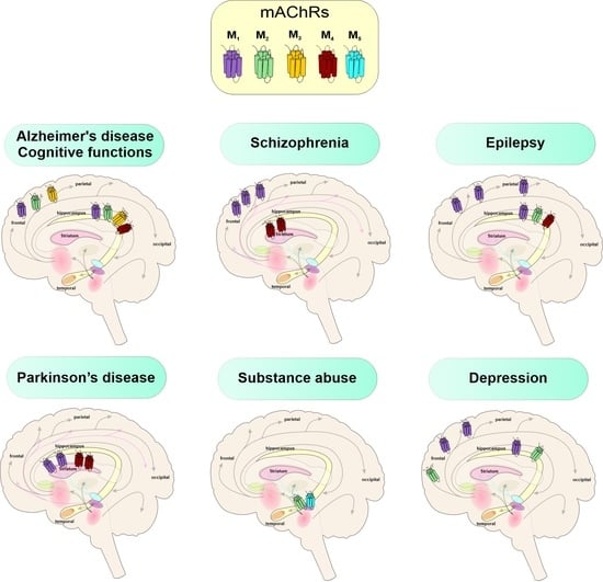
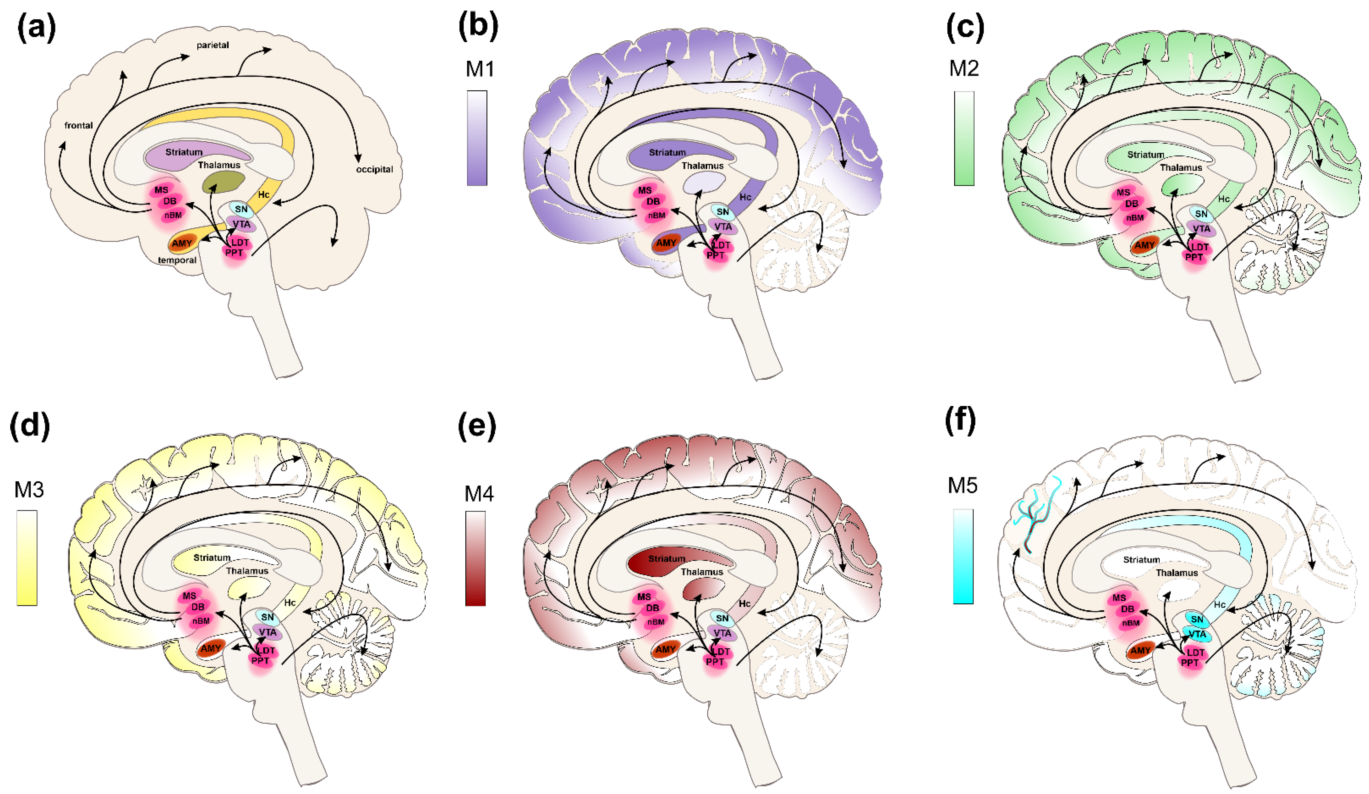
3. Muscarinic Receptors in CNS

Muscarinic receptors are expressed throughout the entire CNS. Based on their subtype and location, they mediate a wide array of physiological processes and thus become pharmacological targets in various pathologies related to altered muscarinic signalling (Table 1). The M1 receptors are predominantly expressed in the neocortex, hippocampus, and striatum [18,19]. Stimulation of M1 signalling facilitates learning and memory [20]. Impaired M1 signalling constitutes the basis for cognitive decline and dysfunction in various forms of dementia including AD and cognitive symptoms of schizophrenia [21,22,23,24,25]. Overstimulation of M1 receptors may lead to seizures and epilepsy [26,27,28]. The M2 receptors are primarily expressed in the hippocampus, cortex, basal forebrain, and thalamus [19]. Their activation leads to neural inhibition and analgesia and also to tremors and hypothermia [26]. Inhibition of M2 receptors, for example, leads to increased locomotion and decreased food intake. The M2 receptors are implied in the management of pain [29] and depression [30]. The M3 receptors are mainly expressed in the peripheral nervous system. In the CNS, their expression is limited to the cortex and basal ganglia [19]. Similarly to M1 receptors, they improve learning and memory [31]. The M4 receptors are primarily expressed in the striatum, neocortex, hippocampus, and basal ganglia [19,24,32]. Similarly to M2 receptors, their activation leads to auto-inhibition of acetylcholine (ACh) release and analgesia [33]. Additionally, it decreases the release of dopamine in the striatum [34] controlling rhythmicity of locomotor activity [35] and having anti-psychotic effects [36]. The M4 receptors are proposed pharmacological targets in schizophrenia [21,24,25,37,38], Parkinson’s disease (PD) [34,39], and pain management [40]. In comparison to other mAChRs, the expression of M5 receptors is scarce and is limited mainly to the ventral tegmental area (VTA), substantia nigra (SN), and brain microvasculature [19,24,31] and to a lesser extent to cerebellum [41]. Activation of M5 receptors in microvasculature results in cerebral vasodilation. The VTA is a part of the mesocorticolimbic circuit that works as a reward system. Activation of M5 receptors in the VTA induces dopamine release [26,27], promoting drug-seeking behaviour and reward [25]. The M5 receptors are proposed pharmacological targets in AD [42,43], schizophrenia [25], and substance abuse [43,44]. For further details see Figure 1 and Table 1.

4. Genomic Effects of Steroids on mAChRs in CNS

Effects exerted by steroids that take place over a longer timescale include genomic action [48]. This accounts for 17β-estradiol and PROG as it represents a fundamental effector in the regulation of the menstrual cycle and cognition in females. Age-related decreases in the 17β-estradiol as well as PROG, 5α-Dihydroprogesterone (5α-DHP), and ALLO levels within CNS in older age [49] are thought to be implicated in cognitive decline and higher risk of Alzheimer’s disease (AD) [50,51,52,53]. These steroidal changes in men are associated with alterations in mAChRs expression levels within the brain.
There are numerous studies on mAChRs expression changes in the human brain, however, the results are often unmatched. Some of the analyses conducted on post-mortem brain tissue described marked decline [54], while other studies show only minor changes in the overall mAChRs population detected in ageing individuals [55]. The results of the more detailed in vivo positron emission tomography (PET) studies in age-related muscarinic decline brought even more contradictions. Both reductions [56,57,58,59] and increases [60] in the densities of mAChRs were reported. These inconsistencies are mainly due to variability in the populations (males and females), selectivity of used ligands, and differences in quantification methods. To avoid such discrepancies [61], the age-related differences between M1 and M4 receptors in the population of healthy women have been examined, employing the single photon emission tomography (SPET) of the M1/M4 selective probe (R, R)[123I]-I-QNB. This study revealed an age-related reduction in mAChRs densities.
To date, many animal and human studies revealed that Estrogen Therapy (ET), if initiated within a specific time window, can bring beneficial effects on cognition and reduce the risk of dementia by enhancing the cholinergic system via increasing choline acetyltransferase (ChAT) activity as well as high-affinity choline uptake [51,62,63]. In animals, mAChRs densities fluctuate during oestrous cycles and it is positively correlated with the levels of estrogens. During the proestrus characterized by the highest levels of estrogen, the mAChRs densities increase, while during diestrous, the mAChRs densities decline together with estrogens levels [64]. It suggests that estrogens regulate mAChRs expression levels by their genomic action.
Ovariectomy is often used in laboratory animals as a model of menopause. It was shown that the removal of ovaries is associated with a decrease in ChAT mRNA and changes in the expression of mAChRs and a decrease in cholinergic fibre density [65,66]. Experimental studies in ovariectomized rats suggested that estrogen level affects mAChRs expression in the cerebral cortex [64], medial basal hypothalamus [67,68], and hippocampus [69,70,71]. Interestingly, a recent study did not find any changes in the density of mAChRs in the brain of ovariectomized rats [72].
Changes in mAChRs expression levels reported across mentioned studies varied, depending on the time post-ovariectomy, the time when the 17β-estradiol replacement was initiated, the 17β-estradiol dose as well as on the time point at which expression was measured. In general, the removal of ovaries resulted in an initial, significant increase in mAChRs expression levels throughout the brain which might be contracted by 17β-estradiol replacement (Table 2). On the contrary, some researchers reported a marked decrease in mAChRs expression levels in mice brains following ovariectomy [65]. In this study, the authors showed that 17β-estradiol-induced improvement in cognitive performance was not correlated with ovariectomy itself nor with the levels of mAChRs, which remained unchanged after 17β-estradiol treatment. Hormonal replacement therapy often comprises a combination of 17β-estradiol and progesterone. In the reported animal studies, 17β-estradiol and progesterone [64] or progesterone alone [63] did not influence mAChRs expression levels. On the contrary, some studies suggest that progesterone may counteract the effects of estrogens [73].
As for human studies, there are very few publications revising estrogen-induced changes in mAChRs expression levels. An example might be the single-photon emission tomography (SPET) study reporting higher mAChRs brain densities in postmenopausal women receiving estrogen therapy [74].
The exact mechanisms and relationship between the changes in the densities of mAChRs and estrogens level are still not fully understood, however, some concepts may partially explain an increase in mAChRs associated with lowered 17β-estradiol levels during menopause or after ovariectomy. Since membrane estrogen receptors ERα, Erβ, and GPR30/GPER1 are co-expressed with mAChRs in cultured hippocampal neurons [75,76], it was hypothesized that the lack of estrogen receptor stimulation could also diminish the ACh release from the cholinergic neurons. As a result, low ACh levels could induce a compensatory increase in mAChRs expression [63,70]. On the contrary, estrogen replacement therapy applied immediately after ovariectomy may prevent the instantaneous upregulation of mAChRs observed in the rodent brain. It might be speculated that increased 17β-estradiol levels provided by the treatment may indirectly stimulate mAChRs gene expression by signalling through tyrosine kinase receptor (trkA) [70,77,78]. Indeed, 17β-estradiol can also influence the mAChRs expression levels by modulation of histone methylation via the mER-activated PI3K/Akt signal transduction pathway [79,80]. Prolonged periods of stress-increasing corticosterone reduced ACh uptake and increased the density of mAChRs in the hippocampus [81]. However, it is more likely that the changes in mAChRs expression levels are due to heterologous regulation by different hormones and transmitters [82].

5. Neurosteroids and Muscarinic Receptors in CNS

5.1. Cognitive Functions

A wide array of evidence suggests that cholinergic neurotransmission via mAChRs plays a key role in multiple cognitive functions [20] including attention [83], learning, and memory formation [84]. Antagonists of mAChRs are well known to cause confusion, disorientation, learning, and memory deficits which support an important role for the cholinergic system in these cognitive functions. Indeed, the administration of non-selective muscarinic receptor antagonist scopolamine has been and still is widely used as a valid pharmacological model of cognitive impairment [85] as well as an experimental compound increasing locomotor activity [86]. On the other hand, M1-preferring agonists, such as WAY-132983 [87] or xanomeline [20], or M1-selective partial agonist HTL0018138 [88] exhibit clear pro-cognitive effects. Attention, memory, and learning are essential for flexibility in responding to changing environments. These processes are highly interconnected as attention powerfully influences what we memorize [89]. Regarding the mechanisms of learning and memory, for relevant information to be acquired from the environment needs to be selected/filtered from many others, to be further encoded, stored, and/or retrieved (episodic memory), and it is regulated by attention. As for implicit memory, attention helps in sculpting what we learn, and working memory is closely related to selective attention at a particular moment [89,90]. Top-down attention (as well as working memory and inhibitory control) highly depend on activity within networks of prefrontal and parietal cortical areas [91,92].
The major role in cognition is attributed to M1 and M3 receptors. The M1 receptor is highly expressed in the cerebral cortex, hippocampus, and striatum, especially in extrasynaptic regions of glutamatergic pyramidal neurons [93]. Its localization is consistent with the cholinergic modulation of glutamatergic neurotransmission and thus neuronal excitability, synaptic plasticity as well as learning and memory processes [94,95,96]. Activation of M1 receptors was found to facilitate long-term potentiation (LTP) and enhance NMDAR activation [97]. The M3 receptor expression in CNS is much less abundant in comparison to M1. The M3-receptor knockout mice are deficient in hippocampus-dependent contextual fear conditioning [98]. Mice strain expressing the phosphorylation-deficient M3 receptor showed a deficit in fear conditioning, suggesting this type of learning depends on receptor phosphorylation/arrestin signalling pathway [99].
NSs improve memory by a mechanism involving modulation of key excitatory glutamatergic NMDA receptors and inhibitory GABAergic signals [100] as well as by affecting ACh release and regulating cholinergic neurotransmission [101,102,103]. The early studies on the influence of steroids on mAChRs function in cognition started with observations that 17β-estradiol upregulates the activity of the acetylcholine-synthesizing enzyme, choline acetyltransferase (ChAT) when administered after ovariectomy [104]. Further, it was reported that treatment with 17β-estradiol prevented ovariectomy-induced deficits in attention and learning [105] and 17β-estradiol was also shown to partially or completely reverse scopolamine-induced memory impairment in spatial memory tasks [62,106,107]. Since 17β-estradiol is needed for adequate functioning of basal forebrain cholinergic systems, a decrease in its levels may be responsible for menopause-associated cognitive decline.
The estrogen facilitates working memory via M2 receptors expressed in GABAergic neurons of the CA1 region of the hippocampus. Estrogen stimulates ACh release and thus activates these M2 receptors reducing GABAergic inhibitory tone on hippocampal pyramidal cells and increasing expression of NMDA receptors in the CA1 region of the hippocampus [108,109] resulting in the improvement of working memory [101]. The effects of 17β-estradiol on learning and memory are rather complex and involve not only muscarinic but also nicotinic and other receptors and signalling pathways [110]. It is disputable whether the improvement of cognition exerted by 17β-estradiol is due to the direct modulation of the M1 receptor alone. Typically, 17β-estradiol is present in the body at rather low concentrations (0.07–2.3 nM during the menstrual cycle and 22 nM during pregnancy). However, 17β-estradiol increases ACh efficacy at the M1 receptor only at concentrations of 100 nM and higher [13]. The levels of PROG are decreasing with age and participate in cognitive decline. It was demonstrated that PROG exerts opposing effects on memory during learning. High levels of PROG disrupt the acquisition phase of learning but facilitate the consolidation phase of memory formation [111]. Given that PROG increases the efficacy of ACh at the M1 and decreases it at M2 receptors, it might account for the improvement in the consolidation of memory formation. On the other hand, negative modulation of M2 receptors in GABAergic neurons may lead to an increased GABAergic tone and inhibition of NMDA receptors disrupting the acquisition phase of learning.
Adrenal stress hormones, such as cortisol (corticosterone in the rat), exert a major influence on brain functions, mainly related to stress reaction, memory, cognition, and sleep [112]. Specifically, glucocorticoids enhance the consolidation of new memories but impair information retrieval from long-term memory [113]. Corticosterone enhances the consolidation of memory associated with emotionally charged and stressful events [114,115]. This augmentation of glucocorticoid-induced memory consolidation is dependent on increased cholinergic activity within the basolateral amygdala (BLA) [116]. Moreover, in animal studies, increased levels of corticosterone within the hippocampus were associated with improved memory consolidation during the awake state, but not during sleep [117].
Stressful stimuli are associated with an increase in choline uptake and ACh release. The levels of glucocorticoids increase with prolonged stress or ageing [118], resulting in hippocampal neuronal loss and memory impairment. During chronic stress, elevated levels of Ach cause an up-regulation of mAChRs [81]. Conversely, a decrease in glucocorticoid levels stimulates hippocampal cholinergic neurotransmission [119], enhancing the process of memory consolidation [115]. In turn, an activation of the M2 (but not M4) receptor enhances corticosterone release [120]. Moreover, corticosterone increases Ach potency at the M2 but decreased it at the M4 receptor in functional experiments in vitro [13].
The PREG and sulphated neurosteroids includincludeding DHEAS, pregnanolone sulphate (PREGS), and estrone sulphate, also affect cholinergic neurotransmission. The PREG increases ACh release in many brain areas (frontal cortex, hippocampus, and amygdala) and improves spatial recognition and memory [102,103]. Sulphated neurosteroids reduced GABAergic recurrent inhibition in the hippocampus via enhancement of cholinergic neurotransmission [109]. The DHEAS and PREG reversed scopolamine-induced amnesia in rats and scopolamine-induced deficits in spatial memory in mice [121,122,123]. Moreover, inhibition of steroid sulfatase may be effective in improving memory by increasing levels of sulphated steroids in the brain [124]. Although sulphated NS improve cognitive processes related to muscarinic signalling, to the best of our knowledge, there is no evidence for the binding of sulphated NSs to mAChRs yet.

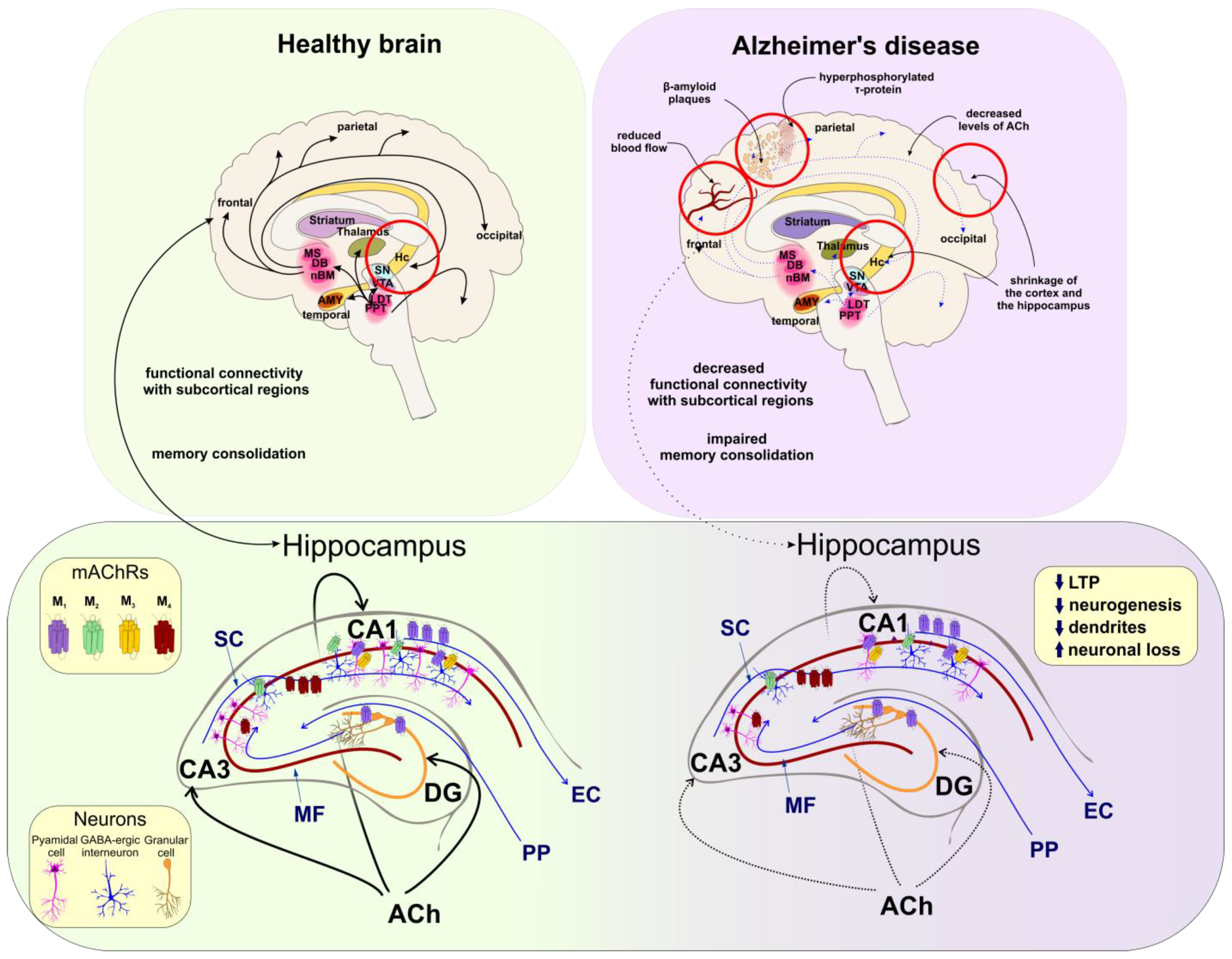
5.2. Alzheimer’s Disease

AD is a multifactorial progressive neurodegenerative disorder histologically defined by the presence of β-amyloid plaques in the brain. AD is associated with the gradual deterioration of memory and cognitive functions, impairment of cholinergic neurotransmission (especially in the cortex and hippocampus), and morphological changes in the brain (decrease in thickness of grey matter and increase in ventricular volume). Indeed, known factors associated with AD development (ageing, genetic factors, vascular diseases), there are many hypotheses concerning AD pathophysiology, including the formation of neurofibrillary tangles of hyperphosphorylated τ-protein inside the neurons (τ-hypothesis), abnormal extracellular deposits of already mentioned amyloid β (amyloid hypothesis), oxidative stress, inflammation, alterations in brain vasculature, cholinergic neuron damage, etc.
The amyloid hypothesis is tightly connected to the deterioration of cholinergic transmission. The amyloid hypothesis is based on the excessive accumulation of the extracellular deposits of beta-amyloid protein (Aβ) within the brain [125]. The Aβ peptide is the product of proteolytic cleavage of the transmembrane amyloid precursor protein (APP) by β- and γ-secretase. The APP is cleaved by these enzymes into several peptides which are 30–51 amino acids long. The most common forms are Aβ40 and Aβ42. The Aβ42 is most prone to oligomerisation. The Aβ42 oligomers are noxious inducing neuroinflammation in microglia cells and resulting in oxidative stress, and neurotoxicity, leading consequently to neurodegeneration [126]. Cleavage of APP by α-secretase precludes cleavage of APP by β-secretase resulting in the production of a soluble harmless non-amyloidogenic peptide (sAPP-α) which is responsible for the modulation of the normal neuronal excitability. Importantly, both pathways are regulated by the presence of membrane CLR [127]. Activation of the muscarinic M1 receptor leads to an increase in α-secretase activity both in vitro and in vivo [128,129,130]. Therefore, therapy by M1 agonists or PAMs was proposed [131]. In addition, activation of M1 was shown to reduce τ-protein phosphorylation, both in vitro and in vivo [132,133,134]. Mutations in the APP gene leading to increased cleavage by β-secretase are the cause of the familial form of AD that is characterized by early onset and fast progression. However, the familial form of AD accounts for less than 3% of AD cases. The majority of AD cases are sporadic forms of AD conditioned by many genetic, environmental, nutritional, and behavioural risk factors. The sporadic form of AD is late onset with slow progression. The major genetic risk factor of sporadic AD is the ε4 allele of apolipoprotein E (ApoE). The ApoE is a glycoprotein mediating lipid transport and endocytosis of CLR and lipoproteins. The ApoEε4 allele is associated with a high risk for AD as it promotes Aβ deposition and τ-protein hyperphosphorylation.
The cholinergic hypothesis of AD was built based on progressive neurofibrillary degeneration of limbic and neocortical cholinergic innervation associated with AD (Figure 2). Cholinesterase inhibitors (e.g., tacrine, donepezil, galantamine, rivastigmine) used as a treatment in AD patients, increase the availability of ACh at synapses and have been proven clinically relevant [135]. A defective coupling of neocortical M1 receptor to G-proteins is associated with cognitive decline in AD and dementia [22]. Additionally, it was suggested that M5 receptors of cerebral microvasculature are involved in the dilation of cerebral blood vessels [42]. Thus, the diminished cholinergic tone in AD, preventing proper vasodilatation, may play a part in the progression of AD. Therefore, stimulation of the M5 receptor may be beneficial in AD therapy [42].
As already mentioned, some neurosteroids (such as PROG) exert neuroprotective effects and depress the level of the hyperphosphorylation of τ-protein [52]. However, direct allosteric modulation of M1 signalling by 17β-estradiol at physiological concentrations is unlikely [13]. Therefore, the favourable effects of long-term estrogen therapy on cholinergic neurotransmission [136] (Table 3) are probably due to its influence on nicotinic acetylcholine receptors via estrogen receptor-β [137]. It might be also a result of an increased level of ACh due to activation of the GPR30 expressed mainly in the cholinergic neurons of the basal forebrain [76].
Figure 2. Changes in the human brain associated with AD. Left: In the healthy brain, projections from the basal forebrain and the brainstem (thick black arrows) supply cortical and subcortical brain regions (respectively) with ACh. Cholinergic neurons support cognitive functions and memory formation by sustaining functional connectivity between frontal cortical regions and the hippocampus (Hc) (black arrow). The Hc excitatory trisynapric circuit projections (blue lines) include pedunculopontine nucleus (PPN) projecting to the diagonal band of Broca (DG) (orange line-granule cells), DG projects to CA3 via MF, and CA3 region sends ith projections to CA1 via SC. (CA3 and CA1 are shown as a red line and pink-pyramidal cells). The CA1 further projects to EC. Activation of M1 (purple) mAChRs expressed mainly in granule cells of DG and M1/M3 (yellow) receptors at pyramidal neurons (pink) of the CA1, increases the activity of NMDARs (not shown) in long-term potentiation (LTP), synaptic plasticity, neurogenesis, memory encoding, and consolidation. The M2 (green) receptors are expressed mainly on interneurons (blue) and their activation reduces CA3 and CA1 activity. M4 (dark red) is expressed presynaptically on glutamatergic terminals. Their activation inhibits Schaffer collateral. Right: In cognitive decline and AD the ACh levels are decreased (thin blue arrows). Due to the accumulation of β-amyloid plaques, τ–protein tangles, neuronal loss in CA1, and reduced neurogenesis, the neocortex and hippocampus lose their volume. Lower part: Hc pathways are less active (thin blue arrows) resulting in impairment of memory. Stimulation of M1 in the DG, CA3, and CA1 as well as M2 on the GABA-ergic interneurons increases the excitability of pyramidal neurons in Hc and supports memory formation. According to [126,138,139].
Ijms 24 00507 g002
Table 3. Effects of the steroids on the muscarinic signalling in cognitive decline and AD.
M1M2
17-β estradiol
(mechanism independent from mAChRs)
↑ ACh levels
↑ LTP
↑ Learning acquisition phase
↑ Memory consolidation
↑ ACh potency
↓ Activation of GABA-ergic neurons (Hc)
↑ Excitability of pyramidal neurons (Hc)
↑ Activation of NMDARs
↑ LTP
↑ Learning acquisition phase
↑ Memory consolidation
Progesterone↑ ACh efficacy
↑ LTP
↑ Learning acquisition phase
↑ Memory consolidation
↓ ACh efficacy
↑ Activation of GABA-ergic neurons (Hc)
↓ Excitability of pyramidal neurons (Hc)
↓ Activation of NMDARs
↓ Learning acquisition phase
Corticosterone ↑ ACh potency
↓ Activation of GABA-ergic neurons (Hc)
↑ Excitability of pyramidal neurons (Hc)
↑ Learning acquisition phase
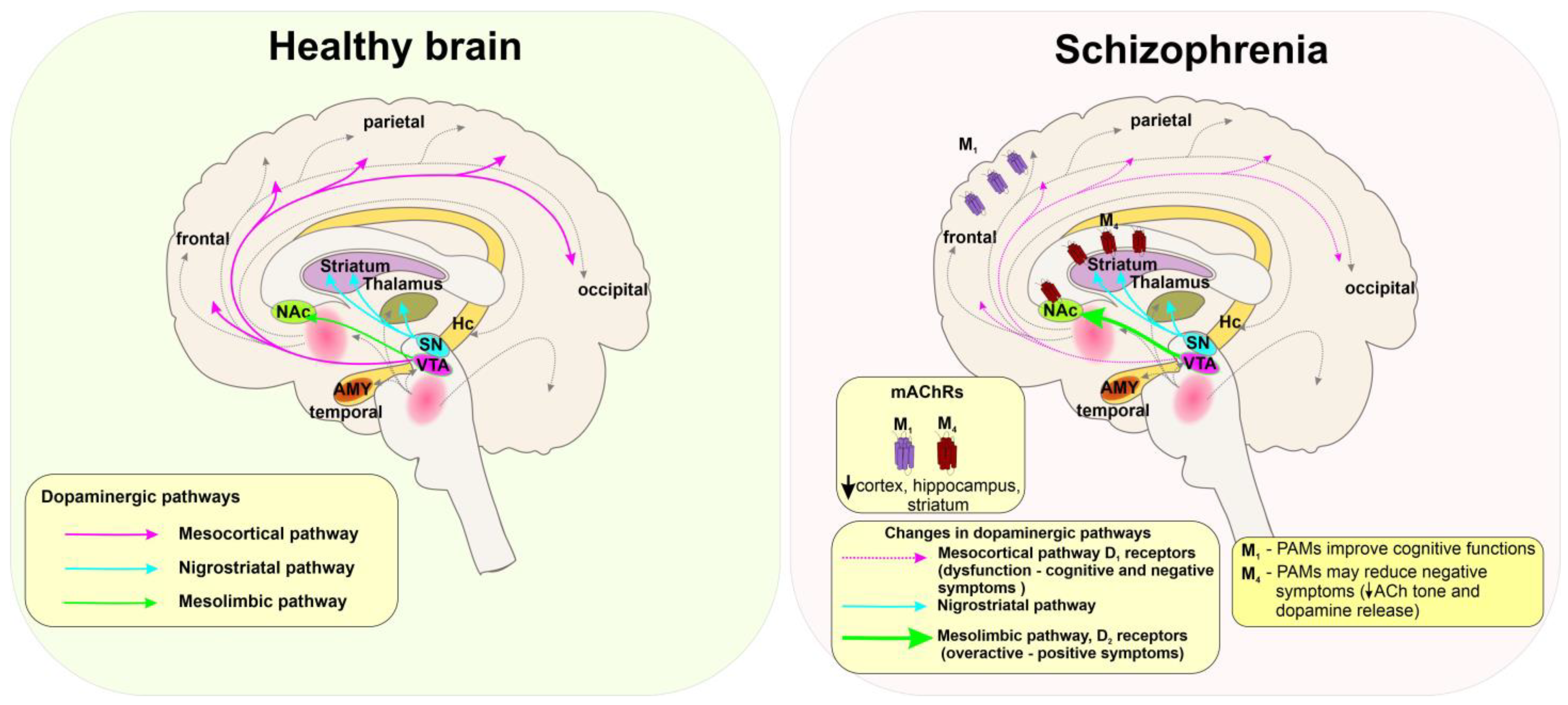
Abbreviations: increase ↑, decrease ↓, LTP-long-term potentiation, hippocampus (Hc).

5.3. Schizophrenia

Schizophrenia is a chronic psychiatric disorder with prevalence mostly in adolescence and early adulthood. Schizophrenia with its complex neurobiological background is accompanied by three categories of symptoms: (i) “Positive symptoms” are behaviours that are not normally present (hallucinations, delusions, and confusion associated with recurrent psychosis). (ii) “Negative symptoms” are fading or no longer present behaviours (anhedonia, social isolation, poverty of speech, loss of motivation). (iii) “Cognitive symptoms” are manifested as cognitive dysfunctions (Figure 3).
The hallmark of schizophrenia is overactive dopaminergic pathways in the central nervous system, especially the mesolimbic area, resulting in positive symptoms [140]. On the other hand, the negative symptoms result from reduced D1 receptor activation in the prefrontal cortex [141] and decreased activity of the nucleus caudatus [142]. Negative symptoms that arise are thought to occur also as a result of overactivation of the prefrontal cortex. The imbalance between increased long-term potentiation (LTP) and deficits in long-term depression (LTD) is often caused by transient inhibition of NMDARs and accompanied by negative and cognitive symptoms of the disorder [25].
In general, the dopaminergic and cholinergic systems are deeply interconnected. The release of ACh in the striatum is stimulated through the release of dopamine [38]. The anticholinergic drugs cause retrograde amnesia, impaired general cognitive performance, and worsen psychosis but also cause a modest improvement of negative symptoms. The pro-cholinergic drugs, such as cholinesterase inhibitors, improve cognitive functions [38]. A commonly used antipsychotic clozapine has a unique property to antagonize D2 receptors and serotonergic 5-HT2A and 5-HT2C receptors and to activate M4 receptors. The active metabolite of clozapine, N-Desmethylclozapine (NDMC), is an M1-preferring agonist [143]. Striatal M4 receptors modulate behaviour. Thus, changes in cholinergic signalling in the striatum contribute to positive and negative symptoms of schizophrenia. The M1 and M4 receptors in the cortex and basal ganglia, M2 in the thalamus and brainstem, and M5 in the brainstem and midbrain (substantia nigra and the VTA) affect dopamine release [144].
The expression level of M1 and M4 receptors is lower in the cortex, hippocampus, and striatum but not in the thalamus of the post-mortem samples from schizophrenia patients [145,146]. Therefore, centrally acting muscarinic agonists or PAMs selective for M1 and/or M4 receptors could be efficacious in treating the positive (psychosis) and negative (cognitive decline) symptoms associated with schizophrenia [21,37,38]. In preclinical studies, the M1/M4-preferring agonist xanomeline [147,148] improved verbal learning and short-term memory function [149,150]. However, xanomeline was withdrawn from clinical studies due to the severe side effects [150]. The M1-preferring allosteric agonist TAK-071 improved cognitive deficits in a rodent model of schizophrenia, exerting fewer side effects than xanomeline [151].
Indeed, 17β-estradiol may play a protective role against the development of schizophrenia [152]. Negative symptoms are less manifested at high 17β-estradiol levels during the menstrual cycle and pregnancy [78]. Treatment with PREG and subsequent increases in levels of ALLO and PREGS reduced the severity of negative and positive symptoms [100,153]. Thus, steroidal PAMs of M1 and M4 receptors have therapeutic potential in schizophrenia. PREG has a high affinity for mAChRs [13], yet its detailed effect on mAChRs concerning schizophrenia needs to be further investigated.
Figure 3. Changes in the human brain associated with schizophrenia. Left: In the healthy brain dopaminergic pathways arising from the ventral tegmental area (VTA) project into the neocortex (mesocortical pathway, pink), and to the nucleus accumbens (NAc) (mesolimbic pathway, green), while substantia nigra (SN) projects into the striatum and thalamus (nigrostriatal pathway, cyan). The distribution of dopaminergic projections greatly overlaps with cholinergic pathways (grey dashed lines). Right: In schizophrenia, dysfunction of the mesocortical pathway and decreased D1 receptors activation (dashed pink lines) are associated with negative symptoms. Overactive mesolimbic pathways (thick green lines) and increased activity of D2 receptors are related to positive symptoms of schizophrenia. Selective modulation of the central M1 and M4 receptors may represent the possible treatment of both negative and positive symptoms of the disease according to [144].
Ijms 24 00507 g003

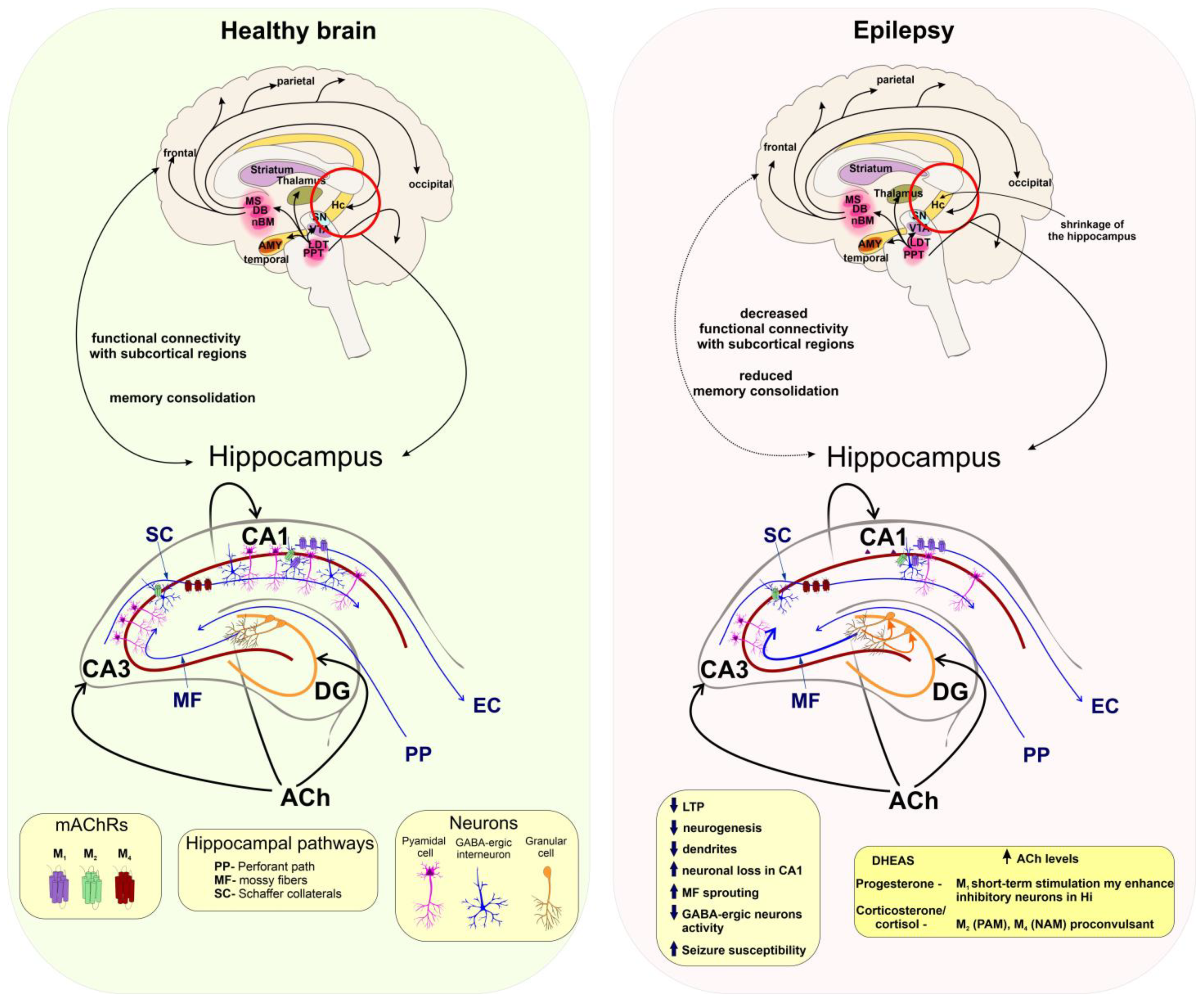
5.4. Seizures and Epilepsy

Overall, epilepsy is a collective name for a group of brain disorders consisting of a complex spectrum of different seizure types and syndromes. According to the International League Against Epilepsy (ILAE), a seizure is defined as “a transient occurrence of signs and/or symptoms due to abnormal excessive or synchronous neuronal activity in the brain.” Epilepsy is defined as a “disease characterized by an enduring predisposition to generate epileptic seizures and by the neurobiological, cognitive, psychological, and social consequences of this condition.” Therefore, a seizure is an event while epilepsy is a disease involving recurrent unprovoked seizures. Despite many advances in epilepsy research, presently an estimated 30% of people with epilepsy show drug resistance to currently available antiepileptic drugs (AEDs) [28]. A rather general explanation for the cause of seizure manifestation is a prolonged imbalance between the levels of excitatory and inhibitory neurotransmission (Figure 4).
Cholinergic neurotransmission plays an essential balancing role in brain excitability. Exposure to organophosphates (such as pesticides or chemical warfare agents) induces cholinergic neurotoxicity which may lead to the development of acute status epilepticus (SE), which may lead to the development of epilepsy [28]. As defined by the ILAE, “SE is a condition resulting either from the failure of the mechanisms responsible for seizure termination or from the initiation of mechanisms which lead to abnormally prolonged seizures. It is a condition that can have long-term consequences, including neuronal death, neuronal injury, and alteration of neuronal networks, depending on the type and duration of seizures”.
Enhancement of cholinergic activity by stimulation of mAChRs, mainly M1 receptor by pilocarpine, produces convulsions in laboratory rodents and is widely used as a model of SE in research laboratories [154]. Similarly, overstimulation of mAChRs following organophosphate poisoning may result in seizures [155]. An increase in ACh levels leads to an increase in susceptibility to seizures in experimental animals. Knockout mice models lacking acetylcholinesterase (AChE) are highly susceptible to seizures (progressing to tonic seizures) [156]. On the other hand, the mice with only partially eliminated AChE in the CNS by knocking out protein anchoring AChE to the membrane were not prone to develop spontaneous seizures and expressed a normal phenotype [157]. In accordance, brain-penetrating M1-selective antagonist VU0255035 suppresses the severity of the seizures and prevents SE after exposure to paraoxon or soman [158]. Repetitive seizure activity was shown to affect mAChRs expression throughout the brain. The expression levels of auto-inhibitory M2 receptors are higher in the neocortex of patients with focal temporal lobe epilepsy (TLE) [159] as well as in the brainstem of pentylenetetrazole-kindled epileptic rats [160] as the result of compensatory mechanisms. The expression levels of stimulatory M3 receptors are lower [161].
The importance of NS and steroid hormones in epileptogenesis was already described in detail [15,28,162]. From the wide range of steroidal compounds affecting susceptibility to seizures 17β-estradiol, PREGS, DHEAS, cortisol, and 11-deoxycortisol, were listed to have pro-convulsant effects. These NSs are acting mainly by negative modulation of GABAA and/or positive modulation of NMDARs. Interestingly, NSs can regulate ACh levels and thus play a crucial role in epileptogenesis. For example, intraperitoneally administered DHEAS enhanced the release of ACh in the rat cortex and hippocampus [102,163]. PROG, pregnanolone, androsterone, etiocholanone, DOC, THDOC, and ALLO are potent allosteric agonists and modulators of the synaptic and extrasynaptic GABAA receptors, enhancing phasic, and tonic neuronal inhibition [28].
A rise in the level of neuroprotective and antiseizure ALLO in the hippocampus has antiseizure effects [164]. Cyclic changes of estrogens and PROG are important in the pathogenesis of catamenial (menstrual cycle-related) epilepsy, as the changes in their levels correlate with the seizure incidence during the perimenstrual or periovulatory period [165].
The short-term selective stimulation of M1 or M4 receptors located mainly at glutamatergic terminals was shown to normalize hippocampal activity [166]. M1 receptors are responsible for tonic cholinergic excitation of CA1 and CA3 pyramidal neurons and M4 receptors modulate presynaptic inhibition of Schaffer collateral EPSPs [167]. The activation of M1 receptors enhances the excitability of fast-spiking putative (parvalbumin-positive) PV1 interneurons contributing to inhibitory tone within hippocampal circuitry [168]. Therefore, temporally-limited positive modulation of hippocampal M1 receptors may contribute to the anticonvulsive effect of PROG. Repetitive stress increases levels of corticosteroids resulting in an increased risk of seizures and epileptogenesis [169]. Consequently, long-lasting exposure to increased levels of corticosterone is often used as a model of chronic stress-inducing limbic epileptogenesis [28]. The M2 and M4 receptors are distributed both pre- and post-synaptically where expression of the M2 muscarinic subtype is characteristic of interneurons [93]. The pro-convulsant effects of corticosterone may be partially attributed to the positive modulation of the M2 and negative modulation of the M4 receptors [161].
Figure 4. Cholinergic projections in the brain and hippocampal circuit in health and epilepsy. Left: In the healthy brain, cholinergic neurotransmission provides functional connectivity between the neocortex and hippocampus (Hc) and thus supports memory consolidation. In the Hc, Ach maintains a balance between excitation and inhibition within neuronal pathways involved in learning and memory (see Figure 2). Cholinergic projections regulate Hc activity within the dentate gyrus (DG), CA3, and CA1 regions of the Hc and M1 receptors (purple) regulate excitability mainly of the pyramidal neurons (pink) and are also expressed on GABA-ergic interneurons (blue) in the CA1. The M2 receptors (green) are expressed mostly presynaptically on GABA-ergic interneurons and their activation supports the CA1 excitability. The M4 receptors on are expressed on glutamatergic neurons of SC and their activation decreases the activation of CA1. Right: In the epileptic brain, Hc circuits are affected by MS sprouting and increased excitability within DG granule cells. Mossy fibres (MF) send more projections to the CA3 and further increases Hc excitability and seizure susceptibility. These eventually lead to neuronal loss in the CA1 and impairment of Hc functions. Short-term stimulation of M1 expressed on interneurons may reduce excitability within CA1. M4 PAMs may inhibit overactive SC and thus CA1 activity. According to [45].
Ijms 24 00507 g004

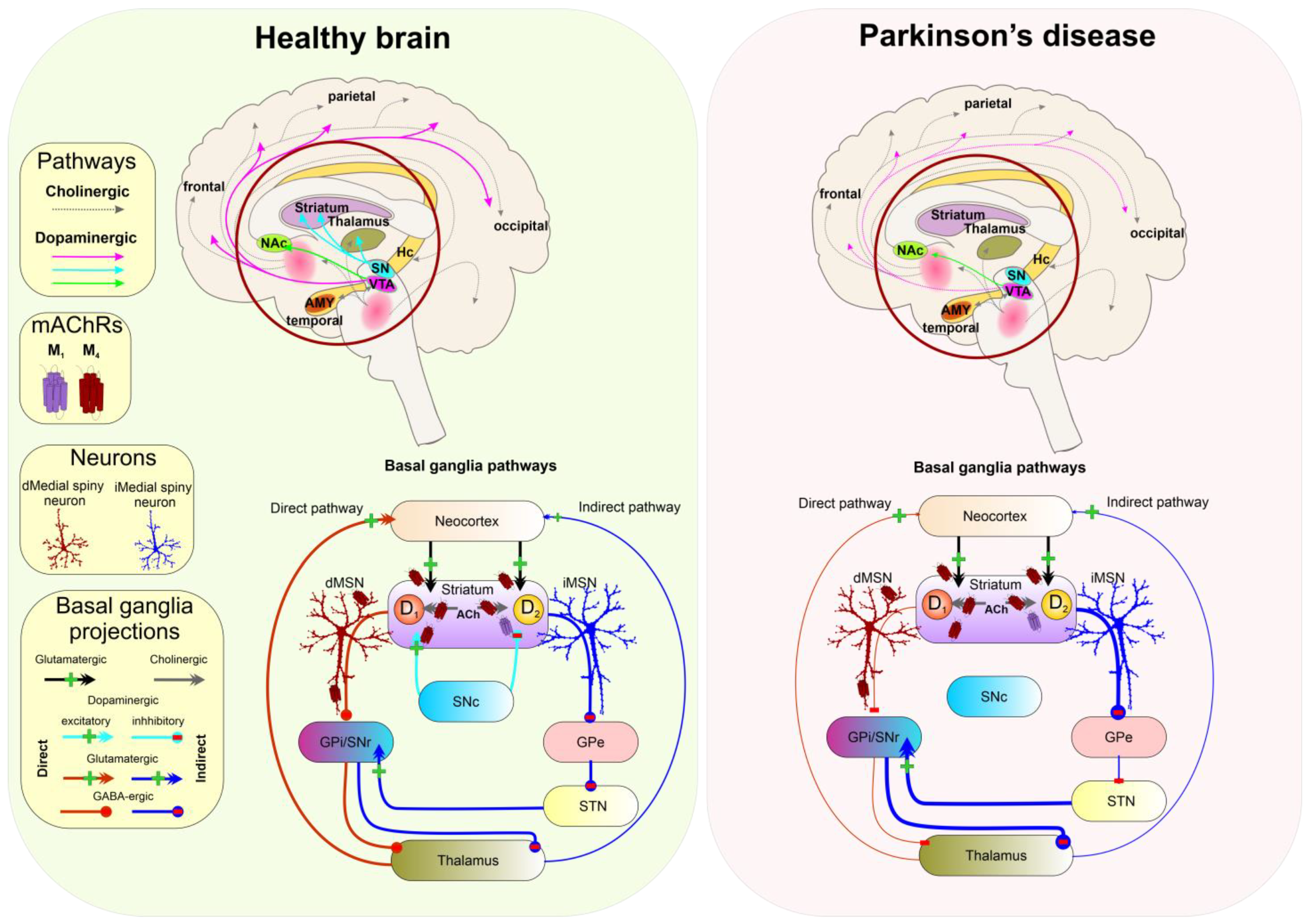
5.5. Parkinson’s Disease

Parkinson’s disease (PD) is associated with dopamine deficiency caused by the loss of dopaminergic neurons within the basal ganglia (BG) structure, substantia nigra pars compacta (SNc), and widespread intracellular protein (α-synuclein) accumulation (Lewy bodies). Decreased dopamine levels in the substantia nigra are responsible for slowness of movements, body rigidity, and rest tremors [170] (Figure 5).
Cholinergic neurotransmission mediated by mAChRs (mainly M4 and M1) and nAChRs is known to regulate dopamine release from the terminals originating from the SNc with the M4 receptor playing a central role [34]. In the striatum, mAChRs and dopaminergic receptors are often colocalised on Substance P-positive neurons, where the M1 receptor is co-expressed with the D2 receptor in stimulating indirect pathways, while the M4 receptor is co-expressed on medial spiny neurons with the D1 receptor in spiny projection neurons (dMSN) that comprise the BG direct pathway. Activation of M4 receptors reduces the glutamatergic tone in the striatum. Moreover, activation of M4 on dMSN leads to a decrease in dopamine release from dopaminergic terminals in SNc. On the cholinergic interneurons activation of M4 receptors may result in decreased tonic firing [173,174]. Thus, selective negative modulation of M4 receptor signalling may be effective in disorders accompanied by reduced dopamine levels (Table 1 and Figure 5). Indeed, NAMs of the M4 receptor can improve symptoms of Parkinson’s disease by increasing dopamine release [25,34]. On the other hand, centrally acting M1 antagonists such as trihexyphenidyl, pirenzepine, or VU0255035 alleviate symptoms of striatal dystonia [175].
NSs exert neuroprotective effects related to PD. In the animal model of PD, PROG, and 17β-estradiol protected against the degeneration of dopaminergic neurons induced by neurotoxin MPTP (1-methyl-4-phenyl-1,2,3,6-tetrahydropyridine) [176]. Interestingly, the levels of neuroprotective NS, such as PROG and ALLO, are lowered in the cerebrospinal fluid of PD patients [177]. Some of the beneficial effects of NSs on PD might be attributed to mild negative modulation of the M4 receptors by 17β-estradiol and PROG as PROG slightly decreases the efficacy of ACh, and 17β-estradiol decreases its potency at this receptor [13].

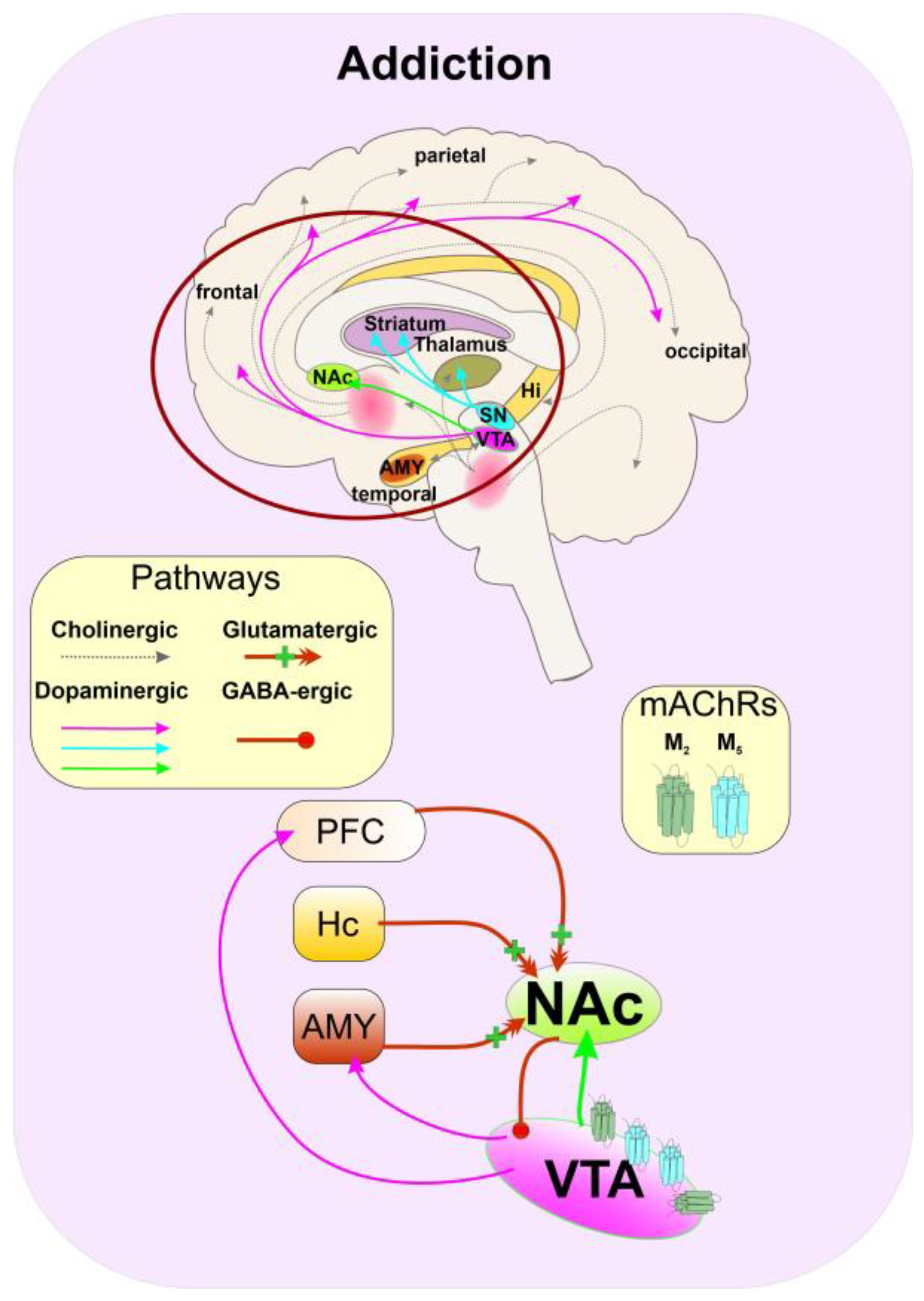
5.6. Substance Abuse

According to the Diagnostic and statistical manual of mental disorders (American Psychiatric Association, DSM-5 2013), substance abuse has several stages, starting with acute drug use and intoxication. Subsequent compulsive and harmful use may develop into dependence and withdrawal syndrome (with or without delirium) and drug-seeking behaviour that may trigger amnesic syndrome, residual and late-onset psychotic disorders, and other mental and behavioural changes. Addiction may be defined as a chronic illness characterized by compulsive drug seeking and use. Overall, substance abuse and addiction are associated with the function of dopamine. Dopamine cell bodies located in the ventral tegmental area (VTA) with their projections to the nucleus accumbens (NAc) and prefrontal cortex (PFC) represent the midbrain reward circuitry [178]. Stimulation of dopamine release within NAc, a region in the basal forebrain (part of the ventral striatum) which is activated by food and water intake, but also by psychomotor stimulants and opiates, produces the feeling of pleasure, euphoria, and reward. By modulation of the various brain regions interconnected with NAc, such as VTA, PFC, hippocampus, and amygdala, dopamine affects brain activity and establishes a mechanism of learned behaviour associated with reward [179] (Figure 6). Addiction develops because of alterations in these reward circuits.
Profound sex differences in course, symptoms, and treatment of substance use disorders indicate that estrogens and PROG are involved in mediating responses to drugs of abuse [180]. Typically, women are more vulnerable than men to the deleterious consequences of drug use at every phase of the addiction process. Exogenous PROG protects against the reinforcing effects and attenuates drug cravings [181]. On the other hand, estrogen enhances the reinforcing effects [182].
Figure 6. Schematic representation of the pathological dopaminergic circuitry in addiction. Repetitive rewarding stimulus induces excessive dopamine release from the ventral tegmental area (VTA) projections (pink lines) to the prefrontal cortex (PFC), amygdala (AMY), and most importantly to medial spiny neurons (MSNs) of nucleus accumbens (NAc) (light green), involved in the direct and indirect motor pathways (see Figure 5). Learned behaviour strengthens excitatory projections (red lines) from AMY, hippocampus (Hc), and PFC to NAc and supports addictive behaviour. MSNs of NAc containing D1 receptors project back to the VTA and inhibit dopamine release. Inhibition or negative modulation of M5 in the VTA might decrease dopamine release and prevent the further development of addiction according to [179,183].
Ijms 24 00507 g006
At least since the “opioid wars” in China, muscarinic antagonists atropine and scopolamine are used to prevent opiate overdose and the development of addiction, as well as support detoxification, indicating the involvement of mAChRs in the process of addiction and substance abuse. Dopamine release within NAc is decreased in M5 receptor knock-out mice. These are less sensitive to the rewarding effects of cocaine, indicating the role of M5 receptors in the regulation of the dopaminergic reward circuitry [184]. In addition, the NAM of the M5 receptor ML375 [185] affected cocaine-related behaviour without impairing behaviour not associated with drug use [25,184]. Probably, a decrease in potency and efficacy of ACh at the M5 receptor by pregnanolone [13] contributes to its beneficial effects on rewarding effects in drug abuse. The M2 receptors are expressed in the pre- and post-synaptic sites in the VTA. Their activation may affect cognitive and goal-based dopamine-mediated behaviour. Negative allosteric modulation of M2 receptors by pregnanolone may further diminish rewards seeking behaviour.

5.7. Depression

By the definition, depression is a mental state of low mood and apathy. Depression affects a person’s feelings, behaviour, and motivation. Major depressive disorder (MDD) is a multifactorial syndrome that involves disruption of mood, cognition, and other processes, including sleep, appetite, and libido. Signs and symptoms of MDD are mainly linked to low levels of brain monoamines dopamine, serotonin, and noradrenaline. In addition, stress and augmented glutamatergic neurotransmission often lead to neurodegenerative changes [186]. Changes in hippocampal neurogenesis, often associated with chronic stress in animal models may be in part mediated by cholinergic dysfunction, which in turn could underlie the cognitive disturbances observed in depression [187].
Antidepressant treatment involves the use of selective reuptake inhibitors of serotonin (SSRI), noradrenaline (NRI), both serotonin and noradrenaline (SNRI) as well as tricyclics and monoamine oxidase inhibitors (MAOI) which regulate and increase monoamine. Pharmacological targeting of the cholinergic system may represent a viable alternative. Muscarinic agonists (e.g., arecoline, oxotremorine), and AChE inhibitors (e.g., physostigmine, donepezil) provoke symptoms of depression. Stress-increased ACh release within the forebrain and activation of the septohippocampal pathway also results in MDD symptoms. Additionally, cholinergic dysfunctions may account for the development of cognitive symptoms associated with MDD [187]. The expression of M2 and/or M4 receptors are decreased in the dorsolateral prefrontal cortex of MDD patients [188]. The single-nucleotide polymorphism of A->T 1890 in the 3′ UTR of the (CHRM2) gene coding for M2 is gender-specific and characteristic of women with MDD [30].
In human studies, antidepressant-like properties of muscarinic antagonist scopolamine were first recorded about 30 years ago during the research of scopolamine as an anti-abuse drug on sleep and mood of alcoholics [189]. Unlike classical antidepressants, scopolamine exerts rapid antidepressant effects within a few days. Although scopolamine is a non-selective muscarinic antagonist, the anti-depressant mechanism of action of scopolamine was found to be dependent on the presence of M1 and M2. The knockout (KO) mice lacking these receptors expressed diminished scopolamine effectiveness in reducing immobility time in the forced swim test. In addition, the effects of M1-preferring antagonist were mitigated in M1 KO mice but not in the mice lacking M2 [190]. In the same study, the M2-preferring antagonist SCH226206 was found to be ineffective in M2 KO mice. Inhibition of M1 receptors in the medial prefrontal cortex (mPFC) was found to play a major role in the rapid antidepressant effects of scopolamine [191].
As the anxiety and symptoms of low mood are temporally linked to events of reproductive transitions in women (menstrual cycle, pregnancy and postpartum, and perimenopause) it is presumed that changes in the mood are rather associated with fluctuations in the levels of estrogens and PROG than by their absolute concentrations [192]. Additionally, women are more likely than men to show an antidepressant response to scopolamine [193]. These findings indicate the interplay between sex hormones and mAChRs in MDD.
PROG is well known for its negative impact on mood and memory. Changes in plasma levels are tightly associated with symptoms of depression or premenstrual dysphoric disorder [194]. These effects of PROG were found to occur independently from the activation of the endocrine PROG receptor. Acute administration of low doses of PROG (which reliably elevates ALLO and PREG levels) had negative effects on mood, while higher doses had no effect [194]. PROG may contribute to the symptoms of depression via modulation of mAChRs by increasing ACh efficacy at M1 receptors while diminishing it at M2 [13]. Positive modulation of post-synaptic M1 and negative modulation of pre-synaptic auto-inhibitory M2 receptors may have a synergistic effect.
As already mentioned, 17β-estradiol increases cholinergic neurotransmission and affects the function of mAChRs enhancing cognitive functions and alleviating scopolamine-induced impairments in attention and memory tasks [136]. It was found that treatment with 17β-estradiol alleviates depressive symptoms in premenopausal women. Under conditions where levels of estrogens are fluctuating and decreasing more rapidly than under normal circumstances, hormone replacement therapy stabilizes 17β-estradiol levels and thus moderates the negative effects of fluctuations of 17β-estradiol levels [195] and 17β-estradiol reduces ACh efficacy at M2 receptors [13] which may lead to augmented cholinergic transmission due to a decrease in M2-mediated auto-inhibition of ACh release. These findings indicate that increase in Ach levels by 17β-estradiol contributes to MDD.

6. Conclusions

The importance of cholinergic neurotransmission and its widespread effects exerted by mAChRs in health and disease is unquestionable. Due to their expression in the CNS and their contribution to the pathology of numerous psychiatric and neurologic diseases and disorders, mAChRs represent possible therapeutic targets. Keeping in mind that the orthosteric binding sites of all five subtypes of mAChRs are structurally the same, selective activation or inhibition of a single subtype by an orthosteric ligand is virtually impossible. Pharmacological targeting of mAChRs by orthosteric ligands is accompanied by more or less severe side effects. The answer to the need for subtype-specific compounds has emerged and is represented by allosteric modulators that bind to the less conserved sites on mAChRs. Recent advances have greatly expanded the set of known allosteric modulators of mAChRs. Cholesterol allosterically modulates many GPCRs including mAChRs. The CLR-binding site differs from the common allosteric binding site on mAChRs located between the second and third extracellular loops and is oriented towards the membrane. Compounds sharing the steroidal scaffold with CLR, such as SHs and NSs, have the potential to allosterically modulate GPCRs. Most recently, it was revealed that corticosterone and PROG, allosterically modulate the functional response of mAChRs at physiologically relevant concentrations. This important finding, not only pointed at the steroids exerting their action via a non-genomic mechanism but also suggested that already well-known beneficial effects of treatments with steroids (17β-estradiol, PROG, corticosterone) may be contributed by allosteric modulation not only of NMDA and GABA receptors but also mAChRs. Modulation of mAChRs by SHs and NSs contributes to various CNS-related disorders and diseases. Namely: Allosteric modulation of M1 and M4 receptors may have a protective effect in the development of schizophrenia. Proconvulsant effects of corticosterone may be partially attributed to positive modulation of the M2 and negative modulation of the M4 receptors. Negative modulation of the M4 receptors PROG and 17β-estradiol may alleviate PD symptoms. Negative modulation of the M5 receptor by pregnanolone may diminish the rewarding effects of drug abuse. Negative modulation of the M2 by 17β-estradiol increasing level of ACh may contribute to MDD. Further, positive allosteric modulation of M1 receptors by NSs has therapeutic potential in cognitive deficits and the development of AD.

7. Perspectives

Pharmacotherapeutic targeting of mAChRs in the CNS is an ongoing challenge. In general, NSs pass the blood-brain barrier. Some of them exert a high affinity for CLR-binding sites at mAChRs. Thus, allosteric modulation of mAChRs via the CLR-binding site/sites represents a new approach for the development of potent and efficacious compounds with therapeutic potential in diseases and disorders associated with altered muscarinic signalling. Development of novel neuroactive steroids acting as allosteric modulators of mAChRs may be expected soon.

Author Contributions

E.S. and A.R. compiled Section 3, Section 4 and Section 5; E.S.-P. and E.K. compiled Section 2 and contributed to Section 4 and Section 5; N.C. contributed to Section 3, Section 4 and Section 5; and J.J. wrote Section 1, Section 6 and Section 7. All authors have read and agreed to the published version of the manuscript.

Funding

Supported by the project National Institute for Neurological Research (Programme EXCELES, ID Project No. LX22NPO5107)—Funded by the European Union—Next Generation EU and by the Czech Academy of Sciences institutional supports RVO:61388963 and RVO:67985823.

Institutional Review Board Statement

Not applicable.

Informed Consent Statement

Not applicable.

Data Availability Statement

Not applicable.

Acknowledgments

We thank Esam El-Fakahany, University of Minnesota College of Pharmacy, and Vladimír Doležal, Institute of Physiology CAS, for the critical reading of the manuscript.

Conflicts of Interest

The authors declare no conflict of interest. The funders had no role in the design of the study; in the writing of the manuscript, or in the decision to publish.

References

  1. Lambert, J.J.; Cooper, M.A.; Simmons, R.D.J.; Weir, C.J.; Belelli, D. Neurosteroids: Endogenous Allosteric Modulators of GABA(A) Receptors. Psychoneuroendocrinology 2009, 34 (Suppl. S1), S48–S58. [Google Scholar] [CrossRef] [PubMed]
  2. Sedlácek, M.; Korínek, M.; Petrovic, M.; Cais, O.; Adamusová, E.; Chodounská, H.; Vyklický, L. Neurosteroid Modulation of Ionotropic Glutamate Receptors and Excitatory Synaptic Transmission. Physiol. Res. 2008, 57 (Suppl. S3), S49–S57. [Google Scholar] [CrossRef] [PubMed]
  3. Thomsen, M.; Sørensen, G.; Dencker, D. Physiological Roles of CNS Muscarinic Receptors Gained from Knockout Mice. Neuropharmacology 2018, 136, 411–420. [Google Scholar] [CrossRef] [PubMed] [Green Version]
  4. Haga, K.; Kruse, A.C.; Asada, H.; Yurugi-Kobayashi, T.; Shiroishi, M.; Zhang, C.; Weis, W.I.; Okada, T.; Kobilka, B.K.; Haga, T.; et al. Structure of the Human M2 Muscarinic Acetylcholine Receptor Bound to an Antagonist. Nature 2012, 482, 547–551. [Google Scholar] [CrossRef] [PubMed] [Green Version]
  5. Kruse, A.C.; Hu, J.; Pan, A.C.; Arlow, D.H.; Rosenbaum, D.M.; Rosemond, E.; Green, H.F.; Liu, T.; Chae, P.S.; Dror, R.O.; et al. Structure and Dynamics of the M3 Muscarinic Acetylcholine Receptor. Nature 2012, 482, 552–556. [Google Scholar] [CrossRef] [Green Version]
  6. Thal, D.M.; Sun, B.; Feng, D.; Nawaratne, V.; Leach, K.; Felder, C.C.; Bures, M.G.; Evans, D.A.; Weis, W.I.; Bachhawat, P.; et al. Crystal Structures of the M1 and M4 Muscarinic Acetylcholine Receptors. Nature 2016, 531, 335–340. [Google Scholar] [CrossRef] [Green Version]
  7. Vuckovic, Z.; Gentry, P.R.; Berizzi, A.E.; Hirata, K.; Varghese, S.; Thompson, G.; van der Westhuizen, E.T.; Burger, W.A.C.; Rahmani, R.; Valant, C.; et al. Crystal Structure of the M5 Muscarinic Acetylcholine Receptor. Proc. Natl. Acad. Sci. USA 2019, 116, 26001–26007. [Google Scholar] [CrossRef] [Green Version]
  8. Jakubík, J.; Randáková, A.; Chetverikov, N.; El-Fakahany, E.E.; Doležal, V. The Operational Model of Allosteric Modulation of Pharmacological Agonism. Sci. Rep. 2020, 10, 14421. [Google Scholar] [CrossRef]
  9. Wootten, D.; Christopoulos, A.; Sexton, P.M. Emerging Paradigms in GPCR Allostery: Implications for Drug Discovery. Nat. Rev. Drug Discov. 2013, 12, 630–644. [Google Scholar] [CrossRef]
  10. Jakubik, J.; El-Fakahany, E.E. Current Advances in Allosteric Modulation of Muscarinic Receptors. Biomolecules 2020, 10, 325. [Google Scholar] [CrossRef]
  11. Jakubík, J.; El-Fakahany, E.E. Allosteric Modulation of GPCRs of Class A by Cholesterol. Int. J. Mol. Sci. 2021, 22, 1953. [Google Scholar] [CrossRef]
  12. Szczurowska, E.; Szánti-Pintér, E.; Randáková, A.; Jakubík, J.; Kudova, E. Allosteric Modulation of Muscarinic Receptors by Cholesterol, Neurosteroids and Neuroactive Steroids. Int. J. Mol. Sci. 2022, 23, 13075. [Google Scholar] [CrossRef]
  13. Dolejší, E.; Szánti-Pintér, E.; Chetverikov, N.; Nelic, D.; Randáková, A.; Doležal, V.; Kudová, E.; Jakubík, J. Neurosteroids and Steroid Hormones Are Allosteric Modulators of Muscarinic Receptors. Neuropharmacology 2021, 199, 108798. [Google Scholar] [CrossRef]
  14. Dolejší, E.; Chetverikov, N.; Szánti-Pintér, E.; Nelic, D.; Randáková, A.; Doležal, V.; El-Fakahany, E.E.; Kudová, E.; Jakubík, J. Neuroactive Steroids, WIN-Compounds and Cholesterol Share a Common Binding Site on Muscarinic Acetylcholine Receptors. Biochem. Pharmacol. 2021, 192, 114699. [Google Scholar] [CrossRef]
  15. Reddy, D.S. Neurosteroids: Endogenous Role in the Human Brain and Therapeutic Potentials. Prog. Brain Res. 2010, 186, 113–137. [Google Scholar] [CrossRef]
  16. Baulieu, E.E.; Robel, P. Neurosteroids: A New Brain Function? J. Steroid Biochem. Mol. Biol. 1990, 37, 395–403. [Google Scholar] [CrossRef]
  17. Jo, D.H.; Abdallah, M.A.; Young, J.; Baulieu, E.E.; Robel, P. Pregnenolone, Dehydroepiandrosterone, and Their Sulfate and Fatty Acid Esters in the Rat Brain. Steroids 1989, 54, 287–297. [Google Scholar] [CrossRef]
  18. Levey, A.I.; Kitt, C.A.; Simonds, W.F.; Price, D.L.; Brann, M.R. Identification and Localization of Muscarinic Acetylcholine Receptor Proteins in Brain with Subtype-Specific Antibodies. J. Neurosci. 1991, 11, 3218–3226. [Google Scholar] [CrossRef] [Green Version]
  19. Abrams, P.; Andersson, K.-E.; Buccafusco, J.J.; Chapple, C.; de Groat, W.C.; Fryer, A.D.; Kay, G.; Laties, A.; Nathanson, N.M.; Pasricha, P.J.; et al. Muscarinic Receptors: Their Distribution and Function in Body Systems, and the Implications for Treating Overactive Bladder. Br. J. Pharmacol. 2006, 148, 565–578. [Google Scholar] [CrossRef] [Green Version]
  20. Bubser, M.; Byun, N.; Wood, M.R.; Jones, C.K. Muscarinic Receptor Pharmacology and Circuitry for the Modulation of Cognition. Handb. Exp. Pharmacol. 2012, 208, 121–166. [Google Scholar] [CrossRef]
  21. Crook, J.M.; Tomaskovic-Crook, E.; Copolov, D.L.; Dean, B. Low Muscarinic Receptor Binding in Prefrontal Cortex from Subjects with Schizophrenia: A Study of Brodmann’s Areas 8, 9, 10, and 46 and the Effects of Neuroleptic Drug Treatment. Am. J. Psychiatry 2001, 158, 918–925. [Google Scholar] [CrossRef] [PubMed]
  22. Tsang, S.W.Y.; Lai, M.K.P.; Kirvell, S.; Francis, P.T.; Esiri, M.M.; Hope, T.; Chen, C.P.L.-H.; Wong, P.T.-H. Impaired Coupling of Muscarinic M1 Receptors to G-Proteins in the Neocortex Is Associated with Severity of Dementia in Alzheimer’s Disease. Neurobiol Aging 2006, 27, 1216–1223. [Google Scholar] [CrossRef] [PubMed]
  23. Potter, P.E.; Rauschkolb, P.K.; Pandya, Y.; Sue, L.I.; Sabbagh, M.N.; Walker, D.G.; Beach, T.G. Pre- and Post-Synaptic Cortical Cholinergic Deficits Are Proportional to Amyloid Plaque Presence and Density at Preclinical Stages of Alzheimer’s Disease. Acta Neuropathol. 2011, 122, 49–60. [Google Scholar] [CrossRef] [PubMed] [Green Version]
  24. Scarr, E. Muscarinic Receptors: Their Roles in Disorders of the Central Nervous System and Potential as Therapeutic Targets. CNS Neurosci. Ther. 2012, 18, 369–379. [Google Scholar] [CrossRef] [PubMed]
  25. Moran, S.P.; Maksymetz, J.; Conn, P.J. Targeting Muscarinic Acetylcholine Receptors for the Treatment of Psychiatric and Neurological Disorders. Trends Pharmacol. Sci. 2019, 40, 1006–1020. [Google Scholar] [CrossRef]
  26. Westfall, T.C. Cholinergic Neurotransmission in the Autonomic and Somatic Motor Nervous System. In Encyclopedia of Neuroscience; Squire, L.R., Ed.; Elsevier: Amsterdam, The Netherlands, 2009; pp. 827–834. ISBN 9780080450469. [Google Scholar]
  27. Saygı Bacanak, M.; Aydın, B.; Cabadak, H.; Nurten, A.; Gören, M.Z.; Enginar, N. Contribution of M1 and M2 Muscarinic Receptor Subtypes to Convulsions in Fasted Mice Treated with Scopolamine and given Food. Behav. Brain Res. 2019, 364, 423–430. [Google Scholar] [CrossRef]
  28. Reddy, D.S. Role of Hormones and Neurosteroids in Epileptogenesis. Front. Cell Neurosci. 2013, 7, 115. [Google Scholar] [CrossRef] [Green Version]
  29. Tata, A.M. Muscarinic Acetylcholine Receptors: New Potential Therapeutic Targets in Antinociception and in Cancer Therapy. Recent Pat. CNS Drug. Discov. 2008, 3, 94–103. [Google Scholar] [CrossRef]
  30. Comings, D.E.; Wu, S.; Rostamkhani, M.; McGue, M.; Iacono, W.G.; MacMurray, J.P. Association of the Muscarinic Cholinergic 2 Receptor (CHRM2) Gene with Major Depression in Women. Am. J. Med. Genet. 2002, 114, 527–529. [Google Scholar] [CrossRef]
  31. Borroto-Escuela, D.O.; Agnati, L.F.; Fuxe, K.; Ciruela, F. Muscarinic Acetylcholine Receptor-Interacting Proteins (MAChRIPs): Targeting the Receptorsome. Curr. Drug Targets 2012, 13, 53–71. [Google Scholar] [CrossRef]
  32. Pancani, T.; Bolarinwa, C.; Smith, Y.; Lindsley, C.W.; Conn, P.J.; Xiang, Z. M4 MAChR-Mediated Modulation of Glutamatergic Transmission at Corticostriatal Synapses. ACS Chem. Neurosci. 2014, 5, 318–324. [Google Scholar] [CrossRef]
  33. Fiorino, D.F.; Garcia-Guzman, M. Muscarinic Pain Pharmacology: Realizing the Promise of Novel Analgesics by Overcoming Old Challenges. Handb. Exp. Pharmacol. 2012, 208, 191–221. [Google Scholar] [CrossRef]
  34. Moehle, M.S.; Conn, P.J. Roles of the M4 Acetylcholine Receptor in the Basal Ganglia and the Treatment of Movement Disorders. Mov. Disord. 2019, 34, 1089–1099. [Google Scholar] [CrossRef]
  35. Riljak, V.; Janisova, K.; Myslivecek, J. Lack of M4 Muscarinic Receptors in the Striatum, Thalamus and Intergeniculate Leaflet Alters the Biological Rhythm of Locomotor Activity in Mice. Brain Struct. Funct. 2020, 225, 1615–1629. [Google Scholar] [CrossRef]
  36. Dean, B.; Scarr, E. Muscarinic M1 and M4 Receptors: Hypothesis Driven Drug Development for Schizophrenia. Psychiatry Res. 2020, 288, 112989. [Google Scholar] [CrossRef]
  37. Crook, J.M.; Tomaskovic-Crook, E.; Copolov, D.L.; Dean, B. Decreased Muscarinic Receptor Binding in Subjects with Schizophrenia: A Study of the Human Hippocampal Formation. Biol. Psychiatry 2000, 48, 381–388. [Google Scholar] [CrossRef]
  38. Raedler, T.J.; Bymaster, F.P.; Tandon, R.; Copolov, D.; Dean, B. Towards a Muscarinic Hypothesis of Schizophrenia. Mol. Psychiatry 2007, 12, 232–246. [Google Scholar] [CrossRef] [Green Version]
  39. Tozzi, A.; de Iure, A.; Tantucci, M.; Durante, V.; Quiroga-Varela, A.; Giampà, C.; Di Mauro, M.; Mazzocchetti, P.; Costa, C.; Di Filippo, M.; et al. Endogenous 17β-Estradiol Is Required for Activity-Dependent Long-Term Potentiation in the Striatum: Interaction with the Dopaminergic System. Front. Cell Neurosci. 2015, 9, 1–14. [Google Scholar] [CrossRef] [Green Version]
  40. De Angelis, F.; Tata, A.M. Analgesic Effects Mediated by Muscarinic Receptors: Mechanisms and Pharmacological Approaches. Cent. Nerv. Syst. Agents Med. Chem. 2016, 16, 218–226. [Google Scholar] [CrossRef]
  41. Piggott, M.; Owens, J.; O’Brien, J.; Paling, S.; Wyper, D.; Fenwick, J.; Johnson, M.; Perry, R.; Perry, E. Comparative Distribution of Binding of the Muscarinic Receptor Ligands Pirenzepine, AF-DX 384, (R,R)-I-QNB and (R,S)-I-QNB to Human Brain. J. Chem. Neuroanat. 2002, 24, 211–223. [Google Scholar] [CrossRef]
  42. Yamada, M.; Lamping, K.G.; Duttaroy, A.; Zhang, W.; Cui, Y.; Bymaster, F.P.; McKinzie, D.L.; Felder, C.C.; Deng, C.X.; Faraci, F.M.; et al. Cholinergic Dilation of Cerebral Blood Vessels Is Abolished in M5 Muscarinic Acetylcholine Receptor Knockout Mice. Proc. Natl. Acad. Sci. USA 2001, 98, 14096–14101. [Google Scholar] [CrossRef] [PubMed] [Green Version]
  43. Bender, A.M.; Garrison, A.T.; Lindsley, C.W. The Muscarinic Acetylcholine Receptor M5: Therapeutic Implications and Allosteric Modulation. ACS Chem. Neurosci. 2019, 10, 1025–1034. [Google Scholar] [CrossRef] [PubMed]
  44. Gould, R.W.; Gunter, B.W.; Bubser, M.; Matthews, R.T.; Teal, L.B.; Ragland, M.G.; Bridges, T.M.; Garrison, A.T.; Winder, D.G.; Lindsley, C.W.; et al. Acute Negative Allosteric Modulation of M5 Muscarinic Acetylcholine Receptors Inhibits Oxycodone Self-Administration and Cue-Induced Reactivity with No Effect on Antinociception. ACS Chem. Neurosci. 2019, 10, 3740–3750. [Google Scholar] [CrossRef] [PubMed]
  45. Ballinger, E.C.; Ananth, M.; Talmage, D.A.; Role, L.W. Basal Forebrain Cholinergic Circuits and Signaling in Cognition and Cognitive Decline. Neuron 2016, 91, 1199–1218. [Google Scholar] [CrossRef] [PubMed] [Green Version]
  46. Bacanak, M.S. Muscarinic M1 and M2 Receptors, Fasting and Seizure Development in Animals. Clin. Exp. Health Sci. 2018, 8, 308–313. [Google Scholar]
  47. Zhang, W.; Yamada, M.; Gomeza, J.; Basile, A.S.; Wess, J. Multiple Muscarinic Acetylcholine Receptor Subtypes Modulate Striatal Dopamine Release, as Studied with M1-M5 Muscarinic Receptor Knock-out Mice. J. Neurosci. 2002, 22, 6347–6352. [Google Scholar] [CrossRef] [Green Version]
  48. Lösel, R.; Wehling, M. Nongenomic Actions of Steroid Hormones. Nat. Rev. Mol. Cell Biol. 2003, 4, 46–56. [Google Scholar] [CrossRef]
  49. Bixo, M.; Andersson, A.; Winblad, B.; Purdy, R.H.; Bäckström, T. Progesterone, 5α-Pregnane-3,20-Dione and 3α-Hydroxy-5α-Pregnane-20-One in Specific Regions of the Human Female Brain in Different Endocrine States. Brain Res. 1997, 764, 173–178. [Google Scholar] [CrossRef]
  50. Janowsky, J.S. The Role of Ovarian Hormones in Preserving Cognition in Aging. Curr. Psychiatry Rep. 2002, 4, 467–473. [Google Scholar] [CrossRef]
  51. Gibbs, R.B. Estrogen Therapy and Cognition: A Review of the Cholinergic Hypothesis. Endocr. Rev. 2010, 31, 224–253. [Google Scholar] [CrossRef] [Green Version]
  52. Guennoun, R. Progesterone in the Brain: Hormone, Neurosteroid and Neuroprotectant. Int. J. Mol. Sci. 2020, 21, 5271. [Google Scholar] [CrossRef]
  53. Caruso, D.; Pesaresi, M.; Abbiati, F.; Calabrese, D.; Giatti, S.; Garcia-Segura, L.M.; Melcangi, R.C. Comparison of Plasma and Cerebrospinal Fluid Levels of Neuroactive Steroids with Their Brain, Spinal Cord and Peripheral Nerve Levels in Male and Female Rats. Psychoneuroendocrinology 2013, 38, 2278–2290. [Google Scholar] [CrossRef]
  54. Nordberg, A.; Alafuzoff, I.; Winblad, B. Nicotinic and Muscarinic Subtypes in the Human Brain: Changes with Aging and Dementia. J. Neurosci. Res. 1992, 31, 103–111. [Google Scholar] [CrossRef]
  55. Davies, P.; Verth, A.H. Regional Distribution of Muscarinic Acetylcholine Receptor in Normal and Alzheimer’s-Type Dementia Brains. Brain Res. 1977, 138, 385–392. [Google Scholar] [CrossRef]
  56. Lee, K.S.; Frey, K.A.; Koeppe, R.A.; Buck, A.; Mulholland, G.K.; Kuhl, D.E. In Vivo Quantification of Cerebral Muscarinic Receptors in Normal Human Aging Using Positron Emission Tomography and [11C]Tropanyl Benzilate. J. Cereb. Blood Flow Metab. 1996, 16, 303–310. [Google Scholar] [CrossRef] [Green Version]
  57. Zubieta, J.K.; Koeppe, R.A.; Frey, K.A.; Kilbourn, M.R.; Mangner, T.J.; Foster, N.L.; Kuhl, D.E. Assessment of Muscarinic Receptor Concentrations in Aging and Alzheimer Disease with [11C]NMPB and PET. Synapse 2001, 39, 275–287. [Google Scholar] [CrossRef]
  58. Dewey, S.L.; Volkow, N.D.; Logan, J.; MacGregor, R.R.; Fowler, J.S.; Schlyer, D.J.; Bendriem, B. Age-Related Decreases in Muscarinic Cholinergic Receptor Binding in the Human Brain Measured with Positron Emission Tomography (PET). J. Neurosci. Res. 1990, 27, 569–575. [Google Scholar] [CrossRef]
  59. Suhara, T.; Inoue, O.; Kobayashi, K.; Suzuki, K.; Tateno, Y. Age-Related Changes in Human Muscarinic Acetylcholine Receptors Measured by Positron Emission Tomography. Neurosci. Lett. 1993, 149, 225–228. [Google Scholar] [CrossRef]
  60. Podruchny, T.A.; Connolly, C.; Bokde, A.; Herscovitch, P.; Eckelman, W.C.; Kiesewetter, D.O.; Sunderland, T.; Carson, R.E.; Cohen, R.M. In Vivo Muscarinic 2 Receptor Imaging in Cognitively Normal Young and Older Volunteers. Synapse 2003, 48, 39–44. [Google Scholar] [CrossRef]
  61. Norbury, R.; Travis, M.J.; Erlandsson, K.; Waddington, W.; Owens, J.; Pimlott, S.; Ell, P.J.; Murphy, D.G.M. In Vivo Imaging of Muscarinic Receptors in the Aging Female Brain with (R,R)[123I]-I-QNB and Single Photon Emission Tomography. Exp. Gerontol. 2005, 40, 137–145. [Google Scholar] [CrossRef]
  62. Gibbs, R.B. Estrogen Replacement Enhances Acquisition of a Spatial Memory Task and Reduces Deficits Associated with Hippocampal Muscarinic Receptor Inhibition. Horm. Behav. 1999, 36, 222–233. [Google Scholar] [CrossRef] [PubMed]
  63. El-Bakri, N.K.; Adem, A.; Suliman, I.A.; Mulugeta, E.; Karlsson, E.; Lindgren, J.U.; Winblad, B.; Islam, A. Estrogen and Progesterone Treatment: Effects on Muscarinic M(4) Receptor Subtype in the Rat Brain. Brain Res. 2002, 948, 131–137. [Google Scholar] [CrossRef] [PubMed]
  64. van Huizen, F.; March, D.; Cynader, M.S.; Shaw, C. Muscarinic Receptor Characteristics and Regulation in Rat Cerebral Cortex: Changes during Development, Aging and the Oestrous Cycle. Eur. J. Neurosci. 1994, 6, 237–243. [Google Scholar] [CrossRef] [PubMed]
  65. Vaucher, E.; Reymond, I.; Najaffe, R.; Kar, S.; Quirion, R.; Miller, M.M.; Franklin, K.B.J. Estrogen Effects on Object Memory and Cholinergic Receptors in Young and Old Female Mice. Neurobiol. Aging 2002, 23, 87–95. [Google Scholar] [CrossRef] [PubMed]
  66. Bartholomeusz, C.F.; Wesnes, K.A.; Kulkarni, J.; Vitetta, L.; Croft, R.J.; Nathan, P.J. Estradiol Treatment and Its Interaction with the Cholinergic System: Effects on Cognitive Function in Healthy Young Women. Horm. Behav. 2008, 54, 684–693. [Google Scholar] [CrossRef]
  67. Rainbow, T.C.; Degroff, V.; Luine, V.N.; McEwen, B.S. Estradiol 17β Increases the Number of Muscarinic Receptors in Hypothalamic Nuclei. Brain Res. 1980, 198, 239–243. [Google Scholar] [CrossRef]
  68. Dohanich, G.P.; Witcher, J.A.; Weaver, D.R.; Clemens, L.G. Alteration of Muscarinic Binding in Specific Brain Areas Following Estrogen Treatment. Brain Res. 1982, 241, 347–350. [Google Scholar] [CrossRef]
  69. Cardoso, C.C.; Pereira, R.T.S.; Koyama, C.A.; Porto, C.S.; Abdalla, F.M.F. Effects of Estrogen on Muscarinic Acetylcholine Receptors in the Rat Hippocampus. Neuroendocrinology 2004, 80, 379–386. [Google Scholar] [CrossRef]
  70. Cardoso, C.C.; Ricardo, V.P.; Frussa-Filho, R.; Porto, C.S.; Abdalla, F.M.F. Effects of 17ß-Estradiol on Expression of Muscarinic Acetylcholine Receptor Subtypes and Estrogen Receptor Alpha in Rat Hippocampus. Eur. J. Pharmacol. 2010, 634, 192–200. [Google Scholar] [CrossRef]
  71. dos Santos Pereira, R.T.; Porto, C.S.; Godinho, R.O.; Abdalla, F.M.F. Effects of Estrogen on Intracellular Signaling Pathways Linked to Activation of Muscarinic Acetylcholine Receptors and on Acetylcholinesterase Activity in Rat Hippocampus. Biochem. Pharmacol. 2008, 75, 1827–1834. [Google Scholar] [CrossRef]
  72. Ch’ng, S.S.; Walker, A.J.; McCarthy, M.; Le, T.-K.; Thomas, N.; Gibbons, A.; Udawela, M.; Kusljic, S.; Dean, B.; Gogos, A. The Impact of Removal of Ovarian Hormones on Cholinergic Muscarinic Receptors: Examining Prepulse Inhibition and Receptor Binding. Brain Sci. 2020, 10, 106. [Google Scholar] [CrossRef]
  73. Aguirre, C.; Jayaraman, A.; Pike, C.; Baudry, M. Progesterone Inhibits Estrogen-Mediated Neuroprotection against Excitotoxicity by down-Regulating Estrogen Receptor-β. J. Neurochem. 2010, 115, 1277–1287. [Google Scholar] [CrossRef] [Green Version]
  74. Norbury, R.; Travis, M.J.; Erlandsson, K.; Waddington, W.; Ell, P.J.; Murphy, D.G.M. Estrogen Therapy and Brain Muscarinic Receptor Density in Healthy Females: A SPET Study. Horm. Behav. 2007, 51, 249–257. [Google Scholar] [CrossRef]
  75. Hösli, E.; Hösli, L. Cellular Localization of Estrogen Receptors on Neurones in Various Regions of Cultured Rat CNS: Coexistence with Cholinergic and Galanin Receptors. Int. J. Dev. Neurosci. 1999, 17, 317–330. [Google Scholar] [CrossRef]
  76. Hammond, R.; Nelson, D.; Gibbs, R.B. GPR30 Co-Localizes with Cholinergic Neurons in the Basal Forebrain and Enhances Potassium-Stimulated Acetylcholine Release in the Hippocampus. Psychoneuroendocrinology 2011, 36, 182–192. [Google Scholar] [CrossRef] [Green Version]
  77. Toran-Allerand, C.D.; Miranda, R.C.; Bentham, W.D.; Sohrabji, F.; Brown, T.J.; Hochberg, R.B.; MacLusky, N.J. Estrogen Receptors Colocalize with Low-Affinity Nerve Growth Factor Receptors in Cholinergic Neurons of the Basal Forebrain. Proc. Natl. Acad. Sci. USA 1992, 89, 4668–4672. [Google Scholar] [CrossRef] [Green Version]
  78. Russell, J.K.; Jones, C.K.; Newhouse, P.A. The Role of Estrogen in Brain and Cognitive Aging. Neurotherapeutics 2019, 16, 649–665. [Google Scholar] [CrossRef]
  79. Bredfeldt, T.G.; Greathouse, K.L.; Safe, S.H.; Hung, M.-C.; Bedford, M.T.; Walker, C.L. Xenoestrogen-Induced Regulation of EZH2 and Histone Methylation via Estrogen Receptor Signaling to PI3K/AKT. Mol. Endocrinol. 2010, 24, 993–1006. [Google Scholar] [CrossRef] [Green Version]
  80. Wilkenfeld, S.R.; Lin, C.; Frigo, D.E. Communication between Genomic and Non-Genomic Signaling Events Coordinate Steroid Hormone Actions. Steroids 2018, 133, 2–7. [Google Scholar] [CrossRef]
  81. Finkelstein, Y.; Koffler, B.; Rabey, J.M.; Gilad, G.M. Dynamics of Cholinergic Synaptic Mechanisms in Rat Hippocampus after Stress. Brain Res. 1985, 343, 314–319. [Google Scholar] [CrossRef]
  82. Myslivecek, J.; Kvetnanský, R. The Effects of Stress on Muscarinic Receptors. Heterologous Receptor Regulation: Yes or No? Auton. Autacoid Pharmacol. 2006, 26, 235–251. [Google Scholar] [CrossRef] [PubMed]
  83. Sarter, M.; Hasselmo, M.E.; Bruno, J.P.; Givens, B. Unraveling the Attentional Functions of Cortical Cholinergic Inputs: Interactions between Signal-Driven and Cognitive Modulation of Signal Detection. Brain Res. Rev. 2005, 48, 98–111. [Google Scholar] [CrossRef] [PubMed]
  84. Solari, N.; Hangya, B. Cholinergic Modulation of Spatial Learning, Memory and Navigation. Eur. J. Neurosci. 2018, 48, 2199–2230. [Google Scholar] [CrossRef] [PubMed] [Green Version]
  85. Blokland, A.; Sambeth, A.; Prickaerts, J.; Riedel, W.J. Why an M1 Antagonist Could Be a More Selective Model for Memory Impairment than Scopolamine. Front. Neurol. 2016, 7, 167. [Google Scholar] [CrossRef] [PubMed] [Green Version]
  86. Chintoh, A.; Fulton, J.; Koziel, N.; Aziz, M.; Sud, M.; Yeomans, J.S. Role of Cholinergic Receptors in Locomotion Induced by Scopolamine and Oxotremorine-M. Pharmacol. Biochem. Behav. 2003, 76, 53–61. [Google Scholar] [CrossRef] [PubMed]
  87. Bartolomeo, A.C.; Morris, H.; Buccafusco, J.J.; Kille, N.; Rosenzweig-Lipson, S.; Husbands, M.G.; Sabb, A.L.; Abou-Gharbia, M.; Moyer, J.A.; Boast, C.A. The Preclinical Pharmacological Profile of WAY-132983, a Potent M1 Preferring Agonist. J. Pharmacol. Exp. Ther. 2000, 292, 584–596. [Google Scholar]
  88. Bakker, C.; Tasker, T.; Liptrot, J.; Hart, E.P.; Klaassen, E.S.; Doll, R.J.; Brown, G.A.; Brown, A.; Congreve, M.; Weir, M.; et al. Safety, Pharmacokinetics and Exploratory pro-Cognitive Effects of HTL0018318, a Selective M1 Receptor Agonist, in Healthy Younger Adult and Elderly Subjects: A Multiple Ascending Dose Study. Alzheimers. Res. Ther. 2021, 13, 87. [Google Scholar] [CrossRef]
  89. Long, N.M.; Kuhl, B.A.; Chun, M.M. Memory and Attention. In Stevens’ Handbook of Experimental Psychology and Cognitive Neuroscience; Wiley: New York, NY, USA, 2018; pp. 1–37. [Google Scholar]
  90. Chun, M.M.; Johnson, M.K. Memory: Enduring Traces of Perceptual and Reflective Attention. Neuron 2011, 72, 520–535. [Google Scholar] [CrossRef] [Green Version]
  91. Bichot, N.P.; Heard, M.T.; DeGennaro, E.M.; Desimone, R. A Source for Feature-Based Attention in the Prefrontal Cortex. Neuron 2015, 88, 832–844. [Google Scholar] [CrossRef] [Green Version]
  92. Thiele, A.; Bellgrove, M.A. Neuromodulation of Attention. Neuron 2018, 97, 769–785. [Google Scholar] [CrossRef] [Green Version]
  93. Levey, A.I. Muscarinic Acetylcholine Receptor Expression in Memory Circuits: Implications for Treatment of Alzheimer Disease. Proc. Natl. Acad. Sci. USA 1996, 93, 13541–13546. [Google Scholar] [CrossRef] [Green Version]
  94. Volpicelli, L.A.; Levey, A.I. Muscarinic Acetylcholine Receptor Subtypes in Cerebral Cortex and Hippocampus. Prog. Brain Res. 2004, 145, 59–66. [Google Scholar] [CrossRef]
  95. Eglen, R.M. Overview of Muscarinic Receptor Subtypes. In Handb Exp Pharmacol; Fryer, A.D., Arthur, C., Nathanson, N.M., Eds.; Springer: Berlin/Heidelberg, Germany, 2012; pp. 3–28. ISBN 0171-2004. [Google Scholar]
  96. Jiang, S.; Li, Y.; Zhang, C.; Zhao, Y.; Bu, G.; Xu, H.; Zhang, Y.-W. M1 Muscarinic Acetylcholine Receptor in Alzheimer’s Disease. Neurosci. Bull. 2014, 30, 295–307. [Google Scholar] [CrossRef] [Green Version]
  97. Buchanan, K.A.; Petrovic, M.M.; Chamberlain, S.E.L.; Marrion, N.V.; Mellor, J.R. Facilitation of Long-Term Potentiation by Muscarinic M(1) Receptors Is Mediated by Inhibition of SK Channels. Neuron 2010, 68, 948–963. [Google Scholar] [CrossRef] [Green Version]
  98. Kim, W.B.; Cho, J.-H. Encoding of Contextual Fear Memory in Hippocampal-Amygdala Circuit. Nat. Commun. 2020, 11, 1382. [Google Scholar] [CrossRef] [Green Version]
  99. Poulin, B.; Butcher, A.; McWilliams, P.; Bourgognon, J.-M.; Pawlak, R.; Kong, K.C.; Bottrill, A.; Mistry, S.; Wess, J.; Rosethorne, E.M.; et al. The M3-Muscarinic Receptor Regulates Learning and Memory in a Receptor Phosphorylation/Arrestin-Dependent Manner. Proc. Natl. Acad. Sci. USA 2010, 107, 9440–9445. [Google Scholar] [CrossRef] [Green Version]
  100. Ratner, M.H.; Kumaresan, V.; Farb, D.H. Neurosteroid Actions in Memory and Neurologic/Neuropsychiatric Disorders. Front. Endocrinol. (Lausanne) 2019, 10, 169. [Google Scholar] [CrossRef]
  101. Daniel, J.M.; Hulst, J.L.; Berbling, J.L. Estradiol Replacement Enhances Working Memory in Middle-Aged Rats When Initiated Immediately after Ovariectomy But Not after a Long-Term Period of Ovarian Hormone Deprivation. Endocrinology 2006, 147, 607–614. [Google Scholar] [CrossRef] [Green Version]
  102. Pallarés, M.; Darnaudéry, M.; Day, J.; Le Moal, M.; Mayo, W. The Neurosteroid Pregnenolone Sulfate Infused into the Nucleus Basalis Increases Both Acetylcholine Release in the Frontal Cortex or Amygdala and Spatial Memory. Neuroscience 1998, 87, 551–558. [Google Scholar] [CrossRef]
  103. Darnaudéry, M.; Koehl, M.; Piazza, P.V.; Le Moal, M.; Mayo, W. Pregnenolone Sulfate Increases Hippocampal Acetylcholine Release and Spatial Recognition. Brain Res. 2000, 852, 173–179. [Google Scholar] [CrossRef]
  104. Luine, V.N.; Khylchevskaya, R.I.; McEwen, B.S. Effect of Gonadal Steroids on Activities of Monoamine Oxidase and Choline Acetylase in Rat Brain. Brain Res. 1975, 86, 293–306. [Google Scholar] [CrossRef] [PubMed]
  105. Singh, M.; Meyer, E.M.; Millard, W.J.; Simpkins, J.W. Ovarian Steroid Deprivation Results in a Reversible Learning Impairment and Compromised Cholinergic Function in Female Sprague-Dawley Rats. Brain Res. 1994, 644, 305–312. [Google Scholar] [CrossRef] [PubMed]
  106. Fader, A.J.; Johnson, P.E.; Dohanich, G.P. Estrogen Improves Working but Not Reference Memory and Prevents Amnestic Effects of Scopolamine of a Radial-Arm Maze. Pharmacol. Biochem. Behav. 1999, 62, 711–717. [Google Scholar] [CrossRef] [PubMed]
  107. Tanabe, F.; Miyasaka, N.; Kubota, T.; Aso, T. Estrogen and Progesterone Improve Scopolamine-Induced Impairment of Spatial Memory. J. Med. Dent. Sci. 2004, 51, 89–98. [Google Scholar] [PubMed]
  108. Daniel, J.M.; Dohanich, G.P. Acetylcholine Mediates the Estrogen-Induced Increase in NMDA Receptor Binding in CA1 of the Hippocampus and the Associated Improvement in Working Memory. J. Neurosci. 2001, 21, 6949–6956. [Google Scholar] [CrossRef] [Green Version]
  109. Steffensen, S.C.; Jones, M.D.; Hales, K.; Allison, D.W. Dehydroepiandrosterone Sulfate and Estrone Sulfate Reduce GABA-Recurrent Inhibition in the Hippocampus via Muscarinic Acetylcholine Receptors. Hippocampus 2006, 16, 1080–1090. [Google Scholar] [CrossRef]
  110. Frick, K.M.; Kim, J. Mechanisms Underlying the Rapid Effects of Estradiol and Progesterone on Hippocampal Memory Consolidation in Female Rodents. Horm. Behav. 2018, 104, 100–110. [Google Scholar] [CrossRef]
  111. Barros, L.A.; Tufik, S.; Andersen, M.L. The Role of Progesterone in Memory: An Overview of Three Decades. Neurosci. Biobehav. Rev. 2015, 49, 193–204. [Google Scholar] [CrossRef]
  112. Ouanes, S.; Popp, J. High Cortisol and the Risk of Dementia and Alzheimer’s Disease: A Review of the Literature. Front. Aging Neurosci. 2019, 11, 43. [Google Scholar] [CrossRef] [Green Version]
  113. de Quervain, D.; Schwabe, L.; Roozendaal, B. Stress, Glucocorticoids and Memory: Implications for Treating Fear-Related Disorders. Nat. Rev. Neurosci. 2017, 18, 7–19. [Google Scholar] [CrossRef]
  114. Schwabe, L.; Joëls, M.; Roozendaal, B.; Wolf, O.T.; Oitzl, M.S. Stress Effects on Memory: An Update and Integration. Neurosci. Biobehav. Rev. 2012, 36, 1740–1749. [Google Scholar] [CrossRef]
  115. Sánchez-Resendis, O.; Medina, A.C.; Serafín, N.; Prado-Alcalá, R.A.; Roozendaal, B.; Quirarte, G.L. Glucocorticoid-Cholinergic Interactions in the Dorsal Striatum in Memory Consolidation of Inhibitory Avoidance Training. Front. Behav. Neurosci. 2012, 6, 33. [Google Scholar] [CrossRef]
  116. Power, A.E.; Roozendaal, B.; McGaugh, J.L. Glucocorticoid Enhancement of Memory Consolidation in the Rat Is Blocked by Muscarinic Receptor Antagonism in the Basolateral Amygdala. Eur. J. Neurosci. 2000, 12, 3481–3487. [Google Scholar] [CrossRef]
  117. Kelemen, E.; Bahrendt, M.; Born, J.; Inostroza, M. Hippocampal Corticosterone Impairs Memory Consolidation during Sleep but Improves Consolidation in the Wake State. Hippocampus 2014, 24, 510–515. [Google Scholar] [CrossRef] [Green Version]
  118. Aguilera, G. HPA Axis Responsiveness to Stress: Implications for Healthy Aging. Exp. Gerontol. 2011, 46, 90–95. [Google Scholar] [CrossRef] [Green Version]
  119. Mizoguchi, K.; Shoji, H.; Ikeda, R.; Tanaka, Y.; Maruyama, W.; Tabira, T. Suppression of Glucocorticoid Secretion Enhances Cholinergic Transmission in Rat Hippocampus. Brain Res. Bull. 2008, 76, 612–615. [Google Scholar] [CrossRef]
  120. Hemrick-Luecke, S.K.; Bymaster, F.P.; Evans, D.C.; Wess, J.; Felder, C.C. Muscarinic Agonist-Mediated Increases in Serum Corticosterone Levels Are Abolished in m(2) Muscarinic Acetylcholine Receptor Knockout Mice. J. Pharmacol. Exp. Ther. 2002, 303, 99–103. [Google Scholar] [CrossRef] [Green Version]
  121. Meziane, H.; Mathis, C.; Ungerer, A.; Paul, S.M. The Neurosteroid Pregnenolone Sulfate Reduces Learning Deficits Induced by Scopolamine and Has Promnestic Effects in Mice Performing an Appetitive Learning Task. Psychopharmacology 1996, 126, 323–330. [Google Scholar] [CrossRef]
  122. Urani, A.; Privat, A.; Maurice, T. The Modulation by Neurosteroids of the Scopolamine-Induced Learning Impairment in Mice Involves an Interaction with Sigma1 (Σ1) Receptors. Brain Res. 1998, 799, 64–77. [Google Scholar] [CrossRef]
  123. Johnson, D.A.; Wu, T.H.; Li, P.K.; Maher, T.J. The Effect of Steroid Sulfatase Inhibition on Learning and Spatial Memory. Brain Res. 2000, 865, 286–290. [Google Scholar] [CrossRef]
  124. Rhodes, M.E.; Li, P.K.; Burke, A.M.; Johnson, D.A. Enhanced Plasma DHEAS, Brain Acetylcholine and Memory Mediated by Steroid Sulfatase Inhibition. Brain Res. 1997, 773, 28–32. [Google Scholar] [CrossRef] [PubMed]
  125. Hardy, J.A.; Higgins, G.A. Alzheimer’s Disease: The Amyloid Cascade Hypothesis. Science 1992, 256, 184–185. [Google Scholar] [CrossRef] [PubMed]
  126. Chen, G.-F.; Xu, T.-H.; Yan, Y.; Zhou, Y.-R.; Jiang, Y.; Melcher, K.; Xu, H.E. Amyloid Beta: Structure, Biology and Structure-Based Therapeutic Development. Acta Pharmacol. Sin. 2017, 38, 1205–1235. [Google Scholar] [CrossRef] [PubMed] [Green Version]
  127. Rudajev, V.; Novotny, J. Cholesterol as a Key Player in Amyloid β-Mediated Toxicity in Alzheimer’s Disease. Front. Mol. Neurosci. 2022, 15, 937056. [Google Scholar] [CrossRef]
  128. Nitsch, R.M.; Slack, B.E.; Wurtman, R.J.; Growdon, J.H. Release of Alzheimer Amyloid Precursor Derivatives Stimulated by Activation of Muscarinic Acetylcholine Receptors. Science 1992, 258, 304–307. [Google Scholar] [CrossRef]
  129. Hock, C.; Maddalena, A.; Raschig, A.; Müller-Spahn, F.; Eschweiler, G.; Hager, K.; Heuser, I.; Hampel, H.; Müller-Thomsen, T.; Oertel, W.; et al. Treatment with the Selective Muscarinic M1 Agonist Talsaclidine Decreases Cerebrospinal Fluid Levels of A Beta 42 in Patients with Alzheimer’s Disease. Amyloid 2003, 10, 1–6. [Google Scholar] [CrossRef]
  130. Cisse, M.; Braun, U.; Leitges, M.; Fisher, A.; Pages, G.; Checler, F.; Vincent, B. ERK1-Independent α-Secretase Cut of β-Amyloid Precursor Protein via M1 Muscarinic Receptors and PKCα/ε. Mol. Cell Neurosci. 2011, 47, 223–232. [Google Scholar] [CrossRef]
  131. Giacobini, E.; Cuello, A.C.; Fisher, A. Reimagining Cholinergic Therapy for Alzheimer’s Disease. Brain 2022, 145, 2250–2275. [Google Scholar] [CrossRef]
  132. Caccamo, A.; Oddo, S.; Billings, L.M.; Green, K.N.; Martinez-Coria, H.; Fisher, A.; LaFerla, F.M. M1 Receptors Play a Central Role in Modulating AD-like Pathology in Transgenic Mice. Neuron 2006, 49, 671–682. [Google Scholar] [CrossRef] [Green Version]
  133. Fisher, A.; Bezprozvanny, I.; Wu, L.; Ryskamp, D.A.; Bar-Ner, N.; Natan, N.; Brandeis, R.; Elkon, H.; Nahum, V.; Gershonov, E.; et al. AF710B, a Novel M1/Σ1 Agonist with Therapeutic Efficacy in Animal Models of Alzheimer’s Disease. Neurodegener. Dis. 2016, 16, 95–110. [Google Scholar] [CrossRef] [Green Version]
  134. Dwomoh, L.; Tejeda, G.S.; Tobin, A.B. Targeting the M1 Muscarinic Acetylcholine Receptor in Alzheimer’s Disease. Neuronal Signal. 2022, 6, NS20210004. [Google Scholar] [CrossRef]
  135. Hampel, H.; Mesulam, M.-M.; Cuello, A.C.; Farlow, M.R.; Giacobini, E.; Grossberg, G.T.; Khachaturian, A.S.; Vergallo, A.; Cavedo, E.; Snyder, P.J.; et al. The Cholinergic System in the Pathophysiology and Treatment of Alzheimer’s Disease. Brain 2018, 141, 1917–1933. [Google Scholar] [CrossRef]
  136. Newhouse, P.; Dumas, J. Estrogen-Cholinergic Interactions: Implications for Cognitive Aging. Horm. Behav. 2015, 74, 173–185. [Google Scholar] [CrossRef] [Green Version]
  137. Fernandez, J.W.; Grizzell, J.A.; Wecker, L. The Role of Estrogen Receptor β and Nicotinic Cholinergic Receptors in Postpartum Depression. Prog. Neuropsychopharmacol. Biol. Psychiatry 2013, 40, 199–206. [Google Scholar] [CrossRef]
  138. Bettio, L.E.B.; Rajendran, L.; Gil-Mohapel, J. The Effects of Aging in the Hippocampus and Cognitive Decline. Neurosci. Biobehav. Rev. 2017, 79, 66–86. [Google Scholar] [CrossRef]
  139. Breijyeh, Z.; Karaman, R. Comprehensive Review on Alzheimer’s Disease: Causes and Treatment. Molecules 2020, 25, 5789. [Google Scholar] [CrossRef]
  140. Kahn, R.S.; Sommer, I.E.; Murray, R.M.; Meyer-Lindenberg, A.; Weinberger, D.R.; Cannon, T.D.; O’Donovan, M.; Correll, C.U.; Kane, J.M.; van Os, J.; et al. Schizophrenia. Nat. Rev. Dis. Prim. 2015, 1, 15067. [Google Scholar] [CrossRef]
  141. Shen, L.-H.; Liao, M.-H.; Tseng, Y.-C. Recent Advances in Imaging of Dopaminergic Neurons for Evaluation of Neuropsychiatric Disorders. J. Biomed. Biotechnol. 2012, 2012, 259349. [Google Scholar] [CrossRef] [Green Version]
  142. O’Donnell, P.; Grace, A.A. Dysfunctions in Multiple Interrelated Systems as the Neurobiological Bases of Schizophrenic Symptom Clusters. Schizophr. Bull. 1998, 24, 267–283. [Google Scholar] [CrossRef]
  143. Sur, C.; Mallorga, P.J.; Wittmann, M.; Jacobson, M.A.; Pascarella, D.; Williams, J.B.; Brandish, P.E.; Pettibone, D.J.; Scolnick, E.M.; Conn, P.J. N-Desmethylclozapine, an Allosteric Agonist at Muscarinic 1 Receptor, Potentiates N-Methyl-D-Aspartate Receptor Activity. Proc. Natl. Acad. Sci. USA 2003, 100, 13674–13679. [Google Scholar] [CrossRef] [Green Version]
  144. Miller, A.D.; Blaha, C.D. Midbrain Muscarinic Receptor Mechanisms Underlying Regulation of Mesoaccumbens and Nigrostriatal Dopaminergic Transmission in the Rat. Eur. J. Neurosci. 2005, 21, 1837–1846. [Google Scholar] [CrossRef] [PubMed]
  145. Scarr, E.; Dean, B. Muscarinic Receptors: Do They Have a Role in the Pathology and Treatment of Schizophrenia? J. Neurochem. 2008, 107, 1188–1195. [Google Scholar] [CrossRef] [PubMed]
  146. Mancama, D.; Arranz, M.J.; Landau, S.; Kerwin, R. Reduced Expression of the Muscarinic 1 Receptor Cortical Subtype in Schizophrenia. Am. J. Med. Genet. B. Neuropsychiatr. Genet. 2003, 119B, 2–6. [Google Scholar] [CrossRef] [PubMed]
  147. Shannon, H.E.; Rasmussen, K.; Bymaster, F.P.; Hart, J.C.; Peters, S.C.; Swedberg, M.D.; Jeppesen, L.; Sheardown, M.J.; Sauerberg, P.; Fink-Jensen, A. Xanomeline, an M(1)/M(4) Preferring Muscarinic Cholinergic Receptor Agonist, Produces Antipsychotic-like Activity in Rats and Mice. Schizophr. Res. 2000, 42, 249–259. [Google Scholar] [CrossRef] [PubMed]
  148. Bymaster, F.P.; Felder, C.; Ahmed, S.; McKinzie, D. Muscarinic Receptors as a Target for Drugs Treating Schizophrenia. Curr Drug Targets CNS Neurol Disord 2002, 1, 163–181. [Google Scholar] [CrossRef]
  149. Bodick, N.C.; Offen, W.W.; Levey, A.I.; Cutler, N.R.; Gauthier, S.G.; Satlin, A.; Shannon, H.E.; Tollefson, G.D.; Rasmussen, K.; Bymaster, F.P.; et al. Effects of Xanomeline, a Selective Muscarinic Receptor Agonist, on Cognitive Function and Behavioral Symptoms in Alzheimer Disease. Arch. Neurol. 1997, 54, 465–473. [Google Scholar] [CrossRef]
  150. Shekhar, A.; Potter, W.Z.; Lightfoot, J.; Lienemann, J.; Dubé, S.; Mallinckrodt, C.; Bymaster, F.P.; McKinzie, D.L.; Felder, C.C. Selective Muscarinic Receptor Agonist Xanomeline as a Novel Treatment Approach for Schizophrenia. Am. J. Psychiatry 2008, 165, 1033–1039. [Google Scholar] [CrossRef]
  151. Sako, Y.; Kurimoto, E.; Mandai, T.; Suzuki, A.; Tanaka, M.; Suzuki, M.; Shimizu, Y.; Yamada, M.; Kimura, H. TAK-071, a Novel M1 Positive Allosteric Modulator with Low Cooperativity, Improves Cognitive Function in Rodents with Few Cholinergic Side Effects. Neuropsychopharmacology 2019, 44, 950–960. [Google Scholar] [CrossRef]
  152. Gogos, A.; Sbisa, A.M.; Sun, J.; Gibbons, A.; Udawela, M.; Dean, B. A Role for Estrogen in Schizophrenia: Clinical and Preclinical Findings. Int. J. Endocrinol. 2015, 2015, 1–16. [Google Scholar] [CrossRef] [Green Version]
  153. Marx, C.E.; Bradford, D.W.; Hamer, R.M.; Naylor, J.C.; Allen, T.B.; Lieberman, J.A.; Strauss, J.L.; Kilts, J.D. Pregnenolone as a Novel Therapeutic Candidate in Schizophrenia: Emerging Preclinical and Clinical Evidence. Neuroscience 2011, 191, 78–90. [Google Scholar] [CrossRef]
  154. Ahmed Juvale, I.I.; Che Has, A.T. The Evolution of the Pilocarpine Animal Model of Status Epilepticus. Heliyon 2020, 6, e04557. [Google Scholar] [CrossRef]
  155. Williamson, J.; Singh, T.; Kapur, J. Neurobiology of Organophosphate-Induced Seizures. Epilepsy Behav. 2019, 101, 106426. [Google Scholar] [CrossRef]
  156. Duysen, E.G.; Stribley, J.A.; Fry, D.L.; Hinrichs, S.H.; Lockridge, O. Rescue of the Acetylcholinesterase Knockout Mouse by Feeding a Liquid Diet; Phenotype of the Adult Acetylcholinesterase Deficient Mouse. Brain Res. Dev. Brain Res. 2002, 137, 43–54. [Google Scholar] [CrossRef]
  157. Farar, V.; Mohr, F.; Legrand, M.; Lamotte d’Incamps, B.; Cendelin, J.; Leroy, J.; Abitbol, M.; Bernard, V.; Baud, F.; Fournet, V.; et al. Near-Complete Adaptation of the PRiMA Knockout to the Lack of Central Acetylcholinesterase. J. Neurochem. 2012, 122, 1065–1080. [Google Scholar] [CrossRef]
  158. Miller, S.L.; Aroniadou-Anderjaska, V.; Pidoplichko, V.I.; Figueiredo, T.H.; Apland, J.P.; Krishnan, J.K.S.; Braga, M.F.M. The M1 Muscarinic Receptor Antagonist VU0255035 Delays the Development of Status Epilepticus after Organophosphate Exposure and Prevents Hyperexcitability in the Basolateral Amygdala. J. Pharmacol. Exp. Ther. 2017, 360, 23–32. [Google Scholar] [CrossRef] [Green Version]
  159. Palomero-Gallagher, N.; Schleicher, A.; Bidmon, H.-J.; Pannek, H.-W.; Hans, V.; Gorji, A.; Speckmann, E.-J.; Zilles, K. Multireceptor Analysis in Human Neocortex Reveals Complex Alterations of Receptor Ligand Binding in Focal Epilepsies. Epilepsia 2012, 53, 1987–1997. [Google Scholar] [CrossRef]
  160. Akyüz, E.; Doğanyiğit, Z.; Paudel, Y.N.; Kaymak, E.; Yilmaz, S.; Uner, A.; Shaikh, M.F. Increased ACh-Associated Immunoreactivity in Autonomic Centers in PTZ Kindling Model of Epilepsy. Biomedicines 2020, 8, 113. [Google Scholar] [CrossRef]
  161. Wang, Y.; Tan, B.; Wang, Y.; Chen, Z. Cholinergic Signaling, Neural Excitability, and Epilepsy. Molecules 2021, 26, 2258. [Google Scholar] [CrossRef]
  162. Reddy, D.S.; Estes, W.A. Clinical Potential of Neurosteroids for CNS Disorders. Trends Pharmacol. Sci. 2016, 37, 543–561. [Google Scholar] [CrossRef] [Green Version]
  163. Rhodes, M.E.; Li, P.K.; Flood, J.F.; Johnson, D.A. Enhancement of Hippocampal Acetylcholine Release by the Neurosteroid Dehydroepiandrosterone Sulfate: An in Vivo Microdialysis Study. Brain Res. 1996, 733, 284–286. [Google Scholar] [CrossRef]
  164. Osborne, D.M.; Frye, C.A. Estrogen Increases Latencies to Seizures and Levels of 5alpha-Pregnan-3alpha-Ol-20-One in Hippocampus of Wild-Type, but Not 5alpha-Reductase Knockout, Mice. Epilepsy Behav. 2009, 16, 411–414. [Google Scholar] [CrossRef] [PubMed] [Green Version]
  165. Reddy, D.S. Neuroendocrine Aspects of Catamenial Epilepsy. Horm. Behav. 2013, 63, 254–266. [Google Scholar] [CrossRef] [PubMed]
  166. Thorn, C.A.; Popiolek, M.; Stark, E.; Edgerton, J.R. Effects of M1 and M4 Activation on Excitatory Synaptic Transmission in CA1. Hippocampus 2017, 27, 794–810. [Google Scholar] [CrossRef] [PubMed] [Green Version]
  167. Dasari, S.; Gulledge, A.T. M1 and M4 Receptors Modulate Hippocampal Pyramidal Neurons. J. Neurophysiol. 2011, 105, 779–792. [Google Scholar] [CrossRef] [PubMed] [Green Version]
  168. Righes Marafiga, J.; Vendramin Pasquetti, M.; Calcagnotto, M.E. GABAergic Interneurons in Epilepsy: More than a Simple Change in Inhibition. Epilepsy Behav. 2021, 121, 106935. [Google Scholar] [CrossRef]
  169. Jones, N.C.; Lee, H.E.; Yang, M.; Rees, S.M.; Morris, M.J.; O’Brien, T.J.; Salzberg, M.R. Repeatedly Stressed Rats Have Enhanced Vulnerability to Amygdala Kindling Epileptogenesis. Psychoneuroendocrinology 2013, 38, 263–270. [Google Scholar] [CrossRef]
  170. Poewe, W.; Seppi, K.; Tanner, C.M.; Halliday, G.M.; Brundin, P.; Volkmann, J.; Schrag, A.-E.; Lang, A.E. Parkinson Disease. Nat. Rev. Dis. Prim. 2017, 3, 17013. [Google Scholar] [CrossRef]
  171. McGregor, M.M.; Nelson, A.B. Circuit Mechanisms of Parkinson’s Disease. Neuron 2019, 101, 1042–1056. [Google Scholar] [CrossRef] [Green Version]
  172. Acharya, S.; Kim, K.-M. Roles of the Functional Interaction between Brain Cholinergic and Dopaminergic Systems in the Pathogenesis and Treatment of Schizophrenia and Parkinson’s Disease. Int. J. Mol. Sci. 2021, 22, 4299. [Google Scholar] [CrossRef]
  173. Moehle, M.S.; Pancani, T.; Byun, N.; Yohn, S.E.; Wilson, G.H.; Dickerson, J.W.; Remke, D.H.; Xiang, Z.; Niswender, C.M.; Wess, J.; et al. Cholinergic Projections to the Substantia Nigra Pars Reticulata Inhibit Dopamine Modulation of Basal Ganglia through the M4 Muscarinic Receptor. Neuron 2017, 96, 1358–1372.e4. [Google Scholar] [CrossRef] [Green Version]
  174. Foster, D.J.; Wilson, J.M.; Remke, D.H.; Mahmood, M.S.; Uddin, M.J.; Wess, J.; Patel, S.; Marnett, L.J.; Niswender, C.M.; Jones, C.K.; et al. Antipsychotic-like Effects of M 4 Positive Allosteric Modulators Are Mediated by CB 2 Receptor-Dependent Inhibition of Dopamine Release. Neuron 2016, 91, 1244–1252. [Google Scholar] [CrossRef] [Green Version]
  175. Eskow Jaunarajs, K.L.; Bonsi, P.; Chesselet, M.F.; Standaert, D.G.; Pisani, A. Striatal Cholinergic Dysfunction as a Unifying Theme in the Pathophysiology of Dystonia. Prog. Neurobiol. 2015, 127–128, 91–107. [Google Scholar] [CrossRef]
  176. Sawada, H.; Ibi, M.; Kihara, T.; Honda, K.; Nakamizo, T.; Kanki, R.; Nakanishi, M.; Sakka, N.; Akaike, A.; Shimohama, S. Estradiol Protects Dopaminergic Neurons in a MPP+Parkinson’s Disease Model. Neuropharmacology 2002, 42, 1056–1064. [Google Scholar] [CrossRef]
  177. Borowicz-Reutt, K. Neurosteroids and Their Neuroprotective Actions. In Proceedings of the Health Science International Conference (HSIC 2017), 4–5 October 2017, Malang, Indonesia; Atlantis Press: Paris, France, 2017; Volume 2. [Google Scholar]
  178. Nestler, E.J. Role of the Brain’s Reward Circuitry in Depression. In International Review of Neurobiology; Academic Press Inc.: Cambridge, MA, USA, 2015; Volume 124, pp. 151–170. ISBN 9780128015834. [Google Scholar]
  179. Cooper, S.; Robison, A.J.; Mazei-Robison, M.S. Reward Circuitry in Addiction. Neurotherapeutics 2017, 14, 687–697. [Google Scholar] [CrossRef] [Green Version]
  180. Moran-Santa Maria, M.M.; Flanagan, J.; Brady, K. Ovarian Hormones and Drug Abuse. Curr. Psychiatry Rep. 2014, 16, 511. [Google Scholar] [CrossRef] [Green Version]
  181. Fox, H.C.; Sofuoglu, M.; Morgan, P.T.; Tuit, K.L.; Sinha, R. The Effects of Exogenous Progesterone on Drug Craving and Stress Arousal in Cocaine Dependence: Impact of Gender and Cue Type. Psychoneuroendocrinology 2013, 38, 1532–1544. [Google Scholar] [CrossRef] [Green Version]
  182. Lynch, W.J.; Roth, M.E.; Mickelberg, J.L.; Carroll, M.E. Role of Estrogen in the Acquisition of Intravenously Self-Administered Cocaine in Female Rats. Pharmacol. Biochem. Behav. 2001, 68, 641–646. [Google Scholar] [CrossRef]
  183. Everitt, B.J.; Robbins, T.W. Neural Systems of Reinforcement for Drug Addiction: From Actions to Habits to Compulsion. Nat. Neurosci. 2005, 8, 1481–1489. [Google Scholar] [CrossRef]
  184. Gunter, B.W.; Gould, R.W.; Bubser, M.; McGowan, K.M.; Lindsley, C.W.; Jones, C.K. Selective Inhibition of M5 Muscarinic Acetylcholine Receptors Attenuates Cocaine Self-Administration in Rats. Addict. Biol. 2018, 23, 1106–1116. [Google Scholar] [CrossRef]
  185. Gentry, P.R.; Kokubo, M.; Bridges, T.M.; Kett, N.R.; Harp, J.M.; Cho, H.P.; Smith, E.; Chase, P.; Hodder, P.S.; Niswender, C.M.; et al. Discovery of the First M5-Selective and CNS Penetrant Negative Allosteric Modulator (NAM) of a Muscarinic Acetylcholine Receptor: (S)-9b-(4-Chlorophenyl)-1-(3,4-Difluorobenzoyl)-2,3-Dihydro-1H-Imidazo[2,1-a]Isoindol-5(9bH)-One (ML375). J. Med. Chem. 2013, 56, 9351–9355. [Google Scholar] [CrossRef] [Green Version]
  186. Wohleb, E.S.; Wu, M.; Gerhard, D.M.; Taylor, S.R.; Picciotto, M.R.; Alreja, M.; Duman, R.S. GABA Interneurons Mediate the Rapid Antidepressant-like Effects of Scopolamine. J. Clin. Investig. 2016, 126, 2482–2494. [Google Scholar] [CrossRef] [PubMed] [Green Version]
  187. Dagytė, G.; Den Boer, J.A.; Trentani, A. The Cholinergic System and Depression. Behav. Brain Res. 2011, 221, 574–582. [Google Scholar] [CrossRef] [PubMed]
  188. Gibbons, A.S.; Scarr, E.; McLean, C.; Sundram, S.; Dean, B. Decreased Muscarinic Receptor Binding in the Frontal Cortex of Bipolar Disorder and Major Depressive Disorder Subjects. J. Affect. Disord. 2009, 116, 184–191. [Google Scholar] [CrossRef] [PubMed] [Green Version]
  189. Gillin, J.C.; Sutton, L.; Ruiz, C.; Darko, D.; Golshan, S.; Risch, S.C.; Janowsky, D. The Effects of Scopolamine on Sleep and Mood in Depressed Patients with a History of Alcoholism and a Normal Comparison Group. Biol. Psychiatry 1991, 30, 157–169. [Google Scholar] [CrossRef]
  190. Witkin, J.M.; Overshiner, C.; Li, X.; Catlow, J.T.; Wishart, G.N.; Schober, D.A.; Heinz, B.A.; Nikolayev, A.; Tolstikov, V.V.; Anderson, W.H.; et al. M1 and M2 Muscarinic Receptor Subtypes Regulate Antidepressant-like Effects of the Rapidly Acting Antidepressant Scopolamine. J. Pharmacol. Exp. Ther. 2014, 351, 448–456. [Google Scholar] [CrossRef] [Green Version]
  191. Navarria, A.; Wohleb, E.S.; Voleti, B.; Ota, K.T.; Dutheil, S.; Lepack, A.E.; Dwyer, J.M.; Fuchikami, M.; Becker, A.; Drago, F.; et al. Rapid Antidepressant Actions of Scopolamine: Role of Medial Prefrontal Cortex and M1-Subtype Muscarinic Acetylcholine Receptors. Neurobiol. Dis. 2015, 82, 254–261. [Google Scholar] [CrossRef] [Green Version]
  192. Standeven, L.R.; McEvoy, K.O.; Osborne, L.M. Progesterone, Reproduction, and Psychiatric Illness. Best Pract. Res. Clin. Obstet. Gynaecol. 2020, 69, 108–126. [Google Scholar] [CrossRef]
  193. Furey, M.L.; Khanna, A.; Hoffman, E.M.; Drevets, W.C. Scopolamine Produces Larger Antidepressant and Antianxiety Effects in Women than in Men. Neuropsychopharmacology 2010, 35, 2479–2488. [Google Scholar] [CrossRef]
  194. Andréen, L.; Sundström-Poromaa, I.; Bixo, M.; Nyberg, S.; Bäckström, T. Allopregnanolone Concentration and Mood--a Bimodal Association in Postmenopausal Women Treated with Oral Progesterone. Psychopharmacology 2006, 187, 209–221. [Google Scholar] [CrossRef]
  195. Wharton, W.; Gleason, C.E.; Olson, S.R.M.S.; Carlsson, C.M.; Asthana, S. Neurobiological Underpinnings of the Estrogen—Mood Relationship. Curr. Psychiatry Rev. 2012, 8, 247–256. [Google Scholar] [CrossRef]
Figure 1. Cholinergic pathways and expression levels of muscarinic receptors in the human brain. (a) The cholinergic inputs to cortical and subcortical structures from the basal forebrain projections, including medial septum (MS), diagonal band of Broca (DB), and nucleus basalis magnocellularis (nBM) with projections (black arrows) to the neocortex (frontal, parietal, occipital), amygdala (AMY), and hippocampus (Hc). The brainstem cholinergic projections from the pedunculopontine nucleus (PPN) and laterodorsal tegmentum (LDT) to the thalamus and the structures of the basal forebrain. The distribution of individual muscarinic receptors subtypes (b) M1 (purple), (c) M2 (green), (d) M3 (yellow), (e) M4 (red), (f) M5 (cyan) with expression levels indicated by colour gradients. According to [45].
Ijms 24 00507 g001
Figure 5. Neural pathways and circuits implicated in Parkinson’s disease. Left: In the healthy brain, the diagonal band of Broca (BG) nuclei provides control over the movements by the direct or indirect inhibition of the excitatory activity of the thalamus by the globus pallidus (GP). The net effect of activation of the direct pathway is a fine-tuned stimulation of the motor cortex, allowing for voluntary motor activity. Excitatory projections from the neocortical cholinergic interneurons (grey arrows) into the striatum and release of dopamine from the substantia nigra pars compacta (SNc) (cyan) activate striatal D1 (excitatory) receptors, which in turn activate inhibitory medial spiny neurons of direct pathway (dMSNs) (dark red). The dMSNs send their projections (red line) to the GPi/SNr and reduce their strong inhibitory tone. Though GPi/SNr send their inhibitory projections (red line) to the thalamus, the GP/SNr inhibitory tone is diminished, allowing for adjusted stimulation of the motor cortex (light red arrow) and controlled movement. The indirect pathway acts along with the direct pathway to prevent overstimulation of the thalamus and thus excessive or involuntary movements. In the indirect pathway, excitatory inputs from the cortex that synapse on cholinergic interneurons and dopamine release from the SNc, stimulate striatal dopaminergic D2 (inhibitory) receptors. This reduces the inhibitory tone of the iMSN projections (blue) on the GPe. Disinhibited GPe sends fewer inhibitory signals to STN. In turn, activated STN sends excitatory projections (blue arrow) to the GPi/SNr which stimulates it to send more inhibitory signals to the thalamus which sends fewer excitatory inputs to the cortex. Activation of the indirect pathway results in inhibition of the motor activity. Right: In the PD, loss of dopaminergic neurons projecting from the SNc results in diminished activation of the dMSN and disinhibition of iMSN, stimulating indirect pathway, resulting in strong inhibition of GPe. Consequently, strong activation of GPi/SNr and inhibition of the thalamus and cortex occur. In the effect, voluntary motor control is lost. Selective negative modulation of the M4 receptors on the corticostriatal projections, dMSN, and cholinergic interneurons (red receptor) could reduce parkinsonian motor disability according to [34,171,172].
Ijms 24 00507 g005
Table 1. Signalling, expression sites, and implications in CNS disorders.
ResponseExpression ActivationInhibitionImplications in CNS Disorders
M1↑ PLC, IP3, DAG, Ca2+ and PKC; depolarization and excitation (EPSPs) neocortex, hippocampus, striatum [18,19] ↑ neuronal depolarization [31], ↑ learning and memory [20], ↓ dopamine release, locomotion, ↑ wakefulness, ↓ delta sleep, ↑ seizure activity [27]↓ REM sleep [46] ↓ dopamine releaseAD [22,23], schizophrenia [24,25], PD [39], cognitive dysfunction [24,25,38], seizures/epilepsy [27,28]
M2↓ cAMP, ↑ GIRKs, ↓ VGCCs, neuronal hyperpolarization hippocampus, cortex, olfactory bulb, basal forebrain, thalamus, cerebellum [19,41]↓ neurotransmitter release, tremor, hypothermia, analgesia [26]↑ locomotor activity, ↑ vasomotor centre, ↓ pituitary, ↓ food intake, ↓ growth hormone, ↓ prolactin, [26]pain management [29], depression [30]
M3↑ PLC, IP3, DAG, Ca2+ and PKC; depolarization and excitation (EPSPs) cortex, basal ganglia, cerebellum [19,41]↑ learning, memory [31]disinhibition of dopamine release [47]
M4↓ adenylyl cyclase, ↓cAMP, ↑ GIRKs, ↓ VGCCs, hyperpolarizationstriatum, neocortexhippocampus basal ganglia [19,24,32]↓ neurotransmitter release, analgesia, ↓ dopamine release [34], anti-psychotic effects [36] schizophrenia [21,24,25,37,38], PD [34,39], pain management [40]
M5↑ PLC, IP3, DAG, Ca2+ and PKC; depolarization and excitation (EPSPs); ↑ PLD2VTA, SN, brain microvasculature, cerebellum [19,24,31,41]↑ cerebral vasodilation, ↑ dopamine release [26,27]; ↑ drug-seeking behaviour and reward [25]↓ cerebral vasodilation [27] ↓ drug-seeking behaviour and reward [25]AD [42,43], schizophrenia [25], substance abuse [43,44]
Abbreviations: AC, adenylyl cyclase; AD, Alzheimer’s disease; DAG, diacylglycerol; EPSP, excitatory postsynaptic potential; GIRK, inwardly rectifying K+ channels; IP3, inositol 1,4,5-trisphosphate; NO, nitric oxide; PD, Parkinson’s disease; PLC, phospholipase C; PLD2, phospholipase D2; PKC, protein kinase C; SN, substantia nigra; VGCC, voltage-gate Ca2+ channel; VTA, ventral tegmental area; ↑- activation and/or increase; ↓-inhibition and/or decrease.
Table 2. Effects of steroids on the expression level of mAChRs.
TreatmentBrain AreaSubtypesMethods Post-Ovariectomy TimeDuration of Hormone TreatmentEffectRef.
proestrus stage (high 17β-estradiol level)rat cerebral cortexall[3H]NMS binding [64]
diestrus stage (low 17β-estradiol level)
EB and PROG replacement after ovariectomy7 days
EB replacement after ovariectomyrat medial basal hypothalamusall[3H]QNB binding 2 days[67]
EB replacement after ovariectomyrat medial basal hypothalamusall[3H]QNB binding14 days [68]
rat medial preoptic area
17β-estradiol deprivation (ovariectomy)rat hippocampus, frontal cortex, hypothalamusM4autoradiography10 days [63]
17β-estradiol replacement2 days10 weeks
progesterone
17β-estradiol deprivation (ovariectomy)rat hippocampusall[3H]QNB binding2, 10, 15 days [69]
EB replacement15 days 7 days0
immediate treatment21 days
17β-estradiol deprivation (ovariectomy)rat hippocampusM1–M5immuno-precipitation15 days [70]
EB replacement15 days 7 days0
immediate treatment21 days
17β-estradiol deprivation (ovariectomy)[3H]QNB binding15 days
EB replacement15 days7 days0
immediate treatment21 days
17β-estradiol deprivation (ovariectomy)rat amygdala, caudate putamen, dorsal hippocampus, motor cortex, retrosplenial cortex, ventromedial hypothalamusM1, M2/M4, M3autoradiography10 days 0
[72]
long-term estrogen therapyhuman left striatum, hippocampus, lateral frontal cortex, thalamusM1 and M4 (SPET) [74]
The effect of 17β-estradiol and progesterone on the expression of mAChRs in the brain. Abbreviations: EB, 17β-estradiol benzoate; SPET, single photon emission tomography; ↑, increase; ↓, decrease; 0, no change.
Disclaimer/Publisher’s Note: The statements, opinions and data contained in all publications are solely those of the individual author(s) and contributor(s) and not of MDPI and/or the editor(s). MDPI and/or the editor(s) disclaim responsibility for any injury to people or property resulting from any ideas, methods, instructions or products referred to in the content.

