Review

Diversity in Genetic Regulation of Bacterial Fimbriae Assembled by the Chaperone Usher Pathway

by
Dharmender K. Gahlot
1,2,3,*,
Nayyer Taheri
4 and
Sheila MacIntyre
1,*
1
School of Biological Sciences, University of Reading, Reading RG6 6EX, UK
2
Department of Molecular Biology, Umeå University, 901 87 Umeå, Sweden
3
Umeå Centre for Microbial Research (UCMR), Umeå University, 901 87 Umeå, Sweden
4
APC Microbiome Institute, University College Cork, T12 K8AF Cork, Ireland
*
Authors to whom correspondence should be addressed.
Int. J. Mol. Sci. 2023, 24(1), 161; https://doi.org/10.3390/ijms24010161
Submission received: 19 September 2022 / Revised: 9 November 2022 / Accepted: 10 November 2022 / Published: 22 December 2022
(This article belongs to the Special Issue Molecular Mechanisms of Infection for Pathogenic Bacteria)

Abstract

:
Bacteria express different types of hair-like proteinaceous appendages on their cell surface known as pili or fimbriae. These filamentous structures are primarily involved in the adherence of bacteria to both abiotic and biotic surfaces for biofilm formation and/or virulence of non-pathogenic and pathogenic bacteria. In pathogenic bacteria, especially Gram-negative bacteria, fimbriae play a key role in bacteria–host interactions which are critical for bacterial invasion and infection. Fimbriae assembled by the Chaperone Usher pathway (CUP) are widespread within the Enterobacteriaceae, and their expression is tightly regulated by specific environmental stimuli. Genes essential for expression of CUP fimbriae are organised in small blocks/clusters, which are often located in proximity to other virulence genes on a pathogenicity island. Since these surface appendages play a crucial role in bacterial virulence, they have potential to be harnessed in vaccine development. This review covers the regulation of expression of CUP-assembled fimbriae in Gram-negative bacteria and uses selected examples to demonstrate both dedicated and global regulatory mechanisms.

1. Introduction

Fimbriae or pili are hair-like proteinaceous appendages that are present on the surface of many commensal and pathogenic bacteria [1,2,3]. They are primarily involved in the adherence of bacterial cells to both abiotic and biotic surfaces and thus have an important role in biofilm formation, initiation of infection via specific host cell binding and in some cases invasion [2,4,5,6,7]. Pili have been classified according to the mechanism by which they are assembled. In Gram-negative bacteria, in addition to conjugative pili, this includes Chaperone-Usher Pathway (CUP) assembled pili [3,8,9,10], Type IV (-4) pili [1,3,11,12], Curli fibres [13,14], pili involved in Type IV secretion system (T4SS) [15] and, in Gram-positive bacteria, sortase assembled pili [1,3,6]. A consequence of the assembly mechanism and final structure of these organelles, is that some pili have other functions in addition to adhesion. Type IV pili are associated with twitching motility via the ability to retract pili and Type IV pili, conjugative pili and T4SS are all involved in DNA transfer. In addition, surface pili, also called needles, of T4SS and Type 3 secretion system (T3SS) transfer effector proteins [1,15,16,17]. In the Enterobacteriaceae, pili/fimbriae assembled by the CU pathway are prevalent [8]. While the term pili often refers to more structured, longer hair-like appendages, and fimbriae to thinner structures, within the CU pathway both terms have been used. The ubiquitous nature of these fimbriae, and differences in binding specificities involved, highlight the crucial role that these structures have in the pathogenesis of many bacterial species [5,8].
Expression of fimbrial operons is generally tightly regulated and fimbriae are often only expressed under specific environmental conditions [8,18,19,20]. This is essential to ensure effective niche colonisation while optimising immune evasion. This review focuses on the expression of fimbriae assembled by the CU pathway, highlighting the diversity of regulatory mechanisms. The review mainly considers the primary level of regulation, which frequently involves regulatory genes directly linked to the fimbrial cluster but in some cases involves control by unlinked gene products, global virulence regulators. Based on sequence analysis and predicted structures, these regulators belong to different families corresponding to the well-studied regulators, AraC/XylS (A/X), LysR, CRP, and OmpR [21]. In addition to responding to environmental factors, regulatory cross-talk has been identified between different fimbrial clusters and also between different physiological process, such as adhesion and swimming. Using selected examples, primarily from the Enterobacteriaceae, common features of CUP fimbriae and their assembly are briefly introduced (for detailed reviews see [8,16,22,23,24] prior to consideration of the diverse mechanisms that control fimbriae expression.

1.1. Fimbriae: Genetic Clusters and Assembly via the CU Pathway

Assembly of CUP fimbriae has been extensively studied in strains of uropathogenic Escherichia coli (UPEC), one of the most common causative agents of the urinary tract infection (UTI) in humans [8,16,17,18,19,22]. Type 1 fimbriae bind mannose residues and in UTI bind to uroplakins in the bladder epithelium. Pap pili bind to the diasaccharide Galα(1-4)Gal linkage on P blood group cells and are important in kidney colonisation and infection. The detailed characterisation of assembly of Pap pili and type 1 fimbriae from these bacteria has highlighted the requirement of a dedicated periplasmic chaperone and outer membrane usher protein for synthesis of each different fimbriae [16,20,22,23,24,25,26,27,28,29,30,31,32]. Surface assembly of CUP fimbriae requires specific binding of the nascent fimbrial subunits by the cognate chaperone in the bacterial periplasm via a process known as donor-strand complementation [23,25,26,27,28]. In this step, a chaperone: subunit complex is formed in which the chaperone G1 β–strand complements a hydrophobic cleft in the partially folded subunit, thus stabilising an assembly competent intermediate. Subsequent targeting of the subunit: chaperone complex to the outer membrane usher is followed by polymerisation of the fimbrial subunits, involving periplasmic domains of the usher protein [23,29,30,31]. Polymerisation is achieved as the N-terminus of an incoming fimbrial subunit displaces the chaperone G1 β–strand to form a non-covalent, but highly stable subunit: subunit interaction, a process termed donor-strand exchange [23,28,29,30,31,33]. Assembly and transfer through the outer membrane usher to form surface located organelles is driven by these protein-protein interactions [30,31,32,33]. With complex pili, such as Pap and type 1 pili, assembly of adhesin and subunits occurs in a highly ordered fashion [8,23,31].
Genes encoding the CUP fimbriae are organised in operons or gene clusters, which can be located either on the bacterial chromosome or on a plasmid [8,34,35]. It has been demonstrated that a single bacterial genome can have multiple fimbrial operons dispersed throughout the genome [35]. A survey of E. coli strains identified an average of 12 fimbrial operons per strain [35] and 17 fimbrial operons have been identified in Proteus mirabilis [36]. Functional operons always contain a gene for the major structural subunit, a periplasmic chaperone, and an outer membrane usher (Figure 1). Most contain additional genes encoding structural proteins, such as minor subunits and terminal adhesins, a few include additional chaperones, and genes encoding regulatory proteins have been identified linked to a number of CUP loci [8,18,24,37].

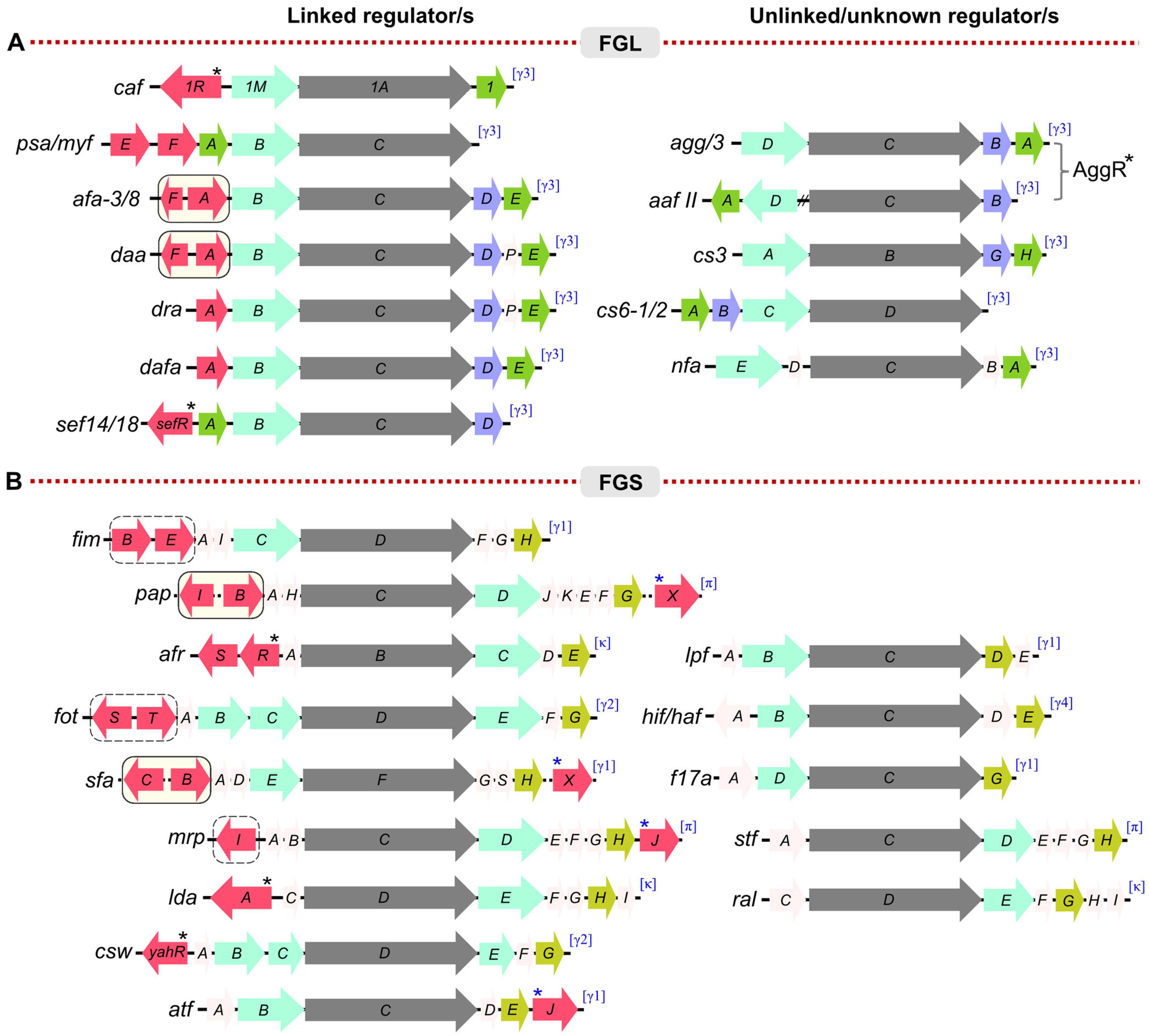
1.2. Diversity in the CUP-Assembled Fimbriae

The CUP fimbriae operons were broadly divided into two structurally and functionally distinct groups, FGL and FGS (Figure 1) [46,47]. In the FGL group, fibre assembly is assisted by a cognate chaperone with an extended donor G1 β–strand that correlates with a longer F1–G1 loop (FGL) and high affinity for the subunit. Well-studied examples of the CUP fimbriae from the FGL group include the F1 capsular antigen [26,27,28,29,30,38,48,49] and Psa fimbriae of Y. pestis [50,51] and the Afa/Dra family of fimbriae from E. coli [8]. Most of the FGL-grouped fimbriae assemble into simple, thin (~2–3 nm diameter), and flexible fibres composed of only one or two subunit types (Figure 1). In contrast, fimbriae (pili) belonging to the well-defined FGS group are generally thicker (~7 nm diameter) and more rigid. These fimbriae have a more complex structure with a major structural pilin subunit that constitutes the shaft of the pilus plus several additional subunits which form a thin, flexible tip fibrillum capped by a single specialised adhesin. This terminal adhesin typically has two domains, both a pilin and an adhesin domain and hence is approximately twice the size of pilin subunits [17,24]. Thus, chaperones of FGS (F1–G1 short loop) fimbrial clusters have a requirement to recognise a number of different subunits and hence a lower specificity of binding to each subunit. Hence, the subgrouping based on chaperone properties, appears to correlate with complexity of fimbrial structure. Phylogenetically, based on usher sequence, the FGS group is subdivided into several clades including, β-, γ1-, γ2-, γ4-, κ-, and π-fimbriae [37]. Unlike FGS, the FGL group is a small monophyletic group, having only one clade, γ3. The α- clade corresponds to CU systems defined as ‘alternate’ CUP, including colonisation factor CS-1 of enterotoxigenic E. coli (ETEC) [37]. The following sections of the review, discuss knowledge of the regulatory mechanisms of expression of some well-characterised examples of CUP fimbriae.

2. Diversity in Regulation: Phase Variation

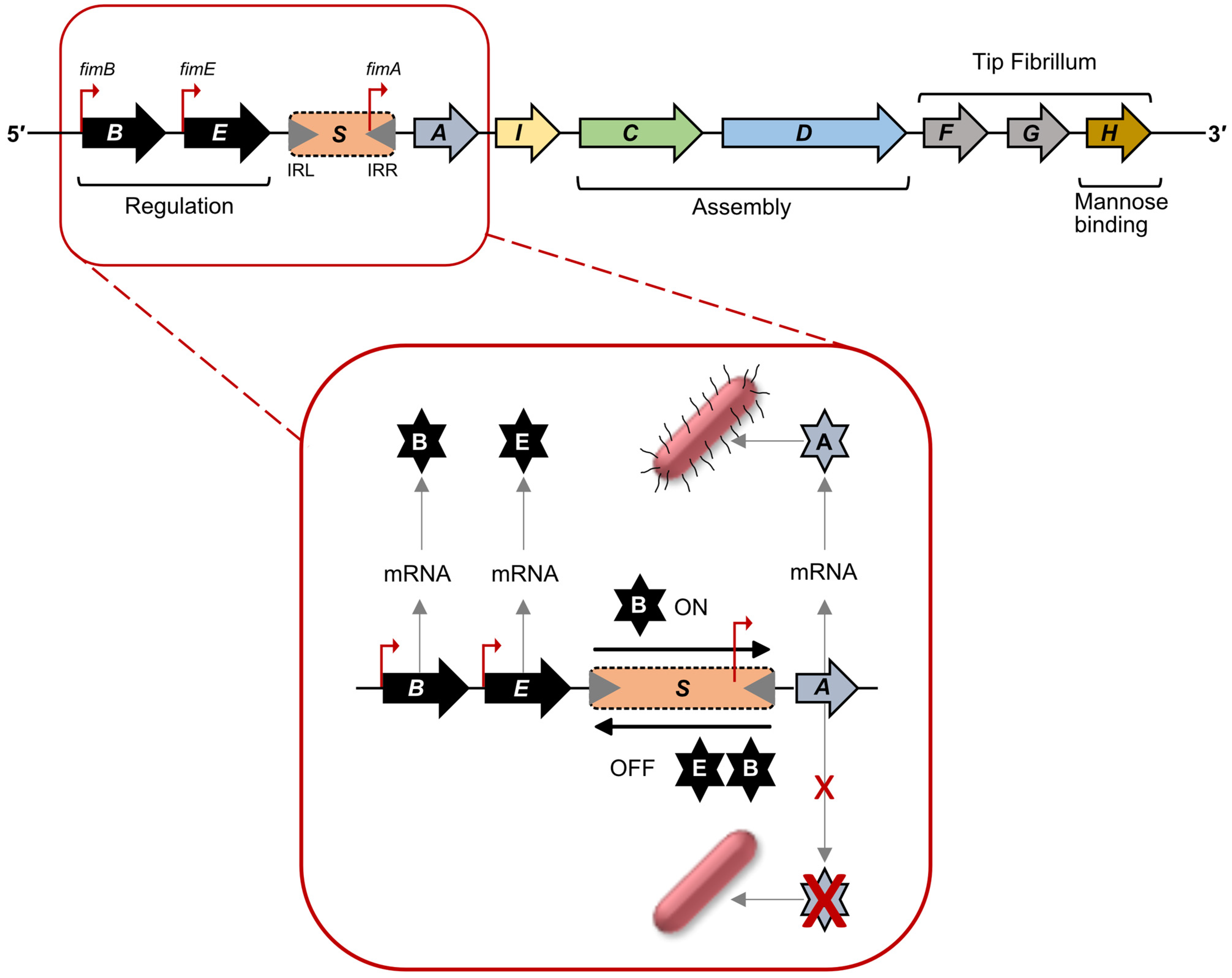
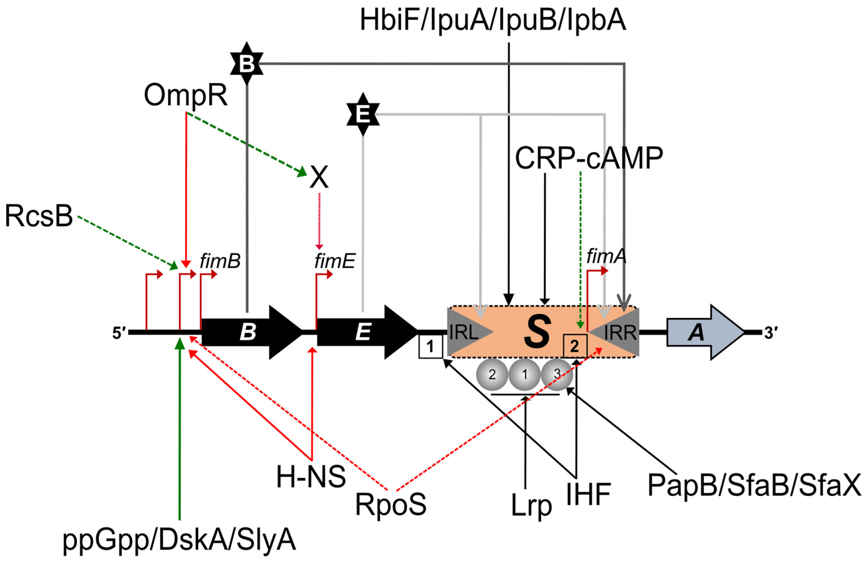
2.1. Regulation of Type 1 Fimbriae Expression: Promoter Inversion

Type 1 fimbriae of UPEC (FGS-grouped fimbriae) are synthesised from a chromosomally encoded gene cluster known as the fim locus. This locus is composed of nine genes, encoding two regulatory proteins, FimB and FimE; a major subunit pilin, FimA; two assembly proteins including a periplasmic chaperone, FimC and an outer membrane usher, FimD; two tip-associated fibrillum subunits, FimF and FimG; a mannose-binding adhesin, FimH, and finally FimI, a subunit responsible for anchoring the pilus to the cell surface (Figure 2) [8,9,25,46,52,53,54]. Expression of the fim locus is controlled by phase variation mediated by inversion of the promoter on a 314 bp DNA fragment, known as fimS, flanked by 9 bp inverted repeats, IRL and IRR (TTGGGGCCA) (Figure 2). When switched to the ON orientation, the fimA promoter leads to transcription and synthesis of the major fimbrial subunit, FimA, plus other structural subunits and assembly genes. In the opposite orientation the fimA promoter is non-functional. Hence, the forward orientation leads to Phase-ON (piliated state), and reverse orientation leads to Phase-OFF (non-piliated state). The regulatory proteins, FimB and FimE influence the orientation of fimS. These are site-specific recombinases, share 48% amino acid identity, and contain a tetrad of conserved amino acids (Arg47, His141, Arg144 and Tyr176; in FimB and Arg41, His136, Arg139 and Tyr171; in FimE), which has been shown to be essential for recombinase activity [52,53,54,55,56,57]. Both bind to regions flanking fimS including the inverted repeats.
FimB can influence the switch of fimS to either Phase-ON or Phase-OFF, while presence of FimE primarily leads to a switch of fimS to the Phase-OFF state (Figure 2). The mechanism and factors influencing the orientation and recombination of the fimS element is not only influenced by the concentrations of FimB and FimE, but also by other factors. Three promoters have been identified for fimB expression [58,59,60], while for fimE a single promoter has been identified [60].
At least 20 additional auxiliary genes are known to influence the expression of type 1 fimbriae (Figure 3) [54]. The product of pilG, an allele of the hns gene, was reported to influence inversion; a mutation in the pilG was shown to increase inversion of fimS up to 100-fold using a fimA-lacZ fusion [61]. The global regulatory protein, H-NS (histone-like nucleoid-structuring protein) was shown to repress transcription of both fimB and fimE genes by binding to promoters of both with high specificity [54,58,60]. H-NS may also directly affect inversion by binding to sequences adjacent to the fimS DNA fragment. In addition to H-NS, integration host factor (IHF) and leucine-responsive protein (Lrp) also influence phase variation [62]. Other site-specific recombinases (HbiF, IpuA, IpuB and IpbA) have been identified as possibly influencing inversion of fimS. There is also crosstalk between regulators of different pili in E. coli. PapB, a regulatory protein of the Pap pili locus, and SfaB from the S pili locus can influence the orientation of fimS by inhibiting the Phase-OFF to the Phase-ON switch, enhancing selective expression of the correct pili at the right time and place. The alarmone, ppGpp also appears to be required for optimum expression of type 1 fimbriae, and the ppGpp alarmone, along with RNA polymerase-binding protein, DksA was shown to stimulate transcription of fimB from the P2 promoter [54]. Moreover, environmental factors such as temperature and pH also play a crucial role in the expression of type 1 fimbriae. Several studies have shown that the phase switching from ON to OFF state surges at a lower temperature [54,63,64]. Under low pH conditions type 1 fimbriae production is inhibited and increases at neutral pH. At neutral pH, proteins such as SlyA or RcsB may promote type 1 fimbriae synthesis by activating fimB and preventing H-NS binding [54].
Phase variation via promoter inversion has been shown to control expression of several other fimbriae, including CS18 fimbriae from enterotoxigenic E. coli (ETEC) and mannose-resistant pili (MR/P) from Proteus mirabilis (Figure 1). The fot cluster, encoding CS18 fimbriae, is regulated by a 312 bp inversion driven by the two site-specific recombinases, FotS and FotT, that share close to 50% sequence identity with FimB and FimE, respectively [39]. Similar sequence identity is not shared between other genes of the two operons. The mrp cluster is controlled by promoter inversion on a 252 bp sequence, flanked by inverted repeats and located between mrpI and mrpA, which encodes the major subunit of MR/P fimbriae. MrpI also shares high homology with FimB and E, but unlike the fim and fot clusters, MrpI is the sole recombinase [40,41]. It is specific for the mrp promoter switch, and can also switch the promoter to either ON or OFF position. Expression of MR/P fimbriae is strongly upregulated in vivo. MrpJ, a MarR-like transcription factor and product of the last gene of the mrpABCDEFGHJ operon (Figure 1) can function as an auto-activator of the operon [42]. Detail of how promoter inversion, mrpJ expression and environmental factors interact to drive promoter orientation to the ON- phase remain to be elucidated.
P. mirabilis, a significant pathogen of urinary tract infections, is highly motile and exhibits characteristic swarming activity. During infection co-ordination between bacterial swimming and adhesion is critical. In accord with cross-talk between expression of fimbrial operons, MrpJ has been shown to decrease flagella production and swimming by repressing transcription of the flagellar master regulator, flhD [36,42]. Recent studies have outlined an MrpJ regulon, involving cross-talk between fimbrial operons, flagella expression and the Type-6 secretion system [42]. The mrpJ gene is present in 10 of the 17 fimbrial operons of P. mirabilis, including the atf locus, shown in Figure 1 [36], and homologues have been identified adjacent to the E. coli sfa locus, encoding S pili associated with infant meningitis, and also downstream from the pap operon in some E. coli strains (Figure 1). PapX similarly decreases flagella production by binding to the flhD promoter [43,65].

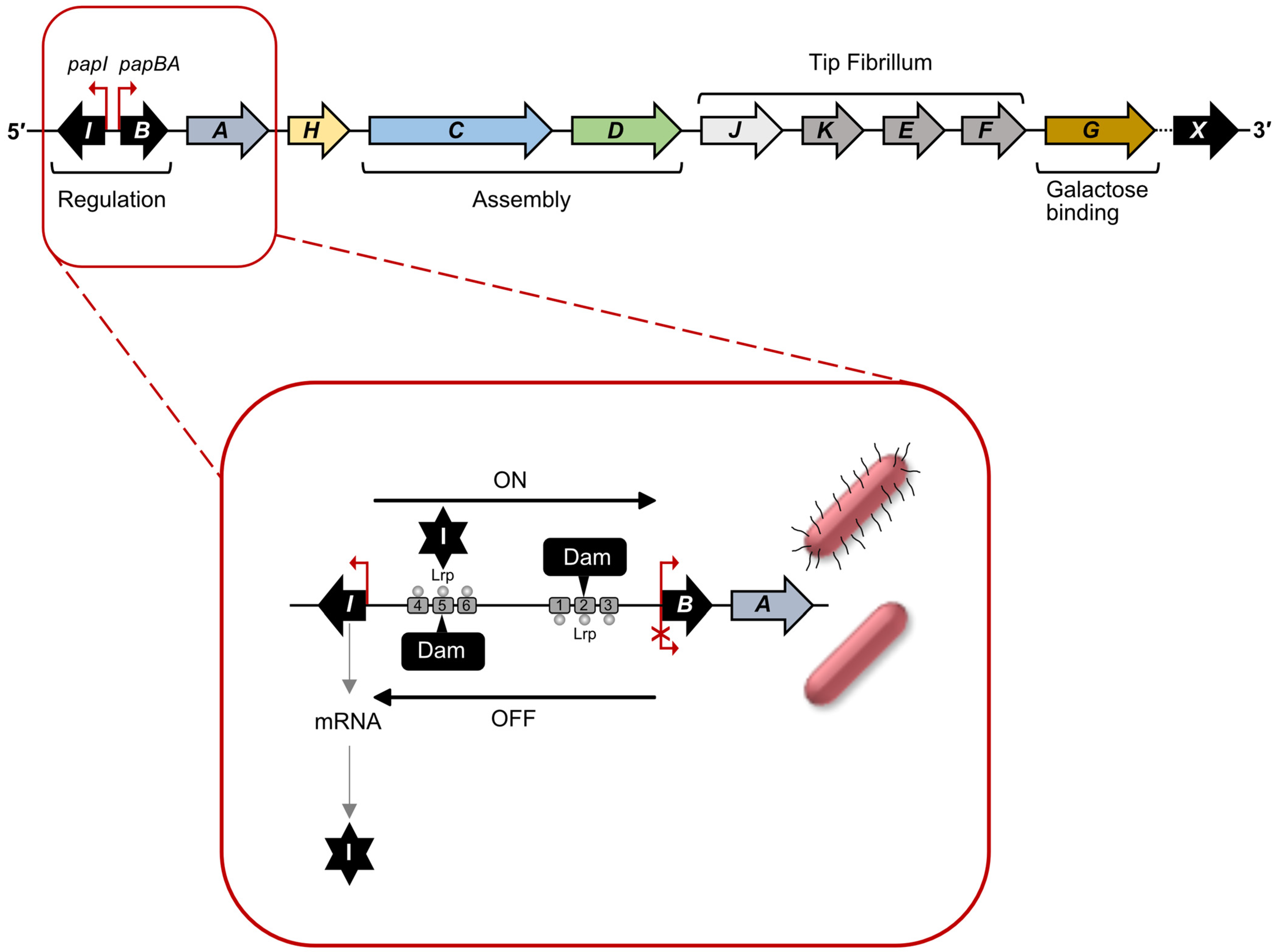
2.2. Regulation of Pap pili Expression: Methylation/Epigenetic Switch

Like type 1 fimbriae, genes encoding Pap pili are also clustered (pap locus) on the chromosome and controlled by phase variation [18,66] The core pap locus contains 11 genes, plus the mrpJ homologue, papX, located a short distance downstream in some strains [8,43,65]. Transcription of the pap locus is controlled by the papBA promoter and regulated by a phase variation (ON and OFF) mechanism via the PapB and PapI regulatory proteins [52]. In Pap pili PapA is the major pilin, while PapJ, PapK, PapE and PapF constitute the tip fibrillum. PapG is a galactose-binding adhesin and PapH is the pilus anchor. The other two gene products, PapD (periplasmic chaperone) and PapC (OM, usher), aid in CUP-based assembly of the Pap pili (Figure 4) [65].
Phase variable control of expression of the pap locus is different from that of the fim locus. In the pap locus, the ON-OFF switch is determined by binding of Lrp to two different Dam methylation sites, within the promoter region. Lrp-binding prevents methylation of the site leading to either activation (Phase-ON) or repression (Phase-OFF) of transcription of the pap gene cluster. The regulatory proteins, PapI (8 kDa) and PapB (12 kDa), positively control the expression of the pap locus in association with the global regulatory proteins, Lrp, CAP and H-NS, as reviewed [18,52]. The intergenic regulatory region (416 bp) between papI and papB contains six Lrp-binding sites that control transcription of both papA and papB (Figure 4). Within this, there are two DNA methylase (Dam) sites, GATCprox and GATCdist located within Lrp-binding sites 2 and 5, respectively. When accessible, these sites are methylated at the A base of the GATC motif, but when Lrp is bound, methylation of the sites is blocked. Lrp-binding proximal to the papBA promoter inhibits transcription of the pap operon, whereas binding at the distal site permits methylation at site 2 and activates transcription of papBA (Phase-ON). Lrp binds with higher affinity to sites 1–3 than to sites 4–6 [52]. The regulator, PapI promotes binding of Lrp to sites 4–6 rather than 1–3, promoting the ON state (Figure 4). Furthermore, pap locus transcription is also under the control of catabolite repression, requiring binding of cAMP-CAP complex, 60 bp upstream of the Lrp-binding site. Similar to regulation of type 1 fimbriae, expression of pap pili is also controlled by several environmental stimuli [44]. Transcription of pap pili is significantly repressed during growth at lower temperatures (<26 °C) in Luria broth. In addition to environmental factors, proteins not belonging to the pap locus have been shown to contribute to the positive regulation of the Pap pilus transcription. For example, the two-component sensor-regulator, CpxAR appears to be activated during misfolding of Pap subunits in the periplasm as well as on binding of Pap pilus to solid surfaces and inhibits transcription by binding close to the GATC sites [67,68].
A relatively new study identified the role of a small RNA—papR—in the regulation of pap locus phase variation, during infection of bladder epithelial cell lines with UPEC, strain UTI89 [69]. Trans-acting papR sRNA acts as a post-transcriptional repressor for papI and Lrp was shown to act as a transcriptional activator of papR expression. Deletion of papR increased bacterial adhesion to both kidney and bladder cells in the absence of type 1 fimbriae [69,70]. Involvement of papR sRNA in control of production of Pap pili facilitates rapid adjustments in response to changing environmental conditions during infection.
A role of Dam methylation/LRP control in phase variable expression has been demonstrated for a number of other fimbrial clusters [18], including E. coli afa/dra family of fimbriae clusters and the E. coli sfa cluster that encodes S pili (Figure 1) [18,71]. E. coli S fimbriae are involved in newborn meningitis and Afa family of fimbriae are common in UPEC strains and form thin surface fibres with an afimbrial appearance [8]. In both cases homologues of PapI and B are involved, AfaF/ AfaA and SfaC/SfaB respectively, and LRP-binding to the PapI homologue promotes expression. Interestingly, analysis of the afa locus in certain clinical strains has identified IS elements of varying length between AfaF and AfaA, which in the case of IS1 insertion led to a strong hybrid promoter and enhanced expression [71].

3. Other DNA Binding Regulators, Dedicated and Global

3.1. pH 6 Antigen, Regulation by a Sensor-Regulator

The FGL-grouped fimbriae are often classified as polyadhesins and show structural and functional differences compared to the FGS-grouped fimbriae [24,47]. The FGL fimbriae are linear polymers of one or two protein subunits. In some cases, the polymer possesses one or two adhesive sites, which are independent of each other and specific to host cell receptors. Thus, permitting polyvalent fastening of bacterial adhesin and host cell receptors that may increase the affinity of binding and enhance adhesion. The chromosomally located psa or myf locus, which encodes the pH 6 antigen in Yersinia spp. is an example of this [50,72,73,74,75]. The pH 6 antigen is expressed at low pH and inside macrophage, hence its name. The psa locus (Figure 1) is composed of five genes, psaE, psaF, psaA, psaB and psaC and encodes a single fimbrial subunit Psa which is assembled to form polyadhesive fibres with two low affinity binding sites for galactosyl residues and phosphatidylcholine [24,50]. Regulation of the psa operon appears to be an unusual example among fimbrial systems. The regulators, PsaE (24 kDa) and PsaF (18 kDa), which are required for transcription of psa, are both located within the inner membrane. PsaE also possesses a cytosolic N-terminal DNA-binding domain with homology to PhoB and OmpR of E. coli, ToxR of Vibrio cholerae and HilA of Salmonella typhimurium [72]. PsaF appears to be important in stabilising PsaE. The C-terminal domain of PsaE lies in the periplasm and interacts with the periplasmic domain of PsaF [72]. Thus PsaE/PsaF regulation has some structural characteristics of a one component sensor-DNA binding regulator system.
In Y. pestis, maximum expression of Psa occurs at 37 °C at pH 6. Understanding the mechanism of sensing temperature and pH is limited. There is evidence that the 5’UTR of psaE contributes to temperature sensing by forming a polyU RNA thermometer-like structure at lower temperature [51]. In addition, the central regulator, RovA (18 kDa), has been shown to interact with promoter regions of psaE and psaA [73,75]. A study by Zhang et al. demonstrated that PhoP and RovA recognise the promoter-proximal regions of psaEF and psaABC [73]. RovA activated psaEF and psaABC, whereas PhoP repressed both psaEF and psaABC through direct association between RovA/PhoP and the target promoter regions. It has been suggested that this reciprocal regulation of psa genes by PhoP and RovA could contribute to the tight regulation of pH 6 antigen expression during infection [73,74].

3.2. Agg and Aaf Fimbriae, Regulation by A/X Family Regulator, AggR

Enteroaggregative E. coli (EAEC) is a diarrhoea-causing pathogen in adults and children [19,76,77,78]. The Aaf I/III and Aaf II fimbriae, encoded on the clusters agg and aaf shown in Figure 1, form thin bundled fimbriae that lead to a characteristic stacked-brick biofilm of these E. coli cells bound to host epithelial cells [76,77]. The operon for the Aaf fimbriae is located on the pAA plasmid and comprises genes encoding a chaperone, aafC, an outer membrane usher, aafD, and a major and minor pilin subunit, aafA and aafB, respectively. Expression of Agg and Aaf fimbriae of EAEC is controlled by AggR, an A/X family transcriptional activator, located 9 kbp upstream from the respective fimbrial locus [79]. AggR dimerizes and binds to an AT rich consensus sequence overlapping the promoter to regulate transcription of agg fimbrial genes [80]. AggR has also been shown to act as a global regulator [81,82]. Forty-four additional AggR targets, including genes encoding dispersin, a surface protein (Aap), dispersin translocator and Aai type-VI secretion system were identified. AggR was found to activate the expression of Agg fimbriae in response to temperature, oxygen tension, and osmolarity as well as the growth media composition [79]. Five variants of Aaf (Aaf/I-Aaf/V) are known and these variants show a high degree of similarity in the CU assembly genes [83]. The differences are mainly in the genes that encode the major fimbrial structural protein, AggA and AafA. Polymers of these subunits bind fibronectin, only the Aaf/V variant Agg5A lacks fibronectin-binding capacity due to changes in pilin structure [83]. Aaf/II from E. coli strain O42 has an unusual cluster organisation with split operons for aafDA and afaB-aafCB locus that otherwise remain functional (Figure 1) [80,84].

3.3. CS and CfaI Fimbriae, Regulation by A/X Family Regulator, Rns

CUP fimbriae produced by enterotoxigenic E. coli (ETEC) were initially identified serologically as major components of Colonisation Factor Antigens (CFA) and further characterised genetically as a dominant proportion of Coli Surface (CS) antigens [70]. At least 20 different CUP-assembled CS fimbrial clusters have been identified so far among ETEC strains, although most strains express only two or three types [70]. Many of these, CFA/I, CS1, CS2, CS4, CS14, CS17, CS19 and PCF071, belong to the alternate CUP fimbriae, assigned as the α clade (based on usher phylogeny [37]), and form rigid fimbriae. Both CS3 and CS6 (Figure 1) belong to the FGL group and γ3 clade [24,70]. The most commonly observed fimbriae are CS1-CS6 and CFA/I, of which the mechanism of regulation of CS1, CS2 and CFA/I have been studied in some detail [8,18,70].
The gene cluster of CS1 fimbriae, cooBACD, is located on a large plasmid, pCOO and regulated in trans by an A/X family transcriptional regulator, Rns [84,85,86]. Rns is encoded on a distinct plasmid, unlinked to any CU system, and is flanked by a transposase encoding gene on one side and pseudogenes on the other [87]. Expression of CFA/I fimbriae is activated by the Rns homolog CfaD/CfaR, which shares 95% amino acid identity with Rns, is functionally interchangeable with Rns and recognises the same binding sites as Rns [18,88]. ETEC fimbriae for which expression is known to be activated by Rns are phylogenetically related and belong to the α clade, CS1, CS2, CS4, CS14, CS17, and CS19 [18,70,89,90,91,92]. There is some evidence that CS3 is under Rns control in a recombinant strain of Vibrio cholerae but not in ETEC [18,93]. In addition to activating the expression of fimbrial genes, Rns positively regulates its own gene expression by direct binding at three sites, centred at −227, +43 and +82 (relative to transcription start site) [86,94]. The involvement of downstream binding sites (at +43 and +82) is often associated with transcription repression. However, it has been shown that activation of Rns requires at least one of the two downstream binding sites and the upstream binding site [86]. Rns activates the expression of several putative virulence genes [95,96]. A DNaseI footprinting located the same Rns binding site immediately upstream of the −35 element of Pcoo promoter (required for the expression of CfaA, CS1 fimbriae) and the promoters of CS17, CS19 and PCFO71 fimbriae [85,92], suggesting Rns may activate transcription by direct contact with RNA polymerase. In addition to the binding site near to −35 promoter element, a second binding site was identified further upstream in all cases (at −88 in CfaA, at −144 in CS1, at −109.5 in CS17 and PCFO71 and at −108.5 in CS19). Each site showed an additive effect on Rns-dependent transcription activation, indicating requirement of the distal site also for full activation of Rns [85,86,92].
Rns homologs, linked to regulation of virulence factors, have been identified in several strains of ETEC and other enteropathogenic bacteria. Proteins having strong homology to Rns are CsvR and FapR from ETEC [97], PerA of enteropathogenic E. coli [98], AggR of EAEC [79,80], ToxT of Vibrio cholerae [99] and VirF of Shigella spp. [100,101]. Among these, AggR, CsvR, PerA, and ToxT regulate the expression of fimbrial genes. The VirF of Shigella spp. activates the expression of icsA and virB genes which have roles in invasion and cell-to-cell spread of bacteria in the host epithelial cells, respectively [100].
Transcription activation by Rns and its homologs is thermo-sensitive and is repressed with involvement of H-NS at low temperatures [8,101]. Like most A/X family regulators, Rns is a two-domain protein, with an N-terminal domain (NTD) and a conserved C-terminal DNA binding domain (DBD) with two predicted HTH motifs [102]. A mutagenesis study using pentapeptide insertions demonstrated that Rns uses both HTH motifs to make DNA contacts and thereby activates the expression of CS1 fimbrial genes [102]. In support of this, a uracil interference study by Munson et al. showed that Rns contacts two major grooves of the DNA to activate transcription [85,86]. Like DBD of several A/X family regulators, Rns DBD is believed to be involved in making RNAP contacts, although the region involved in making such contacts has not been identified [85,90].
The role of Rns-NTD was largely unknown. There was no evidence, in vivo, that Rns responds to any effector molecules [91]. However, N-terminal deletion mutagenesis showed that Rns-NTD is essential for transcription activation both in vivo and in vitro [91]. A truncated version of Rns (first 61 amino acids deleted) also resulted in the complete loss of DNA-binding and transcription activation or repression at the corresponding promoter regions. The Rns-NTD motif from I12-M18 is highly conserved among the closest homologs, sharing about 74% amino acid identity compared to 26% identity in the overall NTDs, suggesting a crucial role of this motif in the overall function of Rns. In support of this, Munson and co-workers isolated two random mutants (I14T and N16D) of Rns-NTD and found their activities were decreased dramatically at the rns promoter, indicating the importance of I14 and N16 residues in transcription activation [91]. It was not clear whether Rns acts primarily as a monomer or dimer with a role of the NTD domain in dimerisation [91,103] or whether it may be involved in effector ligand binding. Interestingly, recently Rns has been crystallised as a dimer in the presence of decanoic acid, providing evidence that Rns-NTD may sense and respond to a fatty acid ligand [45].

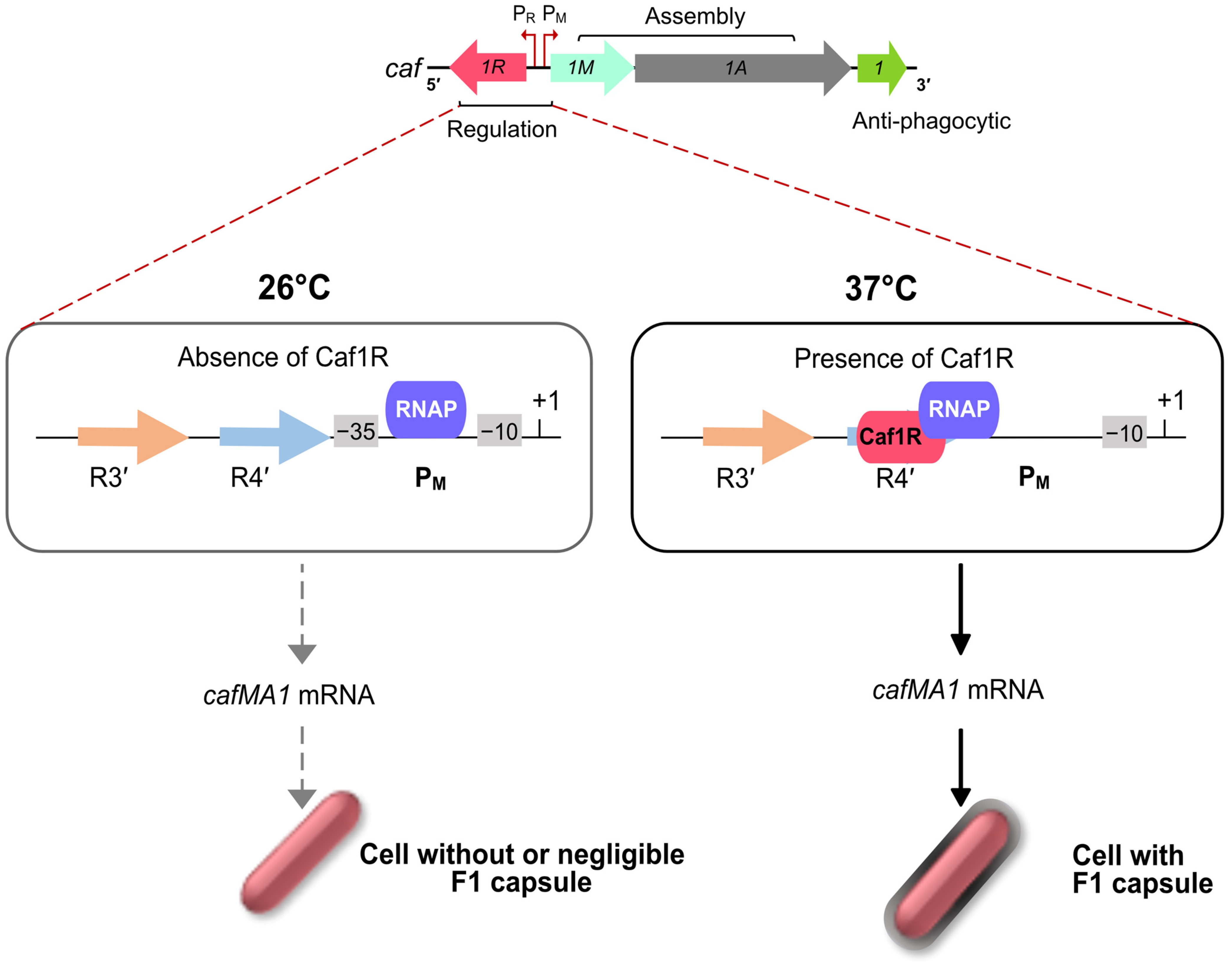
3.4. F1 Capsule, Regulation by A/X Family Regulator, Caf1R

The F1 capsular antigen from Y. pestis comprises long, linear fibres of a single subunit (Caf1) and represents an example of a non-pilus organelle assembled via the FGL (γ3) ushers [24,28,37]. The thin fibres of Caf1 subunit collapse around the bacterial cell forming a capsule-like structure that contributes to the anti-phagocytic arsenal of the bacterium [48,104]. Unlike the psa locus, the caf locus is specific to Y. pestis. Initial sequencing of the caf locus identified the divergently expressed caf1R gene encoding an A/X family regulator, Caf1R [105], but the mechanism of regulation of this key antigen was only more recently elucidated, in 2016, by Kumar, D. [38,49]. Kumar confirmed Caf1R as an essential activator for high level expression of the caf operon and capsule formation. Using promoter-lacZ fusions and Electrophoretic Mobility Shift Assays (EMSA), the Caf1R binding site was identified overlapping the −35 element of the PM promoter, a characteristic of Class II transcriptional activators (Figure 5) [38,49]. Structure-modelling of a Caf1R-DNA binding site complex confirmed location of a virtually non-functional spontaneous point mutation in Caf1R to DNA binding helix6 [38].
Autoregulation is a common feature among A/X family regulators. Examples of autoactivation include Rns [86,91], PerA [106], MarA [107] and AggR [82]. Unlike caf1R, these regulators are primarily ‘unlinked’ to the target operons. Promoter fusion studies also confirmed autoregulation of caf1R [38]. F1 fibres are expressed at 37ºC in the mammalian host but not at 26 °C in the insect host (Figure 5) [48]. Temperature regulation of expression has been reported for a number of fimbriae, with involvement of H-NS contributing to repression at low temperatures [58,64]. The untranslated intergenic region of the caf locus is known to be required for temperature regulation of Caf1R dependent activation of cafMA1 [38] but the mechanism by which this occurs remains to be elucidated.
Transcription activation by A/X family regulators, as seen by Caf1R at PM promoter [38,49], is quite common. In many well studied cases, binding of a small metabolite is required for activation [99,108,109,110,111]. Caf1R also possesses a C-terminal ‘sensing’-like domain, but it remains to be investigated if this binds anything or is involved in dimerisation. Bioinformatic analysis of putative and characterised DBDs of 62 A/X regulators grouped Caf1R DBD with regulators controlling metabolic processes [112]. Unexpectedly, it was aligned very distantly to AggR and VirF, regulators of virulence factors including fimbriae. The Caf1R was most closely aligned with the XylR regulator of E. coli K-12, which is essential for the transcription of genes required to utilise D-xylose and L-arabinose [111]. Whether this relationship to XylR and other regulators of metabolic processes is an indication of some metabolic process also controlled by Caf1R or is an indication of small molecule sensing by Caf1R remains to be seen.
Caf1R-dependent regulation of divergent expression of the caf locus represents a good model for studying the regulation of CUP-fimbriae controlled by a ‘linked’ A/X family regulator. No doubt, there will be many other factors controlling the expression of the caf locus. To fully understand the regulation of F1 expression, it will be important to map the role of other cellular factors on the proposed models [38,49]. Virulence of Y. pestis is still not well understood, and regulators involved in global regulation may be critically important. Hence, even though Caf1R appears to be a dedicated regulator, it is still important to consider a possible role of Caf1R in trans regulation of other virulence or metabolic factors. Furthermore, there is a lot of interest in the development of attenuated vaccines and heterologous expression of F1 [113,114,115]. In this type of vaccine, it is essential to retain optimum expression of F1. For this reason, a complete understanding of factors controlling the expression of F1 is crucial.

4. Conclusions and Perspectives

CUP fimbriae are prevalent in Gram-negative bacteria and are often important virulence determinants, required for adhesion, establishment of colonisation and in some cases invasion. Their structure, regulation and involvement in urinary tract and gastrointestinal infections, in particular, have been studied in detail. CUP fimbriae may be complex structures with a terminal adhesin, or more simple fibres with only one or two subunits which in some cases appear to function as polyadhesins to enhance binding and in others collapse around the cell to form an anti-phagocytic capsule-like structure. Enterobacterial genomes typically include multiple CUP clusters, many of which have different roles during infection. Hence, controlled expression of fimbriae is important to ensure expression in the correct niche while repression, when not required, facilitates evasion of the immune system.
Regulation of expression of fimbrial loci is multi-layered. Fundamentally, phase variation permits a rapid switch from fimbriated cells to non-fimbriated cells, potentially ensuring a guaranteed source of either type of cell. Extensive studies on the recombinase dependent promoter switching of type 1 fimbriae and Dam-methylation of Pap pili have highlighted the significance of global regulator involvement to appropriately moderate phase variation in response to environmental cues, such as temperature, stress, nutritional status [8,18,52,54]. Cross-talk during expression of Pap pili has been shown to contribute to downregulation of type 1 fimbriae. Additionally, an inverse relationship between fimbriae expression and motility is controlled by an additional regulator (e.g., PapX and MrpJ) encoded close to, or part of, the fimbrial cluster [42]. This knowledge provides a starting template to understand control of fimbrial expression in other systems or bacteria where similar regulators of phase variation are identified. But other mechanisms of phase variation of fimbriae expression also exist. Expression of Hif fimbriae of Haemophilus is controlled by slipped strand mispairing and variation of the number of residues between the −10 and −35 elements of the promoter, thus leading to phase variable expression of fimbriae [116].
The γ3 clade (FGL group) of fibrillar structures represent a small group assembled by phylogenetically related chaperones and usher proteins [24,37], but a remarkably diverse range of mechanisms of regulation. Expression of the Aaf fimbriae of aggregative E. coli, is regulated by an unlinked A/X regulator, AggR, and the aaf/agg fimbrial clusters are only one example within the AggR virulence regulon. Further studies will clarify how commonly fimbrial clusters are controlled by unlinked A/X regulators and involvement of fimbrial A/X regulators in global regulation. The Afa/Daa/Dra family of fimbriae are closely related varying primarily in subunit sequence [82]. Regulation of afa and daa loci are known to be dependent on Dam-methylation in a similar manner to the pap operon [71]. The pH 6 antigen and F1 capsule of Y. pestis also have regulator(s) within the fimbrial cluster, but that of the psa locus is reminiscent of a one component-like sensor regulator system [117], while the caf locus is controlled by an A/X family regulator, Caf1R [38,49]. These fimbrial loci, acquired at different times during evolution of Y. pestis [48] have co-opted different regulatory mechanisms. All of these fimbrial loci are important in infection and disease. A detailed understanding of the primary mechanism of regulation, how this is influenced by relevant environmental factors and, importantly, the interplay between expression of these loci and other virulence determinants during infection and disease will provide a solid basis for development of new intervention strategies. In addition to targeting transcription-regulators, adhesins are well documented immunogens with potential to act as vaccine candidates. In depth understanding of regulatory mechanisms will aid in optimising expression which in turn is important in development of attenuated or whole cell killed vaccines.

Author Contributions

Literature analysis and conceptualization, D.K.G. and S.M.; original draft preparation and writing, D.K.G.; art work, D.K.G. and N.T.; review and editing, D.K.G., N.T. and S.M. All authors have read and agreed to the published version of the manuscript.

Funding

This research was funded by the Ministry of Education, formerly Ministry of Human Resource Development (MHRD), Govt. of India, student fellowship to D.K.G. (Grant no. 11015/4/2009-SCDV).

Institutional Review Board Statement

Not applicable.

Informed Consent Statement

Not applicable.

Data Availability Statement

Not applicable.

Acknowledgments

We would like to thank Krishan K. Kapoor, Guru Jambheshwar University of Science & Technology, India and Sun Nyunt Wai, Umeå University, Sweden for their valuable feedbacks and helpful discussions on the manuscript.

Conflicts of Interest

The authors declare no conflict of interest.

References

  1. Proft, T.; Baker, E.N. Pili in Gram-negative and Gram-positive bacteriastructure, assembly and their role in disease. Cell Mol. Life Sci. 2009, 66, 613–635. [Google Scholar] [CrossRef] [PubMed]
  2. Rendon, M.A.; Saldana, Z.; Erdem, A.L.; Monteiro-Neto, V.; Vazquez, A.; Kaper, J.B.; Puente, J.L.; Giron, J.A. Commensal and pathogenic Escherichia coli use a common pilus adherence factor for epithelial cell colonization. Proc. Natl. Acad. Sci. USA 2007, 104, 10637–10642. [Google Scholar] [CrossRef] [Green Version]
  3. Lukaszczyk, M.; Pradhan, B.; Remaut, H. The Biosynthesis and Structures of Bacterial Pili. Subcell. Biochem. 2019, 92, 369–413. [Google Scholar] [PubMed]
  4. Berne, C.; Ducret, A.; Hardy, G.G.; Brun, Y.V. Adhesins Involved in Attachment to Abiotic Surfaces by Gram-Negative Bacteria. Microbiol. Spectr. 2015, 3, 163–199. [Google Scholar] [CrossRef] [PubMed] [Green Version]
  5. Pizarro-Cerda, J.; Cossart, P. Bacterial adhesion and entry into host cells. Cell 2006, 124, 715–727. [Google Scholar] [CrossRef] [PubMed] [Green Version]
  6. Di Martino, P. Bacterial adherence: Much more than a bond. AIMS Microbiol. 2018, 4, 563–566. [Google Scholar] [CrossRef]
  7. Jin, X.; Marshall, J.S. Mechanics of biofilms formed of bacteria with fimbriae appendages. PLoS ONE 2020, 15, e0243280. [Google Scholar] [CrossRef]
  8. Werneburg, G.T.; Thanassi, D.G. Pili Assembled by the Chaperone/Usher Pathway in Escherichia coli and Salmonella. EcoSal Plus 2018, 8. [Google Scholar] [CrossRef] [Green Version]
  9. Pusz, P.; Bok, E.; Mazurek, J.; Stosik, M.; Baldy-Chudzik, K. Type 1 fimbriae in commensal Escherichia coli derived from healthy humans. Acta Biochim. Pol. 2014, 61, 389–392. [Google Scholar] [CrossRef] [Green Version]
  10. Muller, K.H.; Collinson, S.K.; Trust, T.J.; Kay, W.W. Type 1 fimbriae of Salmonella enteritidis. J. Bacteriol. 1991, 173, 4765–4772. [Google Scholar] [CrossRef]
  11. Zhang, Y.; Tennent, J.M.; Ingham, A.; Beddome, G.; Prideaux, C.; Michalski, W.P. Identification of type-4 fimbriae in Actinobacillus pleuropneumoniae. FEMS Microbiol. Lett. 2000, 189, 15–18. [Google Scholar] [CrossRef] [PubMed]
  12. Mattick, J.S.; Whitchurch, C.B.; Alm, R.A. The molecular genetics of type-4 fimbriae in Pseudomonas aeruginosa—A review. Gene 1996, 179, 147–155. [Google Scholar] [CrossRef] [PubMed]
  13. Bhoite, S.; van Gerven, N.; Chapman, M.R.; Remaut, H. Curli Biogenesis: Bacterial Amyloid Assembly by the Type VIII Secretion Pathway. EcoSal Plus 2019, 8. [Google Scholar] [CrossRef] [PubMed]
  14. Barnhart, M.M.; Chapman, M.R. Curli biogenesis and function. Annu. Rev. Microbiol. 2006, 60, 131–147. [Google Scholar] [CrossRef] [PubMed] [Green Version]
  15. Costa, T.R.D.; Harb, L.; Khara, P.; Zeng, L.; Hu, B.; Christie, P.J. Type IV secretion systems: Advances in structure, function, and activation. Mol. Microbiol. 2021, 3, 436–452. [Google Scholar] [CrossRef]
  16. Thanassi, D.G.; Hultgren, S.J. Assembly of complex organelles: Pilus biogenesis in Gram-negative bacteria as a model system. Methods 2000, 20, 111–126. [Google Scholar] [CrossRef] [Green Version]
  17. Thanassi, D.G.; Bliska, J.B.; Christie, P.J. Surface organelles assembled by secretion systems of Gram-negative bacteria: Diversity in structure and function. FEMS Microbiol. Rev. 2012, 36, 1046–1082. [Google Scholar] [CrossRef] [Green Version]
  18. Clegg, S.; Wilson, J.; Johnson, J. More than one way to control hair growth: Regulatory mechanisms in enterobacteria that affect fimbriae assembled by the chaperone/usher pathway. J. Bacteriol. 2011, 193, 2081–2088. [Google Scholar] [CrossRef] [Green Version]
  19. Franca, F.L.; Wells, T.J.; Browning, D.F.; Nogueira, R.T.; Sarges, F.S.; Pereira, A.C.; Cunningham, A.F.; Lucheze, K.; Rosa, A.C.; Henderson, I.R.; et al. Genotypic and phenotypic characterisation of enteroaggregative Escherichia coli from children in Rio de Janeiro, Brazil. PLoS ONE. 2013, 8, e69971. [Google Scholar] [CrossRef]
  20. Thanassi, D.G.; Saulino, E.T.; Hultgren, S.J. The chaperone/usher pathway: A major terminal branch of the general secretory pathway. Curr. Opin. Microbiol. 1998, 1, 223–231. [Google Scholar] [CrossRef]
  21. Perez-Rueda, E.; Tenorio-Salgado, S.; Huerta-Saquero, A.; Balderas-Martinez, Y.I.; Moreno-Hagelsieb, G. The functional landscape bound to the transcription factors of Escherichia coli K-12. Comput. Biol. Chem. 2015, 58, 93–103. [Google Scholar] [CrossRef] [PubMed]
  22. Costa, T.R.; Felisberto-Rodrigues, C.; Meir, A.; Prevost, M.S.; Redzej, A.; Trokter, M.; Waksman, G. Secretion systems in Gram-negative bacteria: Structural and mechanistic insights. Nat. Rev. Microbiol. 2015, 13, 343–359. [Google Scholar] [CrossRef] [PubMed]
  23. Geibel, S.; Waksman, G. The molecular dissection of the chaperone-usher pathway. Biochim. Biophys. Acta. 2014, 1843, 1559–1567. [Google Scholar] [CrossRef] [Green Version]
  24. Zav’yalov, V.; Zavialov, A.; Zav’yalova, G.; Korpela, T. Adhesive organelles of Gram-negative pathogens assembled with the classical chaperone/usher machinery: Structure and function from a clinical standpoint. FEMS Microbiol. Rev. 2010, 34, 317–378. [Google Scholar] [CrossRef] [Green Version]
  25. Choudhury, D.; Thompson, A.; Stojanoff, V.; Langermann, S.; Pinkner, J.; Hultgren, S.J.; Knight, S.D. X-ray structure of the FimC-FimH chaperone-adhesin complex from uropathogenic Escherichia coli. Science 1999, 285, 1061–1066. [Google Scholar] [CrossRef]
  26. MacIntyre, S.; Zyrianova, I.M.; Chernovskaya, T.V.; Leonard, M.; Rudenko, E.G.; Zav’Yalov, V.P.; Chapman, D.A. An extended hydrophobic interactive surface of Yersinia pestis Caf1M chaperone is essential for subunit binding and F1 capsule assembly. Mol. Microbiol. 2001, 39, 12–25. [Google Scholar] [CrossRef] [PubMed] [Green Version]
  27. Zavialov, A.V.; Kersley, J.; Korpela, T.; Zav’yalov, V.P.; MacIntyre, S.; Knight, S.D. Donor strand complementation mechanism in the biogenesis of non-pilus systems. Mol. Microbiol. 2002, 45, 983–995. [Google Scholar] [CrossRef] [PubMed]
  28. Zavialov, A.V.; Berglund, J.; Pudney, A.F.; Fooks, L.J.; Ibrahim, T.M.; MacIntyre, S.; Knight, S.D. Structure and biogenesis of the capsular F1 antigen from Yersinia pestis: Preserved folding energy drives fiber formation. Cell 2003, 113, 587–596. [Google Scholar] [CrossRef] [Green Version]
  29. Dubnovitsky, A.P.; Duck, Z.; Kersley, J.E.; Härd, T.; MacIntyre, S.; Knight, S.D. Conserved hydrophobic clusters on the surface of the Caf1A usher C-terminal domain are important for F1 antigen assembly. J. Mol. Biol. 2010, 403, 243–259. [Google Scholar] [CrossRef]
  30. Yu, X.D.; Dubnovitsky, A.; Pudney, A.F.; Macintyre, S.; Knight, S.D.; Zavialov, A.V. Allosteric mechanism controls traffic in the chaperone/usher pathway. Structure 2012, 20, 1861–1871. [Google Scholar] [CrossRef]
  31. Barnhart, M.M.; Sauer, F.G.; Pinkner, J.S.; Hultgren, S.J. Chaperone-subunit-usher interactions required for donor strand exchange during bacterial pilus assembly. J. Bacteriol. 2003, 185, 2723–2730. [Google Scholar] [CrossRef] [PubMed] [Green Version]
  32. Le Trong, I.; Aprikian, P.; Kidd, B.A.; Thomas, W.E.; Sokurenko, E.V.; Stenkamp, R.E. Donor strand exchange and conformational changes during Escherichia coli fimbrial formation. J. Struct. Biol. 2010, 172, 380–388. [Google Scholar] [CrossRef] [PubMed] [Green Version]
  33. Zyla, D.; Echeverria, B.; Glockshuber, R. Donor strand sequence, rather than donor strand orientation, determines the stability and non-equilibrium folding of the type 1 pilus subunit FimA. J. Biol. Chem. 2020, 295, 12437–12448. [Google Scholar] [CrossRef]
  34. Korea, C.G.; Badouraly, R.; Prevost, M.C.; Ghigo, J.M.; Beloin, C. Escherichia coli K-12 possesses multiple cryptic but functional chaperone-usher fimbriae with distinct surface specificities. Environ. Microbiol. 2010, 12, 1957–1977. [Google Scholar] [CrossRef] [PubMed]
  35. Wurpel, D.J.; Beatson, S.A.; Totsika, M.; Petty, N.K.; Schembri, M.A. Chaperone-usher fimbriae of Escherichia coli. PLoS ONE 2013, 8, e52835. [Google Scholar] [CrossRef] [Green Version]
  36. Schaffer, J.N.; Pearson, M.M. Proteus mirabilis and Urinary Tract Infections. Microbiol. Spectr. 2015, 3, 383–433. [Google Scholar] [CrossRef] [Green Version]
  37. Nuccio, S.P.; Baumler, A.J. Evolution of the Chaperone-Usher assembly pathway: Fimbrial classification goes Greek. Microbiol. Mol. Biol. Rev. 2007, 71, 551–575. [Google Scholar] [CrossRef] [Green Version]
  38. Kumar, D. Caf1R-Mediated Regulation of the F1 Surface Antigen of Yersinia Pestis. Ph.D. Thesis, University of Reading, Reading, UK, 2016. Available online: http://centaur.reading.ac.uk/66018/ (accessed on 18 July 2022).
  39. Honarvar, S.; Choi, B.-K.; Schifferli, D.M. Phase variation of the 987P-like CS18 fimbriae of human enterotoxigenic Escherichia coli is regulated by site-specific recombinases. Mol. Microbiol. 2003, 48, 157–171. [Google Scholar] [CrossRef]
  40. Zhao, H.; Li, X.; Johnson, D.E.; Blomfield, I.; Mobley, H.L. In vivo phase variation of MR/P fimbrial gene expression in Proteus mirabilis infecting the urinary tract. Mol. Microbiol. 1997, 23, 1009–1019. [Google Scholar] [CrossRef]
  41. Li, X.; Lockatell, C.V.; Johnson, D.E.; Mobley, H.L. Identification of MrpI as the sole recombinase that regulates the phase variation of MR/P fimbria, a bladder colonization factor of uropathogenic Proteus mirabilis. Mol. Microbiol. 2002, 45, 865–874. [Google Scholar] [CrossRef]
  42. Bode, N.J.; Debnath, I.; Kuan, L.; Schulfer, A.; Ty, M.; Pearson, M.M. Transcriptional analysis of the MrpJ network: Modulation of diverse virulence-associated genes and direct regulation of mrp fimbrial and flhDC flagellar operons in Proteus mirabilis. Infect. Immun. 2015, 83, 2542–2556. [Google Scholar] [CrossRef] [Green Version]
  43. Simms, A.N.; Mobley, H.L. PapX, a P fimbrial operon-encoded inhibitor of motility in uropathogenic Escherichia coli. Infect. Immun. 2008, 76, 4833–4841. [Google Scholar] [CrossRef] [PubMed] [Green Version]
  44. White-Ziegler, C.A.; Villapakkam, A.; Ronaszeki, K.; Young, S. H-NS controls pap and daa fimbrial transcription in Escherichia coli in response to multiple environmental cues. J. Bacteriol. 2000, 182, 6391–6400. [Google Scholar] [CrossRef] [PubMed] [Green Version]
  45. Midgett, C.R.; Talbot, K.M.; Day, J.L.; Munson, G.P.; Jon Kull, F. Structure of the master regulator Rns reveals an inhibitor of enterotoxigenic Escherichia coli virulence regulons. Sci. Rep. 2021, 11, 15663. [Google Scholar] [CrossRef] [PubMed]
  46. Hung, D.L.; Knight, S.D.; Woods, R.M.; Pinkner, J.S.; Hultgren, S.J. Molecular basis of two subfamilies of immunoglobulin-like chaperones. EMBO J. 1996, 15, 3792–3805. [Google Scholar] [CrossRef] [PubMed]
  47. Zavialov, A.; Zav’yalova, G.; Korpela, T.; Zav’yalov, V. FGL chaperone-assembled fimbrial polyadhesins: Anti-immune armament of Gram-negative bacterial pathogens. FEMS Microbiol. Rev. 2007, 31, 478–514. [Google Scholar] [CrossRef] [Green Version]
  48. MacIntyre, S.; Knight, S.D.; Fooks, L.J. Structure, assembly and applications of the polymeric F1 antigen of Yersinia pestis. In Yersinia: Molecular and Cellular Biology; Carniel, E., Hinnenbusch, B.J., Eds.; Horizon Bioscience: Norfolk, UK, 2004; pp. 363–407. [Google Scholar]
  49. Gahlot, D.K.; Ifill, G.; MacIntyre, S. Optimised Heterologous Expression and Functional Analysis of the Yersinia pestis F1-Capsular Antigen Regulator Caf1R. Int. J. Mol. Sci. 2021, 22, 9805. [Google Scholar] [CrossRef]
  50. Bao, R.; Nair, M.K.; Tang, W.K.; Esser, L.; Sadhukhan, A.; Holland, R.L.; Xia, D.; Schifferli, D.M. Structural basis for the specific recognition of dual receptors by the homopolymeric pH 6 antigen (Psa) fimbriae of Yersinia pestis. Proc. Natl. Acad. Sci. USA 2013, 110, 1065–1070. [Google Scholar] [CrossRef] [Green Version]
  51. Quinn, J.D.; Weening, E.H.; Miner, T.A.; Miller, V.L. Temperature Control of psaA Expression by PsaE and PsaF in Yersinia pestis. J. Bacteriol. 2019, 201, e00217-19. [Google Scholar] [CrossRef] [Green Version]
  52. Blomfield, I.C. The regulation of Pap and type 1 fimbriation in Escherichia coli. Adv. Microb. Physiol. 2001, 45, 1–49. [Google Scholar] [CrossRef]
  53. Katani, R.; Kudva, I.T.; Srinivasan, S.; Stasko, J.B.; Schilling, M.; Li, L.; Cote, R.; DebRoy, C.; Arthur, T.M.; Sokurenko, E.V.; et al. Strain and host-cell dependent role of type 1 fimbriae in the adherence phenotype of super-shed Escherichia coli O157:H7. Int. J. Med. Microbiol. 2021, 311, 151511. [Google Scholar] [CrossRef]
  54. Schwan, W.R. Regulation of fim genes in uropathogenic Escherichia coli. World J. Clin. Infect. Dis. 2011, 1, 17–25. [Google Scholar] [CrossRef] [PubMed]
  55. Saldaña-Ahuactzi, Z.; Soria-Bustos, J.; Martínez-Santos, V.I.; Yañez-Santos, J.A.; Martínez-Laguna, Y.; Cedillo-Ramirez, M.L.; Puente, J.L.; Girón, J.A. The Fis Nucleoid Protein Negatively Regulates the Phase Variation fimS Switch of the type 1 Pilus Operon in Enteropathogenic Escherichia coli. Front. Microbiol. 2022, 13, 882563. [Google Scholar] [CrossRef] [PubMed]
  56. Burns, L.S.; Smith, S.G.; Dorman, C.J. Interaction of the FimB integrase with the fimS invertible DNA element in Escherichia coli in vivo and in vitro. J. Bacteriol. 2000, 182, 2953–2959. [Google Scholar] [CrossRef] [PubMed] [Green Version]
  57. Smith, S.G.; Dorman, C.J. Functional analysis of the FimE integrase of Escherichia coli K-12: Isolation of mutant derivatives with altered DNA inversion preferences. Mol. Microbiol. 1999, 34, 965–979. [Google Scholar] [CrossRef]
  58. Donato, G.M.; Lelivelt, M.J.; Kawula, T.H. Promoter-specific repression of fimB expression by the Escherichia coli nucleoid-associated protein H-NS. J. Bacteriol. 1997, 179, 6618–6625. [Google Scholar] [CrossRef] [Green Version]
  59. Schwan, W.R.; Seifert, H.S.; Duncan, J.L. Analysis of the fimB promoter region involved in type 1 pilus phase variation in Escherichia coli. Mol. Gen. Genet. 1994, 242, 623–630. [Google Scholar] [CrossRef]
  60. Olsen, P.B.; Klemm, P. Localization of promoters in the fim gene cluster and the effect of H-NS on the transcription of fimB and fimE. FEMS Microbiol. Lett. 1994, 116, 95–100. [Google Scholar] [CrossRef]
  61. Spears, P.A.; Schauer, D.; Orndorff, P.E. Metastable regulation of type 1 piliation in Escherichia coli and isolation and characterization of a phenotypically stable mutant. J. Bacteriol. 1986, 168, 179–185. [Google Scholar] [CrossRef] [Green Version]
  62. Rice, P.A.; Yang, S.; Mizuuchi, K.; Nash, H.A. Crystal structure of an IHF-DNA complex: A protein-induced DNA U-turn. Cell 1996, 87, 1295–1306. [Google Scholar] [CrossRef]
  63. Gally, D.L.; Bogan, J.A.; Eisenstein, B.I.; Blomfield, I.C. Environmental regulation of the fim switch controlling type 1 fimbrial phase variation in Escherichia coli K-12: Effects of temperature and media. J. Bacteriol. 1993, 175, 6186–6193. [Google Scholar] [CrossRef] [Green Version]
  64. Olsen, P.B.; Schembri, M.A.; Gally, D.L.; Klemm, P. Differential temperature modulation by H-NS of the fimB and fimE recombinase genes which control the orientation of the type 1 fimbrial phase switch. FEMS Microbiol. Lett. 1998, 162, 17–23. [Google Scholar] [CrossRef] [PubMed] [Green Version]
  65. Sauer, F.G.; Remaut, H.; Hultgren, S.J.; Waksman, G. Fiber assembly by the Chaperone-Usher pathway. Biochim. Biophys. Acta 2004, 1694, 259–267. [Google Scholar] [CrossRef] [PubMed]
  66. Braaten, B.A.; Blyn, L.B.; Skinner, B.S.; Low, D.A. Evidence for a methylation-blocking factor (mbf) locus involved in Pap pilus expression and phase variation in Escherichia coli. J. Bacteriol. 1991, 173, 1789–1800. [Google Scholar] [CrossRef] [PubMed] [Green Version]
  67. Hernday, A.D.; Braaten, B.A.; Broitman-Maduro, G.; Engelberts, P.; Low, D.A. Regulation of the pap epigenetic switch by CpxAR: Phosphorylated CpxR inhibits transition to the phase ON state by competition with Lrp. Mol. Cell 2004, 16, 537–547. [Google Scholar] [CrossRef]
  68. Otto, K.; Silhavy, T.J. Surface sensing and adhesion of Escherichia coli controlled by the Cpx-signaling pathway. Proc. Natl. Acad. Sci. USA 2002, 99, 2287–2292. [Google Scholar] [CrossRef] [Green Version]
  69. Khandige, S.; Kronborg, T.; Uhlin, B.E.; Moller-Jensen, J. sRNA-Mediated Regulation of P-Fimbriae Phase Variation in Uropathogenic Escherichia coli. PLoS Pathog. 2015, 11, e1005109. [Google Scholar] [CrossRef] [Green Version]
  70. Madhavan, T.P.; Sakellaris, H. Colonization factors of enterotoxigenic Escherichia coli. Adv. Appl. Microbiol. 2015, 90, 155–197. [Google Scholar] [CrossRef]
  71. Alvarez-Fraga, L.; Phan, M.D.; Goh, K.G.K.; Nhu, N.T.K.; Hancock, S.J.; Allsopp, L.P.; Peters, K.M.; Forde, B.M.; Roberts, L.W.; Sullivan, M.J.; et al. Differential Afa/Dr Fimbriae Expression in the Multidrug-Resistant Escherichia coli ST131 Clone. mBio 2022, 13, e0351921. [Google Scholar] [CrossRef]
  72. Yang, Y.; Isberg, R.R. Transcriptional regulation of the Yersinia pseudotuberculosis pH 6 antigen adhesin by two envelope-associated components. Mol. Microbiol. 1997, 24, 499–510. [Google Scholar] [CrossRef]
  73. Zhang, Y.; Wang, L.; Fang, N.; Qu, S.; Tan, Y.; Guo, Z.; Qiu, J.; Zhou, D.; Yang, R. Reciprocal regulation of pH 6 antigen gene loci by PhoP and RovA in Yersinia pestis biovar Microtus. Future Microbiol. 2013, 8, 271–280. [Google Scholar] [CrossRef]
  74. Fang, H.; Liu, L.; Zhang, Y.; Yang, H.; Yan, Y.; Ding, X.; Han, Y.; Zhou, D.; Yang, R. BfvR, an AraC-Family Regulator, Controls Biofilm Formation and pH 6 Antigen Production in Opposite Ways in Yersinia pestis Biovar Microtus. Front. Cell. Infect. Microbiol. 2018, 8, 347. [Google Scholar] [CrossRef] [PubMed] [Green Version]
  75. Cathelyn, J.S.; Crosby, S.D.; Lathem, W.W.; Goldman, W.E.; Miller, V.L. RovA, a global regulator of Yersinia pestis, specifically required for bubonic plague. Proc. Natl. Acad. Sci. USA 2006, 103, 13514–13519. [Google Scholar] [CrossRef] [PubMed] [Green Version]
  76. Jonsson, R.; Struve, C.; Boisen, N.; Mateiu, R.V.; Santiago, A.E.; Jenssen, H.; Nataro, J.P.; Krogfelt, K.A. Novel aggregative adherence fimbria variant of enteroaggregative Escherichia coli. Infect. Immun. 2015, 83, 1396–1405. [Google Scholar] [CrossRef] [PubMed] [Green Version]
  77. Berry, A.A.; Yang, Y.; Pakharukova, N.; Garnett, J.A.; Lee, W.C.; Cota, E.; Marchant, J.; Roy, S.; Tuittila, M.; Liu, B.; et al. Structural insight into host recognition by aggregative adherence fimbriae of enteroaggregative Escherichia coli. PLoS Pathog. 2014, 10, e1004404. [Google Scholar] [CrossRef] [Green Version]
  78. Boisen, N.; Struve, C.; Scheutz, F.; Krogfelt, K.A.; Nataro, J.P. New adhesin of enteroaggregative Escherichia coli related to the Afa/Dr/AAF family. Infect. Immun. 2008, 76, 3281–3292. [Google Scholar] [CrossRef] [Green Version]
  79. Nataro, J.P.; Yikang, D.; Yingkang, D.; Walker, K. AggR, a transcriptional activator of aggregative adherence fimbria I expression in enteroaggregative Escherichia coli. J. Bacteriol. 1994, 176, 4691–4699. [Google Scholar] [CrossRef] [Green Version]
  80. Yasir, M.; Icke, C.; Abdelwahab, R.; Haycocks, J.R.; Godfrey, R.E.; Sazinas, P.; Pallen, M.J.; Henderson, I.R.; Busby, S.J.W.; Browning, D.F. Organization and architecture of AggR-dependent promoters from enteroaggregative Escherichia coli. Mol. Microbiol. 2019, 111, 534–551. [Google Scholar] [CrossRef] [Green Version]
  81. Dudley, E.G.; Thomson, N.R.; Parkhill, J.; Morin, N.P.; Nataro, J.P. Proteomic and microarray characterization of the AggR regulon identifies a pheU pathogenicity island in enteroaggregative Escherichia coli. Mol. Microbiol. 2006, 61, 1267–1282. [Google Scholar] [CrossRef]
  82. Morin, N.; Santiago, A.E.; Ernst, R.K.; Guillot, S.J.; Nataro, J.P. Characterization of the AggR regulon in enteroaggregative Escherichia coli. Infect. Immun. 2013, 81, 122–132. [Google Scholar] [CrossRef]
  83. Jonsson, R.; Liu, B.; Struve, C.; Yang, Y.; Jorgensen, R.; Xu, Y.; Jenssen, H.; Krogfelt, K.A.; Matthews, S. Structural and functional studies of Escherichia coli aggregative adherence fimbriae (AAF/V) reveal a deficiency in extracellular matrix binding. Biochim. Biophys. Acta Proteins Proteom. 2017, 1865, 304–311. [Google Scholar] [CrossRef]
  84. Elias Jr, W.P.; Czeczulin, J.R.; Henderson, I.R.; Trabulsi, L.R.; Nataro, J.P. Organization of biogenesis genes for aggregative adherence fimbria II defines a virulence gene cluster in enteroaggregative Escherichia coli. J. Bacteriol. 1999, 181, 1779–1785. [Google Scholar] [CrossRef] [PubMed] [Green Version]
  85. Munson, G.P.; Scott, J.R. Binding site recognition by Rns, a virulence regulator in the AraC family. J. Bacteriol. 1999, 181, 2110–2117. [Google Scholar] [CrossRef] [PubMed] [Green Version]
  86. Munson, G.P.; Scott, J.R. Rns, a virulence regulator within the AraC family, requires binding sites upstream and downstream of its own promoter to function as an activator. Mol. Microbiol. 2000, 36, 1391–1402. [Google Scholar] [CrossRef] [PubMed]
  87. Caron, J.; Coffield, L.M.; Scott, J.R. A plasmid-encoded regulatory gene, rns, required for expression of the CS1 and CS2 adhesins of enterotoxigenic Escherichia coli. Proc. Natl. Acad. Sci. USA 1989, 86, 963–967. [Google Scholar] [CrossRef] [PubMed] [Green Version]
  88. Caron, J.; Scott, J.R. A rns-like regulatory gene for colonization factor antigen I (CFA/I) that controls expression of CFA/I pilin. Infect. Immun. 1990, 58, 874–878. [Google Scholar] [CrossRef] [PubMed] [Green Version]
  89. Pichel, M.; Binsztein, N.; Viboud, G. CS22, a novel human enterotoxigenic Escherichia coli adhesin, is related to CS15. Infect. Immun. 2000, 68, 3280–3285. [Google Scholar] [CrossRef] [Green Version]
  90. Bodero, M.D.R.; Munson, G.P. The Virulence Regulator Rns Activates the Expression of CS14 Pili. Genes 2016, 7, 120. [Google Scholar] [CrossRef] [Green Version]
  91. Basturea, G.N.; Bodero, M.D.; Moreno, M.E.; Munson, G.P. Residues near the amino terminus of Rns are essential for positive autoregulation and DNA binding. J. Bacteriol. 2008, 190, 2279–2285. [Google Scholar] [CrossRef] [Green Version]
  92. Bodero, M.D.; Harden, E.A.; Munson, G.P. Transcriptional regulation of subclass 5b fimbriae. BMC Microbiol. 2008, 8, 180. [Google Scholar] [CrossRef]
  93. Favre, D.; Lüdi, S.; Stoffel, M.; Frey, J.; Horn, M.P.; Dietrich, G.; Spreng, S.; Viret, J.F. Expression of enterotoxigenic Escherichia coli colonization factors in Vibrio cholerae. Vaccine 2006, 24, 4354–4368. Available online: https://www.sciencedirect.com/science/article/pii/S0264410X06002489?via%3Dihub (accessed on 18 July 2022). [CrossRef]
  94. Froehlich, B.; Husmann, L.; Caron, J.; Scott, J.R. Regulation of rns, a positive regulatory factor for pili of enterotoxigenic Escherichia coli. J. Bacteriol. 1994, 176, 5385–5392. [Google Scholar] [CrossRef] [PubMed] [Green Version]
  95. Munson, G.P.; Holcomb, L.G.; Alexander, H.L.; Scott, J.R. In vitro identification of Rns-regulated genes. J. Bacteriol. 2002, 184, 1196–1199. [Google Scholar] [CrossRef] [PubMed] [Green Version]
  96. Pilonieta, M.C.; Bodero, M.D.; Munson, G.P. CfaD-dependent expression of a novel extracytoplasmic protein from enterotoxigenic Escherichia coli. J. Bacteriol. 2007, 189, 5060–5067. [Google Scholar] [CrossRef] [Green Version]
  97. de Haan, L.A.; Willshaw, G.A.; van der Zeijst, B.A.; Gaastra, W. The nucleotide sequence of a regulatory gene present on a plasmid in an enterotoxigenic Escherichia coli strain of serotype O167:H5. FEMS Microbiol. Lett. 1991, 83, 341–346. [Google Scholar] [CrossRef]
  98. Tobe, T.; Schoolnik, G.K.; Sohel, I.; Bustamante, V.H.; Puente, J.L. Cloning and characterization of bfpTVW, genes required for the transcriptional activation of bfpA in enteropathogenic Escherichia coli. Mol. Microbiol. 1996, 21, 963–975. [Google Scholar] [CrossRef] [PubMed]
  99. Lowden, M.J.; Skorupski, K.; Pellegrini, M.; Chiorazzo, M.G.; Taylor, R.K.; Kull, F.J. Structure of Vibrio cholerae ToxT reveals a mechanism for fatty acid regulation of virulence genes. Proc. Natl. Acad. Sci. USA 2010, 107, 2860–2865. [Google Scholar] [CrossRef] [Green Version]
  100. Di Martino, M.L.; Falconi, M.; Micheli, G.; Colonna, B.; Prosseda, G. The Multifaceted Activity of the VirF Regulatory Protein in the Shigella Lifestyle. Front. Mol. Biosci. 2016, 3, 61. [Google Scholar] [CrossRef] [PubMed] [Green Version]
  101. Jordi, B.J.; Dagberg, B.; de Haan, L.A.; Hamers, A.M.; van der Zeijst, B.A.; Gaastra, W.; Uhlin, B.E. The positive regulator CfaD overcomes the repression mediated by histone-like protein H-NS (H1) in the CFA/I fimbrial operon of Escherichia coli. EMBO J. 1992, 11, 2627–2632. [Google Scholar] [CrossRef] [PubMed]
  102. Mahon, V.; Smyth, C.J.; Smith, S.G.J. Mutagenesis of the Rns regulator of enterotoxigenic Escherichia coli reveals roles for a linker sequence and two helix-turn-helix motifs. Microbiology 2010, 156, 2796–2806. [Google Scholar] [CrossRef]
  103. Mahon, V.; Fagan, R.P.; Smith, S.G. Snap denaturation reveals dimerization by AraC-like protein Rns. Biochimie 2012, 94, 2058–2061. [Google Scholar] [CrossRef] [Green Version]
  104. Knight, S.D. Structure and assembly of Yersinia pestis F1 antigen. Adv. Exp. Med. Biol. 2007, 603, 74–87. [Google Scholar] [CrossRef] [PubMed]
  105. Karlyshev, A.V.; Galyov, E.E.; Abramov, V.M.; Zav’yalov, V.P. Caf1R gene and its role in the regulation of capsule formation of Y. pestis. FEBS Lett. 1992, 305, 37–40. [Google Scholar] [CrossRef] [PubMed] [Green Version]
  106. Porter, M.E.; Mitchell, P.; Roe, A.J.; Free, A.; Smith, D.G.; Gally, D.L. Direct and indirect transcriptional activation of virulence genes by an AraC-like protein, PerA from enteropathogenic Escherichia coli. Mol. Microbiol. 2004, 54, 1117–1133. [Google Scholar] [CrossRef] [PubMed]
  107. Martin, R.G.; Jair, K.W.; Wolf, R.E., Jr.; Rosner, J.L. Autoactivation of the marRAB multiple antibiotic resistance operon by the MarA transcriptional activator in Escherichia coli. J. Bacteriol. 1996, 178, 2216–2223. [Google Scholar] [CrossRef] [Green Version]
  108. Schleif, R. AraC protein, regulation of the L-arabinose operon in Escherichia coli, and the light switch mechanism of AraC action. FEMS Microbiol. Rev. 2010, 34, 779–796. [Google Scholar] [CrossRef] [Green Version]
  109. Kolin, A.; Balasubramaniam, V.; Skredenske, J.M.; Wickstrum, J.R.; Egan, S.M. Differences in the mechanism of the allosteric l-rhamnose responses of the AraC/XylS family transcription activators RhaS and RhaR. Mol. Microbiol. 2008, 68, 448–461. [Google Scholar] [CrossRef] [Green Version]
  110. Dominguez-Cuevas, P.; Marin, P.; Busby, S.; Ramos, J.L.; Marques, S. Roles of effectors in XylS-dependent transcription activation: Intramolecular domain derepression and DNA binding. J. Bacteriol. 2008, 190, 3118–3128. [Google Scholar] [CrossRef] [Green Version]
  111. Ni, L.; Tonthat, N.K.; Chinnam, N.; Schumacher, M.A. Structures of the Escherichia coli transcription activator and regulator of diauxie, XylR: An AraC DNA-binding family member with a LacI/GalR ligand-binding domain. Nucleic Acids Res. 2013, 41, 1998–2008. [Google Scholar] [CrossRef] [Green Version]
  112. Schuller, A.; Slater, A.W.; Norambuena, T.; Cifuentes, J.J.; Almonacid, L.I.; Melo, F. Computer-based annotation of putative AraC/XylS-family transcription factors of known structure but unknown function. J. Biomed. Biotechnol. 2012, 2012, 103132. [Google Scholar] [CrossRef]
  113. Derbise, A.; Cerda Marin, A.; Ave, P.; Blisnick, T.; Huerre, M.; Carniel, E.; Demeure, C.E. An encapsulated Yersinia pseudotuberculosis is a highly efficient vaccine against pneumonic plague. PLoS Negl. Trop. Dis. 2012, 6, e1528. [Google Scholar] [CrossRef] [Green Version]
  114. Gallagher, T.B.; Mellado-Sanchez, G.; Jorgensen, A.L.; Moore, S.; Nataro, J.P.; Pasetti, M.F.; Baillie, L.W. Development of a multiple-antigen protein fusion vaccine candidate that confers protection against Bacillus anthracis and Yersinia pestis. PLoS Negl. Trop. Dis. 2019, 13, e0007644. [Google Scholar] [CrossRef] [PubMed]
  115. Sun, W.; Singh, A.K. Plague vaccine: Recent progress and prospects. NPJ Vaccines 2019, 4, 11. [Google Scholar] [CrossRef] [PubMed] [Green Version]
  116. van Ham, S.M.; van Alphen, L.; Mooi, F.R.; van Putten, J.P. Phase variation of H. influenzae fimbriae: Transcriptional control of two divergent genes through a variable combined promoter region. Cell 1993, 73, 1187–1196. [Google Scholar] [CrossRef] [PubMed]
  117. Kinch, L.N.; Cong, Q.; Jaishankar, J.; Orth, K. Co-component signal transduction systems: Fast-evolving virulence regulation cassettes discovered in enteric bacteria. Proc. Natl. Acad. Sci. USA 2022, 119, e2203176119. [Google Scholar] [CrossRef]
Figure 1. Diversity in gene-cluster of the CUP assembled fimbriae of Gram-negative bacteria. Selected fimbrial clusters and regulator genes of the structurally and functionally distinct groups, FGL (A) and FGS (B), where the F1-G1 loop of the chaperone is either Long (as in FGL) or Short (as in FGS). Coloured arrows indicate the orientation of each gene encoding; red (regulator/s where identified), cyan (periplasmic chaperone), grey (outer membrane usher), apple green (adhesin, FGS), jade green (major subunit/adhesin, FGL), purple (additional subunit/invasin, FGL), light pink, additional pilin subunits (FGS) or protein/s of unknown function (FGL). DNA binding regulators belonging to the A/X family of transcriptional factors are indicated by black (*) asterisk, with blue asterisk indicating mrpJ homologues. Regulation by recombinases/promoter inversion is depicted in enclosed dashed boxes. Epigenetic regulation, via Dam-methylation, is indicated by beige boxes with solid lines. Gene clusters are from Escherichia coli, except caf, psa (Yersinia pestis); lpf, sef, stf (Salmonella spp.); mrp, atf (Proteus mirabilis); hif/haf (Haemophilus influenzae). Gene clusters originally sourced from Zav’yalov et al. [24], and adapted [8,18,36,38,39,40,41,42,43,44,45].
Ijms 24 00161 g001
Figure 2. Regulation of type 1 fimbriae expression: promoter inversion. The fim operon indicating key features, promoters and regulatory invertible fimS DNA element. The fimS is flanked by inverted repeats IRL and IRR which are responsible for Phase ON/OFF states. Regulation of the fim operon is illustrated, as pull-out, with FimB- and FimE-mediated inversion of fimS. The FimB influences ON state and piliated bacteria. FimB and FimE both bind to influence switch to non-piliated bacteria.
Ijms 24 00161 g002
Figure 3. Auxiliary proteins known to influence the expression of type 1 fimbriae. A schematic of function/s of key auxiliary proteins in regulation of type 1 fimbriae is depicted. Invertible motifs, IRL and IRR within the fimS switch are indicated by grey triangles. IHFs (1, 2) and Lrp (1–3) binding sites are represented as open boxes and grey-coloured balls, respectively. Orientation and location of the fimB, fimE and fimA genes are also displayed. The identified promoters are shown as bent red arrows. Confirmed and presumed binding (of auxiliary proteins) associated with stimulatory effects is presented by solid and dashed green arrows, respectively; whereas confirmed and presumed binding (of auxiliary proteins) associated with repressing effects are shown by solid and dashed red arrows, respectively. Diagram is based on information from Schwan [54].
Ijms 24 00161 g003
Figure 4. Regulation of Pap pili expression: methylation/epigenetic switch. The pap operon, indicating key features. Genes encoding regulatory proteins, PapI and PapB; pilus assembly proteins, PapH, PapC and PapD; tip fibrillum proteins, PapJ, PapK, PapE and PapF; and adhesin protein, PapG are indicated by different shaded boxes and labelled as per function, the regulator PapX is present in some strains downstream from the pap operon and downregulates motility. Regulatory network of pap operon, as pull-out, depicts the regulatory region of divergently transcribed papBA and papI promoters. Promoter DNA-methylation states with Dam methylation sites, responsible for ON (piliated) and OFF (non-piliated) are indicated. DNA methylation is blocked by Lrp-binding at sites, 4–6 in Phase-ON cells and at sites, 1–3 in Phase-OFF cells.
Ijms 24 00161 g004
Figure 5. Regulation of expression of Y. pestis F1 capsule by A/X family regulator, Caf1R. Caf1R-dependent and independent transcription-regulation of the caf operon at PM promoter. It was suggested that interaction of Caf1R with RNA Polymerase at the R4′ caf DNA motif at 37 °C is vital to initiate transcription of the cafMA1 operon at the PM promoter. Expression of the cafMA1 leads to an abundant level of F1 on the cell surface, giving the appearance of encapsulated cells. At 26 °C, in the absence of Caf1R, there is little or no expression of cafMA1 hence giving the appearance of non-capsulated cells.
Ijms 24 00161 g005
Disclaimer/Publisher’s Note: The statements, opinions and data contained in all publications are solely those of the individual author(s) and contributor(s) and not of MDPI and/or the editor(s). MDPI and/or the editor(s) disclaim responsibility for any injury to people or property resulting from any ideas, methods, instructions or products referred to in the content.

