Article

Protein-Based Oncopanel as Addition to Target Sequencing in Head and Neck Squamous Cell Carcinoma to Individualize Treatment Decisions

by
Adrian von Witzleben
1,
Urs Müller-Richter
2,*,
Katja Maurus
3,
Stephanie Brändlein
3,
Marie-Nicole Theodoraki
1,
Cornelia Brunner
1,
Simon Laban
1,
Jochen Lennerz
4,
Peter Möller
5,
Thomas K. Hoffmann
1,
Johannes Doescher
1,† and
Patrick J. Schuler
1,†
1
Department of Otorhinolaryngology and Head & Neck Surgery, Head and Neck Cancer Center of the Comprehensive Cancer Center Ulm, University Medical Center Ulm, 89075 Ulm, Germany
2
Department of Oral and Maxillofacial Plastic Surgery, Comprehensive Cancer Center Mainfranken, University Hospital of Würzburg, 97080 Würzburg, Germany
3
Institute of Pathology, Comprehensive Cancer Center Mainfranken, University Hospital of Würzburg, 97080 Würzburg, Germany
4
Department of Pathology, Center for Integrated Diagnostics, Massachusetts General Hospital, Harvard Medical School, Boston, MA 02114, USA
5
Institute of Pathology, University Medical Center Ulm, 89075 Ulm, Germany
*
Author to whom correspondence should be addressed.
These authors contributed equally to this work.
Int. J. Mol. Sci. 2022, 23(24), 15835; https://doi.org/10.3390/ijms232415835
Submission received: 3 November 2022 / Revised: 30 November 2022 / Accepted: 11 December 2022 / Published: 13 December 2022
(This article belongs to the Special Issue Molecular Mechanisms and Pathophysiology of Head and Neck Diseases)

Abstract

:
Head and neck squamous cell carcinoma (HNSCC) is a heterogeneous group of cancers and patients have limited therapy options if primary treatment fails. Therefore, additional information about the biology of the tumor is essential. Here we performed a feasibility study of concurrently applying two precision diagnostic tools in a consecutive series of HNSCC patients. We analyzed tumor samples of 31 patients using a genomic (oncomine) and a proteomic, immunohistochemical approach (oncopanel) and compared the result, also in the focus on their overlapping therapeutical targets. We found no strong correlation between the two approaches and observed a higher proportion of marker expression for the immunohistochemical panel. However, both panels show in our HNSCC cohort distinct patterns with druggable targets. The data suggest that both approaches complement one another and can be applied side-by-side to identify the best targets for the development of individual treatment options for HNSCC patients.

1. Introduction

Squamous cell carcinoma of the head and neck (HNSCC) is a common cancer with nearly 900,000 newly diagnosed cases per year and around 450,000 deaths per year [1]. Approximately 20–30% of HNSCCs arise in the oropharynx and a quarter of all HNSCCs are associated with human papillomavirus (HPV) while 60% of oropharyngeal squamous cell carcinomas (OPSCC) are HPV-driven [2]. Infection with high-risk HPV subtypes can lead to the development of OPSCC in individuals regardless of classic risk factors such as alcohol consumption and smoking [3].
Locoregionally advanced HNSCC are treated either surgically with subsequent risk-adapted adjuvant (chemo)radiation therapy (RT) or definitive chemoradiotherapy (CRT) [4]. The modulation of immune checkpoint molecules (ICM) and other therapeutically usable drug targets for the treatment of HNSCC is seeing increasing importance. Treatment with anti-programmed death 1 (PD1) antibodies has been established as the standard of care for platinum-naive recurrent or metastatic (R/M) HNSCC as monotherapy or in combination with cisplatin and 5-fluorouracil [5] and as monotherapy for platinum-refractory R/M HNSCC [6,7]. The combination of anti-PD1/PD-L1 treatments with conventional modalities in a curative setting is currently being investigated in various clinical studies [8,9,10]. Another targeted therapy, Cetuximab, an anti-epithelial growth factor receptor (EGFR) inhibitor, has been used for more than 10 years in R/M HNSCC as a monotherapy or in combination with platinum-based chemotherapeutic agents and taxanes [11,12,13].
Long before immunomodulation was found to be a game changer in the therapy of a subgroup of cancer patients, other therapeutically targetable cancer molecules such as tyrosine-kinase inhibitors, etc., were identified. Those potential therapeutic targets can be identified by analyzing the genetic and transcriptomic tumor landscape in head and neck tumors [4]. However, this approach can be difficult to perform in a clinical setting as it requires multiple steps of processing and is rather expensive. A cheaper and more straightforward approach could be the immunohistochemical analysis for the detection of targetable protein expression. Surface markers are the carriers of biological function and serve as target structures for many drugs. We refer to this approach as oncopanel [14,15]. Until now it was unclear whether the different approaches can detect the same targets, or if there are different signaling patterns detectable on the DNA and protein levels.
In this study, we compare our protein-based immunohistochemistry panel with targeted sequencing data assessing a variety of druggable surface markers in HNSCC patients.

2. Results

2.1. Patient Cohort

The cohort of 31 HNSCC patients contained 23 male and eight female patients. The mean age at diagnosis was 62.6 years. An oropharyngeal tumor was detected in 25 patients, of which 13 were HPV positive, and the other patients (n = 6) suffered from oral cavity cancer. Six patients in our cohort had a recurrence at the time of follow-up. Here we obtained matched recurrence samples from three patients with oropharyngeal cancer. One of these patients died (see individual expression profile below), and all other patients were alive at the time of follow-up. At the time of diagnosis, 54.8% (17/31) of the patients had a T2 tumor stage and 64.5% (20/31) had lymph node metastases. Most patients (28/31, 90.3%) were treated with surgery and risk-adapted radio(chemo)therapy. Primary CRT was given to three patients (9.7%) (patient data and characteristics in Table 1 and Table 2).

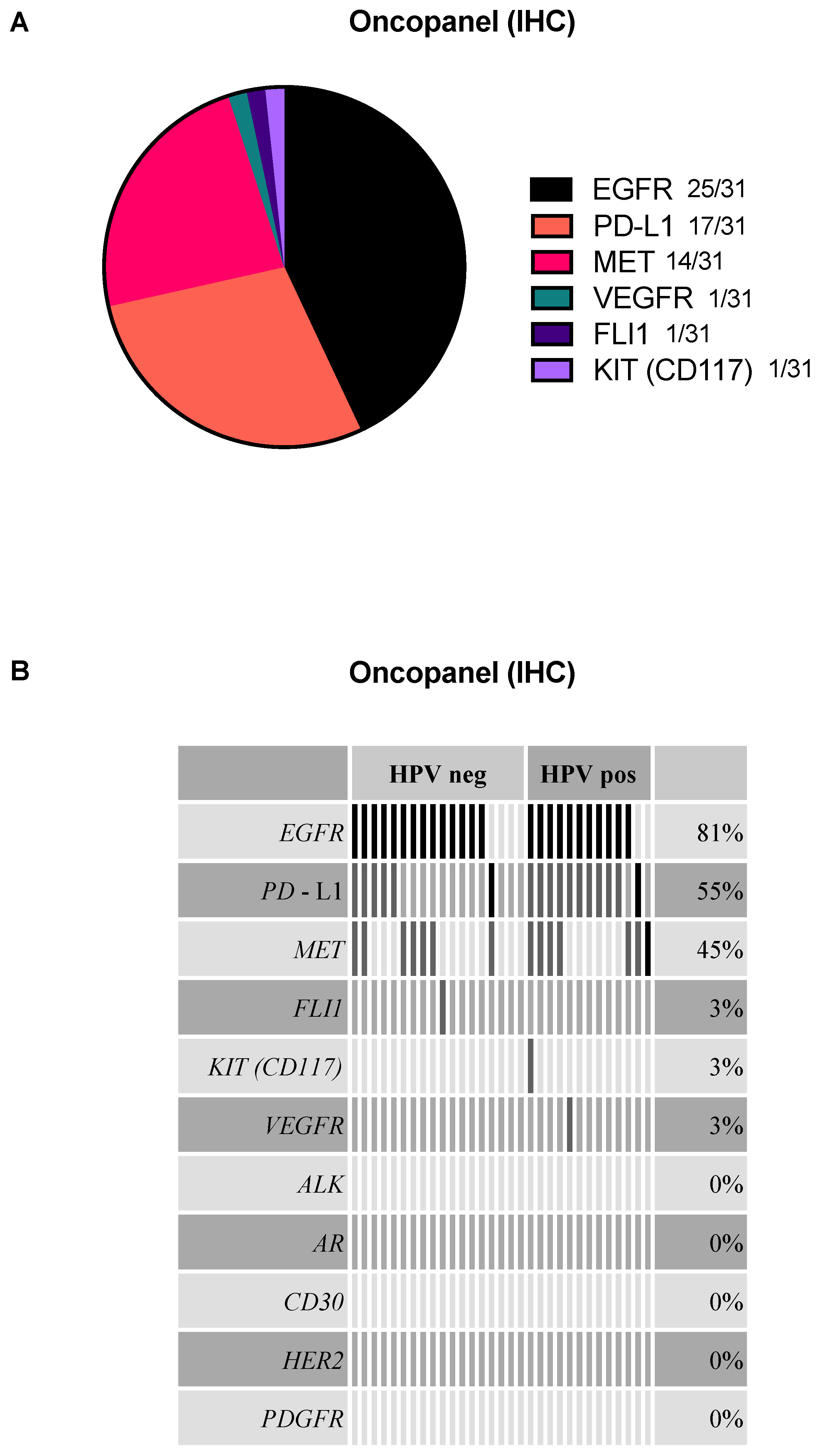
2.2. EGFR, PD-L1, and MET Are the Most Frequently Detected Targets in the Oncopanel

Out of 31 EGFR-stained cases, 25 were positive for EGFR. The second and third most common expression was found for PD-L1 (17/31) and MET (14/31). VEGFR, FLI1, and KIT were only positive in one case each (Figure 1A). Co-expression for EGFR and PD-L1 was seen in most cases (n = 8, 25.8%), followed by EGFR and MET co-expression (n = 5, 16.1%), as well as triple co-expression of EGFR, PD-L1, and MET in five cases (16.1%) (Figure 1B). In our cohort, no expression was found for HER2, ALK, PDGFR, CD30, and AR. The T-status or nodal status had no statistically significant influence on the proteomic results.

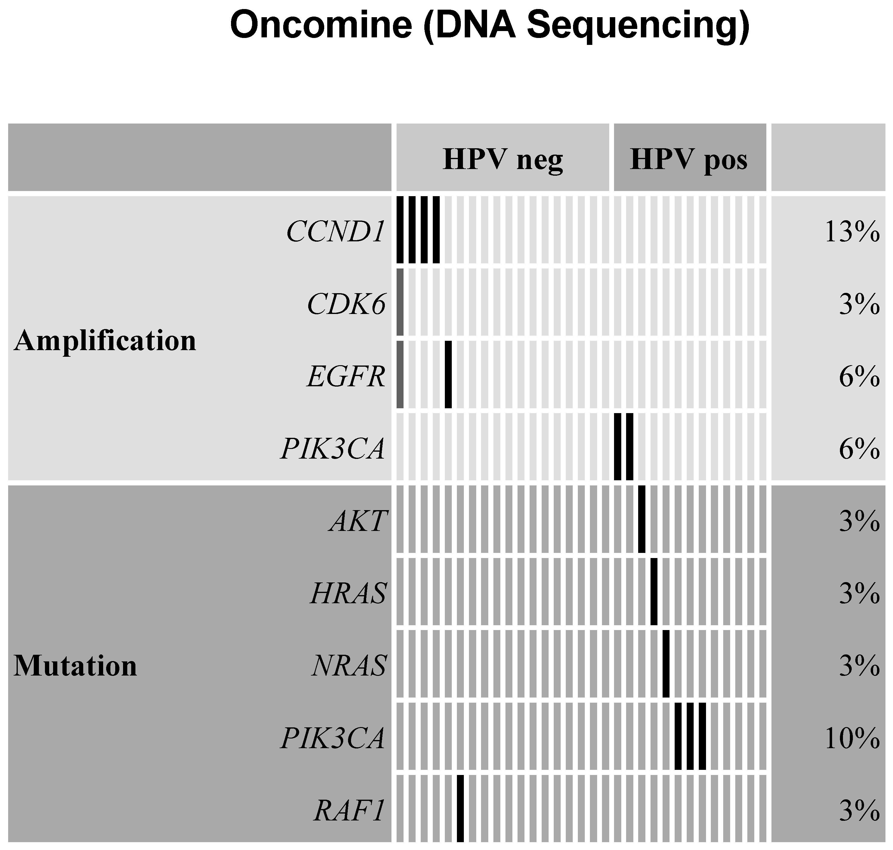
2.3. Amplifications Found for Cell Cycle Genes, EGFR, and PI3-Kinase in Oncomine Targeted Sequencing

Amplification of CCND1 (encoding Cyclin D1) was found in four cases and one of those harbored an additional CDK6 amplification. An EGFR amplification was found in two cases, whereas two cases showed a PIK3CA amplification. Furthermore, we could detect oncogenic mutations within the PIK3CA gene. Mutations affecting the genes RAF1, NRAS, HRAS, AKT, FGFR1, and MTOR were found in one case each (Figure 2). The T-status or nodal status had no statistically significant influence on the genomic results.

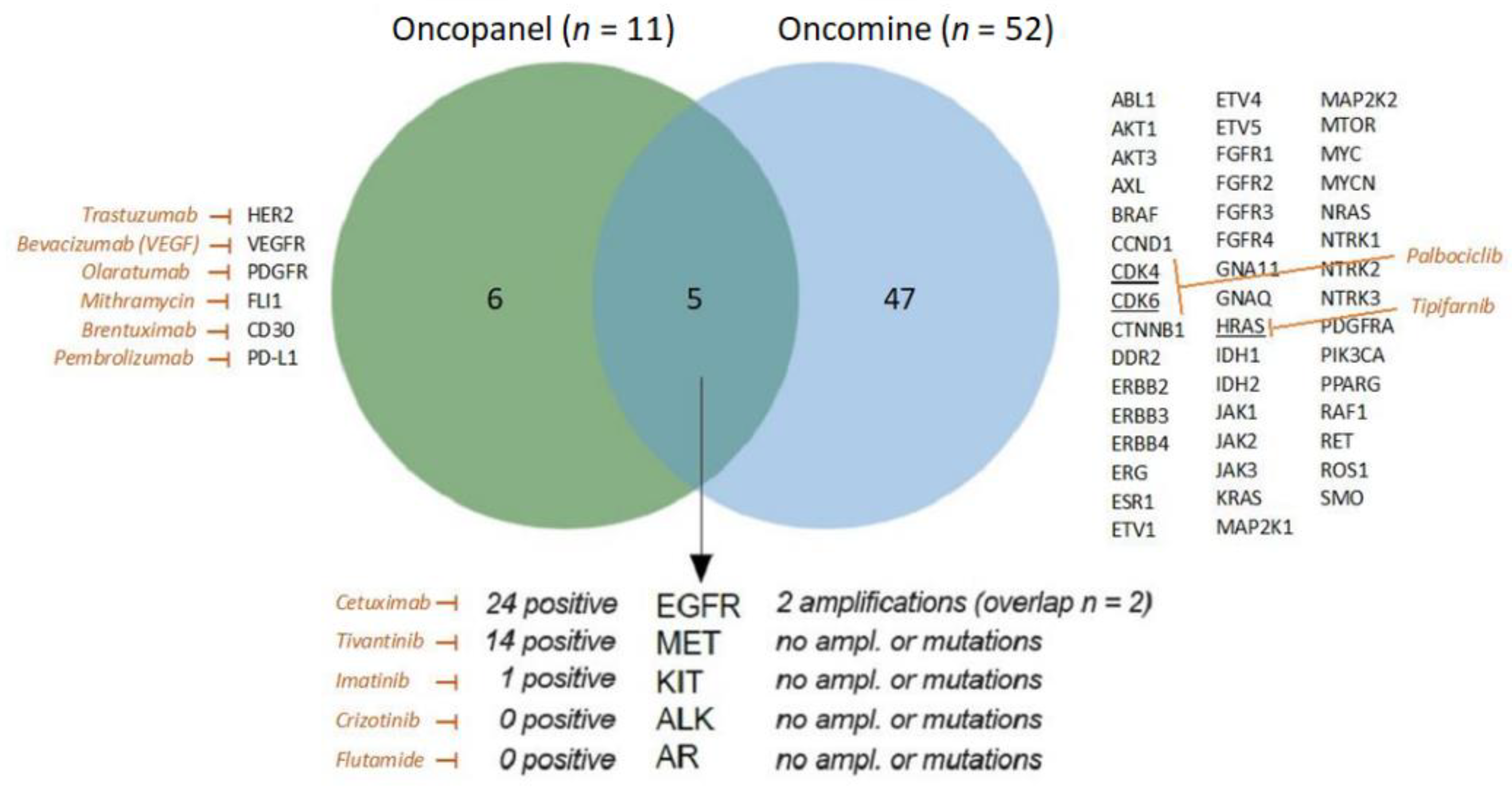
2.4. Overlap of Oncopanel and Oncomine

The two different panels overlap in five genes/markers: EGFR, MET, ALK, KIT, and AR (Figure 3). Within the overlapping markers, only the two EGFR amplifications were matched by IHC analysis.

2.5. HPV-Positive and HPV-Negative HNSCC Analyzed by Oncopanel and Oncomine

A dichotomized analysis of the oncopanel immunohistochemical data showed a similar distribution of EGFR and MET expression in HPV-positive and HPV-negative HNSCC cases. In most HPV-positive cases, we detected a PD-L1 expression as expected (Figure 1B). Comparing the HPV status regarding the oncomine assay, we found CCND1, CDK6, and EGFR amplifications only in HPV-negative cases, while PIK3CA amplifications and mutations were only found in HPV-positive HNSCC (Figure 2).

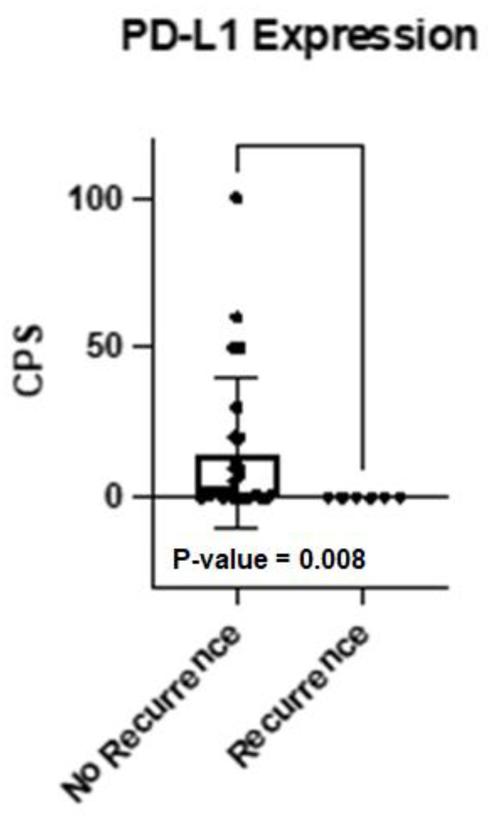
2.6. HNSCC Recurrences Analyzed by Oncopanel and Oncomine

Matching tumor samples were available from three patients (patients #7, #10, and #16, Table 3). The comparison of the primary tumor and its relapse showed stable EGFR expression in two patients (#10 and #16) on the protein level. Interestingly, both cases displayed an acquired PD-L1 expression in the recurrent tumor whereas the primary tumor did not express PD-L1. For the genetic analysis with the Oncomine Focus Panel, amplification of CCND1 was found in the primary and recurrent tumor of #16. Patient #10 developed an amplification for CCND1 beyond progression. Analysis of case #7 did not result in any therapeutical options on the protein or DNA level.

2.7. Characteristics of Recurrent and Deceased Patients

In our cohort, one female patient died shortly after her HNSCC recurrence was detected (#10). This case was positive for EGFR in the oncopanel, but no genetic alterations were found in the oncomine analysis. In the group of six recurrent HNSCC, no mutations were found with the oncomine panel in the primary tumor, whereas out of the 25 HNSCC cases without recurrence seven cases harbored mutations (28.0%). Amplifications were found in 2/6 recurring cases (33.0%) and 5/25 non-recurring cases (20.0%). The protein expression of the oncopanel revealed that no case with a recurrence had PD-L1 expressed in the primary tumor. Using the absolute CPS percentages, a nonparametric Mann–Whitney test revealed a statistically significant difference (p-value = 0.008, Figure 4). Furthermore, 5/6 cases expressed EGFR, and 3/6 cases expressed MET (Supplementary Table S2).

3. Discussion

In this study, we compared an immunohistochemical approach with a sequencing-based oncological panel to detect possible therapeutic targets in a cohort of 31 untreated HNSCC patients. Currently, those approaches are useful in the case of metastatic or recurrent HNSCC, where immunotherapy or other targeted therapies are often the only possible treatment options. However, these personalized targeted approaches can also become an option in curative treatment situations, if patient selection and patient benefit can be further improved.
This study shows the benefit of a straightforward proteomic approach, as the immunohistochemical analysis directly detects the targetable protein expression. Additionally, the surface markers are the carriers of biological function and therefore serve as target structures for many of the drugs. Mutations can lead to different expression patterns of RNA and subsequentially to a different protein expression. However, studies are showing that there is only a low correlation between transcriptomic and the related protein expression for genes of regulation in terms of biological processes [16]. Genome-wide correlation between mRNA expression and protein expression is poor with approximately only 40% [17].
In our study, we saw a variety of different expression patterns on the protein level and different mutation and amplification patterns. This points out the importance of inter-individual differences and illustrates the value of the personalized in-detail analysis of each patient’s tumor tissue.
In the oncopanel analysis, we observed a good proportion of tumors with co-expression of PD-L1 and EGFR. This underlines a possible treatment benefit of combining Cetuximab, an EGFR-blocking antibody, with ICM modulation. The first results of a phase 2 study, which support this promising approach, were recently published (NCT03082534) [18]. In this non-randomized, multicenter study, R/M HNSCC patients (n = 33) received a combination of pembrolizumab (200 mg/3 wk) and cetuximab (400 mg/m2 + 250 mg/m2/wk). After six months the overall response rate was 45% (15/33), which is more than twice as much as compared to the expected response rate with pembrolizumab monotherapy [6]. Assessment of the PD-L1 status revealed that all patients with recurrences had a negative CPS. This finding is in accordance with our previously published paper, where we could show that PD-L1 expression is a protection against the occurrence of recurrences [19].
We found an intracellular molecule CCND1, which is a protein-coding gene for Cyclin D1, to be amplified in four patients. Cyclin D1 is a protein that is involved in the cell cycle and regulates CDK4 and CDK6 as a subunit for the progression through the G1 phase of the cell cycle [20]. If this expression could be verified on the protein level as well, a possible treatment with a CDK4/CDK6 inhibitor such as Palbociclib or Abemaciclib could be attempted. Therefore, the next upgrade for the oncopanel would be an additional IHC marker with Cyclin D1, maybe also with CDK4 and CDK6. A phase I study evaluated Palbociclib in R/M HNSCC safe with tumor responses, and in vitro experiments combined with an mTOR inhibitor show promising anti-tumor effects (Figure 3) [21,22].
With the oncomine assay, we detected an activating mutation of HRAS (Figure 2 and Figure 3), which can be targeted using Tipifarnib a farnesyltransferase-inhibitor. This targeted therapy showed promising results in a phase II clinical trial of R/M HNSCC with a median progression-free survival of 5.6 months on tipifarnib versus 3.6 months on the last prior therapy (NCT02383927) [23].
Interestingly, we could find a different genomic and proteomic pattern in HPV-negative vs. -positive HNSCC. For example, cell cycle aberrations were only found in HPV-negative cases, while PIK3CA amplifications and mutation were solely found in HPV-positive cases. This underlines the importance of stratifying HNSCC in HPV-positive and HPV-negative also in the clinical decision finding.
The two panels were used as available, clinically proven panels containing each therapeutically targetable markers and genes. Therefore, the targets are not overlapping completely, however, they complement each other. The combination of both approaches is in our opinion necessary, as well as complementary, as not every amplification leads to an (over-)expression of protein, and other important regulatory mechanisms are involved. However, it would be interesting if following studies could assess all genes of interest on genomic, transcriptomic, and proteomic levels and could include analyses such as methylome or miRNA screens. Subsequently, a sound statement could be made about how DNA, RNA, and protein expression correlate, as well as how other gene regulatory mechanisms are involved, and how it might differ specifically for each gene of interest. However, this is rather expensive and labor intensive and is probably not feasible in the clinical routine. One key limitation of all DNA-based genotyping assays is that the functional consequences are usually “predicted” rather than directly assessed. Mutation-specific antibodies (REF) or immunohistochemistry in general (PD-1/PD-L1) can overcome this limitation and can directly visualize the pharmacologic targets in situ. Proteins are the carriers of biological function; our approach to combining DNA and protein assessment acknowledges this biological paradigm. Another limitation might be the small number of cases, but, on the other hand, we carefully selected a rather homogeneous cohort for the best comparison within cases.
An additional interesting point to mention is the similar costs to perform the assays. The immunohistochemical oncopanel costs EUR 350 related to the German fee schedule, while the material for the oncomine panel costs for one sample also around EUR 300. Here is to add, that the hands-on time for the sequencing is more labor-intensive than the oncopanel, and the analysis is more complex and needs additional equipment to perform those tasks, such as sequencers, servers, and computing power.
In summary, we conclude that the combination of the oncopanel and the oncomine assay brought up several targetable genetic and expression changes in individual patients, which can lead to the repurposing of available drugs and opens a variety of treatment options for those patients.

4. Materials and Methods

4.1. Study Design and Patient Cohort

Following a previously published study with heavily pre-treated HNSCC patients [15], we now established a prospective cohort of 31 previously untreated HNSCC patients at the time of their primary surgical treatment and used those in this explorative prospective study (Table 1). It was IRB approved (ethics 90/15) and patients signed informed consent before biopsy and any kind of treatment. The samples were collected either at the time of tumor biopsy or at the time of major surgery, and the samples were referred to both immunohistochemistry staining and targeted DNA sequencing (Oncomine Focus Assay, an NGS tumor-specific panel, ThermoFisher, Waltham, MA, USA) (Table 2). Further, for three of the patients a specimen for subsequent analysis at the time of recurrence was available.

4.2. Oncopanel

The oncopanel in this study assessed the surface expression of 11 proteins (EGFR, MET, HER2, ALK, VEGFR, PDGFR, FLI1, KIT (CD117), CD30, AR, PD-L1) as previously published (15). The staining was performed on the Dako Omnis platform (Dako, Glostrup, Denmark) as previously published [14,15]. Due to the very low prevalence of BRAF V600E in non-melanocytic head and neck carcinoma, we modified the oncopanel and excluded this marker. We also excluded PD-1 as a marker because PD-L1 expression has been established as a predictor of response to anti-PD-1 therapy and made the routine assessment of PD-1 expression redundant [6]. Briefly, we used formalin-fixed paraffin-embedded tissues and antibody details are provided in the Supplementary Table S1. Cases were considered positive if more than 10% of tumor cells showed immunopositivity in the appropriate cellular compartment. PD-L1 expression was scored according to the combined positive score (CPS) and cases with a CPS ≥ 1 were considered positive.

4.3. DNA Extraction

Archival formalin-fixed paraffin-embedded (FFPE) tissue specimens of squamous cell carcinoma patients were microdissected and genomic DNA was extracted with the Maxwell RSC Blood DNA Kit after a pre-treatment with a THG1-Thioglycerol/incubation buffer mix for 10 min at 80 °C and subsequent incubation with proteinase K at 65 °C overnight (Promega, Walldorf, Germany). DNA from cryo-preserved material was extracted using the DNeasy Blood and Tissue Kit (Qiagen, Hilden, Germany). DNA was extracted from fresh frozen samples (n = 16) and 15 FFPE samples.

4.4. Next Generation Sequencing (NGS)

To determine the concentration of amplifiable genomic DNA, we performed qPCR of the 34 extracted DNA samples (TaqMan RNase P Detection Reagents Kit, ThermoFisher, Waltham, MA, USA). Libraries were prepared with a multiplex PCR approach using the Oncomine Focus Assay and the Ion AmpliSeq Library Kit 2.0 (both ThermoFisher, Waltham, MA, USA) for 52 genomic targets (Figure 3). Libraries were templated and enriched with the Ion OneTouch 2 and the Ion OneTouch ES automated systems (both ThermoFisher, Waltham, MA, USA). Sequencing was performed using semiconductor sequencing technology (Ion GeneStudio S5, ThermoFisher, Waltham, MA, USA; sequencing quality is shown in Supplementary Table S3). Bam-files were generated by Torrent Suite software, version 5.10, and variant annotation was performed by the Ion Reporter software with default parameters for the Oncomine Focus Assay, version 5.10 (ThermoFisher, Waltham, MA, USA).

4.5. Data Analysis and Statistics

Data were analyzed and graphed using Microsoft Excel for Mac (version 16.54) and GraphPad Prism (version 9.2.0). The upset plot was generated in R (version 4.0.3) with RStudio (version 1.2.5033) and the ComplexHeatmap package using the UpSet function [24]. The analysis and visualization of the oncopanel and oncomine data were performed in the standalone software AVAtar (https://github.com/sysbio-bioinf/avatar, accessed on 10 December 2022) [25]. The Venn diagram was generated using the online javascript jvenn [26].

5. Conclusions

Untreated HNSCC patients display a series of targetable alterations in protein expression and DNA levels, which can be detected by immunohistochemistry and DNA sequencing. Both relatively cost-effective approaches complement one another and can be applied side-by-side to identify the best treatment option for HNSCC patients. Currently, those approaches are used in metastatic or recurrent HNSCC, where immunotherapy or other targeted therapies are often the only possible treatment options. However, these personalized targeted approaches can also become an option in curative treatment situations if patient selection and patient benefit can be further improved.

Supplementary Materials

The following supporting information can be downloaded at: https://www.mdpi.com/article/10.3390/ijms232415835/s1.

Author Contributions

Study concepts, P.J.S., J.L. and J.D.; study design, U.M.-R., P.J.S., C.B. and K.M.; data acquisition, A.v.W. and S.B.; quality control of data, P.M., K.M. and P.J.S.; data analysis, A.v.W., S.B. and J.D.; statistical analysis, A.v.W.; manuscript preparation, A.v.W., J.D. and P.J.S.; manuscript editing, T.K.H., S.L., M.-N.T. and U.M.-R.; manuscript review, A.v.W., U.M.-R., K.M., S.B., M.-N.T., C.B., S.L., J.L., P.M., T.K.H., J.D. and P.J.S. All authors have read and agreed to the published version of the manuscript.

Funding

J.D. was funded by the German Research Association (Deutsche Forschungsgemeinschaft, 451445144).

Institutional Review Board Statement

This study and all experimental protocols were IRB approved (ethics 90/15, Ethikkommission der Universität Ulm, ethics committee, university of Ulm, Germany) and patients signed informed consent before biopsy and any kind of treatment. All methods were carried out in accordance with relevant guidelines and regulations.

Informed Consent Statement

Written informed consent has been obtained from the patients before biopsy and any kind of treatment.

Data Availability Statement

The datasets were submitted to the European Genome-phenome Archive and will be available soon at https://www.ebi.ac.uk/ega/datasets (accession number: EGAS00001006296).

Conflicts of Interest

The authors declare no conflict of interest.

References

  1. Bray, F.; Ferlay, J.; Soerjomataram, I.; Siegel, R.L.; Torre, L.A.; Jemal, A. Global cancer statistics 2018: GLOBOCAN estimates of incidence and mortality worldwide for 36 cancers in 185 countries. CA Cancer J. Clin. 2018, 68, 394–424. [Google Scholar] [CrossRef] [Green Version]
  2. D’Souza, G.; Dempsey, A. The role of HPV in head and neck cancer and review of the HPV vaccine. Prev. Med. 2011, 53 (Suppl. 1), S5–S11. [Google Scholar] [CrossRef] [PubMed] [Green Version]
  3. Sethi, S.; Ali-Fehmi, R.; Franceschi, S.; Struijk, L.; van Doorn, L.J.; Quint, W.; Albashiti, B.; Ibrahim, M.; Kato, I. Characteristics and survival of head and neck cancer by HPV status: A cancer registry-based study. Int. J. Cancer 2012, 131, 1179–1186. [Google Scholar] [CrossRef] [PubMed]
  4. National Comprehensive Cancer Network (NCCN) Guidelines: Head and Neck Cancers. Version 1. Available online: https://www.nccn.org/professionals/physician_gls/pdf/head-and-neck.pdf (accessed on 1 February 2020).
  5. Burtness, B.; Harrington, K.J.; Greil, R.; Soulieres, D.; Tahara, M.; de Castro, G., Jr.; Psyrri, A.; Basté, N.; Neupane, P.; Bratland, Å.; et al. Pembrolizumab alone or with chemotherapy versus cetuximab with chemotherapy for recurrent or metastatic squamous cell carcinoma of the head and neck (KEYNOTE-048): A randomised, open-label, phase 3 study. Lancet 2019, 394, 1915–1928. [Google Scholar] [CrossRef]
  6. Cohen, E.E.W.; Soulieres, D.; Le Tourneau, C.; Dinis, J.; Licitra, L.; Ahn, M.J.; Soria, A.; Machiels, J.P.; Mach, N.; Mehra, R.; et al. Pembrolizumab versus methotrexate, docetaxel, or cetuximab for recurrent or metastatic head-and-neck squamous cell carcinoma (KEYNOTE-040): A randomised, open-label, phase 3 study. Lancet 2019, 393, 156–167. [Google Scholar] [CrossRef] [PubMed]
  7. Ferris, R.L.; Blumenschein, G., Jr.; Fayette, J.; Guigay, J.; Colevas, A.D.; Licitra, L.; Harrington, K.; Kasper, S.; Vokes, E.E.; Even, C.; et al. Nivolumab for Recurrent Squamous-Cell Carcinoma of the Head and Neck. N. Engl. J. Med. 2016, 375, 1856–1867. [Google Scholar] [CrossRef] [PubMed] [Green Version]
  8. Doescher, J.; Busch, C.J.; Wollenberg, B.; Dietz, A.; Wurdemann, N.; Schuler, P.; Hoffmann, T.K.; Laban, S. Immunotherapy for head and neck cancer: Highlights of the 2019 ASCO Annual Meeting. HNO 2019, 67, 905–911. [Google Scholar] [CrossRef]
  9. Doescher, J.; Laban, S.; Schuler, P.J.; Brunner, C.; Hoffmann, T.K. Immunotherapy for head and neck cancers: An update and future perspectives. Immunotherapy 2019, 11, 561–564. [Google Scholar] [CrossRef]
  10. Manukian, G.; Bar-Ad, V.; Lu, B.; Argiris, A.; Johnson, J.M. Combining Radiation and Immune Checkpoint Blockade in the Treatment of Head and Neck Squamous Cell Carcinoma. Front. Oncol. 2019, 9, 122. [Google Scholar] [CrossRef] [Green Version]
  11. Guigay, J.; Auperin, A.; Fayette, J.; Saada-Bouzid, E.; Lafond, C.; Taberna, M.; Geoffrois, L.; Martin, L.; Capitain, O.; Cupissol, D.; et al. Cetuximab, docetaxel, and cisplatin versus platinum, fluorouracil, and cetuximab as first-line treatment in patients with recurrent or metastatic head and neck squamous-cell carcinoma (GORTEC 2014-01 TPExtreme): A multicentre, open-label, randomised, phase 2 trial. Lancet Oncol. 2021, 22, 463–475. [Google Scholar]
  12. Herbst, R.S.; Arquette, M.; Shin, D.M.; Dicke, K.; Vokes, E.E.; Azarnia, N.; Hong, W.K.; Kies, M.S. Phase II multicenter study of the epidermal growth factor receptor antibody cetuximab and cisplatin for recurrent and refractory squamous cell carcinoma of the head and neck. J. Clin. Oncol. 2005, 23, 5578–5587. [Google Scholar] [CrossRef]
  13. Chan, A.T.; Hsu, M.M.; Goh, B.C.; Hui, E.P.; Liu, T.W.; Millward, M.J.; Hong, R.L.; Whang-Peng, J.; Ma, B.B.; To, K.F.; et al. Multicenter, phase II study of cetuximab in combination with carboplatin in patients with recurrent or metastatic nasopharyngeal carcinoma. J. Clin. Oncol. 2005, 23, 3568–3576. [Google Scholar] [CrossRef] [PubMed]
  14. Grunmuller, L.; Thierauf, J.; Weissinger, S.E.; Bergmann, C.; Bankfalvi, A.; Veit, J.; Hoffmann, T.K.; Möller, P.; Lennerz, J.K. Biopanel identifies expression status of targetable proteins in sinonasal melanoma. Pers. Med. 2016, 13, 291–301. [Google Scholar] [CrossRef]
  15. Doescher, J.; Weissinger, S.E.; Schonsteiner, S.S.; Lisson, C.; Bullinger, L.; Barth, T.F.; Leithäuser, F.; Mueller-Richter, U.; Laban, S.; Hoffmann, T.K.; et al. Clinical utility of a protein-based oncopanel in patients with end-stage head and neck cancer. Immunotherapy 2019, 11, 1193–1203. [Google Scholar] [CrossRef]
  16. Guo, Y.; Xiao, P.; Lei, S.; Deng, F.; Xiao, G.G.; Liu, Y.; Chen, X.; Li, L.; Wu, S.; Chen, Y.; et al. How is mRNA expression predictive for protein expression? A correlation study on human circulating monocytes. Acta Biochim. Biophys. Sin. 2008, 40, 426–436. [Google Scholar] [CrossRef] [Green Version]
  17. de Abreu, R.S.; Penalva, L.O.; Marcotte, E.M.; Vogel, C. Global signatures of protein and mRNA expression levels. Mol. BioSyst. 2009, 5, 1512–1526. [Google Scholar] [CrossRef] [Green Version]
  18. Sacco, A.G.; Chen, R.; Worden, F.P.; Wong, D.J.L.; Adkins, D.; Swiecicki, P.; Chai-Ho, W.; Oppelt, P.; Ghosh, D.; Bykowski, J.; et al. Pembrolizumab plus cetuximab in patients with recurrent or metastatic head and neck squamous cell carcinoma: An open-label, multi-arm, non-randomised, multicentre, phase 2 trial. Lancet Oncol. 2021, 22, 883–892. [Google Scholar] [CrossRef]
  19. Doescher, J.; Minkenberg, P.; Laban, S.; Kostezka, U.; von Witzleben, A.; Hoffmann, T.K.; Schuler, P.J.; Weissinger, S.E. Immune checkpoint expression in HNSCC patients before and after definitive chemoradiotherapy. Head Neck 2021, 43, 778–787. [Google Scholar] [CrossRef]
  20. Baldin, V.; Lukas, J.; Marcote, M.J.; Pagano, M.; Draetta, G. Cyclin D1 is a nuclear protein required for cell cycle progression in G1. Genes Dev. 1993, 7, 812–821. [Google Scholar] [CrossRef] [Green Version]
  21. Ku, B.M.; Yi, S.Y.; Koh, J.; Bae, Y.H.; Sun, J.M.; Lee, S.H.; Ahn, J.S.; Park, K.; Ahn, M.J. The CDK4/6 inhibitor LY2835219 has potent activity in combination with mTOR inhibitor in head and neck squamous cell carcinoma. Oncotarget 2016, 7, 14803–14813. [Google Scholar] [CrossRef] [Green Version]
  22. Michel, L.; Ley, J.; Wildes, T.M.; Schaffer, A.; Robinson, A.; Chun, S.E.; Lee, W.; Lewis, J., Jr.; Trinkaus, K.; Adkins, D. Phase I trial of palbociclib, a selective cyclin dependent kinase 4/6 inhibitor, in combination with cetuximab in patients with recurrent/metastatic head and neck squamous cell carcinoma. Oral Oncol. 2016, 58, 41–48. [Google Scholar] [CrossRef]
  23. Ho, A.L.; Brana, I.; Haddad, R.; Bauman, J.; Bible, K.; Oosting, S.; Wong, D.J.; Ahn, M.J.; Boni, V.; Even, C.; et al. Tipifarnib in Head and Neck Squamous Cell Carcinoma with HRAS Mutations. J. Clin. Oncol. 2021, 39, 1856–1864. [Google Scholar] [CrossRef]
  24. Gu, Z.; Eils, R.; Schlesner, M. Complex heatmaps reveal patterns and correlations in multidimensional genomic data. Bioinformatics 2016, 32, 2847–2849. [Google Scholar] [CrossRef] [Green Version]
  25. Volkel, G.; Laban, S.; Furstberger, A.; Kuhlwein, S.D.; Ikonomi, N.; Hoffman, T.K.; Brunner, C.; Neuberg, D.S.; Gaidzik, V.; Döhner, H.; et al. Analysis, identification and visualization of subgroups in genomics. Brief. Bioinform. 2021, 22, bbaa217. [Google Scholar] [CrossRef]
  26. Bardou, P.; Mariette, J.; Escudie, F.; Djemiel, C.; Klopp, C. Jvenn: An interactive Venn diagram viewer. BMC Bioinform. 2014, 15, 293. [Google Scholar] [CrossRef]
Figure 1. Results of the immunohistochemical approach (oncopanel) for 31 patient samples. (A) Pie chart displaying the distribution of marker expression. (B) AVAtar plot shows the markers’ distribution and their co-expression grouped by HPV status. The black bars show the first positive result per sample, while the grey bars depict a co-expression of a target in a specific sample.
Ijms 23 15835 g001
Figure 2. Results of the targeted mutation and amplification analysis using the oncomine panel displayed in an AVAtar graph grouped by HPV status and mutation or amplification. The black bars show the first positive result per sample, while the grey bars depict an additional finding of a target in a specific sample.
Ijms 23 15835 g002
Figure 3. Overlapping genes/markers (n = 5) between the oncopanel (n = 11) and the oncomine panel (n = 52) in a Venn diagram. The results for the overlapping markers are displayed at the bottom. On the sides, a table shows the remaining genes/markers for each table. A selection of possible targets is marked with an example of targeted treatment.
Ijms 23 15835 g003
Figure 4. Bar graph displaying the CPS score as a percentage for all patients grouped by their recurrence status. As all tumor samples of patients with relapse in course of treatment were PD-L1-negative there is a statistically significant difference between those two groups (p = 0.008, Mann–Whitney test).
Ijms 23 15835 g004
Table 1. Tabular patient characteristics with their ID, gender, age, life status, recurrence status, HPV status, localization, TNM Status, and treatment.
PatIDGen-derAgeLife StatusOSRecurrencePFSHPV StatusLocalizationT-StatusN-StatusM-StatusTherapy
1m52alive35no negativeOropharynx310SRG+RT
2m47alive35no negativeOropharynx310SRG+RT
3m65alive35no positiveOropharynx110SRG+CRT
4m61alive26no positiveOropharynx22b0SRG+CRT
5m71alive32yes16positiveOropharynx42c0CRT
6m50alive35no negativeOropharynx200SRG
7f59alive33yes34negativeOropharynx200SRG
8m69alive34no negativeOropharynx23b0SRG+CRT
9m71alive22no negativeOropharynx22b0SRG+RT
10f54dead10yes8negativeOropharynx200SRG+RT
11m68alive32no positiveOropharynx22c0SRG+RT
12m64alive29no positiveOropharynx200SRG
13m54alive29no negativeOropharynx42c0SRG+CRT
14m58alive26yes23negativeOropharynx300CRT
15m56alive28no positiveOropharynx210SRG+CRT
16f56alive27yes5negativeOropharynx42a0SRG
17f38alive25no naOral Cavity200SRG+RT
18m69alive19no naOral Cavity310SRG
19f66alive21no naOral Cavity310SRG+RT
20f69alive19no naOral Cavity200SRG
21m64alive15no positiveOropharynx32c0CRT
22m64alive14no positiveOropharynx210SRG+RT
23m83alive14no positiveOropharynx210SRG+RT
24m76alive16no positiveOropharynx200SRG+RT
25m68alive13no negativeOropharynx22b0SRG
26f64alive1no positiveOropharynx42a0SRG+CRT
27f57alive12no positiveOropharynx200SRG
28m76alive13yes6negativeOropharynx42b0SRG+RT
29m67alive13no positiveOropharynx12a0SRG+RT
30m63alive6no naOral Cavity100SRG+RT
31m64alive6no naOral Cavity200SRG
m = male; f = female; SRG = surgery; RT = radiotherapy; CRT = chemoradiotherapy; OS = overall survival in months; PFS = progression-free survival in months.
Table 2. Summarized patient characteristics.
n%
Primary SiteOropharynx2581%
Oral Cavity619%
Total31
SexMale2374%
Female826%
Agemean (range)62.6 (37.9–83.0)
Tumor StatusT1310%
T21755%
T3619%
T4516%
Nodal StatusN-1135%
N+2065%
Distant MetastasesM031100%
HPV-status (DNA)negative 12
positive13
missing (not OPSCC)6
TreatmentCRT only3 (2 *)
Surgery only9
Surgery+RT6
Surgery+CRT13
HPV = human papillomavirus, RT = radiotherapy, CRT = chemoradiotherapy, * with Pembrolizumab.
Table 3. Tabular comparison of the primary and recurrent tumor tissue for both panels. Patient IDs are displayed as defined in Table 1.
PrimaryRecurrence
Oncopanel (IHC)
Patient 7no targetno target
Patient 10EGFR+EGFR+/PD-L1
Patient 16EGFR+EGFR+/PD-L1
Oncomine (amplification)
Patient 7no targetno target
Patient 10no targetCCND1
Patient 16CCND1CCND1
Publisher’s Note: MDPI stays neutral with regard to jurisdictional claims in published maps and institutional affiliations.

