Review

Similarities and Differences between the Orai1 Variants: Orai1α and Orai1β

Department of Physiology (Cellular Physiology Research Group), Institute of Molecular Pathology Biomarkers (IMPB), University of Extremadura, 10003 Caceres, Spain
*
Authors to whom correspondence should be addressed.
These authors contributed equally to this work.
Int. J. Mol. Sci. 2022, 23(23), 14568; https://doi.org/10.3390/ijms232314568
Submission received: 4 November 2022 / Revised: 16 November 2022 / Accepted: 21 November 2022 / Published: 23 November 2022
(This article belongs to the Special Issue Calcium Signaling in Health and Diseases)

Abstract

:
Orai1, the first identified member of the Orai protein family, is ubiquitously expressed in the animal kingdom. Orai1 was initially characterized as the channel responsible for the store-operated calcium entry (SOCE), a major mechanism that allows cytosolic calcium concentration increments upon receptor-mediated IP3 generation, which results in intracellular Ca2+ store depletion. Furthermore, current evidence supports that abnormal Orai1 expression or function underlies several disorders. Orai1 is, together with STIM1, the key element of SOCE, conducting the Ca2+ release-activated Ca2+ (CRAC) current and, in association with TRPC1, the store-operated Ca2+ (SOC) current. Additionally, Orai1 is involved in non-capacitative pathways, as the arachidonate-regulated or LTC4-regulated Ca2+ channel (ARC/LRC), store-independent Ca2+ influx activated by the secretory pathway Ca2+-ATPase (SPCA2) and the small conductance Ca2+-activated K+ channel 3 (SK3). Furthermore, Orai1 possesses two variants, Orai1α and Orai1β, the latter lacking 63 amino acids in the N-terminus as compared to the full-length Orai1α form, which confers distinct features to each variant. Here, we review the current knowledge about the differences between Orai1α and Orai1β, the implications of the Ca2+ signals triggered by each variant, and their downstream modulatory effect within the cell.

1. Introduction

The calcium ion (Ca2+) is a ubiquitous messenger involved in several physiological events, including immune response, muscle contraction, neuronal transmission, and fertility, among others. Ca2+ is also heavily implicated at the cellular level, where it mediates gene expression, secretion, cell proliferation, and apoptosis. Due to its pleiotropic effects, altered intracellular Ca2+ homeostasis induces pathological conditions [1,2,3].
The human body possesses an intricated machinery that precisely regulates Ca2+ homeostasis, including the store-operated Ca2+ entry (SOCE), a major mechanism of Ca2+ mobilization in the electrically non-excitable and excitable cells, where the release of Ca2+ stored in the intracellular compartments, mainly the endoplasmic reticulum (ER), leads to the activation of Ca2+ channels in the plasma membrane (PM), followed by a massive Ca2+ influx from the extracellular medium [4,5]. SOCE is mediated by two types of channels, the Ca2+ release-activate calcium (CRAC) and the store-operated calcium (SOC) channels. The passing of Ca2+ from the extracellular milieu through the channels generates two distinct currents, Icrac and Isoc, each with identifiable features. Whereas CRAC influx is mediated exclusively by the ER Ca2+ sensor, STIM1, and Orai1 channels, TRPC1 proteins associate with Orai1 and STIM1 to form the SOC channels [5].
Orai1, a pivotal actor of SOCE, was first identified by three independent interference RNA screens in Drosophila S2 cells that related a few Drosophila genes to SOCE, including olf186-F, named Drosophila Orai (dOrai1) [6,7,8]. Feske and coworkers linked their findings in Drosophila to a region of human chromosome 12, containing the human homolog of dOrai1, Orai1 (also termed CRACM1), by the genetic mapping of members of a family that presented severe combined immunodeficiency (SCID). SCID patients express a homozygous R91W point mutation in the Orai1 protein that impaired T-cell activation. Over-expression of wild-type Orai1 in T cells from SCID patients reestablished SOCE [6]. In addition to SCID, gain or loss of Orai1 function has been associated with other diseases, such as autoimmune disorders (reviewed in [9,10,11,12]). Two other members of the Orai family have been identified, Orai2 and Orai3 [7], which can associate with STIM1 and STIM2 to trigger SOCE [13,14,15]. The current evidence supports that native CRAC channels consist of the heteromeric association of Orai1, Orai2, and/or Orai3 (PMID: 28294127, PMID: 29604961, PMID: 31015290), where the presence of Orai2 and Orai3 in native CRAC channels ensures that the magnitude of Ca2+ influx is proportional to the strength of agonist stimulation [16,17]. Despite there being no evidence indicating the composition of native CRAC channels, the stoichiometry of these channels in a given cell type presumably depends on the relative expression of Orai2 and Orai3 [15,17,18].
All three Orai proteins present the same structure: a four-membrane spanning protein containing one intracellular and two extracellular loops and the N- and C-terminus facing the cytoplasm [6,7,8] (Figure 1a). While the transmembrane domains (TM1–4) are highly conserved in all three isoforms, there are clear differences between the cytosolic regions, with 34% and 46% of sequence similarity between the N-terminus and C-terminus, respectively, of Orai1 and Orai3. The extracellular regions of the Orai proteins diverge as well, with an Orai3 third loop longer than the one exhibited by Orai1 and Orai2 (Figure 1b) [19,20,21]. In 2012, the structure of dOrai was crystallized, featuring an unexpected arrangement of six dOrai subunits conforming to the channel [22]. Although tetrameric and pentameric stoichiometries for the human Orai channels have been proposed, it is currently accepted that the Orai1 proteins might form a hexamer [21,23,24,25].
In the hexameric architecture, one dOrai channel is formed by three dimers with a crossing C-termini. The TM1 domains form the inner ring that acts as the ion-conducting pore. TM2–3 generate a central ring that protects the pore from interaction with the lipids in the PM. TM4 arranges as the outer ring, interacting with other PM components. The N-terminal sequence (aa 1–70) of human Orai1 (hOrai1) is further discussed in the following sections. An α-helix of the TM1 domain, known as the extended transmembrane Orai1 N-terminus (ETON, aa 73–90), reaches the cytoplasm and is required for the association with STIM1 and the gating of the channel [26]. The pore region is composed of a basic domain (hOrai1 aa R83, K87, and R91, whose mutation to W induced SCID), a hydrophobic segment (hOrai1 aa L95, F99, and V102), and the selectivity filter, formed by a ring of six glutamates (hOrai1 aa E106). TM1 and TM2 are connected by the first extracellular loop (loop1) that contains an acidic calcium accumulating region (CAR, hOrai1 aa D110, D112, D114) that ensures Ca2+ influx even in low Ca2+ conditions [27]. The only cytosolic Orai1 loop (loop2), located between TM2 and TM3, possesses two modulating domains that control Ca2+ influx through Orai1: the first one (hOrai1 aa 151–154) facilitates Orai1 fast calcium-dependent inactivation (FCDI) [28], and the second region within loop2 is an interacting domain (hOrai1 aa 157–167) with the chaperonin-containing T-complex protein 1 chaperonin complex (CCT) that mediates Orai1 internalization and recycling, thus acting as a regulator of Ca2+ signaling mediated by SOCE [29]. TM3 and TM4 are linked by another extracellular loop (loop3), which contains the unique glycosylation site within Orai1 (hOrai1 aa N223), allowing the interaction with lectins (carbohydrate-binding proteins) in a cell-type dependent manner, which results in the attenuation of SOCE [30]. All loops contain cysteine residues (hOrai1 loop1-C126, loop2-C143, and loop3-C195) that modulate the redox regulation of Orai1 channels as treatment with H2O2 significantly impaired SOCE [31]. The C-terminal region of dOrai extends to the cytoplasm from the TM4 domain bending in a highly conserved region (hOrai1 aa 268–291), known as TM4 extended (TM4x) [32], which results in an antiparallel association between the C-termini of two neighboring dOrai subunits [22]. This hinge region within Orai C-termini contains the initial and main binding and activating site between STIM1 and Orai1 [33,34,35,36,37]. Furthermore, Orai1 C-terminal exhibits a region (hOrai1 aa 260–275), termed as the C-terminus internalization handle (CIH), which exert a negative effect over SOCE by contributing to the internalization of Orai1 during meiosis through caveolin- and dynamin-dependent endocytic pathway [38,39].
Upon cell stimulation by physiological agonists, the reduction in the luminal ER Ca2+ concentration is sensed by STIM1, which suffers a conformational change that allows the activation of Orai1 channels in the plasma membrane [6,7,8,40]. STIM1 binds to Orai1 C-termini (aa 260–275) by an activating region located in the cytosolic STIM1-CC1α3 and -CC2 domain (aa 312–387) [37], which was simultaneously characterized by three independent groups and dubbed as CAD (CRAC activating domain) [34], SOAR (STIM1-Orai1 activating region) [35], and OASF (Orai1 activating small fragment) [36]. Orai1 C-termini is considered the primary binding site with STIM1, as deletion of this fragment completely abolishes the association between STIM1-Orai1 [33,41,42]. Although still a matter of debate (extensively discussed in [43]), several reports have demonstrated that STIM1 interacts with Orai1 N-termini to gate and modulate the opened Orai1 channel [26,34,44,45]. For instance, a recent study has shown that the STIM1-Orai1 N-termini interaction is vital to obtain CRAC currents featuring the hallmarks of a wild-type CRAC current [45]. A typical feature of CRAC channels is the fast inactivation mediated by Ca2+, which limits the number of ions passing through the channel [46]. Other proteins, such as calmodulin [47,48,49] or SARAF [50,51,52], contribute to regulating the Ca2+ flux through Orai1. Ultimately, when the signal responsible for intracellular Ca2+ stores depletion ends, STIM1 disassociates from Orai1 and returns to its coalescent state, and, subsequently, the channel closes, interrupting the Ca2+ influx.
Since the identification of STIM [53] and Orai [6,7,8] proteins as main players for SOCE, several models regarding the stoichiometry of both proteins have been presented [37,54,55,56,57,58,59,60,61,62,63,64]. Such knowledge is key to the creation of new drugs able to fight against conditions arising from altered SOCE [9,12]. It is clear from the beginning that ectopic expression of any of the two proteins in dissimilar ratios led to altered SOCE and CRAC currents [54,57]. In 2011, Hoover and Lewis presented the optimal STIM1:Orai1 ratio as 2:1 [58]. The identification of the dOrai1 crystal structure in 2012 [22], and further studies based on the channel architecture determined that a bimolecular coupling between 1 STIM1 dimer and 2 adjacent Orai1 subunits would suffice to activate the hexameric Orai1 channel [32,37,60,63,65,66], which did not quite fit with the most favorable STIM1:Orai1 ratio [58]. However, new pieces of evidence have been presented against this bimolecular model, favoring a unimolecular association between 1 STIM1 dimer and 1 Orai1 subunit [62,64,67], which fits with the 2:1 optimal ratio. Zhou and coworkers proposed that the formation of the pocket by to Orai1-TM4 through I316 and I319 may be an artifact of dOrai1 crystallization conditions; thus, it would be improbable that 1 STIM1 dimer might bind to and activate 2 neighboring Orai1 subunits [67]. Undoubtedly, this subject requires further investigation, and probably, we will not solve all the questions until the full hOrai1 structure is identified. Fortunately, new technologies, such as neural networks or artificial intelligence systems capable of predicting a protein’s 3D structure from its amino acid sequence [68], might shed some light, helping us to unravel the mystery.
In addition to the Ca2+ influx through CRAC channels, Orai1 is also responsible for the Ca2+ entry mediated by the SOC channels in collaboration with STIM1 and TRPC1 [5,69,70,71,72]. STIM1 activates TRPC1 via the association of a region of STIM1 C-termini polybasic domain (aa 684–685) with two conserved aspartates (aa 639–640) within TRPC1 [70,71]. The activation of SOC channels via receptor-mediated inositol 1,4,5-trisphosphate (IP3) and the subsequent Ca2+ store release generates characteristic changes in cytosolic Ca2+ concentration, where Orai1 conducts the generation of Ca2+ oscillations and TRPC1 is implicated in the frequency of baseline Ca2+ oscillations, supporting, as well, a maintained Ca2+ elevation with higher agonist concentration [72,73]. Orai1-mediated Ca2+ oscillations control NFAT translocation to the nucleus and NFAT-dependent gene expression, probably due to the proximity of NFAT machinery neighboring the pore of Orai1 [72,74,75]. While TRPC1 have no implications in NFAT activation [72], it is involved in the activation of NFκB and the NFκB-dependent gene expression [72,76,77]. Furthermore, TRPC1 Ca2+ entry upon STIM1 activation is crucial for triggering epithelial-to-mesenchymal transition (EMT) in invasive ductal carcinoma breast cancer cells, where SOCE is mainly mediated by TRPC1 channels, and the implication of Orai1 channels is still unknown [78]. Additional studies are needed to understand the physiology of SOC channels, which will help us to finally explain their implication in the pathophysiology of certain diseases, such as cancer.
Moreover, Orai1 supports other non-capacitative Ca2+ entry pathways, as well. For instance, Orai1 associates with STIM1 and Orai3 to form the arachidonate-regulated or LTC4-regulated Ca2+ channel (ARC/LRC) [79,80]. Furthermore, Orai1 might act independently of STIM1 associated with the secretory pathway Ca2+-ATPase-2 (SPCA2) [81,82] or the small conductance Ca2+-activated K+ channel 3 (SK3) [83]. Store-independent Ca2+ entry and its role in physiology and pathophysiology are extensively reviewed in [84,85].
Orai1 is a ubiquitous and complex Ca2+ channel that contributes to several local and global Ca2+ signals, which modulate the downstream effect of many Ca2+-dependent pathways. Alterations in those routes lead to diseases [12], thus, a thorough understanding of Orai1 is required. Recently, two Orai1 variants have been identified [5], which might explain some of the questions that we had but have raised several new ones. Here we review the current knowledge about the Orai1 variants and their implication in Ca2+ signals mediated by Orai1.

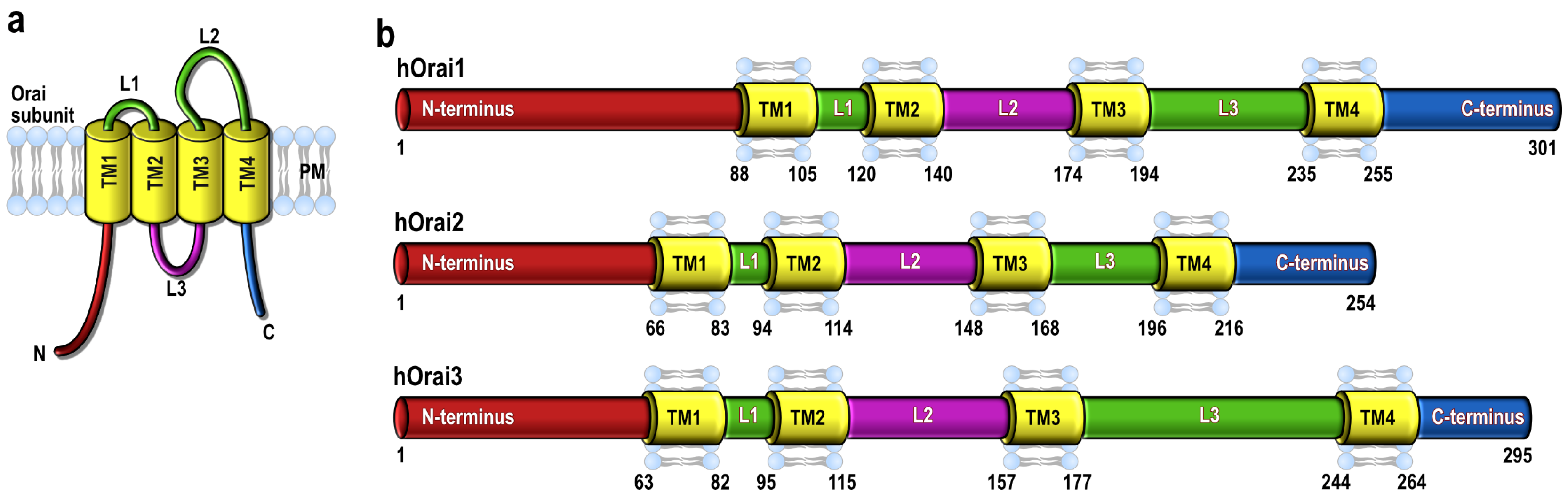
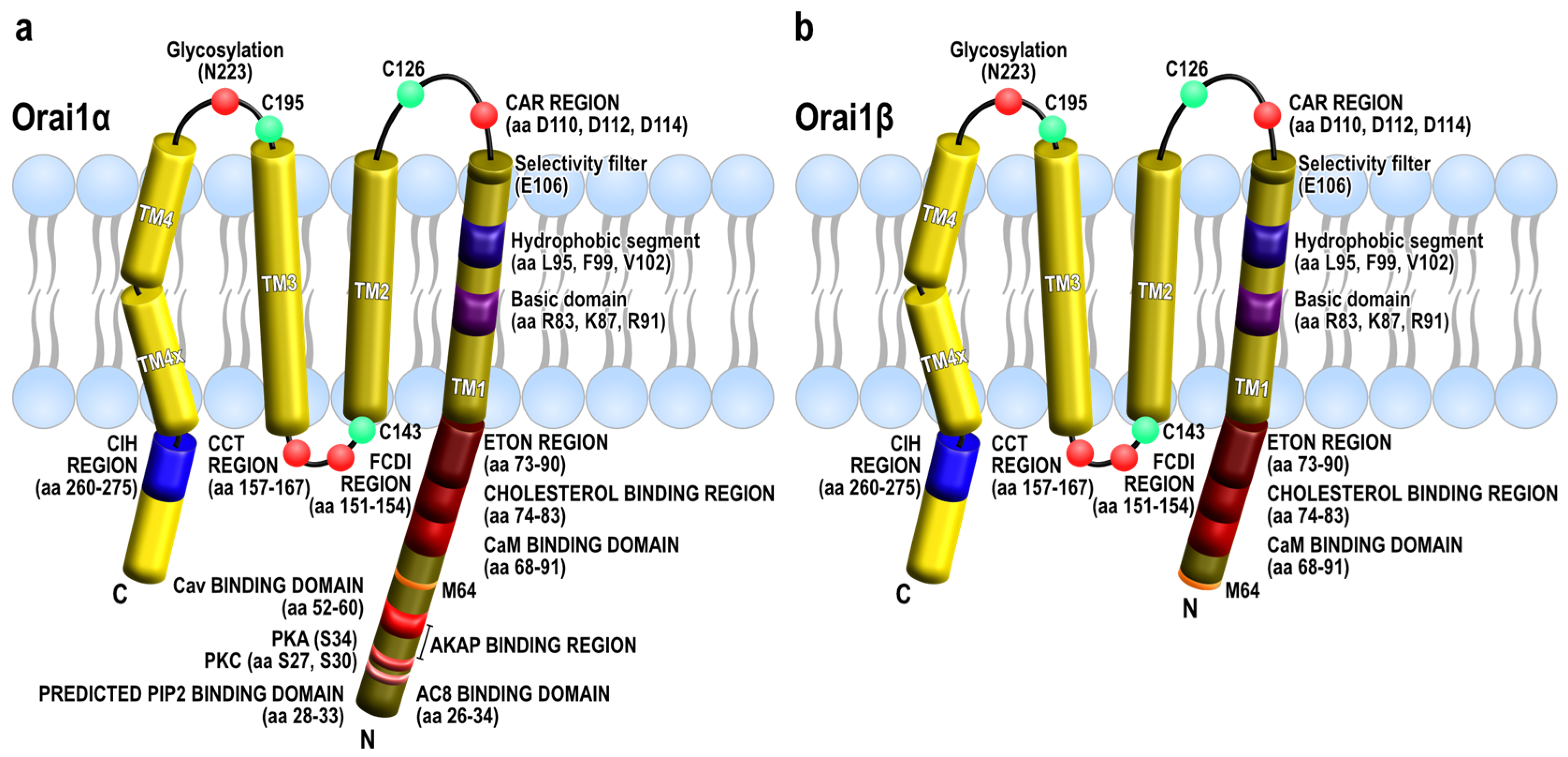
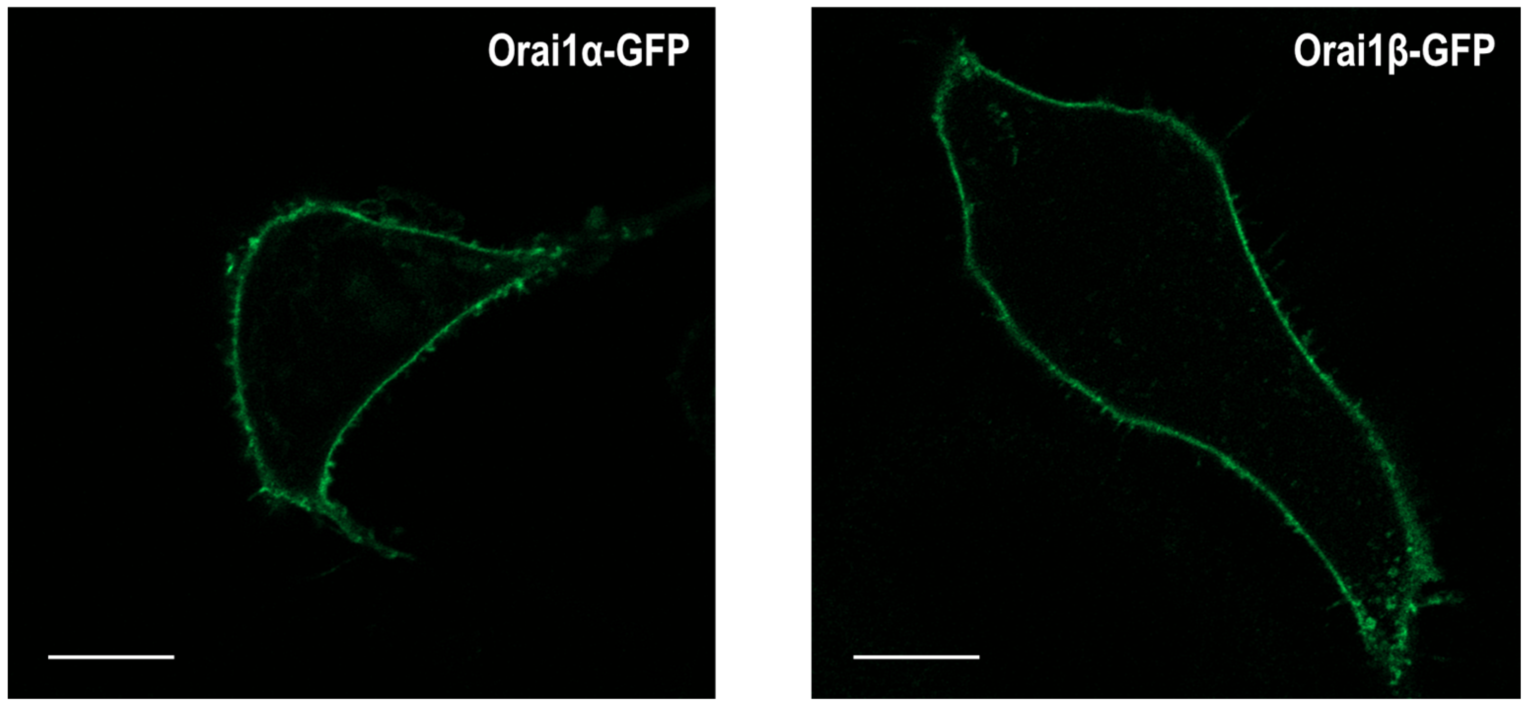
2. Orai1 Variants: Orai1α and Orai1β

Orai proteins are evolutionarily conserved across the animal kingdom. Analysis of the phylogenetic relationships of the Orai proteins in different species has revealed that invertebrates contain single copies of the Orai protein (except for Tribolium castaneum and Apis mellifera, which contain two Orai molecules). The Orai protein found in invertebrates evolved into two Orai proteins in vertebrates, referred to as Orai1 and Orai2 in mammalian cells. In mammals, specifically, duplication of the Orai1 gene led to Orai3, which exhibits closer phylogenetic relationships with Orai1 [86].
At present, no variants of Orai1 derived from alternative pre-messenger RNA splicing have been described; however, two forms of Orai1 have been identified in mammalian cells generated by alternative translation initiation. The human full-length Orai1 variant, named Orai1α, contains 301 amino acids (Figure 2a), while the short form, known as Orai1β (Figure 2b), arises from alternative translation initiation at methionine 64, or even methionine 71, therefore lacking the N-terminal 63 or 70 amino acids present in Orai1α [87,88]. It has been reported that the Kozak sequence for the first methionine in the native 5′-untranslated region is rather weak, leading to the translation of Orai1β from an alternative translation start site, methionines 64 and 71. Accordingly, improvement of the Kozak sequence for the first methionine has been reported to lead to the generation of Orai1α exclusively, as well as mutation of methionines 64 and 71 to alanine or valine [87]. Conversely, mutation of the first methionine results in the production of Orai1β, which strongly suggest that methionines 64 and 71 function as second translation initiation sites in Orai1 [88].
Different functional domains have been identified in the Orai1α exclusive N-terminal region, containing 63 amino acids, which have been associated with the sensitivity to Ca2+-dependent inactivation, protein-protein interaction, or cellular location. Specifically, residues 26–34 in Orai1α are involved in its interaction with adenylyl cyclase 8 (AC8), a Ca2+-modulated cyclase with low affinity for this cation [89]. In addition, Orai1 has been reported to be phosphorylated by PKC at N-terminal serines 27 and 30 residues, an event that is strongly associated with the suppression of SOCE and CRAC channel function [90]. Furthermore, serine 34 is a PKA phosphorylation site reported to mediate Ca2+-dependent Orai1 channel inactivation as a feedback mechanism upon the activation of AC8 [74]. There is also a predicted PIP2-binding domain between amino acids 28–33 whose functional role is still uncertain but might be involved in the distribution of Orai1 in the plasma membrane [91] or the regulation of STIM1-Orai1 interaction by the protein SARAF [92]. Finally, residues 52–60 form a predicted caveolin-binding domain which might regulate the plasma membrane location of Orai1 as it has been reported during meiosis, where Orai1 internalization has been shown to be dependent on its interaction with caveolin [39].
Orai1α and Orai1β are expressed in all the cells investigated, including HEK-293 and HeLa cells, as well as a variety of tumor cells, such as luminal MCF7 and triple-negative MDA-MB-231 breast cancer cells, squamous carcinoma A431 cells, lung adenocarcinoma H441 cells and colorectal adenocarcinoma T84 cells [87,93]. The Orai1α:Orai1β expression ratio varies among the cell lines investigated but mostly ranging from 0.3 to 1 [87,93], thus suggesting that Orai1 mRNA transduction mostly favors the expression of the short variant.
Analysis of Orai1α and Orai1β subcellular locations has revealed that, as expected, both Orai1 variants are predominantly localized in the plasma membrane [87]. Figure 3 shows that Orai1α-GFP and Orai1β-GFP expressed in Orai1-KO HEK-293 cells are exclusively located at the plasma membrane; therefore, the truncation of N-terminal 63 amino acids in Orai1β does not affect its plasma membrane location in resting cells. Furthermore, Fukushima and coworkers demonstrated that Ca2+ store depletion using the SERCA inhibitor thapsigargin or the physiological agonist carbachol resulted in the accumulation of Orai1α and Orai1β into puncta with similar efficiency, thus indicating that Orai1α and Orai1β exhibit a similar subcellular location [87]. Nevertheless, although no differences have been found in the cellular distribution of both Orai1 variants, the mobility of Orai1α and Orai1β in the plasma membrane significantly differs. As analyzed by rates of fluorescence recovery after photobleaching (FRAP), either expressed singly or together, the half-time of Orai1β recovery follows a normal distribution while that for Orai1α exhibits a bimodal distribution, the first one with a half-time similar to that of Orai1β followed by a second, predominant, population with a slower rate of recovery. The nature of the two populations of Orai1α rates of recovery remains uncertain. The observation that Orai1β exhibits the same mobility profile either expressed alone or co-expressed with Orai1α, which is different from the Orai1α mobility profile, suggests that Orai1α and Orai1β do not form heteromeric channels; however, further studies are required to characterize the two Orai1α populations and whether the minor, faster, population of Orai1α might heteromerize with Orai1β under certain conditions, as co-expression of both Orai1 forms slows down their rates of recovery [87].

3. Functional Properties of Orai1α and Orai1β

Using Orai1α- or Orai1β-optimized constructs that results in the production of one of the Orai1 forms, Desai and coworkers demonstrated that both Orai1 variants can rescue SOCE in Orai1-KO mouse embryonic fibroblasts (MEFs), with Orai1α showing smaller efficiency. Similarly, expression of Orai1α or Orai1β in Orai1-KO MEFs and HEK-293 cells restored Icrac but with similar efficiency [5]. The reason for the discrepant efficiencies of both Orai1 variants in SOCE and Icrac was attributed to the dialysis of the intracellular fast Ca2+ chelator BAPTA for Icrac measurements, which impairs Ca2+-dependent inactivation of the Orai1 channels. In fact, analysis of fast Ca2+-dependent inactivation of Orai1α and Orai1β expressed in HEK-293 cells revealed that, in the presence of the slower Ca2+ chelator EGTA, Orai1α produced a smaller current than Orai1β at all potentials tested, an observation that was abrogated in the presence of BAPTA. These findings indicate that, while both variants support Icrac, Orai1α shows a stronger fast Ca2+-dependent inactivation than Orai1β, the latter being barely sensitive to Ca2+-depending inactivation as it behaves similarly in the presence of EGTA and BAPTA [5]. These findings were confirmed in Orai1-KO HEK-293 cells expressing Orai1α or Orai1β using plasmids with the weak thymidine kinase (TK) promoter to reconstitute Orai1 expression at a more physiological level [74]. Zhang et al. also reported that the substitution of extracellular Ca2+ by Ba2+ abrogates the differences between Orai1α and Orai1β, which strongly suggests that the different inactivation rates are mediated by Ca2+ [74].
The functional role of Orai1 variants in the Isoc current has also been investigated. This current is the result of a complex mechanism where Ca2+ influx through Orai-forming CRAC channels recruits TRPC channels to the plasma membrane [94]; therefore, Isoc comprises the currents mediated by CRAC and TRPC1 channels [5,94]. In HEK-293 cells, patch-clamp experiments have demonstrated that both Orai1α and Orai1β support Isoc with similar efficiency [5]. These findings were confirmed by analyzing Mn2+ influx through TRPC1 channels [73]. Mn2+ is used as a surrogate for Ca2+ that enters the cells through TRPC1 but not via Orai1 channels in the presence of extracellular divalent cations, such as Ca2+ [40,73]. However, the involvement of Orai1β in this mechanism seems to be cell-specific, as, in HeLa cells, we have found that Orai1α, but not Orai1β, participates in the recruitment of TRPC1 channels in the plasma membrane and is required for store-operated cation entry through TRPC1. In this cellular model, Orai1β was not found to interact or co-localize with TRPC1 channels, and expression of a dominant negative Orai1β mutant failed to attenuate translocation of TRPC1 to the plasma membrane or Mn2+ influx through TRPC1 channels [73]. The basis for the different involvement of Orai1β in TRPC1 function and Isoc in these cell types is uncertain at present.
One of the most relevant functional differences between both Orai1 forms is that Orai1α, but not Orai1β, has been shown to support Iarc [5], the store-independent Ca2+ selective current activated by arachidonate and its metabolite leukotriene C4 [95]. ARC channels consist of heteropentamers of three Orai1 subunits and two Orai3 subunits [20,79] and require the participation of plasma membrane resident STIM1 [80,96]. In HEK-293 cells, individual expression of Orai1α was found to rescue Iarc in cells transfected with siOrai1, while Orai1β failed to do this.

4. Orai1 Variants and NFAT Activation

Nuclear factor of activated T-cell (NFAT) proteins are widely expressed transcription factors that regulate a plethora of genes involved in immunity, development, and, more recently, having an important function in cancer physiology [97,98]. Four out of five members NFAT(1–4) are activated by phosphatases that respond to rises in cytosolic free-Ca2+ concentration ([Ca2+]i), while only NFAT5 is sensitive to osmotic stress [99]. Ca2+-sensitive NFATs are mainly activated by Orai1/CRAC channels [98,100], but increasing evidence supported its activation by other channels involved in different modalities of Ca2+ influx into the cell, such as L-type [101,102,103], T-type [104,105,106] and members of the TRP family not related with SOCE [100,107,108,109,110,111,112,113]. The amplitude, duration, oscillatory pattern, and source of Ca2+ transients modulate the later activation of NFAT [15,16,114,115,116,117]. Thus, raises of [Ca2+]i promoted by opened PM Ca2+-channels activate the Ca2+-dependent calmodulin (CaM), which contains EF-hand motifs to sense [Ca2+]i, among other proteins. As a result, heavily phosphorylated resting NFAT proteins located in the cytoplasm become dephosphorylated by calcineurin, starting the calcineurin-NFAT pathway [99]. Active dephosphorylated NFAT migrates towards the nucleus, binding there to the promoter region of many genes, both alone or forming complexes with other transcription factors (for instance, AP-1, GATA-4, or MEF-2) [99]. It has been suggested that calcineurin remains bound to NFAT to prevent rephosphorylation during translocation [118]. Rephosphorylation on nuclear NFAT by nuclear kinases (such as GSK3, JNK, p38) implies its inactivation and relocation to the cytoplasm [99]. This way, PM Ca2+-channels can trigger gene transcription through the calcineurin-NFAT pathway.
Focusing on SOCE, Orai1-mediated Icrac trigger NFAT(1, 2 and 4) translocation [6,15,74,75,103,117,119,120,121], while its participation in NFAT(3 and 5) translocation has not been yet reported. However, NFAT5 has been proposed as a powerful regulator of Orai1 expression in megakaryocytes, the precursor cells of blood platelets [122]. As aside, Orai2 and Orai3 are not able (or less able) to activate NFATs translocation themselves in the absence of Orai1 [16,75,119]. Nevertheless, their presence can modulate Orai1-dependent NFATs activation by heteromerization with Orai1 [15,16,17,18,123,124], despite their inability to directly activate NFATs. Therefore, Orai2 and Orai3 seem to act as Orai1 modulators rather than NFAT1 activators [15,16].
Since the identification of Orai1α and Orai1β variants has only recently been reported, previous studies mostly described Orai1α-NFAT interactions. The role of the Orai1α subunit in calmodulin and NFAT1 activation is not only restricted to the generation of local high [Ca2+]i microdomains within ER-PM junctions. The Orai1α region located within the amino acid residues 39–59 (AKAR region) interacts directly or indirectly with the scaffolding protein A-kinase anchoring protein 79 (AKAP79) [75]. Once complexed with Orai1α through AKAR, AKAP79 binds calcineurin and NFAT together, facilitating their interaction, the calcineurin-mediated NFAT1 dephosphorylation, and the subsequent NFAT activation [75,119]. In conclusion, Orai1α not only promotes the initiation but also compartmentalizes the complexed AKAP79/calcineurin-NFAT pathway into high [Ca2+]i microdomains during SOCE.
The Orai1α-AKAP79 interaction seems to be specific since the AKAR region is absent in Orai2 or Orai3 subunits, and they are less able to interact with AKAP79 [75]. Interestingly, Orai1β also lacks this N-terminal region necessary to form a complex with AKAP-79, but the impact of its absence on NFAT activation is controversial. Kar and coworkers proposed that Orai1-AKAP79 interaction is mandatory to drive the activation of NFAT translocation in HEK-293 cells. They reported a relatively weak Orai1β-AKAP79 interaction due to the absence of the AKAR region that, despite the strong Ca2+-influx promoted by Orai1β, is too weak to promote NFAT1 translocation to the nucleus. Thus, Orai1β might fail to trigger NFAT1-dependent gene expression due to the lack of the N-terminal region. This idea is reinforced by the fact that Orai2 and Orai3 also lack AKAR, resulting in much less NFAT1 trafficking to the nucleus. However, it might indicate a remaining residual mechanism able to promote NFAT1 dephosphorylation and translocation to a lesser extent [75]. In summary, Kar et al. suggest that the presence of Orai1β might negatively contribute to Orai1α -mediated NFAT1 activation, comprising then a mechanism to suppress gene expression without interfering with other SOCE functions [75]. Given that, this study implies that raises in [Ca2+]i themselves are not sufficient and that it is mandatory for the formation of Orai1/AKAP-79/calmodulin/NFAT1 complexes to drive NFAT1 translocation.
AKAP79 also binds and recruits NFAT4 near L-type channels in neurons [103]. By extrapolation, one might expect an abrogated Orai1β-NFAT4 interaction as well; however, Zhang and coworkers have reported direct NFAT4 activation by Orai1β also in HEK-293 cells [74]. Zhang et al. show that both Orai1α and Orai1β are equally able to activate NFAT1 [74]. Both variants activate NFAT1 and NFAT4 translocation at high agonist concentrations, but only Orai1β can trigger NFAT4 translocation at physiological agonist concentrations. The proposed mechanism explains how different [Ca2+]i oscillatory frequencies modulate alternative NFAT1/NFAT4 patterns of translocation, which were previously observed using different agonist concentrations [114,117,125,126]. Thus, authors reported that Orai1β are less sensitive to FCDI as previously reported [5,87], which will be important at the end for NFATs modulation. FCDI attenuates Icrac, and it is characteristic of Orai1α [74]. Ca2+-dependent inactivation (CDI) comprises two components; a fast inactivation (FCDI) occurring within milliseconds that is promoted by the high [Ca2+]i microdomain formed near the channel, and a slow inactivation (SCDI) occurring over seconds triggered by overall rises in [Ca2+]i [127]. CDI was previously proposed as a mechanism to avoid toxic [Ca2+]C rises, but this work also proposes that FCDI modulates the frequency of [Ca2+]C oscillations. The lesser sensitivity to CDI exhibited by Orai1β has been attributed to the absence of Ser34, which, in Orai1α, is phosphorylated by protein kinase A (PKA), leading to CDI initiation. The lack of CDI entails the promotion of different oscillatory [Ca2+]i frequencies by Orai1β than those generated by Orai1α. Thus, Orai1α leads to lower frequency [Ca2+]i oscillations than Orai1β, which might underlie the activation of NFAT isoforms at different degrees depending on the agonist concentration [74,117]. Both Orai1 isoforms generate robust Icrac at high agonist stimulation, and both would activate NFAT1 and NFAT4, but only Orai1β would activate NFAT4 and not NFAT1 because NFAT1 does not respond to weak Ca2+-currents, at low physiological agonist stimulation. The translocation dynamics of both activated NFATs are different. Active NFAT4 translocates faster and remains transiently, in an oscillatory mode, inside the nucleus, while NFAT1 remains in a more sustained manner [15,125,126]. This mechanism seems to have a similar goal to the heteromerization of Orai2 or Orai3 with Orai1, the generation of different patterns of NFAT1/NFAT4 activation [15,16], except that the Orai1α/Orai1β heteromerization has not been demonstrated [87]. Although the work suggests that Orai1- and IP3R-dependent Ca2+-currents are important for NFATs activation, it does not clarify which one is the determinant one. Other studies reported that NFAT4 requires Ca2+ influx from two sources, from PM channels and nuclear IP3R, to be activated [117,126]. Nevertheless, their hypothesis implies that raises in [Ca2+]i are sufficient to activate NFATs in contrast to Kar and coworkers and that their oscillatory nature modulates NFATs function. Regarding other NFAT isoforms, there are no studies about the possible interactions between Orai1β and NFAT2 or NFAT3. Orai1α activity triggers NFAT2 translocation [121]. Whether the interaction is mediated by AKAP79 or the NFAT2 is unresponsive to Orai1β activation remains unsolved.

5. Regulation of Orai1α by AC8 in Breast Cancer Cells

Mammalian adenylyl cyclases comprise nine (AC1–9) transmembrane enzymes and one cytosolic isoform (AC10) that catalyze the synthesis of cyclic AMP (cAMP) from ATP [128]. AC isoforms are located throughout the organism, displaying specific locations and presenting cell and tissue-specific expression patterns [129]. ACs are classically activated by G-proteins downstream G-protein coupled receptors, but additionally, AC1 and AC8 are activated by Ca2+ [129,130,131,132,133,134,135,136,137]. cAMP is a basic second messenger that has a major function in cellular physiology. Molecular components of cAMP signaling pathways are highly intracellularly compartmentalized and trigger a wide spectrum of basic cellular functions [138]. PKA, for instance, is one of the cAMP effectors and controls a broad spectrum of downstream signaling cascades [139]. Target molecules of PKA differ depending on where this kinase is subcellularly located. Its compartmentalization is regulated by scaffold proteins that form complexes with PKA to anchor it close to target molecules. Several studies demonstrated that SOCE or SOCE components can trigger AC5, AC6, and AC8 activation [130,131,132,133,134,135,136,137]. AC8 is regulated by Ca2+ and exhibits a low affinity for this ion. This isoform is able to form complexes with Orai1 channels [89,140], both proteins co-localize into lipid rafts [141], and residues 26–34 of the Orai1α N-terminal region [89] directly bind to the N-terminal region of AC8 [142]. The Orai1α region also contains the AKAR region, which is necessary to bind to AKAP79, which recruits PKA. Palmitoylation targets AKAP79 to lipid rafts where Orai1 and AC8 are located [143]. Thus, the N-terminal region of Orai1α recruits AC8/AKAP79/PKA closer, assembling the cAMP signaling complex nearby.
The Orai1-induced AC8 activation triggers a signaling loop ending in Orai1α phosphorylation by PKA. Orai1α-mediated SOCE activates AC8 in HEK293 cells overexpressing the latter, generating local microdomains of high cytoplasmic cAMP concentration ([cAMP]c), that activates the complexed PKA, which, in turn, phosphorylates Orai1α at Ser34. The consequence of Ser34 phosphorylation is the activation of CDI since the mutation of this phosphorylation site (the exchange of a Ser34 by Arg34) abrogates CDI [74]. cAMP signaling is later degraded by phosphodiesterases to finish the signaling pathway [144,145]. Interestingly, Ser34 is also phosphorylated by PKG, having similar results on Orai1 activity [146]. Two additional Ser residues (Ser27 and Ser30) phosphorylated by PKC were described within the N-terminal region, whose function is to reduce Orai1 channel activity [90]. Interestingly, those three phosphorylatable Ser residues are located within the 26–34 residue region that contains the AC8 binding site [89]. Thus, the AC8-dependent cAMP signaling pathway comprises a system to trigger CDI after Orai1 channel activation [74]. By contrast, Orai1β does not bind to AC8 [74,93] due to the lack of the N-terminal region containing both AKAR and AC8 binding regions [74]. Orai1β is, therefore, unable to bind and recruit AKAP-79 and AC8, failing to recruit the AC8/AKAP-79/PKA complex responsible for cAMP-dependent signaling. This interaction, therefore, seems to be selective to Orai1α since neither Orai2 nor Orai3 contain both AKAR and AC8 binding regions as well [89]. As a result, Orai1β is less sensitive to CDI, as depicted in the previous section. The presence/absence of CDI after Orai1 channel activation would shape the amplitude of SOCE and the pattern of Ca2+ oscillations, establishing different NFATs translocation patterns [74]. AC8 participates in CDI since the Orai1α mutant lacking the AC8 binding site still displays a diminished CDI [74]; however, AC8 is not the only inducer of CDI; for instance, SARAF was previously proposed as a regulator of CDI after Orai1 channel activation [147]. In agreement, previous studies revealed that cAMP increases attenuate SOCE [148,149,150]. Hypothetically, the absence of the cAMP signaling complex close to Orai1β-assembled channels might have consequences in Ca2+ signaling. It is known that cAMP modulate Ca2+ homeostasis (extensively discussed in [136]). In summary, the high [cAMP]c microenvironment generated around activated Orai1α subunits regulates not only Icrac but also other Ca2+-handling mechanisms. Therefore, the distinct ability of Orai1 variants to activate AC8 might differentially shape the Ca2+ signals in cells where this cyclase plays a functional role.
It has been reported that embryonic kidney HEK293 cells do not express AC8, but they can recruit AKAP79/PKA and phosphodiesterase 4 (PDE4) complexes nearby activated Orai1 channels in the absence of cAMP microdomains suggesting that there should be an alternative source of cAMP able to trigger PKA activity close to Orai1 [145].
In cancer cells, the cAMP/PKA pathway promotes proliferation, migration, and invasive properties, as well as other aspects of their physiology [151]. Moreover, the Orai1 channel is a well-known regulator of proliferation and migration in breast cancer cells [136,152]. Therefore, the SOCE-dependent cAMP signaling might have increased importance in breast cancer biology since both Orai1 and AC8 have been found to be highly expressed in these cells [93,153,154,155,156]. This is consistent with previous evidence supporting a remodeling of the Ca2+-signaling machinery in cancer [136]. Indeed, both Orai1α and Orai1β were found to be highly expressed in triple-negative breast cancer MDA-MB-231 and luminal MCF7 cell lines [93]. In these cells, AC8 overexpression interferes with PKA, PKC, or PKG-induced Orai1 inactivation, impairing Ser27, 30, or 34 phosphorylation as these residues overlap with the AC8 binding motif at the N-terminal region of Orai1α subunit. AC8 is predominantly overexpressed over Orai1, and AC8 interaction has been reported to restrict the accessibility to the phosphorylatable Ser residues. The consequence is that AC8 enhances Orai1-mediated SOCE in breast cancer cells attenuating Orai1α CDI [136]. Since Orai1β does not interact with AC8, it remains unaffected. Moreover, it has been reported that AC8 knockdown attenuates cell proliferation and migration in breast cancer cell lines, in contrast to non-tumoral breast epithelial cell lines that express lesser AC8, offering a molecular explanation on previous observations reporting a role of Orai1-mediated SOCE in proliferation and migration in breast cancer cells [136,152]. Finally, the transcription factor cAMP response element-binding protein (CREB) is a well-known substrate of PKA. It has been reported that SOCE can also activate CREB [157,158,159], and the induction of CREB activity by AC8 has been demonstrated in neurons [160,161]. Overstimulation of CREB has been associated with cancer [132]. Therefore, Orai1α-AC8 interaction might link SOCE with CREB function, allowing SOCE to modulate CREB-dependent gene expression [162].

6. Conclusions

Orai1 is the key pore-forming protein of the CRAC channels, which mediate the prototypical and best-characterized store-operated current Icrac. In addition, Orai1 promotes store-dependent activation of the Isoc current involving TRPC1. Two variants of Orai1 are expressed at the protein level in mammalian cells. These variants, Orai1α and Orai1β, generated by alternative translation initiation, differ in the N-terminal 63 amino acids lacking in the short form, Orai1β. While these forms have been reported to support Icrac with similar efficiencies, their participation in Isoc depends on the cell type investigated, and Iarc activation is unique to Orai1α. Orai1 variants also differ in their sensitivity to fast Ca2+-dependent inactivation, which explains the different Ca2+ signals mediated by these forms when expressed individually. The absence of the N-terminal 63 amino acids in Orai1β limits the interaction with different partners, including AC8, AKAP79, or caveolin, as well as serine phosphorylation at residues 27, 31 and 34, present in Orai1α. These differences have been proposed to underlie the distinct channel inactivation properties, mobility profiles, and NFAT activation mechanisms. The presence of Orai1α and Oraiβ in a given cell type provides an additional tool to generate Ca2+ signals appropriate to the intensity of agonist stimulation.

Author Contributions

Conceptualization, J.A.R., A.B.-E. and I.J.; writing—original draft preparation, A.B.-E., J.A.R. and I.J.; writing—review and editing, A.B.-E., J.A.R., G.M.S., J.N.-F., A.M., J.S.-C., J.J.L. and I.J.; supervision, J.A.R. and I.J.; funding acquisition, J.A.R. All authors have read and agreed to the published version of the manuscript.

Funding

This research was funded by Agencia Estatal de Investigación PID2019-104084GB-C21/AEI/10.13039/501100011033, and Junta de Extremadura-Fondo Europeo de Desarrollo Regional (FEDER; Grant IB20007 and GR21008) to J.A.R. A.B.-E. is supported by a contract from Junta de Extremadura. J.J.L. is supported by a contract from Junta de Extremadura (TA18011). J.S.-C. and J.N.-F. are supported by contracts from Agencia Estatal de Investigacion.

Institutional Review Board Statement

The study was conducted in accordance with the guidelines of the Declaration of Helsinki and approved by the Institutional Ethical Committee of the University of Extremadura (protocol code 195/2019 and date of approval 24 July 2019).

Informed Consent Statement

Not applicable.

Data Availability Statement

Not applicable.

Conflicts of Interest

The authors declare no conflict of interest.

References

  1. Berridge, M.J.; Bootman, M.D.; Roderick, H.L. Calcium signalling: Dynamics, homeostasis and remodelling. Nat. Rev. Mol. Cell Biol. 2003, 4, 517–529. [Google Scholar] [CrossRef] [Green Version]
  2. Berridge, M.J. Unlocking the secrets of cell signaling. Annu. Rev. Physiol. 2005, 67, 1–21. [Google Scholar] [CrossRef] [Green Version]
  3. Clapham, D.E. Calcium signaling. Cell 2007, 131, 1047–1058. [Google Scholar] [CrossRef] [Green Version]
  4. Putney, J.W., Jr. A model for receptor-regulated calcium entry. Cell Calcium 1986, 7, 1–12. [Google Scholar] [CrossRef]
  5. Desai, P.N.; Zhang, X.; Wu, S.; Janoshazi, A.; Bolimuntha, S.; Putney, J.W.; Trebak, M. Multiple types of calcium channels arising from alternative translation initiation of the Orai1 message. Sci. Signal. 2015, 8, ra74. [Google Scholar] [CrossRef] [Green Version]
  6. Feske, S.; Gwack, Y.; Prakriya, M.; Srikanth, S.; Puppel, S.H.; Tanasa, B.; Hogan, P.G.; Lewis, R.S.; Daly, M.; Rao, A. A mutation in Orai1 causes immune deficiency by abrogating CRAC channel function. Nature 2006, 441, 179–185. [Google Scholar] [CrossRef]
  7. Vig, M.; Peinelt, C.; Beck, A.; Koomoa, D.L.; Rabah, D.; Koblan-Huberson, M.; Kraft, S.; Turner, H.; Fleig, A.; Penner, R.; et al. CRACM1 is a plasma membrane protein essential for store-operated Ca2+ entry. Science 2006, 312, 1220–1223. [Google Scholar] [CrossRef] [Green Version]
  8. Zhang, S.L.; Yeromin, A.V.; Zhang, X.H.; Yu, Y.; Safrina, O.; Penna, A.; Roos, J.; Stauderman, K.A.; Cahalan, M.D. Genome-wide RNAi screen of Ca2+ influx identifies genes that regulate Ca2+ release-activated Ca2+ channel activity. Proc. Natl. Acad. Sci. USA 2006, 103, 9357–9362. [Google Scholar] [CrossRef] [Green Version]
  9. Lacruz, R.S.; Feske, S. Diseases caused by mutations in ORAI1 and STIM1. Ann. N. Y. Acad. Sci. 2015, 1356, 45–79. [Google Scholar] [CrossRef] [Green Version]
  10. Concepcion, A.R.; Vaeth, M.; Wagner, L.E., 2nd; Eckstein, M.; Hecht, L.; Yang, J.; Crottes, D.; Seidl, M.; Shin, H.P.; Weidinger, C.; et al. Store-operated Ca2+ entry regulates Ca2+-activated chloride channels and eccrine sweat gland function. J. Clin. Investig. 2016, 126, 4303–4318. [Google Scholar] [CrossRef]
  11. Vaeth, M.; Eckstein, M.; Shaw, P.J.; Kozhaya, L.; Yang, J.; Berberich-Siebelt, F.; Clancy, R.; Unutmaz, D.; Feske, S. Store-Operated Ca(2+) Entry in Follicular T Cells Controls Humoral Immune Responses and Autoimmunity. Immunity 2016, 44, 1350–1364. [Google Scholar] [CrossRef] [Green Version]
  12. Feske, S. CRAC channels and disease—From human CRAC channelopathies and animal models to novel drugs. Cell Calcium 2019, 80, 112–116. [Google Scholar] [CrossRef]
  13. DeHaven, W.I.; Smyth, J.T.; Boyles, R.R.; Putney, J.W., Jr. Calcium inhibition and calcium potentiation of Orai1, Orai2, and Orai3 calcium release-activated calcium channels. J. Biol. Chem. 2007, 282, 17548–17556. [Google Scholar] [CrossRef] [Green Version]
  14. Lis, A.; Peinelt, C.; Beck, A.; Parvez, S.; Monteilh-Zoller, M.; Fleig, A.; Penner, R. CRACM1, CRACM2, and CRACM3 are store-operated Ca2+ channels with distinct functional properties. Curr. Biol. 2007, 17, 794–800. [Google Scholar] [CrossRef] [Green Version]
  15. Emrich, S.M.; Yoast, R.E.; Xin, P.; Arige, V.; Wagner, L.E.; Hempel, N.; Gill, D.L.; Sneyd, J.; Yule, D.I.; Trebak, M. Omnitemporal choreographies of all five STIM/Orai and IP3Rs underlie the complexity of mammalian Ca2+ signaling. Cell Rep. 2021, 34, 108760. [Google Scholar] [CrossRef]
  16. Yoast, R.E.; Emrich, S.M.; Zhang, X.; Xin, P.; Johnson, M.T.; Fike, A.J.; Walter, V.; Hempel, N.; Yule, D.I.; Sneyd, J.; et al. The native ORAI channel trio underlies the diversity of Ca2+ signaling events. Nat. Commun. 2020, 11, 2444. [Google Scholar] [CrossRef]
  17. Yoast, R.E.; Emrich, S.M.; Trebak, M. The anatomy of native CRAC channel(s). Curr. Opin. Physiol. 2020, 17, 89–95. [Google Scholar] [CrossRef]
  18. Sanchez-Collado, J.; Lopez, J.J.; Cantonero, C.; Jardin, I.; Regodon, S.; Redondo, P.C.; Gordillo, J.; Smani, T.; Salido, G.M.; Rosado, J.A. Orai2 Modulates Store-Operated Ca2+ Entry and Cell Cycle Progression in Breast Cancer Cells. Cancers 2021, 14, 114. [Google Scholar] [CrossRef]
  19. Fahrner, M.; Schindl, R.; Romanin, C. Studies of structure-function and subunit composition of Orai/STIM channel. In Calcium Entry Channels in Non-Excitable Cells; Kozak, J.A., Putney, J.W., Jr., Eds.; CRC Press/Taylor & Francis: Boca Raton, FL, USA, 2018; pp. 25–50. [Google Scholar]
  20. Mignen, O.; Thompson, J.L.; Shuttleworth, T.J. Both Orai1 and Orai3 are essential components of the arachidonate-regulated Ca2+-selective (ARC) channels. J. Physiol. 2008, 586, 185–195. [Google Scholar] [CrossRef]
  21. Shuttleworth, T.J. Orai3—The ‘exceptional’ Orai? J. Physiol. 2012, 590, 241–257. [Google Scholar] [CrossRef]
  22. Hou, X.; Pedi, L.; Diver, M.M.; Long, S.B. Crystal structure of the calcium release-activated calcium channel Orai. Science 2012, 338, 1308–1313. [Google Scholar] [CrossRef] [Green Version]
  23. Demuro, A.; Penna, A.; Safrina, O.; Yeromin, A.V.; Amcheslavsky, A.; Cahalan, M.D.; Parker, I. Subunit stoichiometry of human Orai1 and Orai3 channels in closed and open states. Proc. Natl. Acad. Sci. USA 2011, 108, 17832–17837. [Google Scholar] [CrossRef] [Green Version]
  24. Maruyama, Y.; Ogura, T.; Mio, K.; Kato, K.; Kaneko, T.; Kiyonaka, S.; Mori, Y.; Sato, C. Tetrameric Orai1 is a teardrop-shaped molecule with a long, tapered cytoplasmic domain. J. Biol. Chem. 2009, 284, 13676–13685. [Google Scholar] [CrossRef] [Green Version]
  25. Penna, A.; Demuro, A.; Yeromin, A.V.; Zhang, S.L.; Safrina, O.; Parker, I.; Cahalan, M.D. The CRAC channel consists of a tetramer formed by Stim-induced dimerization of Orai dimers. Nature 2008, 456, 116–120. [Google Scholar] [CrossRef] [Green Version]
  26. Derler, I.; Plenk, P.; Fahrner, M.; Muik, M.; Jardin, I.; Schindl, R.; Gruber, H.J.; Groschner, K.; Romanin, C. The extended transmembrane Orai1 N-terminal (ETON) region combines binding interface and gate for Orai1 activation by STIM1. J. Biol. Chem. 2013, 288, 29025–29034. [Google Scholar] [CrossRef] [Green Version]
  27. Frischauf, I.; Zayats, V.; Deix, M.; Hochreiter, A.; Jardin, I.; Muik, M.; Lackner, B.; Svobodova, B.; Pammer, T.; Litvinukova, M.; et al. A calcium-accumulating region, CAR, in the channel Orai1 enhances Ca2+ permeation and SOCE-induced gene transcription. Sci. Signal. 2015, 8, ra131. [Google Scholar] [CrossRef] [Green Version]
  28. Srikanth, S.; Jung, H.J.; Ribalet, B.; Gwack, Y. The intracellular loop of Orai1 plays a central role in fast inactivation of Ca2+ release-activated Ca2+ channels. J. Biol. Chem. 2010, 285, 5066–5075. [Google Scholar] [CrossRef] [Green Version]
  29. Hodeify, R.; Nandakumar, M.; Own, M.; Courjaret, R.J.; Graumann, J.; Hubrack, S.Z.; Machaca, K. The CCT chaperonin is a novel regulator of Ca(2+) signaling through modulation of Orai1 trafficking. Sci. Adv. 2018, 4, eaau1935. [Google Scholar] [CrossRef] [Green Version]
  30. Dorr, K.; Kilch, T.; Kappel, S.; Alansary, D.; Schwar, G.; Niemeyer, B.A.; Peinelt, C. Cell type-specific glycosylation of Orai1 modulates store-operated Ca2+ entry. Sci. Signal. 2016, 9, ra25. [Google Scholar] [CrossRef]
  31. Bogeski, I.; Kummerow, C.; Al-Ansary, D.; Schwarz, E.C.; Koehler, R.; Kozai, D.; Takahashi, N.; Peinelt, C.; Griesemer, D.; Bozem, M.; et al. Differential redox regulation of ORAI ion channels: A mechanism to tune cellular calcium signaling. Sci. Signal. 2010, 3, ra24. [Google Scholar] [CrossRef]
  32. Zhou, Y.; Cai, X.; Loktionova, N.A.; Wang, X.; Nwokonko, R.M.; Wang, X.; Wang, Y.; Rothberg, B.S.; Trebak, M.; Gill, D.L. The STIM1-binding site nexus remotely controls Orai1 channel gating. Nat. Commun. 2016, 7, 13725. [Google Scholar] [CrossRef] [PubMed] [Green Version]
  33. Muik, M.; Frischauf, I.; Derler, I.; Fahrner, M.; Bergsmann, J.; Eder, P.; Schindl, R.; Hesch, C.; Polzinger, B.; Fritsch, R.; et al. Dynamic coupling of the putative coiled-coil domain of ORAI1 with STIM1 mediates ORAI1 channel activation. J. Biol. Chem. 2008, 283, 8014–8022. [Google Scholar] [CrossRef] [PubMed] [Green Version]
  34. Park, C.Y.; Hoover, P.J.; Mullins, F.M.; Bachhawat, P.; Covington, E.D.; Raunser, S.; Walz, T.; Garcia, K.C.; Dolmetsch, R.E.; Lewis, R.S. STIM1 clusters and activates CRAC channels via direct binding of a cytosolic domain to Orai1. Cell 2009, 136, 876–890. [Google Scholar] [CrossRef] [Green Version]
  35. Yuan, J.P.; Zeng, W.; Dorwart, M.R.; Choi, Y.J.; Worley, P.F.; Muallem, S. SOAR and the polybasic STIM1 domains gate and regulate Orai channels. Nat. Cell Biol. 2009, 11, 337–343. [Google Scholar] [CrossRef] [PubMed] [Green Version]
  36. Muik, M.; Fahrner, M.; Derler, I.; Schindl, R.; Bergsmann, J.; Frischauf, I.; Groschner, K.; Romanin, C. A Cytosolic Homomerization and a Modulatory Domain within STIM1 C Terminus Determine Coupling to ORAI1 Channels. J. Biol. Chem. 2009, 284, 8421–8426. [Google Scholar] [CrossRef] [Green Version]
  37. Stathopulos, P.B.; Schindl, R.; Fahrner, M.; Zheng, L.; Gasmi-Seabrook, G.M.; Muik, M.; Romanin, C.; Ikura, M. STIM1/Orai1 coiled-coil interplay in the regulation of store-operated calcium entry. Nat. Commun. 2013, 4, 2963. [Google Scholar] [CrossRef] [Green Version]
  38. Yu, F.; Sun, L.; Machaca, K. Orai1 internalization and STIM1 clustering inhibition modulate SOCE inactivation during meiosis. Proc. Natl. Acad. Sci. USA 2009, 106, 17401–17406. [Google Scholar] [CrossRef] [Green Version]
  39. Yu, F.; Sun, L.; Machaca, K. Constitutive recycling of the store-operated Ca2+ channel Orai1 and its internalization during meiosis. J. Cell Biol. 2010, 191, 523–535. [Google Scholar] [CrossRef] [Green Version]
  40. Mercer, J.C.; Dehaven, W.I.; Smyth, J.T.; Wedel, B.; Boyles, R.R.; Bird, G.S.; Putney, J.W., Jr. Large store-operated calcium selective currents due to co-expression of Orai1 or Orai2 with the intracellular calcium sensor, Stim1. J. Biol. Chem. 2006, 281, 24979–24990. [Google Scholar] [CrossRef] [Green Version]
  41. Rothberg, B.S.; Wang, Y.; Gill, D.L. Orai channel pore properties and gating by STIM: Implications from the Orai crystal structure. Sci. Signal. 2013, 6, pe9. [Google Scholar] [CrossRef]
  42. Palty, R.; Isacoff, E.Y. Cooperative Binding of Stromal Interaction Molecule 1 (STIM1) to the N and C Termini of Calcium Release-activated Calcium Modulator 1 (Orai1). J. Biol. Chem. 2016, 291, 334–341. [Google Scholar] [CrossRef] [PubMed] [Green Version]
  43. Humer, C.; Romanin, C.; Hoglinger, C. Highlighting the Multifaceted Role of Orai1 N-Terminal- and Loop Regions for Proper CRAC Channel Functions. Cells 2022, 11, 371. [Google Scholar] [CrossRef] [PubMed]
  44. Fahrner, M.; Pandey, S.K.; Muik, M.; Traxler, L.; Butorac, C.; Stadlbauer, M.; Zayats, V.; Krizova, A.; Plenk, P.; Frischauf, I.; et al. Communication between N terminus and loop2 tunes Orai activation. J. Biol. Chem. 2018, 293, 1271–1285. [Google Scholar] [CrossRef] [PubMed] [Green Version]
  45. Derler, I.; Butorac, C.; Krizova, A.; Stadlbauer, M.; Muik, M.; Fahrner, M.; Frischauf, I.; Romanin, C. Authentic CRAC channel activity requires STIM1 and the conserved portion of the Orai N terminus. J. Biol. Chem. 2018, 293, 1259–1270. [Google Scholar] [CrossRef] [PubMed] [Green Version]
  46. Prakriya, M. The molecular physiology of CRAC channels. Immunol. Rev. 2009, 231, 88–98. [Google Scholar] [CrossRef] [Green Version]
  47. Liu, Y.; Zheng, X.; Mueller, G.A.; Sobhany, M.; DeRose, E.F.; Zhang, Y.; London, R.E.; Birnbaumer, L. Crystal structure of calmodulin binding domain of orai1 in complex with Ca2+ calmodulin displays a unique binding mode. J. Biol. Chem. 2012, 287, 43030–43041. [Google Scholar] [CrossRef] [Green Version]
  48. Traxler, L.; Rathner, P.; Fahrner, M.; Stadlbauer, M.; Faschinger, F.; Charnavets, T.; Muller, N.; Romanin, C.; Hinterdorfer, P.; Gruber, H.J. Detailed Evidence for an Unparalleled Interaction Mode between Calmodulin and Orai Proteins. Angew. Chem. Int. Ed. Engl. 2017, 56, 15755–15759. [Google Scholar] [CrossRef] [Green Version]
  49. Maganti, L.; Dutta, S.; Ghosh, M.; Chakrabarti, J. Allostery in Orai1 binding to calmodulin revealed from conformational thermodynamics. J. Biomol. Struct. Dyn. 2019, 37, 493–502. [Google Scholar] [CrossRef]
  50. Palty, R.; Raveh, A.; Kaminsky, I.; Meller, R.; Reuveny, E. SARAF inactivates the store operated calcium entry machinery to prevent excess calcium refilling. Cell 2012, 149, 425–438. [Google Scholar] [CrossRef] [Green Version]
  51. Albarran, L.; Lopez, J.J.; Amor, N.B.; Martin-Cano, F.E.; Berna-Erro, A.; Smani, T.; Salido, G.M.; Rosado, J.A. Dynamic interaction of SARAF with STIM1 and Orai1 to modulate store-operated calcium entry. Sci. Rep. 2016, 6, 24452. [Google Scholar] [CrossRef] [Green Version]
  52. Jardin, I.; Nieto-Felipe, J.; Alvarado, S.; Diez-Bello, R.; Lopez, J.J.; Salido, G.M.; Smani, T.; Rosado, J.A. SARAF and EFHB Modulate Store-Operated Ca2+ Entry and Are Required for Cell Proliferation, Migration and Viability in Breast Cancer Cells. Cancers 2021, 13, 4160. [Google Scholar] [CrossRef] [PubMed]
  53. Roos, J.; DiGregorio, P.J.; Yeromin, A.V.; Ohlsen, K.; Lioudyno, M.; Zhang, S.; Safrina, O.; Kozak, J.A.; Wagner, S.L.; Cahalan, M.D.; et al. STIM1, an essential and conserved component of store-operated Ca2+ channel function. J. Cell Biol. 2005, 169, 435–445. [Google Scholar] [CrossRef] [PubMed] [Green Version]
  54. Soboloff, J.; Spassova, M.A.; Tang, X.D.; Hewavitharana, T.; Xu, W.; Gill, D.L. Orai1 and STIM reconstitute store-operated calcium channel function. J. Biol. Chem. 2006, 281, 20661–20665. [Google Scholar] [CrossRef] [Green Version]
  55. Ji, W.; Xu, P.; Li, Z.; Lu, J.; Liu, L.; Zhan, Y.; Chen, Y.; Hille, B.; Xu, T.; Chen, L. Functional stoichiometry of the unitary calcium-release-activated calcium channel. Proc. Natl. Acad. Sci. USA 2008, 105, 13668–13673. [Google Scholar] [CrossRef] [PubMed] [Green Version]
  56. Mignen, O.; Thompson, J.L.; Shuttleworth, T.J. Orai1 subunit stoichiometry of the mammalian CRAC channel pore. J. Physiol. 2008, 586, 419–425. [Google Scholar] [CrossRef]
  57. Scrimgeour, N.; Litjens, T.; Ma, L.; Barritt, G.J.; Rychkov, G.Y. Properties of Orai1 mediated store-operated current depend on the expression levels of STIM1 and Orai1 proteins. J. Physiol. 2009, 587, 2903–2918. [Google Scholar] [CrossRef]
  58. Hoover, P.J.; Lewis, R.S. Stoichiometric requirements for trapping and gating of Ca2+ release-activated Ca2+ (CRAC) channels by stromal interaction molecule 1 (STIM1). Proc. Natl. Acad. Sci. USA 2011, 108, 13299–13304. [Google Scholar] [CrossRef] [Green Version]
  59. Li, Z.; Liu, L.; Deng, Y.; Ji, W.; Du, W.; Xu, P.; Chen, L.; Xu, T. Graded activation of CRAC channel by binding of different numbers of STIM1 to Orai1 subunits. Cell Res. 2011, 21, 305–315. [Google Scholar] [CrossRef] [Green Version]
  60. Fahrner, M.; Muik, M.; Schindl, R.; Butorac, C.; Stathopulos, P.; Zheng, L.; Jardin, I.; Ikura, M.; Romanin, C. A coiled-coil clamp controls both conformation and clustering of stromal interaction molecule 1 (STIM1). J. Biol. Chem. 2014, 289, 33231–33244. [Google Scholar] [CrossRef] [Green Version]
  61. Perni, S.; Dynes, J.L.; Yeromin, A.V.; Cahalan, M.D.; Franzini-Armstrong, C. Nanoscale patterning of STIM1 and Orai1 during store-operated Ca2+ entry. Proc. Natl. Acad. Sci. USA 2015, 112, E5533–E5542. [Google Scholar] [CrossRef]
  62. Zhou, Y.; Wang, X.; Wang, X.; Loktionova, N.A.; Cai, X.; Nwokonko, R.M.; Vrana, E.; Wang, Y.; Rothberg, B.S.; Gill, D.L. STIM1 dimers undergo unimolecular coupling to activate Orai1 channels. Nat. Commun. 2015, 6, 8395. [Google Scholar] [CrossRef] [PubMed] [Green Version]
  63. Yen, M.; Lewis, R.S. Numbers count: How STIM and Orai stoichiometry affect store-operated calcium entry. Cell Calcium 2019, 79, 35–43. [Google Scholar] [CrossRef] [PubMed]
  64. Baraniak, J.H., Jr.; Zhou, Y.; Nwokonko, R.M.; Jennette, M.R.; Kazzaz, S.A.; Stenson, J.M.; Whitsell, A.L.; Wang, Y.; Trebak, M.; Gill, D.L. Orai channel C-terminal peptides are key modulators of STIM-Orai coupling and calcium signal generation. Cell Rep. 2021, 35, 109322. [Google Scholar] [CrossRef]
  65. Maus, M.; Jairaman, A.; Stathopulos, P.B.; Muik, M.; Fahrner, M.; Weidinger, C.; Benson, M.; Fuchs, S.; Ehl, S.; Romanin, C.; et al. Missense mutation in immunodeficient patients shows the multifunctional roles of coiled-coil domain 3 (CC3) in STIM1 activation. Proc. Natl. Acad. Sci. USA 2015, 112, 6206–6211. [Google Scholar] [CrossRef] [PubMed] [Green Version]
  66. Cai, X.; Zhou, Y.; Nwokonko, R.M.; Loktionova, N.A.; Wang, X.; Xin, P.; Trebak, M.; Wang, Y.; Gill, D.L. The Orai1 Store-operated Calcium Channel Functions as a Hexamer. J. Biol. Chem. 2016, 291, 25764–25775. [Google Scholar] [CrossRef] [Green Version]
  67. Zhou, Y.; Nwokonko, R.M.; Baraniak, J.H., Jr.; Trebak, M.; Lee, K.P.K.; Gill, D.L. The remote allosteric control of Orai channel gating. PLoS Biol. 2019, 17, e3000413. [Google Scholar] [CrossRef] [Green Version]
  68. Jumper, J.; Evans, R.; Pritzel, A.; Green, T.; Figurnov, M.; Ronneberger, O.; Tunyasuvunakool, K.; Bates, R.; Zidek, A.; Potapenko, A.; et al. Highly accurate protein structure prediction with AlphaFold. Nature 2021, 596, 583–589. [Google Scholar] [CrossRef]
  69. Jardin, I.; Lopez, J.J.; Salido, G.M.; Rosado, J.A. Orai1 mediates the interaction between STIM1 and hTRPC1 and regulates the mode of activation of hTRPC1-forming Ca2+ channels. J. Biol. Chem. 2008, 283, 25296–25304. [Google Scholar] [CrossRef] [Green Version]
  70. Zeng, W.; Yuan, J.P.; Kim, M.S.; Choi, Y.J.; Huang, G.N.; Worley, P.F.; Muallem, S. STIM1 gates TRPC channels, but not Orai1, by electrostatic interaction. Mol. Cell 2008, 32, 439–448. [Google Scholar] [CrossRef] [Green Version]
  71. Pani, B.; Ong, H.L.; Brazer, S.C.; Liu, X.; Rauser, K.; Singh, B.B.; Ambudkar, I.S. Activation of TRPC1 by STIM1 in ER-PM microdomains involves release of the channel from its scaffold caveolin-1. Proc. Natl. Acad. Sci. USA 2009, 106, 20087–20092. [Google Scholar] [CrossRef]
  72. Ong, H.L.; Jang, S.I.; Ambudkar, I.S. Distinct contributions of Orai1 and TRPC1 to agonist-induced [Ca2+]i signals determine specificity of Ca2+-dependent gene expression. PLoS ONE 2012, 7, e47146. [Google Scholar] [CrossRef] [PubMed] [Green Version]
  73. Sanchez-Collado, J.; Lopez, J.J.; Jardin, I.; Berna-Erro, A.; Camello, P.J.; Cantonero, C.; Smani, T.; Salido, G.M.; Rosado, J.A. Orai1alpha, but not Orai1beta, co-localizes with TRPC1 and is required for its plasma membrane location and activation in HeLa cells. Cell. Mol. Life Sci. 2022, 79, 33. [Google Scholar] [CrossRef] [PubMed]
  74. Zhang, X.; Pathak, T.; Yoast, R.; Emrich, S.; Xin, P.; Nwokonko, R.M.; Johnson, M.; Wu, S.; Delierneux, C.; Gueguinou, M.; et al. A calcium/cAMP signaling loop at the ORAI1 mouth drives channel inactivation to shape NFAT induction. Nat. Commun. 2019, 10, 1971. [Google Scholar] [CrossRef] [Green Version]
  75. Kar, P.; Lin, Y.P.; Bhardwaj, R.; Tucker, C.J.; Bird, G.S.; Hediger, M.A.; Monico, C.; Amin, N.; Parekh, A.B. The N terminus of Orai1 couples to the AKAP79 signaling complex to drive NFAT1 activation by local Ca2+ entry. Proc. Natl. Acad. Sci. USA 2021, 118, e2012908118. [Google Scholar] [CrossRef]
  76. Paria, B.C.; Malik, A.B.; Kwiatek, A.M.; Rahman, A.; May, M.J.; Ghosh, S.; Tiruppathi, C. Tumor necrosis factor-alpha induces nuclear factor-kappaB-dependent TRPC1 expression in endothelial cells. J. Biol. Chem. 2003, 278, 37195–37203. [Google Scholar] [CrossRef] [PubMed] [Green Version]
  77. Takahashi, Y.; Watanabe, H.; Murakami, M.; Ohba, T.; Radovanovic, M.; Ono, K.; Iijima, T.; Ito, H. Involvement of transient receptor potential canonical 1 (TRPC1) in angiotensin II-induced vascular smooth muscle cell hypertrophy. Atherosclerosis 2007, 195, 287–296. [Google Scholar] [CrossRef]
  78. Schaar, A.; Sukumaran, P.; Sun, Y.; Dhasarathy, A.; Singh, B.B. TRPC1-STIM1 activation modulates transforming growth factor beta-induced epithelial-to-mesenchymal transition. Oncotarget 2016, 7, 80554–80567. [Google Scholar] [CrossRef] [Green Version]
  79. Mignen, O.; Thompson, J.L.; Shuttleworth, T.J. The molecular architecture of the arachidonate-regulated Ca2+-selective ARC channel is a pentameric assembly of Orai1 and Orai3 subunits. J. Physiol. 2009, 587, 4181–4197. [Google Scholar] [CrossRef]
  80. Zhang, X.; Zhang, W.; Gonzalez-Cobos, J.C.; Jardin, I.; Romanin, C.; Matrougui, K.; Trebak, M. Complex role of STIM1 in the activation of store-independent Orai1/3 channels. J. Gen. Physiol. 2014, 143, 345–359. [Google Scholar] [CrossRef] [Green Version]
  81. Faddy, H.M.; Smart, C.E.; Xu, R.; Lee, G.Y.; Kenny, P.A.; Feng, M.; Rao, R.; Brown, M.A.; Bissell, M.J.; Roberts-Thomson, S.J.; et al. Localization of plasma membrane and secretory calcium pumps in the mammary gland. Biochem. Biophys. Res. Commun. 2008, 369, 977–981. [Google Scholar] [CrossRef]
  82. Feng, M.; Grice, D.M.; Faddy, H.M.; Nguyen, N.; Leitch, S.; Wang, Y.; Muend, S.; Kenny, P.A.; Sukumar, S.; Roberts-Thomson, S.J.; et al. Store-independent activation of Orai1 by SPCA2 in mammary tumors. Cell 2010, 143, 84–98. [Google Scholar] [CrossRef] [PubMed] [Green Version]
  83. Chantome, A.; Potier-Cartereau, M.; Clarysse, L.; Fromont, G.; Marionneau-Lambot, S.; Gueguinou, M.; Pages, J.C.; Collin, C.; Oullier, T.; Girault, A.; et al. Pivotal role of the lipid Raft SK3-Orai1 complex in human cancer cell migration and bone metastases. Cancer Res. 2013, 73, 4852–4861. [Google Scholar] [CrossRef] [PubMed] [Green Version]
  84. Cantonero, C.; Sanchez-Collado, J.; Gonzalez-Nunez, M.A.; Salido, G.M.; Lopez, J.J.; Jardin, I.; Rosado, J.A. Store-independent Orai1-mediated Ca(2+) entry and cancer. Cell Calcium 2019, 80, 1–7. [Google Scholar] [CrossRef] [PubMed]
  85. Zhang, X.; Gueguinou, M.; Trebak, M. Store-independent Orai channels regulated by STIM. In Calcium Entry Channels in Non-Excitable Cells; Kozak, J.A., Putney, J.W., Jr., Eds.; CRC Press/Taylor & Francis: Boca Raton, FL, USA, 2018; pp. 197–214. [Google Scholar]
  86. Cai, X. Molecular evolution and structural analysis of the Ca2+ release-activated Ca2+ channel subunit, Orai. J. Mol. Biol. 2007, 368, 1284–1291. [Google Scholar] [CrossRef]
  87. Fukushima, M.; Tomita, T.; Janoshazi, A.; Putney, J.W. Alternative translation initiation gives rise to two isoforms of Orai1 with distinct plasma membrane mobilities. J. Cell Sci. 2012, 125, 4354–4361. [Google Scholar] [CrossRef] [Green Version]
  88. Putney, J.W. Alternative forms of the store-operated calcium entry mediators, STIM1 and Orai1. Curr. Top. Membr. 2013, 71, 109–123. [Google Scholar] [CrossRef]
  89. Willoughby, D.; Everett, K.L.; Halls, M.L.; Pacheco, J.; Skroblin, P.; Vaca, L.; Klussmann, E.; Cooper, D.M. Direct binding between Orai1 and AC8 mediates dynamic interplay between Ca2+ and cAMP signaling. Sci. Signal. 2012, 5, ra29. [Google Scholar] [CrossRef]
  90. Kawasaki, T.; Ueyama, T.; Lange, I.; Feske, S.; Saito, N. Protein kinase C-induced phosphorylation of Orai1 regulates the intracellular Ca2+ level via the store-operated Ca2+ channel. J. Biol. Chem. 2010, 285, 25720–25730. [Google Scholar] [CrossRef] [Green Version]
  91. Calloway, N.; Owens, T.; Corwith, K.; Rodgers, W.; Holowka, D.; Baird, B. Stimulated association of STIM1 and Orai1 is regulated by the balance of PtdIns(4,5)P2 between distinct membrane pools. J. Cell Sci. 2011, 124, 2602–2610. [Google Scholar] [CrossRef] [Green Version]
  92. Maleth, J.; Choi, S.; Muallem, S.; Ahuja, M. Translocation between PI(4,5)P2-poor and PI(4,5)P2-rich microdomains during store depletion determines STIM1 conformation and Orai1 gating. Nat. Commun. 2014, 5, 5843. [Google Scholar] [CrossRef] [Green Version]
  93. Sanchez-Collado, J.; Lopez, J.J.; Jardin, I.; Camello, P.J.; Falcon, D.; Regodon, S.; Salido, G.M.; Smani, T.; Rosado, J.A. Adenylyl Cyclase Type 8 Overexpression Impairs Phosphorylation-Dependent Orai1 Inactivation and Promotes Migration in MDA-MB-231 Breast Cancer Cells. Cancers 2019, 11, 1624. [Google Scholar] [CrossRef] [PubMed] [Green Version]
  94. Cheng, K.T.; Liu, X.; Ong, H.L.; Swaim, W.; Ambudkar, I.S. Local Ca2+ entry via Orai1 regulates plasma membrane recruitment of TRPC1 and controls cytosolic Ca2+ signals required for specific cell functions. PLoS Biol. 2011, 9, e1001025. [Google Scholar] [CrossRef] [PubMed] [Green Version]
  95. Mignen, O.; Shuttleworth, T.J. I(ARC), a novel arachidonate-regulated, noncapacitative Ca(2+) entry channel. J. Biol. Chem. 2000, 275, 9114–9119. [Google Scholar] [CrossRef] [PubMed] [Green Version]
  96. Mignen, O.; Thompson, J.L.; Shuttleworth, T.J. STIM1 regulates Ca2+ entry via arachidonate-regulated Ca2+-selective (ARC) channels without store depletion or translocation to the plasma membrane. J. Physiol. 2007, 579, 703–715. [Google Scholar] [CrossRef] [PubMed]
  97. Kitamura, N.; Kaminuma, O. Isoform-Selective NFAT Inhibitor: Potential Usefulness and Development. Int. J. Mol. Sci. 2021, 22, 2725. [Google Scholar] [CrossRef]
  98. Muller, M.R.; Rao, A. NFAT, immunity and cancer: A transcription factor comes of age. Nat. Rev. Immunol. 2010, 10, 645–656. [Google Scholar] [CrossRef]
  99. Hogan, P.G.; Chen, L.; Nardone, J.; Rao, A. Transcriptional regulation by calcium, calcineurin, and NFAT. Genes Dev. 2003, 17, 2205–2232. [Google Scholar] [CrossRef] [Green Version]
  100. Wu, X.; Eder, P.; Chang, B.; Molkentin, J.D. TRPC channels are necessary mediators of pathologic cardiac hypertrophy. Proc. Natl. Acad. Sci. USA 2010, 107, 7000–7005. [Google Scholar] [CrossRef] [Green Version]
  101. Graef, I.A.; Mermelstein, P.G.; Stankunas, K.; Neilson, J.R.; Deisseroth, K.; Tsien, R.W.; Crabtree, G.R. L-type calcium channels and GSK-3 regulate the activity of NF-ATc4 in hippocampal neurons. Nature 1999, 401, 703–708. [Google Scholar] [CrossRef]
  102. Makarewich, C.A.; Correll, R.N.; Gao, H.; Zhang, H.; Yang, B.; Berretta, R.M.; Rizzo, V.; Molkentin, J.D.; Houser, S.R. A caveolae-targeted L-type Ca2+ channel antagonist inhibits hypertrophic signaling without reducing cardiac contractility. Circ. Res. 2012, 110, 669–674. [Google Scholar] [CrossRef]
  103. Murphy, J.G.; Crosby, K.C.; Dittmer, P.J.; Sather, W.A.; Dell’Acqua, M.L. AKAP79/150 recruits the transcription factor NFAT to regulate signaling to the nucleus by neuronal L-type Ca2+ channels. Mol. Biol. Cell 2019, 30, 1743–1756. [Google Scholar] [CrossRef]
  104. Chiang, C.S.; Huang, C.H.; Chieng, H.; Chang, Y.T.; Chang, D.; Chen, J.J.; Chen, Y.C.; Chen, Y.H.; Shin, H.S.; Campbell, K.P.; et al. The Ca(v)3.2 T-type Ca(2+) channel is required for pressure overload-induced cardiac hypertrophy in mice. Circ. Res. 2009, 104, 522–530. [Google Scholar] [CrossRef] [PubMed] [Green Version]
  105. Markandeya, Y.S.; Phelan, L.J.; Woon, M.T.; Keefe, A.M.; Reynolds, C.R.; August, B.K.; Hacker, T.A.; Roth, D.M.; Patel, H.H.; Balijepalli, R.C. Caveolin-3 Overexpression Attenuates Cardiac Hypertrophy via Inhibition of T-type Ca2+ Current Modulated by Protein Kinase Calpha in Cardiomyocytes. J. Biol. Chem. 2015, 290, 22085–22100. [Google Scholar] [CrossRef] [Green Version]
  106. Kosiorek, M.; Zylinska, L.; Zablocki, K.; Pikula, S. Calcineurin/NFAT signaling represses genes Vamp1 and Vamp2 via PMCA-dependent mechanism during dopamine secretion by Pheochromocytoma cells. PLoS ONE 2014, 9, e92176. [Google Scholar] [CrossRef] [PubMed] [Green Version]
  107. Rosenberg, P.; Hawkins, A.; Stiber, J.; Shelton, J.M.; Hutcheson, K.; Bassel-Duby, R.; Shin, D.M.; Yan, Z.; Williams, R.S. TRPC3 channels confer cellular memory of recent neuromuscular activity. Proc. Natl. Acad. Sci. USA 2004, 101, 9387–9392. [Google Scholar] [CrossRef] [PubMed] [Green Version]
  108. Onohara, N.; Nishida, M.; Inoue, R.; Kobayashi, H.; Sumimoto, H.; Sato, Y.; Mori, Y.; Nagao, T.; Kurose, H. TRPC3 and TRPC6 are essential for angiotensin II-induced cardiac hypertrophy. EMBO J. 2006, 25, 5305–5316. [Google Scholar] [CrossRef] [PubMed]
  109. Bai, H.; Zhu, H.; Yan, Q.; Shen, X.; Lu, X.; Wang, J.; Li, J.; Chen, L. TRPV2-induced Ca2+-calcineurin-NFAT signaling regulates differentiation of osteoclast in multiple myeloma. Cell Commun. Signal. 2018, 16, 68. [Google Scholar] [CrossRef] [Green Version]
  110. Jia, X.; Zhang, H.; Cao, X.; Yin, Y.; Zhang, B. Activation of TRPV1 mediates thymic stromal lymphopoietin release via the Ca2+/NFAT pathway in airway epithelial cells. FEBS Lett. 2014, 588, 3047–3054. [Google Scholar] [CrossRef] [Green Version]
  111. Weber, K.S.; Hildner, K.; Murphy, K.M.; Allen, P.M. Trpm4 differentially regulates Th1 and Th2 function by altering calcium signaling and NFAT localization. J. Immunol. 2010, 185, 2836–2846. [Google Scholar] [CrossRef] [Green Version]
  112. Chen, T.M.; Huang, C.M.; Hsieh, M.S.; Lin, C.S.; Lee, W.H.; Yeh, C.T.; Liu, S.C. TRPM7 via calcineurin/NFAT pathway mediates metastasis and chemotherapeutic resistance in head and neck squamous cell carcinoma. Aging 2022, 14, 5250–5270. [Google Scholar] [CrossRef]
  113. Li, S.; Sun, X.; Wu, H.; Yu, P.; Wang, X.; Jiang, Z.; Gao, E.; Chen, J.; Li, D.; Qiu, C.; et al. TRPA1 Promotes Cardiac Myofibroblast Transdifferentiation after Myocardial Infarction Injury via the Calcineurin-NFAT-DYRK1A Signaling Pathway. Oxid. Med. Cell. Longev. 2019, 2019, 6408352. [Google Scholar] [CrossRef] [PubMed] [Green Version]
  114. Dolmetsch, R.E.; Lewis, R.S.; Goodnow, C.C.; Healy, J.I. Differential activation of transcription factors induced by Ca2+ response amplitude and duration. Nature 1997, 386, 855–858. [Google Scholar] [CrossRef] [PubMed]
  115. Tomida, T.; Hirose, K.; Takizawa, A.; Shibasaki, F.; Iino, M. NFAT functions as a working memory of Ca2+ signals in decoding Ca2+ oscillation. EMBO J. 2003, 22, 3825–3832. [Google Scholar] [CrossRef] [PubMed] [Green Version]
  116. Li, W.; Llopis, J.; Whitney, M.; Zlokarnik, G.; Tsien, R.Y. Cell-permeant caged InsP3 ester shows that Ca2+ spike frequency can optimize gene expression. Nature 1998, 392, 936–941. [Google Scholar] [CrossRef]
  117. Kar, P.; Parekh, A.B. Distinct spatial Ca2+ signatures selectively activate different NFAT transcription factor isoforms. Mol. Cell 2015, 58, 232–243. [Google Scholar] [CrossRef] [Green Version]
  118. Shibasaki, F.; Price, E.R.; Milan, D.; McKeon, F. Role of kinases and the phosphatase calcineurin in the nuclear shuttling of transcription factor NF-AT4. Nature 1996, 382, 370–373. [Google Scholar] [CrossRef] [PubMed]
  119. Kar, P.; Samanta, K.; Kramer, H.; Morris, O.; Bakowski, D.; Parekh, A.B. Dynamic assembly of a membrane signaling complex enables selective activation of NFAT by Orai1. Curr. Biol. 2014, 24, 1361–1368. [Google Scholar] [CrossRef] [Green Version]
  120. Jans, R.; Mottram, L.; Johnson, D.L.; Brown, A.M.; Sikkink, S.; Ross, K.; Reynolds, N.J. Lysophosphatidic acid promotes cell migration through STIM1- and Orai1-mediated Ca2+(i) mobilization and NFAT2 activation. J. Investig. Dermatol. 2013, 133, 793–802. [Google Scholar] [CrossRef] [Green Version]
  121. Hwang, S.Y.; Putney, J.W. Orai1-mediated calcium entry plays a critical role in osteoclast differentiation and function by regulating activation of the transcription factor NFATc1. FASEB J. 2012, 26, 1484–1492. [Google Scholar] [CrossRef] [Green Version]
  122. Sahu, I.; Pelzl, L.; Sukkar, B.; Fakhri, H.; Al-Maghout, T.; Cao, H.; Hauser, S.; Gutti, R.; Gawaz, M.; Lang, F. NFAT5-sensitive Orai1 expression and store-operated Ca2+ entry in megakaryocytes. FASEB J. 2017, 31, 3439–3448. [Google Scholar] [CrossRef]
  123. Vaeth, M.; Yang, J.; Yamashita, M.; Zee, I.; Eckstein, M.; Knosp, C.; Kaufmann, U.; Karoly Jani, P.; Lacruz, R.S.; Flockerzi, V.; et al. ORAI2 modulates store-operated calcium entry and T cell-mediated immunity. Nat. Commun. 2017, 8, 14714. [Google Scholar] [CrossRef] [PubMed] [Green Version]
  124. Tsvilovskyy, V.; Solis-Lopez, A.; Schumacher, D.; Medert, R.; Roers, A.; Kriebs, U.; Freichel, M. Deletion of Orai2 augments endogenous CRAC currents and degranulation in mast cells leading to enhanced anaphylaxis. Cell Calcium 2018, 71, 24–33. [Google Scholar] [CrossRef] [PubMed]
  125. Yissachar, N.; Sharar Fischler, T.; Cohen, A.A.; Reich-Zeliger, S.; Russ, D.; Shifrut, E.; Porat, Z.; Friedman, N. Dynamic response diversity of NFAT isoforms in individual living cells. Mol. Cell 2013, 49, 322–330. [Google Scholar] [CrossRef] [PubMed] [Green Version]
  126. Kar, P.; Mirams, G.R.; Christian, H.C.; Parekh, A.B. Control of NFAT Isoform Activation and NFAT-Dependent Gene Expression through Two Coincident and Spatially Segregated Intracellular Ca2+ Signals. Mol. Cell 2016, 64, 746–759. [Google Scholar] [CrossRef] [Green Version]
  127. Parekh, A.B. Regulation of CRAC channels by Ca2+-dependent inactivation. Cell Calcium 2017, 63, 20–23. [Google Scholar] [CrossRef]
  128. Khannpnavar, B.; Mehta, V.; Qi, C.; Korkhov, V. Structure and function of adenylyl cyclases, key enzymes in cellular signaling. Curr. Opin. Struct. Biol. 2020, 63, 34–41. [Google Scholar] [CrossRef]
  129. Dessauer, C.W.; Watts, V.J.; Ostrom, R.S.; Conti, M.; Dove, S.; Seifert, R. International Union of Basic and Clinical Pharmacology. CI. Structures and Small Molecule Modulators of Mammalian Adenylyl Cyclases. Pharmacol. Rev. 2017, 69, 93–139. [Google Scholar] [CrossRef]
  130. Fagan, K.A.; Graf, R.A.; Tolman, S.; Schaack, J.; Cooper, D.M. Regulation of a Ca2+-sensitive adenylyl cyclase in an excitable cell. Role of voltage-gated versus capacitative Ca2+ entry. J. Biol. Chem. 2000, 275, 40187–40194. [Google Scholar] [CrossRef] [Green Version]
  131. Watson, E.L.; Wu, Z.; Jacobson, K.L.; Storm, D.R.; Singh, J.C.; Ott, S.M. Capacitative Ca2+ entry is involved in cAMP synthesis in mouse parotid acini. Am J. Physiol. 1998, 274, C557–C565. [Google Scholar] [CrossRef]
  132. Parker, T.; Wang, K.W.; Manning, D.; Dart, C. Soluble adenylyl cyclase links Ca2+ entry to Ca2+/cAMP-response element binding protein (CREB) activation in vascular smooth muscle. Sci. Rep. 2019, 9, 7317. [Google Scholar] [CrossRef]
  133. Lefkimmiatis, K.; Srikanthan, M.; Maiellaro, I.; Moyer, M.P.; Curci, S.; Hofer, A.M. Store-operated cyclic AMP signalling mediated by STIM1. Nat. Cell Biol. 2009, 11, 433–442. [Google Scholar] [CrossRef]
  134. Spirli, C.; Mariotti, V.; Villani, A.; Fabris, L.; Fiorotto, R.; Strazzabosco, M. Adenylyl cyclase 5 links changes in calcium homeostasis to cAMP-dependent cyst growth in polycystic liver disease. J. Hepatol. 2017, 66, 571–580. [Google Scholar] [CrossRef] [Green Version]
  135. Motiani, R.K.; Tanwar, J.; Raja, D.A.; Vashisht, A.; Khanna, S.; Sharma, S.; Srivastava, S.; Sivasubbu, S.; Natarajan, V.T.; Gokhale, R.S. STIM1 activation of adenylyl cyclase 6 connects Ca2+ and cAMP signaling during melanogenesis. EMBO J. 2018, 37, e97597. [Google Scholar] [CrossRef]
  136. Sanchez-Collado, J.; Lopez, J.J.; Jardin, I.; Salido, G.M.; Rosado, J.A. Cross-Talk between the Adenylyl Cyclase/cAMP Pathway and Ca(2+) Homeostasis. Rev. Physiol. Biochem. Pharmacol. 2021, 179, 73–116. [Google Scholar] [CrossRef] [PubMed]
  137. Fagan, K.A.; Mahey, R.; Cooper, D.M. Functional co-localization of transfected Ca(2+)-stimulable adenylyl cyclases with capacitative Ca2+ entry sites. J. Biol. Chem. 1996, 271, 12438–12444. [Google Scholar] [CrossRef] [PubMed] [Green Version]
  138. Zaccolo, M.; Zerio, A.; Lobo, M.J. Subcellular Organization of the cAMP Signaling Pathway. Pharmacol. Rev. 2021, 73, 278–309. [Google Scholar] [CrossRef]
  139. Postler, T.S. A most versatile kinase: The catalytic subunit of PKA in T-cell biology. Int. Rev. Cell Mol. Biol. 2021, 361, 301–318. [Google Scholar] [CrossRef]
  140. Willoughby, D.; Cooper, D.M. Organization and Ca2+ regulation of adenylyl cyclases in cAMP microdomains. Physiol. Rev. 2007, 87, 965–1010. [Google Scholar] [CrossRef] [PubMed] [Green Version]
  141. Martin, A.C.; Willoughby, D.; Ciruela, A.; Ayling, L.J.; Pagano, M.; Wachten, S.; Tengholm, A.; Cooper, D.M. Capacitative Ca2+ entry via Orai1 and stromal interacting molecule 1 (STIM1) regulates adenylyl cyclase type 8. Mol. Pharmacol. 2009, 75, 830–842. [Google Scholar] [CrossRef] [Green Version]
  142. Smith, K.E.; Gu, C.; Fagan, K.A.; Hu, B.; Cooper, D.M. Residence of adenylyl cyclase type 8 in caveolae is necessary but not sufficient for regulation by capacitative Ca2+ entry. J. Biol. Chem. 2002, 277, 6025–6031. [Google Scholar] [CrossRef]
  143. Delint-Ramirez, I.; Willoughby, D.; Hammond, G.R.; Ayling, L.J.; Cooper, D.M. Palmitoylation targets AKAP79 protein to lipid rafts and promotes its regulation of calcium-sensitive adenylyl cyclase type 8. J. Biol. Chem. 2011, 286, 32962–32975. [Google Scholar] [CrossRef] [PubMed] [Green Version]
  144. Lugnier, C. The Complexity and Multiplicity of the Specific cAMP Phosphodiesterase Family: PDE4, Open New Adapted Therapeutic Approaches. Int. J. Mol. Sci. 2022, 23, 10616. [Google Scholar] [CrossRef] [PubMed]
  145. Kar, P.; Barak, P.; Zerio, A.; Lin, Y.P.; Parekh, A.J.; Watts, V.J.; Cooper, D.M.F.; Zaccolo, M.; Kramer, H.; Parekh, A.B. AKAP79 Orchestrates a Cyclic AMP Signalosome Adjacent to Orai1 Ca2+ Channels. Function 2021, 2, zqab036. [Google Scholar] [CrossRef] [PubMed]
  146. Wang, Y.; Li, Z.C.; Zhang, P.; Poon, E.; Kong, C.W.; Boheler, K.R.; Huang, Y.; Li, R.A.; Yao, X. Nitric Oxide-cGMP-PKG Pathway Acts on Orai1 to Inhibit the Hypertrophy of Human Embryonic Stem Cell-Derived Cardiomyocytes. Stem. Cells 2015, 33, 2973–2984. [Google Scholar] [CrossRef]
  147. Jardin, I.; Albarran, L.; Salido, G.M.; Lopez, J.J.; Sage, S.O.; Rosado, J.A. Fine-tuning of store-operated calcium entry by fast and slow Ca2+-dependent inactivation: Involvement of SARAF. Biochim. Biophys. Acta Mol. Cell Res. 2018, 1865, 463–469. [Google Scholar] [CrossRef]
  148. Rosado, J.A.; Porras, T.; Conde, M.; Sage, S.O. Cyclic nucleotides modulate store-mediated calcium entry through the activation of protein-tyrosine phosphatases and altered actin polymerization in human platelets. J. Biol. Chem. 2001, 276, 15666–15675. [Google Scholar] [CrossRef] [Green Version]
  149. Heemskerk, J.W.; Feijge, M.A.; Sage, S.O.; Walter, U. Indirect regulation of Ca2+ entry by cAMP-dependent and cGMP-dependent protein kinases and phospholipase C in rat platelets. Eur. J. Biochem. 1994, 223, 543–551. [Google Scholar] [CrossRef]
  150. Cuinas, A.; Garcia-Morales, V.; Vina, D.; Gil-Longo, J.; Campos-Toimil, M. Activation of PKA and Epac proteins by cyclic AMP depletes intracellular calcium stores and reduces calcium availability for vasoconstriction. Life Sci. 2016, 155, 102–109. [Google Scholar] [CrossRef]
  151. Ahmed, M.B.; Alghamdi, A.A.A.; Islam, S.U.; Lee, J.S.; Lee, Y.S. cAMP Signaling in Cancer: A PKA-CREB and EPAC-Centric Approach. Cells 2022, 11, 2020. [Google Scholar] [CrossRef]
  152. Yang, S.; Zhang, J.J.; Huang, X.Y. Orai1 and STIM1 are critical for breast tumor cell migration and metastasis. Cancer Cell 2009, 15, 124–134. [Google Scholar] [CrossRef]
  153. Peretti, M.; Badaoui, M.; Girault, A.; Van Gulick, L.; Mabille, M.P.; Tebbakha, R.; Sevestre, H.; Morjani, H.; Ouadid-Ahidouch, H. Original association of ion transporters mediates the ECM-induced breast cancer cell survival: Kv10.1-Orai1-SPCA2 partnership. Sci. Rep. 2019, 9, 1175. [Google Scholar] [CrossRef] [PubMed] [Green Version]
  154. Azimi, I.; Milevskiy, M.J.G.; Chalmers, S.B.; Yapa, K.; Robitaille, M.; Henry, C.; Baillie, G.J.; Thompson, E.W.; Roberts-Thomson, S.J.; Monteith, G.R. ORAI1 and ORAI3 in Breast Cancer Molecular Subtypes and the Identification of ORAI3 as a Hypoxia Sensitive Gene and a Regulator of Hypoxia Responses. Cancers 2019, 11, 208. [Google Scholar] [CrossRef] [PubMed] [Green Version]
  155. McAndrew, D.; Grice, D.M.; Peters, A.A.; Davis, F.M.; Stewart, T.; Rice, M.; Smart, C.E.; Brown, M.A.; Kenny, P.A.; Roberts-Thomson, S.J.; et al. ORAI1-mediated calcium influx in lactation and in breast cancer. Mol. Cancer Ther. 2011, 10, 448–460. [Google Scholar] [CrossRef] [PubMed] [Green Version]
  156. Motiani, R.K.; Abdullaev, I.F.; Trebak, M. A novel native store-operated calcium channel encoded by Orai3: Selective requirement of Orai3 versus Orai1 in estrogen receptor-positive versus estrogen receptor-negative breast cancer cells. J. Biol. Chem. 2010, 285, 19173–19183. [Google Scholar] [CrossRef] [Green Version]
  157. Rodriguez-Moyano, M.; Diaz, I.; Dionisio, N.; Zhang, X.; Avila-Medina, J.; Calderon-Sanchez, E.; Trebak, M.; Rosado, J.A.; Ordonez, A.; Smani, T. Urotensin-II promotes vascular smooth muscle cell proliferation through store-operated calcium entry and EGFR transactivation. Cardiovasc. Res. 2013, 100, 297–306. [Google Scholar] [CrossRef]
  158. Pulver, R.A.; Rose-Curtis, P.; Roe, M.W.; Wellman, G.C.; Lounsbury, K.M. Store-operated Ca2+ entry activates the CREB transcription factor in vascular smooth muscle. Circ. Res. 2004, 94, 1351–1358. [Google Scholar] [CrossRef] [Green Version]
  159. Takahashi, Y.; Watanabe, H.; Murakami, M.; Ono, K.; Munehisa, Y.; Koyama, T.; Nobori, K.; Iijima, T.; Ito, H. Functional role of stromal interaction molecule 1 (STIM1) in vascular smooth muscle cells. Biochem. Biophys. Res. Commun. 2007, 361, 934–940. [Google Scholar] [CrossRef]
  160. Wei, F.; Qiu, C.S.; Kim, S.J.; Muglia, L.; Maas, J.W.; Pineda, V.V.; Xu, H.M.; Chen, Z.F.; Storm, D.R.; Muglia, L.J.; et al. Genetic elimination of behavioral sensitization in mice lacking calmodulin-stimulated adenylyl cyclases. Neuron 2002, 36, 713–726. [Google Scholar] [CrossRef] [Green Version]
  161. DiRocco, D.P.; Scheiner, Z.S.; Sindreu, C.B.; Chan, G.C.; Storm, D.R. A role for calmodulin-stimulated adenylyl cyclases in cocaine sensitization. J. Neurosci. 2009, 29, 2393–2403. [Google Scholar] [CrossRef] [Green Version]
  162. Maus, M.; Cuk, M.; Patel, B.; Lian, J.; Ouimet, M.; Kaufmann, U.; Yang, J.; Horvath, R.; Hornig-Do, H.T.; Chrzanowska-Lightowlers, Z.M.; et al. Store-Operated Ca2+ Entry Controls Induction of Lipolysis and the Transcriptional Reprogramming to Lipid Metabolism. Cell Metab. 2017, 25, 698–712. [Google Scholar] [CrossRef]
Figure 1. (a) Predicted topology of Orai proteins. (b) A schematic representation of the three members of the Orai family with their structural domains. Abbreviations: L, loop; TM, transmembrane domain.
Ijms 23 14568 g001
Figure 2. Cartoon depicting Orai1α or Orai1β relevant domains. Both variants of Orai1, Orai1α (a) and Orai1β (b), are presented with several key amino acids and domains required for their function and the association with other proteins. Abbreviations as they appear from the N– to the C–termini: AC8, adenylyl cyclase 8; PIP2, phosphatidylinositol bisphosphate; PKC, protein kinase C; PKA, protein kinase A, AKAP, A–kinase anchoring protein; Cav, caveolin; CaM, Calmodulin; ETON, extended transmembrane Orai1 N-terminal region; CAR, Ca2+ accumulating region; C126, C143 and C195, cysteine residues that modulate the redox regulation of Orai1; FCDI, fast Ca2+-dependent inactivation; CCT, chaperonin–containing T–complex protein 1 chaperonin complex; CIH, C-terminus Internalization Handle; TM, transmembrane domain; TM4x, extended transmembrane domain 4.
Ijms 23 14568 g002
Figure 3. Orai1α or Orai1β subcellular location. Representative confocal images of Orai1α-GFP or Orai1β-GFP expressed in Orai1-KO HEK-293 cells. Scale bar: 10 µm.
Ijms 23 14568 g003
Publisher’s Note: MDPI stays neutral with regard to jurisdictional claims in published maps and institutional affiliations.

