Article

iPLA2β-Null Mice Show HCC Protection by an Induction of Cell-Cycle Arrest after Diethylnitrosamine Treatment

1
Department of Internal Medicine IV (Gastroenterology and Infectious Disease), University Hospital Heidelberg, 69120 Heidelberg, Germany
2
Center for Model System and Comparative Pathology, University Hospital Heidelberg, 69120 Heidelberg, Germany
3
Tissuebank of the NCT, Institute of Pathology, University Hospital Heidelberg, 69120 Heidelberg, Germany
*
Author to whom correspondence should be addressed.
Int. J. Mol. Sci. 2022, 23(22), 13760; https://doi.org/10.3390/ijms232213760
Submission received: 10 October 2022 / Revised: 4 November 2022 / Accepted: 8 November 2022 / Published: 9 November 2022

Abstract

:
Group VIA phospholipase A2 (iPLA2β) play diverse biological functions in epithelial cells and macrophages. Global deletion in iPLA2β-null (KO) mice leads to protection against hepatic steatosis in non-alcoholic fatty liver disease, in part, due to the replenishment of the loss of hepatocellular phospholipids. As the loss of phospholipids also occurs in hepatocellular carcinoma (HCC), we hypothesized that global deletion in KO mice may lead to protection against HCC. Here, HCC induced by diethylnitrosamine (DEN) was chosen because DEN causes direct injury to the hepatocytes. Male wild-type (WT) and KO mice at 3–5 weeks of age (12–13 mice/group) were subjected to a single intraperitoneal treatment with 10 mg/kg DEN, and mice were killed 12 months later. Analyses of histology, plasma cytokines, and gene expression were performed. Due to the low-dose DEN used, we observed a liver nodule in 3 of 13 WT and 2 of 12 KO mice. Only one DEN-treated WT mouse was confirmed to have HCC. DEN-treated KO mice did not show any HCC but showed suppressed hepatic expression of cell-cycle cyclinD2 and BCL2 as well as inflammatory markers IL-1β, IL-10, and VCAM-1. Notably, DEN-treated KO mice showed increased hepatic necrosis and elevated levels of plasma lactate dehydrogenase suggesting an exacerbation of liver injury. Thus, global iPLA2β deficiency in DEN-treated mice rendered HCC protection by an induction of cell-cycle arrest. Our results suggest the role of iPLA2β inhibition in HCC treatment.

1. Introduction

Hepatocellular carcinoma (HCC) is the second and sixth cause of cancer-related deaths in men and women, respectively [1]. In spite of the remarkable capacity of the liver to regenerate, most chronic liver injuries induce fibrosis, which can develop into cirrhosis and HCC. While inflammation is one of the key pathogenic mechanisms of HCC [2], metabolic alterations of phospholipids (PLs) seen in blood [3,4] and livers [5,6,7] of HCC patients are considered important events associated with HCC pathogenesis. PLs constitute the bulk of the membrane’s lipid matrix. Their synthesis and metabolism are highly controlled to maintain proper hepatocellular functions and integrity [8,9]. A decrease in PL contents can lead to chronic liver disease such as HCC and non-alcoholic fatty liver disease (NAFLD) in mice [10]. Consistently, a decrease in total PLs is observed in human HCC, predominantly, in males [6]. Human HCC shows an increase in PLs containing saturated fatty acids but specifically a decrease in those containing polyunsaturated fatty acids (PUFAs) [7]. Furthermore, a decrease in lysoPC is reported in human HCC [11] suggesting an important role of PL deacylation and reacylation in this disease. Taken together, hepatic PL synthesis and metabolism play a critical role in the development of HCC.
Group VIA calcium-independent phospholipase A2 (iPLA2β, PLA2G6 or PNPLA9) is a widely expressed enzyme that hydrolyzes PLs at the sn-2 position producing a free fatty acid and a lysoPL [12]. The hydrolysis allows reacylation of PUFAs into PLs that function to maintain membrane homeostasis. Global deletion in iPLA2β-null (KO) mice disturbs membrane homeostasis resulting in major phenotypes of male infertility and neurological disorders [12]. iPLA2 is shown to regulate cell growth [13]; however, the role of iPLA2β on HCC development is complex. In woodchuck hepatitis virus/c-myc mice [14], it is shown that suppressed iPLA2β contributes to chronic inflammation at an early stage; however, iPLA2β is enhanced at the late stage in supporting HCC development. The latter is inconsistent with the findings that global deletion in KO mice leads to protection against xenograft ovarian epithelial cancer [15] and development of mesenteric lymph node lymphoma [16]. On the contrary, we found that KO mice, which were housed in animal rooms infected with novo virus, Helicobactor spp., Helicobactor typhlonius, and Helicobactor Hepaticus, showed an increase in HCC incidence (unpublished results). We surmise that such infection-sensitized HCC could be due to hyperactivated macrophages by global iPLA2β deficiency previously observed in non-alcoholic steatohepatitis [17]. As the loss of hepatocellular PLs is seen in HCC [6,7,11], we hypothesize that these KO mice may be protected from HCC that is primarily induced by a direct injury to the hepatocytes. Here, the commonly used carcinogen, diethylnitrosamine (DEN), was chosen for HCC induction because it is metabolized to generate intermediates that react with nucleic acids resulting in genetic alterations and HCC formation [18]. We demonstrated that DEN-treated KO mice showed an increase in hepatic necrosis without any HCC detected. Thus, pan-iPLA2β inhibitors may be effective for the treatment of not only epithelial, such as ovarian cancers [15], but also HCC.

2. Results and Discussion

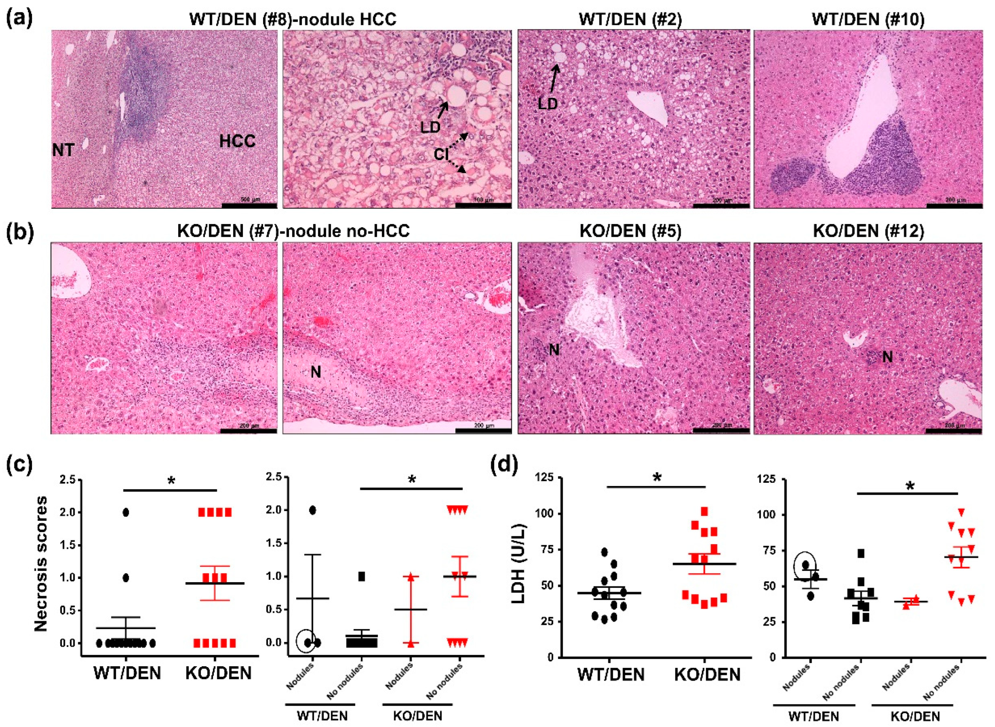
2.1. iPLA2β-Null Mice Show Reduced Body-Weight Gains, No HCC Detectable, and Enhanced Hepatic Necrosis after DEN Treatment

It is known that hepatic pathogenesis induced by DEN is dependent on the sex, age, and the strain of mice, and mice of male gender are recommended for DEN-mediated HCC [19,20]. Compared to other mouse strains, C57BL/6 mice are known to be resistant against HCC [20]; however, in our hands, a single intraperitoneal administration of five three-week-old male C57BL/6 mice with 50 mg/kg DEN was found to be toxic causing death one week later. In our study cohort, we therefore decided to use a much lower DEN dose at 10 mg/kg for treatment of male WT and KO mice at 3–5 weeks of age. Treated mice were harvested for analysis 12 months later. In our previous studies, male KO mice at 12 months old did not show any hepatic abnormalities [21]; therefore, we did not include WT and KO groups without DEN treatment in our cohort.
Due to the very low DEN dose used in our study, only a small size nodule was found in each liver of three from 13 DEN-treated WT (0.24, 0.34, and 0.42 cm diameter) and of two from 12 DEN-treated KO (0.16 and 0.27 cm diameter) mice (Figure 1a). Among these mice, only one mouse, i.e., DEN-treated WT#8 with a 0.34 cm nodule, showed characteristics of HCC cells (Figure 2a). In addition to the very low DEN dose used, mice at 3–5 weeks of age used in our cohort were older than the commonly used, 2 weeks of age [19,20]. We were not able to obtain mice at 2 weeks of age due to a delayed delivery of tail biopsies for genotyping. While 50 mg/kg DEN was toxic to our mice, it was difficult to judge whether 10 mg/kg DEN was insufficient to induce aggressive HCC observed 12 months later. Our animal license also did not include DEN dose-response studies. We therefore decided to perform phenotyping of mice under 10 mg/kg DEN treatment. After sacrifice, only a slight increase in heart weights was found in DEN-treated KO mice (Figure 1b). However, they showed lesser body weight gains in grams (Figure 1c) and % increase from starting body weights (Figure 1d) obtained from total number of mice (left panel) and mice without nodules (right panel). As mice exhibiting HCC are reported to show an increase in body weight gains [22], this parameter was consistently the highest in DEN-treated WT Mouse #8 (the circled value in Figure 1c,d). The observed reduction of this parameter in DEN-treated KO mice, particularly those with nodules, may be indicative of HCC reduction. Despite a few nodules detected in WT and KO mice (Figure 1a), but we were still able to study and compare the abnormalities in their livers by histology, plasma cytokines, and gene expression.
Upon histopathological evaluation of H&E-stained livers of DEN-treated WT mice [23,24], HCC at a grade 3 severity level was observed in the nodule of DEN-treated WT Mouse #8 (Figure 2a). A compression at the border line between HCC and normal tissue (indicated as NT) was observed together with massive lymphoplasmacellular infiltration. At high magnification shown in Figure 2a, macrovesicular lipid droplets (LD indicated by an arrow), cytoplasmic inclusions (CI indicated by dotted arrows), and lymphoplasmacellar infiltration could be observed among morphologically aberrant HCC cells. The nodules of two other DEN-treated WT (Mouse #5 and #13) displayed hyperplastic nodules without any compression characteristics of hepatocellular adenoma or HCC. While the nodule of WT Mouse #5 showed no hepatic abnormalities, that of WT #13 showed some lymphoplasmacellular infiltration (not shown).
Microvesicular steatosis was evident in DEN-treated WT mice with six mice showing grade 1 and four mice showing grade 2 level of steatosis. Grade 2 steatosis with macrovesicular LD was shown for WT Mouse #2 (Figure 2a). With one exception, WT Mouse #10 showed multiple large foci of heteromorphy lymphoid infiltrates beneath some central veins with a suspicion of lymphoma (Figure 2a). Hepatic necrosis at grade 1 and 2 was also observed in two DEN-treated WT mice. No significant abnormalities were detected in the duodenum and spleen of DEN-treated WT mice, while mild lymphoplasmacellular infiltration could be observed in the pancreas of DEN-treated WT with nodules, Mouse #5 and #8 (not shown).
None of DEN-treated KO mice with nodules displayed any compression of adjacent hepatic parenchyma characteristics of HCC or hepatocellular adenoma. Hyperplastic nodules were found in KO Mouse #7 (Figure 2b) and Mouse #10 (Figure 1a). The nodule of KO Mouse #7 displayed a subcapsular area with widespread hepatic necrosis (indicated as N), concomitant with chronic fibrosing active inflammation and microvesicular steatosis grade 1. In addition to KO Mouse #7, seven other DEN-treated KO mice showed hepatic necrosis with grade 1 (shown for KO Mouse #12) and grade 2 (shown for KO Mouse #5) (Figure 2b). By plotting necrosis scores of livers outside the nodules [23], DEN-treated KO mice without nodules showed a significant increase in hepatic necrosis (Figure 2c) and the elevation of plasma LDH (Figure 2d) when compared to DEN-treated WT mice without nodules. Thus, the deficiency may exacerbate DEN-induced liver injury independent of nodule formation. Among mice with nodules, no apparent difference in hepatic necrosis was observed between the genotypes. This indicates no correlation between hepatic necrosis and the formation of DEN-induced nodules observed in WT and KO mice. Overall microvesicular steatosis scores [24] were not different among DEN-treated WT and KO mice (not shown). No abnormalities were found in the pancreas, spleen, and duodenum of DEN-treated KO mice.
Thus, global iPLA2β deficiency led to a reduction of body-weight gains after DEN treatment together with no HCC phenotypes in the nodules. The deficiency however increased hepatic necrosis in mice that did not exhibit nodules. Thus, hepatic necrosis was not correlated with nodular development, but rather related to exacerbation of DEN-induced liver injury in mutant mice without nodules. In addition to reduced body weights, further analyses were needed to phenotype HCC protection in DEN-treated KO mice.

2.2. DEN-Treated iPLA2β-Null Mice Show Attenuated Cell-Cycle and Inflammatory Markers

Even though the majority of DEN-treated WT and KO (10 mice per genotype) did not exhibit any nodules, we continued to perform the analyses of mouse plasma and livers in order to understand KO phenotypes. The analyses of plasma inflammatory cytokines revealed an attenuation of IL-1β levels in DEN-treated KO mice obtained from total number of mice (left panel) and mice without nodules (right panel), while TNF-α, IL-6, IL-10, and IFN-γ levels were not affected (Figure 3a).
Upon analyses of inflammatory genes, DEN-treated KO mice consistently showed attenuated mRNA expression of vascular cell adhesion molecule-1 (VCAM1) obtained from the total number of mice and mice without nodules (Figure 3b). The deficiency attenuated the expression of IL-1β obtained from the total number of mice and IL-10 from the total number of mice and mice without nodules (Figure 3c). IL-1β and IL-10 are recognized as pro-inflammatory mediators of HCC [25], and attenuated IL-10 may reflect reduced hepatocyte regenerative response [26]. Attenuated VCAM-1 expression may lead to the reduction of lymphocyte adhesion to liver endothelium [27] and an increase in apoptosis of inflammatory T cells [28]. Thus, the attenuated levels of IL-1β, IL-10, and VCAM1 were consistent with the lack of HCC observed in DEN-treated KO mice (Figure 1 and Figure 2a,b).
iPLA2β deficiency, however, did not alter mRNA expression of apoptosis markers including apoptosis inducing factor (AIF), caspase3 (Casp3), high-mobility group box1 (HMGB1), BAX, BAD, and TLR3 (Figure 3d) as well as caspase 3/7 activities in liver homogenates (not shown). Remarkably, a significant decrease in mRNA expression of cyclin D2 (CCND2) and B cell lymphoma2 (BCL2) was observed in DEN-treated KO mice obtained from the total number of mice and mice without nodules (Figure 3d). BCL2 is a prognostic indicator in HCC for cell proliferation [29] and epithelial-mesenchymal transition [30]. Suppressed BCL2 is shown to decrease HCC survival by increasing non-apoptotic cell death including ferroptosis [31]. It is thus speculated that such non-apoptotic cell death may be one of the mechanisms for hepatic necrosis and elevation of plasma LDH seen in DEN-treated KO mice without nodules (Figure 2c,d). In line with this notion, genetic or pharmacological inactivation of iPLA2β has been shown to sensitize cells to ferroptosis [32]. As ferroptosis is a form of cell death occurring during therapy of HCC [33], it is thus warranted to further investigate ferroptosis as one of the mechanisms for protection against DEN-induced HCC by hepatocyte-specific iPLA2β deficiency.
CCND2 is one of the G1 cyclins that plays an important role in G1 cell-cycle transition, and their overexpression is implicated in neoplastic transformation [34]. A decrease in hepatic CCND2 expression in DEN-treated KO mice (Figure 3d) implies the presence of a cell-cycle arrest leading to suppressed proliferation of HCC cells. Indeed, suppressed mitosis/proliferation leads to the inability of hepatocytes to regenerate during chemically induced hepatotoxicity [35]. Consistent with the role of iPLA2 on cell growth [13], an inhibition of iPLA2β is shown to induce a cell-cycle arrest in the G1 phase [36], which involves an increase in PLs containing PUFAs [37]. Thus, HCC protection by iPLA2β deficiency could be, in part, due to a cell-cycle arrest through the replenishment of hepatic loss of PUFA-containing PLs.
In Figure 4, we attempted to outline the mechanisms for the reduction of HCC observed in DEN-treated mutants. DEN undergoes metabolism in the liver to produce diazonium ions as alkylating intermediates that react with nucleic acids and proteins to form alkylated macromolecules resulting in a sequence of genetic alterations that transform hepatocytes to HCC cells [18]. By the generation of acetaldehyde and oxidative stress, DEN also initiates hepatocyte injury, inflammation, and necrosis. In response to DEN-induced injury, hepatocytes undergo mitosis for proliferative regeneration, and some selected hepatocytes with genetic alterations may become precursor cells [35,38], including tumor-initiating LGR5 stem cells [39] to form nodules and subsequently HCC.
It is known that PLA2 enzymes participate in mobilization of PUFAs [40] (Figure 4). Cytosolic PLA2 (cPLA2) has specificity to cleave arachidonic acid (AA), which is a substrate of cyclooxygenase 2 (COX2) to generate inflammatory eicosanoids and prostanoids, such as prostaglandin E2 (PGE2). It has been reported that iPLA2β does not couple with COX2 [41]; this decoupling is consistent with the attenuation of hepatic PGE2 in ob/ob mice by iPLA2β deficiency [21]. Similarly, hepatic PGE2 may be attenuated in DEN-treated KO mice, and this attenuation may limit the expansion of LGR5 stem cells leading to HCC protection [42]. iPLA2β preferentially mobilizes ω-3 PUFAs such as docosahexaenoic acid (DHA) [40], which is shown to inhibit HCC by blocking β-catenin and COX2 [43]. DHA is also a precursor of pro-resolution lipids, Resolvins, which are reported to reduce the stemness of HCC [44]. Consistently, ResolvinD2 is shown to be elevated in peritoneal macrophages of KO mice [45]. Furthermore, we recently discovered that hepatocyte-specific iPLA2β deletion led to an increase in hepatic Lipoxin A4 [46], which is shown to suppress HCC via remodeling of tumor microenvironment including LGR5+ compartments [47]. Thus, the proposed elevation of these pro-resolution lipids in DEN-treated KO mice may lead to attenuated inflammation leading to reduced nodular formation and hence HCC protection (Figure 4).
At the cell membrane, iPLA2β deficiency in hepatocytes leads to an accumulation of PLs containing PUFAs [6,7,11], and this would prevent the loss of PLs occurring during preneoplastic progression to HCC (Figure 4). Moreover, these PLs may induce a cell-cycle arrest [36,37], leading to a decrease in regenerative response [35,38]. Alternatively, these membrane PUFA-PLs can be oxidized by 5/15-lipoxygenase to generate corresponding hydroperoxides [PLs(PUFAOOH)], of which in the presence of cellular iron have been identified as mediators of ferroptosis [32]. Increased hepatic ferroptosis/necrosis in DEN-treated KO mice may hamper mitosis and proliferation of preneoplastic cells [35,38].
Regarding the effects of iPLA2β deficiency in macrophages on hepatic inflammation, current data have shown that macrophages or Kupffer cells from KO mice displayed suppressed M1 cytokines, such as, TNF-α and IL-6 [16]. However, Kupffer cells from KO mice with Jo-2-induced liver injury in turn released elevated levels of IL-6 indicating opposing changes towards M1 during injury. For further understanding, mice with macrophage-specific iPLA2β deletion have been generated in our laboratory and initial report has been published in an abstract form [48]. Male macrophage-specific mutants at 12 months of age displayed hepatic necrosis, elevated plasma IL-6, and interstitial infiltration of immune cells in vivo. These results indicate the propensity of iPLA2β-deficient macrophages towards M1 activation perhaps via an induction of ferroptosis in these cells [32]. More relevantly, the promotion of M1-macrophage response is shown to induce protective tumor immunity [49] and works in concert with suicide gene therapy and cisplatin to improve the treatment of HCC [50] and lung cancer [51], respectively. Thus, HCC protection observed in DEN-treated KO mice could be due to the response of the gene deletion specifically in macrophages. Taken together, iPLA2β inactivation in hepatocytes via cell-cycle arrest [36,37] and macrophages via M1 [16,48] in DEN-treated KO mice may contribute to hepatic necrosis as a background. Upon DEN treatment, this background could lead to suppression of mitosis and HCC regeneration resulting HCC protection. In opposition to iPLA2β deficiency in macrophages [16,48], the deficiency in hepatocytes may render protection against hepatic inflammation leading to attenuated preneoplastic regeneration and HCC protection (Figure 4).
Concurrently, unspecific inhibitors of calcium-independent PLA2 have widely been tested in vitro in epithelial breast [52], prostate [53], and intestinal [54] cancer cells. iPLA2β-specific inhibitor, FKGK11, is able to inhibit epithelial ovarian cancer development in xenograft models [55]. Multiple administration of the iPLA2 inhibitor, bromoenol lactone, combined with an anti-cancer drug, paclitaxel, also reduces the number of ovarian tumors [55]. Hence, pan-iPLA2β inhibitors may be therapeutically useful for epithelial cancers including HCC. We speculate that these pan-inhibitors may be less effective, when compared with non-viral HCC, to treat infection/viral-induced HCC due to overt macrophage hyperactivation [16,17,38]. Further experiments are necessary to determine whether FKGK11 as well as hepatocyte- or macrophage-specific deletion of iPLA2β could modulate HCC induced by DEN or DEN combined with high-fat-diet feeding. These results could provide some clues whether pan-iPLA2β inhibitors or hepatocyte-, macrophage-specific iPLA2β inhibitors can represent a class of drugs for therapy of non-viral HCC.

3. Materials and Methods

3.1. Animals and Treatment

KO mice were gifts from Dr. John Turk (Washington University School of Medicine, St. Louis, MO, USA). KO mice were bred and genotyped according to published methods [16,17,18,19,20]. KO mice were bred into C57BL/6 background for at least 20 generations. All mice were bred and kept in the Interfaculty Biomedical Facility of the University of Heidelberg (Im Neuenheimer Feld 347). Male KO (N = 12) and control C57BL/6 (WT, N = 13) mice at 3–5 weeks old were intraperitoneally injected with 10 mg/kg DEN and were killed 12 months later. Body and organ weights were recorded before and after DEN treatment. Blood was collected and liver, spleen, and intestine were fixed in formalin and snap frozen in liquid nitrogen. Studies involving animals have been approved by the University of Heidelberg Institutional Animal Care and Use Committee. This study protocol was reviewed and approved by German Authority in Karlsruhe (Baden-Württemberg Regierungspräsidium Karlsruhe) with approval number G248/11, according to Animal Welfare Laboratory Animal Ordinance (Tierschutz-Versuchstierverordnung, TierSchVersV) from the German Animal Welfare Act (Tierschutzgesetz, TierScG).

3.2. Biochemical Assays

Plasma lactate dehydrogenase (LDH) activities were measured using Randox kits (Krefeld, Germany). Liver homogenates were prepared for determination of caspase 3/7 activity using caspase 3/7Glo kit (Promega, Mannheim, Germany) with a luminometer Lumat LB 9507 (Berthold Technologies, Bad Wildbad, Germany). Caspase 3/7 activity was normalized to mg protein.

3.3. Histology

After overnight fixation in 10% buffered formalin representative specimens of liver, spleen, pancreas, and duodenum were routinely dehydrated, embedded in paraffin, and then cut into 4-μm thick sections. The tissue sections were stained with H&E according to standard protocols. Hepatocellular responses including fatty changes, hepatocellular necrosis, and inflammatory cell infiltration were identified according to published procedures [23,24]. The grading for severity of hepatic necrosis [23] was performed by Drs. Poth and Brobeil using the following scoring system: grade 1 (1–2 foci as minimal), grade 2 (3–6 foci as few), grade 3 (7–12 foci as several), grade 4 (>12 foci as many), and grade 5 (diffuse, severe). Steatosis was scored according to a published protocol by Kleiner et al. [24].

3.4. ELISA

Plasma samples were subjected to determination of cytokines TNF-α, IL-6, IL-10, INF-γ, and IL-1β using ELISA kits from Biolegends (Cologne, Germany).

3.5. Gene Expression

RNA was isolated from liver samples using Gen Elute Miniprep kit from Sigma (Taufkirchen, Germany). cDNAs were prepared from RNA using a Thermo Scientific’s cDNA kit (Karlsruhe, Germany). mRNA expression was analyzed on an Applied Biosystems 7500 using TaqMan® assay-on-demand primers. The expression level was calculated using Δ−Ct transformation method and determined as a ratio of target gene with house-keeping gene GAPDH.

3.6. Statistics

Data was presented as mean ± SEM. By using GraphPad Prism 7 (GraphPad, La Jolla, CA, USA), statistical analysis was performed using the Mann-Whitney U tests with p < 0.05 considered significant or t-tests (p = 0.08) with Welch’s correction with p < 0.1 considered significant (Figure 3).

4. Conclusions

In conclusion, global iPLA2β deficiency in DEN-treated mice limited HCC development with mechanisms involving the induction of cell-cycle arrest and attenuation of inflammatory markers Bcl2, IL-1β, IL-10, and VCAM-1. These results are in line with the reported effects of iPLA2β inactivation in hepatocytes and macrophages. Thus, pan- iPLA2β inhibitors may be effective for treatment of not only epithelial ovarian cancers [15,46] but also HCC.

Author Contributions

Conceptualization, W.C., U.M.; methodology, A.A., T.P. and A.B.; software, A.A., T.P. and W.C.; validation, A.A., T.P. and W.C.; formal analysis, A.A., T.P. and A.B.; investigation, A.A. and T.P.; resources, U.M. and W.C.; data curation, A.A. and T.P.; writing—original draft preparation, A.A.; writing—review and editing, T.P., A.B., U.M. and W.C.; visualization, A.A., T.P. and W.C.; supervision, U.M. and W.C.; project administration, U.M. and W.C.; funding acquisition, W.C. All authors have read and agreed to the published version of the manuscript.

Funding

This work was supported by the Deutsche Forchungsgemeinschaft (CH288/6-2).

Institutional Review Board Statement

Studies involving animals have been approved by the University of Heidelberg Institutional Animal Care and Use Committee. This study protocol was reviewed and approved by German Authority in Karlsruhe (Baden-Württemberg Regierungspräsidium Karlsruhe), approval number G248/11. according to Animal Welfare Laboratory Animal Ordinance from the German Animal Welfare Act.

Informed Consent Statement

Not applicable.

Data Availability Statement

The data that support the findings of this study are openly available online: https//www.wcmat.com/stats/ (accessed on 9 October 2022).

Acknowledgments

We thank John Turk for the iPLA2β-null mice and genotyping method used in this study. We thank Xingya Zhu and Sabine Tuma-Kellner for technical assistance.

Conflicts of Interest

The authors declare no conflict of interest.

References

  1. El-Serag, H.B.; Rudolph, K.L. Hepatocellular carcinoma: Epidemiology and molecular carcinogenesis. Gastroenterology 2007, 132, 2557–2576. [Google Scholar] [CrossRef] [PubMed]
  2. Sun, B.; Karin, M. Inflammation and liver tumorigenesis. Front. Med. 2013, 7, 242–254. [Google Scholar] [CrossRef] [PubMed]
  3. Cotte, A.K.; Vanessa, C.; Aires, V.; Mouillot, T.; Rizk, M.; Vinault, S.; Binquet, C.; Pais de Barros, J.-P.; Hillon, P.; Delmas, D. Phospholipid profiles and hepatocellular carcinoma risk and prognosis in cirrhotic patients. Oncotarget 2019, 10, 2161–2172. [Google Scholar] [CrossRef] [PubMed]
  4. Qiu, J.-F.; Zhang, K.-L.; Zhang, X.-J.; Hu, Y.-J.; Li, P.; Shang, C.-Z.; Wan, J.-B. Abnormalities in plasma phospholipid fatty acid profiles of patients with hepatocellular carcinoma. Lipids 2015, 50, 977–985. [Google Scholar] [CrossRef] [PubMed]
  5. Evangelista, E.B.; Kwee, S.A.; Sato, M.M.; Wang, L.; Rettenmeier, C.; Xie, G.; Jia, W.; Wong, L.L. Phospholipids are a potentially important source of tissue biomarkers for hepatocellular carcinoma: Results of a pilot study involving targeted metabolomics. Diagnostics 2019, 9, 167. [Google Scholar] [CrossRef] [Green Version]
  6. Liu, Z.; Zhang, Z.; Mei, H.; Mao, J.; Zhou, X. Distribution and clinical relevance of phospholipids in hepatocellular carcinoma. Hepatol. Int. 2020, 14, 544–555. [Google Scholar] [CrossRef]
  7. Krautbauer, S.; Meier, E.M.; Rein-Fischboeck, L.; Pohl, R.; Weiss, T.S.; Sigruener, A.; Aslanidis, C.; Liebisch, G.; Buechler, C. Ceramide and polyunsaturated phospholipids are strongly reduced in human hepatocellular carcinoma. Biochim. Biophys. Acta 2016, 1861, 1767–1774. [Google Scholar] [CrossRef]
  8. Hermansson, M.; Hokynar, K.; Somerharju, P. Mechanisms of glycerophospholipid homeostasis in mammalian cells. Prog. Lipid. Res. 2011, 50, 240–257. [Google Scholar] [CrossRef]
  9. Holthuis, J.C.; Menon, A.K. Lipid landscapes and pipelines in membrane homeostasis. Nature 2014, 510, 48–57. [Google Scholar] [CrossRef]
  10. Teng, Y.-W.; Mehedint, M.G.; Garrow, T.A.; Zeisel, S.H. Deletion of betaine-homocysteine S-methyltransferase in mice perturbs choline and 1-carbon metabolism, resulting in fatty liver and hepatocellular carcinomas. J. Biol. Chem. 2011, 286, 36258–36267. [Google Scholar] [CrossRef]
  11. Morita, Y.; Sakaguchi, T.; Ikegami, K.; Goto-Inoue, N.; Hayasaka, T.; Hang, V.T.; Tanaka, H.; Harada, T.; Shibasaki, Y.; Suzuki, A.; et al. Lysophosphatidylcholine acyltransferase 1 altered phospholipid composition and regulated hepatoma progression. J. Hepatol. 2013, 59, 292–299. [Google Scholar] [CrossRef]
  12. Turk, J.; White, T.D.; Nelson, A.J.; Lei, X.; Ramanadham, S. iPLA2β and its role in male fertility, neurological disorders, metabolic disorders, and inflammation. Biochim. Biophys. Acta Mol. Cell Biol. Lipids. 2019, 1864, 846–860. [Google Scholar] [CrossRef]
  13. Hooks, S.B.; Cummings, B.S. Role of Ca2+-independent phospholipase A2 in cell growth and signaling. Biochem. Pharmacol. 2008, 76, 1059–1067. [Google Scholar] [CrossRef] [Green Version]
  14. Li, M.; Li, C.; Liu, W.-X.; Liu, C.; Cui, J.; Li, Q.; Ni, H.; Yang, Y.; Wu, C.; Chen, C.; et al. Dysfunction of PLA2G6 and CYP2C44-associated network signals imminent carcinogenesis from chronic inflammation to hepatocellular carcinoma. J. Mol. Cell Biol. 2017, 9, 489–503. [Google Scholar] [CrossRef] [Green Version]
  15. Li, H.; Zhao, Z.; Wei, G.; Yan, L.; Wang, D.; Zhang, H.; Sandusky, G.E.; Turk, J.; Xu, Y. Group VIA phospholipase A2 in both host and tumor cells is involved in ovarian cancer development. FASEB J. 2010, 24, 4103–4116. [Google Scholar] [CrossRef] [Green Version]
  16. Inhoffen, J.; Tuma-Kellner, S.; Straub, B.; Stremmel, W.; Chamulitrat, W. Deficiency of iPLA₂β primes immune cells for proinflammation: Potential involvement in age-related mesenteric lymph node lymphoma. Cancers 2015, 7, 2427–2442. [Google Scholar] [CrossRef]
  17. Zhu, X.; Gan-Schreier, H.; Otto, A.C.; Cheng, Y.; Staffer, S.; Tuma-Kellner, S.; Ganzha, A.; Liebisch, G.; Chamulitrat, W. iPla2β deficiency in mice fed with MCD diet does not correct the defect of phospholipid remodeling but attenuates hepatocellular injury via an inhibition of lipid uptake genes. Biochim. Biophys. Acta Mol. Cell Biol. Lipids 2019, 1864, 677–687. [Google Scholar] [CrossRef]
  18. George, J.; Tsuchishima, M.; Tsutsumi, M. Molecular mechanisms in the pathogenesis of N-nitrosodimethylamine induced hepatic fibrosis. Cell Death Dis. 2019, 10, 18–27. [Google Scholar] [CrossRef] [Green Version]
  19. Vesselinovitch, S.D.; Mihailovich, N. Kinetics of diethylnitrosamine hepatocarcinogenesis in the infant mouse. Cancer Res. 1983, 43, 4253–4259. [Google Scholar]
  20. Tolba, R.; Kraus, T.; Liedtke, C.; Schwarz, M.; Weiskirchen, R. Diethylnitrosamine (DEN)-induced carcinogenic liver injury in mice. Lab. Anim. 2015, 49 (Suppl. 1), 59–69. [Google Scholar] [CrossRef] [Green Version]
  21. Deng, X.; Wang, J.; Jiao, L.; Utaipan, T.; Tuma-Kellner, S.; Schmitz, G.; Liebisch, G.; Stremmel, W.; Chamulitrat, W. iPLA2β deficiency attenuates obesity and hepatic steatosis in ob/ob mice through hepatic fatty-acyl phospholipid remodeling. Biochim. Biophys. Acta Mol. Cell Biol. Lipids 2016, 1861, 449–461. [Google Scholar] [CrossRef] [PubMed]
  22. Gurses, S.A.; Banskar, S.; Stewart, C.; Trimoski, B.; Dziarski, R.; Gupta, D. Nod2 protects mice from inflammation and obesity-dependent liver cancer. Sci. Rep. 2020, 10, 20519–20536. [Google Scholar] [CrossRef] [PubMed]
  23. Thoolen, B.; Maronpot, R.R.; Harada, T.; Nyska, A.; Rousseaux, C.; Nolte, T.; Malarkey, D.E.; Kaufmann, W.; Küttler, K.; Deschl, U.; et al. Proliferative and nonproliferative lesions of the rat and mouse hepatobiliary system. Toxicol. Pathol. 2010, 38 (Suppl. 7), 5S–81S. [Google Scholar] [CrossRef] [PubMed]
  24. Kleiner, D.E.; Brunt, E.M.; Van Natta, M.; Behling, C.; Contos, M.J.; Cummings, O.W.; Ferrell, L.D.; Liu, Y.-C.; Torbenson, M.S.; Unalp-Arida, A.; et al. Design and validation of a histological scoring system for nonalcoholic fatty liver disease. Hepatology 2005, 41, 1313–1321. [Google Scholar] [CrossRef]
  25. Yang, Y.; Luo, C.; Feng, R.; Bi, S. The TNF-α, IL-1B and IL-10 polymorphisms and risk for hepatocellular carcinoma: A meta-analysis. J. Cancer Res. Clin. Oncol. 2011, 137, 947–952. [Google Scholar] [CrossRef]
  26. Dinant, S.; Veteläinen, R.L.; Florquin, S.; van Vliet, A.K.; van Gulik, T.M. IL-10 attenuates hepatic I/R injury and promotes hepatocyte proliferation. J. Surg. Res. 2007, 141, 176–182. [Google Scholar] [CrossRef]
  27. Wadkin, J.C.R.; Patten, D.A.; Kamarajah, S.K.; Shepherd, E.L.; Novitskaya, V.; Berditchevski, F.; Adams, D.H.; Weston, C.J.; Shetty, S. CD151 supports VCAM-1-mediated lymphocyte adhesion to liver endothelium and is upregulated in chronic liver disease and hepatocellular carcinoma. Am. J. Physiol. Gastrointest. Liver Physiol. 2017, 313, G138–G149. [Google Scholar] [CrossRef] [Green Version]
  28. Afford, S.C.; Humphreys, E.H.; Reid, D.T.; Russell, C.L.; Banz, V.M.; Oo, Y.; Vo, T.; Jenne, C.; Adams, D.H.; Eksteen, B. Vascular cell adhesion molecule 1 expression by biliary epithelium promotes persistence of inflammation by inhibiting effector T-cell apoptosis. Hepatology 2014, 59, 1932–1943. [Google Scholar] [CrossRef]
  29. EI-Emshaty, H.M.; Saad, E.A.; Toson, E.A.; Malak, C.A.; Gadelhak, N.A. Apoptosis and cell proliferation: Correlation with BCL-2 and P53 oncoprotein expression in human hepatocellular carcinoma. Hepatogastroenterology 2014, 61, 1393–1401. [Google Scholar]
  30. Zhao, N.; Sun, B.-C.; Zhao, X.-l.; Liu, Z.-Y.; Sun, T.; Qiu, Z.-Q.; Gu, Q.; Che, N.; Dong, X.-Y. Coexpression of Bcl-2 with epithelial-mesenchymal transition regulators is a prognostic indicator in hepatocellular carcinoma. Med. Oncol. 2012, 29, 2780–2792. [Google Scholar] [CrossRef]
  31. Wang, G.X.; Tu, H.-C.; Dong, Y.; Skanderup, A.J.; Wang, Y.; Takeda, S.; Ganesan, Y.T.; Han, S.; Liu, H.; Hsieh, J.J.; et al. ΔNp63 inhibits oxidative stress-induced cell death, including ferroptosis, and cooperates with the BCL-2 family to promote clonogenic survival. Cell Rep. 2017, 21, 2926–2939. [Google Scholar] [CrossRef] [PubMed] [Green Version]
  32. Sun, W.-Y.; Tyurin, V.A.; Mikulska-Ruminska, K.; Shrivastava, I.H.; Anthonymuthu, T.S.; Zhai, Y.-J.; Pan, M.-H.; Gong, H.-B.; Lu, D.-H.; Sun, J.; et al. Phospholipase iPLA2β averts ferroptosis by eliminating a redox lipid death signal. Nat. Chem. Biol. 2021, 17, 465–476. [Google Scholar] [CrossRef] [PubMed]
  33. Louandre, C.; Ezzoukhry, Z.; Godin, C.; Barbare, J.-C.; Mazière, J.-C.; Chauffert, B.; Galmiche, A. Iron-dependent cell death of hepatocellular carcinoma cells exposed to sorafenib. Int. J. Cancer 2013, 133, 1732–1742. [Google Scholar] [CrossRef] [PubMed]
  34. Takano, Y.; Kato, Y.; van Diest, P.J.; Masuda, M.; Mitomi, H.; Okayasu, I. Cyclin D2 overexpression and lack of p27 correlate positively and cyclin E inversely with a poor prognosis in gastric cancer cases. Am. J. Pathol. 2000, 156, 585–594. [Google Scholar] [CrossRef] [Green Version]
  35. Lee, V.M.; Cameron, R.G.; Archer, M.C. Zonal location of compensatory hepatocyte proliferation following chemically induced hepatotoxicity in rats and humans. Toxicol. Pathol. 1998, 26, 621–627. [Google Scholar] [CrossRef] [PubMed] [Green Version]
  36. Zhang, X.H.; Zhao, C.; Seleznev, K.; Song, K.; Manfredi, J.J.; Ma, Z.A. Disruption of G1-phase phospholipid turnover by inhibition of Ca2+-independent phospholipase A2 induces a p53-dependent cell-cycle arrest in G1 phase. J. Cell Sci. 2006, 119 Pt 6, 1005–1015. [Google Scholar] [CrossRef] [Green Version]
  37. Zhang, X.H.; Zhao, C.; Ma, Z.A. The increase of cell-membranous phosphatidylcholines containing polyunsaturated fatty acid residues induces phosphorylation of p53 through activation of ATR. J. Cell Sci. 2007, 120 Pt 23, 4134–4143. [Google Scholar] [CrossRef] [Green Version]
  38. Sia, D.; Villanueva, A.; Friedman, S.L.; Llovet, J.M. Liver cancer cell of origin, molecular class, and effects on patient prognosis. Gastroenterology 2017, 152, 745–761. [Google Scholar] [CrossRef] [Green Version]
  39. Cao, W.; Li, M.; Liu, J.; Zhang, S.; Noordam, L.; Verstegen, M.M.A.; Wang, L.; Ma, B.; Li, S.; Wang, W.; et al. LGR5 marks targetable tumor-initiating cells in mouse liver cancer. Nat. Commun. 2020, 11, 1961. [Google Scholar] [CrossRef] [Green Version]
  40. Astudillo, A.M.; Balboa, M.A.; Balsinde, J. Selectivity of phospholipid hydrolysis by phospholipase A2 enzymes in activated cells leading to polyunsaturated fatty acid mobilization. Biochim. Biophys. Acta Mol. Cell Biol. Lipids 2019, 1864, 772–783. [Google Scholar] [CrossRef]
  41. Murakami, M.; Kambe, T.; Shimbara, S.; Kudo, I. Functional coupling between various phospholipase A2s and cyclooxygenases in immediate and delayed prostanoid biosynthetic pathways. J. Biol. Chem. 1999, 274, 3103–3115. [Google Scholar] [CrossRef] [Green Version]
  42. Al-Kharusi, M.R.A.; Smartt, H.J.M.; Greenhough, A.; Collard, T.J.; Emery, E.D.; Williams, A.C.; Paraskeva, C. LGR5 promotes survival in human colorectal adenoma cells and is upregulated by PGE2: Implications for targeting adenoma stem cells with NSAIDs. Carcinogenesis 2013, 34, 1150–1157. [Google Scholar] [CrossRef] [PubMed]
  43. Lim, K.; Han, C.; Dai, Y.; Shen, M.; Wu, T. Omega-3 polyunsaturated fatty acids inhibit hepatocellular carcinoma cell growth through blocking beta-catenin and cyclooxygenase-2. Mol. Cancer Ther. 2009, 8, 3046–3055. [Google Scholar] [CrossRef] [PubMed] [Green Version]
  44. Sun, L.; Wang, Y.; Wang, L.; Yao, B.; Chen, T.; Li, Q.; Liu, Z.; Liu, R.; Niu, Y.; Song, T.; et al. Resolvin D1 prevents epithelial-mesenchymal transition and reduces the stemness features of hepatocellular carcinoma by inhibiting paracrine of cancer-associated fibroblast-derived COMP. J. Exp. Clin. Cancer Res. 2019, 38, 170. [Google Scholar] [CrossRef]
  45. Nelson, A.J.; Stephenson, D.J.; Cardona, C.L.; Lei, X.; Almutairi, A.; White, T.D.; Tusing, Y.G.; Park, M.A.; Barbour, S.E.; Chalfant, C.E.; et al. Macrophage polarization is linked to Ca2+-independent phospholipase A2β-derived lipids and cross-cell signaling in mice. J. Lipid Res. 2020, 61, 143–158. [Google Scholar] [CrossRef]
  46. Jansakun, C.; Chunglok, W.; Altamura, S.; Muckenthaler, M.; Staffer, S.; Tuma-Kellner, S.; Merle, U.; Chamulitrat, W. Myeloid- and hepatocyte-specific deletion of group VIA calcium-independent phospholipase A2 leads to dichotomous opposing phenotypes during MCD diet-induced NASH. BBA Mol. Basis Dis. 2022, in press. [Google Scholar] [CrossRef]
  47. Hao, H.; Liu, M.; Wu, P.; Cai, L.; Tang, K.; Yi, P.; Li, Y.; Chen, Y.; Ye, D. Lipoxin A4 and its analog suppress hepatocellular carcinoma via remodeling tumor microenvironment. Cancer Lett. 2011, 309, 85–94. [Google Scholar] [CrossRef]
  48. Klement, L.J.; Jansakun, C.; Tuma-Kellner, S.; Staffer, S.; Altamura, S.; Muckenthaler, M.; Merle, U.; Chamulitrat, W. Macrophage-specific PLA2g6 deficiency exacerbates liver injury during bacterial sepsis via myelopoiesis in male mice. In Proceedings of the German Association of the Study of the Liver, Mannheim, Germany, 28–29 January 2022. [Google Scholar]
  49. Mrad, M.; Imbert, C.; Garcia, V.; Rambow, F.; Therville, N.; Carpentier, S.; Ségui, B.; Levade, T.; Azar, R.; Marine, J.-C.; et al. Downregulation of sphingosine kinase-1 induces protective tumor immunity by promoting M1 macrophage response in melanoma. Oncotarget 2016, 7, 71873–71886. [Google Scholar] [CrossRef] [Green Version]
  50. Tsuchiyama, T.; Nakamoto, Y.; Sakai, Y.; Mukaida, N.; Kaneko, S. Optimal amount of monocyte chemoattractant protein-1 enhances antitumor effects of suicide gene therapy against hepatocellular carcinoma by M1 macrophage activation. Cancer Sci. 2008, 99, 2075–2082. [Google Scholar] [CrossRef] [Green Version]
  51. Yuan, A.; Hsiao, Y.-J.; Chen, H.-Y.; Chen, H.-W.; Ho, C.-C.; Chen, Y.-Y.; Liu, Y.-C.; Hong, T.-H.; Yu, S.-L.; Chen, J.J.W.; et al. Opposite effects of M1 and M2 macrophage subtypes on lung cancer progression. Sci. Rep. 2015, 5, 14273–14285. [Google Scholar] [CrossRef] [Green Version]
  52. Calderon, L.E.; Liu, S.; Arnold, N.; Breakall, B.; Rollins, J.; Ndinguri, M. Bromoenol lactone attenuates nicotine-induced breast cancer cell proliferation and migration. PLoS ONE 2015, 10, e0143277. [Google Scholar] [CrossRef] [PubMed] [Green Version]
  53. Sun, B.; Zhang, X.; Yonz, C.; Cummings, B.S. Inhibition of calcium-independent phospholipase A2 activates p38 MAPK signaling pathways during cytostasis in prostate cancer cells. Biochem. Pharmacol. 2010, 79, 1727–1735. [Google Scholar] [CrossRef]
  54. Liou, J.-Y.; Aleksic, N.; Chen, S.-F.; Han, T.-J.; Shyue, S.-K.; Wu, K.K. Mitochondrial localization of cyclooxygenase-2 and calcium-independent phospholipase A2 in human cancer cells: Implication in apoptosis resistance. Exp. Cell Res. 2005, 306, 75–84. [Google Scholar] [CrossRef]
  55. Li, H.; Zhao, Z.; Antalis, C.; Zhao, Z.; Emerson, R.; Wei, G.; Zhang, S.; Zhang, Z.-Y.; Xu, Y. Combination therapy of an inhibitor of group VIA phospholipase A2 with paclitaxel is highly effective in blocking ovarian cancer development. Am. J. Pathol. 2011, 179, 452–461. [Google Scholar] [CrossRef]
Figure 1. Mice at 3–5 weeks old were treated with a single dose of 10 mg/kg DEN and sacrificed 12 months later. Tissue weights and the diameter of any observable nodules were measured. (a) Left panel shows pictures of a small nodule of DEN-treated WT (Mouse #8, indicated by a black arrow) and KO (Mouse #10, indicated by a white arrow). Right panel shows a plot of nodule diameters of 3 DEN-treated WT mice and 2 of DEN-treated KO mice. (b) Absolute liver, spleen, kidneys, subcutaneous fat, and heart weights. (c) An increase in body weight in grams after one-year DEN treatment obtained from total number of mice (left) and mice with ‘Nodules’ or ‘No nodules’ (right). (d) % increase from starting body weights obtained from total number of mice (left) and mice with ‘Nodules’ or ‘No nodules’ (right). The value with a black circle was from DEN-treated WT Mouse #8. Data are mean ± SEM, N = 12–13 for (bd). * p < 0.05, ** p < 0.01 with Mann-Whitney U tests.
Ijms 23 13760 g001
Figure 2. Mice were treated as described in Figure 1. (a) Histological evaluation of WT Mouse #8 mice treated with DEN showed tumor (HCC) bordering to normal tissue (NT). HCC cells were mixed with lipid droplets (LD indicated by a solid arrow), cytoplasmic inclusions (CI indicated by dotted arrows), and lymphoplasmacellar infiltration. Other abnormalities in DEN-treated WT mice were fatty changes (Mouse #2) and heteromorphous population of lymphocytes and heteromorphous population of lymphocytes suspicious for lymphoma (Mouse #10). (b) Histological evaluation of livers of DEN-treated KO mice showed a compressed necrotic (N) area in hyperplastic nodule of Mouse #7. Focal hepatic necrosis was evident in Mouse #5 and Mouse #12. (c) Necrosis histology scores in livers outside the nodules obtained from total number of mice (left) and mice with ‘Nodules’ or ‘No nodules’ (right). (d) Plasma LDH (U/L) levels obtained from total number of mice (left) and mice with ‘Nodules’ or ‘No nodules’ (right). The value with a black circle was from DEN-treated WT Mouse #8. Data are mean ± SEM; N = 12–13. *, p < 0.05 with Mann-Whitney U tests.
Ijms 23 13760 g002
Figure 3. Mice were treated as described in Figure 1. Data in the left-hand panel were obtained from the total number of mice, and those in right-hand panel(s) were obtained from ‘Nodule’ and ‘No-nodule’ groups. (a) Plasma levels (pg/mL) of cytokines TNF-α, IL-6, IL-10, IFN-γ, and IL-1β. (b) Hepatic mRNA expression of genes related to chemokines (MCP-1, VCAM1, ICAM1, CCR2, CXCL12, and CCR5). (c) Hepatic mRNA expression of inflammatory genes (TNF-α, IL-6, IL-1β, IL-10, and NF-kB). (d) Hepatic mRNA expression of genes related to apoptosis (inducing factor (AIF), caspase3 [Casp3], high-mobility group box1 [HMGB1], BAX, BAD, and TLR3) as well as proliferation and anti-apoptosis (cyclin D2 [CCND2], cFLIP, and BCL2). The value with a black circle was from DEN-treated WT Mouse #8. Data are mean ± SEM; N = 12–13. * p < 0.05 with Mann-Whitney U tests for total number of mice, and t-tests (p = 0.08) with Welch’s correction for mice with or without nodules (p < 0.1 considered significant).
Ijms 23 13760 g003
Figure 4. iPLA2β deficiency in hepatocytes results in an increase in hepatic PUFA-containing PLs leading to an induction of a cell-cycle arrest, suppression of HCC regenerative response, and induction of hepatic necrosis and ferroptosis likely via lipoxygenase-induced oxidation of PLs [PLs(PUFAOOH)]. Similarly, iPLA2β deficiency in macrophages may lead to ferroptosis. As a background, ferroptosis may lead to hepatic necrosis which may limit mitosis and liver cell regeneration. PLs(PUFA) are also precursors of pro-resolution lipoxin A4 and resolvinD2 leading to an attenuated inflammatory response resulting in reduced liver cell regeneration, reduced nodular formation, and HCC protection.
Ijms 23 13760 g004
Publisher’s Note: MDPI stays neutral with regard to jurisdictional claims in published maps and institutional affiliations.

