Article

A Tripartite Efflux System Affects Flagellum Stability in Helicobacter pylori

1
Department of Microbiology, University of Georgia, Athens, GA 30602, USA
2
Microbial Sciences Institute, Yale University, West Haven, CT 06516, USA
3
Department of Microbial Pathogenesis, Yale School of Medicine, New Haven, CT 06536, USA
4
Institute of Bioinformatics, University of Georgia, Athens, GA 30602, USA
*
Author to whom correspondence should be addressed.
These authors contributed equally to this work.
Int. J. Mol. Sci. 2022, 23(19), 11609; https://doi.org/10.3390/ijms231911609
Submission received: 5 September 2022 / Revised: 23 September 2022 / Accepted: 29 September 2022 / Published: 1 October 2022
(This article belongs to the Special Issue Flagella)

Abstract

:
Helicobacter pylori uses a cluster of polar, sheathed flagella for swimming motility. A search for homologs of H. pylori proteins that were conserved in Helicobacter species that possess flagellar sheaths but were underrepresented in Helicobacter species with unsheathed flagella identified several candidate proteins. Four of the identified proteins are predicted to form part of a tripartite efflux system that includes two transmembrane domains of an ABC transporter (HP1487 and HP1486), a periplasmic membrane fusion protein (HP1488), and a TolC-like outer membrane efflux protein (HP1489). Deleting hp1486/hp1487 and hp1489 homologs in H. pylori B128 resulted in reductions in motility and the number of flagella per cell. Cryo-electron tomography studies of intact motors of the Δhp1489 and Δhp1486/hp1487 mutants revealed many of the cells contained a potential flagellum disassembly product consisting of decorated L and P rings, which has been reported in other bacteria. Aberrant motors lacking specific components, including a cage-like structure that surrounds the motor, were also observed in the Δhp1489 mutant. These findings suggest a role for the H. pylori HP1486-HP1489 tripartite efflux system in flagellum stability. Three independent variants of the Δhp1486/hp1487 mutant with enhanced motility were isolated. All three motile variants had the same frameshift mutation in fliL, suggesting a role for FliL in flagellum disassembly.

1. Introduction

Helicobacter pylori, a member of the phylum Campylobacterota, colonizes the human gastric mucosa where it can cause a variety of diseases, including chronic gastritis, peptic and duodenal ulcers, B cell MALT lymphoma and gastric adenocarcinoma [1]. H. pylori possesses a cluster of polar, sheathed flagella that it uses to migrate through the viscous mucus layer covering the stomach epithelium. Flagellar-mediated motility is required for host colonization, as non-motile H. pylori mutants are unable to colonize animal models [2,3].
The bacterial flagellum consists of three distinct parts referred to as the basal body, hook, and filament. The basal body includes a rotary motor and the flagellar protein export apparatus that transports axial components of the flagellum (e.g., rod, hook, and filament proteins) across the inner membrane [4,5]. Components of the archetypical flagellar motor of Escherichia coli and Salmonella enterica include the MS ring, C ring, rod, L ring, P ring, and a stator unit comprised of a complex of the MotA and MotB proteins. The stator unit is the torque generator for the motor and is powered by the proton motive force [6]. The MS and C rings form the rotor and transmit torque through the rod and hook to the filament, which acts as a propeller to push the cell forward [7]. The H. pylori flagellar motor accommodates up to 18 stators [8], making it the largest bacterial flagellar motor described to date. In addition to the conserved core features of flagellar motors, the H. pylori motor contains several accessories not found in the archetypical E. coli/S. enterica motor [8,9,10].
Most Helicobacter species, including H. pylori, possess a membranous sheath surrounding the flagellar filament that is continuous with the outer membrane [8]. Despite the continuity of the sheath and outer membrane, protein compositions of these structures may vary somewhat [11,12,13,14,15]. Proposed roles for the H. pylori flagellar sheath include protecting the filament subunits from dissociation by gastric acid and adherence to host cells [16]. In Vibrio cholerae, the flagellar sheath has been suggested to hide flagellin from Toll-like receptor 5 and thereby prevent triggering of the host innate immune system [17]. Although H. pylori flagellins lack the conserved region that is recognized by Toll-like receptor 5 [18,19], the flagellins of H. pylori and its close relative Campylobacter jejuni are glycosylated with pseudaminic acids, which modulate the host’s immune response in C. jejuni infections [20]. Another proposed function of the flagellar sheath is the generation of outer membrane vesicles (OMVs). Rotation of the sheathed flagellum in Vibrio species releases OMVs that contain the potent immune response stimulant lipopolysaccharide (LPS) [21,22]. In the symbiosis between Vibrio fischeri and the Hawaiian bobtail squid, Euprymna scolopes, LPS associated with the OMVs induces apoptotic cell death in the epithelium of the squid light organ, which is required for normal development of the organ [22]. In addition to communicating with the host innate immune system, OMVs can bind bacteriophage, which appears to afford some protection against phage for bacteria [23,24].
Our understanding of flagellar sheath biosynthesis for any bacterial species is limited [16]. To address this gap in our knowledge, we compared the proteins encoded in the genomes of Helicobacter species that have flagellar sheaths (FS+) against Helicobacter species that have unsheathed flagella (FS) to identify proteins with possible roles in sheath biosynthesis or function. Homologs of nearly 40 proteins were found significantly more frequently in the FS+ Helicobacter species compared to FS Helicobacter species. Four of these proteins, HP1486, HP1487, HP1488 and HP1489, were found exclusively in FS+ Helicobacter species and appear to be part of a tripartite efflux system. HP1486 and HP1487 are predicted transmembrane domains of an ATP-binding cassette (ABC) transporter, HP1488 is a periplasmic membrane fusion protein (MFP), and HP1489 is an outer membrane efflux protein (OEP). Tripartite efflux systems transport a variety of substrates directly from the cytoplasm to the external medium, including proteins, oligosaccharides, antibiotics, metals, large cations, and various xenobiotics [25,26]. Deleting hp1486/hp1487 and hp1489 homologs in H. pylori B128 reduced motility and the number of flagella per cell. Imaging the motors of the Δhp1489 and Δhp1486/hp1487 mutants by cryo-electron tomography (cryo-ET) revealed many of the cells contained flagellum substructures consisting of only the L ring, P ring, basal disk, and outer disk, resembling flagellar disassembly products (referred to as PL-subcomplexes) reported for other bacteria [27,28]. In addition to the PL-subcomplex, the Δhp1489 mutant contained aberrant motors that lacked specific components, which may be flagellum disassembly intermediates. Variants of the Δhp1486/hp1487 mutant with enhanced motility were isolated, and each of the variants had the same frameshift mutation in fliL. The C-terminal domain of FliL forms a ring that encircles the MotB dimer within the stator unit of the H. pylori motor [29]. The frameshift mutation occurred near the beginning of the fliL coding region, which was surprising since fliL is required for motility in H. pylori [29]. We hypothesize the mutation in fliL results in the expression of a truncated FliL that suppresses the motility defect in the Δhp1487/hp1486 mutant. Taken together, our observations suggest roles for the HP1489-HP1486 tripartite efflux system and FliL in the stability and disassembly of the H. pylori flagellar motor.

2. Results

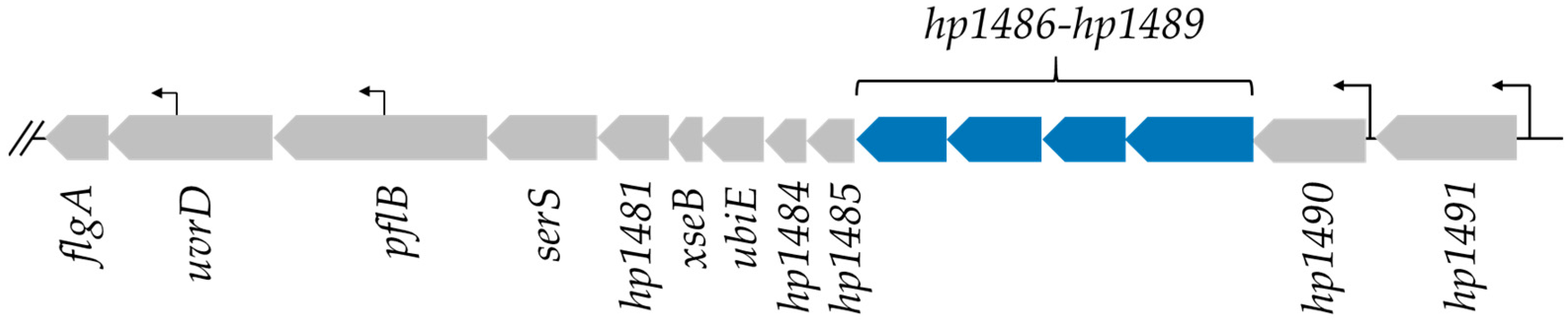
2.1. Identification of H. pylori Proteins That Are Found Preferentially in Helicobacter Species with Flagellar Sheaths

Most Helicobacter species identified to date possess a flagellar sheath, but several Helicobacter species have unsheathed flagella. To identify proteins with possible roles in sheath biosynthesis or function, we searched for homologs of H. pylori proteins that were prevalent in FS+ Helicobacter species but underrepresented in FS Helicobacter species. Using blastp, predicted proteins encoded in the genomes of 35 FS+ Helicobacter species and 9 FS Helicobacter species were examined for homologs of the 1458 predicted proteins encoded in the H. pylori G27 genome. The H. pylori G27 genome was chosen as a reference genome for the analysis since G27 is a model for studies of motility and flagellum biosynthesis in H. pylori. Helicobacter species and strains analyzed are listed in Table S1. The blastp comparison was performed with an E-value cutoff of 10−20. Under the conditions of the null hypothesis (that any protein is equally likely to have a homolog in the FS+ species as in the FS species), the number of homologs in FS+ species is expected to follow the hypergeometric distribution. We therefore used the hypergeometric test (equivalent to one-tail Fisher’s exact test) to obtain a p-value for each protein. False discovery rate (FDR) [30] and a more conservative Bonferroni correction were used for multiple testing correction. Results from the blastp analysis for all 1458 H. pylori G27 proteins are presented in Tables S2 and S3. Forty-two proteins that satisfy the Bonferroni-adjusted cutoff for statistical significance (p-value < 3.4 × 10⁻⁵) in FS+ Helicobacter species are listed in Table 1. An additional 35 proteins qualified as significantly overrepresented in FS+ Helicobacter species when the less conservative FDR correction was applied (Table S2).

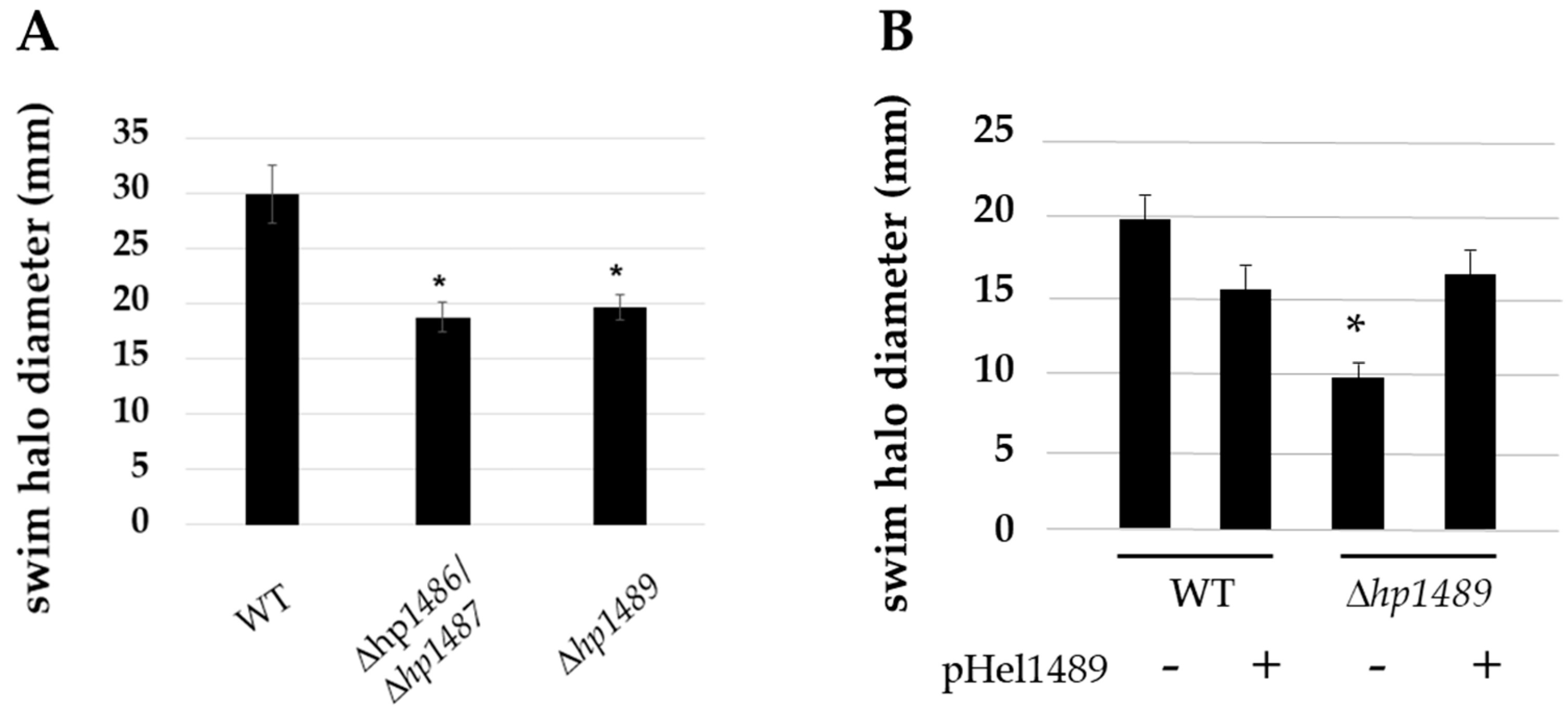
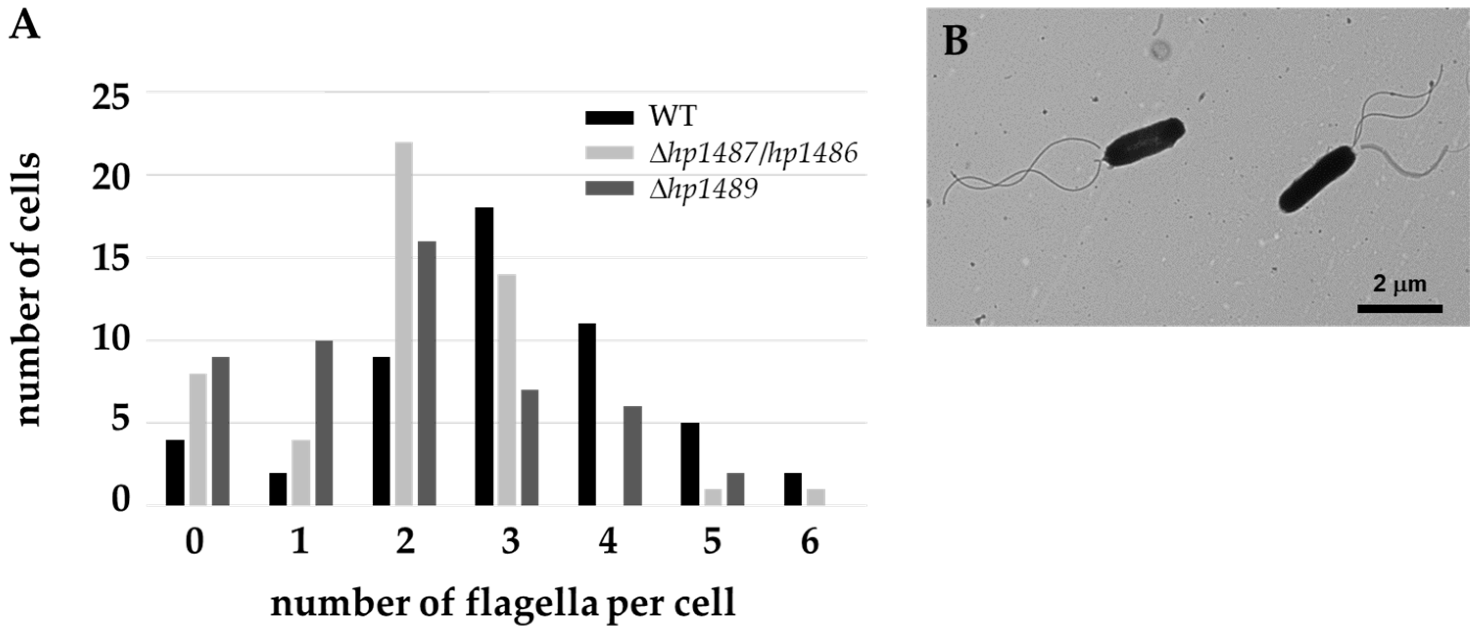
2.2. HP1486, HP1487 and HP1489 Are Required for Robust Motility of H. pylori

To test the predictions from the comparative genomics approach, we targeted some of the identified genes for deletion analysis. Homologs of four proteins, HP1486, HP1487, HP1488 and HP1489, were present in all the FS+ Helicobacter species and absent in all the FS Helicobacter species (Table S2), which made the genes encoding these genes good candidates for further investigation. Based on the functional annotations of the proteins and the linkage of the genes encoding them within an operon (Figure 1), these proteins are predicted to form a tripartite efflux system. Absent in the hp1489-hp1486 locus, however, is a gene encoding the ATPase for the ABC transporter, which presumably is present at a different locus in the genome. The H. pylori 26695 and G27 genomes encode 19 and 18 predicted proteins that contain an ATP-binding domain of an ABC transporter (pfam00005), respectively. Most of the H. pylori genes encoding ATP-binding domain proteins are associated with genes encoding other ABC transporter genes or encode an ABC transporter transmembrane domain, suggesting these genes do not encode the ATPase that functions with HP1486/HP1487 transmembrane domain proteins. A blastp analysis of the sequences of all the ATP-binding domain proteins from H. pylori 26695 versus the genomes of FS+ and FS Helicobacter species failed to identify a clear candidate for the ATPase of the HP1486/HP1487 ABC transporter that was supported by the protein functional annotation or yielded orthologous homologs.
To determine if the HP1489-HP1486 efflux system has a role in flagellar biosynthesis or function in H. pylori, we deleted hp1489 and hp1487-hp1486 in H. pylori B128, and also deleted hp1489 in H. pylori G27, using a sucrose-based counter-selection method [47]. Motilities of all three mutants in soft agar medium were reduced 33 to 50% compared to the wild-type parental strains (Figure 2). Introducing a copy of hp1489 into the H. pylori G27 Δhp1489 mutant on a shuttle vector rescued motility (Figure 2B), indicating loss of hp1489 was responsible for the motility defect in the mutant. Examination of the H. pylori B128 ∆hp1487-hp1486 and Δhp1489 mutants by transmission electron microscopy (TEM) revealed the strains possessed fewer flagella per cell than wild type (Figure 3). Flagella of the Δhp1487-hp1486 and Δhp1489 mutants were sheathed, indicate sheath biosynthesis was not grossly impaired in the mutants. Taken together, these observations suggest the HP1489-HP1486 efflux system is required for normal flagellum biosynthesis and/or motility.

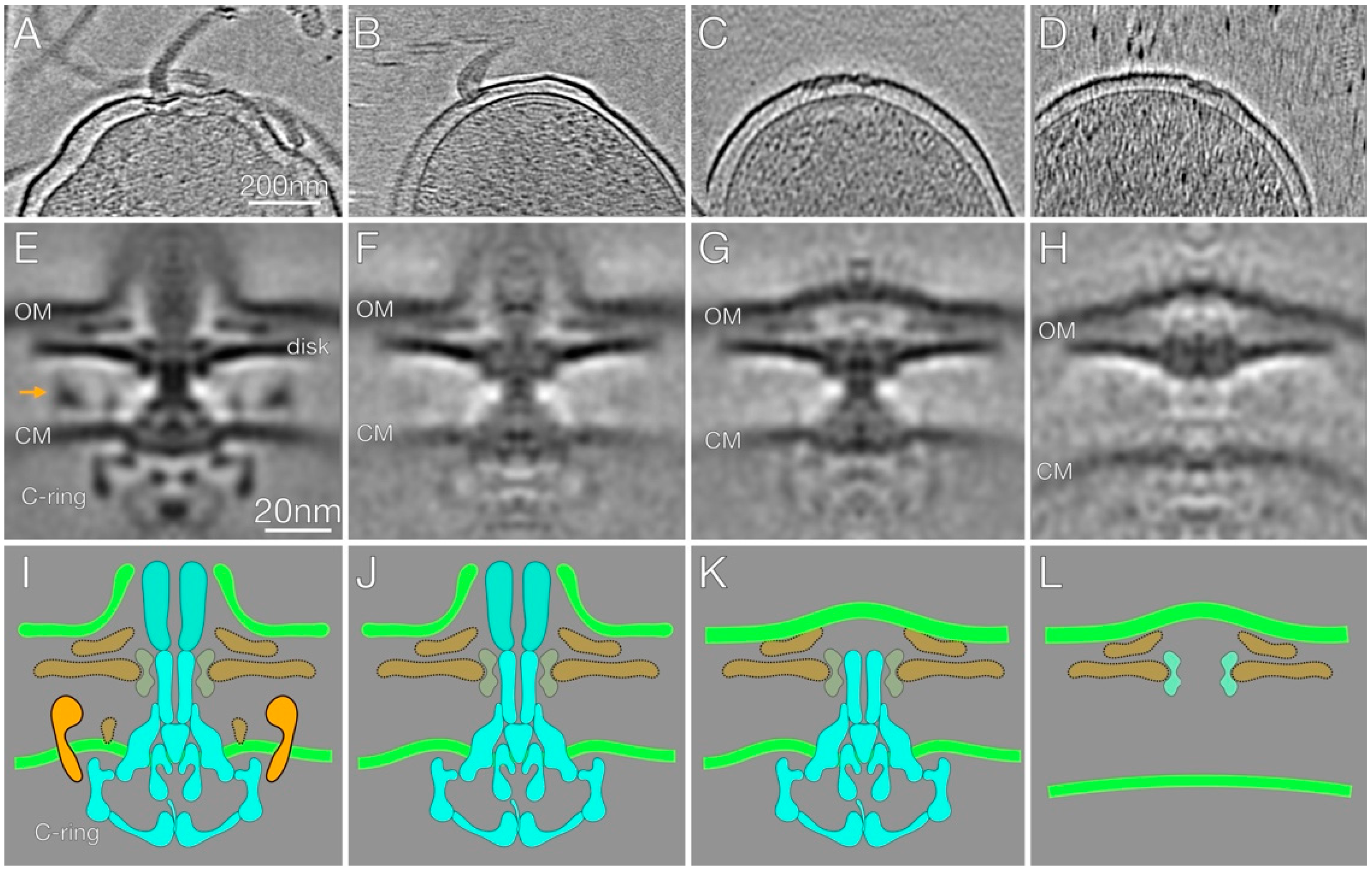
2.3. In-Situ Structures of Flagellar Motors in the Δhp1489 and Δhp1487/hp1486 Mutants Reveal Apparent Flagellum Disassembly Products

The impaired motility and reduced number of flagella in the Δhp1489 and Δhp1487/hp1486 mutants suggested a role for the HP1489-HP1486 efflux system in the assembly or stability of the flagellum. To address this hypothesis, we examined the structure of the flagellar motors of the H. pylori B128 Δhp1489 and Δhp1487/hp1486 mutants by cryo-ET. For the analaysis, we selected 77 subtomograms of motors from 48 reconstructions of Δhp1489 mutant cells and 137 subtomograms of motors from 70 reconstructions of Δhp1487/hp1486 mutant cells, which we analyzed by subtomogram averaging and classification. The resulting motor structures are presented in Figure 4.
Many of the motors of the Δhp1489 and Δhp1487/hp1486 mutants were normal in appearance and attached to a flagellar filament (Figure 4A). These flagella were presumably functional and responsible for the motility observed with the mutants. Several of the motors of the mutants, however, appeared to be incomplete, and often lacked an associated filament (Figure 4B–D). In contrast, such incomplete motors were rarely observed for the H. pylori B128 wild-type strain. To gain a better understanding of the aberrant motors of the Δhp1489 and Δhp1487/hp1486 mutants, we used a multivariate statistical analysis to resolve distinct motor structures that corresponded to the full motor and three specific class averages for the aberrant motors (Figure 4E–H). The number of subtomograms for each class of motor are indicated in Table 2.
In the class 1 motor or full motor, associated hook and flagellar sheath structures were observed (Figure 4A), and the motor contained the core and accessories features reported in previous cryo-ET reconstructions of the intact H. pylori motor (Figure 4E,I) [8]. Similar to the complete motor, the class 2 motor had the associated hook and sheath stuctures (Figure 4B,F,J). In addition, core components of the motor (MS ring, C ring, LP ring, rod and flagellar protein export apparatus), the basal disk, and the outer disk were evident in this class of motor (Figure 4F,J). The cage and other motor components normally found near the inner membrane though were absent in this class of motor (Figure 4F,J).
The class 3 motor lacked the associated hook, filament and flagellar sheath, although the outer membrane above the rod was deformed slightly with a convex bulge (Figure 4C,G,K). Core components of the motor, as well as the basal disk and the outer disk, were visible in the second class of motor, but the cage and motor components normally found near the inner membrane were missing (Figure 4G,K).
The class 4 motor also lacked the associated hook, filament and flagellar sheath (Figure 4D). In contrast to the other classes of motors, the class 4 motor lacked most of the core components of the motor and the only visible structures were the LP ring, the basal disk and the outer disk (Figure 4H,L). As with the class 3 motor, the outer membrane above the rod was deformed slightly with a convex bulge (Figure 4H). The flagellar substructure resembled the outer-membrane-associated relic complexes that result from flagellum disassembly and have been observed in a variety of bacterial species [27,28]. These flagellum disassembly products are referred to as PL-subcomplexes, and so we will refer to this class of motor substructure as the PL-subcomplex.
In bacterial species that lack a flagellar sheath, the PL-subcomplex is associated with a crater-like structure in the outer membrane that is sealed at the bottom by the P ring, L ring proteins or some other undefined molecule [27,28]. The H. pylori PL-subcomplex is not associated with a crater-like structure in the outer membrane, but instead the outer membrane over the PL-subcomplex is convex (Figure 4H) and resembles the PL-subcomplexes observed in Vibrio species [28], which like H. pylori have sheathed flagella.

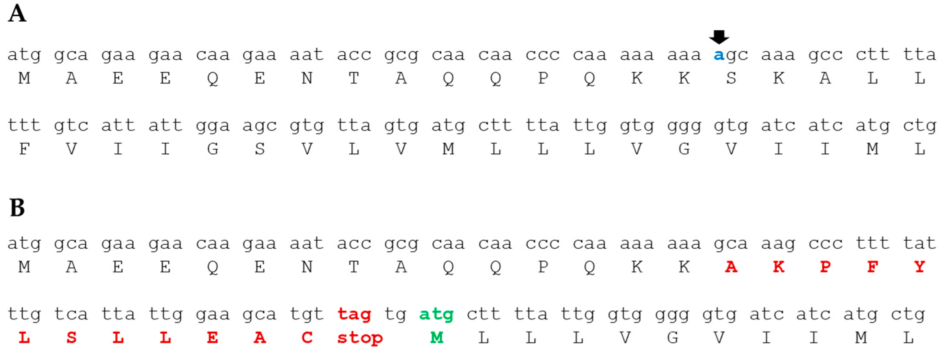
2.4. Isolation of Mutants That Suppress the Motility Defect in the Δhp1487/hp1486 Mutant

To identify mutations that suppress the motility defect associated with loss of the HP1489-HP1486 efflux system, we enriched for variants of the H. pylori B128 ∆hp1487/hp1486 mutant that had enhanced motility in motility agar medium. Three independent motile variants were isolated (designated ESM_MV1 through ESM_MV3) that had motilities in soft agar medium that approximated that of wild type H. pylori B128.
Whole genome sequencing of the Δhp1487/hp1486 motile variants and the Δhp1487/hp1486 parental strain identified several phase variations resulting from the deletions or insertions in homopolymeric runs, as well as single nucleotide polymorphisms (SNPs) present in the motile variants that were not in the parental strain (Table 3). All three Δhp1487/hp1486 motile variants contained the identical frameshift mutation in fliL (Table 3). While some of the alleles appeared in more than one motile variant (e.g., a SNP in murJ), the mutation in fliL was the only allele that occurred in all three motile variants, and moreover, was the only gene that carried a mutation in all three motile variants (Table 3). The genome sequencing data suggested strongly that the mutation in fliL was responsible for the enhance mobility of the Δhp1487/hp1486 motile variants in soft agar medium.
The mutation in fliL in the Δhp1487/hp1486 motile variants was unexpected since fliL is reported to be required for motility in H. pylori [29]. The fliL mutation was a deletion of an A in a homopolymeric run of 9 A residues near the start of the open reading frame of the gene (Figure 5). The frameshift mutation, which was in codon 16, altered the amino acid sequence at the point of the mutation and resulted in a stop codon shortly after the deletion. A couple of nucleotides downstream of the introduced stop codon, however, is a potential start codon in the correct reading frame that would allow for the expression of a truncated FliL protein (Figure 5). Moreover, the ATCATG motif upstream of this potential start codon is a reasonable match for a Shine-Dalgarno sequence and is appropriately positioned relative to the start codon. H. pylori B128 FliL is 183 amino acids in length, and the truncated protein resulting from the potential start codon indicated in Figure 5B is 154 amino acid residues in length.

3. Discussion

3.1. The HP1489-HP1486 Tripartite Efflux System Has an Apparent Role in Stabilizing the H. pylori Flagellar Motor

Using a comparative genomics approach, we found homologs of the HP1489-HP1486 efflux system components were present in all 35 FS+ Helicobacter species but absent in the 9 FS Helicobacter species that we examined. Deletion of hp1489 or hp1487/hp1486 in H. pylori resulted in reduced motility as assessed by the ability of the mutant strains to migrate from the point of inoculation in soft agar medium (Figure 2). In the case of the H. pylori G27 Δhp1489 mutant, motility was restored by introducing a copy of hp1489 into the mutant on the shuttle vector pHel3 (Figure 2), providing strong evidence that the loss of HP1489 was responsible for the motility defect in the original Δhp1489 mutant.
Classification of the motor structures from the Δhp1489 and Δhp1487/hp1486 mutants revealed many of them were missing the cage and other components associated with the inner membrane (Figure 4F,G). At least three distinct classes of aberrant motors were apparent, one of which resembled the PL-subcomplex observed from flagellum disassembly in various bacterial species [27,28]. The relationship between the H. pylori PL-subcomplex and the other two classes of aberrant motors is unclear. The different classes of motors may represent intermediates in the flagellum disassembly pathway. While it is possible the aberrant motors are assembly intermediates, this seems less likely. Qin and co-workers examined the ultrastructure of flagellum assembly intermediates in H. pylori using cryo-ET to visualize over 300 flagellar motors. The earliest flagellum assembly intermediate Qin and co-workers observed had progressed through rod assembly, and this intermediate contained the cage and the other motor components present in the mature H. pylori flagellum [8]. This argues against the aberrant motors in the Δhp1489 mutant being assembly intermediates since they lack the cage.
Two significant questions concerning the HP1489-HP1486 efflux system are: (i) what is the substrate transported by the efflux system; and (ii) how does the efflux system stabilize the flagellum? Regarding the first question, tripartite efflux systems transport a diverse range of proteins, xenobiotics, antibiotics, metal ions and other small molecules [25,26]. While the substrate for the HP1489-HP1486 efflux system is not known, van Amsterdam and co-workers reported that disrupting hp1489 in H. pylori 1061 resulted in increased sensitivity to ethidium bromide [48]. It is not clear from the results of van Amsterdam and co-workers whether the HP1489-HP1486 efflux system transports ethidium bromide, or if loss of the efflux system increases the sensitivity of the hp1489 mutant to ethidium bromide by altering the permeability of the outer membrane. Regarding the second question, the HP1489-HP1486 efflux system may stabilize the flagellum by transporting a molecule that would otherwise accumulate in the cytoplasm of inner membrane and destabilize the flagellum. Alternatively, the transported molecule may be targeted to the outer membrane where it is needed to stabilize the flagellum. It is possible the structure of the HP1489-HP1486 efflux system itself has a role in stabilizing the flagellum. For example, the efflux system may interact with components of the flagellar motor to stabilize the flagellum. Identifying the ATPase that functions with HP1487 and HP1486 will likely prove helpful for examining the validity of the above models for how the efflux system stabilizes the flagellum. The HP1489-HP1486 homologs in Helicobacter anseris MIT 04-9362 are encoded in an operon that contains a gene encoding a predicted ATPase for an ABC transport system. HP0853 is an orphan ATPase (i.e., the gene encoding the protein is not associated with genes encoding transmembrane components of an ABC transport system) and an ortholog of the H. anseris ATPase. We are currently investigating whether HP0853 is the ATPase that functions with HP1487 and HP1486.
The occurrence of the identical frameshift mutation in fliL in all three Δhp1487/hp1486 motile variants strongly suggests the fliL mutation is responsible for suppressing the motility defect in the parental Δhp1487/hp1486 mutant (Table 2). This result was unexpected since fliL was reported to be required for motility in H. pylori [29]. As indicated in Figure 5, the frameshift mutation in fliL is predicted to result in the expression of a short polypeptide corresponding to the first 16 amino acids of the FliL N-terminus plus an additional 12 amino acid residues. The frameshift mutation could also result in the expression of an N-terminal truncation of FliL that is missing the first 29 amino acid residues (Figure 5). It is possible that both truncated FliL proteins are expressed and needed to suppress the motility defect of the Δhp1487/hp1486 mutant. We are currently working to determine if one or both of the truncated FliL proteins are required for suppressing the motility defect of the Δhp1487/hp1486 mutant. We hypothesize that the mutant fliL allele suppresses the motility defect in the Δhp1487/hp1486 mutant by preventing or limiting flagellum disassembly, and studies are planned to examine the validity of this hypothesis. If our hypothesis tests valid, this would implicate FliL in playing a role in flagellum disassembly in H. pylori and perhaps other bacteria. Such a finding would be very exciting as little is known regarding the proteins and factors that control flagellum disassembly.

3.2. Identification of Genes Potentially Involved in Sheath Formation

Although many bacteria possess flagellar sheaths, little is known about the proteins and other factors that contribute to sheath biosynthesis in any bacterial species. To address this gap in our knowledge, we searched for H. pylori proteins that are conserved in FS+ Helicobacter species but are absent or underrepresented in FS Helicobacter species with the idea that some of these proteins have roles in sheath biosynthesis or function. A caveat of the approach is the FS+ and FS Helicobacter species segregate into distinct phylogenic groups [49], which means some of the proteins identified as specifically occurring in FS+ Helicobacter species may be shared among the species of that group even though they have roles unrelated to the sheath, flagellum or motility.
Despite these limitations, the comparative genomics approach enabled the identification of candidates for proteins that have roles in sheath biosynthesis. One such candidate is the cardiolipin synthase ClsC [43]. Cardiolipin is a phospholipid that accumulates at the cell pole and septal regions in rod-shaped bacteria due to its ability to form microdomains that exhibit a high intrinsic curvature and thus have a lower energy upon localization to negatively curved regions of the membrane [50,51]. Given that the flagellar sheath is a long narrow tube with a high degree of negative curvature, it is reasonable to postulate that cardiolipin accumulates in the sheath. Consistent with this hypothesis, preparations of sheathed flagella from H. pylori G27 contain significant amounts of cardiolipin [52]. Thus, FS+ Helicobacter species may need ClsC to satisfy an increased demand for cardiolipin in sheath biosynthesis.
Some of the proteins found preferentially in FS+ Helicobacter species may have roles in mitigating the action of the sheathed flagellum. Rotation of the sheathed flagellum in Vibrio species is a major source of OMVs [21,22], and the generation of these OMVs is important in communication between V. fischeri and its host and symbiotic partner E. scolopes [22]. In contrast, H. pylori has presumably evolved to avoid surveillance by the host innate immune system. Several of the proteins found preferentially in FS+ Helicobacter species enzymes involved in LPS biosynthesis or modification (Table 1), which contribute to the low endotoxin and immunological activity of H. pylori LPS [53,54,55]. These LPS modification enzymes may be a strategy that FS+ Helicobacter species use to avoid activating the host innate immune response by OMVs released from the bacterium during the rotation of the sheathed flagellum.
Several of the proteins conserved in FS+ Helicobacter species belong to the SLR family (Table 1), members of which contain multiple copies of a degenerate amino acid repeat motif that is characteristic of eukaryotic Sel1 proteins [56]. Although the physiological roles of the H. pylori SLR proteins are unclear, some of them bind β-lactam compounds [37,38] and have been suggested to interact with immunomodulatory peptidoglycan fragments to affect the innate immune response [57]. Rotation of the H. pylori sheathed flagellum may result in the release of peptidoglycan fragments from the periplasmic space, and FS+ Helicobacter species may use the peptidoglycan-binding SLR proteins to avoid eliciting a host immune response.

4. Materials and Methods

4.1. Bacterial Strains and Growth Conditions

Escherichia coli DH5α was used for cloning and plasmid construction, and was grown in LB broth or agar medium. Medium was supplemented with ampicillin (100 μg/mL), chloramphenicol (30 μg/mL) or kanamycin (30 μg/mL) when appropriate. H. pylori strains were grown at 37 °C on tryptic soy agar (TSA) supplemented with 5% heat-inactivated horse serum (TSA-HS) under an atmosphere consisting of 10% CO2, 4% O2, and 86% N2. For liquid cultures, H. pylori strains were grown at 37 °C with shaking in Mueller-Hinton broth (Becton, Dickinson and Company, Sparks, MD, USA) (MHB) supplemented with 5% heat-inactivated horse serum (Gibco, Penrose, Aukland, New Zealand) (MHB-HS) under an atmosphere consisting of 5% CO2, 10% H2, 10% O2 and 75% N2. Kanamycin (30 μg/mL) or chloramphenicol (30 μg/mL) was added to the medium used to culture H. pylori when appropriate.

4.2. Strain Construction

Primers used for PCR are listed in Table S4 and strains used in this study are listed in Table 4. Genomic DNA (gDNA) from H. pylori G27 was purified using the Wizard genomic DNA purification kit (Promega, Madison, WI, USA) and used as the template to construct all deletion mutants. Target sequences from gDNA were amplified using Phusion polymerase (New England Biolabs, Ipswich, MA, USA). To facilitate cloning into the pGEM-T Easy vector (Promega) amplicons were incubated with Taq polymerase (Promega) at 72 °C for 10 min to add 3′-A overhangs.
H. pylori B128 hpb128_199g42 (hp1489 homolog) and hpb128_199g40 along with hpb128_199g39 (hp1487 and hp1486 homologs) were deleted as follows. Regions flanking the target genes were amplified from gDNA using primer pairs P117/P118 plus P119/P120 for hp1489, and P177/P178 plus P179/P180 for hp1487/hp1486. The primer pairs introduced complementary sequence to the ends of the amplicons that were used for overlapping PCR. The complementary sequences also included NheI and XhoI restriction sites for a subsequent cloning step. After joining the flanking regions for each target gene by PCR SOEing, the resulting amplicons were cloned into pGEM-T Easy to generate the plasmids pJC049 (carried flanking sequences for hp1489), and pJC076 (carried flanking sequences for hp1487/hp1486). Plasmid pJC038 carries a kanR-sacB cassette in which sacB was under control of the H. pylori ureA promoter [43]. The kanR-sacB cassette from pJC038 was introduced into unique NheI and XhoI sites in plasmids pJC049 and pJC076 to generate the suicide plasmids pJC051 and pJC080, respectively. The suicide plasmids were introduced by natural transformation into H. pylori B128 or H. pylori G27 and transformants were screened for kanamycin resistance. The kanR-sacB cassette in the target genes was removed by transforming the kanamycin-resistant isolates with plasmids pJC049 or pJC076, and transformants in which the kanR-sacB cassette was replaced with the unmarked deletion were isolated following a sucrose-based counter-selection [47]. Regions flanking the targeted genes were amplified by PCR to identify transformants in which the target gene had been deleted, and the resulting amplicons were sequenced (Eton Biosciences) to confirm the target gene had been deleted.

4.3. Motility Assay

Motility was evaluated using a semisolid medium containing MHB-HS, 20 mM MES (2-(4-morpholino)-ethane sulfonic acid) (pH 6.0), 5 μM FeSO4 and 0.4% Noble agar. A minimum of three biological samples and three technical replicates were used to assess the motility of each strain. H. pylori strains grown on TSA supplemented with 10% heat-inactivated horse serum for 4 days were stab-inoculated into the motility agar and incubated at 37 °C under an atmospheric condition consisting of 10% CO2, 4% O2 and 86% N2. The diameters of the resulting swim halos were measured 7 d post-inoculation. The two-sample t test was used to determine statistical significance for the results.

4.4. Complementation of Δhp1489 Mutation

Primers 129 and 130 were used to amplify a ~500 bp region immediately upstream of the start codon for hp1491 from H. pylori G27 gDNA, which included the promoter for the gene. Primers 131 and 132 were used to amplify hp1489 from H. pylori G27 gDNA. The primer pairs introduced complementary sequence to the ends of the amplicons that were used for overlapping PCR. The complementary sequences also included BamHI and XhoI restriction sites for a subsequent cloning step. After joining the flanking regions for each target gene by PCR SOEing, the resulting amplicons were cloned into pGEM-T Easy to generate plasmid pJC083. A BamHI-XhoI fragment bearing the hp1491 promoter fused to hp1489 (Php1491-hp1489) was excised from pJC083 and cloned into the H. pylori shuttle vector pHel3 [60]. The resulting plasmid, pHel1489, was introduced into HP122 (H. pylori G27 Δhp1489) by natural transformation to generate strain HP128.

4.5. Isolation of Variants of ∆hp1487/hp1486 Mutant with Enhanced Motilities

Enrichment of variants of the ∆hp1487/hp1486 mutant with enhanced motility in soft agar medium was accomplished by six rounds of inoculating the mutant in the motility agar medium, allowing the bacteria to migrate from the point of inoculation, picking cells from the edge of the swim halo, and inoculating fresh motility agar medium. Following the last iteration, cells from the edge of the swim halo were streaked onto TSA-HS to obtain single colonies. Three independent enrichments were done and a single motile isolate from each enrichment was saved and characterized.

4.6. Genome Sequencing and Analysis

For whole genome resequencing, H. pylori gDNA was sonicated to approximately 500-bp fragments in a total of 500 ng. Genomic libraries were prepared using the NEBNext Ultra II DNA library prep kit for Illumina (New England Biolabs), including end-repair, adaptor ligation, and addition of an individual i5/i7 index primer. Illumina sequencing of the genomic libraries was done by Genewiz from Azenta Life Sciences. Sequence analysis was performed using the breseq computational pipeline [61]. A reference genome was constructed by mapping reads on an NCBI genome for H. pylori B128 (Accession no.: GCA_007844075.1). Reads were mapped to the reference genome, and a minimum variant frequency of 0.8 was used to call variants and identify SNPs. Variants and SNPs in the strains were compared manually to identify ones that were present only in the suppressor strains.

4.7. Transmission Electron Microscopy

H. pylori strains were grown to late-log phase (OD600 ~1.0) in MHB-HS. Cells from 1 mL of culture were pelleted by centrifugation (550× g) then resuspended in 125 μL of 0.1 M phosphate-buffered saline. Cells were fixed by adding 50 μL of 16% EM grade formaldehyde and 25 μL of 8% EM grade glutaraldehyde to the cell resuspension. Following incubation at room temperature for 5 min, 10 μL of the cell suspension were applied to a 300 mesh, formvar-coated copper grid and incubated at room temperature for 5 min. The cell suspension was wicked off the grids using a filter paper, and the grids were washed 3 times with 10 μL of water. Cells were stained by applying 10 μL of 1% uranyl acetate to the grids for 30 s. After removing the stain with filter paper, the grids were washed three times with 10 μL of water and then air-dried. Cells were visualized using a JEOL JEM 1011 transmission electron microscope. The number of flagella per cell (n = 50) were determined for each strain. A Mann-Whitney U test was used to identify statistically significant differences in number of flagella per cell for the various H. pylori strains.

4.8. Sample Preparation for Cryo-EM Observation

H. pylori strains were cultured on plates for 4 d. The cells on the agar surface were collected then washed with PBS buffer. Colloidal gold solution (10 nm diameter) was added to the diluted H. pylori samples and then deposited on a freshly glow-discharged EM grid for 30 s. The grid was blotted with filter paper and rapidly plunge-frozen in liquid ethane in a homemade plunger apparatus as described [62].

4.9. Cryo-ET Data Collection and Image Processing

The frozen-hydrated specimens of H. pylori strains were transferred to Titan Krios electron microscope (Thermo Fisher) equipped with a 300 kV field emission gun and a Direct Electron Detector. The data was acquired automatically with Tomography from Thermo Fisher. A total dose of 50 e2 is distributed among 35 tilt images covering angles from −51° to +51° at tilt steps of 3°. The tilt series were aligned using gold fiducial markers and volumes reconstructed by the weighted back-projection method using IMOD and Tomo3d software to generate tomograms [63,64]. In total, 207 tomograms from H. pylori B128 WT cells, 48 tomograms from H. pylori B128 Δhp1489 cells and 70 tomograms from the H. pylori B128 Δhp1487/hp1486 double mutant cells were generated.

4.10. Sub-Tomogram Analysis with i3 Packages

Bacterial flagellar motors were detected manually using the i3 program [65,66]. The orientation and geographic coordinates of selected particles were then estimated. In total, 511, sub-tomograms of H. pylori B128 WT, 77 sub-tomograms of H. pylori B128 Δhp1489 mutant and 137 sub-tomograms of the H. pylori B128 Δhp1487/hp1486 double mutant flagellar motors were used to sub-tomogram averaging analysis. The i3 tomographic package was used to generate class averages of the motor as described [62].

Supplementary Materials

The supporting information can be downloaded at: https://www.mdpi.com/article/10.3390/ijms231911609/s1. References [49,67,68,69,70,71,72,73,74,75,76,77,78,79,80,81,82,83,84,85,86,87,88,89,90,91,92,93,94,95,96,97,98,99,100,101,102,103] are cited in the Supplementary Materials.

Author Contributions

Conceptualization, T.R.H. and J.L.; methodology, K.G., J.K.C., S.Z., D.N. and J.M.; validation, K.G., J.K.C., S.Z., D.N. and J.M.; formal analysis, K.G., J.K.C., S.Z., D.N. and J.M.; investigation, K.G., J.K.C., S.Z., D.N. and J.M.; writing—original draft preparation, K.G. and J.K.C.; writing—review and editing, J.M., J.L. and T.R.H.; visualization, S.Z. and J.L.; supervision, J.L. and T.R.H.; project administration, T.R.H.; funding acquisition, T.R.H. and J.L. All authors have read and agreed to the published version of the manuscript.

Funding

This work was supported by NIH grant AI140444 to T.R.H. and NIH grants AI132818 and AI087946 to J.L.

Institutional Review Board Statement

Not applicable.

Informed Consent Statement

Not applicable.

Data Availability Statement

Not applicable.

Acknowledgments

We thank Natividad Ruiz, Vincent Starai and Christine Szymanski for helpful discussions, and Samuel Morris and Winsen Wijaya for technical assistance.

Conflicts of Interest

The authors declare no conflict of interest. The funders had no role in the design of the study; in the collection, analyses, or interpretation of data; in the writing of the manuscript; or in the decision to publish the results.

References

  1. Atherton, J.C.; Blaser, M.J. Coadaptation of Helicobacter pylori and humans: Ancient history, modern implications. J. Clinic. Investig. 2009, 119, 2475–2487. [Google Scholar] [CrossRef] [PubMed] [Green Version]
  2. Eaton, K.A.; Morgan, D.R.; Krakowka, S. Motility as a factor in the colonisation of gnotobiotic piglets by Helicobacter pylori. J. Med. Microbiol. 1992, 37, 123–127. [Google Scholar] [CrossRef] [Green Version]
  3. Ottemann, K.M.; Lowenthal, A.C. Helicobacter pylori uses motility for initial colonization and to attain robust infection. Infect. Immun. 2002, 70, 1984–1990. [Google Scholar] [CrossRef] [PubMed] [Green Version]
  4. Chevance, F.F.; Hughes, K.T. Coordinating assembly of a bacterial macromolecular machine. Nat. Rev. Microbiol. 2008, 6, 455–465. [Google Scholar] [CrossRef] [Green Version]
  5. Macnab, R.M. How bacteria assemble flagella. Annu. Rev. Microbiol. 2003, 57, 77–100. [Google Scholar] [CrossRef]
  6. Manson, M.D.; Tedesco, P.; Berg, H.C.; Harold, F.M.; Van der Drift, C. A protonmotive force drives bacterial flagella. Proc. Natl. Acad. Sci. USA 1977, 74, 3060–3064. [Google Scholar] [CrossRef] [Green Version]
  7. Nakamura, S.; Minamino, T. Flagella-driven motility of bacteria. Biomolecules 2019, 9, 279. [Google Scholar] [CrossRef] [PubMed] [Green Version]
  8. Qin, Z.; Lin, W.T.; Zhu, S.; Franco, A.T.; Liu, J. Imaging the motility and chemotaxis machineries in Helicobacter pylori by cryo-electron tomography. J. Bacteriol. 2017, 199, e00695-16. [Google Scholar] [CrossRef] [Green Version]
  9. Beeby, M.; Ribardo, D.A.; Brennan, C.A.; Ruby, E.G.; Jensen, G.J.; Hendrixson, D.R. Diverse high-torque bacterial flagellar motors assemble wider stator rings using a conserved protein scaffold. Proc. Natl. Acad. Sci. USA 2016, 113, E1917–E1926. [Google Scholar] [CrossRef] [Green Version]
  10. Chen, S.; Beeby, M.; Murphy, G.E.; Leadbetter, J.R.; Hendrixson, D.R.; Briegel, A.; Li, Z.; Shi, J.; Tocheva, E.I.; Muller, A.; et al. Structural diversity of bacterial flagellar motors. EMBO J. 2011, 30, 2972–2981. [Google Scholar] [CrossRef] [PubMed] [Green Version]
  11. Doig, P.; Trust, T.J. Identification of surface-exposed outer membrane antigens of Helicobacter pylori. Infect. Immun. 1994, 62, 4526–4533. [Google Scholar] [CrossRef] [PubMed] [Green Version]
  12. Jones, A.C.; Logan, R.P.H.; Foynes, S.; Cackayne, A.; Wren, B.W.; Penn, C.W. A flagellar sheath protein of Helicobacter pylori is identical to HpaA, a putative N-acetylneuraminyllactose-binding hemagglutinin, but is not an adhesin for AGS cells. J. Bacteriol. 1997, 179, 5643–5647. [Google Scholar] [CrossRef] [PubMed] [Green Version]
  13. Luke, C.J.; Penn, C.W. Identification of a 29 kDa flagellar sheath protein in Helicobacter pylori using a murine monoclonal antibody. Microbiology 1995, 141 Pt 3, 597–604. [Google Scholar] [CrossRef] [PubMed] [Green Version]
  14. Lundstrom, A.M.; Blom, K.; Sundaeus, V.; Bolin, I. HpaA shows variable surface localization but the gene expression is similar in different Helicobacter pylori strains. Microb. Pathog. 2001, 31, 243–253. [Google Scholar] [CrossRef]
  15. Radin, J.N.; Gaddy, J.A.; Gonzalez-Rivera, C.; Loh, J.T.; Algood, H.M.; Cover, T.L. Flagellar localization of a Helicobacter pylori autotransporter protein. mBio 2013, 4, e00613-12. [Google Scholar] [CrossRef] [Green Version]
  16. Chu, J.; Liu, J.; Hoover, T.R. Phylogenetic distribution, ultrastructure, and function of bacterial flagellar sheaths. Biomolecules 2020, 10, 363. [Google Scholar] [CrossRef] [PubMed] [Green Version]
  17. Yoon, S.S.; Mekalanos, J.J. Decreased potency of the Vibrio cholerae sheathed flagellum to trigger host innate immunity. Infect. Immun. 2008, 76, 1282–1288. [Google Scholar] [CrossRef] [Green Version]
  18. Lee, S.K.; Stack, A.; Katzowitsch, E.; Aizawa, S.I.; Suerbaum, S.; Josenhans, C. Helicobacter pylori flagellins have very low intrinsic activity to stimulate human gastric epithelial cells via TLR5. Microbes Infect. 2003, 5, 1345–1356. [Google Scholar] [CrossRef]
  19. Smith, K.D.; Andersen-Nissen, E.; Hayashi, F.; Strobe, K.; Bergman, M.A.; Barrett, S.L.; Cookson, B.T.; Aderem, A. Toll-like receptor 5 recognizes a conserved site on flagellin required for protofilament formation and bacterial motility. Nat. Immunol. 2003, 4, 1247–1253. [Google Scholar] [CrossRef] [PubMed]
  20. Stephenson, H.N.; Mills, D.C.; Jones, H.; Milioris, E.; Copland, A.; Dorrell, N.; Wren, B.W.; Crocker, P.R.; Escors, D.; Bajaj-Elliott, M. Pseudaminic acid on Campylobacter jejuni flagella modulates dendritic cell IL-10 expression via Siglec-10 receptor: A novel flagellin-host interaction. J. Infect. Dis. 2014, 210, 1487–1498. [Google Scholar] [CrossRef] [Green Version]
  21. Aschtgen, M.S.; Lynch, J.B.; Koch, E.; Schwartzman, J.; McFall-Ngai, M.; Ruby, E. Rotation of Vibrio fischeri flagella produces outer membrane vesicles that induce host development. J. Bacteriol. 2016, 198, 2156–2165. [Google Scholar] [CrossRef] [Green Version]
  22. Brennan, C.A.; Hunt, J.R.; Kremer, N.; Krasity, B.C.; Apicella, M.A.; McFall-Ngai, M.J.; Ruby, E.G. A model symbiosis reveals a role for sheathed-flagellum rotation in the release of immunogenic lipopolysaccharide. eLife 2014, 3, e01579. [Google Scholar] [CrossRef] [PubMed]
  23. Manning, A.J.; Kuehn, M.J. Contribution of bacterial outer membrane vesicles to innate bacterial defense. BMC Microbiol. 2011, 11, 258. [Google Scholar] [CrossRef] [PubMed] [Green Version]
  24. Zhang, H.; Li, L.; Zhao, Z.; Peng, D.; Zhou, X. Polar flagella rotation in Vibrio parahaemolyticus confers resistance to bacteriophage infection. Sci. Rep. 2016, 6, 26147. [Google Scholar] [CrossRef] [Green Version]
  25. Hinchliffe, P.; Symmons, M.F.; Hughes, C.; Koronakis, V. Structure and operation of bacterial tripartite pumps. Annu. Rev. Microbiol. 2013, 67, 221–242. [Google Scholar] [CrossRef]
  26. Pasqua, M.; Grossi, M.; Zennaro, A.; Fanelli, G.; Micheli, G.; Barras, F.; Colonna, B.; Prosseda, G. The varied role of efflux pumps of the MFS family in the interplay of bacteria with animal and plant cells. Microorganisms 2019, 7, 285. [Google Scholar] [CrossRef] [Green Version]
  27. Kaplan, M.; Subramanian, P.; Ghosal, D.; Oikonomou, C.M.; Pirbadian, S.; Starwalt-Lee, R.; Mageswaran, S.K.; Ortega, D.R.; Gralnick, J.A.; El-Naggar, M.Y.; et al. In situ imaging of the bacterial flagellar motor disassembly and assembly processes. EMBO J. 2019, 38, e100957. [Google Scholar] [CrossRef] [Green Version]
  28. Kaplan, M.; Sweredoski, M.J.; Rodrigues, J.; Tocheva, E.I.; Chang, Y.W.; Ortega, D.R.; Beeby, M.; Jensen, G.J. Bacterial flagellar motor PL-ring disassembly subcomplexes are widespread and ancient. Proc. Natl. Acad. Sci. USA 2020, 117, 8941–8947. [Google Scholar] [CrossRef] [Green Version]
  29. Tachiyama, S.; Chan, K.L.; Liu, X.; Hathroubi, S.; Peterson, B.; Khan, M.F.; Ottemann, K.M.; Liu, J.; Roujeinikova, A. The flagellar motor protein FliL forms a scaffold of circumferentially positioned rings required for stator activation. Proc. Natl. Acad. Sci. USA 2022, 119, e2118401119. [Google Scholar] [CrossRef]
  30. Benjamini, Y.; Hochber, Y. Controlling the false discovery rate: A practical and powerful approach to muliple testing. J. R. Stat. Soc. Ser. B 1995, 57, 289–300. [Google Scholar] [CrossRef]
  31. McNally, D.J.; Hui, J.P.; Aubry, A.J.; Mui, K.K.; Guerry, P.; Brisson, J.R.; Logan, S.M.; Soo, E.C. Functional characterization of the flagellar glycosylation locus in Campylobacter jejuni 81-176 using a focused metabolomics approach. J. Biol. Chem. 2006, 281, 18489–18498. [Google Scholar] [CrossRef] [PubMed] [Green Version]
  32. Gao, B.; Lara-Tejero, M.; Lefebre, M.; Goodman, A.L.; Galan, J.E. Novel components of the flagellar system in epsilonproteobacteria. mBio 2014, 5, e01349-14. [Google Scholar] [CrossRef] [PubMed] [Green Version]
  33. Appelmelk, B.J.; Martin, S.L.; Monteiro, M.A.; Clayton, C.A.; McColm, A.A.; Zheng, P.; Verboom, T.; Maaskant, J.J.; van den Eijnden, D.H.; Hokke, C.H.; et al. Phase variation in Helicobacter pylori lipopolysaccharide due to changes in the lengths of poly(C) tracts in alpha3-fucosyltransferase genes. Infect. Immun. 1999, 67, 5361–5366. [Google Scholar] [CrossRef] [PubMed] [Green Version]
  34. Stead, C.M.; Zhao, J.; Raetz, C.R.; Trent, M.S. Removal of the outer Kdo from Helicobacter pylori lipopolysaccharide and its impact on the bacterial surface. Mol. Microbiol. 2010, 78, 837–852. [Google Scholar] [CrossRef] [PubMed] [Green Version]
  35. Cullen, T.W.; Giles, D.K.; Wolf, L.N.; Ecobichon, C.; Boneca, I.G.; Trent, M.S. Helicobacter pylori versus the host: Remodeling of the bacterial outer membrane is required for survival in the gastric mucosa. PLoS Pathog. 2011, 7, e1002454. [Google Scholar] [CrossRef] [Green Version]
  36. Hug, I.; Couturier, M.R.; Rooker, M.M.; Taylor, D.E.; Stein, M.; Feldman, M.F. Helicobacter pylori lipopolysaccharide is synthesized via a novel pathway with an evolutionary connection to protein N-glycosylation. PLoS Pathog. 2010, 6, e1000819. [Google Scholar] [CrossRef] [Green Version]
  37. Krishnamurthy, P.; Parlow, M.H.; Schneider, J.; Burroughs, S.; Wickland, C.; Vakil, N.B.; Dunn, B.E.; Phadnis, S.H. Identification of a novel penicillin-binding protein from Helicobacter pylori. J. Bacteriol. 1999, 181, 5107–5110. [Google Scholar] [CrossRef] [Green Version]
  38. Mittl, P.R.; Luthy, L.; Hunziker, P.; Grutter, M.G. The cysteine-rich protein A from Helicobacter pylori is a beta-lactamase. J. Biol. Chem. 2000, 275, 17693–17699. [Google Scholar] [CrossRef] [Green Version]
  39. Alm, R.A.; Bina, J.; Andrews, B.M.; Doig, P.; Hancock, R.E.; Trust, T.J. Comparative genomics of Helicobacter pylori: Analysis of the outer membrane protein families. Infect. Immun. 2000, 68, 4155–4168. [Google Scholar] [CrossRef] [Green Version]
  40. Stahler, F.N.; Odenbreit, S.; Haas, R.; Wilrich, J.; Van Vliet, A.H.; Kusters, J.G.; Kist, M.; Bereswill, S. The novel Helicobacter pylori CznABC metal efflux pump is required for cadmium, zinc, and nickel resistance, urease modulation, and gastric colonization. Infect. Immun. 2006, 74, 3845–3852. [Google Scholar] [CrossRef] [Green Version]
  41. Barison, N.; Cendron, L.; Loconte, V.; Proctor, E.A.; Dokholyan, N.V.; Zanotti, G. Protein HP1028 from the human pathogen Helicobacter pylori belongs to the lipocalin family. Acta Crystallogr. D Biol. Crystallogr. 2013, 69, 1387–1394. [Google Scholar] [CrossRef] [PubMed]
  42. Homuth, G.; Domm, S.; Kleiner, D.; Schumann, W. Transcriptional analysis of major heat shock genes of Helicobacter pylori. J. Bacteriol. 2000, 182, 4257–4263. [Google Scholar] [CrossRef] [PubMed] [Green Version]
  43. Chu, J.K.; Zhu, S.; Herrera, C.M.; Henderson, J.C.; Liu, J.; Trent, M.S.; Hoover, T.R. Loss of a cardiolipin synthase in Helicobacter pylori G27 blocks flagellum assembly. J. Bacteriol. 2019, 201, e00372-19. [Google Scholar] [CrossRef] [PubMed]
  44. Doig, P.; Austin, J.W.; Trust, T.J. The Helicobacter pylori 19.6-kilodalton protein is an iron-containing protein resembling ferritin. J. Bacteriol. 1993, 175, 557–560. [Google Scholar] [CrossRef] [PubMed] [Green Version]
  45. Perez-Gil, J.; Bergua, M.; Boronat, A.; Imperial, S. Cloning and functional characterization of an enzyme from Helicobacter pylori that catalyzes two steps of the methylerythritol phosphate pathway for isoprenoid biosynthesis. Biochim. Biophys. Acta 2010, 1800, 919–928. [Google Scholar] [CrossRef] [PubMed]
  46. Sharma, C.M.; Hoffmann, S.; Darfeuille, F.; Reignier, J.; Findeiss, S.; Sittka, A.; Chabas, S.; Reiche, K.; Hackermuller, J.; Reinhardt, R.; et al. The primary transcriptome of the major human pathogen Helicobacter pylori. Nature 2010, 464, 250–255. [Google Scholar] [CrossRef] [PubMed]
  47. Copass, M.; Grandi, G.; Rappuoli, R. Introduction of unmarked mutations in the Helicobacter pylori vacA gene with a sucrose sensitivity marker. Infect. Immun. 1997, 65, 1949–1952. [Google Scholar] [CrossRef] [Green Version]
  48. van Amsterdam, K.; Bart, A.; van der Ende, A. A Helicobacter pylori TolC Efflux Pump Confers Resistance to Metronidazole. Antimicrob. Agents Chemother. 2005, 49, 1477–1482. [Google Scholar] [CrossRef] [Green Version]
  49. Berthenet, E.; Benejat, L.; Menard, A.; Varon, C.; Lacomme, S.; Gontier, E.; Raymond, J.; Boussaba, O.; Toulza, O.; Ducournau, A.; et al. Whole-genome sequencing and bioinformatics as pertinent tools to support Helicobacteracae taxonomy, based on three strains suspected to belong to novel Helicobacter species. Front. Microbiol. 2019, 10, 2820. [Google Scholar] [CrossRef] [Green Version]
  50. Huang, K.C.; Mukhopadhyay, R.; Wingreen, N.S. A curvature-mediated mechanism for localization of lipids to bacterial poles. PLoS Comput. Biol. 2006, 2, e151. [Google Scholar] [CrossRef]
  51. Renner, L.D.; Weibel, D.B. Cardiolipin microdomains localize to negatively curved regions of Escherichia coli membranes. Proc. Natl. Acad. Sci. USA 2011, 108, 6264–6269. [Google Scholar] [CrossRef] [PubMed] [Green Version]
  52. Chu, J. Understanding the Role of Cardiolipin in Helicobacter pylori Flagellar Synthesis. Ph.D. Thesis, University of Georgia, Athens, Georgia, 2019. [Google Scholar]
  53. Birkholz, S.; Knipp, U.; Nietzki, C.; Adamek, R.J.; Opferkuch, W. Immunological activity of lipopolysaccharide of Helicobacter pylori on human peripheral mononuclear blood cells in comparison to lipopolysaccharides of other intestinal bacteria. FEMS Immunol. Med. Microbiol. 1993, 6, 317–324. [Google Scholar] [CrossRef] [PubMed]
  54. Muotiala, A.; Helander, I.M.; Pyhala, L.; Kosunen, T.U.; Moran, A.P. Low biological activity of Helicobacter pylori lipopolysaccharide. Infect. Immun. 1992, 60, 1714–1716. [Google Scholar] [CrossRef] [PubMed] [Green Version]
  55. Perez-Perez, G.I.; Shepherd, V.L.; Morrow, J.D.; Blaser, M.J. Activation of human THP-1 cells and rat bone marrow-derived macrophages by Helicobacter pylori lipopolysaccharide. Infect. Immun. 1995, 63, 1183–1187. [Google Scholar] [CrossRef] [PubMed] [Green Version]
  56. Mittl, P.R.; Schneider-Brachert, W. Sel1-like repeat proteins in signal transduction. Cell. Signal. 2007, 19, 20–31. [Google Scholar] [CrossRef] [PubMed]
  57. Deml, L.; Aigner, M.; Decker, J.; Eckhardt, A.; Schutz, C.; Mittl, P.R.; Barabas, S.; Denk, S.; Knoll, G.; Lehn, N.; et al. Characterization of the Helicobacter pylori cysteine-rich protein A as a T-helper cell type 1 polarizing agent. Infect. Immun. 2005, 73, 4732–4742. [Google Scholar] [CrossRef] [PubMed] [Green Version]
  58. Israel, D.A.; Salama, N.; Arnold, C.N.; Moss, S.F.; Ando, T.; Wirth, H.P.; Tham, K.T.; Camorlinga, M.; Blaser, M.J.; Falkow, S.; et al. Helicobacter pylori strain-specific differences in genetic content, identified by microarray, influence host inflammatory responses. J. Clin. Investig. 2001, 107, 611–620. [Google Scholar] [CrossRef] [Green Version]
  59. Baltrus, D.A.; Amieva, M.R.; Covacci, A.; Lowe, T.M.; Merrell, D.S.; Ottemann, K.M.; Stein, M.; Salama, N.R.; Guillemin, K. The complete genome sequence of Helicobacter pylori strain G27. J. Bacteriol. 2009, 191, 447–448. [Google Scholar] [CrossRef] [Green Version]
  60. Heuermann, D.; Haas, R. A stable shuttle vector system for efficient genetic complementation of Helicobacter pylori strains by transformation and conjugation. Mol. Gen. Genet. 1998, 257, 519–528. [Google Scholar] [CrossRef]
  61. Deatherage, D.E.; Barrick, J.E. Identification of mutations in laboratory-evolved microbes from next-generation sequencing data using breseq. Meth. Mol. Biol. 2014, 1151, 165–188. [Google Scholar] [CrossRef] [Green Version]
  62. Zhu, S.; Qin, Z.; Wang, J.; Morado, D.R.; Liu, J. In situ structural analysis of the spirochetal flagellar motor by cryo-electron tomography. Meth. Mol. Biol. 2017, 1593, 229–242. [Google Scholar] [CrossRef]
  63. Agulleiro, J.I.; Fernandez, J.J. Tomo3D 2.0—Exploitation of advanced vector extensions (AVX) for 3D reconstruction. J. Struct. Biol. 2015, 189, 147–152. [Google Scholar] [CrossRef] [PubMed]
  64. Kremer, J.R.; Mastronarde, D.N.; McIntosh, J.R. Computer visualization of three-dimensional image data using IMOD. J. Struct. Biol. 1996, 116, 71–76. [Google Scholar] [CrossRef] [PubMed] [Green Version]
  65. Winkler, H. 3D reconstruction and processing of volumetric data in cryo-electron tomography. J. Struct. Biol. 2007, 157, 126–137. [Google Scholar] [CrossRef]
  66. Winkler, H.; Zhu, P.; Liu, J.; Ye, F.; Roux, K.H.; Taylor, K.A. Tomographic subvolume alignment and subvolume classification applied to myosin V and SIV envelope spikes. J. Struct. Biol. 2009, 165, 64–77. [Google Scholar] [CrossRef] [PubMed] [Green Version]
  67. Eppinger, M.; Baar, C.; Linz, B.; Raddatz, G.; Lanz, C.; Keller, H.; Morelli, G.; Gressmann, H.; Achtman, M.; Schuster, S.C. Who ate whom? Adaptive Helicobacter genomic changes that accompanied a host jump from early humans to large felines. PLoS Genet. 2006, 2, e120. [Google Scholar] [CrossRef] [PubMed] [Green Version]
  68. Joosten, M.; Linden, S.; Rossi, M.; Tay, A.C.; Skoog, E.; Padra, M.; Peters, F.; Perkins, T.; Vandamme, P.; Van Nieuwerburgh, F.; et al. Divergence between the highly virulent zoonotic pathogen Helicobacter heilmannii and its closest relative, the low-virulence “Helicobacter ailurogastricus” sp. nov. Infect. Immun 2016, 84, 293–306. [Google Scholar] [CrossRef] [Green Version]
  69. Patterson, M.M.; Schrenzel, M.D.; Feng, Y.; Xu, S.; Dewhirst, F.E.; Paster, B.J.; Thibodeau, S.A.; Versalovic, J.; Fox, J.G. Helicobacter aurati sp. nov., a urease-positive Helicobacter species cultured from gastrointestinal tissues of Syrian hamsters. J. Clin. Microbiol. 2000, 38, 3722–3728. [Google Scholar] [CrossRef] [Green Version]
  70. Baele, M.; Decostere, A.; Vandamme, P.; Van den Bulck, K.; Gruntar, I.; Mehle, J.; Mast, J.; Ducatelle, R.; Haesebrouck, F. Helicobacter baculiformis sp. nov., isolated from feline stomach mucosa. Int. J. Syst. Evol. Microbiol. 2008, 58, 357–364. [Google Scholar] [CrossRef] [Green Version]
  71. Fox, J.G.; Yan, L.L.; Dewhirst, F.E.; Paster, B.J.; Shames, B.; Murphy, J.C.; Hayward, A.; Belcher, J.C.; Mendes, E.N. Helicobacter bilis sp. nov., a novel Helicobacter species isolated from bile, livers, and intestines of aged, inbred mice. J. Clin. Microbiol. 1995, 33, 445–454. [Google Scholar] [CrossRef] [PubMed] [Green Version]
  72. Schott, T.; Rossi, M.; Hanninen, M.L. Genome sequence of Helicobacter bizzozeronii strain CIII-1, an isolate from human gastric mucosa. J. Bacteriol. 2011, 193, 4565–4566. [Google Scholar] [CrossRef] [PubMed] [Green Version]
  73. Stanley, J.; Linton, D.; Burnens, A.P.; Dewhirst, F.E.; Owen, R.J.; Porter, A.; On, S.L.; Costas, M. Helicobacter canis sp. nov., a new species from dogs: An integrated study of phenotype and genotype. J. Gen. Microbiol. 1993, 139, 2495–2504. [Google Scholar] [CrossRef] [PubMed] [Green Version]
  74. Harper, C.G.; Feng, Y.; Xu, S.; Taylor, N.S.; Kinsel, M.; Dewhirst, F.E.; Paster, B.J.; Greenwell, M.; Levine, G.; Rogers, A.; et al. Helicobacter cetorum sp. nov., a urease-positive Helicobacter species isolated from dolphins and whales. J. Clin. Microbiol. 2002, 40, 4536–4543. [Google Scholar] [CrossRef] [PubMed] [Green Version]
  75. Franklin, C.L.; Beckwith, C.S.; Livingston, R.S.; Riley, L.K.; Gibson, S.V.; Besch-Williford, C.L.; Hook, R.R., Jr. Isolation of a novel Helicobacter species, Helicobacter cholecystus sp. nov., from the gallbladders of Syrian hamsters with cholangiofibrosis and centrilobular pancreatitis. J. Clin. Microbiol. 1996, 34, 2952–2958. [Google Scholar] [CrossRef] [PubMed] [Green Version]
  76. Totten, P.A.; Fennell, C.L.; Tenover, F.C.; Wezenberg, J.M.; Perine, P.L.; Stamm, W.E.; Holmes, K.K. Campylobacter cinaedi (sp. nov.) and Campylobacter fennelliae (sp. nov.): Two new Campylobacter species associated with enteric disease in homosexual men. J. Infect. Dis. 1985, 151, 131–139. [Google Scholar] [CrossRef]
  77. Van den Bulck, K.; Decostere, A.; Baele, M.; Vandamme, P.; Mast, J.; Ducatelle, R.; Haesebrouck, F. Helicobacter cynogastricus sp. nov., isolated from the canine gastric mucosa. Int. J. Syst. Evol. Microbiol. 2006, 56, 1559–1564. [Google Scholar] [CrossRef] [Green Version]
  78. Moyaert, H.; Decostere, A.; Vandamme, P.; Debruyne, L.; Mast, J.; Baele, M.; Ceelen, L.; Ducatelle, R.; Haesebrouck, F. Helicobacter equorum sp. nov., a urease-negative Helicobacter species isolated from horse faeces. Int. J. Syst. Evol. Microbiol. 2007, 57, 213–218. [Google Scholar] [CrossRef]
  79. Arnold, I.C.; Zigova, Z.; Holden, M.; Lawley, T.D.; Rad, R.; Dougan, G.; Falkow, S.; Bentley, S.D.; Muller, A. Comparative whole genome sequence analysis of the carcinogenic bacterial model pathogen Helicobacter felis. Genome Biol. Evol. 2011, 3, 302–308. [Google Scholar] [CrossRef] [PubMed] [Green Version]
  80. Smet, A.; Flahou, B.; D’Herde, K.; Vandamme, P.; Cleenwerck, I.; Ducatelle, R.; Pasmans, F.; Haesebrouck, F. Helicobacter heilmannii sp. nov., isolated from feline gastric mucosa. Int. J. Syst. Evol. Microbiol. 2012, 62, 299–306. [Google Scholar] [CrossRef] [PubMed] [Green Version]
  81. Suerbaum, S.; Josenhans, C.; Sterzenbach, T.; Drescher, B.; Brandt, P.; Bell, M.; Droge, M.; Fartmann, B.; Fischer, H.P.; Ge, Z.; et al. The complete genome sequence of the carcinogenic bacterium Helicobacter hepaticus. Proc. Natl. Acad. Sci. USA 2003, 100, 7901–7906. [Google Scholar] [CrossRef] [Green Version]
  82. Hu, S.; Jin, D.; Lu, S.; Liu, S.; Zhang, J.; Wang, Y.; Bai, X.; Xiong, Y.; Huang, Y.; Xu, H.; et al. Helicobacter himalayensis sp. nov. isolated from gastric mucosa of Marmota himalayana. Int. J. Syst. Evol. Microbiol. 2015, 65, 1719–1725. [Google Scholar] [CrossRef] [PubMed]
  83. Shen, Z.; Feng, Y.; Sheh, A.; Everitt, J.; Bertram, F.; Paster, B.J.; Fox, J.G. Isolation and characterization of a novel Helicobacter species, Helicobacter jaachi sp. nov., from common marmosets (Callithrix jaachus). J. Med. Microbiol. 2015, 64, 1063–1073. [Google Scholar] [CrossRef] [PubMed]
  84. Shen, Z.; Feng, Y.; Muthupalani, S.; Sheh, A.; Cheaney, L.E.; Kaufman, C.A.; Gong, G.; Paster, B.J.; Fox, J.G. Novel Helicobacter species H. japonicum isolated from laboratory mice from Japan induces typhlocolitis and lower bowel carcinoma in C57BL/129 IL10-/- mice. Carcinogenesis 2016, 37, 1190–1198. [Google Scholar] [CrossRef] [Green Version]
  85. Fox, J.G.; Boutin, S.R.; Handt, L.K.; Taylor, N.S.; Xu, S.; Rickman, B.; Marini, R.P.; Dewhirst, F.E.; Paster, B.J.; Motzel, S.; et al. Isolation and characterization of a novel helicobacter species, “Helicobacter macacae”, from rhesus monkeys with and without chronic idiopathic colitis. J. Clin. Microbiol. 2007, 45, 4061–4063. [Google Scholar] [CrossRef] [PubMed] [Green Version]
  86. Traverso, F.R.; Bohr, U.R.; Oyarzabal, O.A.; Rohde, M.; Clarici, A.; Wex, T.; Kuester, D.; Malfertheiner, P.; Fox, J.G.; Backert, S. Morphologic, genetic, and biochemical characterization of Helicobacter magdeburgensis, a novel species isolated from the intestine of laboratory mice. Helicobacter 2010, 15, 403–415. [Google Scholar] [CrossRef]
  87. Fox, J.G.; Shen, Z.; Xu, S.; Feng, Y.; Dangler, C.A.; Dewhirst, F.E.; Paster, B.J.; Cullen, J.M. Helicobacter marmotae sp. nov. isolated from livers of woodchucks and intestines of cats. J. Clin. Microbiol. 2002, 40, 2513–2519. [Google Scholar] [CrossRef] [PubMed] [Green Version]
  88. Lee, A.; Phillips, M.W.; O’Rourke, J.L.; Paster, B.J.; Dewhirst, F.E.; Fraser, G.J.; Fox, J.G.; Sly, L.I.; Romaniuk, P.J.; Trust, T.J.; et al. Helicobacter muridarum sp. nov., a microaerophilic helical bacterium with a novel ultrastructure isolated from the intestinal mucosa of rodents. Int. J. Syst. Bacteriol. 1992, 42, 27–36. [Google Scholar] [CrossRef] [PubMed] [Green Version]
  89. Fox, J.G.; Cabot, E.B.; Taylor, N.S.; Laraway, R. Gastric colonization by Campylobacter pylori subsp. mustelae in ferrets. Infect. Immun. 1988, 56, 2994–2996. [Google Scholar] [CrossRef] [PubMed] [Green Version]
  90. Dewhirst, F.E.; Seymour, C.; Fraser, G.J.; Paster, B.J.; Fox, J.G. Phylogeny of Helicobacter isolates from bird and swine feces and description of Helicobacter pametensis sp. nov. Int. J. Syst. Bacteriol. 1994, 44, 553–560. [Google Scholar] [CrossRef] [Green Version]
  91. Dewhirst, F.E.; Fox, J.G.; Mendes, E.N.; Paster, B.J.; Gates, C.E.; Kirkbride, C.A.; Eaton, K.A. ‘Flexispira rappini’ strains represent at least 10 Helicobacter taxa. Int. J. Syst. Evol. Microbiol. 2000, 50 Pt 5, 1781–1787. [Google Scholar] [CrossRef] [PubMed]
  92. Shen, Z.; Mannion, A.; Whary, M.T.; Muthupalani, S.; Sheh, A.; Feng, Y.; Gong, G.; Vandamme, P.; Holcombe, H.R.; Paster, B.J.; et al. Helicobacter saguini, a novel Helicobacter isolated from cotton-top tamarins with ulcerative colitis, has proinflammatory properties and induces typhlocolitis and dysplasia in gnotobiotic IL-10-/- mice. Infect. Immun. 2016, 84, 2307–2316. [Google Scholar] [CrossRef] [PubMed] [Green Version]
  93. Jalava, K.; Kaartinen, M.; Utriainen, M.; Happonen, I.; Hanninen, M.L. Helicobacter salomonis sp. nov., a canine gastric Helicobacter sp. related to Helicobacter felis and Helicobacter bizzozeronii. Int. J. Syst. Bacteriol. 1997, 47, 975–982. [Google Scholar] [CrossRef] [Green Version]
  94. Baele, M.; Decostere, A.; Vandamme, P.; Ceelen, L.; Hellemans, A.; Mast, J.; Chiers, K.; Ducatelle, R.; Haesebrouck, F. Isolation and characterization of Helicobacter suis sp. nov. from pig stomachs. Int. J. Syst. Evol. Microbiol. 2008, 58, 1350–1358. [Google Scholar] [CrossRef] [PubMed] [Green Version]
  95. Mendes, E.N.; Queiroz, D.M.; Dewhirst, F.E.; Paster, B.J.; Moura, S.B.; Fox, J.G. Helicobacter trogontum sp. nov., isolated from the rat intestine. Int. J. Syst. Bacteriol. 1996, 46, 916–921. [Google Scholar] [CrossRef] [PubMed] [Green Version]
  96. Frank, J.; Dingemanse, C.; Schmitz, A.M.; Vossen, R.H.; van Ommen, G.J.; den Dunnen, J.T.; Robanus-Maandag, E.C.; Anvar, S.Y. The complete genome sequence of the murine pathobiont Helicobacter typhlonius. Front. Microbiol. 2015, 6, 1549. [Google Scholar] [CrossRef] [PubMed] [Green Version]
  97. Jeon, W.J.; Dong, H.J.; Shin, J.H.; Kim, I.Y.; Ho, H.; Oh, S.H.; Yoon, Y.M.; Choi, Y.K.; Suh, J.G.; Nam, K.H.; et al. Helicobacter apodemus sp. nov., a new Helicobacter species identified from the gastrointestinal tract of striped field mice in Korea. J. Vet. Sci. 2015, 16, 475–481. [Google Scholar] [CrossRef] [Green Version]
  98. Loman, N.J.; Snyder, L.A.; Linton, J.D.; Langdon, R.; Lawson, A.J.; Weinstock, G.M.; Wren, B.W.; Pallen, M.J. Genome sequence of the emerging pathogen Helicobacter canadensis. J. Bacteriol. 2009, 191, 5566–5567. [Google Scholar] [CrossRef] [Green Version]
  99. Simmons, J.H.; Riley, L.K.; Besch-Williford, C.L.; Franklin, C.L. Helicobacter mesocricetorum sp. nov., a novel Helicobacter isolated from the feces of Syrian hamsters. J. Clin. Microbiol. 2000, 38, 1811–1817. [Google Scholar] [CrossRef] [PubMed] [Green Version]
  100. Stanley, J.; Linton, D.; Burnens, A.P.; Dewhirst, F.E.; On, S.L.; Porter, A.; Owen, R.J.; Costas, M. Helicobacter pullorum sp. nov.-genotype and phenotype of a new species isolated from poultry and from human patients with gastroenteritis. Microbiology 1994, 140 Pt 12, 3441–3449. [Google Scholar] [CrossRef] [Green Version]
  101. Shen, Z.; Fox, J.G.; Dewhirst, F.E.; Paster, B.J.; Foltz, C.J.; Yan, L.; Shames, B.; Perry, L. Helicobacter rodentium sp. nov., a urease-negative Helicobacter species isolated from laboratory mice. Int. J. Syst. Bacteriol. 1997, 47, 627–634. [Google Scholar] [CrossRef] [PubMed] [Green Version]
  102. Collado, L.; Jara, R.; Gonzalez, S. Description of Helicobacter valdiviensis sp. nov., an Epsilonproteobacteria isolated from wild bird faecal samples. Int. J. Syst. Evol. Microbiol. 2014, 64, 1913–1919. [Google Scholar] [CrossRef] [PubMed] [Green Version]
  103. Melito, P.L.; Munro, C.; Chipman, P.R.; Woodward, D.L.; Booth, T.F.; Rodgers, F.G. Helicobacter winghamensis sp. nov., a novel Helicobacter sp. isolated from patients with gastroenteritis. J. Clin. Microbiol. 2001, 39, 2412–2417. [Google Scholar] [CrossRef] [PubMed] [Green Version]
Figure 1. H. pylori 26695 operon that contains genes encoding the HP1486-HP1489 tripartite efflux system. Only the first 15 genes of the potential operon are shown. The larger arrows indicated the predicted open reading frames and are drawn approximately to scale. hp1489, hp1488, hp1487, and hp1486 are indicated in blue. The operon includes two flagellar genes, flgA (encodes chaperone for the P ring protein) and pflB (paralyzed flagella protein B). Other genes of known function are uvrD (encodes DNA helicase II), serS (encodes seryl-tRNA synthetase), xseB (encodes exodeoxyribonuclease VII small subunit), and ubiE (encodes demethylmenaquinone methyltransferase). The remaining genes encode proteins of unknown function, although hp1491 is predicted to encode a phosphate transporter. The smaller arrows indicate identified transcriptional start sites that were identified from the H. pylori 26695 transcriptome [46].
Ijms 23 11609 g001
Figure 2. Motility of H. pylori Δhp1486/hp1487 and Δhp1489 mutants. (A) Motility of H. pylori B128 wild type (WT), H. pylori B128 Δhp1486/hp1487 mutant, and H. pylori B128 Δhp1489 mutant was assessed by inoculating H. pylori strains into soft agar medium and allowing the cells to migrate from the point of inoculation. (B) Motility of H. pylori G27 wild type (WT) and H. pylori G27 Δhp1489 mutant in soft agar medium. The two strains either had (+) or lacked (−) the shuttle vector pHel1489, which carried H. pylori G27 hp1489 under control of its native promoter. Bars indicate the mean of swim halo diameters measured 7 d post-inoculation for at least 9 replicates. Error bars correspond to one standard deviation. For both panels (A,B), the asterisk (*) indicates a significant difference between the mutant and wild type (p-value < 0.0001). Statistical significance was determined using a two-sample t-test.
Ijms 23 11609 g002
Figure 3. Flagella counts for H. pylori B128 wild type, Δhp1489 mutant, and Δhp1487/hp1486 mutant. (A) H. pylori cells were visualized by TEM and flagella were counted for 50 cells. Bar graph indicates number of cells that had the particular number of flagella per cell. Both mutants produced fewer flagella per cell than wild type (p-values < 0.01). Statistical significance was determined using a Mann-Whitney U test. (B) Typical H. pylori cells used for flagellar count data. Cell on the left has two flagella and cell on the right has three flagella.
Ijms 23 11609 g003
Figure 4. Visualization of flagellar motors of the H. pylori B128 Δhp1489 and Δhp1487/hp1486 mutants by cryo-ET. (AD) Representative slices of cryo-ET reconstructions show various aberrant forms of flagellar motor from the mutants. (EH) Central slices of the averaged structures of the specific classes of flagellar motors. Each averaged structure is below the representative slice for the specific class of motor. (IL) The corresponding cartoon illustrations for the various classes of motor structures. The color scheme is: orange, cage; brown, PflA/PflB, P-disk and basal disk; gray, L ring and P ring; cyan, C ring, MS ring, export apparatus, rod, and hook; green, outer membrane/flagellar sheath; light green, inner membrane.
Ijms 23 11609 g004
Figure 5. The fliL frameshift mutation in the motile variants of the Δhp1487/hp1486 mutant. (A) Nucleotide sequence of first 40 codons of wild-type H. pylori B128 fliL and translated amino acid sequence. The A residue that is deleted in all three Δhp1487/hp1486 motile variants is indicated in blue type and by the arrow. (B) Nucleotide and corresponding amino acid sequences of fliL from the Δhp1487/hp1486 motile variants. The altered amino acid sequence following the frameshift mutation is indicated in red type. A potential start codon that brings the nucleotide sequence into the correct reading frame is indicated in green type.
Ijms 23 11609 g005
Table 1. Proteins conserved in Helicobacter species that have flagellar sheaths and underrepresented in Helicobacter species that have unsheathed flagella.
NCBI ReferenceLocus TagGene Known/Proposed FunctionReference
Flagellum function
WP_000742695.1HP0327pseHflagellin glycosylation[31]
WP_000868000.1HPG27_395flgVrequired for motility[32]
Lipopolysaccharide biosynthesis
WP_000487418.1HP0329futAfucosylation of LPS[33]
WP_000433778.1HP0580kdhAKdo-lipid A hydrolase[34]
WP_000898474.1HP1580lpxFlipid A 4′-phosphatase[35]
WP_041201341.1HP1581wecAO-antigen assembly[36]
Sel1-like proteins
WP_000597817.1HP0160hcpDpenicillin-binding protein[37]
WP_000901623.1HP0211hcpApenicillin-binding protein[38]
WP_000111740.1HP0235hcpE
WP_000917816.1HP0628hcpF
WP_000892789.1HP1098hcpC
WP_000540103.1HP1117
WP_000943858.1HPG27_1469hcpG
Outer membrane proteins
WP_041201349.1HP0209hofA [39]
WP_000768629.1HP0486hofC [39]
WP_012552416.1HP0487hofD [39]
WP_000911466.1HP0782hofE [39]
WP_001108270.1HP0788hofF [39]
WP_041201373.1HP0914hofG [39]
WP_000797787.1HP1083hofB [39]
Transport proteins
WP_000788001.1HP0839 fatty acid transport protein[39]
WP_000816869.1HP0970cznBmetal efflux pump[40]
WP_079990419.1HP0971cznCmetal efflux pump[40]
WP_000780237.1HP1028 lipocalin[41]
WP_001008850.1HP1486 ABC-2 family transporter proteinthis study
WP_000489110.1HP1487 ABC-2 family transporter proteinthis study
WP_012552579.1HP1488 membrane fusion proteinthis study
WP_000754037.1HP1489 outer membrane efflux proteinthis study
Miscellaneous functions
WP_000820014.1HP0018 predicted lipoprotein
WP_041201345.1HP0097 predicted lipoprotein
WP_000233964.1HP0111hrcAheat-inducible repressor[42]
WP_000689112.1HP0190clsCcardiolipin synthase[43]
WP_001159179.1HP0199
WP_000114771.1HP0468 DUF5644 domain-containing protein
WP_000462324.1HP0640 poly(A) polymerase
WP_000949206.1HP0653ftnAbacterial non-heme ferritin[44]
WP_000413451.1HP0664 DUF2603 domain-containing protein
WP_001279170.1HP0700dgkAdiacylglycerol kinase
WP_000790557.1HP0827 RNA or ssDNA-binding protein
WP_001268507.1HP0838 putative lipoprotein
WP_012552558.1HP1321 conserved hypothetical ATP-binding protein
WP_000896338.1HP1440ispDFisoprenoid biosynthesis[45]
Table 2. Number of subtomograms for each motor class.
Class 1Class 2Class 3Class 4
Δhp1489 motor number404189
Δhp1487/hp1486 motor number9117156
Table 3. Identified intragenic mutations in the Δhp1487/hp1486 motile variants.
IsolateGene DescriptionLocus TagSequence aImpact bFreq c
ESMV1arsSCV725_RS0308(G)11→12 coding (1261/1281 nt)P421fs84.9%
fliLCV725_04935 (A)9→8 coding (46/552 nt)S16fs96.6%
oipACV725_05780(AG)9→8 coding (51-52/958 nt)S18sf93.3%
glycosyltransferase family 25 proteinCV725_RS05870(G)13→14 coding (539/633 nt)T180fs94.3%
cag pathogenicity island proteinCV725_RS063502 bp→AG coding (2657-2658/5466 nt)N886K96.6%
ESMV2cation:proton antiporterCV725_RS04480GCC→ACC A249T98.9%
fliLCV725_04935 (A)9→8 coding (46/552 nt)S16fs97.3%
murJCV725_06605 (A)9→8 coding (1174/1462 nt)S392fs98.6%
hypothetical proteinCV725_RS06835CTA→CGAL4R100%
hypothetical proteinCV725_RS06835TTC→TCCF5S100%
hypothetical proteinCV725_RS068352 bp→CT coding (17-18/2007 nt)I6L100%
outer membrane beta-barrel proteinCV725_RS07750(TC)9→8 pseudogene (14-15/1989 nt)L5fs96.4%
glycosyltransferase family 8 proteinCV725_RS03290(TC)9→8 pseudogene (95-96/1162 nt)R28fs96.2%
ESMV3fliLCV725_04935 (A)9→8 coding (46/552 nt)S16fs99.7%
murJCV725_06605 (A)9→8 coding (1174/1462 nt)S392fs99.4%
cation:proton antiporterCV725_RS04480GCT→ACTA201T 99.1%
a The numbers in parentheses indicates the position of the mutation (first number) within the entire length of the open reading frame (second number); b Indicates the amino acid position where a different amino acid was introduced or site where a frameshift mutation occurred (fs); c Indicates the percentage of reads at the position that had the particular mutation.
Table 4. H. pylori strains and plasmids used in this study.
StrainRelevant GenotypeSource
B128wild-type[58]
G27wild-type[59]
HP116H. pylori B128 hp1489:: kanR-PureA-sacBthis study
HP119H. pylori B128 ∆hp1489this study
HP174H. pylori B128 hp1487/hp148639:: kanR-PureA-sacBthis study
HP185H. pylori B128 ∆hp1487/hp1486this study
HP121H. pylori G27 hp1489:: kanR-PureA-sacBthis study
HP122H. pylori G27 ∆hp1489this study
HP128HP122 bearing pHel1489this study
PlasmidDescriptionSource
pGEM-T EasyTA cloning vector; ampRPromega
pJC038pGEM-T Easy carrying kanR-PureA-sacB[43]
pJC049pGEM-T Easy carrying flanking regions of hp1489this study
pJC051pJC049 with kanR-PureA-sacB insertionthis study
pJC076pGEM-T Easy carrying flanking regions of hp1487/hp1486this study
pJC080pJC076 with kanR-PureA-sacB insertionthis study
pJC083pGEM-T Easy carrying Php1491-hp1489this study
pHel3H. pylori shuttle vector; kanR[60]
pHel1489pHel3 carrying hp1489this study
Publisher’s Note: MDPI stays neutral with regard to jurisdictional claims in published maps and institutional affiliations.

