Review

Enhancer RNA (eRNA) in Human Diseases

1
MOE Key Laboratory of Metabolism and Molecular Medicine, Department of Biochemistry and Molecular Biology, School of Basic Medical Sciences, Fudan University, Shanghai 200032, China
2
Department of Pathology, School of Basic Medical Sciences, Fudan University, Shanghai 200032, China
*
Authors to whom correspondence should be addressed.
Int. J. Mol. Sci. 2022, 23(19), 11582; https://doi.org/10.3390/ijms231911582
Submission received: 20 August 2022 / Revised: 22 September 2022 / Accepted: 24 September 2022 / Published: 30 September 2022
(This article belongs to the Collection Regulation by Non-coding RNAs)

Abstract

:
Enhancer RNAs (eRNAs), a class of non-coding RNAs (ncRNAs) transcribed from enhancer regions, serve as a type of critical regulatory element in gene expression. There is increasing evidence demonstrating that the aberrant expression of eRNAs can be broadly detected in various human diseases. Some studies also revealed the potential clinical utility of eRNAs in these diseases. In this review, we summarized the recent studies regarding the pathological mechanisms of eRNAs as well as their potential utility across human diseases, including cancers, neurodegenerative disorders, cardiovascular diseases and metabolic diseases. It could help us to understand how eRNAs are engaged in the processes of diseases and to obtain better insight of eRNAs in diagnosis, prognosis or therapy. The studies we reviewed here indicate the enormous therapeutic potency of eRNAs across human diseases.

1. Introduction

The human genome has ~98% non-coding regions, which used to be considered “junk DNA” [1,2]. Using high-throughput sequencing methods, a huge number of RNA transcripts from non-coding regions have been identified and defined as non-coding RNA (ncRNA) [3]. More and more studies have characterized the functional role of ncRNAs in diverse biological processes, suggesting that investigating ncRNA may aid us in comprehensively understanding the human transcriptome.
Non-coding RNA species, including transfer RNAs (tRNAs), small nuclear RNAs (snRNAs) and small nucleolar RNAs (snoRNAs), play constitutive roles in biological processes [4]. For example, tRNAs, typically 70–90 nucleotides (nt) in length, are essential in translation processes, mainly through transferring charged amino acids to initiate or elongate peptides [5]; snRNAs, approximately 150 nt in length, are assembled with many associated proteins to form small nuclear ribonucleoproteins (snRNPs), which dominate pre-mRNA splicing [6]; and snoRNAs, which range in length from 60 to 300 nt, guide the chemical modifications (e.g., pseudo-uridylation and methylation) of different types of RNA, including ribosomal RNAs (rRNAs), snRNAs and mRNAs [7,8,9,10]. Apart from the constitutive ncRNAs, there are other groups of ncRNAs, such as microRNAs (miRNAs), circular RNAs (circRNAs) and long intergenic non-coding RNAs (lincRNAs), which play a regulatory role in gene expression [4]. There are emerging studies revealing the relevance of miRNAs in gene regulation, mostly through their interaction with the 3′ untranslated region (3′ UTR) of target mRNAs, which can trigger mRNA degradation or translational repression [11,12,13]. Circular RNAs can be detected in eukaryotic cells with cell- or tissue-specific expression patterns. They are named after the covalently closed continuous loops which make them much more stable than linear RNAs in cells [14,15,16]. Due to the miRNA response elements they have, circRNAs are able to compete for miRNA binding sites [17]. Therefore, they act as miRNA sponges to induce continuous function loss for miRNAs [17,18]. Long intergenic non-coding RNAs also play pivotal roles in regulatory processes [19,20]. At the transcriptional level, lincRNAs can target transcriptional modulators and DNA duplex [21,22,23]. For example, X-inactive specific transcript (Xist), the key effector in X-chromosome inactivation, is a typical lincRNA. Through transcriptional repression, Xist can inactivate one of the two X-chromosomes entirely in somatic cells of female mammals [24,25]. At the post-transcriptional level, lincRNAs are involved in mRNA splicing, miRNA-mediated suppression and cell signaling-related translation [26,27,28,29]. One lincRNA called Plasmacytoma Variant Translocation-1 (PVT-1) has various impacts on tumorigenesis by downregulating tumor suppressor genes through encoding miRNAs [30,31]. At the epigenetic level, lincRNAs can participate both in covalent modifications through direct interactions with histone- and DNA-modifying factors and in ATP-dependent remodeling processes [20,32,33]. Taking HOTAIR as an example, it promotes tumor cell invasion by altering histone (H3K27) methylation patterns and increasing cancer metastasis in a manner dependent on PRC2 [31,34]. Overall, regulatory ncRNAs are involved in miscellaneous biological processes, varying from cell proliferation to pluripotency, showing a big potential in understanding and diagnosing cancers, inflammation responses and other diseases [17,35].
Non-coding RNAs play critical roles in the gene regulatory network (GRN), which is a term for dynamic interactions contributing to multiple biological processes. In the GRN, ncRNAs, together with proteins, mediate the accessibility and activation of DNA to increase or decrease the expression levels of target genes [36,37]. The ncRNA-mediated alteration of the GRN is associated with different biological processes, such as cell growth, cell differentiation and signaling responses, and thus contributes to human disease development [36,38]. For example, the lncRNA H19 regulates the expression of a cluster of genes in the imprinted genes, including insulin-like growth factor 2 (Igf2), solute carrier family 38 member 4 (Slc38a4) and paternally expressed gene 1 (Peg1), suggesting that the H19-mediated GRN is crucial in regulating the embryo’s development and imprinting pathologies [39,40,41].
Recent research found that one group of ncRNAs, named enhancer RNAs (eRNAs), are transcribed from enhancer regions and highly associated with epigenetic modifications of active enhancers, such as high H3K27ac and H3K27me1 [42,43]. Active enhancers recruit coactivators and transcription factors (TFs) to produce eRNAs [44], which, together with TFs and RNA-binding proteins (RBPs), can mediate gene expression [45,46]. The binding strength of RNA polymerase II also determines the transcriptional levels of their eRNAs [27,47]. For example, P300 binding to enhancers is crucial for the transcription of eRNAs [48]. CBP/P300 inhibitors leads to H3K27Ac reduction at enhancers and thus suppresses eRNA expression and downregulates eRNA target genes [43]. Chromatin loop formation is also vital for adequate eRNA expression. Direct RNA–protein interactions between eRNAs and boundary proteins such as CTCF may help to block loop-extrusion factors such as cohesin [49]. In addition, some tissue- or disease-specific TFs also regulate eRNAs to mediate the GRN. For example, breast cancer cells specifically express TF ESR1, together with its cooperating TF GATA3, inducing global transcriptional network changes by mediating enhancer accessibility as well as eRNA expression in breast cancer cells to promote tumorigenesis [50,51]. In humans, eRNA expression is tightly correlated with the expression of target genes. eRNAs are the markers for active enhancers in the local enhancer-promoter loop and long-range chromatin interactions [49]. Additionally, eRNAs can also serve as scaffolds to stabilize transcriptional complexes by directly mediating target genes [52]. Due to the high-affinity binding sites of eRNA and the potential for cooperative spreading of binding partners in the nucleus, they also perform as the key components in liquid–liquid phase separation [42,53]. Taken together, regulators of eRNAs, through general or specific manners, contribute to the alternation of the GRN by mediating expression in eRNA and eRNA target genes [48,54,55,56,57,58].
Increasing studies have begun to investigate eRNAs in diverse human diseases, such as breast cancers, gastric cancer and pancreatic cancer. The functional roles and clinical liability of eRNA are still continuously being explored in many diseases, particularly in some rare diseases. Herein, we summarized the molecular mechanisms and functional consequences of eRNAs across cancer types, neurodegenerative disorders, cardiovascular diseases and metabolic diseases and discussed the potential clinical utility of eRNAs as biomarkers in a range of diseases.

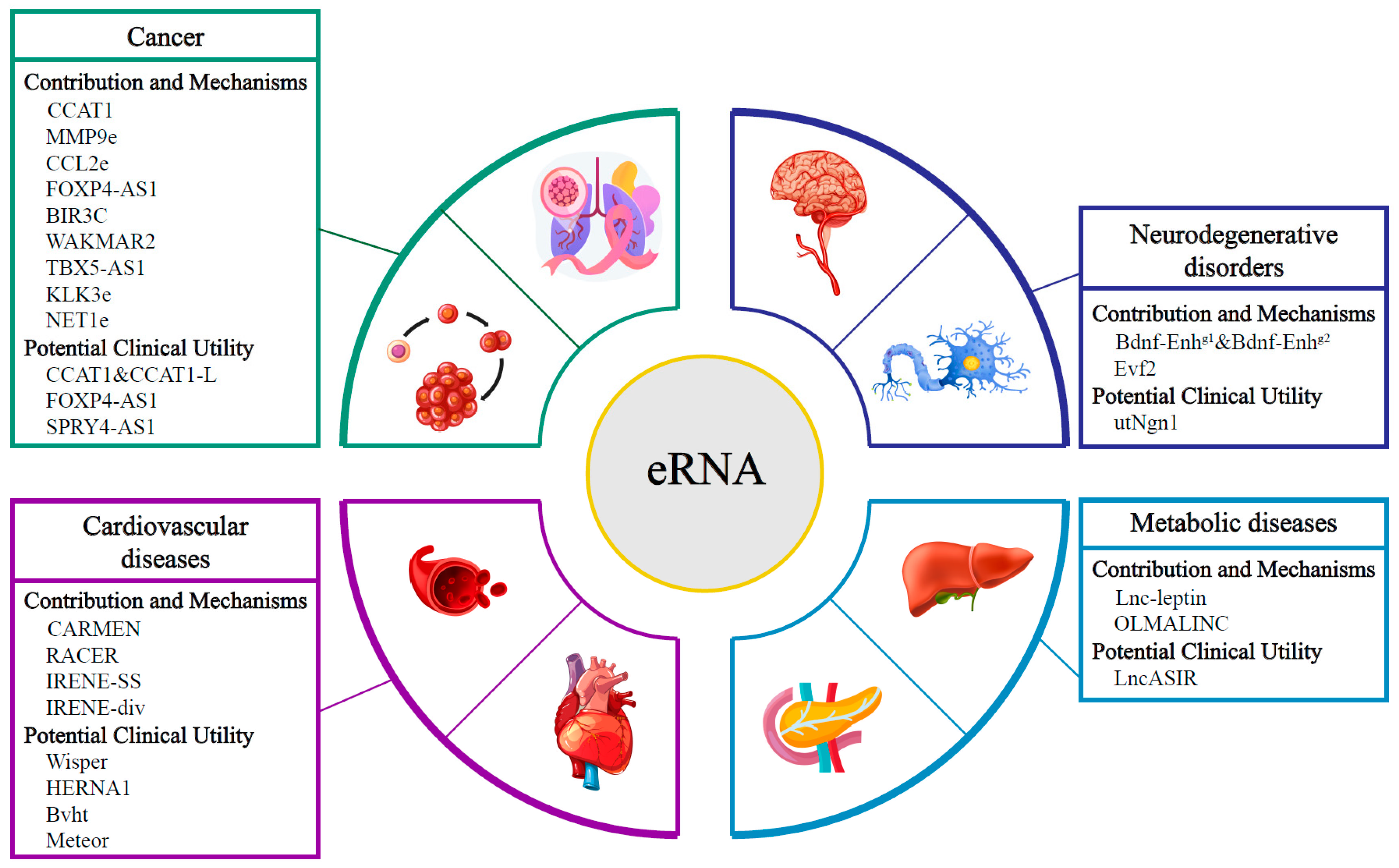
2. Role of eRNAs in Cancers

Cancers are associated with multiple aberrant biological processes, including sustained proliferation, immune escape and apoptosis prevention [59,60]. The aberrant expression of eRNA dysregulates cancer-related genes to promote cancer signaling pathways and results in abnormal cellular responses in tumorigenesis [45,61,62]. In this section, we summarize the contribution and mechanisms of eRNAs in different cancer types, as well as their potential clinical utility for cancer diagnosis and therapies.

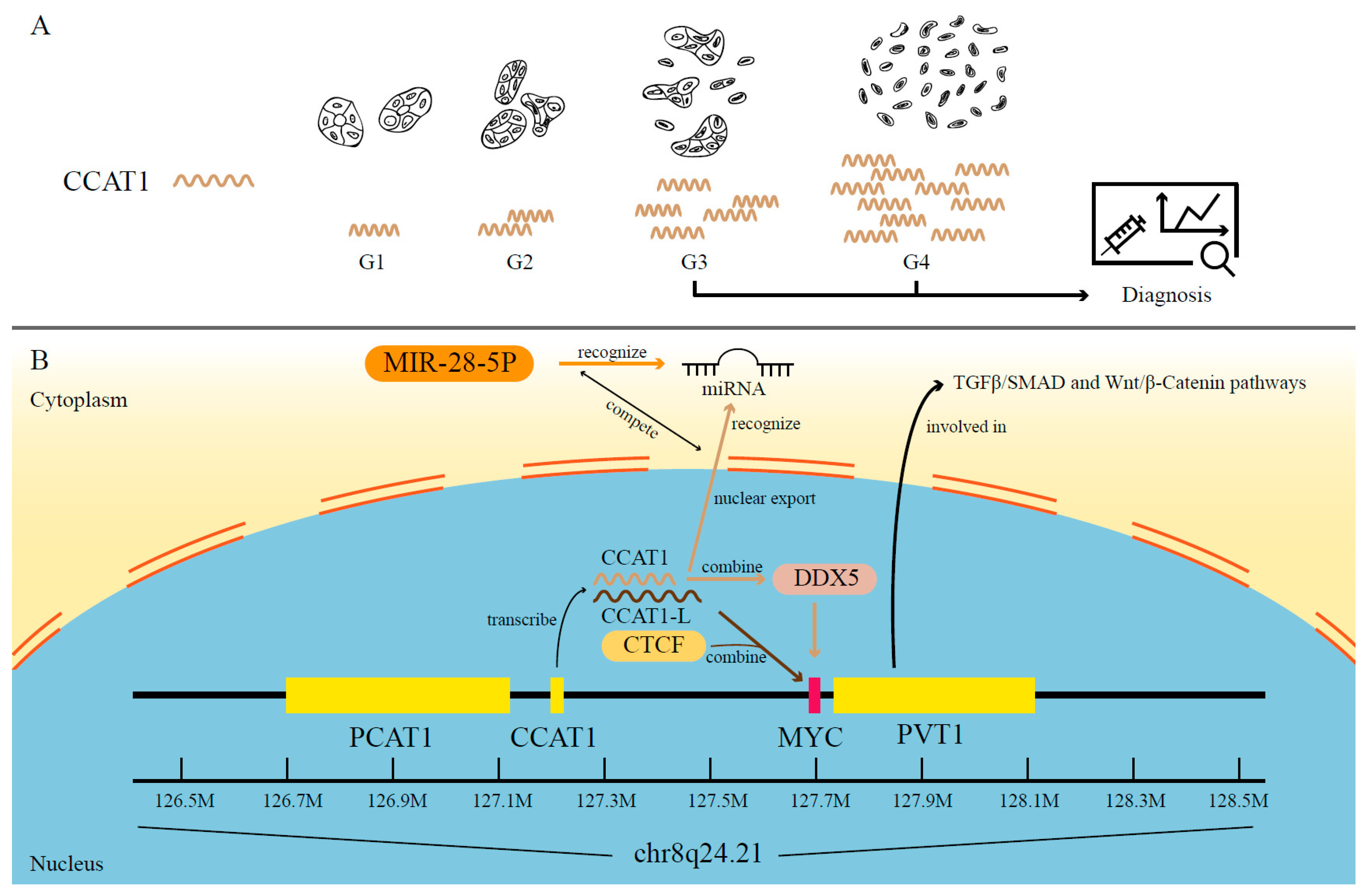
2.1. The Contribution and Mechanisms of eRNAs in Cancer

According to recent studies, eRNAs are highly engaged in oncogene activation and aberrant signaling pathways, which contribute to tumorigenesis [45]. The eRNAs transcribed from the super-enhancer regions on 8q24 serve as a good example of how this works [30,63,64,65,66]. Using the chromosome conformation capture (3C) technique, researchers have found long-range physical interactions between eRNAs from 8q24 and MYC, whose dysregulation is a significant symbol of multiple cancer types [63,67,68,69,70,71]. For example, colon cancer-associated transcript 1 (CCAT1), one well-studied eRNA transcribed from the 8q24 locus, can be activated through long-range interaction with MYC in many cancers, such as colorectal cancer (CRC), prostate adenocarcinoma (PRAD), breast invasive carcinoma (BRCA) and esophageal carcinoma (ESCA) [63,72,73,74]. The evidence has shown that CCAT1 plays its role through different molecular mechanisms in the nucleus and cytoplasm [72]. In the nucleus, CCAT1 directly binds to Asp-Glu-Ala-Asp (DEAD) box helicase 5 (DDX5), which acts as a coactivator involved in MYC gene transcriptional activation and is also able to promote tumor growth [72,75,76,77]. In the cytoplasm, CCAT1, acting as a miRNA sponge, competes with MIR-28-5P for miRNA binding sites to promote prostate cancerous processes (Figure 1B) [72]. Furthermore, in human CRC cells, a long isoform of CCAT1 called CCAT1-L has been observed to upregulate MYC and thus promote tumorigenesis. CCAT1-L achieves such functions by interacting with CTCF and modulating chromatin conformation (Figure 1B) [78]. As for the contribution of eRNAs in aberrant signaling responses, take the variant rs72725854 in a prostate cancer-specific enhancer at the 8q24 locus as an example. The eRNA expression level is significantly affected by this variant and can thus regulate neighboring lncRNAs such as PVT1 [66]. Bioinformatics analysis in human CRC has shown that its expression level is statistically associated with most of the genes within the TGFβ/SMAD and Wnt/β-Catenin pathways (Figure 1B) [66,79]. These pathways are vital to tumorigenesis by contributing to proliferation, differentiation and anti-apoptosis [80,81]. Taken as a whole, the variants, modifications or abnormal activation of 8q24 regions can induce overexpression of its transcribed eRNAs, which then trigger the pathogenesis of multiple human cancers, including CRC, PRAD, BRCA, ESCA and hematological malignancies [65,66,69,70,71,82].
The interaction of eRNAs with Bromodomain and Extraterminal (BET) proteins also showed some insights into cancer development. BET, a protein family containing BRD2, BRD3, BRD4 and BRDT, is regarded as an epigenetic regulator of transcription in carcinogenesis-related biological processes, including cell growth, differentiation and inflammation [83]. In different tissues or pathways, their mechanisms are varied and complicated. BET proteins (e.g., BRD4) can maintain eRNA over-synthesis and the continuous activation of NF-κB as well as the subsequent aberrant inflammatory responses or cell proliferation [84,85,86,87,88]. Recent studies have investigated the direct interplay between BRD4 and certain eRNAs, including eRNAs of MMP9 and CCL2. The tandem bromodomains of BRD4 were found to facilitate the BRD4–eRNA interactions. Conversely, these eRNAs also promote the functions of BRD4 by increasing its binding towards acetylated histone peptides and modulating the occupancy of the BRD4 enhancer to keep the gene active [89].
Another example is FOXP4-AS1, whose upregulation has been identified in different cancers, including hepatocellular carcinoma (HCC), CRC, PRAD and ovarian serous cystadenocarcinoma (OV) [90,91,92,93]. The mechanisms of FOXP4-AS1 in these cancers have also been well studied. For example, in PRAD, the expression of FOXP4-AS1 can upregulate its target gene FOXP4 by acting as a miRNA sponge and sequestering miR-3184-5p. Researchers further determined that the FOXP4-AS1/FOXP4 axis can promote PRAD tumorigenesis [92]. In HCC, FOXP4-AS1 can recruit enhancers of zeste homolog 2 (EZH2) on H3K27me to downregulate the expression of zinc finger CCCH-type containing 12D (ZC3H12D), thus facilitating HCC growth [91]. Such cancer-specific mechanisms indicate a detailed and comprehensive perspective in further research.
Apart from the detailed mechanisms of eRNAs mentioned above, more eRNAs are being detected and studied in different cancer types. By inducing cIAP2 expression, BIR3C eRNA can affect H. pylori infection, which is one of leading causes of gastric cancer [94]. An immune-related eRNA called WAKMAR2 can regulate several downstream target genes in BRCA [95]. In non-small lung cancer cells, the eRNA TBX5-AS1 has been found to regulate the tumor progression through the PI3K/AKT pathway [96,97]. KLK3e, involved in androgen receptor-driven looping, has been discovered to selectively trigger gene activation in PRAD [98]. An important eRNA called NET1e has been observed to have overexpression patterns in PRAD, stomach adenocarcinoma (STAD), liver hepatocellular carcinoma (LIHC), kidney renal clear cell carcinoma (KIRC) and kidney renal papillary cell carcinoma (KIRP) [99,100]. All these examples, even not covering every pattern in eRNA regulation, are fulfilling the understanding of eRNAs in carcinogenesis and cancer growth, which may also shed light on more cancer treatment methods.

2.2. The Potential Clinical Utility of eRNAs in Cancer

Increasing evidence has also shown the significant roles of eRNAs in clinical applications, including acting as biomarkers and therapeutic targets. First, eRNAs can serve as potential biomarkers in diagnosis. For example, in CRC cells, the expression of the eRNA CCAT1 is correlated with differential grades, non-mucinous histology and tumor stages (III and IV) (Figure 1A) [101]. Researchers have also found analogous patterns in other cancer types such as BRCA, in which a correlation has been identified of high CCAT1 expression with lymph node metastases as well as all the diagnostic aspects mentioned above in CRC (Figure 1A) [73]. It should be noted that CCAT1 may provide a limited contribution in detecting CRC at an early stage [102]. In a more recent study, researchers discovered the association between the overexpression of MYC and CCAT1 upregulation before prostate tumor formation, signifying a novel tool for early PC identification [72]. This evidence suggests that eRNAs could be employed as biomarkers in cancer diagnosis. In addition, more studies have focused on the possible utility of eRNAs in cancer prognosis. CCAT1 has also shown its potential to be an independent indicator in CRC and PC to predict poor survival both in recurrence-free survival (RFS) and overall survival (OS) [72,101]. In addition, high expression of FOXP4-AS1 has also been reported to contribute to worse survival and a higher recurrence rate in HCC and CRC patients [91,93]. Bioinformatics methods applied to analyze the TCGA database have also revealed some promising biomarkers for further experiments. For instance, a pan-cancer analysis has displayed that the expression of eRNA SPRY4-AS1 in surgical specimens is associated with survivability in patients with HCC, glioblastoma multiforme (GBM), adrenocortical carcinoma (ACC), brain lower grade glioma (LGG) or mesothelioma (MESO) [103]. Such analysis of the association between eRNA expression levels and survival or relapse rate suggests that eRNAs can be good prognostic biomarkers.
The inhibition of oncogenic eRNA has potential utility in cancer therapy. Studies have found that FOXP4-AS1 knockdown by its targeted shRNA can inhibit CRC cell proliferation and induce apoptosis both in vitro and in vivo [93]. A similar study of eRNAs in BRCA revealed substantial and strong cell growth inhibition in the presence of NET1e knockdown [100]. Apart from eRNA depletion, the analysis of eRNA expression level and drug sensitivity also displayed therapeutic usage. For example, from a TCGA cohort, researchers have observed a strong connection between the response of anti-cancer drugs and eRNA expression through within pathways or cross-pathways, which indicates that changes in eRNA expression may work as a compliment in anti-cancer drug therapies [100]. Furthermore, it has been found that even more hopeful therapeutic potentials are related to BET proteins. In recent years, several BET inhibitors have been developed and tested in clinical trials [104]. These inhibitors all aim in targeting the BRD-acetyl binding pocket to prevent BRD4 from binding to enhancers or super-enhancers [85,104]. According to the discoveries in the direct interaction between certain eRNAs (i.e., MMP9 and CCL2) and BRD4, eRNAs could also be potent therapeutic targets for repressing carcinogenesis [89]. The novel clinical tool targeting eRNAs in this process, together with the inhibitors that have already been put into clinical trials, may shed light on the effect of eRNAs in cancer treatment.
Although researchers have identified the potential clinical utility of eRNAs as biomarkers and therapeutic targets across different cancer types, more comprehensive trials are required [105]. As discoveries of eRNAs are centered in cancer-related studies, the significance of eRNAs in carcinogenesis and their cancer-related clinical relevance would be understood.

3. Role of eRNAs in Neurodegenerative Disorders

Neurodegenerative disorders are caused by the progressive loss of select vulnerable populations of neurons and result in a debilitating loss of sensory, motor and cognitive functions [106,107]. The disorders also share fundamental biological processes related to progressive neuronal dysfunction and death, abnormalities of autophagy and neuroinflammation [106]. Aberrant expression genes involved in those biological processes are vital for interested researchers. Being a critical regulatory element in gene regulation, the eRNAs that respond to neuronal depolarization are worth studying. Among the various neurodegenerative disorders, Alzheimer’s disease (AD), Parkinson’s disease (PD) and Huntington’s disease (HD) have been recently linked to eRNA dysregulation, which may provide a better knowledge of the disease processes and novel possibilities of utilizing eRNAs in clinical usage [108,109,110].

3.1. The Contribution and Mechanisms of eRNAs in Neurodegenerative Disorders

The role of eRNAs in neurodegenerative disorders has recently been observed through various bioinformatic and experiential methods. Several eRNAs are involved in neurodegenerative pathogenesis or regulating the expression of certain genes that have vital roles in these diseases. In addition, bioinformatics investigations have revealed broader expression patterns or enrichment of eRNAs in neurodegenerative disorders, extending the bounds of regulatory elements in neurodegenerative disorders.
It is known that brain-derived neurotrophic factor (BDNF) signaling is impaired in AD brains, while increasing BDNF levels can improve learning and memory [111]. In the intergenic region interacting with BDNF, two putative eRNAs—designated Bdnf-Enhg1 and Bdnf-Enhg2—have been found to regulate BDNF expression by their transcribed eRNAs. More specifically, the expression levels of these putative eRNAs have been observed to have a dramatic increase in tandem with BDNF expression, especially in post-mitotic neuron cells. Further experiments have validated their role in mediating BDNF expression that contributes to dendritic growth [112]. The onset of neurodegenerative diseases has been linked to abnormal BDNF expression regulated by Bdnf-Enhg1 and Bdnf-Enhg2 [111,112,113]. Another study has shown the intermediary but important involvement of eRNAs in pathogenesis by observing the subsequent impact of DNA sequence variants in the enhancer region ANNCR. This change is associated with Apolipoprotein E (APOE), whose ε4 allelic form is a major risk factor for AD. AANCR is folded into an m6A R-loop and partially transcribed into a certain eRNA, which then effectively silences APOE expression and changes the susceptibility to AD [114]. An expression profile study has also revealed a significant enrichment of differentially expressed eRNAs at AD-associated enhancer regions [115].
Some studies have observed the eRNA involvement in other neurodegenerative diseases. For example, in dopamine neurons, several PD-associated variants on chromosome 17q21 have been linked to a putative eRNA expressed from intron 2 of the KANSL1 gene [116]. As KANSL1 dysregulation has been proved to disturb autophagy and thus induce memory impairment and neurodegeneration, the regulatory mechanism of this putative eRNA is worth investigating in PD-associated research [116,117]. Evidence in HD-related studies also displays the importance of eRNAs. For instance, researchers have found that decreased H3K27ac activity at super-enhancers in the R6/11 striatum is able to alter the eRNA production in HD-affected neurons [109]. RNA pol II is then reduced along HD downregulated genes, especially the voltage-gated potassium channels (e.g., Kcnab1, Kcna4 and Kcnj4), causing transcriptional dysregulation of relative neuronal genes and presumably increasing striatal vulnerability in HD [109,118].
Other neurological pathways or circuities—capable of affecting both neurodevelopment and neurodegeneration—have been demonstrated to be associated with eRNAs. For example, a brain-specific ultraconservative eRNA called Evf2 can regulate the expression of the homeodomain TF Dlx5/6 by recruiting DLX and MECP2 in their intergenic enhancer regions. Evf2 mutants reduce the GABAergic interneuron numbers in both early postnatal hippocampal and dentate gyrus and decrease synaptic inhibition in adults’ brains [119,120]. Causing an imbalance between excitatory and inhibitory signaling, GABA-regulated circuit dysfunction is well known for its versatile contribution to neurological diseases, which includes neurodevelopmental disorders such autism and Tourette’s syndrome, neurodegenerative disorders such as HD and epilepsy and other disorders such as dystonia and hepatic encephalopathy [119,121,122,123,124]. Further research into the mechanisms of eRNAs in neurodegenerative disorders is needed to provide a better understanding of these diseases on a comprehensive and more nuanced level.

3.2. The Potential Clinical Utility of eRNAs in Neurodegenerative Disorders

Some studies have further investigated the possible clinical usage of eRNAs in neurogenerative disorders. For example, eRNA impacts HD-related neuronal genes in the striatum, as exemplified in Section 3.1., implying that targeting striatal enhancers can improve eRNA expression levels, which is likely to prevent the repression of neuronal genes in HD patients [118]. In PD rat models, the eRNAs transcribed from super-enhancers (SEs) have also been examined using bioinformatics techniques. A cluster of PD-specific SEs could upregulate SNX5 to prompt ferroptosis levels through the endosomal sorting pathway in PD, which may serve as potential diagnostic markers and therapeutic targets for PD [125].
A more detailed example may shed light on future research directions. An eRNA transcribed from the enhancer region located 5.8–7.0 kb upstream of the mouse neurogenin1 (Neurog1 or Ngn1) gene, designated utNgn1, has been demonstrated to be necessary for effective Ngn1 transcription. Additionally, researchers have identified the elements that can control utNgn1 transcripts and hence indirectly regulate Ngn1 expression: Wnt signaling is capable of upregulating utNgn1 expression, while PcG proteins are capable of downregulating it [126]. As for the function of the Ngn1 gene, the loss of its expression can generate the loss of deep-layer neurons according to Ngn-independent mechanism research [127]. Ngn1 overexpression, on the contrary, promotes premature differentiation and has been considered a means of helping embryonic stem cells (ESCs) in their differentiation into induced neurons [107,126]. The potential therapeutic utility in such a scenario is to replace degenerative neurons with these induced neurons from ESCs [107]. A computational approach has demonstrated the possible contribution of eRNAs derived from super-enhancers in ESC differentiation [128]. Combining the results of these studies, regulating the eRNA utNgn1 to prompt the directional differentiation of ESCs clearly turns out to be potentially useful in stem cell therapy for neurodegenerative disorders.
Thus far, the clinical utility of eRNAs, e.g., prognosis or target therapy, in neurodegenerative disorders is still largely unexplored. These studies have not only revealed the mechanisms of eRNAs in different neurodegenerative disorders but also provided potential approaches to utilizing eRNAs in these diseases.

4. Role of eRNAs in Cardiovascular Diseases

Cardiovascular diseases (CVDs) occur in the blood circulatory system, including the heart and its associated blood vessels [129]. Some studies have revealed that TF genes such as NKX2-5, MESP1, TBX5, MEF2c, HAND2, GATA4 and SRF play critical roles in cardiac gene regulatory networks (GRNs) [130,131]. Most importantly, the eRNAs involved in these cardiac GRNs can perturb them and modulate CVD development [130]. Meanwhile, eRNAs have also shown their potential clinical utility for CVD diagnosis and treatment [132].

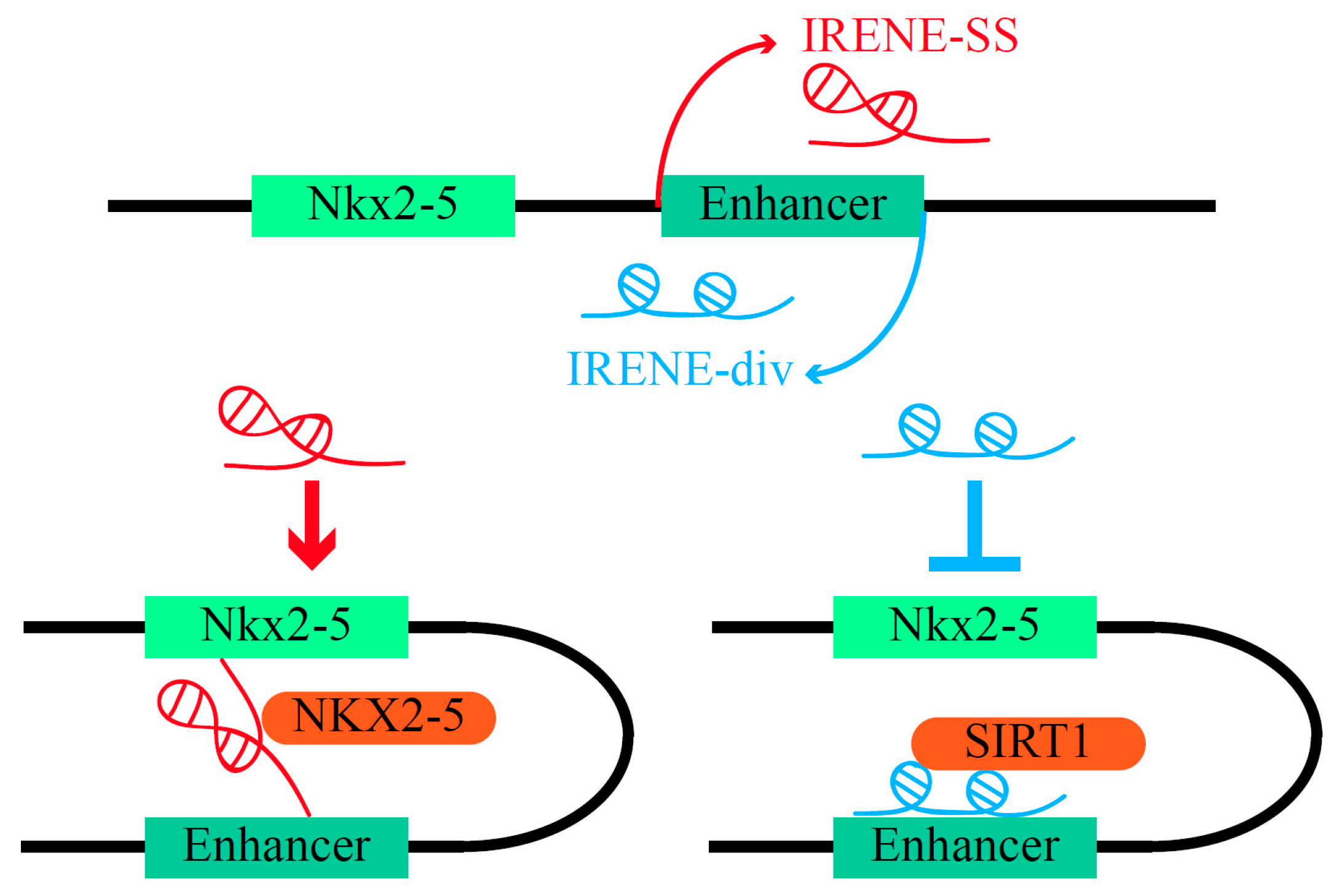
4.1. The Contribution and Mechanisms of eRNAs in Cardiovascular Diseases

Transcriptional alteration can be broadly detected in cardiovascular morphogenesis. Recent studies have revealed that various genes are regulated by eRNAs in these cardiac processes [131]. An eRNA known as CARMEN is a typical example that sparked a flurry of investigation. It maps to the locus proximal to an important region that harbors a cardiac SRF/NKX2.5-bound enhancer in human cardiac precursor cells (CPCs). Computational calculation and experimental validation have corroborated CARMEN as a crucial upregulated eRNA that contributes to both CPC differentiation and cardiovascular pathologies in human hearts. The activity of CARMEN in CPC differentiation is in its interaction with SUZ12 and EZH2, components of polycomb repressive complex 2 (PRC2) [133,134]. In addition, one of CARMEN’s isoforms, named CARMEN3, has been observed to show significant upregulation in the pathologies of two different human CVDs, namely idiopathic dilated cardiomyopathy (DCM) and aortic stenosis (AOS) [134].
Enhancer RNAs are also associated with calcium-handling physiology crucial for maintaining a healthy cardiac rhythm and intact heart activities. In human hearts, the eRNA RACER improves the performance of T-box transcription factor 5 (TBX5), whose malfunction is responsible for cardiac conduction system (CCS) disease through GRN dysregulation. As an upstream regulator of TBX5, RACER maintains normal calcium kinetics by regulating the expression of the TBX5-dependent gene Ryr2 by recruiting Pol2 and stabilizing the structure of the Pol2-Ryr2 promoter [135,136].
Notably, the eRNAs transcribed from different strands of the same enhancer may oppositely exert their regulatory functions according to novel research on Intergenic Regulatory Element Nkx2-5 Enhancers (IRENEs) [137]. These two eRNAs are both transcribed from the enhancer of Nkx2-5, whose mutation has long been viewed as the main cause of congenital heart defects (CHDs) [138,139]. One IRENE eRNA is on the same strand (SS), while the other is in the divergent direction (div). IRENE-SS functions as a typical eRNA to promote Nkx2-5 transcription by enlisting NKX2-5 to its own enhancer. On the contrary, IRENE-div silences the Nkx2-5 enhancer by recruiting the histone deacetylase sirtuin 1 (SIRT1) (Figure 2) [137]. The subcellular distribution of IRENEs exhibits specific patterns: the localization of IRENE-div is dependent on the stage of the transcript’s biogenesis, with the mature form previously in the cytoplasm and the immature form having a subcellular localization similar to that of IRENE-SS. In contrast, 60% of IRENE-SS was found in the nucleus, with 45% of it bound to chromatin [137].

4.2. The Potential Clinical Utility of eRNAs in Cardiovascular Diseases

Understanding the essential roles of eRNAs in CVDs will also pave the road for manipulating eRNAs in related clinical studies. To be specific, eRNAs may be used as therapeutic targets in corresponding cardiovascular pathologies and may be utilized to control the differentiation of precursor stem cells into wanted cardiac cells in regenerative medicine due to their critical roles in cardiac cell development.
For example, Wisper (Wisp2 super-enhancer–associated RNA) has been identified as a cardiac fibroblast (CF)-enriched eRNA that regulates the process of cardiac fibrosis [140]. CFs’ differentiation into myofibroblasts triggered by CF proliferation at first can initiate the pathological process of heart failure, resulting in diseases such as AOS and dilated cardiomyocytes (CM). Additionally, their pathologies are under apoptotic resistance and profibrotic signaling molecular influence [141]. Therapeutic depletion of Wisper in vivo has been proven to generate a significant decrease in the proliferation of CFs and can consequently inhibit cardiac fibrosis. The sequence of Wisper being relatively conserved in humans suggests that it is desirable to explore the potential utility of Wisper as an antifibrotic therapeutic target [140].
Similar clinical utility of eRNAs has also been demonstrated in stress-induced disease development. An eRNA named HERNA1 (hypoxia-inducible enhancer RNA 1) has displayed a great capacity to regulate its neighboring coding genes, such as synaptotagmin XVII and SMG1, by conferring hypoxia responsiveness towards them. Furthermore, as HERNA1 production is robustly induced by pathological stress, antisense oligonucleotides targeting HERNA1 can protect cells against stress-induced pathological hypertrophy and increase overall survival rate [142]. The curative effect of HERNA1 inhibition suggests that it could be a useful post-disease therapeutic target in pressure-overload CVDs, including aortic stenosis (AS) and hypertrophic cardiomyopathy (HCM) [142,143].
Aside from serving as therapeutic targets, eRNAs can also be applied in cardiac regenerative strategies to replenish lost cardiomyocytes [132]. Bvht, transcribed from an important locus enriched with heart-specific enhancers, has been demonstrated to be a requisite eRNA during ESC progression of nascent mesoderm toward a cardiac fate. This decisive step in ESC differentiation occurs by regulating Mesp1, a vital transcriptional factor marking the early cardiac precursor cells [132,144]. Meteor is another eRNA that performs analogous functions in ESC differentiation. Genetically or epigenetically manipulating Meteor has revealed its essential role in mesendoderm specification and subsequent cardiogenic differentiation [145]. Based on these findings, eRNA manipulation shows great promise for guiding the differentiation of precursor cells into desired cardiac cells, which could have intriguing applications in cardiac cell regeneration, providing more chances for cardiac cell therapies.

5. Role of eRNAs in Metabolic Diseases

Dysfunction of major metabolic tissues, e.g., liver and pancreas, could result in a series of metabolic anomalies, which generate metabolic diseases such as obesity, type 2 diabetes (T2D) and hepatic steatosis [146,147,148]. These diseases also show a tight correlation with each other because of the whole integrity of the metabolic network, in which key regulatory elements actively interact [148]. As evidence for the critical involvement of regulatory elements in metabolic networks and related disorders is becoming increasingly abundant, eRNAs—a subset of these elements—have also come to be recognized as a subject worth investigating in metabolic diseases.

5.1. The Contribution and Mechanisms of eRNAs in Metabolic diseases

In studies investigating obesity genesis, researchers have found that an eRNA named Lnc-leptin, transcribed from the enhancer region upstream of the leptin (Lep) gene, is required for Lep expression. Lnc-leptin‘s regulatory effects are based on two different mechanisms: direct interaction with the Lep promoter and acting as a bridge between TFs and histone-modifying proteins [149]. Downregulated Lep expression, caused by Lnc-leptin deficiency, may incur a decrease in leptin, which controls appetite through its effect on the hypothalamus [150,151]. Maternal obesity-associated metabolic and epigenetic alterations can also lead to dampened responses in the offspring’s immune system by reducing the expression of IL6 eRNA and IL10RB eRNA in monocytes [152]. This evidence has emphasized the important engagement of eRNAs in metabolic mechanisms and obesity-associated immune responses.
In lipid metabolism research on hepatic steatosis and non-alcoholic fatty liver disease (NAFLD), the liver expression of an eRNA designated OLMALINC (oligodendrocyte maturation-associated long intergenic noncoding RNA, located at human chromosome 10q24.31) has been proven to be tightly associated with statin use and serum triglycerides (TGs). Such connection relies on OLMALINC’s ability to promote the expression of its adjacent TG-gene recognized as stearoyl-CoA desaturase (SCD), which is known to be up-regulated in hepatic steatosis and NAFLD [153,154]. Notably, OLMALINC has a stable and spliced transcript with a poly-A tail, which indicates its possible secondary functions and a broad usage in future studies [153].
The regulatory function of eRNAs in some specific metabolism-related genes related to a broader range of metabolic diseases may provide a wider understanding of the involvement and even modification of eRNAs. For example, m5C-modified eRNAs in hepatic cells have been found to act as an intermediate substance in the peroxisome proliferator-activated receptor γ coactivator 1 alpha (PGC-1α)-related metabolic network. The dynamic change in the methylation and demethylation of PGC-1α, which plays vital roles in adapted metabolic responses, is caused by methyltransferase SET7/9 and Lysine-specific demethylase 1 (LSD1), corresponding with m5C eRNAs. Moreover, the eRNAs transcribed from the enhancer regions of PGC-1α’s target genes, such as 6-phosphofructokinase (Pfkl) and Sirtuin5 (Sirt5), have been proven to be inducible elements affecting carbamoyl-phosphate synthetase 1 (CPS1) activity and have thus been identified as important sensors of the metabolic state [155]. According to current knowledge on PGC-1α, it has been clearly linked to various metabolic complications, including obesity, T2D and hepatic steatosis [156]. Therefore, understanding how eRNAs work in PGC-1α pathways could aid the intervention of these disorders [155].
Studies have also explored the dysfunction of enhancers in rare diseases. For example, lipodystrophies are referred to as lipid-partitioning disorders. Their primary defect is the loss of functional adipocytes, resulting in ectopic steatosis, severe dyslipidemia and insulin resistance [157,158]. The activation of enhancers regulating the genes involved in lipid metabolic pathways can be influenced by Tmem120a, a transcription factor whose deficiency may suppress the expression of genes associated with lipodystrophies [159]. This evidence indicates the functional and clinical potential of eRNAs in rare diseases.

5.2. The Potential Clinical Utility of eRNAs in Metabolic diseases

Investigations of the eRNAs in metabolic diseases, particularly T2D, show that they are involved in various metabolic pathways. In adipocytes, a well-known nuclear receptor named peroxisome proliferator-activated receptor (PPARγ) regulates adipocyte biology by directly binding to its target genes [160]. An anti-diabetes drug designated rosiglitazone (rosi) can act as a high-affinity activating ligand for PPARγ to treat T2D [161,162]. By recruiting coactivators such as MED1, p300, and CBP, rosi can upregulate certain eRNAs and thus generate an enrichment of these eRNAs at PPARγ binding sites. For example, a series of bidirectional eRNAs transcribed from the enhancers upstream of the Fabp4 locus were observed to be upregulated by rosi. Additionally, 85% of these upregulated eRNAs have PPARγ bound nearby, although there are also rosi-induced downregulated eRNAs whose locations harbor binding sites for other TFs. A better understanding of the complex transcriptional changes caused by rosi may provide solutions and interventions for its severe side effects and toxicities [162].
Insulin responses are always impaired in T2D and subsequent obesity [163]. LncASIR, an eRNA also annotated as a four-exon transcript in the RefSeq database, is transcribed from a super-enhancer region upstream of Lep and has been demonstrated to be a required element for normal insulin signaling pathways. By using dcas9-KRAS and guide RNA, an LncASIR silencing experiment showed a considerable reduction in the insulin-induced gene program in adipocytes. Overexpression of LncASIR alone is not sufficient to increase insulin responses in T2D patients. If LncASIR is induced with its binding protein—polycomb repressive complex (PRC)—together, it might improve the clinical repair of insulin responses [164].

6. Conclusions and Perspectives

Enhancer RNAs are regulated by upstream factors and mediate the expression of their target genes in different diseases. They often harbor similar biological functions and molecular mechanisms. Comprehensive investigations of eRNAs could shed light on their roles in complex GRNs in human diseases. In addition, some eRNAs have shown particular and distinct mechanisms that may indicate new knowledge of their functions. Taking IRENEs as examples, the eRNAs transcribed from the same locus in different strands, designated IRENE-SS and IRENE-div, exert almost opposing functions in regulating their target gene, Nkx2-5 (Figure 2) [137]. Nkx2-5 mutation has long been viewed as the main cause of CHDs, and utilizing the counterbalance effect of IRENEs may be a breaking point for CHD treatment [139]. Thus far, the role of eRNAs in human diseases is still largely unexplored. Furthermore, some eRNAs have shown cascade amplifications in complicated GRNs. For example, the tandem bromodomains of BRD4 can facilitate its binding to certain eRNAs such as MMP9 eRNA. After the binding relation is formed, MMP9 eRNA starts to increase the binding ability towards acetylated histone peptides and modulates the occupancy of the BRD4 enhancer, which then maintains downstream genes’ activation [89]. As BRD4 is highly involved in the biological processes associated with carcinogenesis, such as cell proliferation, differentiation and inflammation, the serial amplification of BRD4-eRNA could provide novel insights about cancer development [86,87]. Similar to the mutual promotion of BRD4 eRNA binding, decreased H3K27ac at super-enhancers in the R6/11 striatum reduces the eRNA production in HD-affected neurons. Conversely, the downregulated eRNA production results in the transcriptional dysregulation of related neuronal genes and presumably increases striatal vulnerability in HD [109,118]. This draws attention to eRNAs’ involvement in multiple biological processes and demonstrates their critical roles in different human diseases.
The systematic study of the functions of eRNA could promote eRNA in potential clinical applications (Figure 3, Table 1). The usage of eRNAs in cancer-related studies can be divided into three potential applications: diagnostic biomarkers, prognostic biomarkers and therapeutic targets. Thus far, these potential applications of eRNAs are still far away from clinical trials, let alone practical clinical uses. Before being applied in clinical trials, their medical potentials mentioned above still need more evidence based on comprehensively investigating their potential toxicity and limitations. Furthermore, eRNAs have been associated with clinical relevance, such as high expression of SPRY4-AS1 associated with poorer survival rates in cancers. The mechanisms and utility of these clinical associations also require further exploration in order to realize their potential application, which also calls for more research focusing on the uncharted capacity of eRNAs in their biological function exploration.

Author Contributions

Original draft preparation, Y.W. (Yunzhe Wang) and Z.Z.; review and editing, Y.W. (Yunzhe Wang), C.Z., Y.W. (Yuxiang Wang), X.L. and Z.Z.; visualization, C.Z. and Y.W. (Yunzhe Wang). All authors have read and agreed to the published version of the manuscript.

Funding

This study was supported by the National Natural Science Foundation of China (32270708 and 82203195) and the Shanghai Pujiang Program (21PJ1401700 to Z.Z. and 20PJ1400700 to Y.W.).

Institutional Review Board Statement

Not applicable.

Informed Consent Statement

Not applicable.

Data Availability Statement

Not applicable.

Acknowledgments

The authors acknowledge Libing Shen for his contribution in English editing throughout this review.

Conflicts of Interest

The authors declare no conflict of interest.

References

  1. Consortium, E.P. An integrated encyclopedia of DNA elements in the human genome. Nature 2012, 489, 57–74. [Google Scholar] [CrossRef] [PubMed]
  2. Mattick, J.S. Non-coding RNAs: The architects of eukaryotic complexity. EMBO Rep. 2001, 2, 986–991. [Google Scholar] [CrossRef] [PubMed]
  3. Behjati, S.; Tarpey, P.S. What is next generation sequencing? Arch. Dis. Child. Educ. Pract. Ed. 2013, 98, 236–238. [Google Scholar] [CrossRef] [PubMed]
  4. Fu, X.D. Non-coding RNA: A new frontier in regulatory biology. Natl. Sci. Rev. 2014, 1, 190–204. [Google Scholar] [CrossRef]
  5. Sharp, S.J.; Schaack, J.; Cooley, L.; Burke, D.J.; Soil, D. Structure and Transcription of Eukaryotic tRNA Gene. RNA Biol. 1985, 18, 738–746. [Google Scholar] [CrossRef]
  6. Matera, A.G.; Wang, Z. A day in the life of the spliceosome. Nat. Rev. Mol. Cell Biol. 2014, 15, 108–121. [Google Scholar] [CrossRef]
  7. Elliott, B.A.; Ho, H.T.; Ranganathan, S.V.; Vangaveti, S.; Ilkayeva, O.; Abou Assi, H.; Choi, A.K.; Agris, P.F.; Holley, C.L. Modification of messenger RNA by 2’-O-methylation regulates gene expression in vivo. Nat. Commun. 2019, 10, 3401. [Google Scholar] [CrossRef]
  8. Kiss, T. Small nucleolar RNA-guided post-transcriptional modification of cellular RNAs. EMBO J. 2001, 20, 3617–3622. [Google Scholar] [CrossRef]
  9. Mattick, J.S.; Makunin, I.V. Non-coding RNA. Hum. Mol. Genet. 2006, 15, R17–R29. [Google Scholar] [CrossRef]
  10. Watkins, N.J.; Bohnsack, M.T. The box C/D and H/ACA snoRNPs: Key players in the modification, processing and the dynamic folding of ribosomal RNA. Wiley Interdiscip. Rev. RNA 2012, 3, 397–414. [Google Scholar] [CrossRef]
  11. Cai, Y.; Yu, X.; Hu, S.; Yu, J. A Brief Review on the Mechanisms of miRNA Regulation. Genom. Proteom. Bioinform. 2009, 7, 147–154. [Google Scholar] [CrossRef]
  12. Khashkhashi Moghadam, S.; Bakhshinejad, B.; Khalafizadeh, A.; Mahmud Hussen, B.; Babashah, S. Non-coding RNA-associated competitive endogenous RNA regulatory networks: Novel diagnostic and therapeutic opportunities for hepatocellular carcinoma. J. Cell Mol. Med. 2022, 26, 287–305. [Google Scholar] [CrossRef] [PubMed]
  13. O’Brien, J.; Hayder, H.; Zayed, Y.; Peng, C. Overview of MicroRNA Biogenesis, Mechanisms of Actions, and Circulation. Front Endocrinol. 2018, 9, 402. [Google Scholar] [CrossRef] [PubMed]
  14. Chen, L.L.; Yang, L. Regulation of circRNA biogenesis. RNA Biol. 2015, 12, 381–388. [Google Scholar] [CrossRef] [PubMed]
  15. Kristensen, L.S.; Andersen, M.S.; Stagsted, L.V.W.; Ebbesen, K.K.; Hansen, T.B.; Kjems, J. The biogenesis, biology and characterization of circular RNAs. Nat. Rev. Genet. 2019, 20, 675–691. [Google Scholar] [CrossRef]
  16. Suzuki, H.; Tsukahara, T. A view of pre-mRNA splicing from RNase R resistant RNAs. Int. J. Mol. Sci. 2014, 15, 9331–9342. [Google Scholar] [CrossRef]
  17. Qu, S.; Yang, X.; Li, X.; Wang, J.; Gao, Y.; Shang, R.; Sun, W.; Dou, K.; Li, H. Circular RNA: A new star of noncoding RNAs. Cancer Lett. 2015, 365, 141–148. [Google Scholar] [CrossRef]
  18. Ebert, M.S.; Sharp, P.A. MicroRNA sponges: Progress and possibilities. RNA 2010, 16, 2043–2050. [Google Scholar] [CrossRef]
  19. Statello, L.; Guo, C.J.; Chen, L.L.; Huarte, M. Gene regulation by long non-coding RNAs and its biological functions. Nat. Rev. Mol. Cell Biol. 2021, 22, 96–118. [Google Scholar] [CrossRef]
  20. Zhang, X.; Wang, W.; Zhu, W.; Dong, J.; Cheng, Y.; Yin, Z.; Shen, F. Mechanisms and Functions of Long Non-Coding RNAs at Multiple Regulatory Levels. Int. J. Mol. Sci. 2019, 20, 5573. [Google Scholar] [CrossRef] [Green Version]
  21. Halley, P.; Kadakkuzha, B.M.; Faghihi, M.A.; Magistri, M.; Zeier, Z.; Khorkova, O.; Coito, C.; Hsiao, J.; Lawrence, M.; Wahlestedt, C. Regulation of the apolipoprotein gene cluster by a long noncoding RNA. Cell Rep. 2014, 6, 222–230. [Google Scholar] [CrossRef] [PubMed]
  22. Luo, S.; Lu, J.Y.; Liu, L.; Yin, Y.; Chen, C.; Han, X.; Wu, B.; Xu, R.; Liu, W.; Yan, P.; et al. Divergent lncRNAs Regulate Gene Expression and Lineage Differentiation in Pluripotent Cells. Cell Stem Cell 2016, 18, 637–652. [Google Scholar] [CrossRef]
  23. Niazi, F.; Valadkhan, S. Computational analysis of functional long noncoding RNAs reveals lack of peptide-coding capacity and parallels with 3′ UTRs. RNA 2012, 18, 825–843. [Google Scholar] [CrossRef]
  24. Johnsson, P.; Lipovich, L.; Grander, D.; Morris, K.V. Evolutionary conservation of long non-coding RNAs; sequence, structure, function. Biochim. Biophys. Acta 2014, 1840, 1063–1071. [Google Scholar] [CrossRef] [PubMed]
  25. Markaki, Y.; Gan Chong, J.; Wang, Y.; Jacobson, E.C.; Luong, C.; Tan, S.Y.X.; Jachowicz, J.W.; Strehle, M.; Maestrini, D.; Banerjee, A.K.; et al. Xist nucleates local protein gradients to propagate silencing across the X chromosome. Cell 2021, 184, 6174–6192.e6132. [Google Scholar] [CrossRef]
  26. Beltran, M.; Puig, I.; Pena, C.; Garcia, J.M.; Alvarez, A.B.; Pena, R.; Bonilla, F.; de Herreros, A.G. A natural antisense transcript regulates Zeb2/Sip1 gene expression during Snail1-induced epithelial-mesenchymal transition. Genes Dev. 2008, 22, 756–769. [Google Scholar] [CrossRef] [PubMed]
  27. Gonzalez, I.; Munita, R.; Agirre, E.; Dittmer, T.A.; Gysling, K.; Misteli, T.; Luco, R.F. A lncRNA regulates alternative splicing via establishment of a splicing-specific chromatin signature. Nat. Struct. Mol. Biol. 2015, 22, 370–376. [Google Scholar] [CrossRef]
  28. Karakas, D.; Ozpolat, B. The Role of LncRNAs in Translation. Noncoding RNA 2021, 7, 16. [Google Scholar] [CrossRef]
  29. Peng, W.X.; Koirala, P.; Mo, Y.Y. LncRNA-mediated regulation of cell signaling in cancer. Oncogene 2017, 36, 5661–5667. [Google Scholar] [CrossRef]
  30. Cui, M.; You, L.; Ren, X.; Zhao, W.; Liao, Q.; Zhao, Y. Long non-coding RNA PVT1 and cancer. Biochem. Biophys. Res. Commun. 2016, 471, 10–14. [Google Scholar] [CrossRef]
  31. Dong, J.; Su, M.; Chang, W.; Zhang, K.; Wu, S.; Xu, T. Long non-coding RNAs on the stage of cervical cancer (Review). Oncol. Rep. 2017, 38, 1923–1931. [Google Scholar] [CrossRef]
  32. Han, P.; Chang, C.P. Long non-coding RNA and chromatin remodeling. RNA Biol. 2015, 12, 1094–1098. [Google Scholar] [CrossRef] [PubMed]
  33. Han, P.; Li, W.; Lin, C.H.; Yang, J.; Shang, C.; Nuernberg, S.T.; Jin, K.K.; Xu, W.; Lin, C.Y.; Lin, C.J.; et al. A long noncoding RNA protects the heart from pathological hypertrophy. Nature 2014, 514, 102–106. [Google Scholar] [CrossRef] [PubMed]
  34. Gupta, R.A.; Shah, N.; Wang, K.C.; Kim, J.; Horlings, H.M.; Wong, D.J.; Tsai, M.C.; Hung, T.; Argani, P.; Rinn, J.L.; et al. Long non-coding RNA HOTAIR reprograms chromatin state to promote cancer metastasis. Nature 2010, 464, 1071–1076. [Google Scholar] [CrossRef] [PubMed]
  35. Yu, C.Y.; Kuo, H.C. The emerging roles and functions of circular RNAs and their generation. J. Biomed. Sci. 2019, 26, 29. [Google Scholar] [CrossRef]
  36. Vijesh, N.; Chakrabarti, S.K.; Sreekumar, J. Modeling of gene regulatory networks: A review. J. Biomed. Sci. Eng. 2013, 06, 223–231. [Google Scholar] [CrossRef]
  37. Emmert-Streib, F.; Dehmer, M.; Haibe-Kains, B. Gene regulatory networks and their applications: Understanding biological and medical problems in terms of networks. Front. Cell Dev. Biol. 2014, 2, 38. [Google Scholar] [CrossRef]
  38. Banf, M.; Rhee, S.Y. Computational inference of gene regulatory networks: Approaches, limitations and opportunities. Biochim. Biophys. Acta Gene Regul. Mech. 2017, 1860, 41–52. [Google Scholar] [CrossRef]
  39. Monnier, P.; Martinet, C.; Pontis, J.; Stancheva, I.; Ait-Si-Ali, S.; Dandolo, L. H19 lncRNA controls gene expression of the Imprinted Gene Network by recruiting MBD1. Proc. Natl. Acad. Sci. USA 2013, 110, 20693–20698. [Google Scholar] [CrossRef]
  40. Gabory, A.; Jammes, H.; Dandolo, L. The H19 locus: Role of an imprinted non-coding RNA in growth and development. BioEssays 2010, 32, 473–480. [Google Scholar] [CrossRef]
  41. Gabory, A.; Ripoche, M.-A.; Le Digarcher, A.; Watrin, F.o.; Ziyyat, A.; Forné, T.; Jammes, H.L.N.; Ainscough, J.F.X.; Surani, M.A.; Journot, L.; et al. H19 acts as a trans regulator of the imprinted gene network controlling growth in mice. Development 2009, 136, 3413–3421. [Google Scholar] [CrossRef] [PubMed]
  42. Shen, Y.; Huang, Z.; Yang, R.; Chen, Y.; Wang, Q.; Gao, L. Insights into Enhancer RNAs: Biogenesis and Emerging Role in Brain Diseases. Neurosci. A Rev. J. Bringing Neurobiol. Neurol. Psychiatry 2021, 10738584211046889. [Google Scholar] [CrossRef] [PubMed]
  43. Raisner, R.; Kharbanda, S.; Jin, L.; Jeng, E.; Chan, E.; Merchant, M.; Haverty, P.M.; Bainer, R.; Cheung, T.; Arnott, D.; et al. Enhancer Activity Requires CBP/P300 Bromodomain-Dependent Histone H3K27 Acetylation. Cell Rep. 2018, 24, 1722–1729. [Google Scholar] [CrossRef] [PubMed]
  44. Lai, F.; Gardini, A.; Zhang, A.; Shiekhattar, R. Integrator mediates the biogenesis of enhancer RNAs. Nature 2015, 525, 399–403. [Google Scholar] [CrossRef]
  45. Lee, J.H.; Xiong, F.; Li, W. Enhancer RNAs in cancer: Regulation, mechanisms and therapeutic potential. RNA Biol. 2020, 17, 1550–1559. [Google Scholar] [CrossRef]
  46. Han, Z.; Li, W. Enhancer RNA: What we know and what we can achieve. Cell Prolif. 2022, 55, e13202. [Google Scholar] [CrossRef]
  47. Mattick, J.S.; De Santa, F.; Barozzi, I.; Mietton, F.; Ghisletti, S.; Polletti, S.; Tusi, B.K.; Muller, H.; Ragoussis, J.; Wei, C.-L.; et al. A Large Fraction of Extragenic RNA Pol II Transcription Sites Overlap Enhancers. PLoS Biol. 2010, 8, e1000384. [Google Scholar] [CrossRef]
  48. Shi, L.; Li, S.; Maurer, K.; Zhang, Z.; Petri, M.; Sullivan, K.E. Enhancer RNA and NFκB-dependent P300 regulation of ADAMDEC1. Mol. Immunol. 2018, 103, 312–321. [Google Scholar] [CrossRef]
  49. de Lara, J.C.-F.; Arzate-Mejía, R.G.; Recillas-Targa, F. Enhancer RNAs: Insights Into Their Biological Role. Epigenetics Insights 2019, 12, 2516865719846093. [Google Scholar] [CrossRef]
  50. Theodorou, V.; Stark, R.; Menon, S.; Carroll, J.S. GATA3 acts upstream of FOXA1 in mediating ESR1 binding by shaping enhancer accessibility. Genome Res. 2013, 23, 12–22. [Google Scholar] [CrossRef] [Green Version]
  51. Bernardo, G.M.; Bebek, G.; Ginther, C.L.; Sizemore, S.T.; Lozada, K.L.; Miedler, J.D.; Anderson, L.A.; Godwin, A.K.; Abdul-Karim, F.W.; Slamon, D.J.; et al. FOXA1 represses the molecular phenotype of basal breast cancer cells. Oncogene 2012, 32, 554–563. [Google Scholar] [CrossRef] [PubMed]
  52. Stees, J.; Varn, F.; Huang, S.; Strouboulis, J.; Bungert, J. Recruitment of Transcription Complexes to Enhancers and the Role of Enhancer Transcription. Biology 2012, 1, 778–793. [Google Scholar] [CrossRef]
  53. Sartorelli, V.; Lauberth, S.M. Enhancer RNAs are an important regulatory layer of the epigenome. Nat. Struct. Mol. Biol. 2020, 27, 521–528. [Google Scholar] [CrossRef] [PubMed]
  54. Spitz, F.; Furlong, E.E.M. Transcription factors: From enhancer binding to developmental control. Nat. Rev. Genet. 2012, 13, 613–626. [Google Scholar] [CrossRef] [PubMed]
  55. Kreibich, E.; Krebs, A.R. Cofactors: A new layer of specificity to enhancer regulation. Trends Biochem. Sci. 2022. [Google Scholar] [CrossRef] [PubMed]
  56. Takagi, H.; Tamura, I.; Fujimura, T.; Doi-Tanaka, Y.; Shirafuta, Y.; Mihara, Y.; Maekawa, R.; Taketani, T.; Sato, S.; Tamura, H.; et al. Transcriptional coactivator PGC-1α contributes to decidualization by forming a histone-modifying complex with C/EBPβ and p300. J. Biol. Chem. 2022, 298, 101874. [Google Scholar] [CrossRef]
  57. Wang, W.; Qiao, S.; Li, G.; Cheng, J.; Yang, C.; Zhong, C.; Stovall, D.B.; Shi, J.; Teng, C.; Li, D.; et al. A histidine cluster determines YY1-compartmentalized coactivators and chromatin elements in phase-separated enhancer clusters. Nucleic Acids Res. 2022, 50, 4917–4937. [Google Scholar] [CrossRef]
  58. Kim, T.-K.; Hemberg, M.; Gray, J.M. Enhancer RNAs: A Class of Long Noncoding RNAs Synthesized at Enhancers: Figure 1. Cold Spring Harb. Perspect. Biol. 2015, 7, a018622. [Google Scholar] [CrossRef]
  59. Hanahan, D. Hallmarks of Cancer: New Dimensions. Cancer Discov. 2022, 12, 31–46. [Google Scholar] [CrossRef]
  60. Hanahan, D.; Weinberg, R.A. Hallmarks of cancer: The next generation. Cell 2011, 144, 646–674. [Google Scholar] [CrossRef] [Green Version]
  61. Herz, H.M.; Hu, D.; Shilatifard, A. Enhancer malfunction in cancer. Mol. Cell 2014, 53, 859–866. [Google Scholar] [CrossRef] [PubMed]
  62. Hamamoto, K.; Fukaya, T. Molecular architecture of enhancer-promoter interaction. Curr. Opin. Cell Biol. 2022, 74, 62–70. [Google Scholar] [CrossRef] [PubMed]
  63. Pomerantz, M.M.; Ahmadiyeh, N.; Jia, L.; Herman, P.; Verzi, M.P.; Doddapaneni, H.; Beckwith, C.A.; Chan, J.A.; Hills, A.; Davis, M.; et al. The 8q24 cancer risk variant rs6983267 shows long-range interaction with MYC in colorectal cancer. Nat. Genet. 2009, 41, 882–884. [Google Scholar] [CrossRef] [PubMed]
  64. Grisanzio, C.; Freedman, M.L. Chromosome 8q24-Associated Cancers and MYC. Genes Cancer 2010, 1, 555–559. [Google Scholar] [CrossRef]
  65. Gu, Y.; Lin, X.; Kapoor, A.; Chow, M.J.; Jiang, Y.; Zhao, K.; Tang, D. The Oncogenic Potential of the Centromeric Border Protein FAM84B of the 8q24.21 Gene Desert. Genes 2020, 11, 312. [Google Scholar] [CrossRef]
  66. Walavalkar, K.; Saravanan, B.; Singh, A.K.; Jayani, R.S.; Nair, A.; Farooq, U.; Islam, Z.; Soota, D.; Mann, R.; Shivaprasad, P.V.; et al. A rare variant of African ancestry activates 8q24 lncRNA hub by modulating cancer associated enhancer. Nat. Commun. 2020, 11, 3598. [Google Scholar] [CrossRef]
  67. Dang, C.V. MYC on the path to cancer. Cell 2012, 149, 22–35. [Google Scholar] [CrossRef]
  68. Meyer, N.; Penn, L.Z. Reflecting on 25 years with MYC. Nat. Rev. Cancer 2008, 8, 976–990. [Google Scholar] [CrossRef]
  69. Heintzman, N.D.; Hon, G.C.; Hawkins, R.D.; Kheradpour, P.; Stark, A.; Harp, L.F.; Ye, Z.; Lee, L.K.; Stuart, R.K.; Ching, C.W.; et al. Histone modifications at human enhancers reflect global cell-type-specific gene expression. Nature 2009, 459, 108–112. [Google Scholar] [CrossRef]
  70. Jia, L.; Landan, G.; Pomerantz, M.; Jaschek, R.; Herman, P.; Reich, D.; Yan, C.; Khalid, O.; Kantoff, P.; Oh, W.; et al. Functional enhancers at the gene-poor 8q24 cancer-linked locus. PLoS Genet. 2009, 5, e1000597. [Google Scholar] [CrossRef] [Green Version]
  71. Sole, X.; Hernandez, P.; de Heredia, M.L.; Armengol, L.; Rodriguez-Santiago, B.; Gomez, L.; Maxwell, C.A.; Aguilo, F.; Condom, E.; Abril, J.; et al. Genetic and genomic analysis modeling of germline c-MYC overexpression and cancer susceptibility. BMC Genom. 2008, 9, 12. [Google Scholar] [CrossRef] [PubMed]
  72. You, Z.; Liu, C.; Wang, C.; Ling, Z.; Wang, Y.; Wang, Y.; Zhang, M.; Chen, S.; Xu, B.; Guan, H.; et al. LncRNA CCAT1 Promotes Prostate Cancer Cell Proliferation by Interacting with DDX5 and MIR-28-5P. Mol. Cancer Ther. 2019, 18, 2469–2479. [Google Scholar] [CrossRef] [PubMed]
  73. Zhang, X.-F.; Liu, T.; Li, Y.; Li, S. Overexpression of long non-coding RNA CCAT1 is a novel biomarker of poor prognosis in patients with breast cancer. Int. J. Clin. Exp. Pathol. 2015, 8, 9440–9445. [Google Scholar]
  74. Hu, M.; Zhang, Q.; Tian, X.H.; Wang, J.L.; Niu, Y.X.; Li, G. lncRNA CCAT1 is a biomarker for the proliferation and drug resistance of esophageal cancer via the miR-143/PLK1/BUBR1 axis. Mol. Carcinog. 2019, 58, 2207–2217. [Google Scholar] [CrossRef] [PubMed]
  75. Clark, E.L.; Hadjimichael, C.; Temperley, R.; Barnard, A.; Fuller-Pace, F.V.; Robson, C.N. p68/DdX5 supports beta-catenin & RNAP II during androgen receptor mediated transcription in prostate cancer. PLoS ONE 2013, 8, e54150. [Google Scholar] [CrossRef]
  76. Clark, E.L.; Coulson, A.; Dalgliesh, C.; Rajan, P.; Nicol, S.M.; Fleming, S.; Heer, R.; Gaughan, L.; Leung, H.Y.; Elliott, D.J.; et al. The RNA helicase p68 is a novel androgen receptor coactivator involved in splicing and is overexpressed in prostate cancer. Cancer Res. 2008, 68, 7938–7946. [Google Scholar] [CrossRef]
  77. Wu, G.; Xing, Z.; Tran, E.J.; Yang, D. DDX5 helicase resolves G-quadruplex and is involved in MYC gene transcriptional activation. Proc. Natl. Acad. Sci. USA 2019, 116, 20453–20461. [Google Scholar] [CrossRef]
  78. Xiang, J.F.; Yin, Q.F.; Chen, T.; Zhang, Y.; Zhang, X.O.; Wu, Z.; Zhang, S.; Wang, H.B.; Ge, J.; Lu, X.; et al. Human colorectal cancer-specific CCAT1-L lncRNA regulates long-range chromatin interactions at the MYC locus. Cell Res. 2014, 24, 513–531. [Google Scholar] [CrossRef]
  79. Shigeyasu, K.; Toden, S.; Ozawa, T.; Matsuyama, T.; Nagasaka, T.; Ishikawa, T.; Sahoo, D.; Ghosh, P.; Uetake, H.; Fujiwara, T.; et al. The PVT1 lncRNA is a novel epigenetic enhancer of MYC, and a promising risk-stratification biomarker in colorectal cancer. Mol. Cancer 2020, 19, 155. [Google Scholar] [CrossRef]
  80. Zhao, M.; Mishra, L.; Deng, C.X. The role of TGF-beta/SMAD4 signaling in cancer. Int. J. Biol. Sci. 2018, 14, 111–123. [Google Scholar] [CrossRef]
  81. Fodde, R.; Brabletz, T. Wnt/beta-catenin signaling in cancer stemness and malignant behavior. Curr. Opin. Cell Biol. 2007, 19, 150–158. [Google Scholar] [CrossRef] [PubMed]
  82. Lancho, O.; Herranz, D. The MYC Enhancer-ome: Long-Range Transcriptional Regulation of MYC in Cancer. Trends Cancer 2018, 4, 810–822. [Google Scholar] [CrossRef] [PubMed]
  83. Morgado-Pascual, J.L.; Rayego-Mateos, S.; Tejedor, L.; Suarez-Alvarez, B.; Ruiz-Ortega, M. Bromodomain and Extraterminal Proteins as Novel Epigenetic Targets for Renal Diseases. Front. Pharmacol. 2019, 10, 1315. [Google Scholar] [CrossRef] [PubMed]
  84. Dey, A.; Chitsaz, F.; Abbasi, A.; Misteli, T.; Ozato, K. The double bromodomain protein Brd4 binds to acetylated chromatin during interphase and mitosis. Proc. Natl. Acad. Sci. USA 2003, 100, 8758–8763. [Google Scholar] [CrossRef] [PubMed]
  85. Loven, J.; Hoke, H.A.; Lin, C.Y.; Lau, A.; Orlando, D.A.; Vakoc, C.R.; Bradner, J.E.; Lee, T.I.; Young, R.A. Selective inhibition of tumor oncogenes by disruption of super-enhancers. Cell 2013, 153, 320–334. [Google Scholar] [CrossRef]
  86. Kanno, T.; Kanno, Y.; LeRoy, G.; Campos, E.; Sun, H.W.; Brooks, S.R.; Vahedi, G.; Heightman, T.D.; Garcia, B.A.; Reinberg, D.; et al. BRD4 assists elongation of both coding and enhancer RNAs by interacting with acetylated histones. Nat. Struct. Mol. Biol. 2014, 21, 1047–1057. [Google Scholar] [CrossRef]
  87. Zou, Z.; Huang, B.; Wu, X.; Zhang, H.; Qi, J.; Bradner, J.; Nair, S.; Chen, L.F. Brd4 maintains constitutively active NF-kappaB in cancer cells by binding to acetylated RelA. Oncogene 2014, 33, 2395–2404. [Google Scholar] [CrossRef]
  88. Hajmirza, A.; Emadali, A.; Gauthier, A.; Casasnovas, O.; Gressin, R.; Callanan, M.B. BET Family Protein BRD4: An Emerging Actor in NFkappaB Signaling in Inflammation and Cancer. Biomedicines 2018, 6, 16. [Google Scholar] [CrossRef]
  89. Rahnamoun, H.; Lee, J.; Sun, Z.; Lu, H.; Ramsey, K.M.; Komives, E.A.; Lauberth, S.M. RNAs interact with BRD4 to promote enhanced chromatin engagement and transcription activation. Nat. Struct. Mol. Biol. 2018, 25, 687–697. [Google Scholar] [CrossRef]
  90. Wu, X.; Xiao, Y.; Zhou, Y.; Zhou, Z.; Yan, W. LncRNA FOXP4-AS1 is activated by PAX5 and promotes the growth of prostate cancer by sequestering miR-3184-5p to upregulate FOXP4. Cell Death Dis. 2019, 10, 472. [Google Scholar] [CrossRef]
  91. Ye, J.; Fu, Y.; Wang, Z.; Yu, J. Long non-coding RNA FOXP4-AS1 facilitates the biological functions of hepatocellular carcinoma cells via downregulating ZC3H12D by mediating H3K27me3 through recruitment of EZH2. Cell Biol. Toxicol. 2021, 1–16. [Google Scholar] [CrossRef] [PubMed]
  92. Liao, C.; Wang, A.; Ma, Y.; Liu, H. Long non-coding RNA FOXP4-AS1 is a prognostic biomarker and associated with immune infiltrates in ovarian serous cystadenocarcinoma. Medicine 2021, 100, e27473. [Google Scholar] [CrossRef] [PubMed]
  93. Li, J.; Lian, Y.; Yan, C.; Cai, Z.; Ding, J.; Ma, Z.; Peng, P.; Wang, K. Long non-coding RNA FOXP4-AS1 is an unfavourable prognostic factor and regulates proliferation and apoptosis in colorectal cancer. Cell Prolif. 2017, 50, e12312. [Google Scholar] [CrossRef] [PubMed]
  94. Chen, Y.; Sheppard, D.; Dong, X.; Hu, X.; Chen, M.; Chen, R.; Chakrabarti, J.; Zavros, Y.; Peek, R.M.; Chen, L.F.H. pylori infection confers resistance to apoptosis via Brd4-dependent BIRC3 eRNA synthesis. Cell Death Dis. 2020, 11, 667. [Google Scholar] [CrossRef]
  95. Wang, L.; Liu, J.; Tai, J.; Zhou, N.; Huang, T.; Xue, Y.; Quan, Z. A prospective study revealing the role of an immune-related eRNA, WAKMAR2, in breast cancer. Sci. Rep. 2021, 11, 15328. [Google Scholar] [CrossRef]
  96. Qu, Q.H.; Jiang, S.Z.; Li, X.Y. LncRNA TBX5-AS1 Regulates the Tumor Progression Through the PI3K/AKT Pathway in Non-Small Cell Lung Cancer. Onco Targets Ther. 2020, 13, 7949–7961. [Google Scholar] [CrossRef]
  97. Cheng, L.; Han, T.; Chen, B.; Nie, K.; Peng, W. TBX5-AS1, an enhancer RNA, is a potential novel prognostic biomarker for lung adenocarcinoma. BMC Cancer 2021, 21, 794. [Google Scholar] [CrossRef]
  98. Hsieh, C.L.; Fei, T.; Chen, Y.; Li, T.; Gao, Y.; Wang, X.; Sun, T.; Sweeney, C.J.; Lee, G.S.; Chen, S.; et al. Enhancer RNAs participate in androgen receptor-driven looping that selectively enhances gene activation. Proc. Natl. Acad. Sci. USA 2014, 111, 7319–7324. [Google Scholar] [CrossRef]
  99. Li, S.; Li, J.; Chen, C.; Zhang, R.; Wang, K. Pan-cancer analysis of long non-coding RNA NEAT1 in various cancers. Genes Dis. 2018, 5, 27–35. [Google Scholar] [CrossRef]
  100. Zhang, Z.; Lee, J.H.; Ruan, H.; Ye, Y.; Krakowiak, J.; Hu, Q.; Xiang, Y.; Gong, J.; Zhou, B.; Wang, L.; et al. Transcriptional landscape and clinical utility of enhancer RNAs for eRNA-targeted therapy in cancer. Nat. Commun. 2019, 10, 4562. [Google Scholar] [CrossRef]
  101. McCleland, M.L.; Mesh, K.; Lorenzana, E.; Chopra, V.S.; Segal, E.; Watanabe, C.; Haley, B.; Mayba, O.; Yaylaoglu, M.; Gnad, F.; et al. CCAT1 is an enhancer-templated RNA that predicts BET sensitivity in colorectal cancer. J. Clin. Investig. 2016, 126, 639–652. [Google Scholar] [CrossRef] [PubMed] [Green Version]
  102. Thean, L.F.; Blocker, C.; Li, H.H.; Lo, M.; Wong, M.; Tang, C.L.; Tan, E.K.W.; Rozen, S.G.; Cheah, P.Y. Enhancer-derived long non-coding RNAs CCAT1 and CCAT2 at rs6983267 has limited predictability for early stage colorectal carcinoma metastasis. Sci. Rep. 2021, 11, 404. [Google Scholar] [CrossRef] [PubMed]
  103. Ye, M.; Wang, S.; Qie, J.B.; Sun, P.L. SPRY4-AS1, A Novel Enhancer RNA, Is a Potential Novel Prognostic Biomarker and Therapeutic Target for Hepatocellular Carcinoma. Front. Oncol. 2021, 11, 765484. [Google Scholar] [CrossRef]
  104. Sun, Y.; Han, J.; Wang, Z.; Li, X.; Sun, Y.; Hu, Z. Safety and Efficacy of Bromodomain and Extra-Terminal Inhibitors for the Treatment of Hematological Malignancies and Solid Tumors: A Systematic Study of Clinical Trials. Front. Pharmacol. 2020, 11, 621093. [Google Scholar] [CrossRef]
  105. Léveillé, N.; Melo, C.A.; Agami, R. Enhancer-associated RNAs as therapeutic targets. Expert Opin. Biol. Ther. 2015, 15, 723–734. [Google Scholar] [CrossRef] [PubMed]
  106. Dugger, B.N.; Dickson, D.W. Pathology of Neurodegenerative Diseases. Cold Spring Harb. Perspect. Biol. 2017, 9, a028035. [Google Scholar] [CrossRef] [PubMed]
  107. Hill, G.W., 3rd; Purcell, E.K.; Liu, L.; Velkey, J.M.; Altschuler, R.A.; Duncan, R.K. Netrin-1-mediated axon guidance in mouse embryonic stem cells overexpressing neurogenin-1. Stem Cells Dev. 2012, 21, 2827–2837. [Google Scholar] [CrossRef] [PubMed]
  108. Alcala-Vida, R.; Awada, A.; Boutillier, A.L.; Merienne, K. Epigenetic mechanisms underlying enhancer modulation of neuronal identity, neuronal activity and neurodegeneration. Neurobiol. Dis. 2021, 147, 105155. [Google Scholar] [CrossRef]
  109. Achour, M.; Le Gras, S.; Keime, C.; Parmentier, F.; Lejeune, F.X.; Boutillier, A.L.; Neri, C.; Davidson, I.; Merienne, K. Neuronal identity genes regulated by super-enhancers are preferentially down-regulated in the striatum of Huntington’s disease mice. Hum. Mol. Genet. 2015, 24, 3481–3496. [Google Scholar] [CrossRef]
  110. Salta, E.; De Strooper, B. Non-coding RNAs with essential roles in neurodegenerative disorders. Lancet Neurol. 2012, 11, 189–200. [Google Scholar] [CrossRef]
  111. Tanila, H. The role of BDNF in Alzheimer’s disease. Neurobiol Dis 2017, 97, 114–118. [Google Scholar] [CrossRef] [PubMed] [Green Version]
  112. Brookes, E.; Alan Au, H.Y.; Varsally, W.; Barrington, C.; Hadjur, S.; Riccio, A. A novel enhancer that regulates Bdnf expression in developing neurons. bioRxiv 2021. [Google Scholar] [CrossRef]
  113. Yu, C.; Li, C.H.; Chen, S.; Yoo, H.; Qin, X.; Park, H. Decreased BDNF Release in Cortical Neurons of a Knock-in Mouse Model of Huntington’s Disease. Sci. Rep. 2018, 8, 16976. [Google Scholar] [CrossRef] [PubMed]
  114. Watts, J.A.; Grunseich, C.; Rodriguez, Y.; Liu, Y.; Li, D.; Burdick, J.T.; Bruzel, A.; Crouch, R.J.; Mahley, R.W.; Wilson, S.H.; et al. A transcriptional mechanism involving R-loop, m6A modification and RNA abasic sites regulates an enhancer RNA of APOE. bioRxiv 2022. [Google Scholar] [CrossRef]
  115. Li, P.; Marshall, L.; Oh, G.; Jakubowski, J.L.; Groot, D.; He, Y.; Wang, T.; Petronis, A.; Labrie, V. Epigenetic dysregulation of enhancers in neurons is associated with Alzheimer’s disease pathology and cognitive symptoms. Nat. Commun. 2019, 10, 2246. [Google Scholar] [CrossRef]
  116. Dong, X.; Liao, Z.; Gritsch, D.; Hadzhiev, Y.; Bai, Y.; Locascio, J.J.; Guennewig, B.; Liu, G.; Blauwendraat, C.; Wang, T.; et al. Enhancers active in dopamine neurons are a primary link between genetic variation and neuropsychiatric disease. Nat. Neurosci. 2018, 21, 1482–1492. [Google Scholar] [CrossRef]
  117. Li, T.; Lu, D.; Yao, C.; Li, T.; Dong, H.; Li, Z.; Xu, G.; Chen, J.; Zhang, H.; Yi, X.; et al. Kansl1 haploinsufficiency impairs autophagosome-lysosome fusion and links autophagic dysfunction with Koolen-de Vries syndrome in mice. Nat. Commun. 2022, 13, 931, Erratum in Nat. Commun. 2022, 13, 1575. [Google Scholar] [CrossRef]
  118. Le Gras, S.; Keime, C.; Anthony, A.; Lotz, C.; De Longprez, L.; Brouillet, E.; Cassel, J.C.; Boutillier, A.L.; Merienne, K. Altered enhancer transcription underlies Huntington’s disease striatal transcriptional signature. Sci. Rep. 2017, 7, 42875. [Google Scholar] [CrossRef]
  119. Bond, A.M.; Vangompel, M.J.; Sametsky, E.A.; Clark, M.F.; Savage, J.C.; Disterhoft, J.F.; Kohtz, J.D. Balanced gene regulation by an embryonic brain ncRNA is critical for adult hippocampal GABA circuitry. Nat. Neurosci. 2009, 12, 1020–1027. [Google Scholar] [CrossRef]
  120. Cajigas, I.; Chakraborty, A.; Swyter, K.R.; Luo, H.; Bastidas, M.; Nigro, M.; Morris, E.R.; Chen, S.; VanGompel, M.J.W.; Leib, D.; et al. The Evf2 Ultraconserved Enhancer lncRNA Functionally and Spatially Organizes Megabase Distant Genes in the Developing Forebrain. Mol. Cell 2018, 71, 956–972.e959. [Google Scholar] [CrossRef]
  121. Foliaki, S.T.; Schwarz, B.; Groveman, B.R.; Walters, R.O.; Ferreira, N.C.; Orru, C.D.; Smith, A.; Wood, A.; Schmit, O.M.; Freitag, P.; et al. Neuronal excitatory-to-inhibitory balance is altered in cerebral organoid models of genetic neurological diseases. Mol. Brain 2021, 14, 156. [Google Scholar] [CrossRef] [PubMed]
  122. Hsu, Y.T.; Chang, Y.G.; Chern, Y. Insights into GABAAergic system alteration in Huntington’s disease. Open Biol. 2018, 8, 180165. [Google Scholar] [CrossRef] [PubMed]
  123. Garret, M.; Du, Z.; Chazalon, M.; Cho, Y.H.; Baufreton, J. Alteration of GABAergic neurotransmission in Huntington’s disease. CNS Neurosci. Ther. 2018, 24, 292–300. [Google Scholar] [CrossRef]
  124. Deidda, G.; Bozarth, I.F.; Cancedda, L. Modulation of GABAergic transmission in development and neurodevelopmental disorders: Investigating physiology and pathology to gain therapeutic perspectives. Front. Cell Neurosci. 2014, 8, 119. [Google Scholar] [CrossRef]
  125. Si, W.; Huang, Z.; Li, X.; Zhao, L.; Ji, Y.; Li, H.; Liu, X.; Ye, S.; Chen, D.; Liu, H.; et al. Super-enhancer-driven Sorting Nexin 5 expression promotes dopaminergic neuronal ferroptosis in Parkinson’s disease models. Biochem. Biophys. Res. Commun. 2021, 567, 35–41. [Google Scholar] [CrossRef] [PubMed]
  126. Onoguchi, M.; Hirabayashi, Y.; Koseki, H.; Gotoh, Y. A noncoding RNA regulates the neurogenin1 gene locus during mouse neocortical development. Proc. Natl. Acad. Sci. USA 2012, 109, 16939–16944. [Google Scholar] [CrossRef] [PubMed]
  127. Schuurmans, C.; Armant, O.; Nieto, M.; Stenman, J.M.; Britz, O.; Klenin, N.; Brown, C.; Langevin, L.M.; Seibt, J.; Tang, H.; et al. Sequential phases of cortical specification involve Neurogenin-dependent and -independent pathways. EMBO J. 2004, 23, 2892–2902. [Google Scholar] [CrossRef]
  128. Chang, H.C.; Huang, H.C.; Juan, H.F.; Hsu, C.L. Investigating the role of super-enhancer RNAs underlying embryonic stem cell differentiation. BMC Genom. 2019, 20, 896. [Google Scholar] [CrossRef]
  129. Thiriet, M. Cardiovascular Disease: An Introduction. In Vasculopathies; Biomathematical and Biomechanical Modeling of the Circulatory and Ventilatory Systems; Springer: Berlin/Heidelberg, Germany, 2018; pp. 1–90. [Google Scholar]
  130. Ounzain, S.; Pezzuto, I.; Micheletti, R.; Burdet, F.; Sheta, R.; Nemir, M.; Gonzales, C.; Sarre, A.; Alexanian, M.; Blow, M.J.; et al. Functional importance of cardiac enhancer-associated noncoding RNAs in heart development and disease. J. Mol. Cell Cardiol. 2014, 76, 55–70. [Google Scholar] [CrossRef]
  131. Ounzain, S.; Pedrazzini, T. Super-enhancer lncs to cardiovascular development and disease. Biochim. Biophys. Acta 2016, 1863, 1953–1960. [Google Scholar] [CrossRef]
  132. Ounzain, S.; Pedrazzini, T. The promise of enhancer-associated long noncoding RNAs in cardiac regeneration. Trends Cardiovasc. Med. 2015, 25, 592–602. [Google Scholar] [CrossRef] [PubMed] [Green Version]
  133. Cho, Y.J.; Kim, S.H.; Kim, E.K.; Han, J.W.; Shin, K.-H.; Hu, H.; Kim, K.S.; Choi, Y.D.; Kim, S.; Lee, Y.H.; et al. Prognostic implications of polycomb proteins ezh2, suz12, and eed1 and histone modification by H3K27me3 in sarcoma. BMC Cancer 2018, 18, 158. [Google Scholar] [CrossRef] [PubMed]
  134. Ounzain, S.; Micheletti, R.; Arnan, C.; Plaisance, I.; Cecchi, D.; Schroen, B.; Reverter, F.; Alexanian, M.; Gonzales, C.; Ng, S.Y.; et al. CARMEN, a human super enhancer-associated long noncoding RNA controlling cardiac specification, differentiation and homeostasis. J. Mol. Cell Cardiol. 2015, 89, 98–112. [Google Scholar] [CrossRef]
  135. Yang, X.H.; Nadadur, R.D.; Hilvering, C.R.E.; Bianchi, V.; Werner, M.; Mazurek, S.R.; Gadek, M.; Shen, K.M.; Goldman, J.A.; Tyan, L.; et al. Transcription-factor-dependent enhancer transcription defines a gene regulatory network for cardiac rhythm. eLife 2017, 6, e31683. [Google Scholar] [CrossRef]
  136. Arnolds, D.E.; Liu, F.; Fahrenbach, J.P.; Kim, G.H.; Schillinger, K.J.; Smemo, S.; McNally, E.M.; Nobrega, M.A.; Patel, V.V.; Moskowitz, I.P. TBX5 drives Scn5a expression to regulate cardiac conduction system function. J. Clin. Investig. 2012, 122, 2509–2518. [Google Scholar] [CrossRef]
  137. Salamon, I.; Serio, S.; Bianco, S.; Pagiatakis, C.; Crasto, S.; Chiariello, A.M.; Conte, M.; Cattaneo, P.; Fiorillo, L.; Felicetta, A.; et al. Divergent Transcription of the Nkx2-5 Locus Generates Two Enhancer RNAs with Opposing Functions. iScience 2020, 23, 101539. [Google Scholar] [CrossRef]
  138. McElhinney, D.B.; Geiger, E.; Blinder, J.; Benson, W.; Goldmuntz, E. NKX2.5 Mutations in Patients with Congenital Heart Disease. J. Am. Coll. Cardiol. 2003, 42, 1650–1655. [Google Scholar] [CrossRef]
  139. Schott, J.-J.; Benson, D.W.; Basson, C.T.; William Pease, G.; Silberbach, M.; Moak, J.P.; Maron, B.J.; Seidman, C.E.; Seidman, J.G. Congenital Heart Disease Caused by Mutations in the Transcription Factor NKX2-5. Science 1998, 281, 108–111. [Google Scholar] [CrossRef]
  140. Micheletti, R.; Plaisance, I.; Abraham, B.J.; Sarre, A.; Ting, C.-C.; Alexanian, M.; Maric, D.; Maison, D.; Nemir, M.; Young, R.A.; et al. The long noncoding RNA Wisper controls cardiac fibrosis and remodeling. Sci. Transl. Med. 2017, 9, eaai9118. [Google Scholar] [CrossRef]
  141. Prabhu, S.D.; Frangogiannis, N.G. The Biological Basis for Cardiac Repair After Myocardial Infarction: From Inflammation to Fibrosis. Circ. Res. 2016, 119, 91–112. [Google Scholar] [CrossRef]
  142. Mirtschink, P.; Bischof, C.; Pham, M.-D.; Sharma, R.; Khadayate, S.; Rossi, G.; Fankhauser, N.; Traub, S.; Sossalla, S.; Hagag, E.; et al. Inhibition of the Hypoxia-Inducible Factor 1α–Induced Cardiospecific HERNA1 Enhance-Templated RNA Protects From Heart Disease. Circulation 2019, 139, 2778–2792. [Google Scholar] [CrossRef] [PubMed]
  143. Mirtschink, P.; Krek, W. Hypoxia-driven glycolytic and fructolytic metabolic programs: Pivotal to hypertrophic heart disease. Biochim. Biophys. Acta 2016, 1863, 1822–1828. [Google Scholar] [CrossRef] [PubMed]
  144. Klattenhoff, C.A.; Scheuermann, J.C.; Surface, L.E.; Bradley, R.K.; Fields, P.A.; Steinhauser, M.L.; Ding, H.; Butty, V.L.; Torrey, L.; Haas, S.; et al. Braveheart, a long noncoding RNA required for cardiovascular lineage commitment. Cell 2013, 152, 570–583. [Google Scholar] [CrossRef] [PubMed]
  145. Alexanian, M.; Maric, D.; Jenkinson, S.P.; Mina, M.; Friedman, C.E.; Ting, C.C.; Micheletti, R.; Plaisance, I.; Nemir, M.; Maison, D.; et al. A transcribed enhancer dictates mesendoderm specification in pluripotency. Nat. Commun. 2017, 8, 1806. [Google Scholar] [CrossRef] [PubMed]
  146. Cao, H. Adipocytokines in obesity and metabolic disease. J. Endocrinol. 2014, 220, T47–T59. [Google Scholar] [CrossRef]
  147. O’Rahilly, S. Human genetics illuminates the paths to metabolic disease. Nature 2009, 462, 307–314. [Google Scholar] [CrossRef]
  148. den Boer, M.; Voshol, P.J.; Kuipers, F.; Havekes, L.M.; Romijn, J.A. Hepatic steatosis: A mediator of the metabolic syndrome. Lessons from animal models. Arterioscler. Thromb. Vasc. Biol. 2004, 24, 644–649. [Google Scholar] [CrossRef]
  149. Lo, K.A.; Huang, S.; Walet, A.C.E.; Zhang, Z.-c.; Leow, M.K.-S.; Liu, M.; Sun, L. Adipocyte Long-Noncoding RNA Transcriptome Analysis of Obese Mice Identified Lnc-Leptin, Which Regulates Leptin. Diabetes 2018, 67, 1045–1056. [Google Scholar] [CrossRef]
  150. Qadir, M.I.; Ahmed, Z. lep Expression and Its Role in Obesity and Type-2 Diabetes. Crit. Rev. Eukaryot. Gene Expr. 2017, 27, 47–51. [Google Scholar] [CrossRef]
  151. Zhang, Y.; Proenca, R.; Maffei, M.; Barone, M.; Leopold, L.; Friedman, J.M. Positional cloning of the mouse obese gene and its human homologue. Nature 1994, 372, 425–432. [Google Scholar] [CrossRef]
  152. Sureshchandra, S.; Wilson, R.M.; Rais, M.; Marshall, N.E.; Purnell, J.Q.; Thornburg, K.L.; Messaoudi, I. Maternal Pregravid Obesity Remodels the DNA Methylation Landscape of Cord Blood Monocytes Disrupting Their Inflammatory Program. J. Immunol. 2017, 199, 2729–2744. [Google Scholar] [CrossRef] [PubMed] [Green Version]
  153. Benhammou, J.N.; Ko, A.; Alvarez, M.; Kaikkonen, M.U.; Rankin, C.; Garske, K.M.; Padua, D.; Bhagat, Y.; Kaminska, D.; Kärjä, V.; et al. Novel Lipid Long Intervening Noncoding RNA, Oligodendrocyte Maturation-Associated Long Intergenic Noncoding RNA, Regulates the Liver Steatosis Gene Stearoyl-Coenzyme A Desaturase As an Enhancer RNA. Hepatol. Commun. 2019, 3, 1356–1372. [Google Scholar] [CrossRef] [PubMed]
  154. Kotronen, A.; Seppanen-Laakso, T.; Westerbacka, J.; Kiviluoto, T.; Arola, J.; Ruskeepaa, A.L.; Oresic, M.; Yki-Jarvinen, H. Hepatic stearoyl-CoA desaturase (SCD)-1 activity and diacylglycerol but not ceramide concentrations are increased in the nonalcoholic human fatty liver. Diabetes 2009, 58, 203–208. [Google Scholar] [CrossRef] [PubMed]
  155. Aguilo, F.; Li, S.; Balasubramaniyan, N.; Sancho, A.; Benko, S.; Zhang, F.; Vashisht, A.; Rengasamy, M.; Andino, B.; Chen, C.H.; et al. Deposition of 5-Methylcytosine on Enhancer RNAs Enables the Coactivator Function of PGC-1alpha. Cell Rep. 2016, 14, 479–492. [Google Scholar] [CrossRef]
  156. Rius-Perez, S.; Torres-Cuevas, I.; Millan, I.; Ortega, A.L.; Perez, S. PGC-1alpha, Inflammation, and Oxidative Stress: An Integrative View in Metabolism. Oxid. Med. Cell. Longev. 2020, 2020, 1452696. [Google Scholar] [CrossRef]
  157. Abhimanyu Garg, M.D. Acquired and Inherited Lipodystrophies. N. Engl. J. Med. 2004, 350, 1220–1234. [Google Scholar] [CrossRef]
  158. Akinci, B.; Sahinoz, M.; Oral, E. Lipodystrophy Syndromes: Presentation and Treatment; MDText.com, Inc.: South Dartmouth, MA, USA, 2000. [Google Scholar]
  159. Czapiewski, R.; Batrakou, D.G.; de Las Heras, J.I.; Carter, R.N.; Sivakumar, A.; Sliwinska, M.; Dixon, C.R.; Webb, S.; Lattanzi, G.; Morton, N.M.; et al. Genomic loci mispositioning in Tmem120a knockout mice yields latent lipodystrophy. Nat. Commun. 2022, 13, 321. [Google Scholar] [CrossRef]
  160. Lefterova, M.I.; Zhang, Y.; Steger, D.J.; Schupp, M.; Schug, J.; Cristancho, A.; Feng, D.; Zhuo, D.; Stoeckert, C.J., Jr.; Liu, X.S.; et al. PPARgamma and C/EBP factors orchestrate adipocyte biology via adjacent binding on a genome-wide scale. Genes Dev. 2008, 22, 2941–2952. [Google Scholar] [CrossRef]
  161. Kahn, B.B.; McGraw, T.E. Rosiglitazone, PPARγ, and Type 2 Diabetes. N. Engl. J. Med. 2010, 363, 2667–2669. [Google Scholar] [CrossRef]
  162. Step, S.E.; Lim, H.W.; Marinis, J.M.; Prokesch, A.; Steger, D.J.; You, S.H.; Won, K.J.; Lazar, M.A. Anti-diabetic rosiglitazone remodels the adipocyte transcriptome by redistributing transcription to PPARgamma-driven enhancers. Genes Dev. 2014, 28, 1018–1028. [Google Scholar] [CrossRef]
  163. Ye, J. Mechanisms of insulin resistance in obesity. Front. Med. 2013, 7, 14–24. [Google Scholar] [CrossRef] [PubMed] [Green Version]
  164. Degirmenci, U.; Li, J.; Lim, Y.C.; Siang, D.T.C.; Lin, S.; Liang, H.; Sun, L. Silencing an insulin-induced lncRNA, LncASIR, impairs the transcriptional response to insulin signalling in adipocytes. Sci. Rep. 2019, 9, 5608. [Google Scholar] [CrossRef] [PubMed] [Green Version]
Figure 1. The mechanisms of eRNAs transcribed from chr8q24.21. (A) Utilizing CCAT1 as a diagnostic biomarker to distinguish CRC stages (III and IV). (B) The different mechanisms of CCAT1 and CCAT1L in the nucleus and the cytoplasm, as well as the impact on eRNA and follow-up pathways by a variant inside the PVT1 region.
Ijms 23 11582 g001
Figure 2. The opposing functions and mechanisms of IRENEs (IRENE-SS and IRENE-div) in regulating Nkx2-5. IRENE-SS, acting as a typical eRNA, enlists NKX2-5 and upregulates Nkx2-5 expression. IRENE-div, on the contrary, recruits SIRT1 to Nkx2-5 enhancer and silences Nkx2-5 expression.
Ijms 23 11582 g002
Figure 3. Identified eRNAs in cancer, neurodegenerative disorders, cardiovascular disease and metabolic diseases. In cancers, various eRNAs contribute to carcinogenesis, including CCAT1, MMP9e and other eRNAs mentioned in the figure. CCAT1 and FOXP4-AS1 also show their potential utility in cancer diagnosis, prognosis and therapy. In neurodegenerative disorders, Bdnf-Enhg1, Bdnf-Enhg2 and Evf2 are involved in the pathologies of AD, PD and/or HD. The eRNA utNgn1 has shown its possible medical usage. In cardiovascular diseases, CARMEN, RACER and IRENEs have demonstrated their contribution to different cardiac-associated pathways; Wisper, HERNA1, Bvht and Meteor may be applied in future therapies. In metabolic diseases, Lnc-leptin and OLMALINC can regulate gene expressions related to metabolic pathologies. Overexpression of LncASIR, together with inducing its binding protein, may be applied in T2D treatment.
Ijms 23 11582 g003
Table 1. Detectable eRNAs, targets and experimental or clinical resources in different human diseases.
Identified eRNADisease TypeTarget GeneExperimental or Clinical ResourcesReference
CCAT1CRC, PRAD, BRCA and ESCAMYCPatient and/or surgical samples and cell lines[72,73,74,78]
MMP9 and CCL2 eRNABRCA, ESCA and PAADBrd4Cell lines (SW480)[86]
FOXP4-AS1HCC, CRC, PRAD and OVFOXP4Patient and/or surgical samples and cell lines (PC-3, DU145, VCaP, LNCaP, RWPE-1, MHCC-97H, HepG2, LM3, SMMC-7721, DLD-1, HT-29, HCT116, SW480, Lovo)[91,92,93]
BIR3CGastric cancercIAP2Cell lines (AGS, MKN28, MKN45)[94]
WAKMAR2Breast cancerIL27RA, RAC2, FABP7, IGLV1-51, IGHA1 and IGHDPatient samples and cell lines (MB-231, MCF7)[95]
TBX5-AS1Lung cancer non-small cellsPI3K and AKTPatient samples and cell lines (16HBE, A549, H1299, NCI-H520)[96,97]
KLK3ePRADPSACell lines (LNCaP, VCaP, COS-7)[98]
NET1ePRAD, STAD, LIHC, KIRC and KIRPDownstream miRNAs (let-7e, miR-34, miR-98, miR-107, etc.)Patient samples[100]
Bdnf-Enhg1 and Bdnf-Enhg2ADBdnfMice models[112]
Evf2HD and epilepsyDlx5/6Rabbit models and cell lines (mouse EL250, PL253 and PL452)[120]
utNgn1NeurogenesisNeurog1Mice models and ESC lines[126]
IRENEsCHDNkx2-5Mice models and cell lines (human RUES2 cells and iPSC-derived CMs, mouse HL-1)[137]
WisperAOS and CMWisp2Patient samples and mice models[140]
HERNA1AS and HCMsynaptotagmin XVII and SMG1Patient samples, mice models and cell lines (HEK-293T cells)[142]
BvhtCVDMesp1ESC lines[144]
MeteorCVDEomes, T, Gsc, Gata4 and Isl1Mice models and ESC lines[145]
Lnc-leptinObesityLepMice models[149]
OLMALINCHepatic steatosis and NAFLDSCDPatient samples and cell lines (HepG2 and Fa2N4)[153]
LncASIRT2DPI3K, Fabp4, Glut4 and Srebp1cMice models[164]
Publisher’s Note: MDPI stays neutral with regard to jurisdictional claims in published maps and institutional affiliations.

