Article

Balanced Force Field ff03CMAP Improving the Dynamics Conformation Sampling of Phosphorylation Site

1
State Key Laboratory of Microbial Metabolism, Joint International Research Laboratory of Metabolic and Developmental Sciences, Department of Bioinformatics and Biostatistics, National Experimental Teaching Center for Life Sciences and Biotechnology, School of Life Sciences and Biotechnology, Shanghai Jiao Tong University, Shanghai 200240, China
2
Shanghai Center for Bioinformation Technology, Shanghai 200240, China
*
Author to whom correspondence should be addressed.
These authors contributed equally to this work.
Int. J. Mol. Sci. 2022, 23(19), 11285; https://doi.org/10.3390/ijms231911285
Submission received: 12 July 2022 / Revised: 8 September 2022 / Accepted: 11 September 2022 / Published: 25 September 2022
(This article belongs to the Collection Computational Studies of Biomolecules)

Abstract

:
Phosphorylation plays a key role in plant biology, such as the accumulation of plant cells to form the observed proteome. Statistical analysis found that many phosphorylation sites are located in disordered regions. However, current force fields are mainly trained for structural proteins, which might not have the capacity to perfectly capture the dynamic conformation of the phosphorylated proteins. Therefore, we evaluated the performance of ff03CMAP, a balanced force field between structural and disordered proteins, for the sampling of the phosphorylated proteins. The test results of 11 different phosphorylated systems, including dipeptides, disordered proteins, folded proteins, and their complex, indicate that the ff03CMAP force field can better sample the conformations of phosphorylation sites for disordered proteins and disordered regions than ff03. For the solvent model, the results strongly suggest that the ff03CMAP force field with the TIP4PD water model is the best combination for the conformer sampling. Additional tests of CHARMM36m and FB18 force fields on two phosphorylated systems suggest that the overall performance of ff03CMAP is similar to that of FB18 and better than that of CHARMM36m. These results can help other researchers to choose suitable force field and solvent models to investigate the dynamic properties of phosphorylation proteins.

1. Introduction

Post-translational modification (PTM) is common in nature [1]. It refers to the process by which a chemical group or small protein is covalently bound to an amino acid or protein. PTM plays a key role in mediating cellular signaling [2], regulation [3], and metabolic activities [4]. Therefore, the study of the mechanistic role of post-translational modifications has become a popular research field. Until now, more than 5000 types of PTM have been identified. Among all these types of PTM, the most common and pervasive one is phosphorylation [1]. It is estimated that one-third of all proteins in eukaryotic cells, including human cells, undergo phosphorylation and dephosphorylation mediated by roughly 500 putative kinases and 150 phosphatases. Phosphorylation modifications occur mainly on serine, threonine, and tyrosine, while some other residues can sometimes undergo O- and N-phosphorylation.
Reversible phosphorylation plays an important function in the cell. The reversible phosphorylation process is involved in signal transduction pathways, amplification, integration during the cell cycle, and reactions to external effectors. Moreover, specific phosphorylation controls protein turnover [5], localization (e.g., nuclear transportation [6]), chromatin remodeling [7], and the assembly of protein complexes [8,9]. In these biological processes, phosphorylation often functions by affecting residue–residue interactions. Compared with the 20 types of canonical residues, phosphorylated residues have novel physicochemical properties and negative charges to form electrostatic interactions. For example, the salt bridge between phosphoserine and lysine can stabilize the helical structure, especially when it is on the N-terminal end of the helix [10].
Previous statistical research found that many phosphorylation sites are located in disordered regions, which are preferred substrates of protein kinases [1]. Intrinsically disordered proteins (or intrinsically disordered regions) refer to peptides (or fragments) that lack a well-defined three-dimensional structure in solution under physiological conditions [11]. Phosphorylation in disordered proteins and regions is involved in the transition between disordered and folded states, changes in the association state, and the activation or deactivation of a protein [12,13,14,15]. For example, in the Ets-1 domain, phosphorylation can trigger disordered-to-folded transitions [16]. Recent research found that phosphorylation can also induce folding of the disordered translational inhibitor 4E-BP2 [14,17].
To detect phosphorylated protein structures and their regulation mechanisms, there are a series of experimental and computational methods that can be used. X-ray, NMR (Nuclear Magnetic Resonance), SAXS (Nuclear Magnetic Resonance) and FRET (Förster resonance energy transfer) can be used to detect phosphorylated protein structures [18]. The experimental measurements include atom coordinates, chemical shifts, scalar couplings, and so on. However, these experimental methods only reflect the average structural insights and have difficulty in tracing the motion of proteins, thus have the limitation of revealing mechanisms at the atomistic level.
Phosphorylation also plays a key role in plant biology [19]. In plant cells, many proteins undergo post-translational modifications and form the mature proteoforms that accumulate to form the observed proteome [20]. Plant PTM Viewer, a PTM database for plants, contains 437,404 PTMs matching 103,992 proteins from plants, of which 74.7% of modifications are phosphorylations [21]. In Arabidopsis, at least 7603 nonredundant proteins have been experimentally identified as phosphorylated at 42,649 different sites [19]. Phosphorylation can function as a molecular switch in regulation, and many signaling pathways work through phosphorylation. A well-studied example is the MAP kinase network, consisting of a hierarchy in which MAP triple-kinases phosphorylate and activate MAPK kinases, which in turn phosphorylate and activate MAPKs at conserved Thr and Tyr residues [22].
Using molecular dynamics (MD) to study the dynamical properties is an important complement to experimental methods [12]. Recently, many studies have used molecular dynamics simulations to study structural change via phosphorylation. Parameters for major types of phosphorylation (phosphorylated serine, threonine, and tyrosine) can be found in most well-known force fields. In the AMBER force field series, the ff9x family contains phosphorylated parameters from Homeyer et al. [23]. The AMBER ff03 family has the post-translational modification parameter Forcefield_PTM from Duan et al. [24]. Another popular force field family, CHARMM, includes phosphorylated parameters for version 27, 36 and 36 m [25,26]. The Gromos force field family also includes two sets of modified residue parameters [27,28,29]. These force fields have already been used in modified protein structural research.
The accuracy of chosen force-field and phosphorylation parameters is the key for better dynamic simulation results. Unfortunately, recent studies carried out on phosphorylated residue parameters showed that the simulation result did not fit well with experimental data for dipeptides or disordered proteins. Vymetal et al. simulated the dipeptide structures of the top three phosphorylated amino acids with three commonly used force fields with their corresponding parameters for phosphorylation, the ff99SBildn and ff03 in AMBER, and CHARMM36m. Results showed that dihedral angle distributions and the trends of the secondary structure changes for modifications were inconsistent [30]. Both the ratios in the α-helical, β-sheet and disordered regions, and the distribution of dihedral angles φ/ψ, showed significant inconsistency among the three force fields. For J-coupling constants, the results showed that the CHARMM36m force field had a greater difference from the experimental value compared to the other two force fields. Furthermore, all three force fields were not accurate enough. In another study, simulation of the SN15 peptides was carried out with ff99SBildn and CHARMM36m. The results of CHARMM36m were worse than those of ff99SBildn for SAXS values. However, the results of the AMBER force field were not perfect where the proportion of α-helix is higher than the actual conformation [31].
Considering that most of the popular major force fields (ff99SBildn [32], ff03 [33] and ff14SB [34]) are more suitable for folded proteins, while phosphorylation sites are more likely to be located in disordered regions, we conjecture that the balanced force field ff03CMAP [35] might outperform other force fields in the simulation of phosphorylated proteins. Compared with the ff03 force field, ff03CMAP adds a correction map (CMAP) parameter to adjust the dihedral angle potential energy, which affects the conformation of the proteins in simulation, achieving improvements in the simulation of disordered proteins without affecting the accuracy of folded proteins [35]. The CMAP method has been widely used to improve the accuracy of force fields [36,37], especially for disordered proteins [38,39,40,41,42,43,44]. The solvent model also affects protein conformations in simulations, therefore we also tested two water models of TIP4P-Ew [45] and TIP4P-D [46] with the two force fields to show a combination effect on phosphorylated protein simulation. To fully cover all kinds of phosphorylated proteins, we chose test systems ranging from dipeptides, disordered proteins, folded proteins, and disordered-folded protein complexes. This diverse test set can adequately cover most types of phosphorylated proteins. The results indicate that the combination between ff03CMAP and TIP4PD can better sample the conformations of phosphorylation sites for disordered proteins or disordered regions than ff03.

2. Material and Methods

2.1. Phosphorylated Protein Structural Propensity

At first, statistical analysis was carried out for all phosphorylated proteins in the PDB database [47]. This analysis was performed to determine the propensity of modification sites, which can help us to better understand the characteristics of the PTM protein structure.
In this statistical analysis, all proteins with phosphorylated serine, threonine, and tyrosine were collected. For each type of phosphorylation, the amount of phosphorylated protein and phosphorylated sites were calculated. We also compared the statistics with former research on the Swiss-Prot database [1]. We used the DSSP program [48,49] to assign the secondary structure of proteins and phosphorylation sites. In the DSSP prediction results, H, G and I were classified as helixes, B, E, T, S were considered as sheets, and blank was considered a disordered region. We calculated the protein secondary structure propensity by calculating the ratio between disordered residues and all residues. Differences between the secondary structure composition in protein and modification sites can present the structural tendency of modification sites.

2.2. Molecular Dynamics Simulation

After understanding the secondary structure tendency of post-translational modification sites, molecular dynamics simulations were conducted on various selected phosphorylated modified proteins, including dipeptides, disordered proteins, folded proteins and complexes of disordered and folded proteins. The test set covered all typical types of phosphorylated proteins. In simulation tests, we used 2 different force fields with 2 different solvent models to compare their performances on phosphorylated proteins. For force fields, we chose the widely-used ff03 force field and the ff03CMAP force field, which has a well-balanced performance between folded and disordered proteins. For solvent models, TIP4P-Ew and TIP4P-D were chosen. In previous ff03CMAP simulation tests, TIP4P-Ew performed well in folded proteins while TIP4P-D performed better in disordered proteins. With the phosphorylated residue parameters from Forcefield_PTM [24], we can compare these 4 different methods in simulating modified peptides/proteins to determine the best force field and solvent model set in phosphorylated protein simulations. In addition, to obtain a broader view of the performance of ff03 and ff03CMAP, we included the tests for two other force fields combined with their recommended water models: CHARMM36m [50] (with TIP3P) and FB18 [51] (with TIP3PFB). The simulation systems and conditions are gathered in Table 1.
Initial structures of the G-X-G systems were built in the LEaP module in the AMBER 18 suite [61] and other structures were taken from the PDB database. All simulation systems were neutralized and solvated in octahedron boxes of TIP4P-Ew or TIP4P-D solvent models with 10 Å to the edge. We checked that the water boxes were big enough to keep the minimum distance between periodic images greater than the cutoff value, as shown in Figure S1. All bonds involving hydrogen atoms were constrained with the SHAKE algorithm [62]. The van der Waals interactions and short-range electrostatic interactions were truncated with 8 Å cutoff. The Particle-Mesh Ewald (PME) algorithm was used to calculate long-range electrostatic interactions [63]. Initial structures were relaxed by 3000 steps of minimization with the steepest descent method, followed by 3000 steps of minimization with the conjugate gradient algorithm. The minimized structures were then heated up to 298.15 K over 50 ps at constant volume, followed by another heating with the same temperature over 100 ps at a constant pressure of 1 bar, and equilibrated over 100 ps at the constant pressure. The production simulations were performed in the pmemd.cuda [64] with simulation time corresponding to Table 1. The simulation temperature was set to room temperature. As postulated in previous research [65,66], ionic concentration had significant impacts on the stability of solute’s intramolecular salt bridges and in turn changed the conformation ensemble. In this study, sodium ions were set to neutralized protein charges.

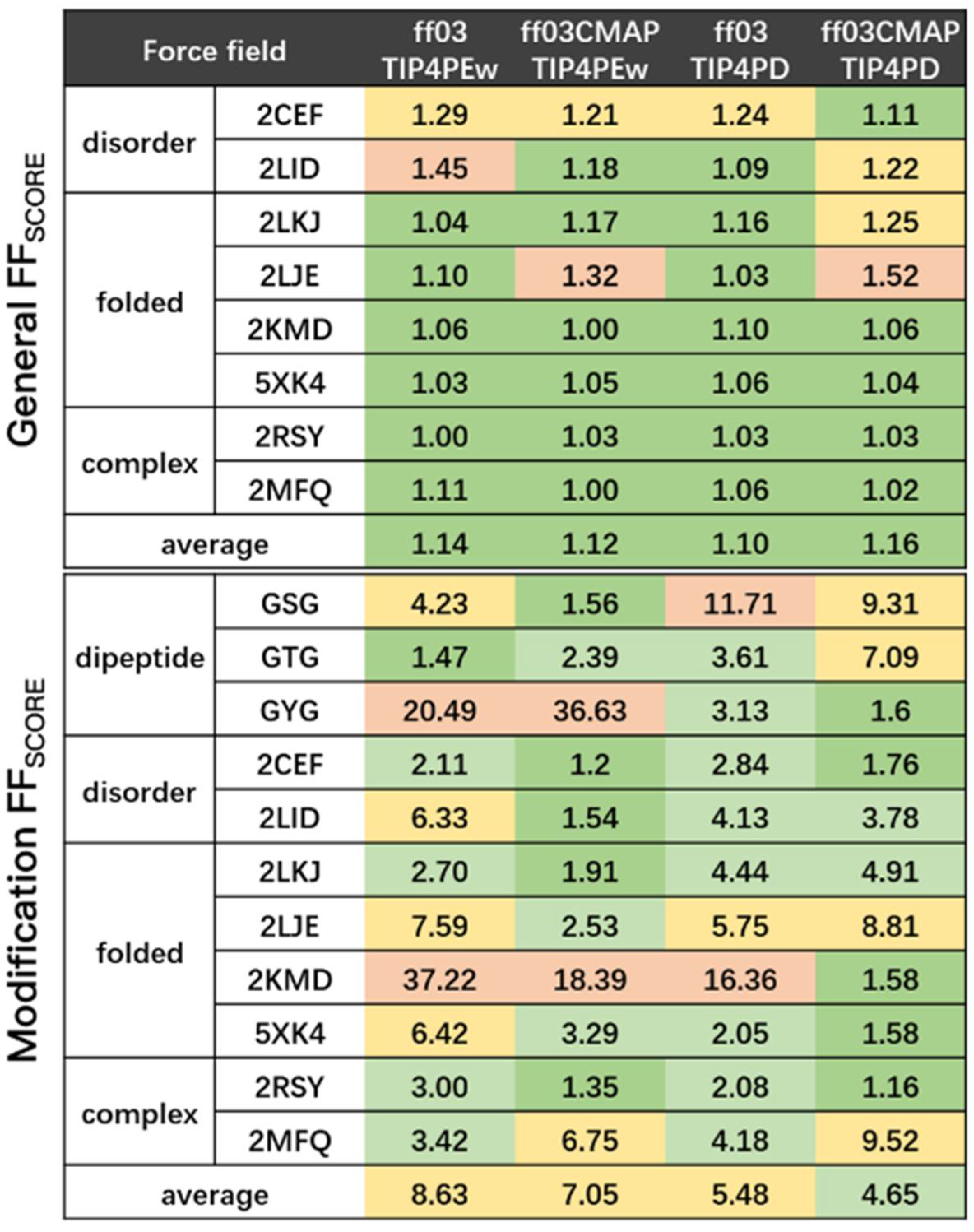
2.3. Evaluation Metrics

To compare the performance of various force fields and solvent models, we used a series of quantification metrics to evaluate simulation results. For dynamical properties, RMSD, RMSF and radius of gyration (Rg) were used for evaluation, and all of these metrics were calculated by CPPTRAJ in AmberTools [67]. For protein structural properties, chemical shifts, secondary structures and contact maps were used in comparison. Chemical shifts were calculated by SPARTA+ [68] for Cα, Cβ, C, N, Hα and HN atom types [68]. The detailed experimental data are listed in Table S1. Secondary structure and contact maps were also calculated by CPPTRAJ in AmberTools [67]. The PyMOL molecular visualization system was used to present structures for all simulated proteins [69].
In addition, the quantification metric force field was used for the benchmark for all methods. The force field score was calculated as in Equation (1) [70]
FF score   = 1 N N i = 1 FF rmsd rmsd norm  
where N is the number of classes of experimental measurements, FF rmsd is the RMSD of the i   th class for simulated and experimental values, and rmsd norm is the lowest RMSD of i   th class in all force fields. Based on the force field score, which considers the overall properties of a protein, we crafted another metric called the modification site force field score to evaluate the experimental values corresponding to post-translational modification site. The modification site force field score was defined as in Equation (2)
ModificationFF score   = 1 N N i = 1 ModificationFF rmsd rmsd norm  
where ModificationFF rmsd is the RMSD of the i   th class for simulated and experimental values in modified residues, and rmsd norm is the lowest RMSD of i   th class among all force fields in modified residues. According to this metric, FFscore and ModificationFFscore are always larger than or equal to 1, and 1 is the best score theoretically.

3. Results

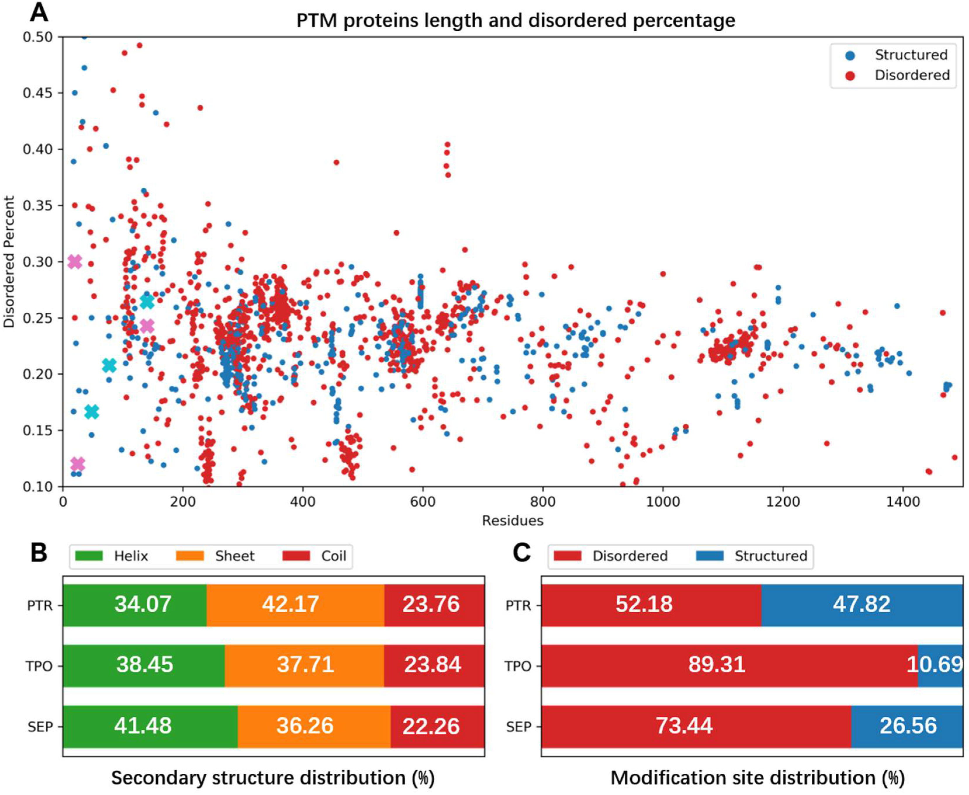
3.1. Structural Statistics of Phosphorylated Proteins in PDB Database

As we compared the secondary structure propensity of phosphorylation modifications in PDB, we found distinct secondary structure propensity of the modification sites. Phosphorylated proteins are widely distributed in both disordered and folded proteins. Most of the proteins havd a folded conformation and only a small number of proteins were disordered (>50% disordered region) (shown in Figure 1A). In all three kinds of phosphorylated proteins, secondary structures were distributed similarly among the helix, sheet and coil. The helix and sheet take up around 40%, and the coil usually takes up the smallest part at 22~23% (Figure 1B). On the contrary, the secondary structure distribution of phosphorylated sites was significantly different. Most of the phosphorylation sites occurred in disordered regions; 52.18% of phosphotyrosine, 73.44% of phosphoserine, and 89.31% of phosphothreonine were located in disordered regions (Figure 1C). Considering the low percentage of coil structure in the overall structure, there is a clear tendency for phosphorylation to occur in disordered regions.
To sum up, phosphorylated proteins in PDB had a balanced distribution on major secondary structures, but phosphorylation sites tended to be located in disordered regions. Therefore, the precise sampling for disordered regions should be important. The findings suggested that a balanced force field for phosphorylated proteins’ simulation might achieve better accuracy in proteins with both folded and disordered regions.

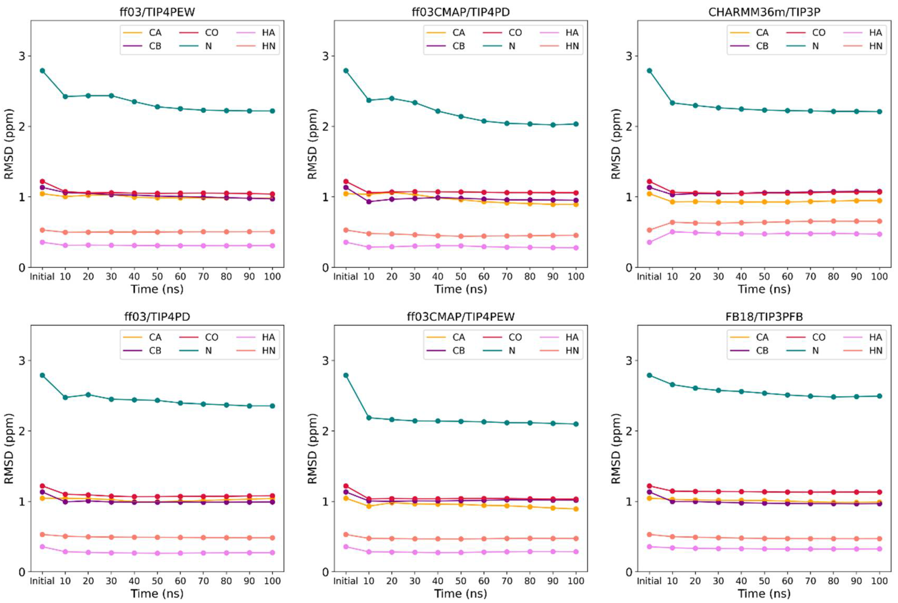
3.2. Convergence of Simulation System

In order to evaluate the convergence, five independent trajectories of 100 ns each were obtained for each system. Convergence of the simulation needs to be considered first because it reflects the adequacy of the simulation time. We calculated the RMSE between the secondary chemical shifts of experimental value and the cumulative average value over time during the simulation. The results show that the simulation of the representative Ets1 system converges under ff03CMAP, ff03, FB18, and CHARMM36m force fields after 100 ns (shown in Figure 2). At the same time, we also compared the performance of sampling between multiple short trajectories (5*100 ns) and a single long trajectory (1000 ns). The results indicate that multiple short trajectories have better sampling ability than a single long trajectory (shown in Figure S2).

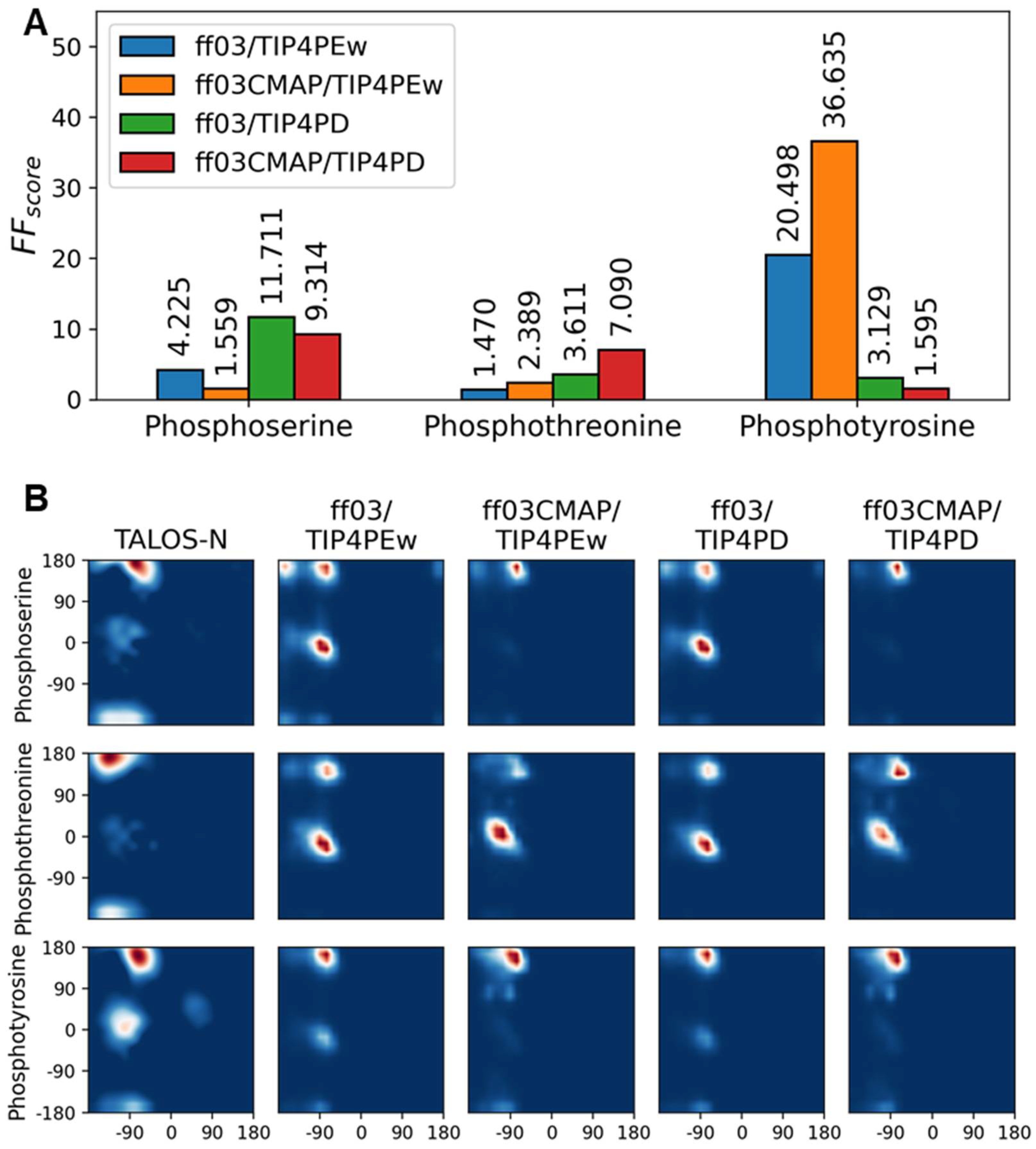
3.3. Phosphorylated Dipeptides

The simulations of phosphoserine, phosphothreonine and phosphotyrosine dipeptides behaved inconsistently in all four force fields. Using the FFscore as the metric, two force fields with TIP4Pew in phosphoserine, ff03/TIP4Pew in phosphothreonine, and ff03CMAP/TIP4PD in phosphotyrosine had the best performance. None of the four force fields performed optimally in all three types of phosphorylation (shown in Figure 3A).
In addition to chemical shifts, Ramachandran plots in Figure 3B presented the backbone dihedral angle distribution of all simulated dipeptides. TALOS-N [71]-predicted distributions from experimental chemical shifts were used as references. In reference Ramachandran plots, phosphoserine and phosphotyrosine were enriched in the PPII region, while phosphothreonine was enriched in the sheet region. Only simulation results from ff03CMAP/TIP4PD had a PPII- and sheet-like distribution, while those from other force fields had high helical propensity in phosphoserine and phosphothreonine.
In general, all force fields have similar accuracy and inconsistent performance in all three types of phosphorylated dipeptides, and ff03CMAP has a small advantage over ff03 in force field scores and dihedral distributions. This is consistent with the previous studies of Vymetal J et al. [30], showing that force fields showed inconsistent performance on phosphorylated dipeptides. Therefore, further evaluation was performed for modified disordered proteins and folded proteins.

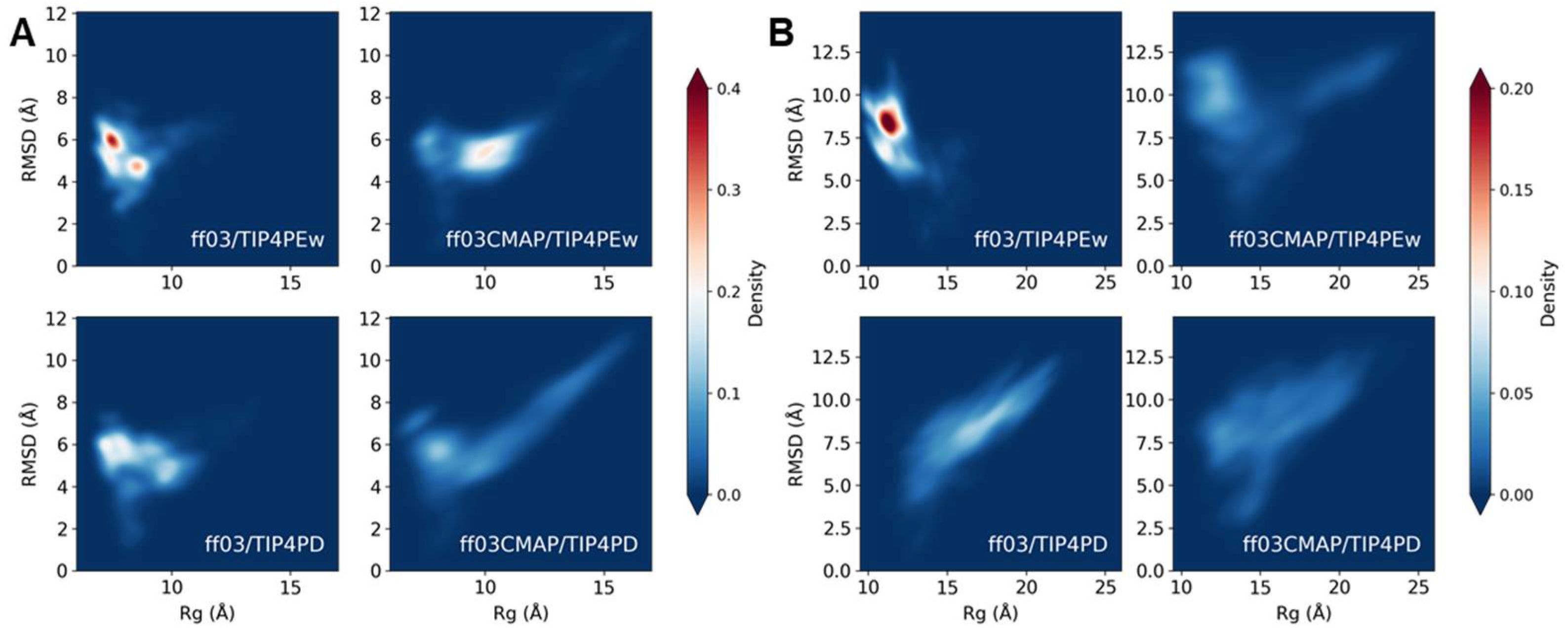
3.4. Phosphorylated Disordered Proteins

As discovered in statistical analysis through PDB, most phosphorylation sites were located in disordered regions. Further simulation tests on phosphorylated disordered proteins can serve as paradigms (references) for further (future) research. Two modified disordered proteins, tissue factor cytoplasmic tail (TF, PDB:2CEF) [53] and vitellogenin (Vg, PDB:2LID) [54], were selected as simulation targets.
Figure 4 shows the RMSD of chemical shifts, overall force field score (FFscore) and modification site force field score (Modification Score). Overall force field scores show that ff03/TIP4PD and ff03CMAP/TIP4PD were the best, while modification site force field scores preferred ff03CMAP/TIP4PEw. This elucidated that if we focus on the modification site, the performance of ff03CMAP is better than that of the ff03 force field. Radius of gyration (Rg) and end-to-end distance distribution showed that four force fields have a huge difference in simulated Rg distribution (shown in Figure S3). ff03/TIP4PEw has the lowest average Rg which represents a more condensed status, while ff03CMAP/TIP4PD has the largest average and the widest range of Rg, which represents the extended structure of disordered proteins. In simulated ensembles’ secondary structure, ff03CMAP tends to have a more disordered structure than ff03, while the difference between TIP4PEw and TIP4PD is not significant (shown in Figure S4).
Sampling is another important factor for disordered protein simulation. The RMSD-Rg free energy landscape indicator can help to predict the sampling efficiency of different force fields. For disordered proteins, a wider energy landscape means the simulated structure will be more reasonable. As shown in Figure 3B, the energy landscape of ff03/TIP4PEw constrained in several conformations, while other force fields obtained a more adequate sampled conformation. With a better sampled landscape, ff03CMAP was able to obtain a more disordered conformation.
We compared the Ramachandran plot between simulation and the TALOS-N prediction from experimental data. The results show that ff03CMAP/TIP4PD has a propensity to be distributed in the PPII region, similar to the pattern found in phosphorylated dipeptides. This propensity has a superimposed effect, which means when we use the combination of ff03CMAP and TIP4PD, it will have a higher proportion in the PPII region. Analysis of hydrogen bonds also showed a difference between ff03 and ff03CMAP, as ff03CMAP has less hydrogen bonds in all residues and modification sites (shown in Figure S5). After comparing chemical shifts, force field scores, Rg distributions, secondary structures, and energy landscapes, we can conclude that ff03CMAP/TIP4PD is the best system for phosphorylated disordered proteins.

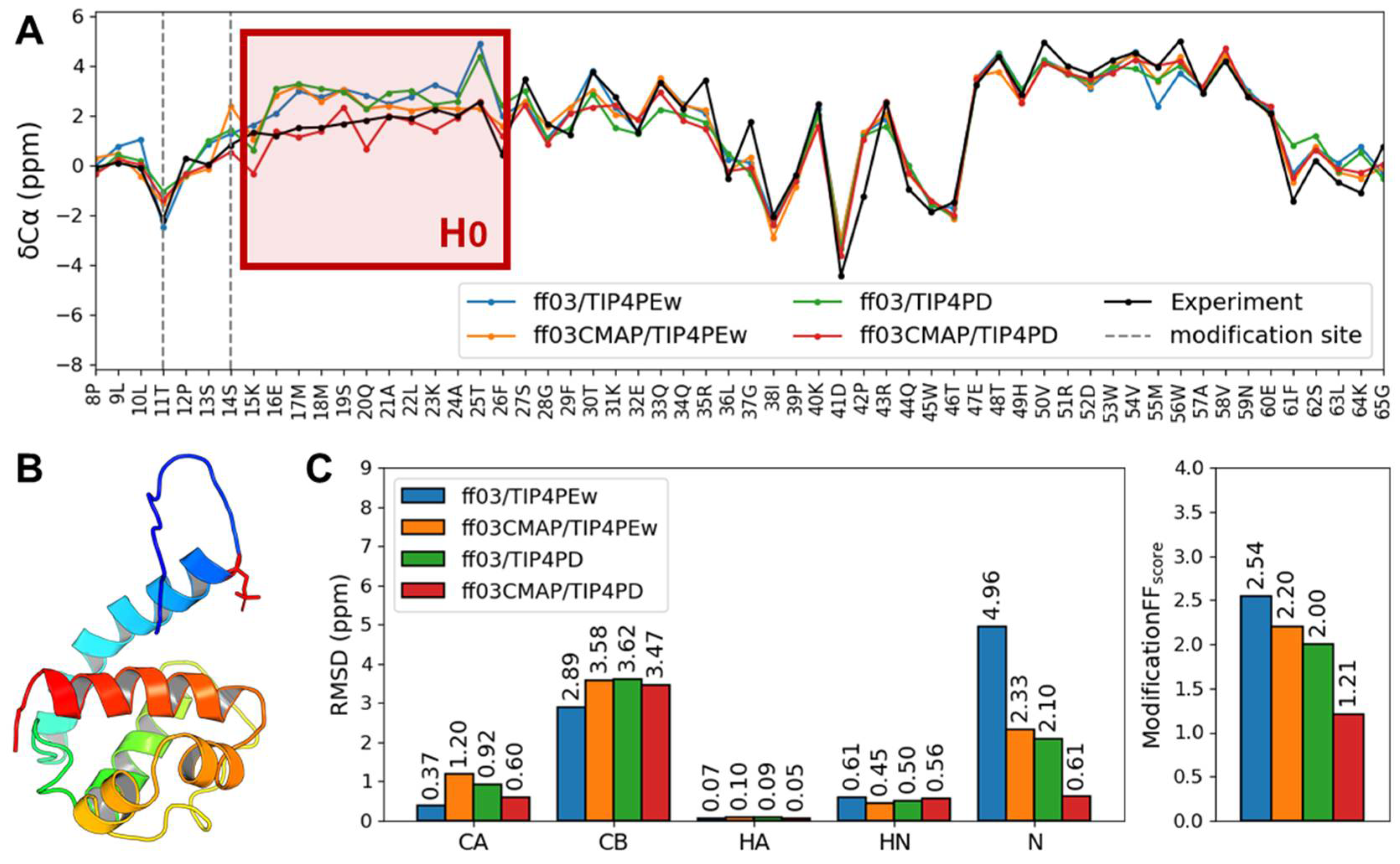
3.5. Phosphorylated Folded Proteins and Complexes

Folded protein takes up the largest part of phosphorylated proteins. In nature, most protein function depends on specific structures, indicating that they are typically folded proteins with a defined, stable tertiary structure. In structural analysis, we found phosphorylation sites were usually located at the loop region (and also disordered regions) in a folded protein, or sometimes at a disordered protein which was bound to a folded protein. Therefore, we chose four folded proteins and two folded-disordered protein complexes as simulation targets for evaluation.
First, we compared the chemical shifts and force field scores. The overall force field scores of four force field systems had similar values and were close to 1. This means that from a holistic perspective, four force fields all have good performance, which is consistent with the previous works [35]. However, the modification site force field score was not unanimous among all four simulation systems. In folded modified proteins and folded-disordered protein complexes, ff03CMAP obviously had a better modification force field score than the ff03 force field. In 2LKJ and 2LJE, ff03CMAP/TIP4PEw had the lowest modification site force field score, and in 2KMD and 5XK4, ff03CMAP/TIP4PD had the lowest modification site force field score.
In order to compare, two representative proteins were chosen for phosphorylated Ets1 (PDB: 2KMD) [57] and Ubiquitin (PDB: 5XK4) [58]. Ets1 is a reaction component of the Ras system. The two phosphorylated residues on Ets1 N terminal disordered region provide this motif with negative charge and repel its neighbor H0 motif from the negatively charged protein surface [57]. Therefore, the properties of the H0 helix should be key information for the simulation. In Figure 5A, we find that the secondary chemical shift for the H0 helix with ff03CMAP/TIP4PD has the best agreement with the experimental one among these force fields. RMSF (root mean square fluctuation) also shows that ff03CMAP has a relatively higher fluctuation than ff03 simulated ensembles (shown in Figure S6). Additionally, the hydrogen bond analysis of Ets1 shows that ff03CMAP/TIP4PD had the least hydrogen bonds connected with two modification sites (shown in Figure S7). All of these results reflect that ff03CMAP/TIP4PD have a higher disorder propensity than other force fields, having secondary chemical shifts closer to zero, fewer hydrogen bonds and higher RMSF.
For phosphorylated ubiquitin, multiple sites of phosphorylation can act as a pH sensor in the cell [58]. These multi-phosphorylated ubiquitin can be found around mitochondria. In phosphorylated ubiquitin, its conformational switch between protonated and unprotonated form plays a key role in its function, and we chose unprotonated single phosphorylated ubiquitin as the target-simulated protein. The result of chemical shifts and force field scores indicated that phosphorylated ubiquitin had low force field scores and was similar to the folded protein Ets1. However, at the modified serine on the disordered region, the modification site force field score of ff03CMAP/TIP4PD was obviously better than other force fields (Figure 5B). In addition, concerning the Ramachandran plot of modification sites, we also discovered that ff03/TIP4PEw, which were the benchmark methods in previous research, formed an unusual distribution of dihedral angles (shown in Figure S8).
We also tested the performance of CHARMM36m [50] and FB18 [51] force fields on the folded protein Ets1 and the disordered protein TF, the chemical shift RMSD and force field scores of which are shown in Figures S9 and S10. The results show that FB18 had better performance than CHARMM36m for both systems, and close with the performance of ff03CMAP. For the disordered protein TF, ff03CMAP/TIP4PD had the lowest general force field score, while FB18 had the lowest modification force field score. For folded protein Ets-1, all force fields except for CHARMM36m had a relative optimal general force field score (less than 1.1), while ff03CMAP/TIP4PD had the lowest modification force field score.

4. Discussion

In our research, the main goal is to find an ideal force field for molecular dynamics simulations of post-translationally modified proteins. First, we studied the structural features of post-translationally modified proteins by analyzing the PDB protein structure database. We found that phosphorylation modifications, which occur most predominantly in post-translational modifications, have a distinct tendency to occur in the disordered regions of the protein. The conventional force fields for folded proteins sometimes might have inaccurate simulations of disordered proteins or disordered regions. Furthermore, previous studies have shown that existing force fields for post-translational modification are not accurate and consistent enough to simulate post-translationally modified peptides or proteins [29]. Therefore, we introduced the balanced ff03CMAP force field, which can perform well in molecular dynamics simulations of both folded and disordered proteins, to compare with the conventional ff03 force field.
By simulating a series of phosphorylated dipeptides, disordered proteins, folded proteins, and disordered-folded protein complexes, we summarized the results of the simulations of several force fields and focused on the discussion of the ff03CMAP force field and ff03 force field with TIP4PEw and TIP4PD solvent models (shown in Figure 6).
The comparison with force fields, including CHARMM36m and FB18 on folded proteins, indicates that the ForceField_PTM compatible ff03 force field family is ideal for the simulation of phosphorylated proteins in a broader view of force fields. For the ff03 force field family, the ff03CMAP force field showed improvement in simulation results of phosphorylated disordered proteins with less difference from experimental values compared with the ff03 force field, and TIP4PD was better between the two solvent models. In folded proteins, the two force fields performed similarly, but when the post-translational modification site was located in the disordered region, the modification site simulation results were closer to the experimental values using the ff03CMAP force field. In the disordered-folded protein complexes, the test results were similar to those of the folded protein, and the post-translational modification site on the occurring disordered protein was closer to the experimental value in the simulation results of ff03CMAP. Overall, the simulations using the ff03CMAP force field outperformed the ff03 force field when simulating biomolecules with post-translational modification sites located on disordered proteins or disordered regions.
Based on previous studies on potential charge in post-translational modification sites, it has been widely accepted that charge is an important component in the process of modeling post-translational modification sites [24,70]. In this research, our analysis reveals that the vast majority of phosphorylation occurs in the disordered region of the protein, which leads us to consider introducing CMAP energy for dihedral angles to improve the simulation of phosphorylated proteins. CMAP energy has been widely used to simulate disordered proteins [38,39,40,41,42,43,44] and can also be used to improve the balanced force field for disordered and structural proteins [35]. We have tried using the balanced force field to simulate phosphorylated proteins with both structured and disordered regions, and indeed the simulation of the modification sites in the disordered region was more accurate than other force field. This further illustrates that the methods used to develop disordered protein force fields can be used to improve the simulation of phosphorylated proteins.
However, the ff03CMAP force field also has limitations. Considering the simulation results of dipeptides, we found that neither the ff03 force field nor the ff03CMAP force field was the consistent result for all three post-translational modifications. Similarly, in the protein simulations, we found that the dihedral angle distribution at the post-translational modification sites still notably differed from the predicted dihedral angle distribution. ff03CMAP still needs further improvement in the dihedral angle distribution at the post-translational modification sites.

Supplementary Materials

The following supporting information can be downloaded at: https://www.mdpi.com/article/10.3390/ijms231911285/s1.

Author Contributions

Conceptualization, B.Z. and H.-F.C.; methodology, B.Z. and G.S.; software, B.Z. and G.S.; validation, B.Z. and G.S.; formal analysis, B.Z. and G.S.; investigation, B.Z. and G.S.; data curation, B.Z. and G.S.; writing—original draft preparation, B.Z. and G.S.; writing—review and editing, H.-F.C.; visualization, H.-F.C.; supervision, H.-F.C.; project administration, H.-F.C.; funding acquisition, Y H.-F.C. All authors have read and agreed to the published version of the manuscript.

Funding

This research received no external funding.

Data Availability Statement

Not applicable.

Acknowledgments

This work was supported by the Center for HPC at Shanghai Jiao Tong University, the National Key Research and Development Program of China (2020YFA0907700) and the National Natural Science Foundation of China (21977068 and 32171242).

Conflicts of Interest

The authors declare no conflict of interest.

References

  1. Khoury, G.A.; Baliban, R.C.; Floudas, C.A. Proteome-Wide Post-Translational Modification Statistics: Frequency Analysis and Curation of the Swiss-Prot Database. Sci. Rep. 2011, 1, 90. [Google Scholar] [CrossRef] [PubMed]
  2. Ardito, F.; Giuliani, M.; Perrone, D.; Troiano, G.; Muzio, L.L. The Crucial Role of Protein Phosphorylation in Cell Signaling and Its Use as Targeted Therapy (Review). Int. J. Mol. Med. 2017, 40, 271–280. [Google Scholar] [CrossRef] [PubMed]
  3. Terman, J.R.; Kashina, A. Post-Translational Modification and Regulation of Actin. Curr. Opin. Cell Biol. 2013, 25, 30–38. [Google Scholar] [CrossRef] [PubMed]
  4. Jenuwein, T.; Allis, C.D. Translating the Histone Code. Science 2001, 293, 1074–1080. [Google Scholar] [CrossRef]
  5. Zecha, J.; Gabriel, W.; Spallek, R.; Chang, Y.-C.; Mergner, J.; Wilhelm, M.; Bassermann, F.; Kuster, B. Linking Post-Translational Modifications and Protein Turnover by Site-Resolved Protein Turnover Profiling. Nat. Commun. 2022, 13, 165. [Google Scholar] [CrossRef]
  6. Appella, E.; Anderson, C.W. Post-Translational Modifications and Activation of P53 by Genotoxic Stresses. Eur. J. Biochem. 2001, 268, 2764–2772. [Google Scholar] [CrossRef]
  7. Govin, J.; Caron, C.; Lestrat, C.; Rousseaux, S.; Khochbin, S. The Role of Histones in Chromatin Remodelling during Mammalian Spermiogenesis. Eur. J. Biochem. 2004, 271, 3459–3469. [Google Scholar] [CrossRef]
  8. Almeida, K.H.; Sobol, R.W. A Unified View of Base Excision Repair: Lesion-Dependent Protein Complexes Regulated by Post-Translational Modification. DNA Repair 2007, 6, 695–711. [Google Scholar] [CrossRef]
  9. Csizmok, V.; Forman-Kay, J.D. Complex Regulatory Mechanisms Mediated by the Interplay of Multiple Post-Translational Modifications. Curr. Opin. Struct. Biol. 2018, 48, 58–67. [Google Scholar] [CrossRef]
  10. Elbaum, M.B.; Zondlo, N.J. OGlcNAcylation and Phosphorylation Have Similar Structural Effects in α-Helices: Post-Translational Modifications as Inducible Start and Stop Signals in α-Helices, with Greater Structural Effects on Threonine Modification. Biochemistry 2014, 53, 2242–2260. [Google Scholar] [CrossRef]
  11. Dunker, A.K.; Lawson, J.D.; Brown, C.J.; Williams, R.M.; Romero, P.; Oh, J.S.; Oldfield, C.J.; Campen, A.M.; Ratliff, C.M.; Hipps, K.W.; et al. Intrinsically Disordered Protein. J. Mol. Graph. Model. 2001, 19, 26–59. [Google Scholar] [CrossRef]
  12. Bah, A.; Forman-Kay, J.D. Modulation of Intrinsically Disordered Protein Function by Post-Translational Modifications. J. Biol. Chem. 2016, 291, 6696–6705. [Google Scholar] [CrossRef] [PubMed]
  13. Owen, I.; Shewmaker, F. The Role of Post-Translational Modifications in the Phase Transitions of Intrinsically Disordered Proteins. Int. J. Mol. Sci. 2019, 20, 5501. [Google Scholar] [CrossRef]
  14. Bah, A.; Vernon, R.M.; Siddiqui, Z.; Krzeminski, M.; Muhandiram, R.; Zhao, C.; Sonenberg, N.; Kay, L.E.; Forman-Kay, J.D. Folding of an Intrinsically Disordered Protein by Phosphorylation as a Regulatory Switch. Nature 2015, 519, 106–109. [Google Scholar] [CrossRef]
  15. Wu, J.; Li, D.; Liu, X.; Li, Q.; He, X.; Wei, J.; Li, M.; Rehman, A.U.; Xia, Y.; Wu, C.; et al. IDDB: A Comprehensive Resource Featuring Genes, Variants and Characteristics Associated with Infertility. Nucleic Acids Res. 2021, 49, D1218–D1224. [Google Scholar] [CrossRef]
  16. Slupsky, C.M.; Gentile, L.N.; Donaldson, L.W.; Mackereth, C.D.; Seidel, J.J.; Graves, B.J.; McIntosh, L.P. Structure of the Ets-1 Pointed Domain and Mitogen-Activated Protein Kinase Phosphorylation Site. Proc. Natl. Acad. Sci. USA 1998, 95, 12129–12134. [Google Scholar] [CrossRef] [PubMed]
  17. Zeng, J.; Jiang, F.; Wu, Y. Mechanism of Phosphorylation-Induced Folding of 4E-BP2 Revealed by Molecular Dynamics Simulations. J. Chem. Theory Comput. 2017, 13, 320–328. Available online: https://pubs.acs.org/doi/abs/10.1021/acs.jctc.6b00848?casa_token=C_nyHiGjT5kAAAAA:hbjhgAWkh2t36_rprkez0_2tUqifD0M5tOpTMqg0fMBfLYdIEt0iq2U5dvI5lji5LSyLcHtifeKX1Bi- (accessed on 16 April 2022). [CrossRef]
  18. Humphrey, S.J.; James, D.E.; Mann, M. Protein Phosphorylation: A Major Switch Mechanism for Metabolic Regulation. Trends Endocrinol. Metab. 2015, 26, 676–687. [Google Scholar] [CrossRef]
  19. Millar, A.H.; Heazlewood, J.L.; Giglione, C.; Holdsworth, M.J.; Bachmair, A.; Schulze, W.X. The Scope, Functions, and Dynamics of Posttranslational Protein Modifications. Annu. Rev. Plant Biol. 2019, 70, 119–151. [Google Scholar] [CrossRef]
  20. Smith, L.M.; Kelleher, N.L. Proteoform: A Single Term Describing Protein Complexity. Nat. Methods 2013, 10, 186–187. [Google Scholar] [CrossRef] [Green Version]
  21. Willems, P.; Horne, A.; Van Parys, T.; Goormachtig, S.; De Smet, I.; Botzki, A.; Van Breusegem, F.; Gevaert, K. The Plant PTM Viewer, a Central Resource for Exploring Plant Protein Modifications. Plant J. 2019, 99, 752–762. [Google Scholar] [CrossRef] [PubMed]
  22. Zulawski, M.; Schulze, W.X. The Plant Kinome. In Plant Phosphoproteomics: Methods and Protocols; Methods in Molecular Biology; Schulze, W.X., Ed.; Springer: New York, NY, USA, 2015; pp. 1–23. ISBN 978-1-4939-2648-0. [Google Scholar]
  23. Homeyer, N.; Horn, A.H.; Lanig, H.; Sticht, H. AMBER Force-Field Parameters for Phosphorylated Amino Acids in Different Protonation States: Phosphoserine, Phosphothreonine, Phosphotyrosine, and Phosphohistidine. J. Mol. Modeling 2006, 12, 281–289. [Google Scholar] [CrossRef] [PubMed]
  24. Khoury, G.A.; Thompson, J.P.; Smadbeck, J.; Kieslich, C.A.; Floudas, C.A. Forcefield_PTM: Ab Initio Charge and AMBER Forcefield Parameters for Frequently Occurring Post-Translational Modifications. J. Chem. Theory Comput. 2013, 9, 5653–5674. [Google Scholar] [CrossRef]
  25. Brooks, B.R.; Bruccoleri, R.E.; Olafson, B.D.; States, D.J.; Swaminathan, S.; Karplus, M. CHARMM: A Program for Macromolecular Energy, Minimization, and Dynamics Calculations. J. Comput. Chem. 1983, 4, 187–217. [Google Scholar] [CrossRef]
  26. Brooks, B.R.; Brooks, C.L., III; Mackerell, A.D., Jr.; Nilsson, L.; Petrella, R.J.; Roux, B.; Won, Y.; Archontis, G.; Bartels, C.; Boresch, S.; et al. CHARMM: The Biomolecular Simulation Program. J. Comput. Chem. 2009, 30, 1545–1614. [Google Scholar] [CrossRef] [PubMed]
  27. Margreitter, C.; Petrov, D.; Zagrovic, B. Vienna-PTM Web Server: A Toolkit for MD Simulations of Protein Post-Translational Modifications. Nucleic Acids Res. 2013, 41, W422–W426. [Google Scholar] [CrossRef] [PubMed]
  28. Petrov, D.; Margreitter, C.; Grandits, M.; Oostenbrink, C.; Zagrovic, B. A Systematic Framework for Molecular Dynamics Simulations of Protein Post-Translational Modifications. PLoS Comput. Biol. 2013, 9, e1003154. [Google Scholar] [CrossRef] [PubMed]
  29. Margreitter, C.; Reif, M.M.; Oostenbrink, C. Update on Phosphate and Charged Post-Translationally Modified Amino Acid Parameters in the GROMOS Force Field. J. Comput. Chem. 2017, 38, 714–720. [Google Scholar] [CrossRef]
  30. Vymetal, J.; Jurásková, V.; Vondrášek, J. AMBER and CHARMM Force Fields Inconsistently Portray the Microscopic Details of Phosphorylation. J. Chem. Theory Comput. 2018, 15, 665–679. [Google Scholar] [CrossRef]
  31. Rieloff, E.; Skepö, M. Phosphorylation of a Disordered Peptide—Structural Effects and Force Field Inconsistencies. J. Chem. Theory Comput. 2020, 16, 1924–1935. [Google Scholar] [CrossRef]
  32. Lindorff-Larsen, K.; Piana, S.; Palmo, K.; Maragakis, P.; Klepeis, J.L.; Dror, R.O.; Shaw, D.E. Improved Side-Chain Torsion Potentials for the Amber Ff99SB Protein Force Field. Proteins Struct. Funct. Bioinform. 2010, 78, 1950–1958. [Google Scholar] [CrossRef] [PubMed]
  33. Duan, Y.; Wu, C.; Chowdhury, S.; Lee, M.C.; Xiong, G.; Zhang, W.; Yang, R.; Cieplak, P.; Luo, R.; Lee, T.; et al. A Point-Charge Force Field for Molecular Mechanics Simulations of Proteins Based on Condensed-Phase Quantum Mechanical Calculations. J. Comput. Chem. 2003, 24, 1999–2012. [Google Scholar] [CrossRef]
  34. Maier, J.A.; Martinez, C.; Kasavajhala, K.; Wickstrom, L.; Hauser, K.E.; Simmerling, C. Ff14SB: Improving the Accuracy of Protein Side Chain and Backbone Parameters from Ff99SB. J. Chem. Theory Comput. 2015, 11, 3696–3713. [Google Scholar] [CrossRef] [PubMed]
  35. Zhang, Y.; Liu, H.; Yang, S.; Luo, R.; Chen, H.-F. Well-Balanced Force Field Ff03CMAP for Folded and Disordered Proteins. J. Chem. Theory Comput. 2019, 15, 6769–6780. [Google Scholar] [CrossRef] [PubMed]
  36. Tian, C.; Kasavajhala, K.; Belfon, K.A.A.; Raguette, L.; Huang, H.; Migues, A.N.; Bickel, J.; Wang, Y.; Pincay, J.; Wu, Q.; et al. Ff19SB: Amino-Acid-Specific Protein Backbone Parameters Trained against Quantum Mechanics Energy Surfaces in Solution. J. Chem. Theory Comput. 2020, 16, 528–552. [Google Scholar] [CrossRef]
  37. Mackerell, A.D., Jr. Empirical Force Fields for Biological Macromolecules: Overview and Issues. J. Comput. Chem. 2004, 25, 1584–1604. [Google Scholar] [CrossRef]
  38. Song, D.; Luo, R.; Chen, H.-F. The IDP-Specific Force Field Ff14IDPSFF Improves the Conformer Sampling of Intrinsically Disordered Proteins. J. Chem. Inf. Model. 2017, 57, 1166–1178. [Google Scholar] [CrossRef]
  39. Chen, J.; Liu, H.; Cui, X.; Li, Z.; Chen, H.-F. RNA-Specific Force Field Optimization with CMAP and Reweighting. J. Chem. Inf. Model. 2022, 62, 372–385. [Google Scholar] [CrossRef]
  40. Song, D.; Liu, H.; Luo, R.; Chen, H.-F. Environment-Specific Force Field for Intrinsically Disordered and Ordered Proteins. J. Chem. Inf. Model. 2020, 60, 2257–2267. [Google Scholar] [CrossRef]
  41. Yang, S.; Liu, H.; Zhang, Y.; Lu, H.; Chen, H. Residue-Specific Force Field Improving the Sample of Intrinsically Disordered Proteins and Folded Proteins. J. Chem. Inf. Model. 2019, 59, 4793–4805. [Google Scholar] [CrossRef]
  42. Liu, H.; Song, D.; Lu, H.; Luo, R.; Chen, H.-F. Intrinsically Disordered Protein-Specific Force Field CHARMM36IDPSFF. Chem. Biol. Drug Des. 2018, 92, 1722–1735. [Google Scholar] [CrossRef]
  43. Ye, W.; Ji, D.; Wang, W.; Luo, R.; Chen, H.-F. Test and Evaluation of Ff99IDPs Force Field for Intrinsically Disordered Proteins. J. Chem. Inf. Model. 2015, 55, 1021–1029. [Google Scholar] [CrossRef]
  44. Wang, W.; Ye, W.; Jiang, C.; Luo, R.; Chen, H.-F. New Force Field on Modeling Intrinsically Disordered Proteins. Chem. Biol. Drug Des. 2014, 84, 253–269. [Google Scholar] [CrossRef]
  45. Horn, H.W.; Swope, W.C.; Pitera, J.W.; Madura, J.D.; Dick, T.J.; Hura, G.L.; Head-Gordon, T. Development of an Improved Four-Site Water Model for Biomolecular Simulations: TIP4P-Ew. J. Chem. Phys. 2004, 120, 9665–9678. [Google Scholar] [CrossRef]
  46. Piana, S.; Donchev, A.G.; Robustelli, P.; Shaw, D.E. Water Dispersion Interactions Strongly Influence Simulated Structural Properties of Disordered Protein States. J. Phys. Chem. B 2015, 119, 5113–5123. [Google Scholar] [CrossRef]
  47. Berman, H.M.; Westbrook, J.; Feng, Z.; Gilliland, G.; Bhat, T.N.; Weissig, H.; Shindyalov, I.N.; Bourne, P.E. The Protein Data Bank. Nucleic Acids Res. 2000, 28, 235–242. [Google Scholar] [CrossRef]
  48. Kabsch, W.; Sander, C. Dictionary of Protein Secondary Structure: Pattern Recognition of Hydrogen-Bonded and Geometrical Features. Biopolym. Orig. Res. Biomol. 1983, 22, 2577–2637. [Google Scholar] [CrossRef]
  49. Touw, W.G.; Baakman, C.; Black, J.; Te Beek, T.A.; Krieger, E.; Joosten, R.P.; Vriend, G. A Series of PDB-Related Databanks for Everyday Needs. Nucleic Acids Res. 2015, 43, D364–D368. [Google Scholar] [CrossRef]
  50. Huang, J.; Rauscher, S.; Nawrocki, G.; Ran, T.; Feig, M.; de Groot, B.L.; Grubmüller, H.; MacKerell, A.D. CHARMM36m: An Improved Force Field for Folded and Intrinsically Disordered Proteins. Nat. Methods 2017, 14, 71–73. [Google Scholar] [CrossRef]
  51. Stoppelman, J.P.; Ng, T.T.; Nerenberg, P.S.; Wang, L.-P. Development and Validation of AMBER-FB15-Compatible Force Field Parameters for Phosphorylated Amino Acids. J. Phys. Chem. B 2021, 125, 11927–11942. [Google Scholar] [CrossRef]
  52. Bienkiewicz, E.A.; Lumb, K.J. Random-Coil Chemical Shifts of Phosphorylated Amino Acids. J. Biomol. NMR 1999, 15, 203–206. [Google Scholar] [CrossRef] [PubMed]
  53. Sen, M.; Herzik, M., Jr.; Craft, J.W., Jr.; Creath, A.L.; Agrawal, S.; Ruf, W.; Legge, G.B. Spectroscopic Characterization of Successive Phosphorylation of the Tissue Factor Cytoplasmic Region. Open Spectrosc. J. 2009, 3, 58. [Google Scholar] [CrossRef] [PubMed]
  54. Havukainen, H.; Underhaug, J.; Wolschin, F.; Amdam, G.; Halskau, Ø. A Vitellogenin Polyserine Cleavage Site: Highly Disordered Conformation Protected from Proteolysis by Phosphorylation. J. Exp. Biol. 2012, 215, 1837–1846. [Google Scholar] [CrossRef] [PubMed]
  55. Deshmukh, L.; Meller, N.; Alder, N.; Byzova, T.; Vinogradova, O. Tyrosine Phosphorylation as a Conformational Switch a CASE Study of Integrin Β3 Cytoplasmic Tail. J. Biol. Chem. 2011, 286, 40943–40953. [Google Scholar] [CrossRef]
  56. Chua, G.-L.; Tang, X.-Y.; Amalraj, M.; Tan, S.-M.; Bhattacharjya, S. Structures and Interaction Analyses of Integrin AMβ2 Cytoplasmic Tails. J. Biol. Chem. 2011, 286, 43842–43854. [Google Scholar] [CrossRef] [PubMed]
  57. Nelson, M.L.; Kang, H.-S.; Lee, G.M.; Blaszczak, A.G.; Lau, D.K.; McIntosh, L.P.; Graves, B.J. Ras Signaling Requires Dynamic Properties of Ets1 for Phosphorylation-Enhanced Binding to Coactivator CBP. Proc. Natl. Acad. Sci. USA 2010, 107, 10026–10031. [Google Scholar] [CrossRef] [PubMed]
  58. Dong, X.; Gong, Z.; Lu, Y.-B.; Liu, K.; Qin, L.-Y.; Ran, M.-L.; Zhang, C.-L.; Liu, Z.; Zhang, W.-P.; Tang, C. Ubiquitin S65 Phosphorylation Engenders a PH-Sensitive Conformational Switch. Proc. Natl. Acad. Sci. USA 2017, 114, 6770–6775. [Google Scholar] [CrossRef]
  59. Tanaka, H.; Akagi, K.; Oneyama, C.; Tanaka, M.; Sasaki, Y.; Kanou, T.; Lee, Y.-H.; Yokogawa, D.; Dobenecker, M.-W.; Nakagawa, A.; et al. Identification of a New Interaction Mode between the Src Homology 2 Domain of C-Terminal Src Kinase (Csk) and Csk-Binding Protein/Phosphoprotein Associated with Glycosphingolipid Microdomains. J. Biol. Chem. 2013, 288, 15240–15254. [Google Scholar] [CrossRef]
  60. Zeng, L.; Kuti, M.; Mujtaba, S.; Zhou, M.-M. Structural Insights into FRS2α PTB Domain Recognition by Neurotrophin Receptor TrkB. Proteins Struct. Funct. Bioinform. 2014, 82, 1534–1541. [Google Scholar] [CrossRef]
  61. Lee, T.-S.; Cerutti, D.S.; Mermelstein, D.; Lin, C.; LeGrand, S.; Giese, T.J.; Roitberg, A.; Case, D.A.; Walker, R.C.; York, D.M. GPU-Accelerated Molecular Dynamics and Free Energy Methods in Amber18: Performance Enhancements and New Features. J. Chem. Inf. Model. 2018, 58, 2043–2050. [Google Scholar] [CrossRef]
  62. Ryckaert, J.-P.; Ciccotti, G.; Berendsen, H.J.C. Numerical Integration of the Cartesian Equations of Motion of a System with Constraints: Molecular Dynamics of n-Alkanes. J. Comput. Phys. 1977, 23, 327–341. [Google Scholar] [CrossRef]
  63. Darden, T.; York, D.; Pedersen, L. Particle Mesh Ewald: An N·log(N) Method for Ewald Sums in Large Systems. J. Chem. Phys. 1993, 98, 10089–10092. [Google Scholar] [CrossRef]
  64. Salomon-Ferrer, R.; Götz, A.W.; Poole, D.; Grand, S.L.; Walker, R.C. Routine Microsecond Molecular Dynamics Simulations with AMBER on GPUs. 2. Explicit Solvent Particle Mesh Ewald. J. Chem. Theory Comput. 2013, 9, 3878–3888. Available online: https://pubs.acs.org/doi/abs/10.1021/ct400314y (accessed on 16 April 2022). [CrossRef] [PubMed]
  65. Rieloff, E.; Skepö, M. Molecular Dynamics Simulations of Phosphorylated Intrinsically Disordered Proteins: A Force Field Comparison. Int. J. Mol. Sci. 2021, 22, 10174. [Google Scholar] [CrossRef]
  66. Rieloff, E.; Skepö, M. The Effect of Multisite Phosphorylation on the Conformational Properties of Intrinsically Disordered Proteins. Int. J. Mol. Sci. 2021, 22, 11058. [Google Scholar] [CrossRef] [PubMed]
  67. Roe, D.R.; Cheatham, T.E., III. PTRAJ and CPPTRAJ: Software for Processing and Analysis of Molecular Dynamics Trajectory Data. J. Chem. Theory Comput. 2013, 9, 3084–3095. [Google Scholar] [CrossRef]
  68. Shen, Y.; Bax, A. SPARTA+: A Modest Improvement in Empirical NMR Chemical Shift Prediction by Means of an Artificial Neural Network. J. Biomol. NMR 2010, 48, 13–22. [Google Scholar] [CrossRef] [PubMed]
  69. Schrödinger, L. The PyMOL Molecular Graphics System, Version 1.8; Schrodinger, LLC: New York, NY, USA, 2015. [Google Scholar]
  70. Robustelli, P.; Piana, S.; Shaw, D.E. Developing a Molecular Dynamics Force Field for Both Folded and Disordered Protein States. Proc. Natl. Acad. Sci. USA 2018, 115, E4758–E4766. [Google Scholar] [CrossRef]
  71. Shen, Y.; Bax, A. Protein Backbone and Sidechain Torsion Angles Predicted from NMR Chemical Shifts Using Artificial Neural Networks. J. Biomol. NMR 2013, 56, 227–241. [Google Scholar] [CrossRef] [Green Version]
Figure 1. Structural analysis of phosphorylated proteins in PDB. Distributions of phosphorylated proteins’ disorder percentage and protein length (A), total protein secondary structure (B) and modification site secondary structure (C). In (A), each point represents a phosphorylated protein in the PDB database, and its color shows whether it had a phosphorylated residue in a disordered region (red) or not (blue). Selected simulation systems are marked by a cross in pink (modification in disordered region) and cyan (modification in folded region). In (B), the white numbers show the three kinds of secondary structures’ percentage in all residues in modified proteins. In (C), we present the two kinds (helix and sheet are considered as structured) of secondary structures’ percentage in all three kinds of phosphorylation site. (B,C) show that the phosphorylation is usually located in disordered regions.
Ijms 23 11285 g001
Figure 2. Time-dependent RMS error of chemical shift for Ets1 between experiment data and simulated ones with six force fields.
Ijms 23 11285 g002
Figure 3. Simulation results of phosphorylated dipeptides. Comparison of force field scores (A) present the inconsistency of different force fields. Predicted and simulated Ramachandran plots (B) showed simulated distribution of backbone dihedral angle distribution difference between experiments and simulations.
Ijms 23 11285 g003
Figure 4. Energy landscape in MD simulation of phosphorylated disordered proteins. Comparison of different force field energy landscapes of phosphorylated disordered protein TF (A) and Vg (B) showed sampling efficiency advantage of ff03CMAP force field with both solvent models.
Ijms 23 11285 g004
Figure 5. Simulation results of phosphorylated folded protein Ets1. Secondary chemical shifts of Ets1 around H0 region (A) showed only ff03CMAP/TIP4PD simulated the H0 helix best, whose conformation was greatly affected by close-site phosphorylations. Ets1 structure and its phosphorylation sites are shown in (B). Chemical shift RMSD of modified residues and modification force field scores (C) also support that ff03CMAP/TIP4PD performed best in phosphorylated residues.
Ijms 23 11285 g005
Figure 6. Overall force field score and modification site force field score of all simulated systems. Green represents a General FFscore lower than 1.2 and Modification score lower than 2. Light green represents a Modification FFscore between 2 and 5. Yellow represents a General FFscore from 1.2 to 1.3 and Modification FFscore from 5 to 10. Red represents a General FFscore higher than 1.3 and Modification Ffscore higher than 10.
Ijms 23 11285 g006
Table 1. All simulated phosphorylated systems and simulation conditions.
SystemDescriptionModification TypeLengthInitial StructureIonsNumber of WatersSimulation Time (ns)Number of Tested Force FieldsNumber of Trajectories
Dipeptide
GpSG [52]Phosphoserine dipeptidepSer3Extended2 Na+96250041
GpTG [52]Phosphothreonine dipeptidepThr3Extended2 Na+81150041
GpYG [52]Phosphotyrosine dipeptidepTyr3Extended2 Na+108950041
Disordered Protein
TF [53]Cytoplasmic Tail of Tissue Factor2*pSer192CEF2 Na+251110065
Vg [54]VitellogeninpSer352LID7 Na+631610045
Folded Protein
β3 [55]β3 cytoplasmic tailpSer242LKJ1 Na+330310045
αM [56]αM cytoplasmic tail2*pTyr472LJE2 Na+382810045
Ets1 [57]Ets1pSer, pThr1112KMD9 Na+764410065
100041
p-Ubiquitin [58]Phosphorylated ubiquitinpSer765XK42 Na+361310045
Complex
SH2 [59]SH2 domain of Csk in complex with a phosphopeptide from CbppTyr1372RSY7 Na+826710045
TrkB [60]FRS2a PTB domain with neurotrophin receptor TrkBpTyr1382MFQ7 Na+875610045
Publisher’s Note: MDPI stays neutral with regard to jurisdictional claims in published maps and institutional affiliations.

