Review

New Advances in Using Virus-like Particles and Related Technologies for Eukaryotic Genome Editing Delivery

1
School of Physical Education, Hangzhou Normal University, Hangzhou 311121, China
2
Wake Forest Institute for Regenerative Medicine, Wake Forest University Health Sciences, Winston-Salem, NC 27157, USA
*
Author to whom correspondence should be addressed.
Int. J. Mol. Sci. 2022, 23(15), 8750; https://doi.org/10.3390/ijms23158750
Submission received: 14 July 2022 / Revised: 29 July 2022 / Accepted: 31 July 2022 / Published: 6 August 2022
(This article belongs to the Collection Feature Papers in “Molecular Biology”)

Abstract

:
The designer nucleases, including Zinc Finger Nuclease (ZFN), Transcription Activator-Like Effector Nuclease (TALEN), and Clustered Regularly Interspaced Short Palindromic Repeats/CRISPR-associated (CRISPR/Cas), have been widely used for mechanistic studies, animal model generation, and gene therapy development. Clinical trials using designer nucleases to treat genetic diseases or cancers are showing promising results. Despite rapid progress, potential off-targets and host immune responses are challenges to be addressed for in vivo uses, especially in clinical applications. Short-term expression of the designer nucleases is necessary to reduce both risks. Currently, delivery methods enabling transient expression of designer nucleases are being pursued. Among these, virus-like particles as delivery vehicles for short-term designer nuclease expression have received much attention. This review will summarize recent developments in using virus-like particles (VLPs) for safe delivery of gene editing effectors to complement our last review on the same topic. First, we introduce some background information on how VLPs can be used for safe and efficient CRISPR/Cas9 delivery. Then, we summarize recently developed virus-like particles as genome editing vehicles. Finally, we discuss applications and future directions.

1. A Brief Introduction to Designer Endonucleases

Designer endonucleases enable scientists to edit target genomes and achieve gene knockout and DNA addition with unprecedented precision [1]. Early gene editing technologies achieved DNA editing through physical and chemical mutagenesis and homologous recombination. But these methods often fell short in efficiency, specificity, and practicality. The emergence of Zinc Finger Nuclease (ZFN) and Transcription Activator-Like Effector Nuclease (TALEN) greatly improved the precision and the practicality of gene editing. Nowadays, the most popular gene editing tool is the Clustered Regularly Interspaced Short Palindromic Repeats/CRISPR-associated (CRISPR/Cas) system. The development of these designer endonucleases enables researchers to make good use of non-homologous end-joining and homologous recombination for gene editing. However, there are still challenges to be overcome, such as off-target effects and immune responses [2,3]. One strategy to reduce these risks is transient delivery of the nucleases.
In the past several years, many groups have developed various lentivirus- or retrovirus-like particles (VLPs) for delivering mRNA, protein, or ribonucleoprotein of designer endonucleases to improve genome editing safety in mammalian cells and animals. Since our last review on VLPs as delivery tools for genome editing [4], many new developments have been reported [5,6,7,8,9,10,11,12,13,14,15,16]. In this review, we will complement our previous review [4] in light of these new discoveries.
Since many VLP systems used interactions between aptamer and aptamer-binding proteins (ABPs) to recruit RNA or protein into VLPs, we begin with background information about bacteriophage aptamers and ABPs. Building on the information on lentiviral capsid proteins in the previous review [4], our discussions on aptamer and ABP will help readers to better understand the mechanisms for using VLPs for safe CRISPR/Cas9 delivery. Following the summary of new developments in using VLPs to deliver designer nucleases in forms of protein, ribonucleoprotein (RNP), or RNA for genome editing, we introduce the applications of various types of VLPs in genome editing, with emphasis on in vivo applications.
Currently, the commonly used designer endonucleases include ZFN, TALEN and CRISPR/Cas endonucleases. ZFN is an artificially designed nucleic acid endonuclease that contains 3–6 Cys2-His2 zinc finger protein tandem for specific DNA recognition and a non-specific nucleic acid endonuclease from FokI (the carboxy-terminal 96 amino acid residues) for DNA cleavage [17]. Each Fok I monomer is linked to a zinc finger protein to form a ZFN to recognize, bind and cleave a specific site. When the 2 monomeric ZFNs bind DNA in the opposite directions and the 2 recognition sites are at the proper distance (6~8 bp), the 2 FokI domains interact to form an enzymatic active unit and cleave the target DNA to form a double-strand break. As a first-generation gene editing tool, ZFNs have the disadvantages of relatively low gene editing efficiency and high toxicity. TALEN is a second-generation gene editing tool, similarly consisting of DNA recognition domains and FokI nucleic acid endonuclease domains [18,19]. Compared with ZFN, TALEN has a simpler process for design, better gene editing efficiency, and lower toxicity. However, due to the presence of many repeats in the sequence coding for the DNA-binding domains, the resultant DNA tends to be unstable.
The CRISPR/Cas system, as the third-generation artificial endonuclease, has been available since August 2012 [20]. It has become widely used by scientists worldwide in various fields for its simple design, low cost, and high efficiency of gene editing in eukaryotic cells [21,22,23,24]. CRISPR/Cas is an adaptive immune system that recognizes and specifically degrades foreign invading nucleic acid sequences in bacteria and archaea [25]. According to the number of Cas gene effector proteins, CRISPR consists of 2 classes and 5 types, a total of 16 subtypes [26]. Type II Cas proteins are represented by CRISPR/Cas9 proteins, which require only one protein to perform cleavage. During long-term evolution, bacterial immune systems store sequences of viral genomes or plasmids in their own DNA in the form of spacer sequences. The spacer sequences are transcribed into CRISPR RNA (crRNA). Once an invading DNA with a sequence complementary to crRNA (the Protospacer) is detected, Cas protein can be directed to digest the target sequence and resist invasion. The CRISPR/Cas9 endonucleases need two components to be enzymatically active: Cas9 nuclease and single-guide RNA (sgRNA), a fusion RNA from crRNA and trans-activating CRISPR RNA (tracrRNA) [20,27]. Cas9 protein will form a complex with sgRNA as Cas9:sgRNA ribonucleoprotein. When the target sequence is completely complementary to crRNA, the Cas9:sgRNA complex is activated to cleave the target region and cause a double-strand break [28].
The advantages of the CRISPR/Cas system are efficient and specific cleavage of target sites, simultaneous editing at multiple locations, and easy preparation. With these advantages, CRISPR/Cas9 endonucleases can be applied to human gene therapy [29,30,31,32], new drug development, and other biomedical research fields. This new gene editing system has been used in research on a wide range of major human diseases, including cancer, infectious diseases, and genetic and autoimmune diseases, and it facilitates personalized clinical treatment. Many CRISPR-based gene therapy clinical trials have been registered on ClinicalTrials.gov, and some reported promising results [33,34,35,36]. In addition, the CRISPR/Cas system is also widely used in animal model generation [37], crop improvement [38], microbial genome editing [39], and gene expression regulation [40,41,42].
However, the potential of off-target effects and immune responses to the bacteria-derived Cas9 protein induce challenges in in vivo and clinical applications of the CRISPR/Cas system, especially when expressed in cells for the long-term [43,44,45,46]. These challenges hinder the progress of gene editing toward clinical treatments [2,47]. Developing delivery strategies featuring short-term designer nuclease expression is one way to resolve the challenges. Lenti- or retrovirus-like particles (VLPs) can be developed for efficient delivery of CRISPR/Cas effectors for short-term endonuclease expression.

2. Background Knowledge for Developing VLPs as Genome Editing Delivery Vehicles

An understanding of the capsids of lentiviruses and retroviruses is needed to learn the principles of VLPs. Please see our previous review [4] and others for more information on these vectors [48,49]. In many types of VLPs, the specific interactions between aptamers and ABPs were utilized to deliver protein or RNA by VLPs for genome editing. Here, we give a brief introduction to aptamers and ABPs used by various groups in developing VLPs.

RNA Aptamers and Aptamer-Binding Proteins (ABPs)

The study of RNA or DNA aptamers started even before Andy Ellington first coined the term “aptamer” [50]. Aptamers are selective affinity reagents that can be used in a wide range of research, diagnostic, and therapeutic applications. They are usually 20~60 nucleotide single-stranded DNA or RNA molecules that can selectively bind to a specific target [51], including proteins, peptides, carbohydrates, small molecules, metal ions and even live cells. Here, we focus on RNA aptamers used in developing VLPs for genome editing delivery. At least four pairs of RNA aptamers/ABPs—MS2/MS2 coat protein (MCP) [52], PP7/PP7 coat protein (PCP) [53], com/Com [54], and BoxB/λ N22 peptide [55]—have been used in VLP-mediated RNA or RNP delivery (Table 1). Each aptamer specifically binds to its ABP with high affinity.
Aptamer MS2 and ABP MCP (MS2 coat protein) were found in bacteriophage MS2, one of a group of single-stranded RNA coliphages, including the bacteriophages MS2, R17, and f2. These bacteriophages contain only three genes, coding for synthetase, major coat protein, and maturation protein, respectively [56]. Aptamer MS2, adopting a stem–loop motif, is a 19 nt sequence found in the operon of synthetase (Figure 1). Specific binding of MCP to MS2 aptamer represses the translation of synthetase [52,57]. The interaction of MS2 and MCP was first used in studying cellular RNA localization in yeast [58] and subsequently used in dead Cas9 mediated gene regulation [59,60,61] and DNA labeling [62]. Recently, it has been used for VLP-mediated delivery of Cas9 mRNA or RNPs [5,52,63,64].
PP7 is a single-strand RNA bacteriophage of Pseudomonas aeruginosa that is distantly related to coliphage MS2 (Figure 1) [53]. Aptamer PP7 is a short sequence found in the translation initiation region of bacteriophage PP7 replicase gene; similar to aptamer MS2, it also adopts a stem–loop structure. Aptamer PP7 is specifically bound by PP7 coat protein (PCP) with an equilibrium dissociation constant of approximately 1 nm to repress the translation of synthetase [53]. Aptamer PP7 and PCP have been used in yeast [65] and mammalian cells [66] for RNA imaging.
Aptamer BoxB and ABP λ peptide N22 were found in bacteriophage λ (Figure 1). Peptide N22 is the amino terminal part of the λ phage N gene product and specifically binds to RNA hairpin BoxB in N protein-regulated genes to counteract transcription regulation [67]. An engineered λ N22 peptide showed greatly improved affinity to λ aptamer BoxB [55], and this aptamer/ABP pair was used to develop tools for genomic DNA labeling [62].
The RNA aptamer com and its interacting protein Com were found in the bacteriophage Mu (Figure 1) [68]. The sequence of the com aptamer was in the translational initiation region of mom gene. Binding Com protein to the com sequence stimulates the translation of mom gene [54]. The com/Com interaction has been used in dead Cas9-mediated gene regulation [60] and DNA labeling [62].

3. Recently Developed VLPs as Safe Genome Editing Delivery Vehicles

Since our last review of various types of VLPs developed for genome editing delivery [4], many new types of VLPs have been developed for different purposes. These newly developed VLPs and those introduced in our last review are listed in Table 2 (RNP delivery) and Table 3 (RNA delivery). In the following subsections, we will only introduce VLPs reported after the publication of our last review. Readers are referred to our previous review and the original papers listed in the Tables for more information on prior VLPs. We will introduce these recently reported VLPs for RNP delivery (Table 2) and mRNA delivery and mRNA/sgRNA co-delivery (Table 3).

3.1. Using VLPs to Deliver Proteins or Ribonucleoproteinss

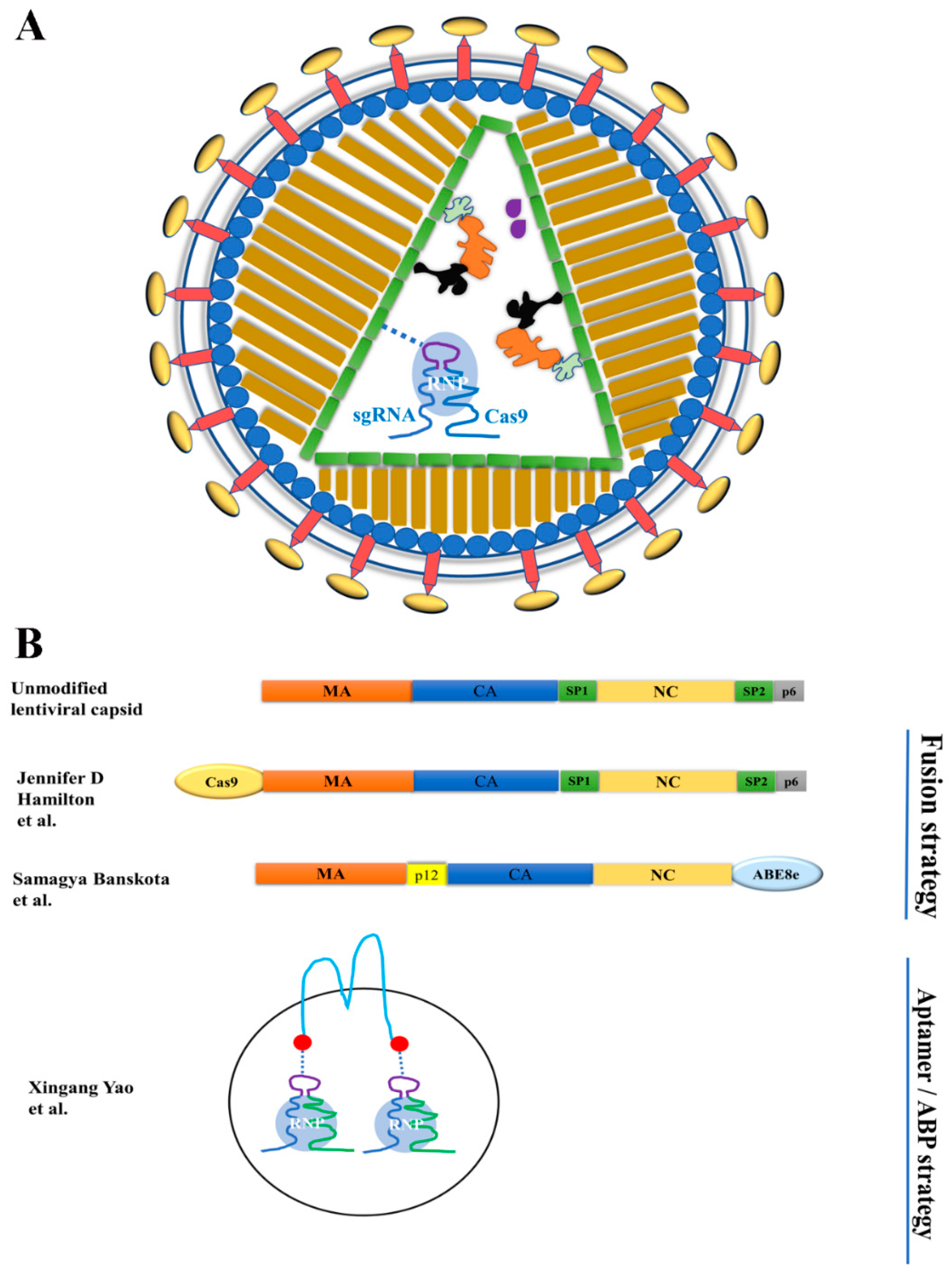
Before being developed as genome editing delivery tools, VLPs were used for protein delivery for the purposes of viral-particle tracking and cell elimination [69,70,71]. The need for transient expression of genome editing effectors promoted the use of VLPs to deliver designer nuclease proteins or ribonucleoproteins (RNPs). Two strategies were used to package cargoes into the particles: the fusion strategy and the aptamer/ABP interaction strategy (Table 2). The fusion strategy was first used by Cai et al. to deliver ZFN and TALEN proteins by VLPs [72]. It was subsequently used by Choi et al. for packaging and delivering Cas9 RNPs (the Cas9:sgRNA complex) [73]. The fusion strategy was first used by Lyu et al. to deliver Cas9 RNPs by VLPs [63]. Here, we also include work using exosomes as delivery vehicles for genome editing [5] since these particles are similar in size to VLPs and also have membranes obtained from the producing cells. Figure 2 illustrates the packaging strategies of the recently reported VLPs.
Table 2. VLP-mediated nuclease protein and RNP delivery.
ReferenceMechanism for Nuclease RecruitmentCapsid TypeEditing Effectors DeliveredExperiment StageGene/Tissue
The fusion strategyCai et al. [72] Fusing editing effector to the N-terminus of GagLVZFN and TALENIn vitroGFP/CCR5/AAVS1cell
Choi et al. [73]Fusing Cas9 protein to the N-terminus of GagLVSpCas9In vitroLTR/CD4/CCR5cell
Mangeot et al. [74]Fusing Cas9 to the C-terminus of MLV GagMLVSpCas9In vivoMYD88/DDX3/GFP/Hpd/Fto/Tyr/LoxPcell
Gee et al. [75]Fusing FKBP12 to Gag, fusing FRB to SpCas9. FKBP12/AP21967/FRB interaction brings SpCas9 to GagLVSpCas9In vivoDMDcell and mouse
Indikova et al. [76]Fusing Cas9 to the C-terminus of VprLVSpCas9In vitroGFP/EMX1/FANCF/HEKs1/HEKs3cell
Hamilton et al. [10]Fusing Cas9 to N-terminal Gag structural proteinLVSpCas9In vitroB2M/TRAC—cell
Banskota et al. [12]Fusing ABE8e to C-terminus of GagMLVSpCas9 and ABEIn vivoBCL11A/COL7A1—cell
Pcsk 9/Dnmt 1—mouse
The Aptamer/ABP StrategyLyu et al. [63,77]Forming a three-component complex: NC-Com/aptamer-sgRNA/Cas9 proteinLVSpCas9 and ABEIn vitroIL2RG/HBB—cell
Lu et al. [78]SaCas9
Yao et al. [5]Forming a three-component complex: CD63-Com/aptamer-sgRNA/Cas9 proteinExosomeSaCas9
SpCas9
ABE
In vivoDMD/IL2RG/HBB—cell
DMD—mouse
Highlighted papers were covered in our previous review [4].

3.1.1. VLP-Mediated Nuclease Delivery Using the Fusion Strategy

VLPs for Co-Delivery of Cas9 RNPs and LV Genomic RNA

Recently, VLPs were harnessed to deliver Cas9 RNPs and DNA templates for both targeted genetic disruption and stable gene addition [10]. In this strategy, Cas9 was fused to the C-terminus of the HIV Gag protein with cleavage linker sequences. The fusion protein will be incorporated into the VLPs formed with the help of unmodified Gag and Pol products. In addition to Cas9 RNPs, lentiviral genomic RNA will also be packaged via the interaction between NC protein and the packaging signal in the lentiviral genomic RNA. Sequences in the genomic RNA can be reverse-transcribed into DNA and integrated into the host genome for long-term gene expression or serve as DNA templates for homologous recombination if the VLPs are integration-defective (with an inactivated integrase). These VLPs could be especially useful in creating chimeric antigen receptor (CAR) T cells with therapeutically relevant gene disruption (e.g., B2M or TRAC). Indeed, pseudotyping the particles with different envelope proteins shifts the tropism of the particles, when transducing T cells.

Recent Applications of Previously Reported VLPs

Recently, there are also new applications of VLPs originally reported by Mangeot et al. [74]. Vindry et al. used this system to prepare VLPs to deliver Cas9 RNPs targeting the selenocysteine-tRNA[Ser]Sec gene in multiple cell types [79]. They observed >80% genome editing efficiency in multiple cell types tested, confirming the efficiency of the VLPs. Gutierrez-Guerrero et al. explored the co-pseudotyping of the VLPs developed by Mangeot et al. [74] with baboon endogenous virus (BaEV) envelope glycoprotein and VSV-G, and they achieved relatively high gene editing efficiency (>25%) in human B, T cells and hematopoietic stem and progenitor cells [9]. Thus, work from multiple groups shows that VLPs can be pseudotyped with different envelope proteins to improve gene editing efficiency in peculiar cell types.

VLPs for Delivering Base Editors

Base editors (BEs), which mediate targeted single-nucleotide conversions, can minimize the risks of generating double-strand breaks compared with artificial endonucleases. They were commonly delivered by plasmid DNA, AAV (needs to be split into two AAV vectors due to the large size of adenine base editors), and LV. Previously Lyu et al. developed VLPs to deliver an adenine base editor (ABE), using the interactions between aptamer and ABP [77]. They found that VLP-delivered ABEs did not show sgRNA-independent RNA off-target effects. Recently, Banskota et al. used a different strategy to deliver base editors by VLPs and called these VLPs engineered DNA-free virus-like particles (eVLP) [12]. They fused ABE8e to the C-terminus of the Friend murine leukemia virus (FMLV) gag polyprotein via a linker peptide to allow for cleavage by the FMLV protease upon particle maturation. They optimized the cleavage site to improve cargo release, added a nucleus export signal to improve cytoplasmic availability of the Gag-cargo for packaging, and adjusted the ratio of Gag-cargo and unmodified Gag to further improve particle generation and base editing activities. These efforts resulted in high efficiency of base editing. The authors successfully used these VLPs for in vivo base editing in mice. Systemically delivered eVLPs efficiently edited the gene Pcsk9 in mouse liver and resulted in a 78% reduction in serum Pcsk9 protein levels [12].

3.1.2. Delivering Cas9 and Base Editor RNPs by Exosomes Using the ABP/Aptamer Interaction Strategy

Previously, Lyu et al. and Lu et al. used the specific interactions between aptamers and ABPs to package and deliver Cas9 and ABE RNPs by VLPs [63,78,80]. Recently, they adapted the strategy to package and deliver Cas9 and ABE RNPs by extracellular vesicles (exosomes) [5], which is an alternative delivery vehicle to VLPs. Although exosomes are different from VLPs in that exosomes do not have virus-derived capsids, exosomes do have similarities with VLPs in size (~100 nm) and membranes. The authors fused ABP Com to the N- and C-termini of CD63, a membrane protein abundant in exosomes [81,82], and showed that SaCas9 RNPs, SpCas9 RNPs, and ABE RNPs can all be enriched and delivered by exosomes. Exosome-delivered RNPs achieved moderate gene edit efficiencies in HEK-293T cells, MDA-MB-231 cells, and human muscle progenitor cells. Compared with delivering RNPs by VLPs, delivering RNPs by exosomes usually needs more HEK-293T cells to generate particles for comparable genome editing efficiencies on the same target. In the fusion strategy to use VLPs for RNP delivery, both VLPs and exosomes are most likely produced. However, since there is a mechanism to enrich RNPs into the VLPs but not exosomes, the vast majority of the genome editing activities are from the VLPs.

3.2. VLPs for Delivering Cas9 mRNAs

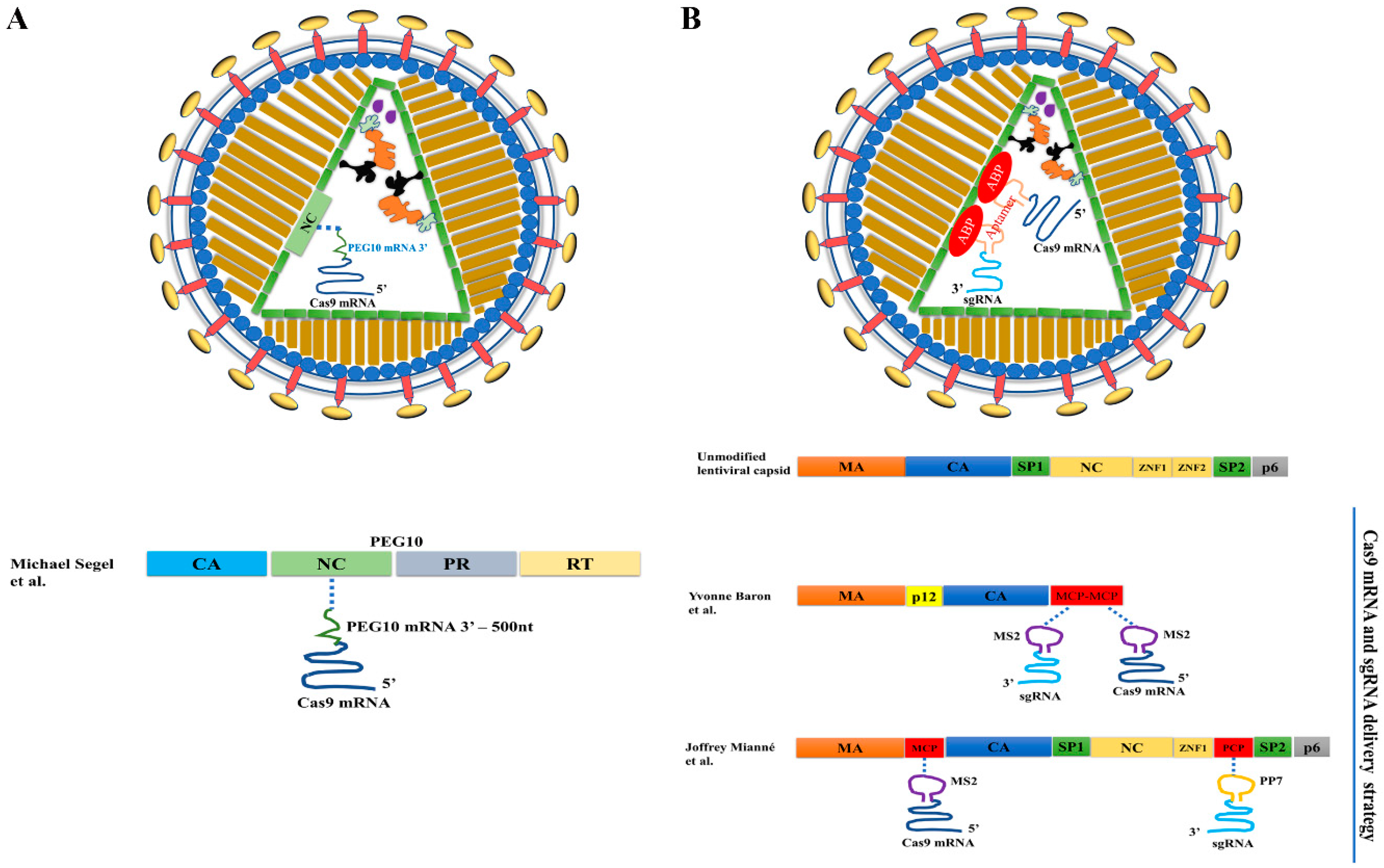
In the past, VLPs for mRNA delivery were developed from viral capsids [15,64,83,84,85]. Recently, endogenous retrotransposon proteins were used to develop VLPs for the same purpose [11]. Segel et al. screened Gag homologs in mouse and human genomes and found that PEG10 could form capsids with RNA packaging capability. They developed a selective endogenous encapsulation for cellular delivery (SEND) system consisting of three modules: the capsid assembling protein PEG10, the cargo mRNA bearing a cis element interacting with PEG10, and the endogenous fusogenic protein SYNA (to replace commonly used VSV-G) for cell entry and escape from the endosome system. The SEND system can efficiently package and deliver SpCas9 mRNA bearing a PEG10-interacting cis element (Figure 3A). Consistent with previous observations [15,63,84], sgRNA alone could not be functionally delivered by these VLPs. Surprisingly, unmodified sgRNA could be functionally delivered by SEND in the presence of SpCas9 mRNA bearing a PEG10-interacting cis element, but the mechanism behind this delivery is unknown. Future in vivo applications of this endogenous VLP system in genome editing will likely be forthcoming.

3.3. VLPs for Co-Delivery of Cas9 mRNA and sgRNA

Several studies have found that sgRNA packaged alone could not be functionally delivered by VLPs [11,15,63,84], possibly because sgRNA is very unstable in cells unless complexed with Cas9 protein [86]. Accordingly, most mRNA-delivering VLPs do not co-deliver sgRNA, except the recently reported endogenous VLP (SEND) system [11], but its mechanism for sgRNA delivery is unclear.
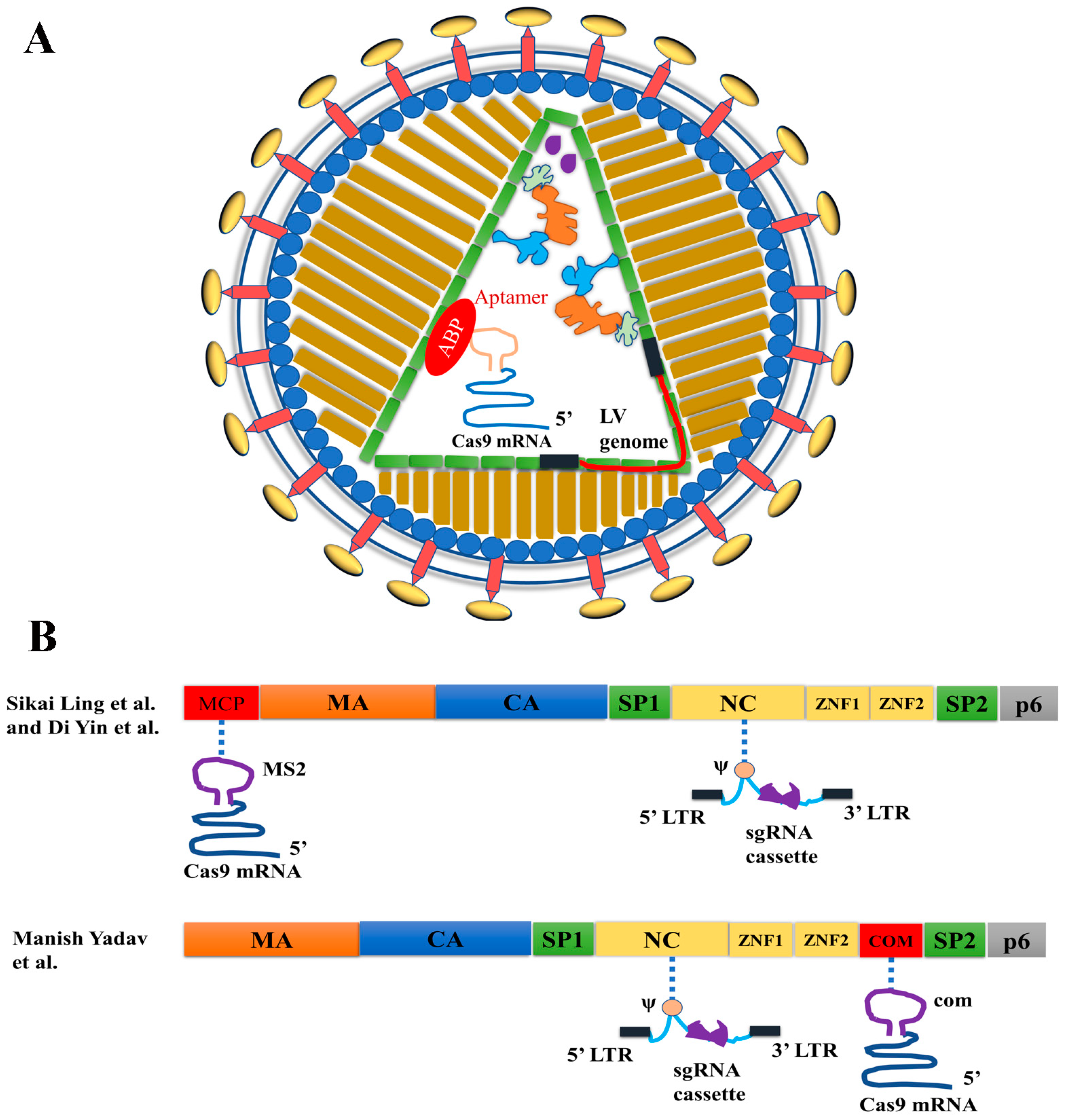
Two different strategies were used to develop VLPs for co-delivering Cas9 mRNA and sgRNA (Table 3). In the first strategy, RNA aptamers were added to Cas9 mRNA and sgRNA, and the aptamer/ABP interactions were used to co-package Cas9 mRNA and sgRNA in VLPs (Figure 3B) [14,15,16]. In the second strategy, Cas9 mRNA was packaged via aptamer/ABP interactions by adding RNA aptamers in the Cas9 mRNA 3′ untranslated region (UTR), whereas the sgRNA expression cassette was packaged as part of the lentiviral genomic RNA via the interactions between nucleocapsid (NC) protein and lentiviral genomic RNA packaging signal ψ (Figure 4). The sgRNA expression cassette was then reverse-transcribed into DNA to mediate sgRNA expression [6,7,13].
Table 3. Virus-like particle (VLP) mediated RNA delivery.
ReferenceVirus TypeCapsid ModificationRNA PackageCopy Number
mRNAMock et al. [83]LVNot modifiedTALEN mRNA2 copies
Prel et al. [85]LVMCP replaced the second zinc finger domain of NCSpCas9 mRNA~6 copies
Lu et al. [64]LVMCP inserted after the second zinc finger domain of NCSaCas9 mRNA50~100 copies
Lindel et al. [84]Foamy VirusesNot modifiedSpCas9 mRNA60 copies
mRNA or mRNA & sgRNASegel et al. [11]Endogenous retrotransposonNo modification of endogenous Gag homologSpCas9 mRNA
(and sgRNA)
Not available
mRNA & sgRNAKnopp et al. [14] Murine Leukemia Virus or Rous sarcoma virus Two copies of MCP replaced NC SpCas9 mRNA and sgRNANot available
Baron et al. [15]
Mianné et al. [16]LVMCP inserted the N terminus of CA and PCP replaced ZNF2SpCas9 mRNA and sgRNA1.43 copies
hybrid mRNA & sgRNALing et al. [6]LVMCP inserted the N terminus of Gag SpCas9 mRNA and sgRNA3~4 copies
Yin et al. [7]
Yadav et al. [13]LVMCP inserted after the second zinc finger domain of NCSaCas9 mRNA and sgRNANot available
Highlighted papers were covered in our previous review [4].

3.3.1. Adding Aptamers to Cas9 mRNA and sgRNA for Co-Delivery

Knopp et al. originally used VLPs developed from gammaretroviral murine leukemia virus to package Cas9 mRNA or co-package Cas9 mRNA and sgRNA [15]. Recently the same group used VLPs developed from alpharetroviral Rous sarcoma virus for improved efficiency [14]. In both studies, the authors replaced the nucleocapsid protein within Gag protein with 2 copies of MCP and added 2 copies of MS2 aptamer in the 3′ UTR of SpCas9 mRNA, and in various positions of sgRNA. The idea was to recruit Cas9 mRNA and sgRNA into the VLPs via MS2/MCP interactions. These VLPs successfully created DNA mutations in various murine and human cell lines, including human T cells, primary human fibroblasts, and cord-blood-derived CD34+ stem and progenitor cells [14,15].
The RNA-delivery VLP system originally reported by Prel et al. [85] was also recently modified for co-delivering Cas9 mRNA and sgRNA to human induced pluripotent stem cells [16]. In this method, two ABPs, MCP and PCP, were inserted into matrix protein (MA) and nucleocapsid protein, respectively; and two aptamers, PP7 and MS2, were inserted into sgRNA and Cas9 mRNA, respectively. The idea was to package sgRNA via PP7/PCP interactions and package Cas9 mRNA via MS2/MCP interactions. The authors observed efficient genome editing by this co-delivery method.
In the studies described above using aptamer/ABP interactions to co-deliver Cas9 mRNA and sgRNA [14,15,16], delivering Cas9 RNPs rather than or in addition to Cas9 mRNA/sgRNA cannot be ruled out. Several lines of evidence suggest that in these VLPs, the packaged Cas9 RNPs could be the main contributor to gene editing activities. First, several reports showed that sgRNA alone could not be functionally delivered by VLPs [11,15,63,84], and it is difficult to explain how sgRNA could be stabilized in the presence of Cas9 mRNA if Cas9 protein is not involved. Second, when co-expressing aptamer-negative Cas9 mRNA with aptamer-positive sgRNA in VLP producing cells, the aptamer/ABP interaction was enough to recruit Cas9 RNPs into VLPs [63]. This was because in VLP-producing cells, Cas9 proteins were translated from Cas9 mRNAs, and the former have an intrinsic affinity with sgRNAs that are recruited into VLPs via aptamer/ABP interactions. Indeed, Mianne et al. observed that VLPs prepared with aptamer-negative Cas9 mRNA and aptamer-positive sgRNA had 80% of the gene editing activity of VLPs prepared with Cas9 mRNA and sgRNA that were both aptamer-positive [16]. This finding suggests that approximately 80% of the genome editing activity could be contributed by packaged Cas9 RNPs. Thus, in these VLPs, a mixture of Cas9 RNPs, Cas9 mRNAs, and sgRNAs could have been delivered, and Cas9 RNPs might have contributed to the majority of the genome editing activities.

3.3.2. VLP and LV Hybrid Particles for Cas9 mRNA and sgRNA Co-Delivery

Two groups used a different strategy to co-deliver Cas9 mRNA and sgRNA by VLPs [6,7,13] (Figure 4). Ling et al. and Yin et al., both from the Cai group, fused MCP to the N-terminus of Gag and added multiple copies of the MS2 aptamer to the 3′ UTR of SpCas9 mRNA [6,7]. The SpCas9 mRNA was packaged by interactions between MCP and MS2. Simultaneous transfection of sgRNA-expressing lentiviral transfer plasmid DNA into the VLP-producing cells produced hybrid VLP/LV particles containing SpCas9 mRNA and sgRNA-expressing LV genomic RNA. Since the integrase bears a D64V mutation, the sgRNA-expression cassette will not be integrated. In one study, the authors used these hybrid particles to target vascular endothelial growth factor A (Vegfa) to suppress aberrant development of blood vessels in a mouse model of wet age-related macular degeneration [6]. In another study, the authors used these hybrid particles to target herpes simplex virus type 1 (HSV-1) DNA and then remove integrated viral DNA from cells in the eye, successfully blocking herpetic stromal keratitis [7]. In both studies, delivering the particles to the eyes of mice produced evident therapeutic effects [6,7].
Yadav et al. used a similar strategy to co-deliver Cas9 mRNA and sgRNA [13] but a different way to incorporate ABP into lentiviral capsids. They inserted ABP after the second zinc finger domain of the NC protein, which preserved capsid assembly efficiency [63,64,78]. They compared the four pairs of aptamer/ABPs listed in Table 1 for VLP-mediated RNA delivery and found that com aptamer and Com ABP was the most efficient pair for this purpose. They similarly expressed sgRNA from a sgRNA-expressing cassette contained in the LV genomic RNA. Co-delivery of Cas9 mRNA and sgRNA by these hybrid VLPs edited the genome more efficiently than delivering RNPs by VLPs. This observation suggests the value of co-delivering Cas9 mRNA and sgRNA.

4. Applications of VLPs in Genome Editing

VLPs as delivery vehicles for genome editing are still in preclinical development. However, successful applications have been reported in clinically relevant primary cells and disease animal models. Below, we summarize these applications.

4.1. In Vitro Applications in Clinically Relevant Blood Cells

Currently, genome editing in human blood cells relies on electroporation of Cas9 RNPs into them, which may affect cell viability. VLPs can deliver Cas9 RNPs into human hematopoietic stem cells to disrupt the Myd88 gene [74], whose mutation causes immunodeficiency. More recently, VLPs (‘Nanoblades’) were co-pseudotyped with VSV-G and baboon endogenous virus envelope glycoproteins to achieve better genome editing efficacy in human B, T, and CD34+ hematopoietic stem and progenitor cells; they showed little effects on cell viability or differentiation [9]. In another study, VLPs efficiently knocked out genes in primary CD4+ T cells [76].
Currently, generating CAR-expressing T cells with gene disruption depends on transduction of retroviral vectors and electroporation of Cas9 RNPs. Hybrid VLPs containing Cas9 RNPs and CAR-expressing lentiviral genomic RNAs have been used for simultaneous CAR insertion and gene (for example B2M or TRAC) disruption [10]. Pseudotyping the VLPs with the HIV-1 envelope glycoprotein increased the transduction of CD4+ T cells compared to CD8+ T cells (53.20% versus 2.51% gene disruption efficiency).

4.2. In Vivo Applications of VLPs in Mice

VLPs have been used to deliver Cas9 RNPs into mouse zygotes to disrupt the tyrosinase (Tyr) gene, and insertions and deletions were observed in 16 of 40 blastocysts injected [74]. In addition, the authors delivered VLPs of loxp-targeting Cas9 RNPs into the perivitelline space of R26R-EYFP embryos, and successfully removed the “lox-stop-lox” cassette to enable EYFP expression in mice. Finally, they delivered VLPs with RNPs targeting the hydroxyphenylpyruvate dioxygenase (Hpd) gene into Fah-/- mice via retro-orbital injection and obtained gene disruption in 7% to 13% of alleles [74].
In another study, Dnmt1-targeting ABE RNPs were delivered by VLPs into the central nervous system of neonatal mice via cerebroventricular injections. The authors observed over 50% base editing efficiency in VLP-transduced cells [12]. By delivering Pcsk9-targeting ABE RNPs into adult mice via retro-orbital injections, they achieved 63% editing in the liver and a 78% reduction in serum Pcsk9 protein levels. Finally, by delivering Rpe65-targeting ABE RNPs into the eyes of rd12 mice with a silence mutation that causes loss of vision, the authors observed 11.5% to 21% base editing and rescued visual function [12].
Cas9 mRNA and sgRNA-delivering VLP/LV hybrid particles were also used in vivo [6,7]. In both studies, the particles were delivered to the eyes of disease mouse models to disrupt the disease-causing DNA: Vegfa, which resulted in abnormal blood vessel formation in one study [6] and HSV-1 viral DNA causing infection in the eye in the other study [7]. Phenotypic improvements were observed after VLP local delivery.
Two types of Cas9 RNP-delivery particles, NanoMEDIC [75] and engineered exosomes [5], have been injected into muscle tissues of Duchenne muscular dystrophy (DMD) mouse models and achieved detectable levels of genome editing. Although local muscle delivery may not be an idea for DMD treatment, these studies demonstrated that these particles can be used for delivery into the muscle tissue.
In summary, VLPs have successfully delivered Cas9 RNPs, ABE RNPs, or Cas9 mRNA/sgRNA into mouse eyes, central nervous system, and muscle via local injections and into mouse livers via systemic injections [5,6,7,12,74]. Combined with tissue-specific pseudotypes, VLPs may be used for CRISPR/Cas9 delivery for genome editing in other organs.

5. Summary

This review presents various VLPs developed for delivering RNAs or RNPs for genome editing. The common feature for all of the VLPs is the short-term expression of the effectors. When the format of cargoes is considered, all VLPs can be classified as RNA VLPs and RNP VLPs. The two types of VLPs each have their own advantages and disadvantages: RNP VLPs are all needed in the particles for genome editing, whereas some RNA VLPs (except for those co-delivering Cas9 mRNA and sgRNA) need sgRNAs to be provided separately. The number of effectors packaged in RNP VLPs is pre-determined and controllable, whereas the number of effectors expressed from RNA VLPs may reach a much higher level during a short period of time. This high level of effectors is not desirable when off-targets are concerned, but it could be desirable when CRISPR/Cas9 cleavage is inhibited by heterochromatin [87,88,89] and more effectors are needed to achieve desirable genome editing efficiency [90].
RNPs are packaged into capsids via two different mechanisms: the fusion mechanism and the aptamer/ABP interaction mechanism. With the fusion mechanism, fusing a large protein to Gag may impair capsid assembly efficiency and result in effector degradation by proteinase. With the aptamer/ABP interaction mechanism, inserting a small ABP into the NC protein of Gag has little effect on capsid assembly, whereas adding aptamers in sgRNA may impair genome editing activity with some target sequences. In addition, the release of RNPs from the ABPs cannot be controlled. It is hard to simply claim that one method is superior to another. The users have to decide which method best meets their specific needs.
Although VLPs have not been tested in clinical trials for genome editing, they have been successfully used in vitro in clinically relevant human cells and in vivo in disease mouse models. We hope that in the near future VLPs are tested as delivery vehicles for genome editing in clinical applications to benefit patients.
One common issue in using VLP as a genome editing delivery vehicle is the dependence of VSV-G or other viral envelope proteins to facilitate cell entry and endosomal escape. VSV-G and other viral envelopes could be toxic at high concentrations [91]. In addition, pre-existing immune responses to VSV-G or other viral envelope proteins may decrease in vivo genome editing efficiency. Recently it was found that mouse SYNA protein showed similar activities as VSV-G when working with retrotransposon derived Gag-like proteins [11]. It would be interesting to determine whether SYNA can replace VSV-G to facilitate cell entry and endosomal escape of VLPs. Using nonviral proteins to facilitate VLP cell entry and endosomal escape will improve safety and efficiency.
Finally, we would like to take this opportunity to encourage authors to try their best to include “virus-like particles (VLPs)” as one of the keywords while reporting related findings. Currently over twenty types of VLPs have been developed as genome editing delivery vehicles (see Table 2 and Table 3). Many authors gave a unique term to the VLPs they developed. If there is not a common term to refer to these different types of particles, unnecessary confusion could be caused. We suggest that authors try their best to use “VLP” (short form for “virus-like particles”) for general particles. “RNP VLP”, “mRNA VLP”, and “mRNA/sgRNA VLP” can be used to specify VLPs packaged with RNPs, mRNA, or mRNA plus sgRNA, respectively. We believe that unified terms will be helpful to the development of this field.

Author Contributions

B.L. conceived the subject and structure of the review. P.L. and B.L. wrote the paper. All authors have read and agreed to the published version of the manuscript.

Funding

This work is funded by Hangzhou Normal University Research Startup 2021QDL029 (P.L.).

Institutional Review Board Statement

No human subjects were involved in this work thus an IRB is not required.

Informed Consent Statement

No human subjects were involved in this work thus Informed Consent is irrelevant.

Acknowledgments

The authors thank Anthony Atala, Xingang Yao, Zuyan Lu, Kyung Whan Yoo, and Manish Yadav at Wake Forest Institute for Regenerative Medicine for their contributions to the recent work from this group discussed in this review. The authors thank Tiantian Feng for her contribution to proofreading the manuscript.

Conflicts of Interest

The authors declare no conflict of interest. The funders had no role in the design of the study; in the collection, analysis, or interpretation of data; in the writing of the manuscript, or in the decision to publish the results.

Abbreviations

DNADeoxyribonucleic acid
cDNAComplementary DNA
RNARibonucleic acid
gRNAGuide RNA
sgRNA
mRNA
Single guide RNA
Messenger RNA
ZFNZinc finger endonuclease
TALENTranscription activator-like effector nuclease
CRISPR/CasClustered Regularly Interspaced Short Palindromic Repeats/CRISPR-associated/CRISPR-associated
PAMProtospacer adjacent motif
ABEAdenine base editor
LVLentiviral vector
AAVAdeno-associated virus-derived vector
IDLVIntegration-defective lentiviral vector
FMLVFriend murine leukemia virus
HIV-1Human immunodeficiency virus type 1
GagGroup-specific antigen
MAMatrix protein
CACapsid protein
NCNucleocapsid protein
RNPRibonucleoprotein
INDELInsertion and deletion
VLPVirus-like particle
ABPAptamer binding protein
MCPMS2 coating protein
PCPPP7 coating protein

References

  1. Woolf, T.M. Therapeutic repair of mutated nucleic acid sequences. Nat. Biotechnol. 1998, 16, 341–344. [Google Scholar] [CrossRef]
  2. Fu, Y.; Foden, J.A.; Khayter, C.; Maeder, M.L.; Reyon, D.; Joung, J.K.; Sander, J.D. High-frequency off-target mutagenesis induced by CRISPR-Cas nucleases in human cells. Nat. Biotechnol. 2013, 31, 822–826. [Google Scholar] [CrossRef] [Green Version]
  3. Nelson, C.E.; Wu, Y.; Gemberling, M.P.; Oliver, M.L.; Waller, M.A.; Bohning, J.D.; Robinson-Hamm, J.N.; Bulaklak, K.; Castellanos Rivera, R.M.; Collier, J.H.; et al. Long-term evaluation of AAV-CRISPR genome editing for Duchenne muscular dystrophy. Nat. Med. 2019, 25, 427–432. [Google Scholar] [CrossRef]
  4. Lyu, P.; Wang, L.; Lu, B. Virus-Like Particle Mediated CRISPR/Cas9 Delivery for Efficient and Safe Genome Editing. Life 2020, 10, 366. [Google Scholar] [CrossRef]
  5. Yao, X.; Lyu, P.; Yoo, K.; Yadav, M.K.; Singh, R.; Atala, A.; Lu, B. Engineered extracellular vesicles as versatile ribonucleoprotein delivery vehicles for efficient and safe CRISPR genome editing. J. Extracell. Vesicles 2021, 10, e12076. [Google Scholar] [CrossRef]
  6. Ling, S.; Yang, S.; Hu, X.; Yin, D.; Dai, Y.; Qian, X.; Wang, D.; Pan, X.; Hong, J.; Sun, X.; et al. Lentiviral delivery of co-packaged Cas9 mRNA and a Vegfa-targeting guide RNA prevents wet age-related macular degeneration in mice. Nat. Biomed. Eng. 2021, 5, 144–156. [Google Scholar] [CrossRef] [PubMed]
  7. Yin, D.; Ling, S.; Wang, D.; Dai, Y.; Jiang, H.; Zhou, X.; Paludan, S.R.; Hong, J.; Cai, Y. Targeting herpes simplex virus with CRISPR-Cas9 cures herpetic stromal keratitis in mice. Nat. Biotechnol. 2021, 39, 567–577. [Google Scholar] [CrossRef] [PubMed]
  8. Fichter, C.; Aggarwal, A.; Wong, A.K.H.; McAllery, S.; Mathivanan, V.; Hao, B.; MacRae, H.; Churchill, M.J.; Gorry, P.R.; Roche, M.; et al. Modular Lentiviral Vectors for Highly Efficient Transgene Expression in Resting Immune Cells. Viruses 2021, 13, 1170. [Google Scholar] [CrossRef]
  9. Gutierrez-Guerrero, A.; Abrey Recalde, M.J.; Mangeot, P.E.; Costa, C.; Bernadin, O.; Perian, S.; Fusil, F.; Froment, G.; Martinez-Turtos, A.; Krug, A.; et al. Baboon Envelope Pseudotyped “Nanoblades” Carrying Cas9/gRNA Complexes Allow Efficient Genome Editing in Human T, B, and CD34(+) Cells and Knock-in of AAV6-Encoded Donor DNA in CD34(+) Cells. Front. Genome Ed. 2021, 3, 604371. [Google Scholar] [CrossRef] [PubMed]
  10. Hamilton, J.R.; Tsuchida, C.A.; Nguyen, D.N.; Shy, B.R.; McGarrigle, E.R.; Sandoval Espinoza, C.R.; Carr, D.; Blaeschke, F.; Marson, A.; Doudna, J.A. Targeted delivery of CRISPR-Cas9 and transgenes enables complex immune cell engineering. Cell Rep. 2021, 35, 109207. [Google Scholar] [CrossRef]
  11. Segel, M.; Lash, B.; Song, J.; Ladha, A.; Liu, C.C.; Jin, X.; Mekhedov, S.L.; Macrae, R.K.; Koonin, E.V.; Zhang, F. Mammalian retrovirus-like protein PEG10 packages its own mRNA and can be pseudotyped for mRNA delivery. Science 2021, 373, 882–889. [Google Scholar] [CrossRef] [PubMed]
  12. Banskota, S.; Raguram, A.; Suh, S.; Du, S.W.; Davis, J.R.; Choi, E.H.; Wang, X.; Nielsen, S.C.; Newby, G.A.; Randolph, P.B.; et al. Engineered virus-like particles for efficient in vivo delivery of therapeutic proteins. Cell 2022, 185, 250–265.e216. [Google Scholar] [CrossRef] [PubMed]
  13. Yadav, M.K.; Atala, A.; Lu, B. Developing all-in-one virus-like particles for Cas9 mRNA/single guide RNA co-delivery and aptamer-containing lentiviral vectors for improved gene expression. BioRxiv 2022. [Google Scholar] [CrossRef] [PubMed]
  14. Baron, Y.; Sens, J.; Lange, L.; Nassauer, L.; Klatt, D.; Hoffmann, D.; Kleppa, M.J.; Barbosa, P.V.; Keisker, M.; Steinberg, V.; et al. Improved alpharetrovirus-based Gag.MS2 particles for efficient and transient delivery of CRISPR-Cas9 into target cells. Mol. Ther. Nucleic Acids 2022, 27, 810–823. [Google Scholar] [CrossRef]
  15. Knopp, Y.; Geis, F.K.; Heckl, D.; Horn, S.; Neumann, T.; Kuehle, J.; Meyer, J.; Fehse, B.; Baum, C.; Morgan, M.; et al. Transient Retrovirus-Based CRISPR/Cas9 All-in-One Particles for Efficient, Targeted Gene Knockout. Mol. Ther. Nucleic Acids 2018, 13, 256–274. [Google Scholar] [CrossRef] [PubMed] [Green Version]
  16. Mianne, J.; Nasri, A.; Van, C.N.; Bourguignon, C.; Fieldes, M.; Ahmed, E.; Duthoit, C.; Martin, N.; Parrinello, H.; Louis, A.; et al. CRISPR/Cas9-mediated gene knockout and interallelic gene conversion in human induced pluripotent stem cells using non-integrative bacteriophage-chimeric retrovirus-like particles. BMC Biol. 2022, 20, 8. [Google Scholar] [CrossRef]
  17. Kim, Y.G.; Cha, J.; Chandrasegaran, S. Hybrid restriction enzymes: Zinc finger fusions to Fok I cleavage domain. Proc. Natl. Acad. Sci. USA 1996, 93, 1156. [Google Scholar] [CrossRef] [Green Version]
  18. Li, T.; Huang, S.; Jiang, W.Z.; Wright, D.; Spalding, M.H.; Weeks, D.P.; Yang, B. TAL nucleases (TALNs): Hybrid proteins composed of TAL effectors and FokI DNA-cleavage domain. Nucleic Acids Res. 2011, 39, 359–372. [Google Scholar] [CrossRef] [Green Version]
  19. Moscou, M.J.; Bogdanove, A.J. A simple cipher governs DNA recognition by TAL effectors. Science 2009, 326, 1501. [Google Scholar] [CrossRef]
  20. Jinek, M.; Chylinski, K.; Fonfara, I.; Hauer, M.; Doudna, J.A.; Charpentier, E. A programmable dual-RNA-guided DNA endonuclease in adaptive bacterial immunity. Science 2012, 337, 816–821. [Google Scholar] [CrossRef]
  21. Cong, L.; Ran, F.A.; Cox, D.; Lin, S.; Barretto, R.; Habib, N.; Hsu, P.D.; Wu, X.; Jiang, W.; Marraffini, L.A.; et al. Multiplex genome engineering using CRISPR/Cas systems. Science 2013, 339, 819–823. [Google Scholar] [CrossRef] [PubMed] [Green Version]
  22. Cho, S.W.; Kim, S.; Kim, J.M.; Kim, J.S. Targeted genome engineering in human cells with the Cas9 RNA-guided endonuclease. Nat. Biotechnol. 2013, 31, 230–232. [Google Scholar] [CrossRef] [PubMed]
  23. Mali, P.; Yang, L.; Esvelt, K.M.; Aach, J.; Guell, M.; DiCarlo, J.E.; Norville, J.E.; Church, G.M. RNA-guided human genome engineering via Cas9. Science 2013, 339, 823–826. [Google Scholar] [CrossRef] [PubMed] [Green Version]
  24. Jinek, M.; East, A.; Cheng, A.; Lin, S.; Ma, E.; Doudna, J. RNA-programmed genome editing in human cells. eLife 2013, 2, e00471. [Google Scholar] [CrossRef] [Green Version]
  25. Barrangou, R.; Fremaux, C.; Deveau, H.; Richards, M.; Boyaval, P.; Moineau, S.; Romero, D.A.; Horvath, P. CRISPR provides acquired resistance against viruses in prokaryotes. Science 2007, 315, 1709–1712. [Google Scholar] [CrossRef]
  26. Koonin, E.V.; Makarova, K.S.; Zhang, F. Diversity, classification and evolution of CRISPR-Cas systems. Curr. Opin. Microbiol. 2017, 37, 67–78. [Google Scholar] [CrossRef]
  27. Doudna, J.A.; Charpentier, E. Genome editing. The new frontier of genome engineering with CRISPR-Cas9. Science 2014, 346, 1258096. [Google Scholar] [CrossRef]
  28. Doench, J.G.; Hartenian, E.; Graham, D.B.; Tothova, Z.; Hegde, M.; Smith, I.; Sullender, M.; Ebert, B.L.; Xavier, R.J.; Root, D.E. Rational design of highly active sgRNAs for CRISPR-Cas9-mediated gene inactivation. Nat. Biotechnol. 2014, 32, 1262–1267. [Google Scholar] [CrossRef] [Green Version]
  29. Nelson, C.E.; Hakim, C.H.; Ousterout, D.G.; Thakore, P.I.; Moreb, E.A.; Castellanos Rivera, R.M.; Madhavan, S.; Pan, X.; Ran, F.A.; Yan, W.X.; et al. In vivo genome editing improves muscle function in a mouse model of Duchenne muscular dystrophy. Science 2016, 351, 403–407. [Google Scholar] [CrossRef] [Green Version]
  30. Tabebordbar, M.; Zhu, K.; Cheng, J.K.W.; Chew, W.L.; Widrick, J.J.; Yan, W.X.; Maesner, C.; Wu, E.Y.; Xiao, R.; Ran, F.A.; et al. In vivo gene editing in dystrophic mouse muscle and muscle stem cells. Science 2016, 351, 407–411. [Google Scholar] [CrossRef] [Green Version]
  31. Long, C.; Amoasii, L.; Mireault, A.A.; McAnally, J.R.; Li, H.; Sanchez-Ortiz, E.; Bhattacharyya, S.; Shelton, J.M.; Bassel-Duby, R.; Olson, E.N. Postnatal genome editing partially restores dystrophin expression in a mouse model of muscular dystrophy. Science 2016, 351, 400–403. [Google Scholar] [CrossRef] [PubMed] [Green Version]
  32. Mirgayazova, R.; Khadiullina, R.; Chasov, V.; Mingaleeva, R.; Miftakhova, R.; Rizvanov, A.; Bulatov, E. Therapeutic Editing of the TP53 Gene: Is CRISPR/Cas9 an Option? Genes 2020, 11, 704. [Google Scholar] [CrossRef] [PubMed]
  33. Gillmore, J.D.; Gane, E.; Taubel, J.; Kao, J.; Fontana, M.; Maitland, M.L.; Seitzer, J.; O’Connell, D.; Walsh, K.R.; Wood, K.; et al. CRISPR-Cas9 In Vivo Gene Editing for Transthyretin Amyloidosis. N. Engl. J. Med. 2021, 385, 493–502. [Google Scholar] [CrossRef]
  34. Frangoul, H.; Altshuler, D.; Cappellini, M.D.; Chen, Y.S.; Domm, J.; Eustace, B.K.; Foell, J.; de la Fuente, J.; Grupp, S.; Handgretinger, R.; et al. CRISPR-Cas9 Gene Editing for Sickle Cell Disease and β-Thalassemia. N. Engl. J. Med. 2021, 384, 252–260. [Google Scholar] [CrossRef] [PubMed]
  35. Kaiser, J. CRISPR Helps a Blind Woman See, But Doesn’t Help All Patients. Available online: https://www.science.org/content/article/crispr-helps-blind-woman-see-doesn-t-help-all-patients (accessed on 29 September 2021).
  36. Stadtmauer, E.A.; Fraietta, J.A.; Davis, M.M.; Cohen, A.D.; Weber, K.L.; Lancaster, E.; Mangan, P.A.; Kulikovskaya, I.; Gupta, M.; Chen, F.; et al. CRISPR-engineered T cells in patients with refractory cancer. Science 2020, 367, eaba7365. [Google Scholar] [CrossRef] [PubMed]
  37. Wang, H.; Yang, H.; Shivalila, C.S.; Dawlaty, M.M.; Cheng, A.W.; Zhang, F.; Jaenisch, R. One-step generation of mice carrying mutations in multiple genes by CRISPR/Cas-mediated genome engineering. Cell 2013, 153, 910–918. [Google Scholar] [CrossRef] [PubMed] [Green Version]
  38. Bortesi, L.; Fischer, R. The CRISPR/Cas9 system for plant genome editing and beyond. Biotechnol. Adv. 2015, 33, 41–52. [Google Scholar] [CrossRef]
  39. Zhang, Z.T.; Jiménez-Bonilla, P.; Seo, S.O.; Lu, T.; Jin, Y.S.; Blaschek, H.P.; Wang, Y. Bacterial Genome Editing with CRISPR-Cas9: Taking Clostridium beijerinckii as an Example. Methods Mol. Biol. 2018, 1772, 297–325. [Google Scholar] [CrossRef]
  40. Qi, L.S.; Larson, M.H.; Gilbert, L.A.; Doudna, J.A.; Weissman, J.S.; Arkin, A.P.; Lim, W.A. Repurposing CRISPR as an RNA-guided platform for sequence-specific control of gene expression. Cell 2013, 152, 1173–1183. [Google Scholar] [CrossRef] [Green Version]
  41. Gilbert, L.A.; Larson, M.H.; Morsut, L.; Liu, Z.; Brar, G.A.; Torres, S.E.; Stern-Ginossar, N.; Brandman, O.; Whitehead, E.H.; Doudna, J.A.; et al. CRISPR-mediated modular RNA-guided regulation of transcription in eukaryotes. Cell 2013, 154, 442–451. [Google Scholar] [CrossRef] [Green Version]
  42. Bikard, D.; Jiang, W.; Samai, P.; Hochschild, A.; Zhang, F.; Marraffini, L.A. Programmable repression and activation of bacterial gene expression using an engineered CRISPR-Cas system. Nucleic Acids Res. 2013, 41, 7429–7437. [Google Scholar] [CrossRef] [Green Version]
  43. Kim, S.; Kim, D.; Cho, S.W.; Kim, J.; Kim, J.S. Highly efficient RNA-guided genome editing in human cells via delivery of purified Cas9 ribonucleoproteins. Genome Res. 2014, 24, 1012–1019. [Google Scholar] [CrossRef] [PubMed] [Green Version]
  44. Rees, H.A.; Komor, A.C.; Yeh, W.H.; Caetano-Lopes, J.; Warman, M.; Edge, A.S.B.; Liu, D.R. Improving the DNA specificity and applicability of base editing through protein engineering and protein delivery. Nat. Commun. 2017, 8, 15790. [Google Scholar] [CrossRef] [Green Version]
  45. Hsu, P.D.; Scott, D.A.; Weinstein, J.A.; Ran, F.A.; Konermann, S.; Agarwala, V.; Li, Y.; Fine, E.J.; Wu, X.; Shalem, O.; et al. DNA targeting specificity of RNA-guided Cas9 nucleases. Nat. Biotechnol. 2013, 31, 827–832. [Google Scholar] [CrossRef] [PubMed]
  46. Shalem, O.; Sanjana, N.E.; Hartenian, E.; Shi, X.; Scott, D.A.; Mikkelson, T.; Heckl, D.; Ebert, B.L.; Root, D.E.; Doench, J.G.; et al. Genome-scale CRISPR-Cas9 knockout screening in human cells. Science 2014, 343, 84–87. [Google Scholar] [CrossRef] [PubMed] [Green Version]
  47. Cradick, T.J.; Fine, E.J.; Antico, C.J.; Bao, G. CRISPR/Cas9 systems targeting beta-globin and CCR5 genes have substantial off-target activity. Nucleic Acids Res. 2013, 41, 9584–9592. [Google Scholar] [CrossRef] [Green Version]
  48. Maetzig, T.; Galla, M.; Baum, C.; Schambach, A. Gammaretroviral vectors: Biology, technology and application. Viruses 2011, 3, 677–713. [Google Scholar] [CrossRef] [Green Version]
  49. Kartikeyan, S.; Bharmal, R.N.; Tiwari, R.P.; Bisen, P.S. HIV and AIDS: Basic Elements and Priorities; Springer: Dordrecht, The Netherlands, 2007; pp. XIV–418. [Google Scholar]
  50. Ellington, A.D.; Szostak, J.W. In vitro selection of RNA molecules that bind specific ligands. Nature 1990, 346, 818–822. [Google Scholar] [CrossRef]
  51. Keefe, A.D.; Pai, S.; Ellington, A. Aptamers as therapeutics. Nat. Rev. Drug Discov. 2010, 9, 537–550. [Google Scholar] [CrossRef]
  52. Fouts, D.E.; True, H.L.; Celander, D.W. Functional recognition of fragmented operator sites by R17/MS2 coat protein, a translational repressor. Nucleic Acids Res. 1997, 25, 4464–4473. [Google Scholar] [CrossRef] [Green Version]
  53. Lim, F.; Downey, T.P.; Peabody, D.S. Translational repression and specific RNA binding by the coat protein of the Pseudomonas phage PP7. J. Biol. Chem. 2001, 276, 22507–22513. [Google Scholar] [CrossRef] [PubMed] [Green Version]
  54. Wulczyn, F.G.; Kahmann, R. Translational stimulation: RNA sequence and structure requirements for binding of Com protein. Cell 1991, 65, 259–269. [Google Scholar] [CrossRef]
  55. Austin, R.J.; Xia, T.; Ren, J.; Takahashi, T.T.; Roberts, R.W. Designed arginine-rich RNA-binding peptides with picomolar affinity. J. Am. Chem. Soc. 2002, 124, 10966–10967. [Google Scholar] [CrossRef] [PubMed] [Green Version]
  56. Atkins, J.F.; Gesteland, R.F. The synthetase gene of the RNA phages R17, MS2 and f2 has a single UAG terminator codon. Mol. Gen. Genet. 1975, 139, 19–31. [Google Scholar] [CrossRef] [PubMed]
  57. Peabody, D.S.; Ely, K.R. Control of translational repression by protein-protein interactions. Nucleic Acids Res. 1992, 20, 1649–1655. [Google Scholar] [CrossRef] [PubMed] [Green Version]
  58. Bertrand, E.; Chartrand, P.; Schaefer, M.; Shenoy, S.M.; Singer, R.H.; Long, R.M. Localization of ASH1 mRNA particles in living yeast. Mol. Cell 1998, 2, 437–445. [Google Scholar] [CrossRef] [Green Version]
  59. Mali, P.; Aach, J.; Stranges, P.B.; Esvelt, K.M.; Moosburner, M.; Kosuri, S.; Yang, L.; Church, G.M. CAS9 transcriptional activators for target specificity screening and paired nickases for cooperative genome engineering. Nat. Biotechnol. 2013, 31, 833–838. [Google Scholar] [CrossRef]
  60. Zalatan, J.G.; Lee, M.E.; Almeida, R.; Gilbert, L.A.; Whitehead, E.H.; La Russa, M.; Tsai, J.C.; Weissman, J.S.; Dueber, J.E.; Qi, L.S.; et al. Engineering complex synthetic transcriptional programs with CRISPR RNA scaffolds. Cell 2015, 160, 339–350. [Google Scholar] [CrossRef] [Green Version]
  61. Konermann, S.; Brigham, M.D.; Trevino, A.E.; Joung, J.; Abudayyeh, O.O.; Barcena, C.; Hsu, P.D.; Habib, N.; Gootenberg, J.S.; Nishimasu, H.; et al. Genome-scale transcriptional activation by an engineered CRISPR-Cas9 complex. Nature 2015, 517, 583–588. [Google Scholar] [CrossRef] [Green Version]
  62. Ma, H.; Tu, L.C.; Naseri, A.; Huisman, M.; Zhang, S.; Grunwald, D.; Pederson, T. Multiplexed labeling of genomic loci with dCas9 and engineered sgRNAs using CRISPRainbow. Nat. Biotechnol. 2016, 34, 528–530. [Google Scholar] [CrossRef] [Green Version]
  63. Lyu, P.; Javidi-Parsijani, P.; Atala, A.; Lu, B. Delivering Cas9/sgRNA ribonucleoprotein (RNP) by lentiviral capsid-based bionanoparticles for efficient ‘hit-and-run’ genome editing. Nucleic Acids Res. 2019, 47, e99. [Google Scholar] [CrossRef] [Green Version]
  64. Lu, B.; Javidi-Parsijani, P.; Makani, V.; Mehraein-Ghomi, F.; Sarhan, W.M.; Sun, D.; Yoo, K.W.; Atala, Z.P.; Lyu, P.; Atala, A. Delivering SaCas9 mRNA by lentivirus-like bionanoparticles for transient expression and efficient genome editing. Nucleic Acids Res. 2019, 47, e44. [Google Scholar] [CrossRef] [Green Version]
  65. Larson, D.R.; Zenklusen, D.; Wu, B.; Chao, J.A.; Singer, R.H. Real-time observation of transcription initiation and elongation on an endogenous yeast gene. Science 2011, 332, 475–478. [Google Scholar] [CrossRef] [PubMed] [Green Version]
  66. Wu, B.; Chao, J.A.; Singer, R.H. Fluorescence fluctuation spectroscopy enables quantitative imaging of single mRNAs in living cells. Biophys. J. 2012, 102, 2936–2944. [Google Scholar] [CrossRef] [PubMed] [Green Version]
  67. Lazinski, D.; Grzadzielska, E.; Das, A. Sequence-specific recognition of RNA hairpins by bacteriophage antiterminators requires a conserved arginine-rich motif. Cell 1989, 59, 207–218. [Google Scholar] [CrossRef]
  68. Wulczyn, F.G.; Bolker, M.; Kahmann, R. Translation of the bacteriophage Mu mom gene is positively regulated by the phage com gene product. Cell 1989, 57, 1201–1210. [Google Scholar] [CrossRef] [Green Version]
  69. Kaczmarczyk, S.J.; Sitaraman, K.; Young, H.A.; Hughes, S.H.; Chatterjee, D.K. Protein delivery using engineered virus-like particles. Proc. Natl. Acad. Sci. USA 2011, 108, 16998–17003. [Google Scholar] [CrossRef] [Green Version]
  70. Peretti, S.; Schiavoni, I.; Pugliese, K.; Federico, M. Cell death induced by the herpes simplex virus-1 thymidine kinase delivered by human immunodeficiency virus-1-based virus-like particles. Mol. Ther. 2005, 12, 1185–1196. [Google Scholar] [CrossRef]
  71. Joo, K.I.; Wang, P. Visualization of targeted transduction by engineered lentiviral vectors. Gene Ther. 2008, 15, 1384–1396. [Google Scholar] [CrossRef]
  72. Cai, Y.; Bak, R.O.; Mikkelsen, J.G. Targeted genome editing by lentiviral protein transduction of zinc-finger and TAL-effector nucleases. eLife 2014, 3, e01911. [Google Scholar] [CrossRef]
  73. Choi, J.G.; Dang, Y.; Abraham, S.; Ma, H.; Zhang, J.; Guo, H.; Cai, Y.; Mikkelsen, J.G.; Wu, H.; Shankar, P.; et al. Lentivirus pre-packed with Cas9 protein for safer gene editing. Gene Ther. 2016, 23, 627–633. [Google Scholar] [CrossRef]
  74. Mangeot, P.E.; Risson, V.; Fusil, F.; Marnef, A.; Laurent, E.; Blin, J.; Mournetas, V.; Massouridès, E.; Sohier, T.J.M.; Corbin, A.; et al. Genome editing in primary cells and in vivo using viral-derived Nanoblades loaded with Cas9-sgRNA ribonucleoproteins. Nat. Commun. 2019, 10, 45. [Google Scholar] [CrossRef] [PubMed] [Green Version]
  75. Gee, P.; Lung, M.S.Y.; Okuzaki, Y.; Sasakawa, N.; Iguchi, T.; Makita, Y.; Hozumi, H.; Miura, Y.; Yang, L.F.; Iwasaki, M.; et al. Extracellular nanovesicles for packaging of CRISPR-Cas9 protein and sgRNA to induce therapeutic exon skipping. Nat. Commun. 2020, 11, 1334. [Google Scholar] [CrossRef] [PubMed] [Green Version]
  76. Indikova, I.; Indik, S. Highly efficient ‘hit-and-run’ genome editing with unconcentrated lentivectors carrying Vpr.Prot.Cas9 protein produced from RRE-containing transcripts. Nucleic Acids Res. 2020, 48, 8178–8187. [Google Scholar] [CrossRef] [PubMed]
  77. Lyu, P.; Lu, Z.; Cho, S.I.; Yadav, M.; Yoo, K.; Atala, A.; Kim, J.S.; Lu, B. Adenine base editor ribonucleoproteins delivered by lentivirus-like particles show high on-target base editing and undetectable RNA off-target activities. CRISPR J. 2021, 4, 69–81. [Google Scholar] [CrossRef] [PubMed]
  78. Lu, Z.; Yao, X.; Lyu, P.; Yadav, M.; Yoo, K.; Atala, A.; Lu, B. Lentiviral Capsid-Mediated Streptococcus pyogenes Cas9 Ribonucleoprotein Delivery for Efficient and Safe Multiplex Genome Editing. CRISPR J. 2021, 4, 914–928. [Google Scholar] [CrossRef]
  79. Vindry, C.; Guillin, O.; Mangeot, P.E.; Ohlmann, T.; Chavatte, L. A Versatile Strategy to Reduce UGA-Selenocysteine Recoding Efficiency of the Ribosome Using CRISPR-Cas9-Viral-Like-Particles Targeting Selenocysteine-tRNA([Ser]Sec) Gene. Cells 2019, 8, 574. [Google Scholar] [CrossRef] [Green Version]
  80. Lyu, P.; Yoo, K.W.; Yadav, M.K.; Atala, A.; Aartsma-Rus, A.; Putten, M.V.; Duan, D.; Lu, B. Sensitive and reliable evaluation of single-cut sgRNAs to restore dystrophin by a GFP-reporter assay. PLoS ONE 2020, 15, e0239468. [Google Scholar] [CrossRef]
  81. Escola, J.M.; Kleijmeer, M.J.; Stoorvogel, W.; Griffith, J.M.; Yoshie, O.; Geuze, H.J. Selective enrichment of tetraspan proteins on the internal vesicles of multivesicular endosomes and on exosomes secreted by human B-lymphocytes. J. Biol. Chem. 1998, 273, 20121–20127. [Google Scholar] [CrossRef] [Green Version]
  82. Kobayashi, T.; Vischer, U.M.; Rosnoblet, C.; Lebrand, C.; Lindsay, M.; Parton, R.G.; Kruithof, E.K.; Gruenberg, J. The tetraspanin CD63/lamp3 cycles between endocytic and secretory compartments in human endothelial cells. Mol. Biol. Cell 2000, 11, 1829–1843. [Google Scholar] [CrossRef] [Green Version]
  83. Mock, U.; Riecken, K.; Berdien, B.; Qasim, W.; Chan, E.; Cathomen, T.; Fehse, B. Novel lentiviral vectors with mutated reverse transcriptase for mRNA delivery of TALE nucleases. Sci. Rep. 2014, 4, 6409. [Google Scholar] [CrossRef] [PubMed] [Green Version]
  84. Lindel, F.; Dodt, C.R.; Weidner, N.; Noll, M.; Bergemann, F.; Behrendt, R.; Fischer, S.; Dietrich, J.; Cartellieri, M.; Hamann, M.V.; et al. TraFo-CRISPR: Enhanced Genome Engineering by Transient Foamy Virus Vector-Mediated Delivery of CRISPR/Cas9 Components. Mol. Ther. Nucleic Acids 2019, 18, 708–726. [Google Scholar] [CrossRef] [PubMed] [Green Version]
  85. Prel, A.; Caval, V.; Gayon, R.; Ravassard, P.; Duthoit, C.; Payen, E.; Maouche-Chretien, L.; Creneguy, A.; Nguyen, T.H.; Martin, N.; et al. Highly efficient in vitro and in vivo delivery of functional RNAs using new versatile MS2-chimeric retrovirus-like particles. Mol. Ther. Methods Clin. Dev. 2015, 2, 15039. [Google Scholar] [CrossRef]
  86. Ma, H.; Tu, L.C.; Naseri, A.; Huisman, M.; Zhang, S.; Grunwald, D.; Pederson, T. CRISPR-Cas9 nuclear dynamics and target recognition in living cells. J. Cell Biol. 2016, 214, 529–537. [Google Scholar] [CrossRef] [PubMed] [Green Version]
  87. Hinz, J.M.; Laughery, M.F.; Wyrick, J.J. Nucleosomes Inhibit Cas9 Endonuclease Activity in Vitro. Biochemistry 2015, 54, 7063–7066. [Google Scholar] [CrossRef]
  88. Horlbeck, M.A.; Witkowsky, L.B.; Guglielmi, B.; Replogle, J.M.; Gilbert, L.A.; Villalta, J.E.; Torigoe, S.E.; Tjian, R.; Weissman, J.S. Nucleosomes impede Cas9 access to DNA in vivo and in vitro. eLife 2016, 5, e12677. [Google Scholar] [CrossRef]
  89. Isaac, R.S.; Jiang, F.; Doudna, J.A.; Lim, W.A.; Narlikar, G.J.; Almeida, R. Nucleosome breathing and remodeling constrain CRISPR-Cas9 function. eLife 2016, 5, e13450. [Google Scholar] [CrossRef]
  90. Kallimasioti-Pazi, E.M.; Thelakkad Chathoth, K.; Taylor, G.C.; Meynert, A.; Ballinger, T.; Kelder, M.J.E.; Lalevee, S.; Sanli, I.; Feil, R.; Wood, A.J. Heterochromatin delays CRISPR-Cas9 mutagenesis but does not influence the outcome of mutagenic DNA repair. PLoS Biol. 2018, 16, e2005595. [Google Scholar] [CrossRef] [Green Version]
  91. Hoffmann, M.; Wu, Y.J.; Gerber, M.; Berger-Rentsch, M.; Heimrich, B.; Schwemmle, M.; Zimmer, G. Fusion-active glycoprotein G mediates the cytotoxicity of vesicular stomatitis virus M mutants lacking host shut-off activity. J. Gen. Virol. 2010, 91, 2782–2793. [Google Scholar] [CrossRef]
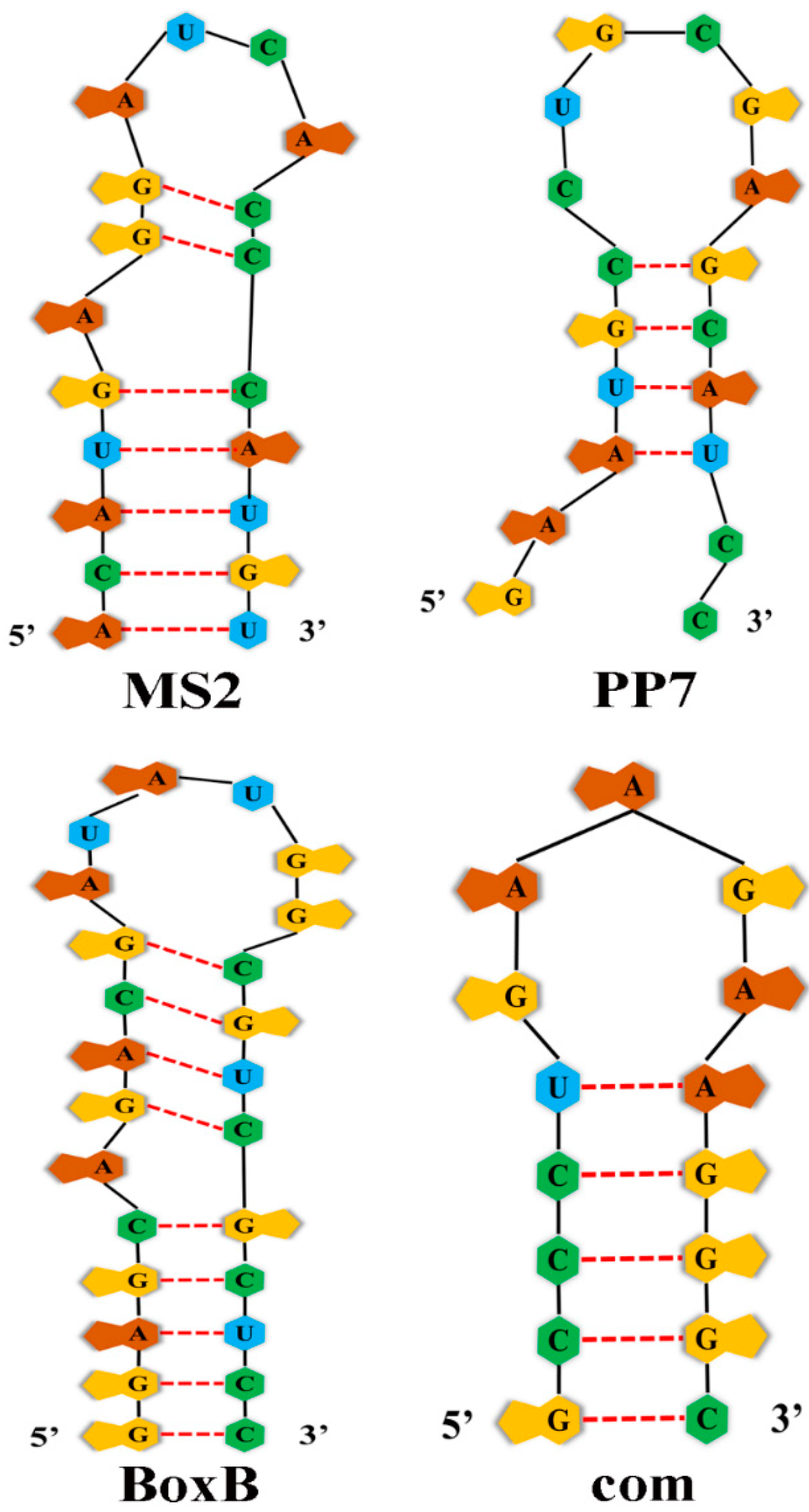
Figure 1. Structure of four RNA aptamers.
Ijms 23 08750 g001
Figure 2. Strategies for modifying the Gag protein for designer nuclease RNP delivery by VLPs. (A) Diagram illustrating protein- or RNP-delivering VLPs. The RNA, if present, does not contain a long terminal repeat, so reverse transcription cannot happen. (B) Strategy for packaging proteins and RNPs into extracellular vesicles. A dashed line indicates non-covalent interactions. The VLPs and exosomes were reported in references [5], [10] and [12] respectively.
Ijms 23 08750 g002
Figure 3. Using VLPs to deliver mRNA for genome editing. (A) Viral capsid-derived RNA-delivering VLPs. Aptamers are added in Cas9 mRNA and sgRNA so that they can be packaged via aptamer/ABP interactions. The RNAs do not contain a long terminal repeat, so reverse transcription cannot happen. (B) Mammalian transposon protein-derived VLPs for RNA delivery. The VLPs were reported in references [11], [15] and [16] respectively.
Ijms 23 08750 g003
Figure 4. Strategies for modifying the Gag protein for mRNA and sgRNA delivery by VLP/LV hybrid particles. (A) Diagram illustrating mRNA- and sgRNA-co-delivery of VLP/LV hybrid particles. The mRNAs can only serve as the templates for translation since they do not contain viral sequences necessary for reverse transcription, so reverse transcription cannot happen. (B) Different approaches to Gag modification for Cas9-mRNA and sgRNA-expression cassette co-delivery. The VLPs were reported in references [6], [7] and [13] respectively.
Ijms 23 08750 g004
Table 1. Aptamer and aptamer-binding protein (ABP) used for VLP-mediated RNA/RNP delivery.
AptamerAptamer Sequence
(5′ to 3′)
ABPSize of ABPReference
MS2ACAUGAGGAUCACCCAUGUMCP117 AA[52]
PP7GAAUGCCUGCGAGCAUCCPCP121 AA[53]
BoxBGGAGCAGACGAUAUGGCGUCGCUCCλ N2222 AA[55]
comGCCCUGAAGAAGGGCCom62 AA[54]
Publisher’s Note: MDPI stays neutral with regard to jurisdictional claims in published maps and institutional affiliations.