Article

Possibility of Venous Serum Cl Concentration ([Cl]s) as a Marker for Human Metabolic Status: Correlation of [Cl]s to Age, Fasting Blood Sugar (FBS), and Glycated Hemoglobin (HbA1c)

1
Medical Research Institute, Kyoto Industrial Health Association, Kyoto 604-8472, Japan
2
Research Organization of Science and Technology, Ritsumeikan University, Kusatsu 525-8577, Japan
3
Graduate School of Medical Science, Kyoto Prefectural University of Medicine, Kyoto 802-8566, Japan
4
Luis Pasteur Center for Medical Research, Kyoto 606-8225, Japan
*
Author to whom correspondence should be addressed.
Int. J. Mol. Sci. 2021, 22(20), 11111; https://doi.org/10.3390/ijms222011111
Submission received: 17 September 2021 / Revised: 9 October 2021 / Accepted: 12 October 2021 / Published: 15 October 2021

Abstract

:
The HCO3 concentration in venous serum ([HCO3]s) is a factor commonly used for detecting the body pH and metabolic conditions. To exactly detect [HCO3]s, the venous CO2 pressure should be kept as it is in the vein. The [HCO3]s measurement is technically complicated to apply for huge numbers of almost heathy persons taking only basic medical examinations. The summation of [HCO3]s and the venous serum Cl concentration ([Cl]s) is approximately constant; therefore, we studied if [Cl]s could be a marker detecting metabolic conditions instead of [HCO3]s. Venous blood was obtained from persons taking basic medical examinations (the number of persons = 107,630). Older persons showed higher values of [Cl]s, fasting blood sugar (FBS), and glycated hemoglobin (HbA1c) than younger ones. [Cl]s showed positive correlation to age and negative correlation to FBS and HBA1c. The negative correlation of [Cl]s to FBS/HbA1c was obvious in persons with high FBS/HbA1c, leading us to an idea that persons with high FBS/HbA1c show high [HCO3]s, which might be caused by low activity of carbonic anhydrase in the lung observed in persons with diabetes mellitus under acidotic conditions. Taken together, an easily measured serum electrolyte, [Cl]s, could be a useful marker estimating metabolic conditions.

1. Introduction

The metabolism is one of the most important functions maintaining our life activities. To exactly detect the metabolic condition, we have to measure various factors such as O2 consumption, CO2 production, pH in venous serum, HCO3 concentration in venous serum ([HCO3]s), fasting blood sugar (FBS), and glycated hemoglobin (HbA1c), etc. [1,2,3,4]. However, even though O2 consumption and CO2 production are measured, certain momentary values of O2 consumption and CO2 production are not enough to estimate the metabolic condition of whole body, but continuous measurements of O2 consumption and CO2 production are required to detect the relatively chronic metabolic condition of whole body [1,2,3,4]. On the one hand, concentrations of electrolytes such as H+ and HCO3 in the venous serum show the relatively chronic status of metabolic conditions [5,6,7,8,9], although acute changes in metabolic conditions would also affect H+ and HCO3 concentrations in the venous serum with a time lag dependent on the degree and the time duration of the acute metabolic changes. Even though these measurements could provide crucial information on the metabolic status, these measurements require technically complicated processes. On the other hand, to obtain information on metabolic conditions of huge numbers of persons taking only basic medical examinations not including [HCO3]s measurements, we should find out another index measurable using a technique easily adaptable to huge numbers of persons. Here, we considered that the venous serum Cl concentration ([Cl]s) could be an index indicating metabolic conditions, since Cl is an easily measurable index and [Cl]s changes to the opposite direction with the change in [HCO3]s. We assumed that the total amount of [Cl]s and [HCO3]s would be approximately constant, as described as follows. The source of HCO3 is CO2 produced in metabolic cells such as myocytes, hepatocytes, and renal epithelial cells, moving into erythrocytes [6,10]. The CO2 in erythrocytes is converted to H+ and HCO3 via a carbonic anhydrase (CA)-facilitated process (CO2 + H2O ⟶ H+ HCO3) [6,10,11]. The HCO3 is excreted into the serum by exchanging HCO3 with serum Cl, which is incorporated into erythrocytes, via a Cl/HCO3 anion exchanger (AE)-mediated process [6,10,12,13,14,15,16,17]. The AE-mediated process leads to a decrease in [Cl]s associated with an increase in [HCO3]s. Thus, in the present study, we tried to clarify if [Cl]s could be an index for metabolic conditions instead of [HCO3]s, although the kidneys and lungs regulate the HCO3 concentration leading us to consider the function of the kidneys and lungs at evaluating [Cl]s as an index for metabolic conditions [18].
In the present study, we indicated that: (1) Older persons show higher values of [Cl]s, FBS, and HbA1c than younger ones; (2) [Cl]s changes with positive correlation to the change of age and negatively correlated to the change of FBS and HbA1c with the order of correlation intensity, age > HbA1c > FBS; (3) [Cl]s of persons with extremely high FBS or/and HbA1c changes more negatively correlated to FBS and HbA1c than that with normal or moderately high FBS or/and HbA1c. These observations led us to the following idea: (1) Older persons show low [HCO3]s due to low production of CO2; (2) persons with extremely high FBS or/and HbA1c would show high [HCO3]s due to high production of CO2; (3) high [HCO3]s might be also caused by slow conversion of H+ and HCO3 to CO2 and H2O (H+ + HCO3 ⟶ CO2 + H2O) via CA-medicated processes in the lung of persons with high leveled FBS or/and HbA1c; and (4) this might be due to low activity of CA observed in capillary endothelia of the lung in diabetes mellitus (DM) patients with high leveled FBS or/and HbA1c.

2. Results

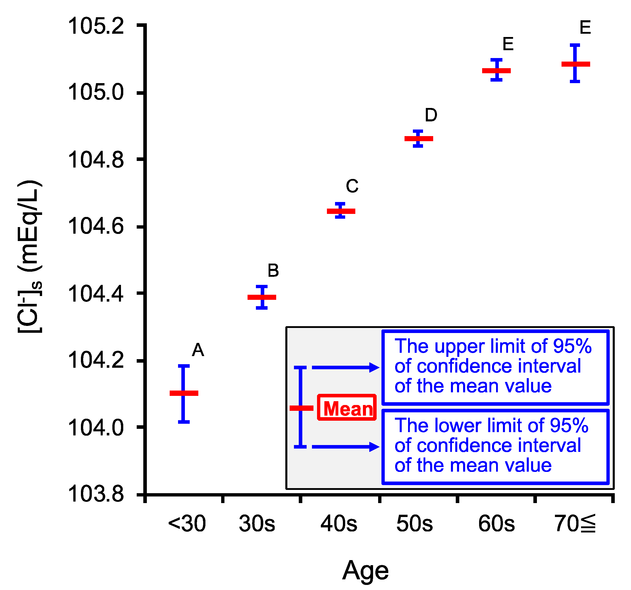
2.1. Age-Dependent Changes in Venous Serum Cl Concentration ([Cl]s)

We firstly studied if the [Cl]s would change in an age-dependent manner. To clarify this point in persons taking medical examinations (the number of persons (n) = 107,630), we categorized the age of persons taking medical examinations into six groups as shown in Table 1; the number of persons (n) in each group is also shown in Table 1. The [Cl]s significantly increased with the age up to 60s (Figure 1), reaching a plateau value in the persons with the ages of 60s and over 70 years old (70≦); we detected no significant difference between 60s and 70≦ (Figure 1. The minimum mean value of [Cl]s was observed at the age <30 (104.10 mEq/L; 95% confidence interval (CI) = 104.02–104.19 mEq/L in Figure 1). On the one hand, the maximum mean value of [Cl]s was observed at the age 60s (105.07 mEq/L; 95% CI = 105.04–105.10 mEq/L; Figure 1) and 70≦ (105.09 mEq/L; 95% CI = 105.03–105.14 mEq/L; Figure 1): no significant difference of the mean [Cl]s values was observed between these two groups, 60s and 70≦ (Figure 1). The difference between the mean [Cl]s values at the ages of all persons in the present study is within only 1 mEq/L; i.e., the minimum and maximum mean values of [Cl]s among the six groups were, respectively, 104.10 and 105.09 mEq/L (Figure 1). Nevertheless, the mean value of [Cl]s significantly increased in an age-dependent manner up to the 60s (Figure 1). The observation shown in Figure 1 suggests that the age-dependent change in [Cl]s would have some physiological meanings.
We considered a possibility that [Cl]s could be an index indicating metabolic conditions in medical examinations based on the following reason. CO2 produced in metabolic cells moves into erythrocytes, and is converted to H+ and HCO3 (H+ + HCO3 ⟶ CO2 + H2O) via CA-medicated processes in erythrocytes. The H+ produced from CO2 is bound to hemoglobin (Hb), while the HCO3 produced from CO2 in erythrocytes is excreted to the serum in blood (the extracellular space of erythrocytes) by AE expressed on the plasma membrane of erythrocytes. The AE participates in HCO3 excretion from erythrocytes to the extracellular space (the serum in blood) and simultaneously Cl uptake into erythrocytes from the serum of blood around metabolic cells [6,10,15,16,19,20,21,22,23,24,25]. To clarify the relationship between tissue metabolisms and [Cl]s, we studied the age-dependent change in venous serum fasting blood sugar (FBS) and HbA1c, which have correlation to tissue metabolism, although [Cl]s and [HCO3]s are also affected by the respiration in the lung.

2.2. Age-Dependent Changes in Venous Serum Fasting Blood Sugar Concentration (FBS)

We studied if FBS would change in an age-dependent manner. FBS significantly increased in an age-dependent manner up to the age 70≦ (Figure 2) similar to that in [Cl]s, although the age-dependent increase in [Cl]s reached a plateau level at the age 60s (Figure 1).

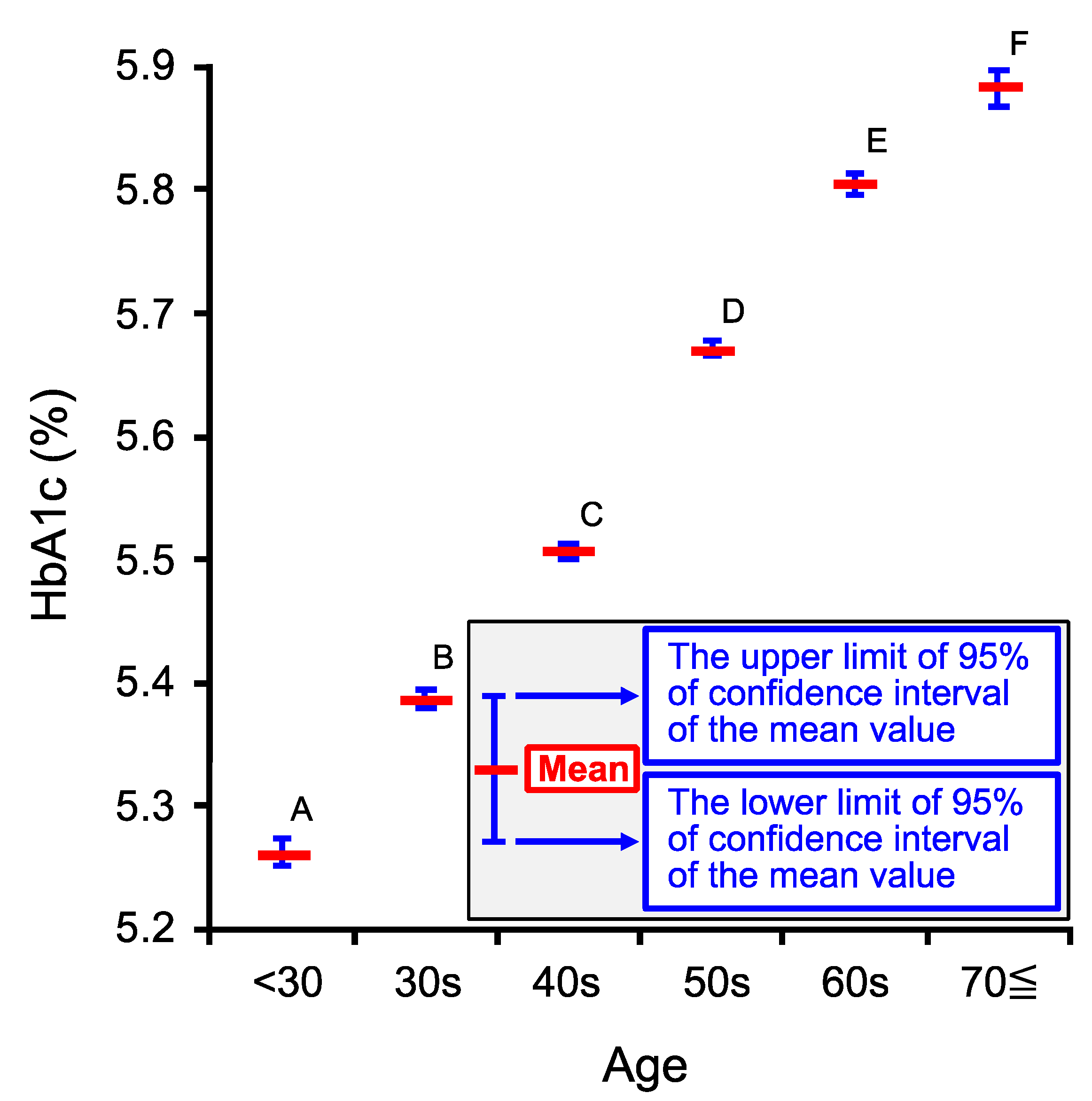
2.3. Age-Dependent Changes in Venous Hemoglobin A1c (HbA1c)

We further studied if HbA1c would change in an age-dependent manner. HbA1c significantly increased in an age-dependent manner up to the age 70≦ as shown in Figure 3. This age-dependent phenomenon observed in HbA1c (Figure 3) seems to be similar to that in FBS. However, the increase in HbA1c from the age 60s to 70≦ (Figure 3) seems to be larger in degree than that in FBS (Figure 2). This phenomenon would be due to the increase in post-prandial blood sugar (PBS) levels of persons with age 70≦ from 60s being larger in degree than that in FBS. This is so-called “impaired glucose tolerance” caused by deficiency in insulin secretion responding to elevation of blood sugar or/and insulin resistance occurring much more severely in 70≦ than in 60s. The “impaired glucose tolerance” influences PBS but not FBS.

2.4. Relationship among [Cl]s, Age, FBS and HbA1c

Although our observations indicate that [Cl]s, FBS, and HbA1c significantly increase in an age-dependent manner, we have no information on the relationship among [Cl]s, FBS, and HbA1c. Therefore, we tried to clarify the relationship among [Cl]s, age, FBS, and HbA1c using Equation (1) (see Section 4.5 in Materials and Methods). [Cl]s showed significantly positive correlation to age ( C AFH Age > 0; Table 2), but significantly negative correlation to FBS or HbA1c ( C AFH FBS < 0 and C AFH HbA 1 c < 0; Table 2). However, it is unclear which factor, age, FBS, or HbA1c, most effectively influenced [Cl]s, since age, FBS, and HbA1c had different units and these factors could not be compared to each other. To clarify this point, we normalized the values of [Cl]s, age, FBS, and HbA1c (see Section 4.6 in Materials and Methods).
The N[Cl]s-influencing coefficient of each factor, C N AFH Age , C N AFH FBS , or C N AFH HbA 1 c , is significantly different from each other. Nage had significantly a positive effect on N[Cl]s ( C N AFH Age >0 in Table 3), while both NFBS and NHbA1c had significantly negative effects on N[Cl]s. Among the absolute values of coefficients, C N AFH Age , C N AFH FBS , and C N AFH HbA 1 c , the largest one was C N AFH Age (Table 3); i.e., Nage was the most effective factor on N[Cl]s, NHbA1c was the next effective one on N[Cl]s, and NFBS was the most non-effective one influencing N[Cl].
We further analyzed the correlation of [Cl]s, FBS, or HbA1c to age using the normalized data, N[Cl]s, NFBS, NHbA1c, and Nage (see Section 4.7 in Materials and Methods). All three coefficients, C N Age Age , F N Age Age , and H N Age Age , were significantly larger than 0, and each coefficient was significantly different from each other (Table 4). HbA1c was the most Nage-dependent factor (Table 4). FBS depended on Nage almost similar to HbA1c, but significantly less dependent on Nage than HbA1c (Table 4). [Cl]s least depended on Nage (Table 4). The value of the Nage-dependent coefficient for N[Cl]s ( C N Age Age ; see Table 4) was smaller than that of the NFBS/NHbA1c-independent, Nage-dependent coefficient for N[Cl]s ( C N AFH Age ; see Table 3), since C N Age Age contains FBS/HbA1c-dependent factors negatively influencing [Cl]s (see Section 4.8 in Materials and Methods).
Table 4. The mean value of age-dependent coefficients, C N Age Age , F N Age Age , and H N Age Age , for [ Cl ] N s , F N BS , and H N bA 1 c , respectively, shown in Equations (3)–(5).
Coefficient
C N A g e A g e
F N A g e A g e
H N A g e A g e
UL of 95% CI0.12260.27070.2870
Mean0.11670.26490.2812
LL of 95% CI0.11080.25910.2754
C N Age Age , F N Age Age , and H N Age Age are respectively the Nage-dependent coefficients for N[Cl]s, NFBS, or NHbA1c. The upper limit (UL) and the lower limit (LL) of 95% confidence interval (CI) of the mean value of the coefficient are also shown. n = 107,630.

2.5. Relationship between [Cl]s and FBS

As shown in Table 3 and Table 4, it is suggested that [Cl]s is negatively correlated to FBS. Therefore, we next analyzed the relationship between [Cl]s and FBS by categorizing FBS into three ranges, (1) FBS < 100, (2) 100 ≦ FBS < 126, and (3) 126 mg/dL ≦ FBS using Equation (9) (see Section 4.9 in Materials and Methods). The coefficient ( C FBS FBS ) in each group of 1) FBS < 100, 2) 100 ≦ FBS < 126, or 3) 126 mg/dL ≦ FBS was significantly different from 0 (Table 5). The value of C FBS FBS in the group of FBS < 100 mg/dL was significantly different from that in the group of 100 ≦ FBS < 126 or 126 mg/dL ≦ FBS (i.e., FBS ≧ 100 mg/dL), while the values of C FBS FBS in the groups of 100 ≦ FBS < 126, and 126 mg/dL ≦ FBS were not significantly different (Table 5). In the group of FBS < 100 mg/dL, [Cl]s increased as FBS was elevated, while [Cl]s decreased as FBS was elevated in the group of FBS ≧ 100 mg/dL (the groups of 100 ≦ FBS < 126, and 126 mg/dL ≦ FBS).

2.6. Relationship between [Cl]s and HbA1c

As shown in Table 3 and Table 4, it is suggested that [Cl]s is negatively correlated to HbA1c. Therefore, we next analyzed the relationship between [Cl]s and HbA1c by categorizing HbA1c into three ranges, (1) HbA1c < 5.6%, (2) 5.6% ≦ HbA1c < 6.5%, and (3) 6.5% ≦ HbA1c using Equation (10) (see Section 4.10 in Materials and Methods). Each coefficient, C HbA 1 c HbA 1 c , in the group of (1) HbA1c < 5.6%, (2) 5.6% ≦ HbA1c < 6.5% or (3) 6.5% ≦ HbA1c is significantly smaller than 0 (Table 6): i.e., C HbA 1 c HbA 1 c in each HbA1c range is negative. No significant difference of C HbA 1 c HbA 1 c was observed between HbA1c < 5.6% and 5.6% ≦ HbA1c < 6.5%, while C HbA 1 c HbA 1 c in the group of 6.5% ≦ HbA1c was significantly different from that in the group of HbA1c < 5.6% or 5.6% ≦ HbA1c < 6.5% (i.e., HbA1c < 6.5%; Table 6). These observations indicate that persons with HbA1c ≧ 6.5% showed [Cl]s decreases in a significantly large degree as HbA1c were elevated compared with those with HbA1c < 6.5% (Table 6).

2.7. Relationship between FBS and HbA1c
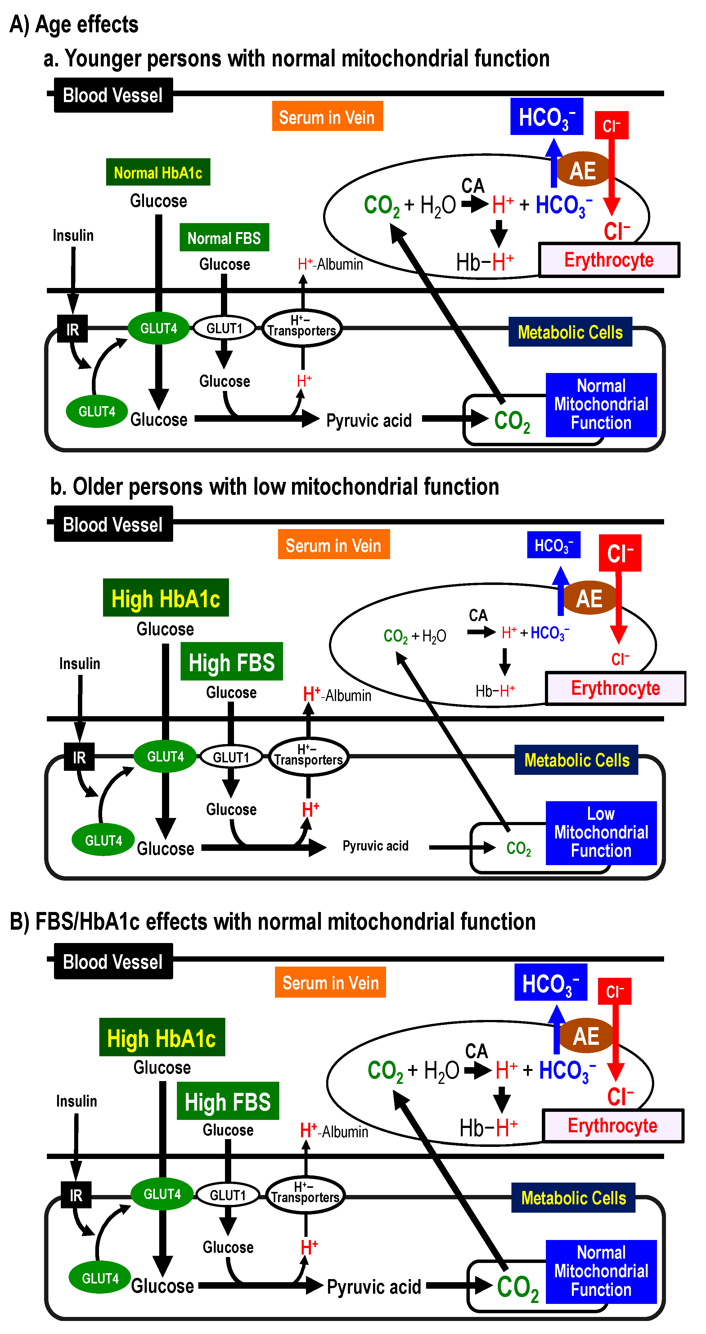
We next analyzed the relationship between FBS and HbA1c using Equation (11) (see Section 4.11 in Materials and Methods), although it is well known that FBS and HbA1c show positive correlation. We also analyzed the relationship between FBS and HbA1c using the normalized data with Equation (12) (see Section 4.11 in Materials and Methods). Both F FBS HbA 1 c and F N FBS HbA 1 c are significantly larger than 0 (Table 7), suggesting that HbA1c changes had a positive correlation to the change in FBS. F N FBS HbA 1 c enabled us to realize the relationship between FBS and HbA1c; the change of FBS in its 78% weight would influence the change in HbA1c.
We further analyzed the relationship between FBS and HbA1c by categorizing FBS into three ranges, (1) FBS < 100 mg/dL, (2) 100 mg/dL ≦ FBS < 126 mg/dL, and (3) 126 mg/dL ≦ FBS using Equations (11) and (12). Table 8 shows the analyzed results using original and normalized data. F FBS HbA 1 c was significantly different from each other among the group, (1) FBS < 100, (2) 100 ≦ FBS < 126 mg/dL, and (3) 126 mg/dL ≦ FBS (Table 8: Tukey–Kramer’s HSD). It is obvious that F N FBS HbA 1 c is significantly different from each other among the group, (1) FBS < 100 mg/dL, (2) 100 mg/dL ≦ FBS < 126 mg/dL, and (3) 126 mg/dL ≦ FBS (Table 8: Tukey–Kramer’s HSD). Interestingly, F FBS HbA 1 c in persons with FBS ≧ 100 mg/dL is much larger than that with FBS < 100 mg/dL; a similar observation is obviously seen in F N FBS HbA 1 c . These observations indicate that HbA1c in persons with high FBS (FBS ≧ 100 mg/dL) would be positively correlated to FBS in a much larger degree than that with normal FBS (FBS < 100 mg/dL). In another word, HbA1c in persons with normal FBS (FBS < 100 mg/dL) show relatively little correlation to FBS compared with that with high FBS (FBS ≧ 100 mg/dL).

2.8. Possible Mechanims of [Cl]s Changes by Age, FBS, and HbA1c and Clincially Significant Meanings of [Cl]s

We indicated possible mechanisms inducing the phenomena observed in the present study and clinical significances of [Cl]s suggested by the observations in the present study.

2.8.1. Possible Mechanisms of [Cl]s Changes by Age

Figure 4A shows possible mechanisms of age-dependent changes in [Cl]s. Figure 4a shows the case of younger persons. Younger persons have normal mitochondrial function [26,27,28,29]. Glucose is metabolized into pyruvic acid, and then CO2 is produced from the pyruvic acid in mitochondria with normal function. The produced CO2 moves into erythrocytes, and is converted into H+ and HCO3 via a CA-facilitated process. The HCO3 is exchanged with serum Cl via a Cl/HCO3 anion exchanger (AE). These processes lead to low [Cl]s. Figure 4b shows cases of older persons. Mitochondrial function is lower in older persons compared to younger ones [26,27,28,29]. In older persons, the amount of CO2 produced in mitochondria becomes low due to low mitochondrial function. Thus, the amount of H+ and HCO3 produced from CO2 becomes low. These processes keep high [Cl]s.

2.8.2. Possible Mechanisms of [Cl]s Changes by FBS and HbA1c

Figure 4B shows possible mechanisms of FBS/HbA1c-dependent changes in [Cl]s. The analytical results shown in Table 5 and Table 6 indicate that [Cl]s would decrease as FBS or HbA1c increases in persons with FBS (≧100 mg/dL) or HbA1c of all ranges. However, we have no information on the definite reason why [Cl]s would decrease as FBS or HbA1c increases in almost healthy persons except cases of FBS < 100 mg/dL. A decrease in [Cl]s would be due to an increase in [HCO3]s converted from CO2 produced in metabolic cells associated with an increase in [H+] converted from CO2 [6,10,19,30,31]. This means that [HCO3]s would increase as FBS or/and HbA1c become larger via elevation of CO2 production except cases of FBS < 100 mg/dL under the condition with normal mitochondrial function (Figure 4B): i.e., under the normal mitochondrial function, [Cl]s would decrease associated with an increase [HCO3]s when FBS and HbA1c are elevated, since the elevation of FBS and HbA1c would increase glucose metabolism resulting in large production of CO2 under the normal mitochondrial function with normal glucose transport function across the plasma membrane of metabolic cells (Figure 4B).
On one hand, we have observed a contrary phenomenon in persons with FBS < 100 mg/dL that [Cl]s would increase according to elevation of FBS (Table 5) compared with the phenomenon that [Cl]s would decrease according to elevation of FBS or/and HbA1c in persons with FBS ≧ 100 mg/dL and all HbA1c ranges (Table 5 and Table 6). As well known, HbA1c shows the average of blood sugar (glucose) level during one–two months [32,33,34], while FBS shows literally the blood sugar level at the fasting state [32,33,34]. If [Cl]s would correlate to chronic metabolic states, [Cl]s would show stronger correlation to HbA1c than FBS. Indeed, this point is confirmed by the analytical results shown in Table 3. Further, to confirm the relationship between FBS and HbA1c, we analyzed the relationship (Table 8). F N FBS HbA 1 c , a coefficient of FBS influencing HbA1c using the normalized data, is much smaller in persons with FBS < 100 mg/dL than that with FBS ≧ 100 mg/dL. This means that FBS shows much stronger correlation to the average of blood glucose sugar (glucose) levels for chronic time duration indicated as HbA1c in persons with FBS ≧ 100 mg/dL than that in FBS < 100 mg/dL (Table 8). Therefore, the phenomenon of [Cl]s increases according to FBS elevation in persons with FBS < 100 mg/dL unlike FBS ≧ 100 mg/dL would be due to the weak correlation of FBS to chronic blood sugar levels (HbA1c) in persons with FBS < 100 mg/dL (Table 8); i.e., FBS would not strongly reflect the average of blood glucose sugar (glucose) levels unlike HbA1c in persons with the normal FBS level (FBS < 100 mg/dL). These observations on the relationship between [Cl]s and HbA1c indicate the following possibilities regarding the body conditions: (1) Elevation of HbA1c associated with diminution of [Cl]s suggests normality of mitochondrial function with hyperphagia; (2) elevation of HbA1c associated with augmentation of [Cl]s suggests abnormality of mitochondrial function and disorder of glucose uptake into metabolic cells mainly due to aging-induced disorders of mitochondrial function and glucose uptake into metabolic cells (Figure 4).

2.8.3. Clinically Significant Meanings of [Cl]s Values

Based on these observations, we recognize the clinically significant meanings of low [Cl]s in almost healthy persons as follows: (1) the normality of glucose uptake into metabolic cells and glucose metabolism in metabolic cells; (2) appearance of slight insulin resistance via the reduction of interstitial fluid pH dependent on high HbA1c. Thus, we suggest that reduced values of [Cl]s could be a clinically useful marker as recognition of glucose uptake, metabolism and slight insulin resistance in almost healthy persons combining the value of HbA1c. Clinically significant meanings of [Cl]s values are summarized in Table 9.

3. Discussion

The analytical results in the present study indicate that: (1) [Cl]s, FBS, and HbA1c significantly increase with age; (2) [Cl]s shows positive correlation to age, and negative correlation to FBS and HbA1c especially in persons with high FBS (≧126 mg/dL) and HbA1c (≧6.5%); (3) the most [Cl]s-influencing factor is age among three factors, age, FBS, and HbA1c (c.f., Figure 4A summarizes age effects on [Cl]s, FBS, and HbA1c, and Figure 4B summarizes FBS/HbA1c effects on [Cl]s in persons with normal mitochondrial function).
The change in [Cl]s would depend on the production of CO2 in metabolic cells such as myocytes, hepatocytes, renal epithelial cells, etc. CO2 produced in metabolic cells moves into erythrocytes, then CO2 is converted to H+ and HCO3 (CO2 + H2O ⟶ H+ HCO3) in erythrocytes via a CA-facilitated process [6,10]. H+ produced from CO2 in erythrocytes bounds to Hb, while HCO3 produced from CO2 in erythrocytes is excreted to the serum in blood (the extracellular space of erythrocytes) via the AE-mediated process, participating in uptake of Cl into erythrocytes from the serum in blood [6,10]. CAs expressed in erythrocytes are I and II isozymes of CAs: CAI and CAII [35]. This Cl movement into erythrocytes across the plasma membrane is well-known as “Cl shift”: (1) in erythrocytes, the Cl concentration increases associated with a decrease of HCO3 concentration; (2) in the serum, [Cl]s concentration decreases associated with an increase of [HCO3]s. Thus, elevation of CO2 production in metabolic cells would increase [HCO3]s associated with a decrease of [Cl]s in the serum of blood [6,7,8]. Compared with younger persons, older persons show smaller O2 uptake due to slower O2 uptake kinetics [26], limitation of oxygen delivery [27], and low rates of electron transfer and O2 uptake in mitochondria [28]. These reports suggest that the amount of CO2 production would be lower in older persons than that in younger ones, since CO2 is produced from O2 in mitochondria. Further, mitochondria dysfunction appears in an age-dependent manner [28,29]. Mitochondria dysfunction leads to low O2 consumption resulting in low production of CO2. Based on these reports, the elevated [Cl]s observed in older persons would be due to mitochondrial dysfunction, which is also observed in persons with cancers and diabetes [36,37,38], thus [Cl]s continuously (even once or twice a year) measured with easy techniques would be useful as a marker detecting mitochondrial function.
Both FBS and HbA1c show the age-dependent increases (Figure 2 and Figure 3). However, the age-dependent increase of HbA1c (Figure 3) from 60s to 70≦ looks larger in degree than that of FBS (Figure 2). The larger age-dependent increase in HBA1c than FBS from 60s to 70≦ would be due to a larger increase in PBS than FBS occurring in 70≦ compared with 60≦ caused by impaired glucose tolerance or/and insulin resistance, affecting PBS but not FBS. Mitochondrial dysfunction appearing in an age-dependent manner [28,29] induces the glycolysis-based metabolic condition associated with production of large amounts of protons (H+), causing acidification of the interstitial fluid [6,7,8,10,19,36,37,38,39,40,41]. This acidification causes insulin resistance via reduction of insulin affinity to its receptor [6,7,8,10,19,39,40,41], resulting in a larger increase in HbA1c due to elevation of PBS compared with elevation of insulin-independently controlled FBS from the age of 60s to 70≦. The absolute value of coefficient of NHbA1c influencing [Cl]s being a little bit but significantly larger than that of NFBS (Table 3) would be explained by the characteristics of HbA1c reflecting the average blood sugar level during one–two months before the blood-sampled time [33,34,42,43] unlike FBS literally showing the fasting blood sugar level at the blood-sampled [33,34]. Therefore, based on the observation that [Cl]s shows a relatively stronger correlation to HbA1c than FBS, it is suggested that [Cl]s would depend on the average blood sugar level reflecting the metabolic condition.
In addition, we should consider cases of diabetic ketoacidosis [10,30,44]. Diabetic ketoacidosis occurs under conditions that glucose is not available as energy sources [10,30,44]. When glucose in not available as energy source, another energy source is required: e.g., a free fatty acid is one of major energy sources at unavailability of glucose. Metabolism of free fatty acids produces ketone bodies [45]. Beta-hydroxybutyric acid (CH3-CH(OH)-CH2-COOH), one of the most major ketone bodies (~70% of total ketone bodies), is produced from free fatty acids released from adipocytes [45], and then is dissociated into beta-hydroxybutyrate (CH3-CH(OH)-CH2-COO) and H+ (CH3-CH(OH)-CH2-COOH ⟶ CH3-CH(OH)-CH2-COO + H+) [46]. Under this condition, little amounts of HCO3 are produced from glucose metabolism associated with a large amount of ketone bodies such as beta-hydroxybutyrate (CH3-CH(OH)-CH2-COO), the concentration of which increases in the serum. In this case, the serum HCO3 or Cl doesn’t change unlike the case of glucose metabolism that CO2 produced in metabolic cells moves into erythrocytes, dissociating into HCO3 + H+ via a CA-mediated process, which leads to an increased [HCO3]s and a decreased [Cl]s via an AE-mediated exchange pathway. The metabolism of free fatty acids produces a large amount of H+ dissociated from ketone bodies at glucose unavailable states, leading to acidosis; it is called normochloremic ketoacidosis with high anion gap, which occurs in patients with severe DM [10,30,44,47,48].
In addition to this explanation on the relationship among [Cl]s, FBS and HbA1c, we should also consider another cause for an increase of [HCO3]s with elevation of FBS or/and HbA1c: i.e., the CO2 excretion capacity into the atmosphere through expiration should be considered [10]. Most parts of CO2 produced in metabolic cells are excreted into the atmosphere through expiration in the lung [10]. The decrease in amounts of CO2 excretion into the atmosphere causes an increase in [HCO3]s. Therefore, we should consider a possibility that the amount of CO2 excretion would decrease as FBS or/and HbA1c are elevated. The CO2 produced in metabolic cells moves into erythrocytes [6,10,19,30,31]. Then, CAs facilitate the converting process of CO2 to H+ and HCO3 (CO2 + H2O ⟶ H+ HCO3) in erythrocytes: I and II isozymes of CA (CAI and CAII) are expressed in erythrocytes [35]. The HCO3 is excreted from erythrocytes to the serum in blood (the extracellular space of erythrocytes) via the AE-mediated pathway, while the produced H+ bounds to Hb (c.f., Figure 4) [6,10,19,30,31]. In the lung, the Hb-bound H+ and HCO3 transported into erythrocytes from the serum via the AE-mediated reversed pathway (c.f., Figure 4) are converted to CO2 and H2O (H+ + HCO3 ⟶ CO2 + H2O) via a CA-facilitated pathway in erythrocytes [6,10,19,30,31]. CA is also expressed in capillary endothelia of the lung [49,50]. The CA expressed in capillary endothelia of the lung contributes to the converting process of H+ and HCO3 dissolved in the serum (several percent of total produced CO2) to CO2 + H2O (H+ + HCO3 ⟶ CO2 + H2O). The activity of CA expressed in capillary endothelia of the lung has been reported to be lower in DM patients than healthy persons [51]. These reports [49,50,51] lead us to an idea that high [HCO3]s might be also caused by slow conversion of H+ and HCO3 to CO2 and H2O (H+ + HCO3 ⟶ CO2 + H2O) via CA-medicated processes in the lung of persons with high leveled FBS or/and HbA1c; (2) the decelerated converting process of H+ and HCO3 to CO2 and H2O in the lung would keep high [HCO3]s in DM patients; (3) the high [HCO3]s in DM patients would keep low [Cl]s; (4) the activity of CA would become lower as FBS or/and HbA1c increase; (5) if so, [Cl]s would decrease associated with elevation of FBS or/and HbA1c due to the low activity of CA under the FBS/HbA1c-elevated condition; (6) the lower activity of CA in persons with high FBS or/and HbA1c might cause acidotic conditions in blood and interstitial fluids, causing the insulin resistance [6,7,8,9,10,19,30,41].
Here, we should also consider the aging effect on gas exchange in the lung [52,53,54] including disorders of gas exchange such as chronic obstructive pulmonary disease (COPD) [54]. Symptoms of COPD are well known to progress with age [52]. Patients with COPD show difficulty to excrete CO2 into the atmosphere [55]. At the early stage of COPD, CO2 retention in the body occurs due to difficulty of CO2 excretion into the atmosphere in the lung [55]. Disorders of gas exchange cause low O2 availability in metabolic cells associated with low CO2 production, resulting in reduction of life activity due to low energy (ATP) supply [52,53,54,55]. Patients suffering from severe COPD would show dyspnea, therefore it is relatively easy to diagnose COPD using various diagnostic devices such as CT scan, etc. [55]. However, it is difficult to diagnose COPD or find symptoms of COPD especially at the early stage. Therefore, [Cl]s could be a screening maker to find out patients staying in a very early stage of COPD just by taking basic medical examinations adaptable for huge numbers of persons, although confirmed diagnosis for COPD definitely requires advanced medical diagnostic devices such as CT scan.
In addition to aging effects on the lung function, we should also consider aging effects on the kidney function. Aging decreases glomerular filtration rate (GFR) [56,57]. The age-dependent decrease in GFR diminishes the filtrating amount of serum Na+ and Cl [56,57], stimulating the secretion of renin followed by activation of the renin-angiotensin-aldosterone (RAA) system [57]. Thus, the activation of RAA system caused by the age-dependent decrease in GFR would be considered as another cause of [Cl]s increases with age.
In the present study, we analyzed the correlation among [Cl]s, age, FBS, and HbA1c, and tried to clarify the physiological and/or pathophysiological meanings of the change in [Cl]s. We especially focused on the relationship between the metabolic condition and [HCO3]s by measuring [Cl]s. To clarify this point, [HCO3]s should be ideally measured. However, most persons showing no or little health problems without any serious symptoms usually take only basic medical examinations without [HCO3]s measurements due to its technical complication. Therefore, under the condition, an easily measurable index, [Cl]s, would be useful to estimate [HCO3]s reflecting metabolic conditions adaptable to huge numbers of persons.

4. Materials and Methods

4.1. Subjects

Data were obtained from persons taking medical examinations at Kyoto Industrial Health Association from 1 April 2011 to 31 March 2017. Written information regarding the present study was provided on WEB of Kyoto Industrial Health Association announcing to persons taking medical examination that they can opt out their own data from the present study. The number (n) of the persons participating in the present study was 107,630; the average of age, 51.61 ± 0.04 (mean ± standard error) years old (18–96); male, n = 71,423, 51.76 ± 0.04 (mean ± standard error) years old (18–96); female, n = 36,207, 51.26 ± 0.06 (mean ± standard error) years old (18–89).

4.2. Fasting Blood Samples

Blood samples were obtained from veins of persons with fasting for more than 5 h who took medical examinations at Kyoto Industrial Health Association. We excluded persons taking any DM treatments.

4.3. Measurements of [Cl]s, FBS and HbA1c

[Cl]s, FBS, and HbA1c were measured at the laboratory of Kyoto Industrial Health Association. [Cl]s was measured using a Cl-selective electrode, A&T Corporation, Yokohama 221-0056, Japan. HbA1c was assayed using high-performance liquid chromatography and was expressed as a National Glycohemoglobin Standardization Program unit.

4.4. Statistical Analysis

The statistical analysis was performed by a software, JMP 8.0 using Tukey–Kramer’s honestly significant difference (HSD). Data are shown as the mean values with the upper and lower limits of the 95% confidence interval (CI) of the mean values except the presentation of age.

4.5. Relationship among [Cl]s, Age, FBS and HbA1c

The relationship among [Cl]s, age, FBS, and HbA1c was analyzed assuming that the following equation would hold.
[ Cl ] s = C AFH Age   age + C AFH FBS   FBS + C AFH HbA 1 c   HbA 1 c + C AFH Int
Here, C AFH Age , C AFH FBS , and C AFH HbA 1 c are respectively [Cl]s-influencing coefficients of age, FBS, and HbA1c; C AFH Int is the intersection value of [Cl]s at age, FBS, and HbA1c = 0; C AFH Age , C AFH FBS , C AFH HbA 1 c , and C AFH Int are constant.

4.6. The Relationship among Normalized Data, N[Cl]s, NAge, NFBS, and NHbA1c

Age, FBS, and HbA1c had different units; therefore, it was impossible to determine which factor, age, FBS, or HbA1c, most effectively influences [Cl]s. To clarify this point, we normalized the values of [Cl]s, age, FBS, and HbA1c by setting each mean value of [Cl]s, age, FBS or HbA1c = 0 with each standard deviation = 1. Here, we respectively represent the normalized data of [Cl]s, age, FBS, and HbA1c as N[Cl]s, Nage, NFBS, and NHbA1c. The relationship among [Cl]s, age, FBS, and HbA1c was analyzed assuming that the following equation would hold.
[ Cl ] N s = C N AFH Age   a N ge + C N AFH FBS   F N BS + C N AFH HbA 1 c   H N bA 1 c + C N AFH Int
Here, C N AFH Age , C N AFH FBS , and C N AFH HbA 1 c are respectively N[Cl]s-influencing coefficients of Nage, NFBS, and NHbA1c, and I N AFH Int is the intersection value of N[Cl] at Nage, NFBS, and NHbA1c = 0; C N AFH Age , C N AFH FBS , C N AFH HbA 1 c , and C N AFH Int are constant.

4.7. Correlation of [Cl]s, FBS or HbA1c to Age Using the Normalized Data, N[Cl]s, NFBS, NHbA1c, and NAge

The analysis was performed using Equations (3)–(5), respectively.
[ Cl ] N s = C N Age Age   a N ge + C N Age Int = C N Age Age   a N ge
[ FBS ] N = F N Age Age   a N ge + F N Age Int = F N Age Age   a N ge
[ HbA 1 c ] N = H N Age Age   a N ge + H N Age Int = H N Age Age   a N ge
Here, C N Age Age , F N Age Age , and H N Age Age are respectively the Nage-dependent coefficients for N[Cl]s, NFBS, or NHbA1c, and C N Age Int , F N Age Int , and H N Age Int are respectively the intersection values of N[Cl]s, NFBS, and NHbA1c at Nage = 0: C N Age Int , F N Age Int or H N Age Int (the intersection value of N[Cl]s, NFBS, or NHbA1c at Nage = 0) would be ideally 0, since all the mean values of [Cl]s, age, FBS, and HbA1c were normalized to be = 0.

4.8. Age-Dependent Factor and FBS/HbA1c-Dependent Factor Influencing N[Cl]s

Substituting Equations (3)–(5) into Equation (2), the following equation was obtained ( C N AFH Int = 0).
[ Cl ] N s = C N AFH Age   a N ge + C N AFH FBS   F N BS + C N AFH HbA 1 c   H N bA 1 c + C N AFH Int = C N AFH Age   a N ge + C N AFH FBS   F N Age Age   a N ge + C N AFH HbA 1 c   H N Age Age   a N ge = ( C N AFH Age + C N AFH FBS   F N Age Age + C N AFH HbA 1 c   H N Age Age )   a N ge = C N Age Age   a N ge  
Thus, Equation (7) functions.
C N Age Age = C N AFH Age + C N AFH FBS   F N Age Age + C N AFH HbA 1 c   H N Age Age
This means that the Nage-dependent coefficient ( C N Age Age ) for [Cl]s consists of C N AFH Age , C N AFH FBS   F N Age Age and C N AFH HbA 1 c   H N Age Age . Here, C N AFH Age is the NFBS/NHbA1c-independent, Nage-dependent coefficient for N[Cl]s; C N AFH FBS   F N Age Age , the NFBS/Nage-dependent coefficient for N[Cl]s; C N AFH HbA 1 c   H N Age Age , the NHbA1c/Nage-dependent coefficient for N[Cl]s. Equation (8) functions, since C N Age Age > 0 , C N AFH Age > 0 , C N AFH FBS < 0   F N Age Age > 0 , C N AFH HbA 1 c < 0 , and H N Age Age > 0 in Equation (7) (see Table 3 and Table 4).
C N Age Age < C N AFH Age

4.9. The Relationship between [Cl]s and FBS

The relationship between [Cl]s and FBS was analyzed using Equation (9).
[ Cl ] s = C FBS FBS   FBS + I FBS FBS
Here, C FBS FBS is a [Cl]s-influencing coefficient of FBS, I FBS FBS is the intersection value of [Cl]s at FBS = 0, and C FBS FBS and I FBS FBS are constant.

4.10. The Relationship between [Cl]s and HbA1c

The relationship between [Cl]s and HbA1c was analyzed using Equation (10).
[ Cl ] s = C HbA 1 c HbA 1 c   HbA 1 c + I HbA 1 c HbA 1 c
Here, C HbA 1 c HbA 1 c is a coefficient of HbA1c influencing [Cl]s, I HbA 1 c HbA 1 c is the intersection value of [Cl]s at HbA1c = 0, and C HbA 1 c HbA 1 c and I HbA 1 c HbA 1 c are constant.

4.11. Relationship between FBS and HbA1c

The relationship between FBS and HbA1c was analyzed using Equation (11), and also using the normalized data with Equation (12).
HbA 1 c = F FBS HbA 1 c   FBS + I FBS HbA 1 c
H N bA 1 c = F N FBS HbA 1 c   F N BS + I N FBS HbA 1 c
Here, F FBS HbA 1 c is a HbA1c-influencing coefficient of FBS, I FBS HbA 1 c is the intersection value of HbA1c at FBS = 0, F N FBS HbA 1 c is H N bA 1 c -influencing coefficient of F N BS , I FBS HbA 1 c is the intersection value of H N bA 1 c at F N BS = 0, and F FBS HbA 1 c , I FBS HbA 1 c , F N FBS HbA 1 c , and I N FBS HbA 1 c are constant.

5. Conclusions

The present study indicates that: (1) the values of [Cl]s, FBS, and HbA1c are larger in older persons than younger ones; (2) [Cl]s shows positive correlation to age, and negative correlation to FBS and HbA1c especially in persons with high FBS (≧126 mg/dL) and HbA1c (≧6.5%); (3) the most [Cl]s-influencing factor is “age” among three factors, age, FBS, and HbA1c. [Cl]s would be a marker of metabolism and insulin resistance, and show mitochondrial function combining information on FBS/HbA1c. Figure 4 and Table 9 summarize the conclusion obtained from the present study.

Author Contributions

Conceptualization, Y.M.; methodology, Y.M. and K.Y.; validation, Y.M., K.Y., N.I., H.K., M.M., A.M., Y.T. and T.N.; formal analysis, Y.M. and K.Y.; data curation, N.I., H.K. and M.M.; writing—original draft preparation, Y.M.; writing—review and editing, Y.M.; visualization, Y.M.; supervision, Y.M.; project administration, Y.M.; funding acquisition, Y.M. All authors have read and agreed to the published version of the manuscript.

Funding

This work was supported by Grants-in-Aid for Scientific Research (B) from Japan Society of the Promotion of Science (JSPS KAKENHI JP18H03182 and JP21H03368 to Y.M.).

Institutional Review Board Statement

This study was approved by the local research ethics committee of Kyoto Industrial Health Association (Approval No. S18-0005).

Informed Consent Statement

Written information regarding the present study was provided on 15 March 2019 at WEB of Kyoto Industrial Health Association announcing to persons taking medical examination that they can opt out their own data on medical examinations from the present study, and the written information has still been provided at the WEB. The persons who did not opt out by the date of the present manuscript submitted to the journal were considered to provide consent for study participation in the present study.

Acknowledgments

The authors thank Norimitsu Nishida and Yoshifumi Mukai at Kyoto Industrial Health Association preparing the data analyzed in the present study.

Conflicts of Interest

The authors declare no conflict of interest.

Abbreviations

AEanion exchanger
CAcarbonic anhydrase
CAIcarbonic anhydrase I
CAIIcarbonic anhydrase II
CIconfidence interval
[Cl]svenous serum Cl concentration
COPDchronic obstructive pulmonary disease
DMdiabetes mellitus
FBSfasting blood sugar
GFRglomerular filtration rate
Hbhemoglobin
HbA1cglycated hemoglobin
[HCO3]svenous serum HCO3 concentration
HSDhonestly significant difference
LLlower limit
PBSpostprandial blood sugar
RAArenin-angiotensin-aldosterone
ULupper limit

References

  1. Altobelli, E.; Angeletti, P.M.; Marziliano, C.; Mastrodomenico, M.; Giuliani, A.R.; Petrocelli, R. Potential therapeutic effects of curcumin on glycemic and lipid profile in uncomplicated type 2 diabetes-A meta-analysis of randomized controlled trial. Nutrients 2021, 13, 404. [Google Scholar] [CrossRef] [PubMed]
  2. Whitworth, J.W.; Hayes, S.M.; Andrews, R.J.; Fonda, J.R.; Beck, B.M.; Hanlon, L.B.; Fortier, C.B.; Milberg, W.P.; McGlinchey, R.E. Cardiorespiratory fitness Is associated with better cardiometabolic health and lower PTSD severity in post-9/11 veterans. Mil. Med. 2020, 185, e592–e596. [Google Scholar] [CrossRef] [PubMed] [Green Version]
  3. Xu, B.; Fu, J.; Qiao, Y.; Cao, J.; Deehan, E.C.; Li, Z.; Jin, M.; Wang, X.; Wang, Y. Higher intake of microbiota-accessible carbohydrates and improved cardiometabolic risk factors: A meta-analysis and umbrella review of dietary management in patients with type 2 diabetes. Am. J. Clin. Nutr. 2021, 113, 1515–1530. [Google Scholar] [CrossRef] [PubMed]
  4. Cunha-Guimaraes, J.P.; Guarino, M.P.; Timóteo, A.T.; Caires, I.; Sacramento, J.F.; Ribeiro, M.J.; Selas, M.; Santiago, J.C.P.; Mota-Carmo, M.; Conde, S.V. Carotid body chemosensitivity: Early biomarker of dysmetabolism in humans. Eur. J. Endocrinol. 2020, 182, 549–557. [Google Scholar] [CrossRef]
  5. Peng, C.C.; Tu, Y.K.; Lee, G.Y.; Chang, R.H.; Huang, Y.; Bukhari, K.; Tsai, Y.C.; Fu, Y.; Huang, H.K.; Munir, K.M. Effects of proton pump inhibitors on glycemic control and incident diabetes: A systematic review and meta-analysis. J. Clin. Endocrinol. Metab. 2021. [Google Scholar] [CrossRef]
  6. Marunaka, Y. Roles of interstitial fluid pH and weak organic acids in development and amelioration of insulin resistance. Biochem. Soc. Trans. 2021, 49, 715–726. [Google Scholar] [CrossRef] [PubMed]
  7. Aoi, W.; Marunaka, Y. The importance of regulation of body fluid pH in the development and progression of metabolic diseases. In Advances in Medicine and Biology; Berhardt, L.V., Ed.; Nova Publishers: Hauppauge, NY, USA, 2014; Volume 77, pp. 177–189. [Google Scholar]
  8. Aoi, W.; Marunaka, Y. Importance of pH homeostasis in metabolic health and diseases: Crucial role of membrane proton transport. BioMed Res. Int. 2014, 2014, 598986. [Google Scholar] [CrossRef] [Green Version]
  9. Aoi, W.; Iwasa, M.; Marunaka, Y. Metabolic functions of flavonoids: From human epidemiology to molecular mechanism. Neuropeptides 2021, 88, 102163. [Google Scholar] [CrossRef]
  10. Marunaka, Y. The proposal of molecular mechanisms of weak organic acids intake-induced improvement of insulin resistance in diabetes mellitus via elevation of interstitial fluid pH. Int. J. Mol. Sci. 2018, 19, 3244. [Google Scholar] [CrossRef] [Green Version]
  11. Lee, D.; Hong, J.H. The Fundamental role of bicarbonate transporters and associated carbonic anhydrase enzymes in maintaining ion and pH homeostasis in non-secretory organs. Int. J. Mol. Sci. 2020, 21, 339. [Google Scholar] [CrossRef] [Green Version]
  12. Remigante, A.; Morabito, R.; Marino, A. Band 3 protein function and oxidative stress in erythrocytes. J. Cell Physiol. 2021, 236, 6225–6234. [Google Scholar] [CrossRef] [PubMed]
  13. Hosogi, S.; Miyazaki, H.; Nakajima, K.; Ashihara, E.; Niisato, N.; Kusuzaki, K.; Marunaka, Y. An inhibitor of Na+/H+ exchanger (NHE), ethyl-isopropyl amiloride (EIPA), diminishes proliferation of MKN28 human gastric cancer cells by decreasing the cytosolic Cl concentration via DIDS-sensitive pathways. Cell. Physiol. Biochem. 2012, 30, 1241–1253. [Google Scholar] [CrossRef] [PubMed]
  14. Shiozaki, A.; Hikami, S.; Ichikawa, D.; Kosuga, T.; Shimizu, H.; Kudou, M.; Yamazato, Y.; Kobayashi, T.; Shoda, K.; Arita, T.; et al. Anion exchanger 2 suppresses cellular movement and has prognostic significance in esophageal squamous cell carcinoma. Oncotarget 2018, 9, 25993–26006. [Google Scholar] [CrossRef] [PubMed]
  15. Shiozaki, A.; Kudou, M.; Ichikawa, D.; Shimizu, H.; Arita, T.; Kosuga, T.; Konishi, H.; Komatsu, S.; Fujiwara, H.; Okamoto, K.; et al. Expression and role of anion exchanger 1 in esophageal squamous cell carcinoma. Oncotarget 2017, 8, 17921–17935. [Google Scholar] [CrossRef] [PubMed] [Green Version]
  16. Shiozaki, A.; Ichikawa, D.; Otsuji, E.; Marunaka, Y. Cellular physiological approach for treatment of gastric cancer. World J. Gastroenterol. 2014, 20, 11560–11566. [Google Scholar] [CrossRef] [PubMed]
  17. Zimna, A.; Kaczmarska, M.; Szczesny-Malysiak, E.; Wajda, A.; Bulat, K.; Alcicek, F.C.; Zygmunt, M.; Sacha, T.; Marzec, K.M. An insight into the stages of ion leakage during red blood cell storage. Int. J. Mol. Sci. 2021, 22, 2885. [Google Scholar] [CrossRef] [PubMed]
  18. Perez Ruiz de Garibay, A.; Kellum, J.A.; Honigschnabel, J.; Kreymann, B. Respiratory and metabolic acidosis correction with the ADVanced Organ Support system. Intensive Care Med. Exp. 2019, 7, 56. [Google Scholar] [CrossRef] [PubMed]
  19. Marunaka, Y. Roles of interstitial fluid pH in diabetes mellitus: Glycolysis and mitochondrial function. World J. Diabetes 2015, 6, 125–135. [Google Scholar] [CrossRef]
  20. Mohebbi, N.; Perna, A.; van der Wijst, J.; Becker, H.M.; Capasso, G.; Wagner, C.A. Regulation of two renal chloride transporters, AE1 and pendrin, by electrolytes and aldosterone. PLoS ONE 2013, 8, e55286. [Google Scholar] [CrossRef] [Green Version]
  21. Jennings, M.L. Stoichiometry of a half-turnover of band 3, the chloride transport protein of human erythrocytes. J. Gen. Physiol. 1982, 79, 169–185. [Google Scholar] [CrossRef] [Green Version]
  22. Falke, J.J.; Pace, R.J.; Chan, S.I. Chloride binding to the anion transport binding sites of band 3. A 35Cl NMR study. J. Biol. Chem. 1984, 259, 6472–6480. [Google Scholar] [CrossRef]
  23. Wolpaw, E.W.; Martin, D.L. A membrane protein in LRM55 glial cells cross-reacts with antibody to the anion exchange carrier of human erythrocytes. Neurosci. Lett. 1986, 67, 42–47. [Google Scholar] [CrossRef]
  24. Kim, H.R.; Yew, N.S.; Ansorge, W.; Voss, H.; Schwager, C.; Vennström, B.; Zenke, M.; Engel, J.D. Two different mRNAs are transcribed from a single genomic locus encoding the chicken erythrocyte anion transport proteins (band 3). Mol. Cell. Biol. 1988, 8, 4416–4424. [Google Scholar] [CrossRef] [Green Version]
  25. Verlander, J.W.; Madsen, K.M.; Low, P.S.; Allen, D.P.; Tisher, C.C. Immunocytochemical localization of band 3 protein in the rat collecting duct. Am. J. Physiol. Cell Physiol. 1988, 255, F115–F125. [Google Scholar] [CrossRef]
  26. Grassi, B. Oxygen uptake kinetics: Old and recent lessons from experiments on isolated muscle in situ. Eur. J. Appl. Physiol. 2003, 90, 242–249. [Google Scholar] [CrossRef]
  27. Murias, J.M.; Paterson, D.H. Slower V . O2 kinetics in older individuals: Is it inevitable? Med. Sci. Sports Exerc. 2015, 47, 2308–2318. [Google Scholar] [CrossRef] [PubMed] [Green Version]
  28. Navarro, A.; Boveris, A. The mitochondrial energy transduction system and the aging process. Am. J. Physiol. Cell Physiol. 2007, 292, C670–C686. [Google Scholar] [CrossRef] [PubMed]
  29. Ghosh, R.; Vinod, V.; Symons, J.D.; Boudina, S. Protein and mitochondria quality control mechanisms and cardiac aging. Cells 2020, 9, 933. [Google Scholar] [CrossRef] [PubMed]
  30. Aoi, W.; Zou, X.; Xiao, J.B.; Marunaka, Y. Body fluid pH balance in metabolic health and possible benefits of dietary alkaline foods. eFood 2020, 1, 12–23. [Google Scholar] [CrossRef] [Green Version]
  31. Marunaka, Y.; Niisato, N.; Zou, X.; Xiao, J.B.; Nakahari, T. Food intake targeting and improving acidity in diabetes and cancer. Food Front. 2020, 1, 9–12. [Google Scholar] [CrossRef] [Green Version]
  32. Chadda, K.R.; Cheng, T.S.; Ong, K.K. GLP-1 agonists for obesity and type 2 diabetes in children: Systematic review and meta-analysis. Obes. Rev. 2021, 22, e13177. [Google Scholar] [CrossRef]
  33. Carlson, A.L.; Criego, A.B.; Martens, T.W.; Bergenstal, R.M. HbA1c: The glucose management indicator, time in range, and standardization of continuous glucose monitoring reports in clinical practice. Endocrinol. Metab. Clin. N. Am. 2020, 49, 95–107. [Google Scholar] [CrossRef]
  34. Chehregosha, H.; Khamseh, M.E.; Malek, M.; Hosseinpanah, F.; Ismail-Beigi, F. A view beyond HbA1c: Role of continuous glucose monitoring. Diabetes Ther. 2019, 10, 853–863. [Google Scholar] [CrossRef] [Green Version]
  35. Alim, Z. 1H-indazole molecules reduced the activity of human erythrocytes carbonic anhydrase I and II isoenzymes. J. Biochem. Mol. Toxicol. 2018, 32, e22194. [Google Scholar] [CrossRef] [PubMed]
  36. Fais, S.; Marunaka, Y. The acidic microenvironment: Is it a phenotype of all cancers? A focus on multiple myeloma and some analogies with diabetes mellitus. Cancers 2020, 12, 3226. [Google Scholar] [CrossRef]
  37. Gillies, R.J.; Pilot, C.; Marunaka, Y.; Fais, S. Targeting acidity in cancer and diabetes. Biochim. Biophys. Acta Rev. Cancer 2019, 1871, 273–280. [Google Scholar] [CrossRef] [PubMed]
  38. Pillai, S.R.; Damaghi, M.; Marunaka, Y.; Spugnini, E.P.; Fais, S.; Gillies, R.J. Causes, consequences, and therapy of tumors acidosis. Cancer Metastasis Rev. 2019, 38, 205–222. [Google Scholar] [CrossRef] [PubMed]
  39. Hosogi, S.; Ohsawa, M.; Kato, I.; Kuwahara, A.; Inui, T.; Inui, A.; Marunaka, Y. Improvement of diabetes mellitus symptoms by intake of ninjin’yoeito. Front. Nutr. 2018, 5, 112. [Google Scholar] [CrossRef]
  40. Hayata, H.; Miyazaki, H.; Niisato, N.; Yokoyama, N.; Marunaka, Y. Lowered extracellular pH is involved in the pathogenesis of skeletal muscle insulin resistance. Biochem. Biophys. Res. Commun. 2014, 445, 170–174. [Google Scholar] [CrossRef]
  41. Aoi, W.; Hosogi, S.; Niisato, N.; Yokoyama, N.; Hayata, H.; Miyazaki, H.; Kusuzaki, K.; Fukuda, T.; Fukui, M.; Nakamura, N.; et al. Improvement of insulin resistance, blood pressure and interstitial pH in early developmental stage of insulin resistance in OLETF rats by intake of propolis extracts. Biochem. Biophys. Res. Commun. 2013, 432, 650–653. [Google Scholar] [CrossRef]
  42. Malandrucco, I.; Russo, B.; Picconi, F.; Menduni, M.; Frontoni, S. Glycemic status assessment by the latest glucose monitoring technologies. Int. J. Mol. Sci. 2020, 21, 8243. [Google Scholar] [CrossRef]
  43. Byrne, F.L.; Martin, A.R.; Kosasih, M.; Caruana, B.T.; Farrell, R. The role of hyperglycemia in endometrial cancer pathogenesis. Cancers 2020, 12, 1191. [Google Scholar] [CrossRef]
  44. Long, B.; Lentz, S.; Koyfman, A.; Gottlieb, M. Euglycemic diabetic ketoacidosis: Etiologies, evaluation, and management. Am. J. Emerg. Med. 2021, 44, 157–160. [Google Scholar] [CrossRef]
  45. Persson, B. Determination of plasma acetoacetate and D-beta-hydroxybutyrate in new-born infants by an enzymatic fluorometric micro-method. Scand. J. Clin. Lab. Investig. 1970, 25, 9–18. [Google Scholar] [CrossRef]
  46. Gosmanov, A.R.; Gosmanova, E.O.; Dillard-Cannon, E. Management of adult diabetic ketoacidosis. Diabetes Metab. Syndr. Obes. 2014, 7, 255–264. [Google Scholar] [CrossRef] [Green Version]
  47. Fenves, A.Z.; Emmett, M. Approach to patients with high anion gap metabolic acidosis: Core curriculum 2021. Am. J. Kidney Dis. 2021, 78, 590–600. [Google Scholar] [CrossRef] [PubMed]
  48. Kraut, J.A.; Madias, N.E. Serum anion gap: Its uses and limitations in clinical medicine. Clin. J. Am. Soc. Nephrol. 2007, 2, 162–174. [Google Scholar] [CrossRef] [PubMed] [Green Version]
  49. Hanson, M.A.; Nye, P.C.; Torrance, R.W. Studies on the localization of pulmonary carbonic anhydrase in the cat. J. Physiol. 1981, 319, 93–109. [Google Scholar] [CrossRef] [PubMed]
  50. Lossow, K.; Hermans-Borgmeyer, I.; Behrens, M.; Meyerhof, W. Genetic labeling of Car4-expressing cells reveals subpopulations of Type III taste cells. Chem. Senses 2017, 42, 747–758. [Google Scholar] [CrossRef]
  51. Bayrak, B.B.; Tunali, S.; Bal-Demirci, T.; Ulkuseven, B.; Yanardag, R. Glycoprotein levels and oxidative lung injury in experimental diabetes: Effect of oxovanadium(IV) complex based on thiosemicarbazone. Toxicol. Mech. Methods 2021, 31, 581–588. [Google Scholar] [CrossRef]
  52. Easter, M.; Bollenbecker, S.; Barnes, J.W.; Krick, S. Targeting aging pathways in chronic obstructive pulmonary disease. Int. J. Mol. Sci. 2020, 21, 6924. [Google Scholar] [CrossRef]
  53. Fuschillo, S.; Paris, D.; Tramice, A.; Ambrosino, P.; Palomba, L.; Maniscalco, M.; Motta, A. Metabolomic profiling of exhaled breath condensate and plasma/serum in chronic obstructive pulmonary disease. Curr. Med. Chem. 2021. [Google Scholar] [CrossRef] [PubMed]
  54. Balnis, J.; Korponay, T.C.; Jaitovich, A. AMP-activated protein kinase (AMPK) at the crossroads between CO2 retention and skeletal muscle dysfunction in chronic obstructive pulmonary disease (COPD). Int. J. Mol. Sci. 2020, 21, 955. [Google Scholar] [CrossRef] [PubMed] [Green Version]
  55. Mathews, A.M.; Wysham, N.G.; Xie, J.; Qin, X.; Giovacchini, C.X.; Ekström, M.; MacIntyre, N.R. Hypercapnia in advanced chronic obstructive pulmonary disease: A secondary analysis of the National Emphysema Treatment Trial. Chronic Obstr. Pulm. Dis. 2020, 7, 336–345. [Google Scholar] [CrossRef] [PubMed]
  56. Ennis, J.L.; Luo, D.; Asplin, J.R.; Coe, F.L. A laboratory-based algorithm to predict future kidney function decline in older adults with reduced estimated glomerular filtration rate. Clin. Nephrol. 2019, 92, 113–122. [Google Scholar] [CrossRef]
  57. Schmitt, R.; Melk, A. Molecular mechanisms of renal aging. Kidney Int. 2017, 92, 569–579. [Google Scholar] [CrossRef] [PubMed]
Figure 1. Age-dependent changes in venous serum Cl concentration ([Cl]s). The ages of persons taking medical examinations were categorized into six groups as shown in Table 1. Red horizontal bars at the persons’ ages show the mean [Cl]s values of persons at the ages. The upper and lower blue horizontal bars at the persons’ ages show respectively the upper and lower limits of the 95% confidence interval (CI) for the mean [Cl]s values of persons at the ages. [Cl]s increased in an age-dependent manner up to the 60s. Labels A, B, C, D, and E show the statistical difference: the mean [Cl]s values of the groups labeled with different characters are significantly different from each other at a level of p < 0.05, while the mean [Cl]s values of the groups labeled with the same character are not significantly different at a level of p ≧ 0.05 (the mean [Cl]s values of persons’ age = 60s and 70≦ were not significantly different). The statistical test was performed by Tukey–Kramer’s honestly significant difference (HSD).
Ijms 22 11111 g001
Figure 2. Age-dependent changes in venous serum fasting blood sugar concentration (FBS). The ages of persons taking medical examinations were categorized into six groups as shown in Table 1. Red horizontal bars show the mean values of FBS of persons at the ages. The upper and lower blue horizontal bars at the persons’ ages show respectively the upper and lower limits of the 95% confidence interval (CI) for the mean values of FBS of persons at the ages. FBS increased in an age-dependent manner up to 70 years old (70≦). Labels A, B, C, D, E and F show the statistical difference: the mean values of FBS of the groups labeled with different characters are significantly different from each other at a level of p < 0.05. The statistical test was performed by Tukey–Kramer’s HSD.
Ijms 22 11111 g002
Figure 3. Age-dependent changes in venous HbA1c. The ages of persons taking medical examinations were categorized into six groups as shown in Table 1. Red horizontal bars at the persons’ ages show the mean values of HbA1c of persons at the ages. The upper and lower blue horizontal bars at the persons’ ages show respectively the upper and lower limits of the 95% confidence interval (CI) for the mean values of HbA1c of persons at the ages. The HbA1c increased in an age-dependent manner up to 70 years old (70≦). Labels, A, B, C, D, E, and F show the statistical difference: The mean values of HbA1c of the groups labeled with different characters are significantly different from each other at a level of p < 0.05. The statistical test was performed by Tukey–Kramer’s HSD.
Ijms 22 11111 g003
Figure 4. Summary. (A) Age effects on [Cl]s. (a) Younger persons with normal mitochondrial function. Glucose is metabolized into pyruvic acid, and then CO2 is produced from the pyruvic acid in mitochondria with normal function. The produced CO2 moves into erythrocytes, and is converted into H+ and HCO3 via a CA-facilitated process. The HCO3 is exchanged with serum Cl via a Cl/HCO3 anion exchanger (AE). These processes lead to low [Cl]s. (b) Older persons with low mitochondrial function. The amount of CO2 produced in mitochondria becomes low due to low mitochondrial function. Thus, the amount of H+ and HCO3 produced from CO2 becomes low. These processes keep high [Cl]s. (B) FBS/HbA1c effects on [Cl]s. with normal mitochondrial function. Glucose is metabolized into pyruvic acid, and then CO2 is produced from the pyruvic acid in mitochondria with normal function. The produced CO2 moves into erythrocytes, and is converted into H+ and HCO3 via a CA-facilitated process. The HCO3 is exchanged with serum Cl via a Cl/HCO3 anion exchanger (AE). In cases of high FBS/HBA1c with normal mitochondrial function, large amounts of CO2 are produced, resulting in production of large amounts of HCO3. These processes lead to low [Cl]s.
Ijms 22 11111 g004
Table 1. Age category and the number of persons in each category.
Age Category<3030s40s50s60s70≦
Meaning of
Age Category
(years old)
Age < 3030 ≦ Age < 4040 ≦ Age < 5050 ≦ Age < 6060 ≦ Age < 7070 ≦ Age
Number of
persons (n)
187814,30035,45729,17520,3446476
We categorized the age of persons taking medical examinations into six groups; (1) younger than 30 years old (<30), (2) equal to or older than 30 years old and younger than 40 years old (30s), (3) equal to or older than 40 years old and younger than 50 years old (40s), (4) equal to or older than 50 years old and younger than 60 years old (50s), (5) equal to or older than 60 years old and younger than 70 years old (shown as 60s), and (6) equal to or older than 70 years old (shown as 70≦).
Table 2. The mean values of coefficients in Equation (1) for the relationship among [Cl]s, age, FBS, and HbA1c.
Coefficient C A F H A g e
(mEq/L/year)
C A F H F B S
(mEq/L/>mg/dL)
C A F H H b A 1 c
(mEq/L/%)
C A F H I n t
(mEq/L)
UL of 95% CI0.0312−0.00727−0.311106.1
Mean0.0300−0.00837−0.345106.0
LL of 95% CI0.0289−0.00947−0.379105.9
C AFH Age , C AFH FBS , and C AFH HbA 1 c are respectively [Cl]s-influencing coefficients of age, FBS, and HbA1c; C AFH Int is the intersection value of [Cl]s at age, FBS, and HbA1c = 0. The upper limit (UL) and the lower limit (LL) of 95% confidence interval (CI) of the mean value of the coefficient are also shown. n = 107,630.
Table 3. The mean value of the coefficient in Equation (2) for the relationship among [Cl]s, age, FBS, and HbA1c using normalized data of [Cl]s, age, FBS, and HbA1.
Coefficient C N A F H A g e
(mEq/L/year)
C N A F H F B S
(mEq/L/mg/dL)
C N A F H H b A 1 c
(mEq/L/%)
C N A F H I n t
(mEq/L)
UL of 95% CI0.1693−0.0626−0.08750.0059
Mean0.1631−0.0721−0.09700.0000
LL of 95% CI0.1569−0.0816−0.1065−0.0059
C N AFH Age , C N AFH FBS , and C N AFH HbA 1 c are respectively N[Cl]s-influencing coefficients of Nage, NFBS, and NHbA1c, and I N AFH Int is the intersection value of N[Cl] at Nage, NFBS, and NHbA1c = 0. The upper limit (UL) and the lower limit (LL) of the 95% confidence interval (CI) of the mean value of the coefficient are also shown. n = 107,630.
Table 5. The mean value of the coefficient in Equation (9) for the relationship between [Cl]s and FBS.
FBS (mg/dL)FBS < 100100 ≦ FBS < 126126 ≦ FBS
n59,92241,6336075
C FBS FBS UL of 95% CI0.0281−0.0128−0.0186
Mean0.0252−0.0161−0.0202
LL of 95% CI0.0222−0.0194−0.0219
C FBS FBS is a [Cl]s-influencing coefficient of FBS. The upper limit (UL) and the lower limit (UL) of 95% confidence interval (CI) of the mean value of the [Cl]s-influencing coefficient of FBS in persons whose FBS was categorized into each range are also shown. Total number = 107,630.
Table 6. The mean value of the coefficient in Equation (9) for the relationship between [Cl]s and HbA1c.
HbA1c (%)HbA1c < 5.65.6 ≦ HbA1c < 6.56.5 ≦ HbA1c
n57,18944,6995742
C HbA 1 c HbA 1 c UL of 95% CI−0.0188−0.1394−0.5794
Mean−0.1082−0.2332−0.6312
LL of 95% CI−0.1976−0.3269−0.6830
C HbA 1 c HbA 1 c is a coefficient of HbA1c influencing [Cl]s. The upper limit (UL) and the lower limit (LL) of 95% confidence interval (CI) of the mean value of the coefficient of FBS influencing [Cl]s in persons whose [FBS] was categorized into each range are shown.
Table 7. The mean values of coefficients in Equations (11) and (12) for the relationship between FBS and HbA1c.
Coefficient F F B S H b A 1 c F N F B S H b A 1 c
UL of 95% CI0.0217250.787159
Mean0.0214620.783447
LL of 95% CI0.0211990.779735
F FBS HbA 1 c is a HbA1c-influencing coefficient of FBS; F N FBS HbA 1 c is H N bA 1 c -influencing coefficient of F N BS . The upper limit (UL) and the lower limit (LL) of 95% confidence interval (CI) of the mean value of the coefficient are also shown. n = 107,630.
Table 8. The mean value of the coefficient in Equations (11) and (12) for the relationship between [Cl]s and FBS.
FBS (mg/dL)FBS < 100100 ≦ FBS < 126126 ≦ FBS
n59,92241,6336075
F FBS HbA 1 c UL of 95% CI0.01140.02660.0278
Mean0.01100.02610.0273
LL of 95% CI0.01060.02560.0267
F N FBS HbA 1 c UL of 95% CI0.35000.81460.8518
Mean0.33740.79850.8351
LL of 95% CI0.32470.78240.8185
The upper limit (UL) and the lower limit (UL) of 95% confidence interval (CI) of the mean value of the [Cl]s-influencing coefficient of FBS in persons whose FBS was categorized into each range are also shown.
Table 9. Clinically significant meanings of [Cl]s values HbA1c.
Low [Cl]sHigh [Cl]s
Normal HbA1cHigh HbA1cNormal HbA1c *High HbA1c
Glucose metabolism
(Mitochondrial function)
NormalNormalLowLow
Insulin resistance + + + +
Insulin resistance: , no insulin resistance; + , slight insulin resistance; + + , a little bit severe insulin resistance. * High HbA1c with high [Cl]s, this status would be caused by diet with low carbohydrates.
Publisher’s Note: MDPI stays neutral with regard to jurisdictional claims in published maps and institutional affiliations.

