Article

Strain-Relief Patterns and Kagome Lattice in Self-Assembled C60 Thin Films Grown on Cd(0001)

School of Physical Science and Technology, Southwest University, Chongqing 400715, China
*
Author to whom correspondence should be addressed.
Int. J. Mol. Sci. 2021, 22(13), 6880; https://doi.org/10.3390/ijms22136880
Submission received: 18 May 2021 / Revised: 18 June 2021 / Accepted: 20 June 2021 / Published: 26 June 2021
(This article belongs to the Special Issue Nanoparticles: From Synthesis to Properties)

Abstract

:
We report an ultra-high vacuum low-temperature scanning tunneling microscopy (STM) study of the C60 monolayer grown on Cd(0001). Individual C60 molecules adsorbed on Cd(0001) may exhibit a bright or dim contrast in STM images. When deposited at low temperatures close to 100 K, C60 thin films present a curved structure to release strain due to dominant molecule–substrate interactions. Moreover, edge dislocation appears when two different wavy structures encounter each other, which has seldomly been observed in molecular self-assembly. When growth temperature rose, we found two forms of symmetric kagome lattice superstructures, 2 × 2 and 4 × 4, at room temperature (RT) and 310 K, respectively. The results provide new insight into the growth behavior of C60 films.

1. Introduction

The structures and growth process of C60 monolayer grown on metal surfaces have attracted widespread interest in the past decades due to the unique physical and chemical properties [1,2,3]. In the fullerene family, C60 was the first member to be created and most extensively discussed. C60 molecules form various self-assembled structures when deposited on different types of substrate, such as Au [4,5,6,7,8,9], Ag [10,11,12,13,14], Cu [15,16], Pb [17], Pd [18], P [19], Al [20], graphene [21,22,23,24], WSe2 [25], Si [26,27,28,29], and Ge [30,31,32]. Many results indicate that C60 molecules are easy to nucleate at the terrace edge and may form a close-packed structure under appropriate conditions, regardless of the types of substrate, such as the 2√3 × 2√3 R30° domain on Au(111) [33,34] and the 4 × 4 superstructure on graphene/Cu(111) [22].
Strain plays a central role in governing the structures of self-assembled thin films when organic molecules are deposited onto solid surfaces [35,36,37,38]. Strain derives from the competition between molecule-molecule interactions and molecule-substrate interactions. Currently, several strain relaxation mechanisms are known, such as step bunching [39], faceting [40], misfit dislocations [41,42], and the formation of periodic domain boundaries [43,44], due to the dominant molecule–substrate interactions of the different layers [45]. In the STM images of the C60 monolayer, the individual C60 molecules appear as a bright or dim contrast [4,5,6,7,8,9,10,11,12,13,14,15,16]. The various arrangements of the bright and dim C60 molecules combined with the multiple molecular orientations lead to the formation of a series of superstructures in the C60 monolayer, such as 2 × 2 [8,25,26], 3 × 3 [5,21], √19 × √19 [28,29], 7 × 7 [4,5,9], and a triple-stripe phase [18]. Moreover, the kagome lattices built from intersecting triangles and hexagons are observed in some C60 monolayers [8,13,23]. It is necessary to study the behavior of bright or dim contrast in kagome lattice when the C60 thin film is under strain.
In divalent metal Cd, there is a Coulomb repulsion between the 4d electrons and conduction band electrons, which leads to electrons with strong anisotropy in the mean free path and heat conduction. Recently, the Cd(0001) surface was shown to be a good substrate for observing and investigating strain phenomena of epitaxial films [46,47].
In this study, we used Cd(0001) thin films grown on Si(111)-7 × 7 as a substrate to explore the interface structures of the C60-Cd system. The STM study demonstrated that the C60 thin films on the Cd(0001) surface presented diversity at different growth temperatures. When the growth temperature was close to 100 K, an unexpected wavy structure driven by compressive strain appeared. Two lines of the molecular arrangements were curved and corresponded to the √3 directions of the Cd(0001) surface. Moreover, an edge dislocation was observed when two neighboring wavy domains with different directions were encountered. When the growth temperature increased, two kinds of symmetric kagome lattice, superstructures (2 × 2 and 4 × 4) were identified in the C60 domains, where individual C60 molecules exhibited a bright or dim contrast.

2. Results and Discussion

2.1. An Individual C60 Seven-Molecule Cluster at Two Bias Voltage

When a small amount of C60 molecules was deposited on the Cd(0001) surface at 100 K, they formed individual small clusters. Figure 1A shows a typical C60 heptamer (seven-molecule cluster) with one central C60 molecule surrounded by six peripheral molecules. At the bias of 1.2 V, except for the central molecule with a dim protrusion, the other six peripheral C60 molecules revealed a similar bright contrast. When the bias was reduced to 0.5 V, the upper-right C60 molecule marked by the arrow became dim and revealed a two-lobe shape (Figure 1B). We speculate that the main mechanism for the contrast changes of this molecule arises from the modification of the molecular orientation during low-bias scanning. To the best of our knowledge, such forms of isolated C60 heptamers were not reported in previous experiments.

2.2. Wavy Structure of the C60 Submonolayer Appears at 100 K

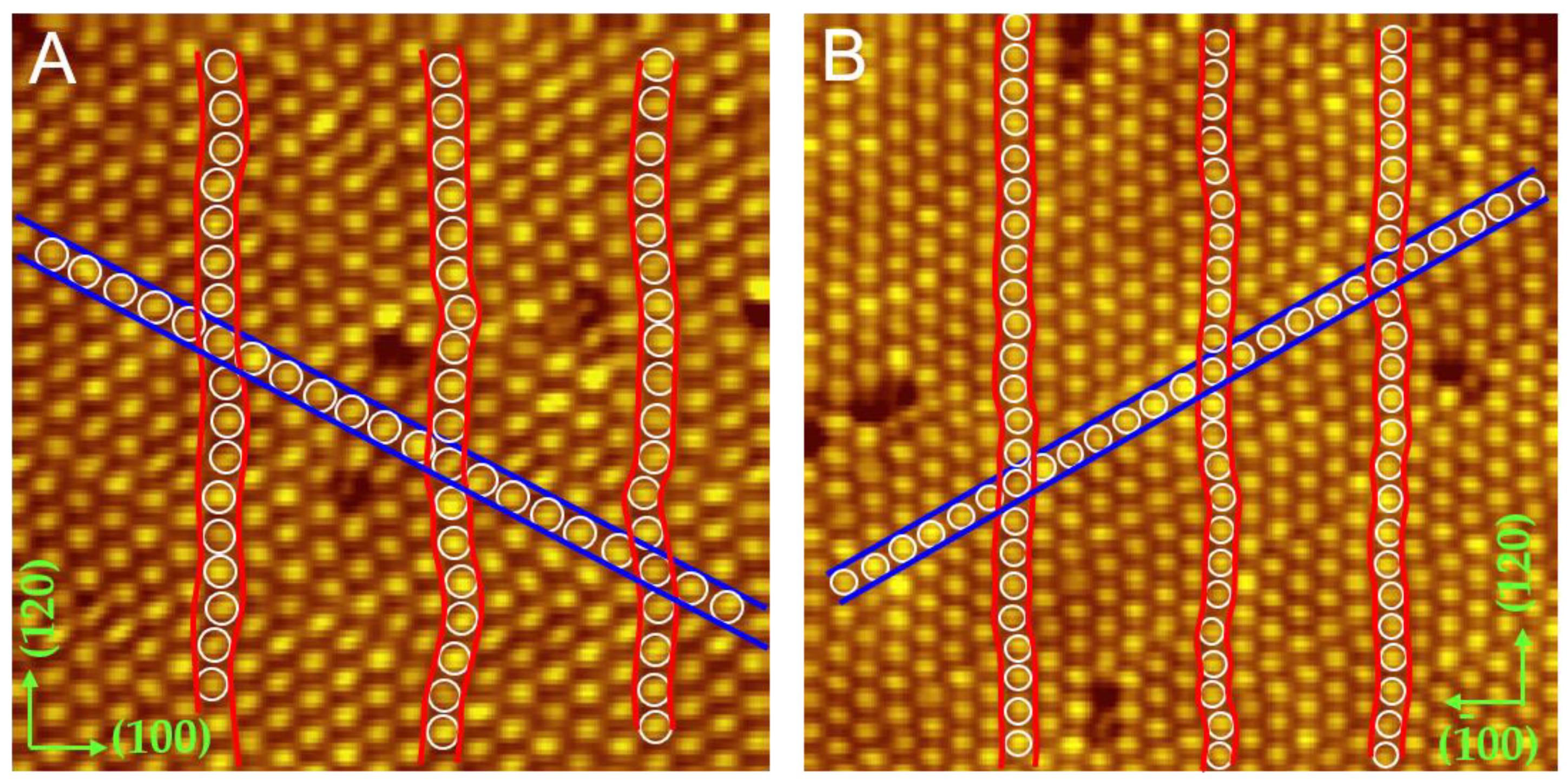
As molecule coverage increased, an unexpected wavy structure of the C60 submonolayer appeared. Figure 2A is the STM image of a wavy domain where the C60 molecules in the (120) direction present a wavy arrangement while the molecules in the (1 1 ¯ 0) direction are arranged in a straight line. Both directions correspond to the √3 directions of the Cd(0001) surface. The three rows marked with red curves demonstrate the wavy arrangement clearly. Figure 2B displays another wavy domain and the direction of straight alignment has an angle of 60 degrees with respect to Figure 2A. The intermolecular spacing is 9.8 ± 0.1 Å in the straight rows and 9.4 ± 0.1 Å in the wavy rows. Both are apparently smaller than the preferred spacing (10.02 Å) of the (111) plane in fcc C60 crystals [48], indicating a 2% compressive strain in straight molecular rows, and a 6.3% compressive strain in wavy molecular rows.
When C60 submonolayers were subjected to external forces, C60 molecules correspond with stable orientations to attach to the substrate. However, it was not enough to release the stress by orientation and a wavy structure appeared. Driven by the compressive stress, the individual C60 molecules deviated from linear arrangement, and the molecular rows became wavy in order to release the strain. In early studies, when the molecule-substrate force worked as a dominant role, slightly curved stripes were also observed in C60 films deposited on an Si(111) 4 × 1 In surface [49]. In addition to unidirectional wavy structures, we observed another strain relaxation pattern through the formation of edge dislocations in the C60 monolayer.

2.3. High-Resolution Topological Graph of the Edge Dislocation

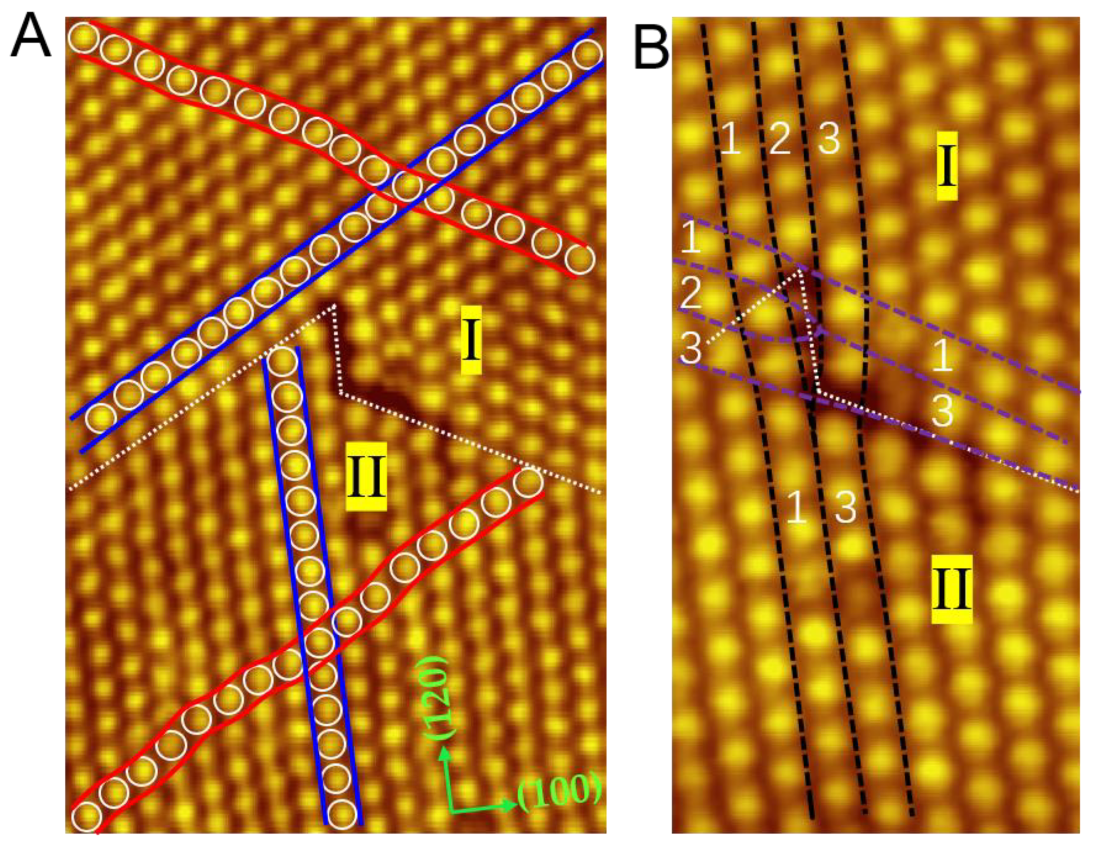
Figure 3A shows another area of the wavy structures comprising two neighboring wavy domains (I and II) with different directions for the straight alignments. The straight arrangements and wavy arrangements in Domain I and II are marked with blue curves and red lines, respectively. The straight arrangement in Domain II is in the (120) direction, while the straight arrangement in Domain I is in the (210) direction. When these domains encounter each other, an edge dislocation appears at the domain boundaries, tagged with white dotted lines. To the best of our knowledge, edge dislocation is seldomly precisely observed in molecular self-assembly. A similar phenomenon was achieved in the work of Klyachko, who found the edge dislocation in the third layer of C60 films grown on Ge(100) [31]. Unfortunately, the detail of the edge dislocation was hard to obtain due to the absence of a high-resolution STM diagram.
Figure 3B depicts the close-up view of the edge dislocation. Represented by black dotted lines, Domain I has an extra C60 row compared to Domain II. The same situation also occurs along the purple dotted lines. This observation indicates that the edge dislocation may occur in the monolayer regime and provides an intuitive STM diagram for the study of edge dislocations. Moreover, we noticed that this type of dislocation is absent when Cd(0001) substrate is kept at RT during C60 deposition.

2.4. Two Regular Domains in R26° and R33°

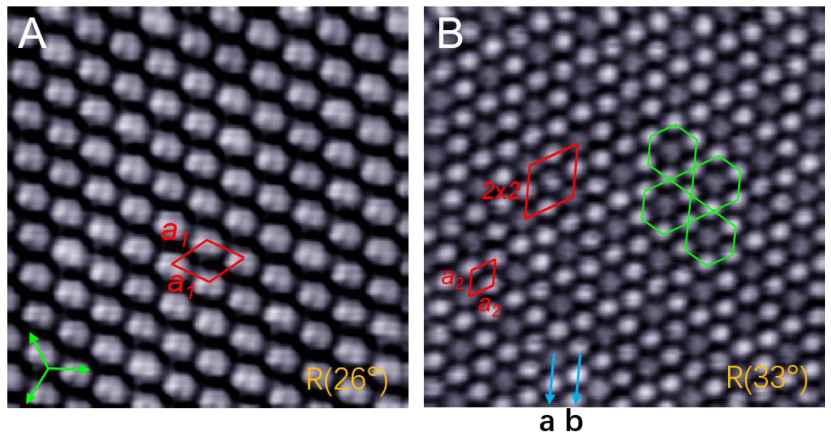
When growth temperature is raised to RT, two types of domain appear in the C60 monolayer. Figure 4A shows the topography of a R26° domain, in which all the C60 molecules adopt the same orientation. Each molecule reveals a two-lobe contrast corresponding to the C60 orientation with a 6:6 bond facing upward, similar to the C60 molecule in Au(111) and graphene [4,22]. The molecules in this domain exhibit a hexagonal lattice constant a1 = 10.0 ± 0.1 Å, close to the same value (10.02 Å) in C60 crystals [48]. This means that there is almost no strain when C60 molecules adopt the same orientation. Figure 4B shows a R33° domain that is composed of symmetric kagome lattices. Inside this domain, each dim C60 molecule is surrounded by six bright molecules, constituting a head-to-head arrangement of hexagonal rings. In other words, the C60 monolayer is composed of two types of molecular rows: In row-b, all the molecules show a bright protrusion, while in row-a, one bright and one dim C60 molecule alternately appear. As a result, the bright and dim molecules constitute a 2 × 2 superstructure, with each unit cell containing four molecules. The measured intermolecular distance is a2 = 10.2 ± 0.1 Å, indicating a 2% tensile strain in the R33° domain. These types of symmetric kagome lattices also exist in the reported C60 domains in Au(111) [8,23].

2.5. A larger 4 × 4 Superstructure of Kagome Lattice in an R44° Domain

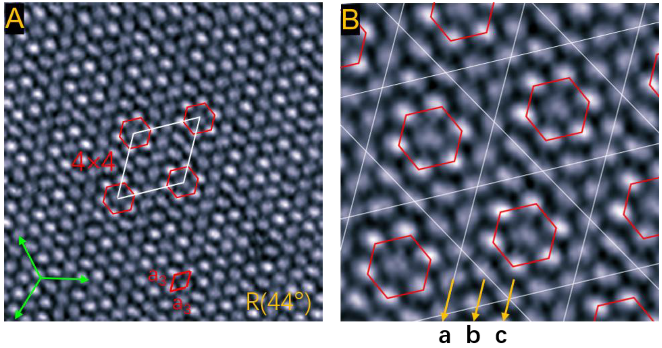
When the growth temperature rises to 310 K, the C60 monolayer presents a larger kagome lattice with a 4 × 4 superstructure, shown in Figure 5A. The intermolecular distance is a3 = 10.5 ± 0.1 Å, much larger than the value (10.02 Å) in C60 crystals. The tensile strain is as large as 5%. These properties indicate that the packing density of C60 molecules decreases with an increase in growth temperature due to thermal expansion of the C60 layer. As displayed in Figure 5B, the six trigonal regions distribute symmetrically around the central hexagon, constituting the kagome lattice. Each kagome unit cell contains sixteen molecules: a C60 heptamer located at the inner hexagon and nine peripheral molecules. The heptamer consists of a central dim molecule and six bright surrounding molecules. We noticed that the C60 monolayer in this domain is composed of three types of molecular rows: all C60 molecules in row-a show a dim contrast; two dim molecules and two bright molecules are alternately arranged in row-b; one bright and one dim molecule appear alternately in row-c.

3. Materials and Methods

The experiments were performed in an ultra-high vacuum low-temperature scanning tunneling microscopy (Unisoku, Japan) with a base pressure close to 1.5 × 10⁻¹⁰ Torr. A clean surface of Si(111)-7 × 7 was obtained through thermal flashing to 1550 K after uninterrupted degassing at 820 K for more than 7 hours. The flat and smooth Cd(0001) thin film was treated by depositing 15-20 monolayers onto the surface of Si-7 × 7. C60 molecules were evaporated from a homemade Tantalum boat at a rate of 0.4 ML/min onto the Cd(0001) thin films. After the process was completed, C60/Cd/Si was transferred into the STM observation chamber. The entire data were obtained in a constant current mode at 78 K (liquid nitrogen temperature).

4. Conclusions

In summary, our research showed the morphology of C60 molecules on Cd(0001) substrate in three different growth temperatures (100 K, RT, and 310 K). STM studies demonstrated that C60 molecules are bright or dim in the thin film. At 100 K, we found the isolated C60 seven-molecule cluster, which indicates that the C60 heptamer can exist alone. As the molecule coverage increased, an unexpected wavy structure appeared, which suggests the C60 submonolayer was subjected to a large compressive strain. As a normal reaction, when under large external stress, the film of atoms or molecules may present larger bulk density or form superstructures. In this study, the arrangement of C60 curved instead of the aforementioned action. Additionally, an edge dislocation was captured when two different wavy structures encountered each other, which may develop a new phenomenon to study edge dislocation. Two types of symmetric kagome lattices were found at elevated growth temperatures, the 2 × 2 superstructure in the R33°domain with a 2% tensile strain at RT, and the 4 × 4 superstructure in the R44° domain with a 5% tensile strain at 310 K. Our results provide a new routine to fabricate and investigate pressure-resistant materials.

Author Contributions

J.W. designed the experiments; Z.W., D.Y. and M.S. performed the experiments; Z.W., K.S. and J.W. wrote the manuscript; M.T., Z.L. and J.Y. revised the manuscript. All authors have read and agreed to the published version of the manuscript.

Funding

This work was supported by the National Natural Science Foundation of China (grant Nos. 11874304, 11574253, 11604269, 11674323, and 11804282) and the Fundamental Research Funds for the Central Universities (XDJK2020B054).

Institutional Review Board Statement

Not applicable.

Informed Consent Statement

Not applicable.

Data Availability Statement

The data presented in this study are available on request from the corresponding author.

Conflicts of Interest

The authors declare no conflict of interest.

References

  1. Wachowiak, A.; Yamachika, R.; Khoo, K.H.; Wang, Y.; Grobis, M.; Lee, D.H.; Louie, S.G.; Crommie, M.F. Visualization of the molecular jahn-teller effect in an insulating K4C60 monolayer. Science 2005, 310, 468–470. [Google Scholar] [CrossRef] [PubMed]
  2. Feng, M.; Zhao, J.; Petek, H. Atomlike, Hollow-core–bound molecular orbitals of C60. Science 2008, 320, 359–362. [Google Scholar] [CrossRef] [PubMed]
  3. Mitrano, M.; Cantaluppi, A.; Nicoletti, D.; Kaiser, S.; Perucchi, A.; Lupi, S.; Pietro, P.D.; Pontiroli, D.; Riccò, M.; Clark, S.R.; et al. Possible light-induced superconductivity in K3C60 at high temperature. Nature 2016, 530, 461–464. [Google Scholar] [CrossRef]
  4. Schull, G.; Berndt, R. Orientationally ordered (7 × 7) superstructure of C60 on Au(111). Phys. Rev. Lett. 2007, 99, 226105. [Google Scholar] [CrossRef] [PubMed]
  5. Gardener, J.A.; Briggs, G.A.D.; Castell, M.R. Scanning tunneling microscopy studies of C60 monolayers on Au(111). Phys. Rev. B 2009, 80, 235434. [Google Scholar] [CrossRef]
  6. Wang, Y.Y.; Yamachika, R.; Wachowiak, A.; Grobis, M.; Khoo, K.H.; Lee, D.H.; Louie, S.G.; Crommie, M.F. Novel Orientational Ordering and Reentrant Metallicity in KxC60 Monolayers for 3 ≤ x ≤ 5. Phys. Rev. Lett. 2007, 99, 086402. [Google Scholar] [CrossRef] [Green Version]
  7. Tang, L.; Xie, Y.C.; Guo, Q.M. Complex orientational ordering of C60 molecules on Au(111). J. Chem. Phys. 2011, 135, 114702. [Google Scholar] [CrossRef]
  8. Tang, L.; Guo, Q.M. Orientational ordering of the second layer of C60 molecules on Au(111). Phys. Chem. Chem. Phys. 2012, 14, 3323–3328. [Google Scholar] [CrossRef] [PubMed]
  9. Shin, H.; Schwarze, A.; Diehl, R.D.; Pussi, K.; Colombier, A.; Gaudry, E.; Ledieu, J.; McGuirk, G.M.; Serkovic, L.L.N.; Fournee, V.; et al. Structure and dynamics of C60 molecules on Au(111). Phys. Rev. B 2014, 89, 245428. [Google Scholar] [CrossRef] [Green Version]
  10. Grobis, M.; Lu, X.; Crommie, M.F. Local electronic properties of a molecular monolayer: C60 on Ag(001). Phys. Rev. B 2002, 66, 161408. [Google Scholar] [CrossRef]
  11. Pai, W.W.; Hsu, C.L. Ordering of an incommensurate molecular layer with adsorbate-induced reconstruction:C60/Ag(100). Phys. Rev. B 2003, 68, 121403. [Google Scholar] [CrossRef]
  12. Hsu, C.L.; Pai, W.W. Aperiodic incommensurate phase of a C60 monolayer on Ag(100). Phys. Rev. B 2003, 68, 245414. [Google Scholar] [CrossRef]
  13. Pai, W.W.; Hsu, C.L.; Lin, K.C.; Sin, L.Y.; Tang, T.B. Characterization and control of molecular ordering on adsorbate-induced reconstructed surfaces. Appl. Surf. Sci. 2005, 241, 194–198. [Google Scholar] [CrossRef]
  14. Grobis, M.; Yamachika, R.; Wachowiak, A.; Lu, X.H.; Crommie, M.F. Phase separation and charge transfer in a K-doped C60 monolayer on Ag(001). Phys. Rev. B 2007, 80, 073410. [Google Scholar] [CrossRef] [Green Version]
  15. Abel, M.; Dmitriev, A.; Fasel, R.; Lin, N.; Barth, J.V.; Kern, K. Scanning tunneling microscopy and x-ray photoelectron diffraction investigation of C60 films on Cu(100). Phys. Rev. B 2003, 67, 245407. [Google Scholar] [CrossRef] [Green Version]
  16. Wong, S.S.; Pai, W.W.; Chen, C.H.; Lin, M.T. Coverage-dependent adsorption superstructure transition of C60/Cu(001). Phys. Rev. B 2010, 82, 125442. [Google Scholar] [CrossRef] [Green Version]
  17. Li, H.I.; Franke, K.J.; Pascual, J.I.; Bruch, L.W.; Diehl, R.D. Origin of Moiré structures in C60 on Pb(111) and their effect on molecular energy levels. Phys. Rev. B 2009, 80, 085415. [Google Scholar] [CrossRef] [Green Version]
  18. Weckesser, J.; Cepek, C.; Fasel, R.; Barth, J.V.; Baumberger, F.; Greber, T.; Kern, K. Binding and ordering of C60 on Pd(110): Investigations at the local and mesoscopic scale. J. Chem. Phys. 2001, 115, 9001–9009. [Google Scholar] [CrossRef]
  19. Cui, X.X.; Han, D.; Guo, H.L.; Zhou, L.W.; Qiao, J.S.; Liu, Q.; Cui, Z.H.; Li, Y.F.; Lin, C.W.; Cao, L.M.; et al. Realizing nearly-free-electron like conduction band in a molecular film through mediating intermolecular van der waals interactions. Nat. Commun. 2019, 10, 3374. [Google Scholar] [CrossRef] [PubMed] [Green Version]
  20. Ledieu, J.; Gaudry, E.; Weerd, M.C.D.; Gille, P.; Diehl, R.D.; Fournee, V. C60 superstructure and carbide formation on the Al-terminated Al9Co2(001) surface. Phys. Rev. B 2015, 91, 155418. [Google Scholar] [CrossRef] [Green Version]
  21. Li, G.; Zhou, H.T.; Pan, L.D.; Zhang, Y.; Mao, J.H.; Zou, Q.; Guo, H.M.; Wang, Y.L.; Du, S.X.; Gao, H.J. Self-assembly of C60 monolayer on epitaxially grown, nanostructured graphene on Ru(0001) surface. Appl. Phys. Lett. 2012, 100, 013304. [Google Scholar] [CrossRef] [Green Version]
  22. Jung, M.; Shin, D.B.; Sohn, S.D.; Kwon, S.Y.; Park, N.J.; Shin, H.J. Atomically resolved orientational ordering of C60 molecules on epitaxial graphene on Cu(111). Nanoscale 2014, 6, 11835–11840. [Google Scholar] [CrossRef] [PubMed] [Green Version]
  23. Han, S.; Guan, M.X.; Song, C.L.; Wang, Y.L.; Ren, M.Q.; Meng, S.; Ma, X.C.; Xue, Q.K. Visualizing molecular orientational ordering and electronic structure in CsnC60 fulleride films. Phys. Rev. B 2020, 101, 085413. [Google Scholar] [CrossRef] [Green Version]
  24. Chen, C.H.; Zheng, H.S.; Mills, A.; Heflin, J.R.; Tao, C.G. Temperature evolution of quasi-one-dimensional C60 nanostructures on rippled graphene. Sci. Rep. 2015, 5, 14336. [Google Scholar] [CrossRef] [Green Version]
  25. Santos, E.J.G.; Scullion, D.; Chu, X.S.; Li, D.O.; Guisinger, N.P.; Wang, Q.H. Rotational superstructure in van der waals heterostructure of self-assembled C60 monolayer on the WSe2 surface. Nanoscale 2017, 9, 13245–13256. [Google Scholar] [CrossRef] [Green Version]
  26. Wang, H.Q.; Zeng, C.G.; Wang, B.; Hou, J.G. Orientational configurations of the C60 molecules in the (2 × 2) superlattice on a solid C60 (111) surface at low temperature. Phys. Rev. B 2001, 63, 085417. [Google Scholar] [CrossRef]
  27. Nakaya, M.; Aono, M.; Nakayama, T. Molecular-scale size tuning of covalently bound assembly of C60 molecules. ACS Nano. 2011, 5, 7830–7837. [Google Scholar] [CrossRef] [PubMed]
  28. Matetskiy, A.V.; Gruznev, D.V.; Zotov, A.V.; Saranin, A.A. Modulated C60 monolayers on Si(111)√3 × √3-Au reconstructions. Phys. Rev. B 2011, 83, 195421. [Google Scholar] [CrossRef]
  29. Gruznev, D.V.; Matetskiy, A.V.; Bondarenko, L.V.; Utas, O.A.; Zotov, A.V.; Saranin, A.A.; Chou, J.P.; Wei, C.M.; Lai, M.Y.; Wang, Y.L. Stepwise self-assembly of C60 mediated by atomic scale moire’ magnifiers. Nat. Commun. 2013, 4, 1679. [Google Scholar] [CrossRef] [PubMed] [Green Version]
  30. Xu, H.; Chen, D.M.; Creager, W.N. C60-induced reconstruction of the Ge(111) surface. Phys. Rev. B 1994, 50, 8454–8459. [Google Scholar] [CrossRef] [PubMed]
  31. Klyachko, D.V.; Lopez-Castillo, J.M.; Jay-Gerin, J.P.; Chen, D.M. Stress relaxation via the displacement domain formation in films of C60 on Ge(100). Phys. Rev. B 1999, 60, 9026–9036. [Google Scholar] [CrossRef] [Green Version]
  32. Fanetti, M.; Gavioli, L.; Cepek, C.; Sancrotti, M. Orientation of C60 molecules in the (3√3 × 3√3) R30° and (√13 × √13) R14° phases of C60/Ge(111) single layers. Phys. Rev. B 2008, 77, 085420. [Google Scholar] [CrossRef]
  33. Torrelles, X.; Pedio, M.; Cepek, C.; Felici, R. (2√3 × 2√3) R30° induced self-assembly ordering by C60 on a Au(111) surface: X-ray diffraction structure analysis. Phys. Rev. B 2012, 86, 075461. [Google Scholar] [CrossRef]
  34. Passens, M.; Waser, R.; Karthaeuser, S. Enhanced fullerene-Au(111) coupling in (2√3 × 2√3) R30° superstructures with intermolecular interactions. Beilstein J. Nanotechnol. 2015, 6, 1421–1431. [Google Scholar] [CrossRef] [PubMed] [Green Version]
  35. Thürmer, K.; Huang, R.Q.; Bartelt, N.C. Surface self-organization caused by dislocation networks. Science 2006, 311, 1272–1274. [Google Scholar] [CrossRef]
  36. Lu, Y.F.; Przybylski, M.; Trushin, O.; Wang, W.H.; Barthel, J.; Granato, E.; Ying, S.C.; Nissila, T.A. Strain relief in Cu-Pd heteroepitaxy. Phys. Rev. Lett. 2005, 94, 146105. [Google Scholar] [CrossRef] [Green Version]
  37. Hsu, P.J.; Finco, A.; Schmidt, L.; Kubetzka, A.; Bergmann, K.V.; Wiesendanger, R. Guiding spin spirals by local uniaxial strain relief. Phys. Rev. Lett. 2016, 116, 017201. [Google Scholar] [CrossRef] [PubMed]
  38. Realpe, H.; Peretz, E.; Shamir, N.; Mintz, M.H.; Shneck, R.Z.; Manassen, Y. Islands as nanometric probes of strain distribution in heterogeneous surfaces. Phys. Rev. Lett. 2010, 104, 056102. [Google Scholar] [CrossRef] [PubMed]
  39. Liu, F.; Tersoff, J.; Lagally, M.G. Self-organization of steps in growth of strained films on vicinal substrates. Phys. Rev. Lett. 1998, 80, 1268–1271. [Google Scholar] [CrossRef]
  40. Mo, Y.W.; Savage, D.E.; Swartzentruber, B.S.; Lagally, M.G. Kinetic pathway in stranski-krastanov growth of Ge on Si(001). Phys. Rev. Lett. 1990, 65, 1020–1023. [Google Scholar] [CrossRef]
  41. Mansour, K.A.; Buchsbaum, A.; Ruffieux, P.; Schmid, M.; Gröning, P.; Varga, P.; Fasel, R.; Gröning, O. Fabrication of a well-ordered nanohole array stable at room temperature. Nano Lett. 2008, 8, 2035–2040. [Google Scholar] [CrossRef]
  42. Jankowski, M.; Wormeester, H.; Zandvliet, H.J.W.; Poelsema, B. Temperature dependent formation and evolution of the interfacial dislocation network of Ag/Pt (111). Phys. Rev. B 2014, 89, 235402. [Google Scholar] [CrossRef]
  43. Lai, M.Y.; Wang, Y.L. Metal/semiconductor incommensurate structure with a rare domain configuration exhibiting p31m symmetry. Phys. Rev. B 2000, 61, 12608–12611. [Google Scholar] [CrossRef]
  44. Zegenhagen, J.; Fontes, E.; Grey, F.; Patel, J.R. Microscopic structure, discommensurations, and tiling of Si(111)/Cu “5 × 5”. Phys. Rev. B 1992, 46, 1860–1863. [Google Scholar] [CrossRef] [PubMed]
  45. Gorji, N.E.; Tanner, B.K.; Vijayaraghavan, R.K.; Danilewsky, A.N.; McNally, P.J. Nondestructive, in situ mapping of die surface displacements in encapsulated IC chip packages using x-ray diffraction imaging techniques. In Proceedings of the IEEE Electronic Components and Technology Conference, Orlando, FL, USA, 30 May–2 June 2017. [Google Scholar] [CrossRef]
  46. Xie, Z.B.; Wang, Y.L.; Tao, M.L.; Sun, K.; Tu, Y.B.; Yuan, H.K.; Wang, J.Z. Ordered array of CoPc-vacancies filled with single-molecule rotors. Appl. Surf. Sci. 2018, 439, 462–467. [Google Scholar] [CrossRef]
  47. Yang, D.X.; Wang, Z.L.; Shi, M.X.; Sun, K.; Tao, M.L.; Yang, J.Y.; Wang, J.Z. Orientational transition of tin phthalocyanine molecules in the self-assembled monolayer. J. Phys. D Appl. Phys. 2020, 53, 154004. [Google Scholar] [CrossRef]
  48. Heiney, P.A.; Fischer, J.E.; Mcghie, A.R.; Romanow, W.J.; Denenstein, A.M.; Mccauley, J.P.; Smith, A.B.; Cox, D.E. Orientational ordering transition in solid C60. Phys. Rev. Lett. 1991, 66, 2911–2914. [Google Scholar] [CrossRef]
  49. Kotlyar, V.G.; Olyanich, D.A.; Utas, T.V.; Zotov, A.V.; Saranin, A.A. Self-assembly of C60 fullerenes on quasi-one-dimensional Si(111)4×1-in surface. Surf. Sci. 2012, 606, 1821–1824. [Google Scholar] [CrossRef]
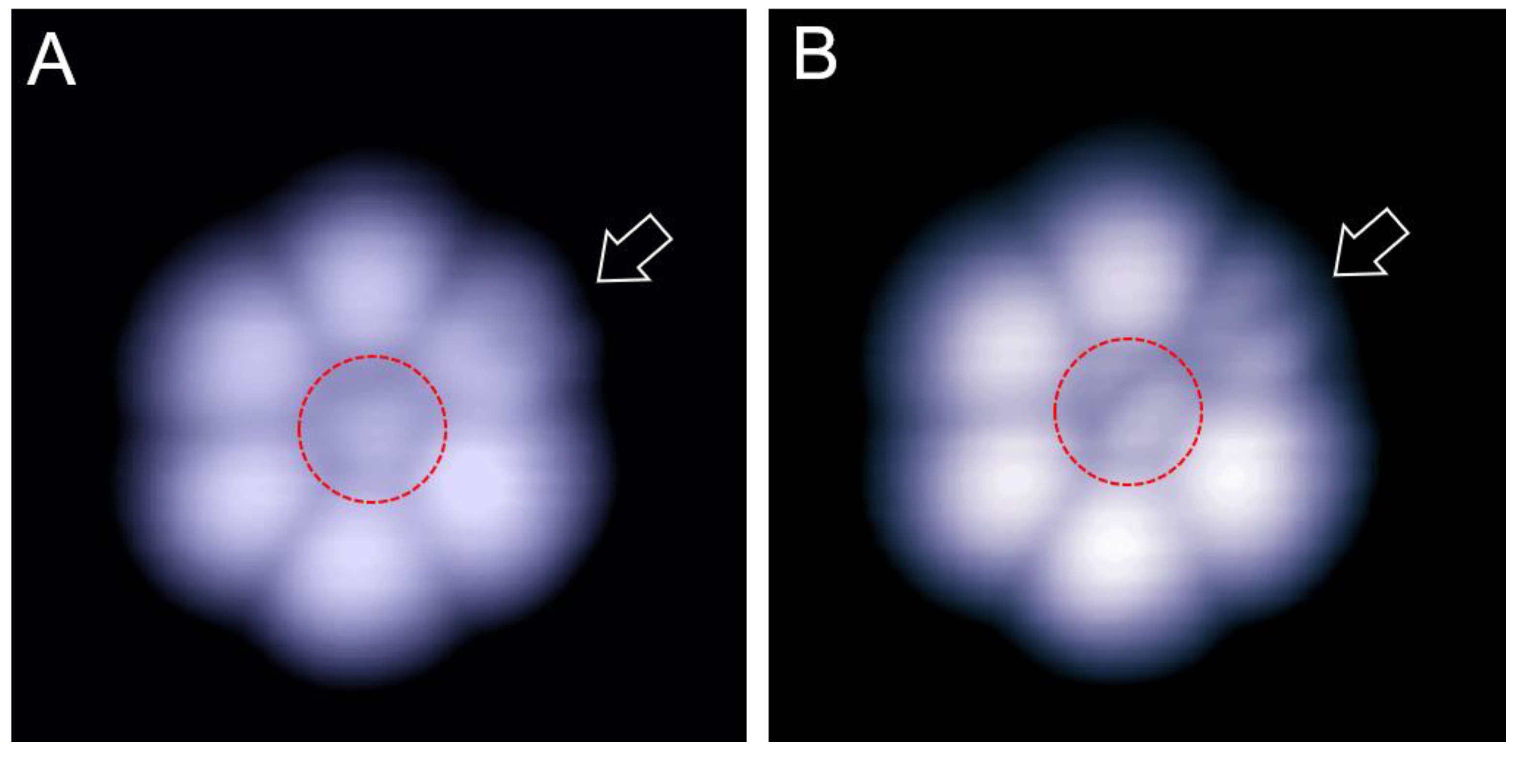
Figure 1. Isolated C60 heptamer form on the Cd(0001) surface at 100 K. (A,B) The empty-state STM images (6 × 6 nm) recorded at 1.2 and 0.5 V, respectively. The central molecule which is marked by a red circle manifests a triangle shape at 1.2 V(A), and a two-lobe motif at 0.5 V (B). The peripheral C60 molecule marked by the arrow also shows a two-lobe motif in (B).
Ijms 22 06880 g001
Figure 2. Wavy structure of the C60 submonolayer driven by compressive strain. (A) A wavy domain showing the wavy arrangement in the (120) direction, and the straight arrangement in the (1 1 ¯ 0) directions, 0.9 V, 20 × 20 nm. Both directions correspond to the √3 directions of the Cd(0001) surface. (B) Another domain showing the wavy alignment of C60 molecules, 0.4 V, 22.7 × 22.7 nm. The direction of straight alignment has an angle of 60 degrees with respect to that in (A).
Ijms 22 06880 g002
Figure 3. Two neighboring wavy domains (I and II) with different directions for the straight alignments. (A) Edge dislocation appears at the domain boundaries (white dotted lines), 23 × 15 nm, 2.8 V. (B) Close-up view of the edge dislocation as marked by the black and purple dotted curves, 7.5 × 13.5 nm, 3.0 V.
Ijms 22 06880 g003
Figure 4. Two regular domains were obtained by deposition at room temperature. (A) Homogeneous orientation of C60 molecules in a R26° domain, 1.2 V, 10 × 10 nm. (B) The symmetric kagome lattice which is marked by hexagons in green reveals a 2 × 2 superstructure in an R33° domain, 2.0 V, 15 × 15 nm. Arrow b represents the molecular rows with bright molecules, and arrow a represents the rows with one bright and one dim molecules alternately arranged.
Ijms 22 06880 g004
Figure 5. Kagome lattice formed at 310 K. (A) Symmetric kagome networks reveal a 4 × 4 superstructure in an R44° domain. The rhombus shows the primitive cell of the superstructure. 20 × 20 nm, 1.2 V. (B) Close-up view of the kagome network with each unit cell containing sixteen C60 molecules, 1.2 V, 10 × 10 nm. The molecular rows a, b, and c contain dim molecules, alternate two bright and two dim molecules, alternate one bright and one dim molecule, respectively.
Ijms 22 06880 g005
Publisher’s Note: MDPI stays neutral with regard to jurisdictional claims in published maps and institutional affiliations.