Article

Isolation and Characterization of Two Klebsiella pneumoniae Phages Encoding Divergent Depolymerases

by
Pilar Domingo-Calap
1,2,*,
Beatriz Beamud
3,
Lucas Mora-Quilis
1,
Fernando González-Candelas
1,3,4 and
Rafael Sanjuán
1
1
Institute for Integrative Systems Biology, I2SysBio, Universitat de València-CSIC, 46980 Paterna, Spain
2
Department of Genetics, Universitat de València, 46980 Paterna, Spain
3
FISABIO-Salud Pública, Generalitat Valenciana, 46020 Valencia, Spain
4
CIBER in Epidemiology and Public Health, 46020 Valencia, Spain
*
Author to whom correspondence should be addressed.
Int. J. Mol. Sci. 2020, 21(9), 3160; https://doi.org/10.3390/ijms21093160
Submission received: 24 March 2020 / Revised: 25 April 2020 / Accepted: 26 April 2020 / Published: 30 April 2020
(This article belongs to the Special Issue Bacteriophage—Molecular Studies)

Abstract

:
The emergence of multidrug-resistant bacteria is a major global health concern. The search for new therapies has brought bacteriophages into the spotlight, and new phages are being described as possible therapeutic agents. Among the bacteria that are most extensively resistant to current antibiotics is Klebsiella pneumoniae, whose hypervariable extracellular capsule makes treatment particularly difficult. Here, we describe two new K. pneumoniae phages, πVLC5 and πVLC6, isolated from environmental samples. These phages belong to the genus Drulisvirus within the family Podoviridae. Both phages encode a similar tail spike protein with putative depolymerase activity, which is shared among other related phages and probably determines their ability to specifically infect K. pneumoniae capsular types K22 and K37. In addition, we found that phage πVLC6 also infects capsular type K13 and is capable of striping the capsules of K. pneumoniae KL2 and KL3, although the phage was not infectious in these two strains. Genome sequence analysis suggested that the extended tropism of phage πVLC6 is conferred by a second, divergent depolymerase. Phage πVLC5 encodes yet another putative depolymerase, but we found no activity of this phage against capsular types other than K22 and K37, after testing a panel of 77 reference strains. Overall, our results confirm that most phages productively infected one or few Klebsiella capsular types. This constitutes an important challenge for clinical applications.

1. Introduction

Klebsiella pneumoniae is an encapsulated Gram-negative bacterium that can be found as a free-living organism, in natural environments. In addition, K. pneumoniae is an opportunistic pathogen causing infections mainly in the lungs and urinary tract, although meningitis and sepsis can occur in severe cases. Due to extensive antibiotic use and favorable transmission opportunities, multidrug-resistant K. pneumoniae strains, particularly carbapenem-resistant strains [1,2], have spread in hospitals and the community. K. pneumoniae belongs to the ESKAPE priority group defined by the WHO, which also includes Enterococcus faecium, Staphylococcus aureus, Acinetobacter baumannii, Pseudomonas aeruginosa, and Enterobacter spp. [3]. Thus, K. pneumoniae is considered to be an important target for the development of new therapies.
Bacteriophages (phages) provide an interesting therapeutic alternative, since they infect bacteria in a specific manner, avoiding dysbiosis [4]. However, in the case of highly variable bacteria such as K. pneumoniae, the use of phages becomes challenging. It has been proposed that phage infectivity is strongly determined by the bacterial capsular type [5], making it difficult to treat a wide range of K. pneumoniae strains, since most of the phages described so far are specific to one or few capsular types. Based on this, the use of phage cocktails has been suggested, in which the interactions between phages can generate synergies that improve bacterial lysis and limit the emergence of resistances [6].
Due to the high heterogeneity of K. pneumoniae, it often becomes difficult to determine the virulence factors of this species [7]. Nevertheless, the capsule of K. pneumoniae is one such virulence factor, and hypervirulence has been associated with differences in the expression of genes involved in the capsule, or with modified capsule phenotypes [8]. Most K. pneumoniae phages have been found to encode depolymerases, which are enzymes capable of digesting the exopolysaccharide capsule by cleaving glycosidic linkages [9,10]. Disruption or capsule loss makes bacteria susceptible to phage infection or antibiotic treatment, suggesting a therapeutic potential for phage depolymerases [5,11,12,13,14]. Although most described phages encode a single depolymerase and, as a result, infect one or few capsular types, some phages have been reported to encode multiple depolymerases. To date, the most salient case is that of phage ØK64-1, which encodes nine different depolymerases [15]. Overall, a limited number of enzymes in active particular capsular types have been described [16]. In addition to their potential therapeutic applications, phage-borne depolymerases have been suggested for capsular typing [17]. For these reasons, discovering new Klebsiella phage depolymerases against distinct capsular types is an interesting topic with potential biomedical applications.
Recently, our group has isolated and characterized four new phages of K. pneumoniae (πVLC1 to πVLC4) from environmental samples in Valencia, Spain [18]. All of these were closely related phylogenetically, belonged to the genus Drulisvirus, encoded only one depolymerase, and showed a very narrow capsular type range. Using a similar protocol, here we isolated two new phages with high lytic activity, which we named πVLC5 and πVLC6. As opposed to our previously described phages, these new isolates were capable of infecting several reference capsular types of Klebsiella spp. Whole genome analysis revealed that πVLC5 and πVLC6 encoded divergent depolymerases, potentially explaining their host tropisms.

2. Results

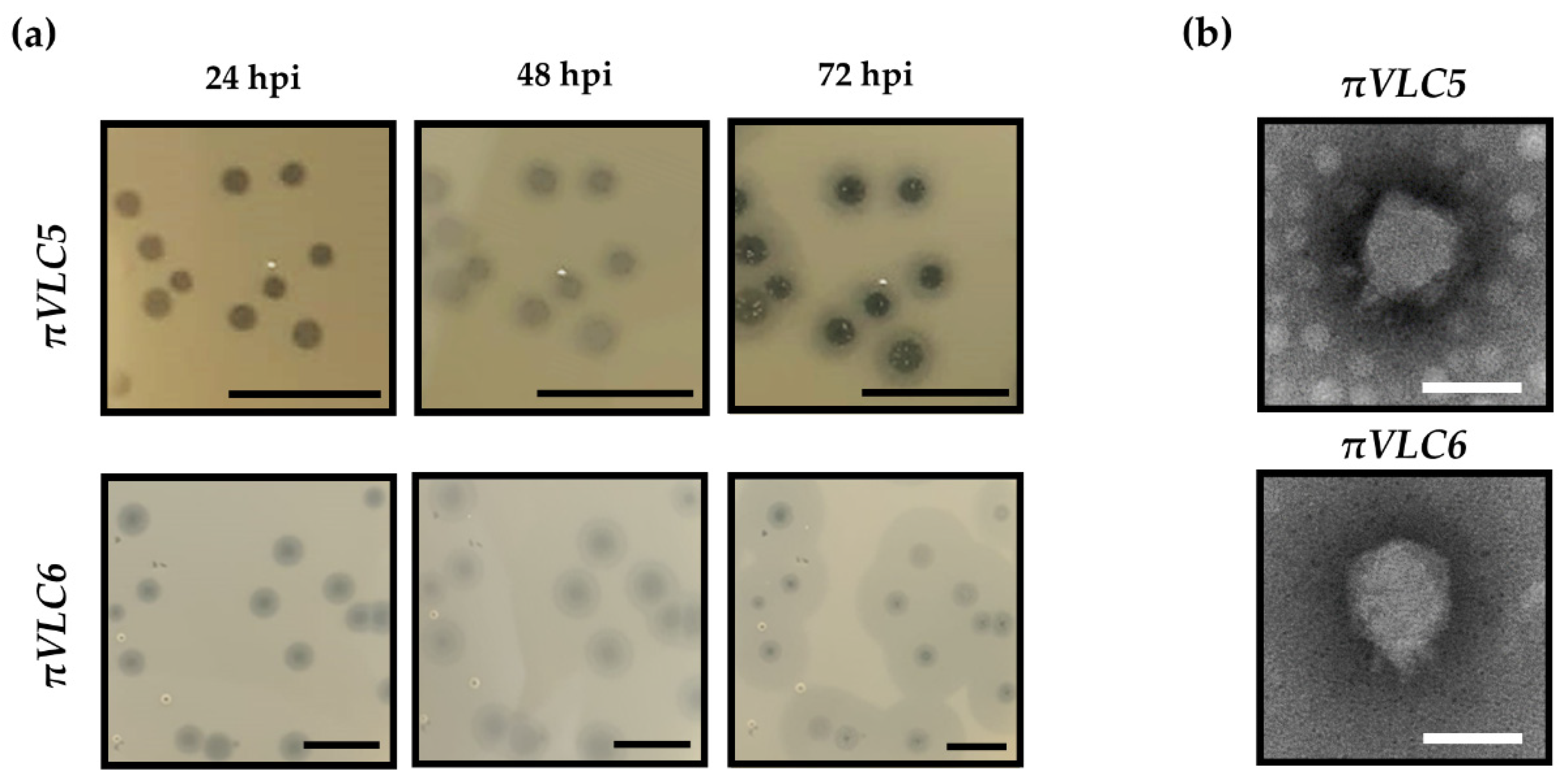
2.1. Isolation and Phenotypic Characterization of Two New Klebsiella Pneumoniae Phages

Phage πVLC5 was obtained from a water sample in an irrigation ditch near a wastewater treatment plant, whereas phage πVLC6 was isolated from a river, near its outflow to the sea, in an area where waste discharged by nearby villages accumulates. Our initial screening was carried out using a multidrug-resistant clinical isolate of K. pneumoniae as host, which belongs to capsular type K22. Once plaques were observed, they were further isolated and plaque-purified. Amplification of the phages in liquid medium was carried out to obtain a high-titer lysate and to proceed with detailed phage characterization. Plaque assays showed that the amplified phage πVLC5 reached a titer of 2.4 × 10¹¹ plaque forming units (PFU) per mL, while πVLC6 reached 1.0 × 10¹⁰ PFU/mL. For both phages, large plaques were observed, which were surrounded by a halo that is often interpreted as an indicator of depolymerase-mediated digestion of bacterial capsules (Figure 1a). Haloes are formed because depolymerases can diffuse and degrade capsules in nearby bacteria even if lysis does not occur in these areas. This has been previously shown in other Klebsiella spp. [18] and in phages infecting other encapsulated bacteria, such as Escherichia coli or Acinetobacter baumannii [19,20]. The haloes of phage πVLC6 were wider than those of πVLC5, whereas lysed areas were larger for πVLC5, suggesting that these were two distinct phages. Transmission electron microscopy micrographs obtained from high-titer lysates showed that the virion morphology of the two phages was similar and typical of podoviruses, with an icosahedral head of approximately 50 nm and a small tail of about 10 nm (Figure 1b).

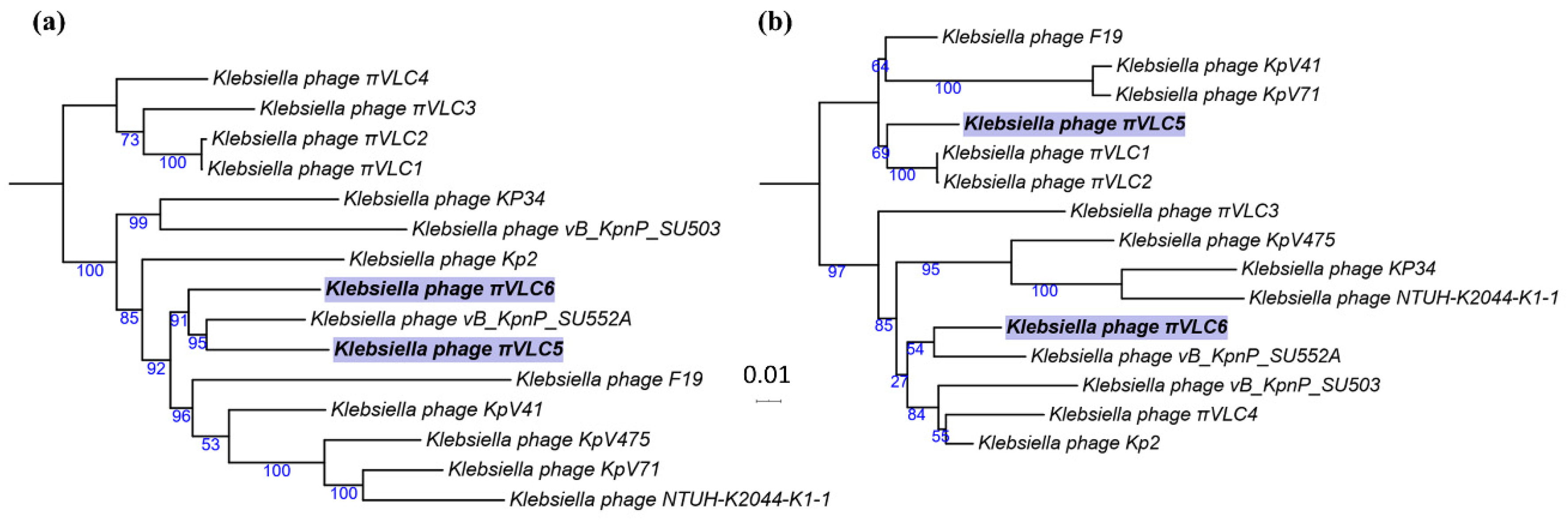
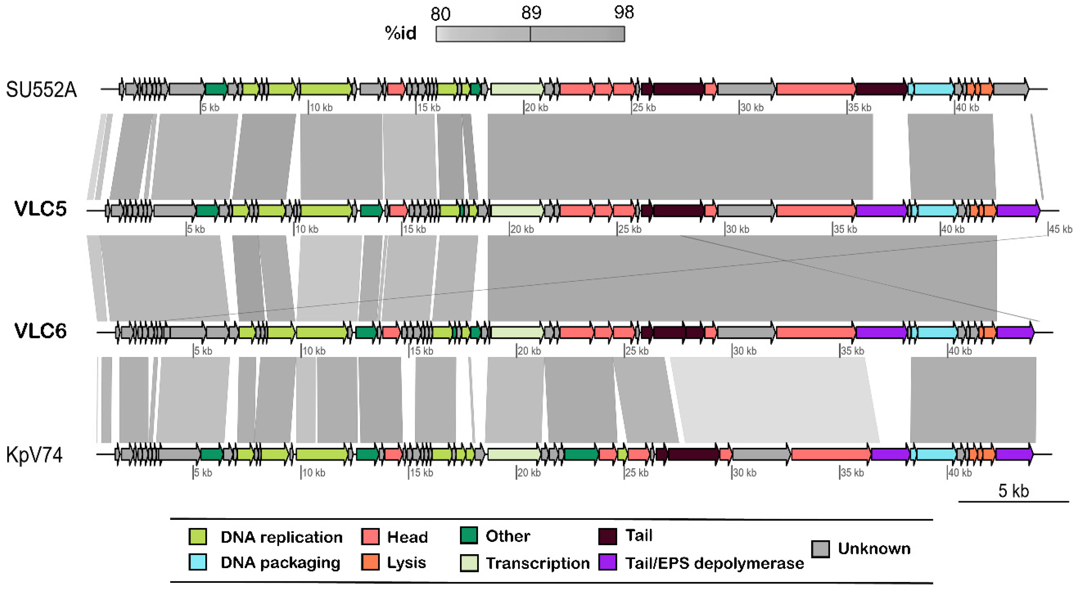
2.2. Genome Sequencing and Comparative Genomics

Genome sequencing of πVLC5 yielded a single contig of 44,932 bp (54.3% G+C content) containing 58 putative coding sequences (CDSs). For phage πVLC6, a single contig was also obtained with 44,294 bp (54.2% G+C content) and 60 CDSs. The two phages showed a pairwise nucleotide identity of 92.5%. BLAST analysis showed that the two phages belonged to the family Podoviridae, genus Drulisvirus. To verify this, we obtained a multiple sequence alignment of all publicly available classified drulisviruses (n = 9), including the four additional genomes recently published by our group [18]. Klebsiella phages πVLC5 and πVLC6 were found to be collinear and similar to the rest of drulisviruses. To determine the evolutionary relationships among these phages, maximum likelihood (ML) phylogenies of whole-genome and RNA polymerase sequences were built. The whole-genome tree showed that Klebsiella phages πVLC5 and πVLC6 were members of a well-supported cluster within drulisviruses, but that πVLC5 was closer to the Klebsiella phage Vb_KpnP_SU552A [21] (Figure 2a). According to this phylogeny, both phages were phylogenetically separated from the previously described πVLC1-4 phages. Yet, the RNA polymerase tree was not consistent with the whole genome tree and suggested a closer relationship between πVLC5 and the previously described phages πVLC1 and πVLC2 (Figure 2b).

2.3. Functional Annotation of K. pneumoniae Phages πVLC5 and πVLC6

Twenty-four conserved proteins were found among drulisviruses, including Klebsiella phages πVLC5 and πVLC6. These included proteins involved in DNA replication (DNA polymerase and DNA-dependent RNA polymerase, 5’-3’ exonuclease, helicase, and primase), DNA packaging (DNA maturase A and B), head proteins (tegument, collar, capsid, internal virion, and core proteins), tail tubular proteins (A and B), enzymatic activities (phosphoesterase and peptidase), lysis proteins (spanin and holin), and additional, hypothetical proteins. Most differences were observed in the tail fiber proteins, where similarity dropped drastically between phages πVLC5, πVLC6, and the rest of drulisviruses, with a few exceptions (Figure 3, Table S1). Both phages contained two putative tail fiber proteins, with the second located after the lysis cassette, as previously observed [22].
The first tail-fiber protein was completely shared between the πVLC5 and πVLC6 phages (ORF49 and ORF51, respectively) and had a high structural similarity to the tail spike protein of bacteriophage phiAB6 (e-value 3.9 × 10⁻³³). Hence, we annotated both ORFs as tail spikes. In addition, these two ORFs showed high sequence similarity with the tail fiber protein of the Pantoea bacteriophage LIMElight (e-values 6.0 × 10−40 and 4.0 × 10−40, respectively), available from the depolymerases database. These ORFs contained two sequence domains. Positions 13–140 matched significantly with bacteriophage T7 tail fiber protein, whereas positions 308–610 matched with a pectin lyase domain (Table 1), where the depolymerase activity is expected to reside.
Phage πVLC5 encoded a second tail fiber/spike protein (ORF58) that also matched significantly with the sequences in the depolymerase database, the best match being with the tail fiber protein of the Klebsiella phage KP32 (e-value 1.4 × 10−55). InterProScan5 revealed three different domains in this ORF—a pectin lyase, an endosialidase chaperone, and a DNA-binding domain. In this case, the closest protein structure determined by HHpred was the tail spike of an Acinetobacter phage (Table 1). In turn, the second tail fiber protein of phage πVLC6 (ORF58) was similar to a putative pectate lyase of Serratia phage phiMAM1. Surprisingly, no protein domains were found with InterProScan5 for this ORF. However, the ORF matched significantly with the protein structure of bacteriophage CBA120 tail spike (Table 1).

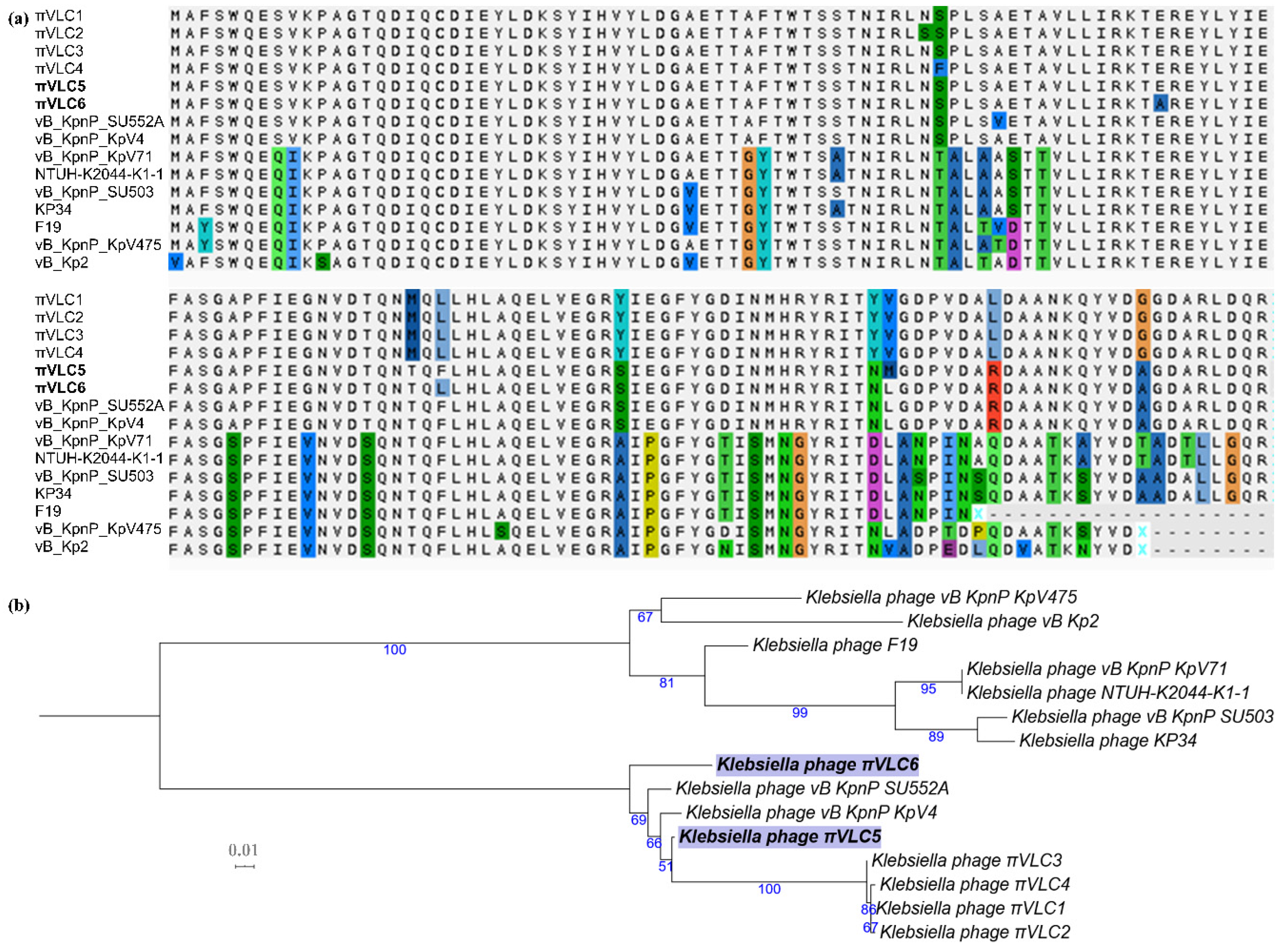
2.4. Divergence of Putative Depolymerase Sequences of Klebsiella Phages πVLC5 and πVLC6

To elucidate the degree of divergence and find putative orthologs for the different tail fibers/spikes of phages πVLC5 and πVLC6, the above four ORFs were extracted and searched against a custom phage database with BLASTN. The tail fiber/spike shared among πVLC5 and πVLC6 (ORF49 and ORF51) was also present in the rest of Drulisvirus analyzed (Figure 4), including non-Klebsiella phages. However, only the first domain was highly conserved, whereas the second domain (where the putative enzymatic activity resides) was absent in all of these sequences except for the πVLC phages and partly in Klebsiella phage KP32 (Table S1). The common region of the tail fiber was not restricted to this genus. In fact, it was similar to the tail fibers of Przondovirus and Phikmvvirus phages, all in the Autographivirinae subfamily (Table S1). Remarkably, the average nucleotide identity values dropped in this region, ranging from 70.7% to 99.8%, when compared to the rest of the genome (from 89.5% to 99.8%). Therefore, this protein seemed to be unique to the πVLC phages, given that the other phages only showed small regions of limited sequence identity. However, the ML phylogenetic tree of the core region of this ORF did not group the novel phages πVLC5 and πVLC6, with the rest of πVLC phages (Figure 4).
Regarding the additional tail fiber/spikes of πVLC5 and πVLC6, only three significant hits were found for the phage πVLC5 ORF58 (Table S1). The best hit was a hypothetical protein of the Klebsiella phage KOX1 with a sequence coverage of 51% and 68% nucleotide identity. This hit contained the pectin lyase domain. The second hit was the tail fiber protein of the Klebsiella phage NJS1 (13% coverage and 67% identity). Klebsiella phages KOX1 and NJS1 belonged to the genus Webervirus of the family Siphoviridae. The last hit was almost non-significant (only 2% coverage but 89% of identity), with a protein of the Vibrio phage 1.164.O. For phage πVLC6 ORF58, we found three hits that covered the entire ORF, all from bacteriophages of the same genus, with levels of identity ranging from 94.8% to 95.7% (Table S1).

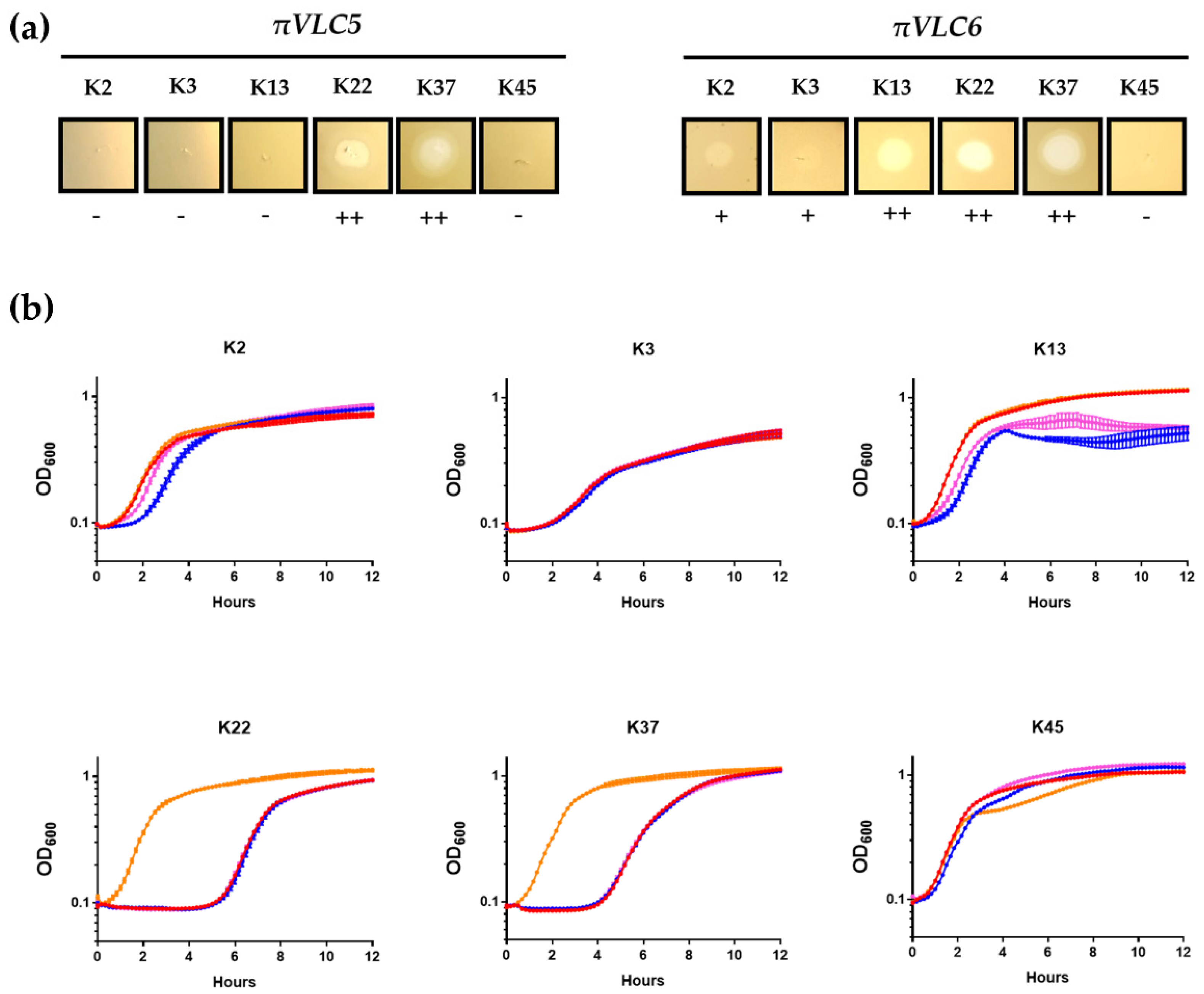
2.5. Determination of the Host Range of the Two Klebsiella Pneumoniae Phages πVLC5 and πVLC6

A panel of 77 reference strains of Klebsiella spp. purchased from the Statens Serum Institute (Copenhagen, Denmark) was used to test the infectivity of the newly discovered phages using spot tests in semi-solid medium and OD measurements in liquid cultures. Overall, the results of the spot tests were consistent with those obtained in liquid infections (Figure 5, Table S2). Phage πVLC5 had a lytic ability against capsular types K22 and K37, whereas phage πVLC6 lysed capsular types K22, K37, and K13 capsular types. Liquid cultures also showed that, for K22 and K37, phage-resistant bacteria emerged at around 5 h post-inoculation (hpi) and eventually reached densities similar to those of uninfected cultures at approximately 10 hpi, a process that could not be prevented using a cocktail of the two phages. In K13, πVLC6-mediated bacterial lysis occurred less efficiently but, on the other hand, the OD values remained lower than those of uninfected cultures even at 12 hpi. For K2 and, to a lesser extent, K3, spot tests revealed that phage πVLC6 produced a slight reduction in the turbidity of the bacterial lawns. Liquid infections also indicated a slight growth delay in K2 cultures inoculated with πVLC6.
In bacterial strains where spot tests revealed an effect of the phage, phage progeny production was evaluated by inoculating low-density cultures (OD ca. 0.05) with ca. 105 PFU (as determined in K22 plaque assays). At 4 hpi, phage titration showed that πVLC5 and πVLC6 did not produce any progeny in K2 and K3 cultures, since the endpoint titers were similar or lower than those at inoculation. The absence of phage progeny was also verified in semi-solid cultures inoculated by the spot test method. In contrast, K22 and K37 were productively infected by both phages, yielding >108 PFU/mL of πVLC5 and >109 PFU/mL of πVLC6. In addition, phage πVLC6 was able to produce >109 PFU/mL in K13, as opposed to phage πVLC5.

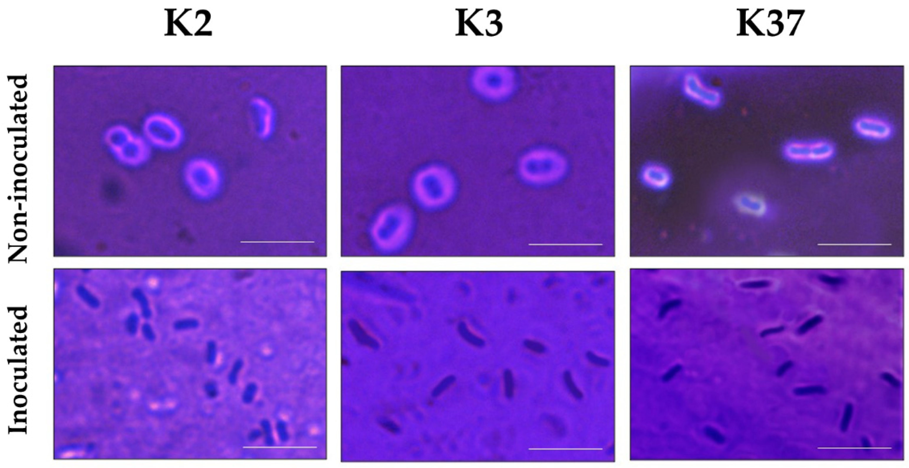
2.6. Depolymerase Activity of Phage πVLC6

Phage πVLC6 did not infect the reference strains of capsular types K2 and K3, as indicated by the absence of phage progeny. However, πVLC6 did produce a halo in K3 lawns (and less obviously, also in K2 lawns) and slightly retarded the growth of K2 cultures. We hypothesized that this effect might be due to the ability of a πVLC6 depolymerase to digest the K2 and K3 capsules. This depolymerase activity could be encoded by ORF58, which showed no homologous gene in πVLC5. To test this, we sampled the halo formed by πVLC6 in K2 and K3 lawns for microscopy. For comparison, we also analyzed regions of the plate where no phage was added. These samples were stained with crystal violet to visualize bacteria and with nigrosin to provide background contrast. In non-inoculated K2 and K3 bacteria, capsules were intact and easily observable as non-stained areas around each cell (Figure 6). In contrast, capsules were obviously absent in K2 and K3 bacteria taken from the halo regions of the cultures inoculated with phage πVLC6. Our results confirmed the degradation of the exopolysaccharide capsule by the action of a depolymerase encoded by πVLC6, despite the inability of the phage to infect and lyse these strains.

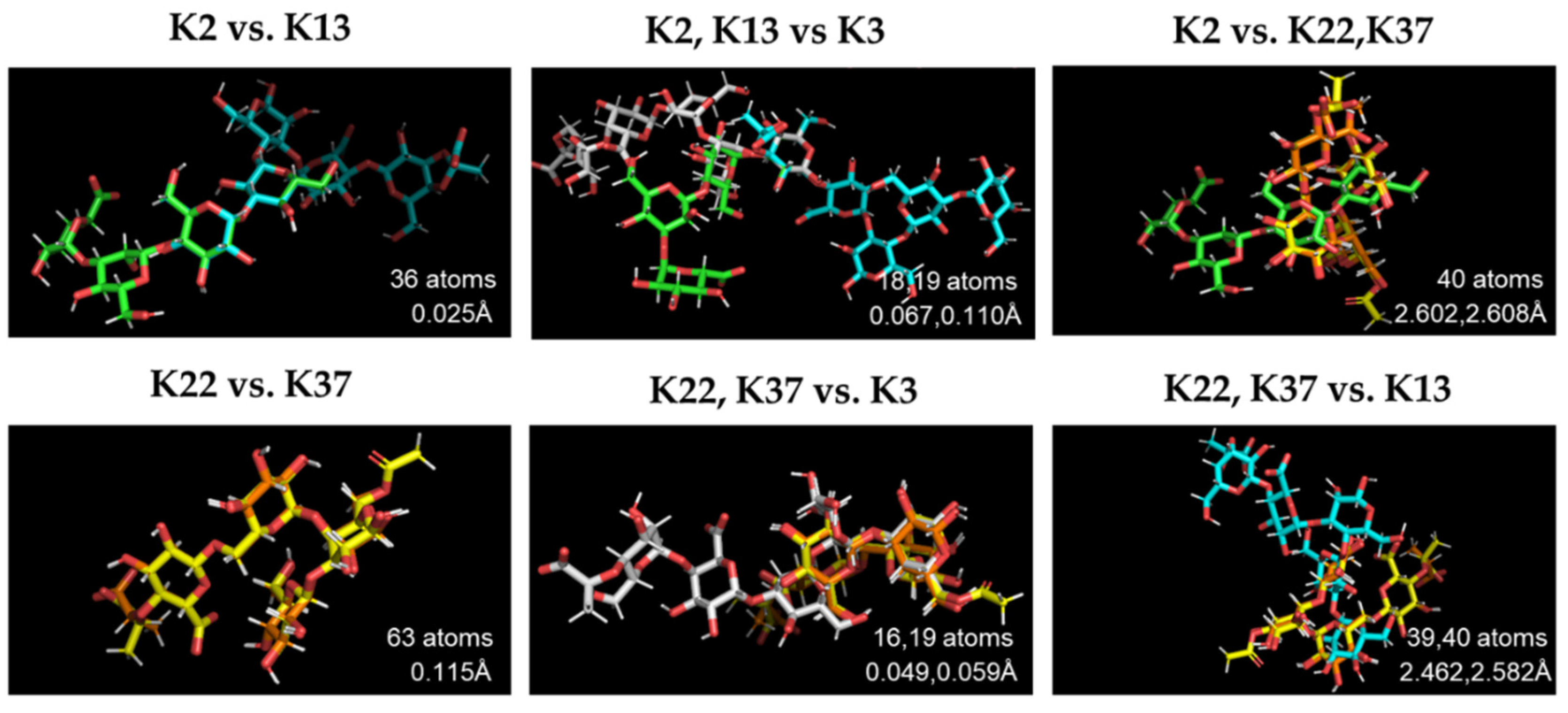
2.7. Structural Comparison of K Antigens

Host-range experiments revealed that both phages πVLC5 and πVLC6 were able to infect capsular types K22 and K37. Additionally, phage πVLC6 could infect K13 and digest K2 and K3 capsules. As depolymerases are thought to be antigenically specific, we wanted to determine whether these five capsular types shared some structural backbone that could make them recognizable by common enzymes. The K. pneumoniae capsule is a polysaccharide matrix of repeated units with an acidic component. K22 and K37 have the same sequence of sugars, with the exception of the presence of OAc substituents in K22 [24]. Approximately 66% of the atoms of K22 and K37 align properly, with low values of RMSD (0.115 Å) as expected (Figure 7). K2 and K13 shared the units of glucose, mannose, and glucuronic acid [25,26]. This could be observed in the overlapping of 36 atoms in the structural pairwise alignment with an RMSD value of 0.025 Å. The RMSD values between these two capsular types, K2 and K13, and those described above, K22 and K37, were much higher (>2.5 Å), showing a poor overlap of their molecular structures. Interestingly, antigen K3 overlapped partially with both clusters (K22/K37 and K2/K13), with RMSD ranging from 0.049 Å to 0.110 Å. The repeated units of the K3 structure were made of pyruvic acetal-bearing pentasaccharide containing galactose, mannose, and glucuronic acid [27]. Thus, K3 shared the sugars mannose with K2/K13 and galactose with K22/K37.

3. Discussion

The K. pneumoniae exopolysaccharide capsule is a major virulence factor and a source of diversity that complicates treatment [28]. Still relatively little is known about the diversity of Klebsiella sp. phages and the molecular mechanisms allowing infection of different capsular types. Although previous studies have proposed that depolymerases play a key role in the recognition and entry of phages in these bacteria, additional data are needed to establish more general patterns. Recently, our group has phenotypically and molecularly characterized four new Drulisviruses infecting capsular type K22 [18]. Despite the fact that this capsular type is not highly prevalent, it has been found among multi-drug-resistant clinical isolates. Moreover, Klebsiella capsular diversity is widely distributed, meaning that no single capsular type monopolizes a large fraction of clinical isolates and that, consequently, it would be necessary to target most types for efficiently combating this pathogen. Here, we have characterized two new phages capable of infecting not only K22, but also other capsular types.
Phylogenetic analyses of whole and core genomes of the genus Drulisvirus group the new phages together, along with the previously described Klebsiella phage Vb_KpnP_SU552A. However, the phylogeny of the RNA polymerase does not support this grouping, placing πVLC5 with other πVLC phages. These results, taken together with the lack of homology in one of the tail spikes, suggest a certain degree of mosaic-like genome structure for drulisviruses. Horizontal acquisition of tail spike genes seems especially relevant in the case of ORF58 of the Klebsiella phage πVLC5 and ORF58 of πVLC6. The former presents a unique structure among the phage sequences described so far, as it is only distantly related to other phages from a different family. On the contrary, the second tail spike of Klebsiella phage πVLC6 is also present in four other phages of the same genus, with a high level of identity. Regarding the shared tail spike present in all members of the genus Drulisvirus and some related phages (Przondovirus and Phikmvvirus), the enzymatic domain is only found in the πVLC phages, all of which share the ability to infect K22 capsular types. Interestingly, phage πVLC5 was sampled in the same place as phages πVLC1 and πVLC2, and these three phages seemed to form a monophyletic group according to the RNA polymerase phylogeny. However, they differed in their depolymerase domains and whole-genome phylogenetic position. This suggests that gene exchange events were involved in adaption to specific ecological niches [22].
Both phages described were capable of lysing capsular types K22 and K37. In addition, in both cases, lysis haloes were observed around plaques, presumably resulting from the action of their depolymerases [29]. Klebsiella strains presenting capsular types K22 and K37 shared most of the genes involved in the synthesis of the capsule polysaccharides, except for one gene that showed a frameshift mutation [30] and the overall biochemical structure as explained above. This could explain why both phages, πVLC5 and πVLC6, were able to infect these two capsular types. In principle, the depolymerase shared between these two phages (and the phages previously described by our group) should be responsible for degrading the exopolysaccharides shared between K22 and K37. The ability of the phage πVLC6 to also infect capsular type K13 and digest K2 capsule should be conferred by its second depolymerase, due to the divergence shown between the capsular biochemical structures. This second putative depolymerase presented high nucleotide identity (96% and 100% of coverage) with the second tail-fiber protein of bacteriophage vB_KpnP_KpV74 [16]. Solovieva et al., cloned and expressed this protein (kpv74_56, APZ82768.1) and showed that it was a functional depolymerase against K2 strains and one K13 strain, thus, sharing not only the nucleotide identity but also host tropism with the second depolymerase of bacteriophage πVLC6 [22]. The association between K2 and K13 capsule digestion was also supported by previous work showing cross-infectivity between the K2 and K13 types [31]. The additional interaction of phage πVLC6 with the K3 strain was less expected, as the biochemical structure of this antigen overlapped only partially with K22/K37 and K2/K13, so both enzymes might be able to recognize the K3 antigen. However, there are no reported cases of cross-reaction involving these antigens. To our knowledge, the only depolymerase confirmed to have K3 tropism is that of the Klebsiella phage KP32, KP32gp37 [5]. Interestingly, this protein is the only one described so far that presents the identity of the enzymatic domain of the first tail spike shared by all πVLC phages (Table S1). Thus, the diversity of this enzymatic domain as well as the role of the second tail spike should be further explored, in order to explain K3 tropism. Lastly, we did not find any additional host specificity of πVLC5, despite the presence of a highly divergent depolymerase. Screening against additional novel capsular types, which are constantly being described [32], might reveal its role.
In conclusion, the two new phages πVLC5 and πVLC6, showed interesting aspects of the Drulisvirus evolution and suggest that host range expansion can occur via the acquisition of divergent depolymerases. Yet, few broad-range Klebsiella phages have been described so far. Further prospection of environmental samples might reveal new, more broadly-acting phages. In future work, it would also be interesting to explore resistance mechanisms, since in most cases bacterial growth fully relapses after few hours. In vitro resistance conferred by capsule loss might be inconsequential in vivo, since de-capsulated mutants tend to be avirulent [33]. Finally, testing synergistic effects between phages and antibiotics is yet another promising line of research [34,35].

4. Materials and Methods

4.1. Bacteria

The clinical isolate 1210 obtained from Hospital La Fe (Valencia, Spain) was used as a host to test environmental samples for phage discovery. In addition, 77 reference strains of Klebsiella spp. purchased from the Statens Serum Institute (Copenhagen, Denmark; Table S3) were used to determine the range of infection of the isolated phages.

4.2. Isolation of Phages from Environmental Samples

A water sample obtained from a river and another from an irrigation ditch were used to isolate phages. For this purpose, 50 mL of water were taken and kept at room temperature until processing in the laboratory, typically 1 h later. After vortexing, samples were centrifuged (4000× g, 10 min) and supernatants filtered through 0.45 μm and 0.22 μm, to remove the cells and debris. Then, 1 mL of the filtered samples was added to 500 μL of K. pneumoniae clinical isolate 1210 and plated on Petri dishes, through the top agar method. Plates were incubated at 37 °C for 20 h. Isolated plaques of different morphology were then recovered by aspiration with a micropipette and stored at −70 °C. In order to check the isolated plaques and purify them, two additional plaque assays and plaque picking steps were performed. The plaque-purified phages were amplified in liquid media by infecting an exponential growth phase culture of the bacterium (OD600 = 0.4). After lysis, bacteria and debris were removed by centrifugation (13,000× g, 3 min twice) and the supernatants were aliquoted and stored at −70 °C.

4.3. Electron Microscopy

High titer lysates were centrifuged and supernatants were filtered through 0.22 µm to remove bacteria and debris. A drop from each lysate was then deposited onto a carbon-coated Formvar supported by a 300 mesh copper grid. After air drying for 30 min, excess liquid was removed with filter paper. Phages were negatively stained with 2% phosphotungstic acid and examined under an electron microscope Jeol JEM-1010.

4.4. DNA Isolation and Genome Sequencing

DNA was extracted by treating high titer filtered lysates with DNAse I to remove non-encapsidated DNA and, following DNAse I inactivation, DNA extraction was performed using a commercial kit (QIAamp, QIAGEN). DNA was then fragmented by sonication and sequenced in an Illumina MiSeq machine yielding 250 paired-end reads, which were screened for contaminants using Kraken 2 [36] and then assembled with SPAdes v3.9.1 [37]. Contigs smaller than 1000 nucleotides were discarded, resulting in a single contig for each genome. PHASTER [38] and BLAST were used to determine the closest sequences in the databases. These were downloaded, and we used ProgressiveMAUVE [39] to obtain a whole-genome multiple sequence alignment. Complete genome sequences are available at Genbank (accession No. MT197175-MT197176).

4.5. Genome Annotation

Phages were annotated using PHANOTATE [40], Glimmer v.3.0 [41], and Prodigal [42]. Gene callings and start and stop coordinates were compared with a custom script available at https://github.com/BBeamud/. Nucleotide sequences for the putative ORFs were extracted with seqtk subseq (https://github.com/lh3/seqtk) and used for functional annotation. A phage nucleotide database was built with makeblastdb from the one available at http://millardlab.org/bioinformatics/bacteriophage-genomes/. This database contains all non-redundant phage proteins sequences deposited in GenBank and their annotation (August, 2019). Phages πVLC were added to this database [18]. The best BLASTN hit of non-hypothetical proteins, when possible, was retrieved with an e-value cutoff of 10−5. We also searched for temperate behavior in our phage genomes, such as mobile genetic elements, antibiotic resistance or virulence genes, and any kind of bacterial gene. Local databases from ICEberg v.2.0 (http://db-mml.sjtu.edu.cn/ICEberg/), the Comprehensive Antibiotic Resistance Database (CARD, https://card.mcmaster.ca/), Virulence factors of Pathogenic Bacteria (VFDB, http://www.mgc.ac.cn/VFs/main.htm), and BacteriaDB (in-house database, 2016) were used to accomplish this using BLASTN, with an e-value cutoff of 10−5. Lastly, CDSs were searched against a local database constructed with depolymerase proteins included in Pires et al. [43]. InterProScan5 [44] and HHpred [45] were used for further checking of domains and protein structures when a significant similarity was found.

4.6. Comparative Genomics

Whole-genome average nucleotide identity (ANI) values between the new Klebsiella phages and the other Drulisvirus phages were obtained using pANIto (https://github.com/sanger-pathogens/panito). Conserved and unique genes among the Drulisvirus were obtained with Proteinortho v.6 [46], by comparing the previously extracted nucleotide CDSs and the ones available from GenBank. Maximum likelihood (ML) phylogenies were constructed with 1000 fast bootstrap pseudo-replicates using the GTR+G+I substitution model in IQ-TREE v.1.6.5 [47].

4.7. Determination of Host Range

Spot tests were performed by adding 2 µL drops of each high titer phage to the bacterial lawns of the 77 reference Klebsiella spp. strains in top-agar semi-solidified media, and incubating plates at 37 °C for 24 h. In addition, we inoculated cultures of the 77 reference strains in w-96 plates with 105 PFU of phage and incubated cultures at 37 °C in a plate reader (Multiskan) for 10 h, to determine the strength of lysis based OD measurements taken every 10 min.

4.8. Determination of Phage Progeny Production

Approximately 105 PFU were used to inoculate 500 µL of log-phase bacterial cultures (OD600 = 0.05) in a Thermoshaker, and the cultures were incubated at 250 rpm and 37 °C. At 4 hpi, the cultures were centrifuged (13,000× g, 2 min) to remove bacteria and debris, and the supernatants were used for titration. To accomplish this, the supernatants were serially diluted in LB, and then used to perform plaque assays in 1210 K. pneumoniae cultures. A similar approach was used to determine phage progeny production in semi-solid cultures. Spots with ca. 105 PFU were used to inoculate bacterial cultures, that were incubated at 37 °C for 8 h. After this, a micropipette was used to recover the phages from the spot, which were resuspended in LB for titration.

4.9. Capsule Light Microscopy

Bacteria were grown in semi-solidified media in petri dishes to perform spot tests with phage suspensions (≥107 PFU/mL). After 24 h at 37 °C, a smear loop was used to sample bacteria from the haloes and non-inoculated regions of the plate. Samples were fixed with 2.5% formaldehyde for 20 min, in the presence of 100 mM lysine, which prevented capsule loss during fixation [48]. After removing the fixative agent through centrifugation and washing with low-salt TE buffer, 10% nigrosin was mixed with the bacterial suspension on a glass slide and 1% crystal violet was added. The preparations were examined through a light microscopy and the presence of capsules was assessed by the exclusion of both nigrosin and crystal violet around the cells.

4.10. Structural Comparison of K Antigens

To determine if the capsular K antigens that showed any type of interaction with the novel phages were structurally similar, we downloaded the atomic structure of each antigen from https://iith.ac.in/K-PAM/k_antigen.html [49]. Program database (PDB) files were loaded and aligned with the PyMOL Molecular Graphics System (Version 2.0 Schrödinger, LLC). The ‘align’ command performed a structural superposition with several rounds of refinement to discard structural outliers. We obtained the number of aligned atoms and the root-mean-square deviation of atomic positions (RMSD) between the pairwise-alignments of K antigens.

Supplementary Materials

The following are available online at https://www.mdpi.com/1422-0067/21/9/3160/s1. Table S1: Results of the BLASTN search of the tail fiber sequences for phages πVLC5 and πVLC6 against a database of phage CDSs. Table S2: Determination of Klebsiella phages πVLC5 and πVLC6 host range in semi-solid and liquid. Table S3: Klebsiella species and K-serotypes of the 77 K-type reference strains from the Statens Serum Institute (Copenhagen, Denmark).

Author Contributions

Conceptualization, P.D.-C. and R.S.; Funding acquisition, P.D.-C., F.G.-C., and R.S.; Investigation, P.D.-C., B.B., and L.M.-Q.; Methodology, P.D.-C., B.B., L.M.-Q., and R.S.; Supervision, P.D.-C., F.G.-C., and R.S.; Writing—original draft, P.D.-C. and B.B.; Writing—review and editing, P.D.-C., F.G.-C., and R.S. All authors have read and agreed to the published version of the manuscript.

Funding

This research was funded by the ESCMID Research Grant 20200063 to P.D-C., ERC Consolidator Grant 724519 Vis-a-Vis to R.S, and project BFU2017-89594R from MICINN to F.G-C. B.B was funded by a PhD fellowship from Spanish MCIU FPU16/02139.

Acknowledgments

We thank Carles Úbeda for the clinical K. pneumoniae 1210 strain, Justine Vienne for help with phage hunting, and María Duran for technical assistance.

Conflicts of Interest

The authors declare no conflict of interest.

References

  1. Holt, K.E.; Wertheim, H.; Zadoks, R.N.; Baker, S.; Whitehouse, C.A.; Dance, D.; Jenney, A.; Connor, T.R.; Hsu, L.; Severin, J.; et al. Genomic analysis of diversity, population structure, virulence, and antimicrobial resistance in Klebsiella pneumoniae, an urgent threat to public health. Proc. Natl. Acad. Sci. USA 2015, 112, E3574–E3581. [Google Scholar] [CrossRef] [PubMed] [Green Version]
  2. Effah, C.Y.; Sun, T.; Liu, S.; Wu, Y. Klebsiella pneumoniae: An increasing threat to public health. Ann. Clin. Microbiol. Antimicrob. 2020, 19, 1–9. [Google Scholar] [CrossRef] [PubMed]
  3. Founou, R.C.; Founou, L.L.; Essack, S.Y. Clinical and economic impact of antibiotic resistance in developing countries: A systematic review and meta-analysis. PLoS ONE 2017, 12, e0189621. [Google Scholar] [CrossRef] [PubMed] [Green Version]
  4. Domingo-Calap, P.; Georgel, P.; Bahram, S. Back to the future: Bacteriophages as promising therapeutic tools. HLA 2016, 87, 133–140. [Google Scholar] [CrossRef] [PubMed]
  5. Majkowska-Skrobek, G.; Latka, A.; Berisio, R.; Squeglia, F.; Maciejewska, B.; Briers, Y.; Drulis-Kawa, Z. Phage-Borne Depolymerases Decrease Klebsiella pneumoniae Resistance to Innate Defense Mechanisms. Front. Microbiol. 2018, 9, 2517. [Google Scholar] [CrossRef] [Green Version]
  6. Kortright, K.E.; Chan, B.K.; Koff, J.L.; Turner, P.E. Phage Therapy: A Renewed Approach to Combat Antibiotic-Resistant Bacteria. Cell Host Microbe 2019, 25, 219–232. [Google Scholar] [CrossRef] [Green Version]
  7. Imai, K.; Ishibashi, N.; Kodana, M.; Tarumoto, N.; Sakai, J.; Kawamura, T.; Takeuchi, S.; Taji, Y.; Ebihara, Y.; Ikebuchi, K.; et al. Clinical characteristics in blood stream infections caused by Klebsiella pneumoniae, Klebsiella variicola, and Klebsiella quasipneumoniae: A comparative study, Japan, 2014–2017. BMC Infect. Dis. 2019, 19, 946. [Google Scholar] [CrossRef] [Green Version]
  8. Palacios, M.; Miner, T.A.; Frederick, D.R.; Sepúlveda, V.; Quinn, J.D.; Walker, K.A.; Miller, V.L. Identification of Two Regulators of Virulence That Are Conserved inKlebsiella pneumonia Classical and Hypervirulent Strains. mBio 2018, 9, e01443-18. [Google Scholar] [CrossRef] [Green Version]
  9. Adams, M.H.; Park, B.H. An enzyme produced by a phage-host cell system. Virology 1956, 2, 719–736. [Google Scholar] [CrossRef]
  10. Rieger-Hug, D.; Stirm, S. Comparative study of host capsule depolymerases associated with Klebsiella bacteriophages. Virology 1981, 113, 363–378. [Google Scholar] [CrossRef]
  11. Lin, T.-L.; Hsieh, P.-F.; Huang, Y.-T.; Lee, W.-C.; Tsai, Y.-T.; Su, P.-A.; Pan, Y.-J.; Hsu, C.-R.; Wu, M.-C.; Wang, J.-T. Isolation of a Bacteriophage and Its Depolymerase Specific for K1 Capsule of Klebsiella pneumoniae: Implication in Typing and Treatment. J. Infect. Dis. 2014, 210, 1734–1744. [Google Scholar] [CrossRef] [PubMed] [Green Version]
  12. Majkowska-Skrobek, G.; Latka, A.; Berisio, R.; Maciejewska, B.; Squeglia, F.; Romano, M.; Lavigne, R.; Struve, C.; Drulis-Kawa, Z. Capsule-Targeting Depolymerase, Derived from Klebsiella KP36 Phage, as a Tool for the Development of Anti-Virulent Strategy. Viruses 2016, 8, 324. [Google Scholar] [CrossRef] [PubMed]
  13. Hsieh, P.-F.; Lin, H.-H.; Lin, T.-L.; Chen, Y.-Y.; Wang, J.-T. Two T7-like Bacteriophages, K5-2 and K5-4, Each Encodes Two Capsule Depolymerases: Isolation and Functional Characterization. Sci. Rep. 2017, 7, 4624. [Google Scholar] [CrossRef] [PubMed]
  14. Wu, Y.; Wang, R.; Xu, M.; Liu, Y.; Zhu, X.; Qiu, J.; Liu, Q.; He, P.; Li, Q. A Novel Polysaccharide Depolymerase Encoded by the Phage SH-KP152226 Confers Specific Activity Against Multidrug-Resistant Klebsiella pneumoniae via Biofilm Degradation. Front. Microbiol. 2019, 10, 2768. [Google Scholar] [CrossRef] [PubMed] [Green Version]
  15. Pan, Y.-J.; Lin, T.-L.; Chen, C.-C.; Tsai, Y.-T.; Cheng, Y.-H.; Chen, Y.-Y.; Hsieh, P.-F.; Lin, Y.-T.; Wang, J.-T. Klebsiella Phage ΦK64-1 Encodes Multiple Depolymerases for Multiple Host Capsular Types. J. Virol. 2017, 91, e02457-16. [Google Scholar] [CrossRef] [Green Version]
  16. Pan, Y.-J.; Lin, T.-L.; Chen, Y.-Y.; Lai, P.-H.; Tsai, Y.-T.; Hsu, C.-R.; Hsieh, P.-F.; Lin, Y.-T.; Wang, J.-T. Identification of three podoviruses infecting Klebsiella encoding capsule depolymerases that digest specific capsular types. Microb. Biotechnol. 2019, 12, 472–486. [Google Scholar] [CrossRef] [Green Version]
  17. Knecht, L.E.; Veljkovic, M.; Fieseler, L. Diversity and Function of Phage Encoded Depolymerases. Front. Microbiol. 2020, 10, 2949. [Google Scholar] [CrossRef]
  18. Domingo-Calap, P.; Beamud, B.; Vienne, J.; González-Candelas, F.; Sanjuán, R. Isolation of Four Lytic Phages Infecting Klebsiella pneumoniae K22 Clinical Isolates from Spain. Int. J. Mol. Sci. 2020, 21, 425. [Google Scholar] [CrossRef] [Green Version]
  19. Morales, A.C.H.; Lessor, L.L.; Wood, T.L.; Migl, D.; Mijalis, E.M.; Cahill, J.; Russell, W.K.; Young, R.; Gill, J.J. Genomic and Biochemical Characterization of Acinetobacter Podophage Petty Reveals a Novel Lysis Mechanism and Tail-Associated Depolymerase Activity. J. Virol. 2018, 92, e01064-17. [Google Scholar] [CrossRef] [Green Version]
  20. Mushtaq, N.; Redpath, M.B.; Luzio, J.P.; Taylor, P.W. Treatment of experimental Escherichia coli infection with recombinant bacteriophage-derived capsule depolymerase. J. Antimicrob. Chemother. 2005, 56, 160–165. [Google Scholar] [CrossRef] [Green Version]
  21. Eriksson, H.; Maciejewska, B.; Latka, A.; Majkowska-Skrobek, G.; Hellstrand, M.; Melefors, Ö.; Wang, J.-T.; Kropinski, A.M.; Drulis-Kawa, Z.; Nilsson, A. A Suggested New Bacteriophage Genus, “Kp34likevirus”, within the Autographivirinae Subfamily of Podoviridae. Viruses 2015, 7, 1804–1822. [Google Scholar] [CrossRef] [PubMed]
  22. Solovieva, E.V.; Myakinina, V.P.; Kislichkina, A.A.; Krasilnikova, V.M.; Verevkin, V.V.; Mochalov, V.V.; Lev, A.; Fursova, N.K.; Volozhantsev, N.V. Comparative genome analysis of novel Podoviruses lytic for hypermucoviscous Klebsiella pneumoniae of K1, K2, and K57 capsular types. Virus Res. 2018, 243, 10–18. [Google Scholar] [CrossRef] [PubMed]
  23. Guy, L.; Kultima, J.R.; Andersson, S.G.E. genoPlotR: Comparative gene and genome visualization in R. Bioinformatics 2010, 26, 2334–2335. [Google Scholar] [CrossRef] [PubMed] [Green Version]
  24. Parolis, L.A.; Parolis, H.; Niemann, H.; Stirm, S. Primary structure of Klebsiella serotype K22 capsular polysaccharide: Another glycan containing 4-O-[(S-1-carboxyethyl]-d-glucuronic acid. Carbohydr. Res. 1988, 179, 301–314. [Google Scholar] [CrossRef]
  25. Sutherland, I.W. The Exopolysaccharides of Klebsiella Serotype 2 Strains as Substrates for Phage-induced Polysaccharide Depolymerases. J. Gen. Microbiol. 1972, 70, 331–338. [Google Scholar] [CrossRef] [Green Version]
  26. Niemann, H.; Frank, N.; Stirm, S. Klebsiella serotype-13 capsular polysaccharide: Primary structure and depolymerization by a bacteriophage-borne glycanase. Carbohydr. Res. 1977, 59, 165–177. [Google Scholar] [CrossRef] [Green Version]
  27. Dutton, G.G.; Parolis, H.; Joseleau, J.-P.; Marais, M.-F. The use of bacteriophage depolymerization in the structural investigation of the capsular polysaccharide from Klebsiella serotype K3. Carbohydr. Res. 1986, 149, 411–423. [Google Scholar] [CrossRef]
  28. Paczosa, M.K.; Mecsas, J. Klebsiella pneumoniae: Going on the Offense with a Strong Defense. Microbiol. Mol. Biol. Rev. 2016, 80, 629–661. [Google Scholar] [CrossRef] [Green Version]
  29. Sutherland, I.W.; Hughes, K.A.; Skillman, L.C.; Tait, K. The interaction of phage and biofilms. FEMS Microbiol. Lett. 2004, 232, 1–6. [Google Scholar] [CrossRef]
  30. Pan, Y.-J.; Lin, T.-L.; Chen, Y.-H.; Hsu, C.-R.; Hsieh, P.-F.; Wu, M.-C.; Wang, J.-T. Capsular Types of Klebsiella pneumoniae Revisited by wzc Sequencing. PLoS ONE 2013, 8, e80670. [Google Scholar] [CrossRef]
  31. Pieroni, P.; Rennie, R.P.; Ziola, B.; Deneer, H.G. The use of bacteriophages to differentiate serologically cross-reactive isolates of Klebsiella pneumoniae. J. Med. Microbiol. 1994, 41, 423–429. [Google Scholar] [CrossRef] [PubMed] [Green Version]
  32. Bellich, B.; Lagatolla, C.; Rizzo, R.; D’Andrea, M.M.; Rossolini, G.M.; Cescutti, P. Determination of the capsular polysaccharide structure of the Klebsiella pneumoniae ST512 representative strain KPB-1 and assignments of the glycosyltransferases functions. Int. J. Biol. Macromol. 2020, 155, 315–323. [Google Scholar] [CrossRef] [PubMed]
  33. Lawlor, M.S.; Hsu, J.; Rick, P.D.; Miller, V.L. Identification of Klebsiella pneumonia virulence determinants using an intranasal infection model. Mol. Microbiol. 2005, 58, 1054–1073. [Google Scholar] [CrossRef]
  34. Domingo-Calap, P.; Delgado-Martínez, J. Bacteriophages: Protagonists of a Post-Antibiotic Era. Antibiotics 2018, 7, 66. [Google Scholar] [CrossRef] [PubMed] [Green Version]
  35. Bansal, S.; Harjai, K.; Chhibber, S. Depolymerase improves gentamicin efficacy during Klebsiella pneumoniae induced murine infection. BMC Infect. Dis. 2014, 14, 456. [Google Scholar] [CrossRef] [PubMed] [Green Version]
  36. Wood, D.E.; Lu, J.; Langmead, B. Improved metagenomic analysis with Kraken 2. Genome Biol. 2019, 20, 257. [Google Scholar] [CrossRef] [PubMed] [Green Version]
  37. Bankevich, A.; Nurk, S.; Antipov, D.; Gurevich, A.; Dvorkin, M.; Kulikov, A.; Lesin, V.M.; Nikolenko, S.; Pham, S.; Prjibelski, A.D.; et al. SPAdes: A New Genome Assembly Algorithm and Its Applications to Single-Cell Sequencing. J. Comput. Biol. 2012, 19, 455–477. [Google Scholar] [CrossRef] [Green Version]
  38. Arndt, D.; Grant, J.R.; Marcu, A.; Sajed, T.; Pon, A.; Liang, Y.; Wishart, D.S. PHASTER: A better, faster version of the PHAST phage search tool. Nucleic Acids Res. 2016, 44, W16–W21. [Google Scholar] [CrossRef] [Green Version]
  39. Darling, A.E.; Mau, B.; Perna, N.T. progressiveMauve: Multiple Genome Alignment with Gene Gain, Loss and Rearrangement. PLoS ONE 2010, 5, e11147. [Google Scholar] [CrossRef] [Green Version]
  40. McNair, K.; Zhou, C.; Dinsdale, E.A.; Souza, B.; Edwards, R.A. PHANOTATE: A novel approach to gene identification in phage genomes. Bioinformatics 2019, 35, 4537–4542. [Google Scholar] [CrossRef] [Green Version]
  41. Delcher, A. Improved microbial gene identification with GLIMMER. Nucleic Acids Res. 1999, 27, 4636–4641. [Google Scholar] [CrossRef]
  42. Hyatt, D.; Chen, G.; Locascio, P.F.; Land, M.; Larimer, F.W.; Hauser, L. Prodigal: Prokaryotic gene recognition and translation initiation site identification. BMC Bioinform. 2010, 11, 119. [Google Scholar] [CrossRef] [Green Version]
  43. Pires, D.P.; Oliveira, H.; Melo, L.D.R.; Cerqueira, M.A.; Azeredo, J. Bacteriophage-encoded depolymerases: Their diversity and biotechnological applications. Appl. Microbiol. Biotechnol. 2016, 100, 2141–2151. [Google Scholar] [CrossRef] [Green Version]
  44. Jones, P.; Binns, D.; Chang, H.Y.; Fraser, M.; Li, W.; McAnulla, C.; McWilliam, H.; Maslen, J.; Mitchell, A.L.; Nuka, G.; et al. InterProScan 5: Genome-scale protein function classification. Bioinformatics 2014, 30, 1236–1240. [Google Scholar] [CrossRef] [PubMed] [Green Version]
  45. Zimmermann, L.; Stephens, A.; Nam, S.-Z.; Rau, D.; Kübler, J.; Lozajic, M.; Gabler, F.; Soeding, J.; Lupas, A.N.; Alva, V. A Completely Reimplemented MPI Bioinformatics Toolkit with a New HHpred Server at its Core. J. Mol. Biol. 2018, 430, 2237–2243. [Google Scholar] [CrossRef] [PubMed]
  46. Lechner, M.; Findeiss, S.; Müller, L.; Marz, M.; Stadler, P.F.; Prohaska, S. Proteinortho: Detection of (Co-)orthologs in large-scale analysis. BMC Bioinform. 2011, 12, 124. [Google Scholar] [CrossRef] [PubMed] [Green Version]
  47. Nguyen, L.-T.; Schmidt, H.A.; Von Haeseler, A.; Minh, B.Q. IQ-TREE: A Fast and Effective Stochastic Algorithm for Estimating Maximum-Likelihood Phylogenies. Mol. Biol. Evol. 2014, 32, 268–274. [Google Scholar] [CrossRef] [PubMed]
  48. Jacques, M.; Graham, L. Improved preservation of bacterila capsule for electron microscopy. J. Electron Microsc. Tech. 1989, 11, 167–169. [Google Scholar] [CrossRef] [PubMed]
  49. Patro, L.P.P.; Sudhakar, K.U.; Rathinavelan, T. K-PAM: A unified platform to distinguish Klebsiella species K- and O-antigen types, model antigen structures and identify hypervirulent strains. bioRxiv 2019. [Google Scholar] [CrossRef]
Figure 1. Initial characterization of Klebsiella phages πVLC5 and πVLC6. (a) Plaque assays of the two phages. Plaques were allowed to develop in soft agar media at 37 °C (hpi: hours post inoculation). Notice that haloes around plaques kept expanding even though the lysis did not proceed further, suggesting passive diffusion of depolymerase activity. Scale bar: 1 cm. (b) Transmission electron micrographs of the two novel K. pneumoniae phages. Scale bar: 50 nm.
Ijms 21 03160 g001
Figure 2. Maximum likelihood (ML) phylogenetic tree of the Drulisvirus genus. (a) Whole-genome and (b) RNA polymerase. Numbers indicate bootstrap support values (1000 pseudo-replicates). The two new phages are highlighted in purple. The scale bar represents 0.01 sequence divergence (percent nucleotide substitutions).
Ijms 21 03160 g002
Figure 3. Genome alignment of the Klebsiella phages πVLC5 and πVLC6 with the previously characterized Klebsiella phages vB_KpnP_SU552A (closest relative) and vB_KpnP_KpV74. Arrows represent coding sequences (CDSs). The different shades of gray represent different identity levels. The figure was obtained using genoPlotR [23].
Ijms 21 03160 g003
Figure 4. Conserved tail fibers/spikes of available drulisviruses (a). Amino acid sequence alignment of the conserved region of the tail fiber/spike. Variant amino acids are highlighted in different colors. (b). Nucleotide ML tree of the conserved region of the tail fiber/spike. Numbers indicate bootstrap values (1000 pseudo-replicates). The scale bar represents 0.01 sequence divergence (percent nucleotide substitutions).
Ijms 21 03160 g004
Figure 5. Lytic activity of Klebsiella phages πVLC5 and πVLC6 in different capsular types. (a) Spot tests of the two K. pneumoniae phages in K2, K3, K13, K22, K37, and K45 Klebsiella sp. reference strains. All 77 reference strains were assayed, but only those showing phage activity are shown, except for K45, which is included as an example of a non-susceptible strain. Plaques were allowed to develop in soft agar media overnight at 37 °C. ++: obvious lysis; +: halo-like turbidity reduction; -: no effect. (b) OD measurements of capsular types K2, K3, K13, K22, K37, and K45 Klebsiella spp. Orange—non-infected control; red—πVLC5; blue—πVLC6; pink—πVLC5 + πVLC6. Error bars indicate the SEM.
Ijms 21 03160 g005
Figure 6. Klebsiella phage πVLC6 depolymerase activity against K2 and K3 strains. Cultures were doubly stained with nigrosin and crystal violet. Capsules appeared as non-stained areas around cells. Inoculation with phage πVLC6 produced a nearly complete stripping of the capsules. K37 strain was used as a positive control. Scale bar: 5 µm.
Ijms 21 03160 g006
Figure 7. Structural superposition of Klebsiella K antigens K2, K3, K13, K22, and K37. The number of atoms that align between the pair-wise structures and the RMSD values are indicated. Green—K2 (85 atoms), white—K3 (112 atoms), blue—K13 (112 atoms), yellow—K22 (98 atoms), and orange—K37 (93 atoms).
Ijms 21 03160 g007
Table 1. Depolymerase motifs identified in Klebsiella phages πVLC5 and πVLC6.
PhageORFProtein Size (aa)ToolMotif (aa)FamilyIdentifiere-Value
πVLC5ORF49 (36103–38478)792InterProScan513–140Bacteriophage T7 tail fiber proteinIPR0056045.1 × 10−14
319–617Pectin lyaseIPR0123343.2 × 10−10
HHpred2–578phiAB6 tail spike5JSD_B2.1 × 10−23
234–789Putative tail fiber; Tail spike, hydrolase5W6S_A2.3 × 10−13
ORF58 (42631–44634)668InterProScan516–365Pectin lyaseIPR0123341.0 × 10−15
544–603Chaperone of endosialidaseIPR0303927.3 × 10−37
544–667Winged helix-like DNA-binding domainIPR0363885.8 × 10−6
HHpred26–665Tail spike protein Acinetobacter phage6EU4_B6.0 × 10−34
πVLC6ORF51 (35765–38140)792InterProScan513–140Bacteriophage T7 tail fiber proteinIPR0056041.7 × 10−14
319–617Pectin lyaseIPR0123349.1 × 10−10
HHpred1–578phiAB6 tail spike5JSD_B6.3 × 10−23
ORF58 (42292–44025)578HHpred1–576Bacteriophage CBA120 tail spike6EU4_B2.1 × 10−41

