Review

Mitochondrial E3 Ubiquitin Ligase Parkin: Relationships with Other Causal Proteins in Familial Parkinson’s Disease and Its Substrate-Involved Mouse Experimental Models

1
Department of Pathological Cell Biology, Medical Research Institute, Tokyo Medical and Dental University (TMDU), 1-5-45 Yushima, Bunkyo-ku, Tokyo 113-8510, Japan
2
Department of Stress Response Science, Center for Advanced Medical Research, Graduate School of Medicine, Hirosaki University, 5 Zaifu-cho, Hirosaki, Aomori 036-8562, Japan
3
Department of Biochemistry and Molecular Biology, Graduate School of Medical Science, Kyoto Prefectural University of Medicine, Kawaramachi-Hirokoji, Kamigyo-ku, Kyoto 602-8566, Japan
4
Department of Molecular and Chemical Life Sciences, Graduate School of Life Sciences, Tohoku University, Sendai 980-8578, Japan
*
Author to whom correspondence should be addressed.
Int. J. Mol. Sci. 2020, 21(4), 1202; https://doi.org/10.3390/ijms21041202
Submission received: 22 January 2020 / Revised: 7 February 2020 / Accepted: 9 February 2020 / Published: 11 February 2020
(This article belongs to the Special Issue Animal Models of Parkinson's Disease and Related Disorders)

Abstract

:
Parkinson’s disease (PD) is a common neurodegenerative disorder. Recent identification of genes linked to familial forms of PD has revealed that post-translational modifications, such as phosphorylation and ubiquitination of proteins, are key factors in disease pathogenesis. In PD, E3 ubiquitin ligase Parkin and the serine/threonine-protein kinase PTEN-induced kinase 1 (PINK1) mediate the mitophagy pathway for mitochondrial quality control via phosphorylation and ubiquitination of their substrates. In this review, we first focus on well-characterized PINK1 phosphorylation motifs. Second, we describe our findings concerning relationships between Parkin and HtrA2/Omi, a protein involved in familial PD. Third, we describe our findings regarding inhibitory PAS (Per/Arnt/Sim) domain protein (IPAS), a member of PINK1 and Parkin substrates, involved in neurodegeneration during PD. IPAS is a dual-function protein involved in transcriptional repression of hypoxic responses and the pro-apoptotic activities.

1. Introduction

Parkinson’s disease (PD) is the second most common neurodegenerative disease and is characterized by progressive resting tremors, rigidity, bradykinesia, gait disturbances, postural instability, and dementia [1,2]. The motor symptoms of PD are associated with the degeneration of dopaminergic neurons in the substantia nigra pars compacta (SNpc). Although most PD cases are sporadic, approximately 10% are familial cases [3]. The pathogenesis of sporadic PD is yet to be established, but it is suggested that genetic predispositions and environmental toxins causing mitochondrial dysfunction and oxidative stress are involved [1,2,3]. In the early 1980s, the first evidence linking PD to mitochondrial dysfunction appeared with the observation that exposure to 1-methyl-4-phenyl-1,2,3,6-tetrahydropyridine (MPTP) causes dopaminergic neurodegeneration [4]. MPTP is a synthetic by-product of meperidine production. The product of MPTP oxidation, 1-methyl-4-phenylpyridinium (MPP+), selectively enters dopaminergic neurons via the dopamine transporters and inhibits mitochondrial complex I, a key component of the mitochondrial respiratory chain [5]. Subsequently, MPP+ induces oxidative stress and dopaminergic neuronal death [6]. Exposure to other toxins, including 6-OHDA, or the pesticides rotenone and paraquat, also results in the loss of nigrostriatal dopaminergic neurons [7,8]. They produce effects similar to parkinsonian phenotypes in animal models and humans. In MPTP-induced mouse models, there are two typical dosing regimens for MPTP, acute and subacute [9]. In the former type, mice are intraperitoneally injected four times with MPTP (15–20 mg/kg) at 2-h intervals (6-h period) within a single day. Dopaminergic neuron loss can be observed at 3–7 days after the last dose of MPTP. For the latter type, mice were intraperitoneally injected once a day with MPTP (30 mg/kg) for five consecutive days. Dopaminergic neuron loss can be observed 7 days after the last dose of MPTP. The concentrations of MPTP and MPP+ in plasma, striatum, and cortex were studied in mice [10]. Elevated concentrations of MPP+ in plasma and the striatum are maintained for 72 h and 12 h after administration, respectively. N-methylmercaptoimidazole increases the amount of MPTP delivered from the peripheral nervous system to the central nervous system [10].
Accumulated genetic analyses have identified that several gene mutations are associated with an early onset of parkinsonism, including α-synuclein (SNCA), (PARK1 and PARK4), Parkin (PARK2), PTEN-induced kinase 1 (PINK1) (PARK6), DJ-1 (PARK7), leucine rich repeat kinase 2 (LRRK2) (PARK8), ATP13A2 (PARK9), HtrA2/Omi (PARK13), PLA2G6 (PARK14), VPS35 (PARK17), coiled-coil-helix-coiled-coil-helix (CHCH) domain 2 (CHCHD2) (PARK22) [3,11]. Among these, mutations of park genes including Parkin, PINK1, HtrA2/Omi and CHCHD2 are directly involved in mitochondrial dysfunction [12,13,14,15]. PINK1, HtrA2/Omi, and CHCHD2 proteins have a positively charged mitochondrial targeting sequence (MTS) in their N-terminus that forms an amphipathic α-helix (Figure 1). Meanwhile, Parkin does not have an MTS, but does translocate to the mitochondria during mitochondrial dysfunction (Figure 1). Studies of these genes using knockout mice and knockout cell lines demonstrate a relationship between these proteins and mitochondrial quality control [12,13,14,15,16]. Autosomal recessive mutations in the Parkin gene (PARK2), which encodes an E3 ubiquitin ligase, are the most common cause of early onset of PD [1,2,3]. Recently, increased in vitro studies examining the molecular mechanisms of the PINK1/Parkin pathway using the mitochondrial uncouplers, carbonyl cyanide m-chlorophenyl hydrazone (CCCP) and carbonyl cyanide p-(trifluoromethoxy) phenyl hydrazone (FCCP) [13,14]. In healthy mitochondria, PINK1 is translocated and imported to mitochondria via its N-terminal MTS. There, it undergoes cleavage by the mitochondrial processing peptidase (MPP) in the inner membrane to form a 60 kDa intermediate [17]. Following, a presenilin-associated rhomboid-like protein (PARL) and/or matrix-AAA (m-AAA) proteases cleave it to generate a 52 kDa processed form of PINK1 that attached to the inner membrane [17,18]. The cleavage site by PARL has been mapped to Ala103, near the MTS in PINK1 [17,19]. N-terminally cleaved PINK1 is then degraded by the ubiquitin/proteasome system in cytosol. Under these conditions, Parkin remains inactive in the cytosol [13]. Upon mitochondrial damage, such as oxidative stress and experimental CCCP treatment, the membrane potential of mitochondria is decreased; hence, PINK1 is not imported into mitochondria and accumulates on the outer mitochondrial membrane (OMM) to activate Parkin [13,18]. PINK1-mediated phosphorylation of the ubiquitin and the ubiquitin-like (UBL) domain of Parkin enable its E3 ubiquitin ligase functions in concert with E2 ubiquitin-conjugating enzymes [20,21,22,23]. Poly-ubiquitin with p-Ser65 bound to OMM substrates acts as a Parkin receptor; further recruiting Parkin to the mitochondria. These poly-ubiquitin chains can be cleaved by de-ubiquitinating enzymes, including USP15 and USP30, to reverse PINK1/Parkin functions [24,25]. Individual OMM proteins decorated with poly-ubiquitin can be extracted from the mitochondrial membrane and degraded by the 26 S proteasomes. Severely damaged mitochondria are decorated by large numbers of poly-ubiquitin chains that signal for the accumulation of the ubiquitin-binding cargo receptors, NDP52, optineurin, and p62 [26]. NDP52 can bind LC3 in phagophores and recruit the autophagy initiator complex, Fip200-Ulk1, leading to progression of the mitochondrial autophagy (mitophagy) [26,27]. Poly-ubiquitin chains also serves as tags for the accumulation of RabGEF1 on mitochondria, leading to activation of Rab5-Rab7 signaling and the subsequent recruitment of Atg9A vesicles to mitochondria [28]. Autophagosomes containing damaged mitochondria fuse with lysosomes, leading to the generation of autolysosomes, where the cargo is degraded [26]. Recently, our group developed a mitochondria-selective pH-sensitive small-molecule fluorescent dye, Mtphagy Dye, which is incorporated into mitochondria and emits a strong red fluorescence in autolysosomes upon acidification during mitophagy [29]. Mtphagy Dye is a useful tool for easy visualization and detection of mitophagy.
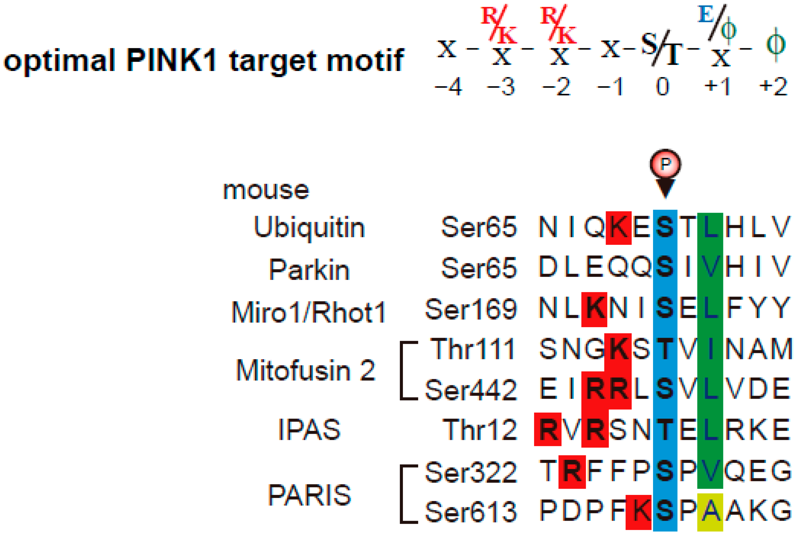
PINK1 was initially named PTEN-induced “putative” kinase-1, and the topology of PINK1 on/in mitochondria was unknown. Although tumor necrosis factor type 1 receptor–associated protein (TRAP1) and the serine protease HtrA2/Omi have been identified as putative PINK1 targets [30,31], TRAP1 and HtrA2/Omi are localized primarily in the mitochondrial matrix and mitochondrial intermembrane space, respectively. Furthermore, previous reports suggest that the kinase domain of mitochondrial PINK1 faces the cytoplasm [32]. Subsequently, increasing numbers of studies using CCCP treatment have investigated the substrates of PINK1 phosphorylation, including Miro1/Rhot1 [33], PINK1 (autophosphorylation) [34], Parkin [20,21], Mitofusin 2 [35], Ubiquitin [22,23], inhibitory PAS (Per/Arnt/Sim) domain protein (IPAS) [36], Parkin-Interacting Substrate (PARIS) [37], and drosophila MIC60/Mitofilin [38]. In addition to LC-MS/MS phosphorylation analyses, some studies used Phos-tag gel mobility shift assays, a powerful and useful tool for detecting the phosphorylation of proteins [39]. Our group has reported well-characterized PINK1 phosphorylation motifs [36] and updates them in this review (Figure 2). Almost all PINK1 target sites in Mus musculus have hydrophobic amino acids at position +2 after the phosphorylation site. In addition, hydrophobic amino acids or acidic amino acids at position +1 after phosphorylation sites are preferred by PINK1. Basic amino acids at positions −3 and −2 (in some cases −5, −4 and −1) are preferred, expect for Parkin. The phosphorylation sites of PARIS are Ser322-P and Ser613-P. Both serine sites have a proline at position +1. As a pS/T-P sequence is preferred by mitogen activated protein (MAP) kinases and cyclin-dependent kinases (CDK), the phosphorylations of PARIS could be regulated by MAP kinases similar to HtrA2/Omi (chapter 2). Thus, PINK1 phosphorylation consensus motifs are valuable for predicting the phosphorylation sites of PINK1-binding proteins and bioinformatical approaches for detecting mitochondrial PINK1-target proteins.
As mentioned above, many studies report the activation mechanism of the PINK1/Parkin pathway and the pathogenesis of PD using cultured cells and mouse models. Next, we will review our findings including the relationships between Parkin and HtrA2/Omi in Parkin-transgenic (Parkin-Tg) mice [40].

2. Parkin-Tg Introduction into mnd2 Mice; Neurodegeneration in HtrA2/Omi Mutant Mice Is Not Rescued by Parkin Transgene Expression

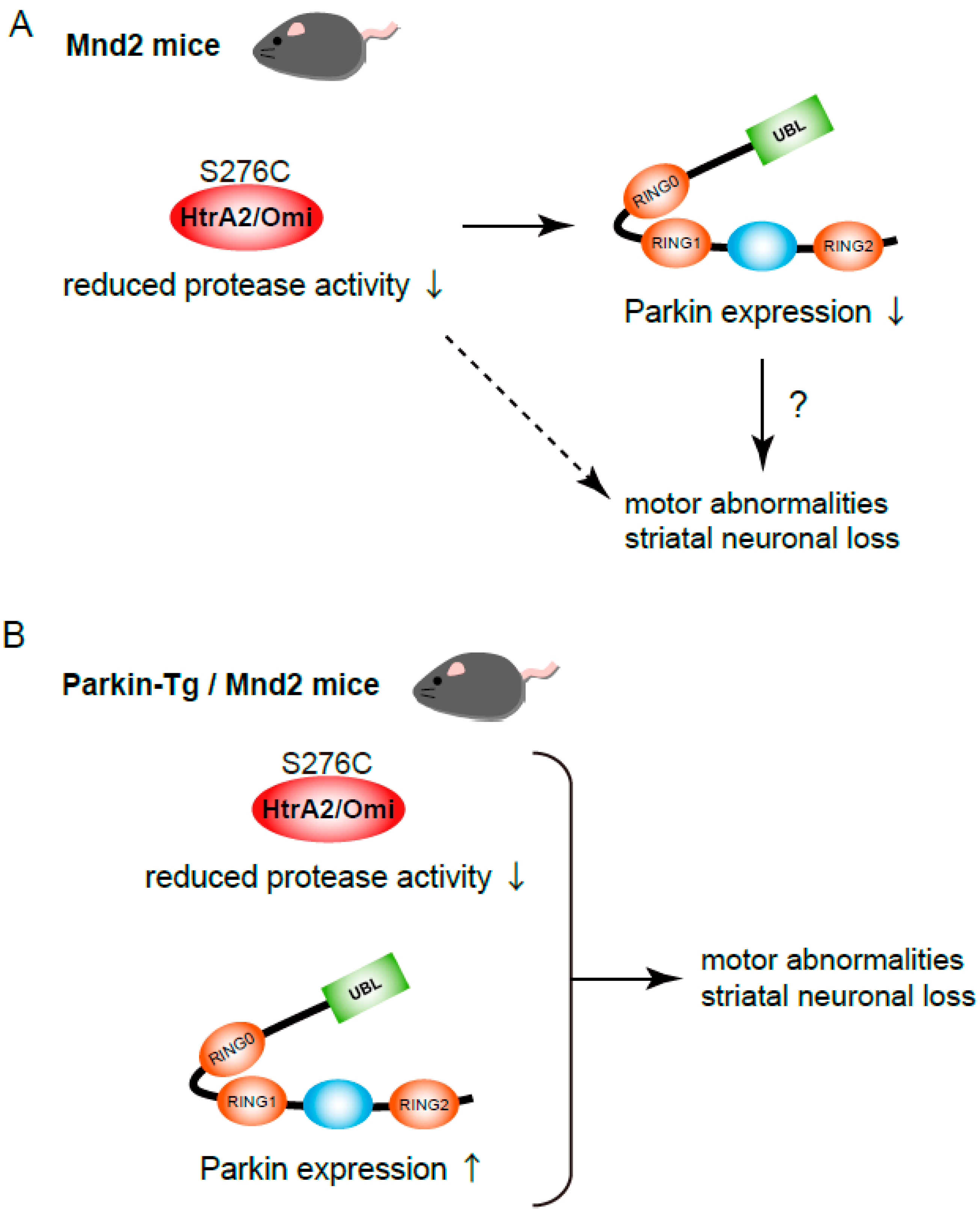
HtrA2/Omi is a serine protease localized in mitochondria (Figure 1) [12]. HtrA2/Omi is expressed as a 49 kDa proenzyme that is targeted to the intermembrane space of the mitochondria where it undergoes proteolytic maturation via cleavage of the first 133 N-terminal residues [41]. HtrA2/Omi contains an N-terminal trypsin-like protease domain and a C-terminal PDZ domain (Figure 1). Ser306 is the active site of the protease domain and mutation of Ser306 inactivates HtrA2/Omi [12]. Ser142 and Ser400 are the known phosphorylation sites of HtrA2/Omi [30,42]. Upon oxidative stress, p38 MAP kinase phosphorylates Ser142 of HtrA2/Omi in a PINK1-depdenent manner. CDK5 phosphorylates Ser400 of HtrA2/Omi in a p38-dependent manner, resulting in the enhancement of HtrA2/Omi proteolytic activity and increased resistance of cells to mitochondrial stress. Meanwhile, mature HtrA2/Omi is released from mitochondria into the cytosol where it is bound to the inhibitor of apoptosis proteins (IAPs); promoting cell death upon apoptotic induction [43,44]. HtrA2/Omi has therefore been proposed to be a pro-apoptotic protein. However, more importantly, HtrA2/Omi is required for maintaining mitochondrial function. This opinion is supported by studies employing HtrA2/Omi-deficient mice, motor neuron degeneration mutant (mnd2) mice [12]. Mnd2 mice possess a non-functional missense mutation at Ser276 to Cys in the protease domain of HtrA2/Omi, close to Ser306 [12]. Protease activity of HtrA2/Omi is greatly reduced in the tissues of mnd2 mice. Mnd2 mutant cells are more vulnerable to Ca2+-induced permeability transitions and mitochondrial membrane permeabilization [12]. Mnd2 mice exhibit weight loss, muscle wasting, neurodegeneration, involution of the spleen and thymus, and finally die within 40 days of birth, and therefore are considered to be a useful animal model for PD [45]. Neuronal cell death in mnd2 mice exhibits features of both apoptosis and necrosis, whereas motor abnormalities and striatal neuronal loss in mnd2 mice are not prevented by overexpression of the anti-apoptotic protein Bcl-2 [46] or cyclophilin D deficiency [47]. Interestingly, Parkin overexpression prevents both neurotoxin-induced and mutant α-synuclein-induced PD models [48,49,50]. Injection of lentiviral vector encoding human Parkin has neuroprotective effects in 6-hydroxydopamine (6-OHDA) rat models for PD; rats overexpressing Parkin display behavioral improvements [48]. Parkin expression protects dopaminergic neurons against the toxicity associated with mutant α-synuclein in vitro [49] and in vivo [50]; thus, Parkin deficiency and α-synuclein mutations are linked to the common pathogenic symptoms associated neuronal cell death. In addition, Parkin overexpression protects neurons from tau-induced dopaminergic degeneration in rats [51]. As mentioned above, both Parkin and PINK1 play a role in the elimination of damaged mitochondria by mitophagy [14], and these functions may be the mechanism by which Parkin protects against PD. With this hypothesis in mind, we examined the expression levels of these proteins in mnd2 mouse brains [40]. Parkin protein levels in the striatum were dramatically reduced in the mnd2 mice at 4 weeks after birth. The decrease started from 2 weeks after birth before neurodegenerative symptoms were observable. Although Parkin protein levels in hippocampus were also decreased in mnd2 mice, the decrease was weaker than that in striatum. The protein levels of α-synuclein, another PD-related protein, were not changed in mnd2 mice when compared to wild-type (WT) littermates. These results suggest that the decrease of Parkin expression may be involved in the neurodegenerative disorders of mnd2 mice (Figure 3) [40], and we hypothesize that when Parkin expression is compensated for by overexpression, neurodegeneration can be delayed or recovered. Thus, we generated Parkin-Tg mice where Parkin was specifically expressed in the brain by regulation of a prion promoter [40]. We observed increased levels of Parkin in striatum, hippocampus, and cerebral cortex in Parkin-Tg mice. Parkin-Tg mice have no remarkable abnormalities or neurodegenerative symptoms. Parkin-Tg mice were then crossed with mnd2 mice to generate Parkin-Tg/mnd2 mice. Both Parkin-Tg/mnd2 mice and mnd2 mice were smaller than WT mice. The body weight of Parkin-Tg/mnd2 and mnd2 barely increased over the first 2 weeks after birth and was about 50% of WT mice after 4 weeks. Body weight trends in mnd2 and Parkin-Tg/mnd2 mice were almost identical. Previous reports indicate that mnd2 mice died within 40 days after birth. Therefore, we examined whether the Parkin transgene prolongs the survival rate of mnd2 mice. However, the average survival of mnd2 and Parkin-Tg/mnd2 mice was the same (mnd2: 30.5 ± 9.14 days and Parkin-Tg/mnd2: 27.8 ± 8.16 days). To examine behaviors reflecting neuromuscular abnormalities, hanging wire tests were performed. Mice need balance and grip strength to remain on the wire net during the test. WT mice remained on the wire net for longer than 60 s, while mnd2 mice and Parkin-Tg/mnd2 mice dropped within 17 s. These results indicate that the Parkin transgene does not rescue the disabilities of mnd2 mice [40]. Taken together, although Parkin protein levels in the striatum is decreased in mnd2 mice, the Parkin transgene did not eliminate the neurodegenerative disorders in the mnd2 mice (Figure 3). However, the observation that Parkin protein levels are significantly decreased in striatum of mnd2 mice is an interesting finding; this suggests that HtrA2/Omi controls the transcriptional level of Parkin mRNA and/or the stability of Parkin proteins due to post-translational regulation. The latter is more likely, because HtrA2/Omi is a protease that can cleave other proteins involved in Parkin removal. Further studies are necessary to elucidate the mechanism(s). As the Parkin transgene does not eliminate the neurodegenerative disorders in the mnd2 mice, either the Parkin protein does not play a major role during neurodegeneration in mnd2 mice or the Parkin transgene alone is not sufficient to rescue mitochondrial dysfunction. In latter pattern, PINK1 up-regulation is necessary for further activation of Parkin.

3. One of PINK1/Parkin-Target Proteins, IPAS; Its Molecular Mechanism of Pro-Apoptotic Activation, and Its Deficient Mouse Model

Some reports have identified multiple Parkin substrates [52], such as the Pael receptor [53], Miro1/Rhot1 [33], FAF1 [54], Mitofusin 2 [35], IPAS [36], BNIP3L [55], PARIS [37], and MITOL/March5 [56]. These Parkin substrates are also phosphorylated by PINK1 which induces Parkin-mediated ubiquitination. Parkin interacts with and ubiquitinates these proteins, leading to their degradation, except for MITOL/March5. Parkin-mediated ubiquitination of MITOL/March5 causes its translocation to peroxisomes [56]. As mentioned above, MPTP inhibits mitochondrial complex I activity, resulting in an increased reactive oxygen species generation. In addition, MPTP inhibits the ubiquitin ligase activity of Parkin due to S-nitrosylation in vitro and in vivo [57]. Hence, MPTP is useful for experimental PD mouse models (for relating Parkin substrates) and Parkin substrates accumulate in the SNpc of MPTP-treated mice and their knockout mice are protected from MPTP-induced neuronal degeneration [36,54,58]. This review focuses on our findings regarding IPAS, a substrate of both PINK1 and Parkin enzyme activity [36].
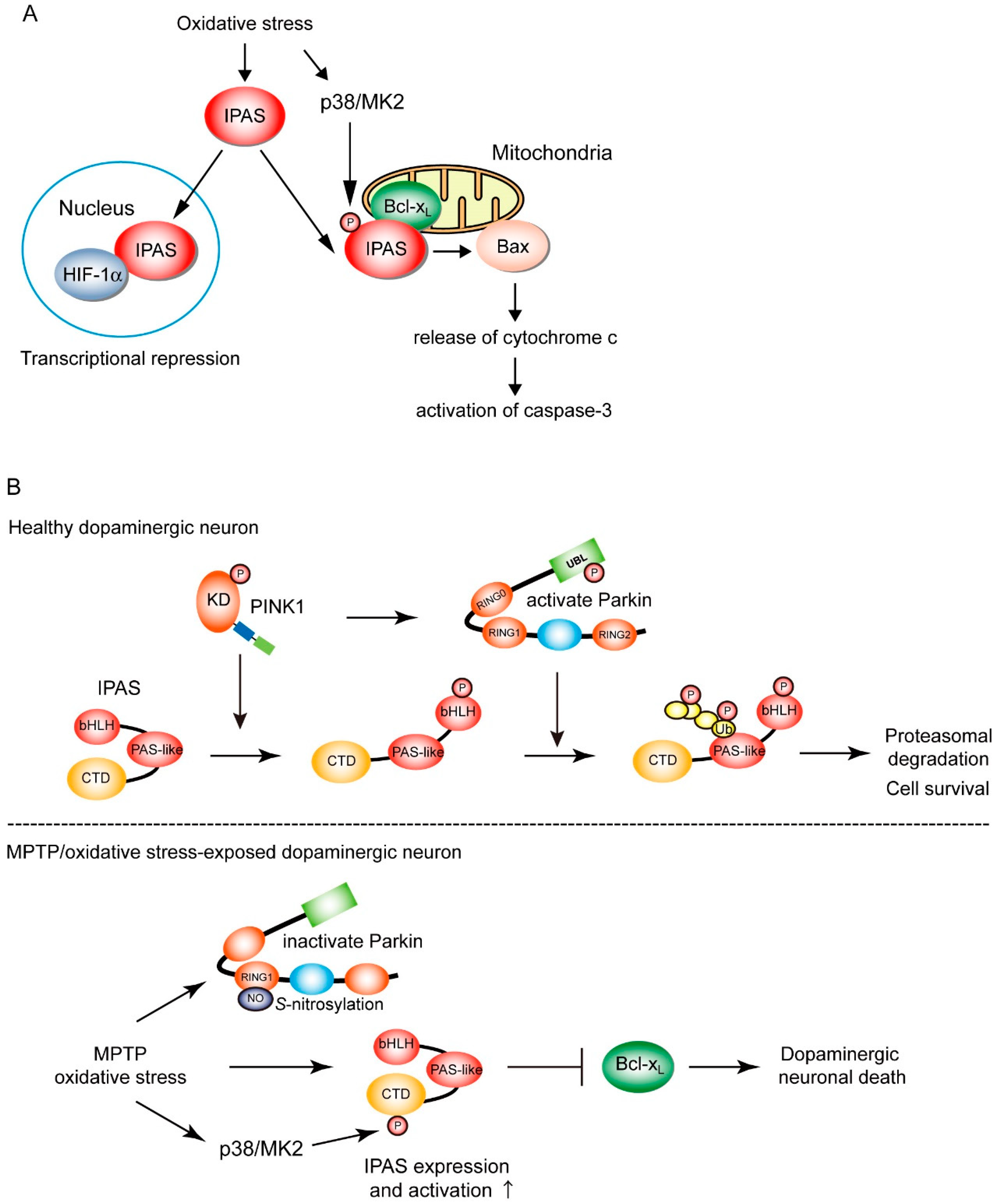
IPAS was first reported as a potent negative regulator of hypoxia-inducible factor-1 (HIF-1) [59], a master regulator of mammalian oxygen homeostasis [60]. IPAS directly binds to HIF-1α, an oxygen-sensitive subunit of HIF-1, preventing their binding to hypoxia response elements (HRE) localized in the transcription-control region of subordinate genes. The binding of IPAS to HIF-1α-like factor (HLF, also known as HIF-2α and EPAS1) has also been observed. IPAS is one of the alternatively spliced variants of HIF-3α, a family member of HIF-1α [61]. Expression of IPAS is highly tissue specific. Makino et al. found that IPAS is constitutively expressed in the Purkinje cells of the cerebellum and corneal epithelium where IPAS was involved in the regulation of angiogenesis [59]. Our observation strongly suggested that IPAS is inducibly expressed in the neurons but not in glial cells of the central nervous system [36]. To our knowledge, IPAS is expressed only in PC12 cells but no other cell culture lines, suggesting the expression of IPAS to be tissue specific [62]. In addition, IPAS is induced by both sustained hypoxia and intermittent hypoxia-induced oxidative stress/NF-κB signaling via the activation of HIF-1 [59,62]. Once induced, IPAS localizes to the nucleus and functions as a negative feedback inhibitor of HIF family-dependent hypoxic responses (Figure 4A). As induction of IPAS is also dependent on the TNF-alpha/NF-κB signaling pathway, there is crosstalk between inflammation pathways and HIF-dependent hypoxic responses [63]. We reported a novel function of IPAS as a pro-apoptotic factor acting on mitochondria [64]. Detailed analyses using subcellular localization studies demonstrate that IPAS is localized to both the nucleus and mitochondria. IPAS has a bipartite-like nuclear localization signal and nuclear export signal in its N- and C-terminal region, respectively, and may shuttle between the nucleus and cytosol [65]. Other HIF-3α variants have nuclear localization signal, but they neither have nuclear export signal nor shuttle between the nucleus and cytosol [66,67]. Mitochondrial IPAS interacts with pro-survival Bcl-2 proteins, including Bcl-xL, Bcl-w, and Mcl-1, but not Bcl-2 and A1a, through its C-terminal domain (CTD). The conformation change in IPAS occurs by binding of Bcl-xL [68]. The physical interactions between IPAS and pro-survival factors results in the dissociation of Bax from pro-survival factor and facilitates the translocation of Bax to mitochondria. Bax translocation leads to the release of cytochrome c from the mitochondria, activation of caspase-3, and finally apoptosis. These findings demonstrate that IPAS is a dual-function protein involved in both transcription repression and apoptosis (Figure 4A), although other HIF-3α variants are involved in transcription regulation [66].
We have sought post-translational modifications of IPAS, by which the pro-apoptotic activities of IPAS may be regulated. We observe that the phosphorylation of IPAS Ser184 by MAP kinase-activated protein kinase 2 (MK2 or MAP-KAPK2), a direct substrate of p38 MAP kinase, enhances the pro-apoptotic activity of IPAS (Figure 4A) [69]. Perinuclear clustering of mitochondria and activation of caspase-3 induced by IPAS expression increased by ultraviolet B irradiation (312 nm), which activates the p38 MAP kinase cascade; this enhancement was completely blocked by a p38 MAP kinase inhibitor, a MK2 inhibitor, and siRNA treatment against MK2. Mass spectrometry analysis revealed that several serine residues including Ser184, Ser219, and Ser223 in the CTD of IPAS are phosphorylated. Of these serine residues, Ser219 and Ser223 match with the p38 MAP kinase consensus sequence, PXpS/TP. The minimal consensus for MK2 phosphorylation has been reported to be RXXpS/T [70]; Ser184 (RMKpS) completely matches with this consensus motif. Among these identified phosphorylation sites, replacement of Ser184 by Ala alone led to a total loss of enhanced perinuclear clustering of mitochondria and the activation of caspase-3 in UVB-irradiated cells. Furthermore, replacement of Ser184 by Asp, a phosphomimetic mutation, led to the enhancement of abnormality of mitochondria and the activation of caspase-3 without UVB irradiation. Notably, the interaction between the IPAS phosphomimetic mutant and Bcl-xL was up-regulated, indicating the phosphorylation of IPAS Ser184 enhances its affinity for pro-survival Bcl-2 family proteins. Collectively, the pro-apoptotic activity of IPAS is regulated by the p38 MAP kinase/MK2 signaling pathway (Figure 4A).
Finally, we investigated physiological and pathological processes in which the pro-apoptotic activity of IPAS is involved. Considering that IPAS is induced by oxidative stress in neural tissues and that the pro-apoptotic activity of mitochondrial IPAS is activated by oxidative stress, we suggest a relationship between IPAS-induced neural cell death and neurodegenerative disorders. PD researchers have identified mitochondrial targets for both PINK1 phosphorylation and Parkin ubiquitination, such as Miro1/Rhot1 and Mitofusin2. Therefore, we hypothesize that IPAS is also a good mitochondrial substrate for PINK1 and Parkin. Our studies confirmed this hypothesis [36,71]. When cultured cells (SH-SY5Y and HeLa cells) were treated with CCCP, in the presence of MG132 to inhibit proteasome activity, mitochondrial IPAS was bound to Parkin via its CTD. IPAS is subsequently ubiquitinated by Parkin at the position lys167–169 in the second PAS-like domain (Figure 4B). The poly-ubiquitin chains of IPAS are predominantly K48 linkages, which are known as a tag for proteasomal degradation of target proteins. K63 linkages on IPAS were not observed after CCCP treatment. The degradation of mitochondrial IPAS, but not nuclear IPAS, was rapidly increased by CCCP treatment. Pathogenic missense mutations of Parkin present in RING0, RING1, and IBR, and RING2 domains exhibited a little or no ubiquitination activity for IPAS. However, the R42P mutant of Parkin enhances IPAS ubiquitination for unknown reasons. Perhaps, Parkin (R42P) may enhance interactions with substrates, as IPAS is markedly bound to Parkin (R42P) without CCCP treatment. Of note, CCCP treatment also increases the interactions between IPAS and PINK1. Moreover, IPAS is phosphorylated by PINK1 at several sites including Thr12. The sequence flanking Thr12 closely matches with the PINK1 phosphorylation consensus motifs (Figure 2). PINK1 siRNA treatment reduces both IPAS phosphorylation and the binding of IPAS to Parkin. Moreover, replacement of Thr12 by Ala leads to reduced interactions between IPAS and Parkin as well as a total loss of IPAS ubiquitination by Parkin; suggesting that IPAS phosphorylation by PINK1 induces conformation changes in IPAS to make its PAS-like domain accessible to Parkin. Treatment of cells with CCCP completely abolished IPAS-induced apoptosis; notably, the inhibition of apoptosis was reversed by a concomitant knockdown of Parkin. Parkin WT, but not a ligase-deficient mutant (T415N), also decreases the IPAS-induced apoptosis. Collectively, these results strongly suggest that IPAS is a mitochondrial substrate in the PINK1-Parkin pathway and is stabilized when PINK1-Parkin activity is decreased. Up-regulation of IPAS leads to apoptosis in neural cells (Figure 4B) [36].
We next sought evidence for causal relationship between IPAS expression and PD pathogenesis. IPAS mRNA and protein are rapidly and strongly induced after at four injections of MPTP. Immunohistochemical analysis indicate that induced IPAS proteins are expressed mainly in the cytoplasm region, but not in nucleus, of tyrosine hydroxylase (TH)-expressing dopaminergic neurons in the SNpc. Therefore, we made mice which lack the IPAS-specific 16th exon. In homozygous mice (IPAS16Δ/16Δ), endogenous IPAS loses its pro-apoptotic activity. The mutant mice were fully viable, fertile, apparently normal in appearance and behavior, and the expression of other HIF-3α splicing variants appeared to be normal. Acute administration of MPTP to IPAS16Δ/16Δ mice caused a modest decrease in the number of TH-positive neurons, whereas MPTP significantly reduced TH-positive neurons in WT littermates. These results demonstrate that the pro-apoptotic activity of IPAS is involved in MPTP-dependent degeneration of dopaminergic neurons in the SNpc.
Some studies demonstrate that MK2-deficient mice are resistant to MPTP-induced neurodegeneration in the SNpc when compared with WT mice [72] and that the selective activation of p38 signaling by MPTP treatment occurs in dopaminergic neurons within the SNpc [73]. From these findings, we speculate that involvement of p38/MK2 signaling in neural death may be derived, at least partly, from the pro-apoptotic activity of IPAS by phosphorylation IPAS Ser184 in the SNpc of mice (Figure 4B). Next, expression levels of IPAS in the SNpc neurons of patients with sporadic PD were investigated [36]. Formalin-fixed, paraffin-embedded sections of the midbrain of six patients and six neurologically normal control individuals were analyzed by immunohistochemistry. The intracellular distribution of IPAS was similar to that observed in the TH-positive neurons of MPTP-treated mice. The intensity of IPAS immunostaining was significantly greater in the neurons of sporadic PD patients versus control individuals. These results suggest that IPAS contributes to the neurodegeneration of dopaminergic neurons in the SNpc of patients with sporadic PD as well as MPTP-treated mouse PD models (Figure 4B) [36].

4. Conclusions

In this review, we first focus on well-characterized PINK1 phosphorylation motifs. We also describe our findings for Parkin-Tg expression in mnd2 mouse models. Although Parkin protein levels in the striatum are decreased in mnd2 mice, the Parkin transgene did not rescue the neurodegenerative disorders in mnd2 mice. Recently, new mutations of CHCHD2 have been identified in familial and sporadic PD cases [11]. Although CHCHD2 primarily localizes to the intermembrane spaces of mitochondria, a pathogenic point mutation of CHCHD2 induces the translocation of CHCHD2 into cytosol where it aggregates with α-synuclein [74]. Under these conditions, the possibility exists that Parkin-Tg can prevent neurodegeneration by inhibiting aggregation of CHCHD2 and α-synuclein. Our Parkin-Tg mice are a powerful tool for elucidating these questions. Third, we describe our findings regarding IPAS, a newly identified target of the PINK1/Parkin pathway. IPAS is a bi-organellar factor with roles in nuclear transcription repression and mitochondrial pro-apoptotic function. This conversion is controlled by its subcellular localization via binding to anchoring proteins, and post-translational modification by other signaling pathways, such as p38 MAP kinase/MK2 signaling. The next challenge is a finding of the nuclear/mitochondrial anchoring proteins for IPAS by which IPAS bi-organellar function is regulated. Experiments using knockout mice lacking these proteins will dramatically enhance our understanding of novel pathogenic mechanism in PD.

Author Contributions

S.T. wrote the manuscript; S.K., T.Y., K.-i.Y., and S.S. edited the manuscript. All authors have read and agreed to the published version of the manuscript.

Funding

This research was funded by a Grant-in-Aid for Scientific Research (C) (26430051, 18K06210), Grant-in-Aid for young scientist in TMDU and Grant-in-Aid for studies of the intractable diseases in Medical Research Institute in TMDU.

Acknowledgments

We are grateful to K. Sogawa for valuable suggestions. We thank members of laboratory of Tohoku University and TMDU for their helpful discussion.

Conflicts of Interest

The authors declare no conflict of interest.

References

  1. Exner, N.; Lutz, A.K.; Haass, C.; Winklhofer, K.F. Mitochondrial dysfunction in Parkinson’s disease: Molecular mechanisms and pathophysiological consequences. EMBO J. 2012, 31, 3038–3062. [Google Scholar] [CrossRef] [PubMed] [Green Version]
  2. Moon, H.E.; Paek, S.H. Mitochondrial dysfunction in Parkinson’s disease. Exp. Neurobiol. 2015, 24, 103–116. [Google Scholar] [CrossRef] [PubMed]
  3. Klein, C.; Westenberger, A. Genetics of Parkinson’s disease. Cold Spring Harb. Perspect. Med. 2012, 2, a008888. [Google Scholar] [CrossRef] [PubMed] [Green Version]
  4. Langston, J.W.; Ballard, P.; Tetrud, J.W.; Irwin, I. Chronic Parkinsonism in humans due to a product of meperidine-analog synthesis. Science 1983, 219, 979–980. [Google Scholar] [CrossRef] [Green Version]
  5. Singer, T.P.; Ramsay, R.R.; McKeown, K.; Trevor, A.; Castagnoli, N.E., Jr. Mechanism of the neurotoxicity of 1-methyl-4-phenylpyridinium (MPP+), the toxic bioactivation product of 1-methyl-4-phenyl-1,2,3,6-tetrahydropyridine (MPTP). Toxicology 1988, 49, 17–23. [Google Scholar] [CrossRef]
  6. Javitch, J.A.; D’Amato, R.J.; Strittmatter, S.M.; Snyder, S.H. Parkinsonism-inducing neurotoxin, N-methyl-4-phenyl-1,2,3,6-tetrahydropyridine: Uptake of the metabolite N-methyl-4-phenylpyridine by dopamine neurons explains selective toxicity. Proc. Natl. Acad. Sci. USA 1985, 82, 2173–2177. [Google Scholar] [CrossRef] [Green Version]
  7. Berry, C.; La Vecchia, C.; Nicotera, P. Paraquat and Parkinson’s disease. Cell Death Differ. 2010, 17, 1115–1125. [Google Scholar] [CrossRef] [Green Version]
  8. Bove, J.; Perier, C. Neurotoxin-based models of Parkinson’s disease. Neuroscience 2012, 211, 51–76. [Google Scholar] [CrossRef]
  9. Jackson-Lewis, V.; Przedborski, S. Protocol for the MPTP mouse model of Parkinson’s disease. Nat. Protoc. 2007, 2, 141–151. [Google Scholar] [CrossRef]
  10. Chiba, K.; Horii, H.; Kubota, E.; Ishizaki, T.; Kato, Y. Effects of N-methylmercaptoimidazole on the disposition of MPTP and its metabolites in mice. Eur. J. Pharmacol. 1990, 180, 59–67. [Google Scholar] [CrossRef]
  11. Funayama, M.; Ohe, K.; Amo, T.; Furuya, N.; Yamaguchi, J.; Saiki, S.; Li, Y.; Ogaki, K.; Ando, M.; Yoshino, H.; et al. CHCHD2 mutations in autosomal dominant late-onset Parkinson’s disease: A genome-wide linkage and sequencing study. Lancet Neurol. 2015, 14, 274–282. [Google Scholar] [CrossRef]
  12. Jones, J.M.; Datta, P.; Srinivasula, S.M.; Ji, W.; Gupta, S.; Zhang, Z.; Davies, E.; Hajnoczky, G.; Saunders, T.L.; Van Keuren, M.L.; et al. Loss of Omi mitochondrial protease activity causes the neuromuscular disorder of mnd2 mutant mice. Nature 2003, 425, 721–727. [Google Scholar] [CrossRef] [PubMed] [Green Version]
  13. Matsuda, N. Phospho-ubiquitin: Upending the PINK-Parkin-ubiquitin cascade. J. Biochem. 2016, 159, 379–385. [Google Scholar] [CrossRef] [PubMed] [Green Version]
  14. Truban, D.; Hou, X.; Caulfield, T.R.; Fiesel, F.C.; Springer, W. PINK1, parkin, and mitochondrial quality control: What can we learn about Parkinson’s disease pathobiology? J. Parkinson’s Dis. 2017, 7, 13–29. [Google Scholar] [CrossRef] [PubMed] [Green Version]
  15. Imai, Y.; Meng, H.; Shiba-Fukushima, K.; Hattori, N. Twin CHCH proteins, CHCHD2, and CHCHD10: Key molecules of Parkinson’s disease, amyotrophic lateral sclerosis, and frontotemporal dementia. Int. J. Mol. Sci. 2019, 20, 908. [Google Scholar] [CrossRef] [Green Version]
  16. Meng, H.; Yamashita, C.; Shiba-Fukushima, K.; Inoshita, T.; Funayama, M.; Sato, S.; Hatta, T.; Natsume, T.; Umitsu, M.; Takagi, J.; et al. Loss of Parkinson’s disease-associated protein CHCHD2 affects mitochondrial crista structure and destabilizes cytochrome c. Nat. Commun. 2017, 8, 15500. [Google Scholar] [CrossRef] [Green Version]
  17. Sim, C.H.; Gabriel, K.; Mills, R.D.; Culvenor, J.G.; Cheng, H.C. Analysis of the regulatory and catalytic domains of PTEN-induced kinase-1 (PINK1). Hum. Mutat. 2012, 33, 1408–1422. [Google Scholar] [CrossRef]
  18. Matsuda, N.; Sato, S.; Shiba, K.; Okatsu, K.; Saisho, K.; Gautier, C.A.; Sou, Y.S.; Saiki, S.; Kawajiri, S.; Sato, F.; et al. PINK1 stabilized by mitochondrial depolarization recruits Parkin to damaged mitochondria and activates latent Parkin for mitophagy. J. Cell Biol. 2010, 189, 211–221. [Google Scholar] [CrossRef]
  19. Deas, E.; Plun-Favreau, H.; Gandhi, S.; Desmond, H.; Kjaer, S.; Loh, S.H.; Renton, A.E.; Harvey, R.J.; Whitworth, A.J.; Martins, L.M.; et al. PINK1 cleavage at position A103 by the mitochondrial protease PARL. Hum. Mol. Genet. 2011, 20, 867–879. [Google Scholar] [CrossRef]
  20. Kondapalli, C.; Kazlauskaite, A.; Zhang, N.; Woodroof, H.I.; Campbell, D.G.; Gourlay, R.; Burchell, L.; Walden, H.; Macartney, T.J.; Deak, M.; et al. PINK1 is activated by mitochondrial membrane potential depolarization and stimulates Parkin E3 ligase activity by phosphorylating Serine 65. Open Biol. 2012, 2, 120080. [Google Scholar] [CrossRef] [Green Version]
  21. Shiba-Fukushima, K.; Imai, Y.; Yoshida, S.; Ishihama, Y.; Kanao, T.; Sato, S.; Hattori, N. PINK1-mediated phosphorylation of the Parkin ubiquitin-like domain primes mitochondrial translocation of Parkin and regulates mitophagy. Sci. Rep. 2012, 2, 1002. [Google Scholar] [CrossRef] [PubMed]
  22. Koyano, F.; Okatsu, K.; Kosako, H.; Tamura, Y.; Go, E.; Kimura, M.; Kimura, Y.; Tsuchiya, H.; Yoshihara, H.; Hirokawa, T.; et al. Ubiquitin is phosphorylated by PINK1 to activate parkin. Nature 2014, 510, 162–166. [Google Scholar] [CrossRef] [PubMed]
  23. Shiba-Fukushima, K.; Arano, T.; Matsumoto, G.; Inoshita, T.; Yoshida, S.; Ishihama, Y.; Ryu, K.Y.; Nukina, N.; Hattori, N.; Imai, Y. Phosphorylation of mitochondrial polyubiquitin by PINK1 promotes Parkin mitochondrial tethering. PLoS Genet. 2014, 10, e1004861. [Google Scholar] [CrossRef] [PubMed]
  24. Bingol, B.; Tea, J.S.; Phu, L.; Reichelt, M.; Bakalarski, C.E.; Song, Q.; Foreman, O.; Kirkpatrick, D.S.; Sheng, M. The mitochondrial deubiquitinase USP30 opposes parkin-mediated mitophagy. Nature 2014, 510, 370–375. [Google Scholar] [CrossRef] [PubMed]
  25. Cornelissen, T.; Haddad, D.; Wauters, F.; Van Humbeeck, C.; Mandemakers, W.; Koentjoro, B.; Sue, C.; Gevaert, K.; De Strooper, B.; Verstreken, P.; et al. The deubiquitinase USP15 antagonizes Parkin-mediated mitochondrial ubiquitination and mitophagy. Hum. Mol. Genet. 2014, 23, 5227–5242. [Google Scholar] [CrossRef] [Green Version]
  26. Malik, B.R.; Maddison, D.C.; Smith, G.A.; Peters, O.M. Autophagic and endo-lysosomal dysfunction in neurodegenerative disease. Mol. Brain 2019, 12, 100. [Google Scholar] [CrossRef]
  27. Ravenhill, B.J.; Boyle, K.B.; von Muhlinen, N.; Ellison, C.J.; Masson, G.R.; Otten, E.G.; Foeglein, A.; Williams, R.; Randow, F. The cargo receptor NDP52 initiates selective autophagy by recruiting the ULK complex to cytosol-invading bacteria. Mol. Cell 2019, 74, 320–329. [Google Scholar] [CrossRef] [Green Version]
  28. Yamano, K.; Wang, C.; Sarraf, S.A.; Munch, C.; Kikuchi, R.; Noda, N.N.; Hizukuri, Y.; Kanemaki, M.T.; Harper, W.; Tanaka, K.; et al. Endosomal Rab cycles regulate Parkin-mediated mitophagy. eLife 2018, 7, e31326. [Google Scholar] [CrossRef]
  29. Iwashita, H.; Torii, S.; Nagahora, N.; Ishiyama, M.; Shioji, K.; Sasamoto, K.; Shimizu, S.; Okuma, K. Live cell imaging of mitochondrial autophagy with a novel fluorescent small molecule. ACS Chem. Biol. 2017, 12, 2546–2551. [Google Scholar] [CrossRef]
  30. Plun-Favreau, H.; Klupsch, K.; Moisoi, N.; Gandhi, S.; Kjaer, S.; Frith, D.; Harvey, K.; Deas, E.; Harvey, R.J.; McDonald, N.; et al. The mitochondrial protease HtrA2 is regulated by Parkinson’s disease-associated kinase PINK1. Nat. Cell Biol. 2007, 9, 1243–1252. [Google Scholar] [CrossRef]
  31. Pridgeon, J.W.; Olzmann, J.A.; Chin, L.S.; Li, L. PINK1 protects against oxidative stress by phosphorylating mitochondrial chaperone TRAP1. PLoS Biol. 2007, 5, e172. [Google Scholar] [CrossRef] [PubMed]
  32. Zhou, C.; Huang, Y.; Shao, Y.; May, J.; Prou, D.; Perier, C.; Dauer, W.; Schon, E.A.; Przedborski, S. The kinase domain of mitochondrial PINK1 faces the cytoplasm. Proc. Natl. Acad. Sci. USA 2008, 105, 12022–12027. [Google Scholar] [CrossRef] [PubMed] [Green Version]
  33. Wang, X.; Winter, D.; Ashrafi, G.; Schlehe, J.; Wong, Y.L.; Selkoe, D.; Rice, S.; Steen, J.; LaVoie, M.J.; Schwarz, T.L. PINK1 and Parkin target Miro for phosphorylation and degradation to arrest mitochondrial motility. Cell 2011, 147, 893–906. [Google Scholar] [CrossRef] [Green Version]
  34. Okatsu, K.; Oka, T.; Iguchi, M.; Imamura, K.; Kosako, H.; Tani, N.; Kimura, M.; Go, E.; Koyano, F.; Funayama, M.; et al. PINK1 autophosphorylation upon membrane potential dissipation is essential for Parkin recruitment to damaged mitochondria. Nat. Commun. 2012, 3, 1016. [Google Scholar] [CrossRef] [Green Version]
  35. Chen, Y.; Dorn, G.W., II. PINK1-phosphorylated mitofusin 2 is a Parkin receptor for culling damaged mitochondria. Science 2013, 340, 471–475. [Google Scholar] [CrossRef] [Green Version]
  36. Torii, S.; Kasai, S.; Suzuki, A.; Todoroki, Y.; Yokozawa, K.; Yasumoto, K.; Seike, N.; Kiyonari, H.; Mukumoto, Y.; Kakita, A.; et al. Involvement of inhibitory PAS domain protein in neuronal cell death in Parkinson’s disease. Cell Death Discov. 2015, 1, 15015. [Google Scholar] [CrossRef] [Green Version]
  37. Lee, Y.; Stevens, D.A.; Kang, S.U.; Jiang, H.; Lee, Y.I.; Ko, H.S.; Scarffe, L.A.; Umanah, G.E.; Kang, H.; Ham, S.; et al. PINK1 primes parkin-mediated ubiquitination of PARIS in dopaminergic neuronal survival. Cell Rep. 2017, 18, 918–932. [Google Scholar] [CrossRef] [Green Version]
  38. Tsai, P.I.; Lin, C.H.; Hsieh, C.H.; Papakyrikos, A.M.; Kim, M.J.; Napolioni, V.; Schoor, C.; Couthouis, J.; Wu, R.M.; Wszolek, Z.K.; et al. PINK1 phosphorylates MIC60/Mitofilin to control structural plasticity of mitochondrial crista junctions. Mol. Cell 2019, 69, 744–756. [Google Scholar] [CrossRef] [Green Version]
  39. Kinoshita, E.; Kinoshita-Kikuta, E.; Takiyama, K.; Koike, T. Phosphate-binding tag, a new tool to visualize phosphorylated proteins. Mol. Cell. Proteom. 2006, 5, 749–757. [Google Scholar] [CrossRef] [Green Version]
  40. Yoshida, T.; Mizuta, T.; Shimizu, S. Neurodegeneration in mnd2 mutant mice is not prevented by parkin transgene. Biochem. Biophys. Res. Commun. 2010, 402, 676–679. [Google Scholar] [CrossRef]
  41. Faccio, L.; Fusco, C.; Chen, A.; Martinotti, S.; Bonventre, J.V.; Zervos, A.S. Characterization of a novel human serine protease that has extensive homology to bacterial heat shock endoprotease HtrA and is regulated by kidney ischemia. J. Biol. Chem. 2000, 275, 2581–2588. [Google Scholar] [CrossRef] [PubMed] [Green Version]
  42. Fitzgerald, J.C.; Camprubi, M.D.; Dunn, L.; Wu, H.C.; Ip, N.Y.; Kruger, R.; Martins, L.M.; Wood, N.W.; Plun-Favreau, H. Phosphorylation of HtrA2 by cyclin-dependent kinase-5 is important for mitochondrial function. Cell Death Differ. 2012, 19, 257–266. [Google Scholar] [CrossRef] [PubMed] [Green Version]
  43. Suzuki, Y.; Imai, Y.; Nakayama, H.; Takahashi, K.; Takio, K.; Takahashi, R. A serine protease, HtrA2, is released from the mitochondria and interacts with XIAP, inducing cell death. Mol. Cell 2001, 8, 613–621. [Google Scholar] [CrossRef]
  44. Martins, L.M.; Iaccarino, I.; Tenev, T.; Gschmeissner, S.; Totty, N.F.; Lemoine, N.R.; Savopoulos, J.; Gray, C.W.; Creasy, C.L.; Dingwall, C.; et al. The serine protease Omi/HtrA2 regulates apoptosis by binding XIAP through a reaper-like motif. J. Biol. Chem. 2002, 277, 439–444. [Google Scholar] [CrossRef] [Green Version]
  45. Jones, J.M.; Albin, R.L.; Feldman, E.L.; Simin, K.; Schuster, T.G.; Dunnick, W.A.; Collins, J.T.; Chrisp, C.E.; Taylor, B.A.; Meisler, M.H. mnd2: A new mouse model of inherited motor neuron disease. Genomics 1993, 16, 669–677. [Google Scholar] [CrossRef] [Green Version]
  46. Rathke-Hartlieb, S.; Schlomann, U.; Heimann, P.; Meisler, M.H.; Jockusch, H.; Bartsch, J.W. Progressive loss of striatal neurons causes motor dysfunction in MND2 mutant mice and is not prevented by Bcl-2. Exp. Neurol. 2002, 175, 87–97. [Google Scholar] [CrossRef]
  47. Ideguchi, K.; Shimizu, S.; Okumura, M.; Tsujimoto, Y. Cyclophilin D-dependent mitochondrial permeability transition is not involved in neurodegeneration in mnd2 mutant mice. Biochem. Biophys. Res. Commun. 2010, 393, 264–267. [Google Scholar] [CrossRef]
  48. Vercammen, L.; Van der Perren, A.; Vaudano, E.; Gijsbers, R.; Debyser, Z.; Van den Haute, C.; Baekelandt, V. Parkin protects against neurotoxicity in the 6-hydroxydopamine rat model for Parkinson’s disease. Mol. Ther. 2006, 14, 716–723. [Google Scholar] [CrossRef]
  49. Petrucelli, L.; O’Farrell, C.; Lockhart, P.J.; Baptista, M.; Kehoe, K.; Vink, L.; Choi, P.; Wolozin, B.; Farrer, M.; Hardy, J.; et al. Parkin protects against the toxicity associated with mutant alpha-synuclein: Proteasome dysfunction selectively affects catecholaminergic neurons. Neuron 2002, 36, 1007–1019. [Google Scholar] [CrossRef] [Green Version]
  50. Manfredsson, F.P.; Burger, C.; Sullivan, L.F.; Muzyczka, N.; Lewin, A.S.; Mandel, R.J. rAAV-mediated nigral human parkin over-expression partially ameliorates motor deficits via enhanced dopamine neurotransmission in a rat model of Parkinson’s disease. Exp. Neurol. 2007, 207, 289–301. [Google Scholar] [CrossRef]
  51. Klein, R.L.; Dayton, R.D.; Henderson, K.M.; Petrucelli, L. Parkin is protective for substantia nigra dopamine neurons in a tau gene transfer neurodegeneration model. Neurosci. Lett. 2006, 401, 130–135. [Google Scholar] [CrossRef] [Green Version]
  52. Hattori, N.; Mizuno, Y. Twenty years since the discovery of the parkin gene. J. Neural Transm. (Vienna) 2017, 124, 1037–1054. [Google Scholar] [CrossRef]
  53. Imai, Y.; Soda, M.; Inoue, H.; Hattori, N.; Mizuno, Y.; Takahashi, R. An unfolded putative transmembrane polypeptide, which can lead to endoplasmic reticulum stress, is a substrate of Parkin. Cell 2001, 105, 891–902. [Google Scholar] [CrossRef] [Green Version]
  54. Sul, J.W.; Park, M.Y.; Shin, J.; Kim, Y.R.; Yoo, S.E.; Kong, Y.Y.; Kwon, K.S.; Lee, Y.H.; Kim, E. Accumulation of the parkin substrate, FAF1, plays a key role in the dopaminergic neurodegeneration. Hum. Mol. Genet. 2013, 22, 1558–1573. [Google Scholar] [CrossRef] [Green Version]
  55. Gao, F.; Chen, D.; Si, J.; Hu, Q.; Qin, Z.; Fang, M.; Wang, G. The mitochondrial protein BNIP3L is the substrate of PARK2 and mediates mitophagy in PINK1/PARK2 pathway. Hum. Mol. Genet. 2015, 24, 2528–2538. [Google Scholar] [CrossRef] [Green Version]
  56. Koyano, F.; Yamano, K.; Kosako, H.; Kimura, Y.; Kimura, M.; Fujiki, Y.; Tanaka, K.; Matsuda, N. Parkin-mediated ubiquitylation redistributes MITOL/March5 from mitochondria to peroxisomes. EMBO Rep. 2019, 20, e47728. [Google Scholar] [CrossRef]
  57. Yao, D.; Gu, Z.; Nakamura, T.; Shi, Z.Q.; Ma, Y.; Gaston, B.; Palmer, L.A.; Rockenstein, E.M.; Zhang, Z.; Masliah, E.; et al. Nitrosative stress linked to sporadic Parkinson’s disease: S-nitrosylation of parkin regulates its E3 ubiquitin ligase activity. Proc. Natl. Acad. Sci. USA 2004, 101, 10810–10814. [Google Scholar] [CrossRef] [Green Version]
  58. Imai, Y.; Inoue, H.; Kataoka, A.; Hua-Qin, W.; Masuda, M.; Ikeda, T.; Tsukita, K.; Soda, M.; Kodama, T.; Fuwa, T.; et al. Pael receptor is involved in dopamine metabolism in the nigrostriatal system. Neurosci. Res. 2007, 59, 413–425. [Google Scholar] [CrossRef]
  59. Makino, Y.; Cao, R.; Svensson, K.; Bertilsson, G.; Asman, M.; Tanaka, H.; Cao, Y.; Berkenstam, A.; Poellinger, L. Inhibitory PAS domain protein is a negative regulator of hypoxia-inducible gene expression. Nature 2001, 414, 550–554. [Google Scholar] [CrossRef]
  60. Hirota, K.; Semenza, G.L. Regulation of hypoxia-inducible factor 1 by prolyl and asparaginyl hydroxylases. Biochem. Biophys. Res. Commun. 2005, 338, 610–616. [Google Scholar] [CrossRef]
  61. Makino, Y.; Uenishi, R.; Okamoto, K.; Isoe, T.; Hosono, O.; Tanaka, H.; Kanopka, A.; Poellinger, L.; Haneda, M.; Morimoto, C. Transcriptional up-regulation of inhibitory PAS domain protein gene expression by hypoxia-inducible factor 1 (HIF-1): A negative feedback regulatory circuit in HIF-1-mediated signaling in hypoxic cells. J. Biol. Chem. 2007, 282, 14073–14082. [Google Scholar] [CrossRef] [PubMed] [Green Version]
  62. Torii, S.; Kobayashi, K.; Takahashi, M.; Katahira, K.; Goryo, K.; Matsushita, N.; Yasumoto, K.; Fujii-Kuriyama, Y.; Sogawa, K. Magnesium deficiency causes loss of response to intermittent hypoxia in paraganglion cells. J. Biol. Chem. 2009, 284, 19077–19089. [Google Scholar] [CrossRef] [PubMed] [Green Version]
  63. Goryo, K.; Torii, S.; Yasumoto, K.; Sogawa, K. Tumour necrosis factor-alpha suppresses the hypoxic response by NF-kappaB-dependent induction of inhibitory PAS domain protein in PC12 cells. J. Biochem. 2011, 150, 311–318. [Google Scholar] [CrossRef] [PubMed]
  64. Torii, S.; Goto, Y.; Ishizawa, T.; Hoshi, H.; Goryo, K.; Yasumoto, K.; Fukumura, H.; Sogawa, K. Pro-apoptotic activity of inhibitory PAS domain protein (IPAS), a negative regulator of HIF-1, through binding to pro-survival Bcl-2 family proteins. Cell Death Differ. 2011, 18, 1711–1725. [Google Scholar] [CrossRef] [Green Version]
  65. Torii, S.; Sakaki, K.; Otomo, M.; Saka, K.; Yasumoto, K.; Sogawa, K. Nucleocytoplasmic shuttling of IPAS by its unique nuclear import and export signals unshared with other HIF-3alpha splice variants. J. Biochem. 2013, 154, 561–567. [Google Scholar] [CrossRef]
  66. Ravenna, L.; Salvatori, L.; Russo, M.A. HIF3alpha: The little we know. FEBS J. 2016, 283, 993–1003. [Google Scholar] [CrossRef] [Green Version]
  67. Yao, Q.; Zhang, P.; Lu, L.; Liu, Y.; Li, Y.; Duan, C. Nuclear localization of Hif-3alpha requires two redundant NLS motifs in its unique C-terminal region. FEBS Lett. 2018, 592, 2769–2775. [Google Scholar] [CrossRef] [Green Version]
  68. Kasai, S.; Kajimoto, S.; Ito, Y.; Saito, T.; Yasumoto, K.; Tokunaga, M.; Sakata-Sogawa, K.; Fukumura, H.; Sogawa, K. Conformational changes in inhibitory PAS domain protein associated with binding of HIF-1alpha and Bcl-xL in living cells. J. Biochem. 2017, 161, 291–296. [Google Scholar]
  69. Kasai, S.; Richardson, M.J.E.; Torii, S.; Yasumoto, K.; Shima, H.; Igarashi, K.; Itoh, K.; Sogawa, K.; Murayama, K. Increase in proapoptotic activity of inhibitory PAS domain protein via phosphorylation by MK2. FEBS J. 2017, 284, 4115–4127. [Google Scholar] [CrossRef]
  70. Yamamoto, H.; Higa-Nakamine, S.; Noguchi, N.; Maeda, N.; Kondo, Y.; Toku, S.; Kukita, I.; Sugahara, K. Desensitization by different strategies of epidermal growth factor receptor and ErbB4. J. Pharmacol. Sci. 2014, 124, 287–293. [Google Scholar] [CrossRef] [Green Version]
  71. Kasai, S.; Torii, S.; Kakita, A.; Sogawa, K. Inhibitory PAS domain protein is a substrate of PINK1 and Parkin and mediates cell death in a Parkinson’s disease model. Cell Death Dis. 2015, 6, e1886. [Google Scholar] [CrossRef] [Green Version]
  72. Thomas, T.; Timmer, M.; Cesnulevicius, K.; Hitti, E.; Kotlyarov, A.; Gaestel, M. MAPKAP kinase 2-deficiency prevents neurons from cell death by reducing neuroinflammation—Relevance in a mouse model of Parkinson’s disease. J. Neurochem. 2008, 105, 2039–2052. [Google Scholar] [CrossRef]
  73. Karunakaran, S.; Saeed, U.; Mishra, M.; Valli, R.K.; Joshi, S.D.; Meka, D.P.; Seth, P.; Ravindranath, V. Selective activation of p38 mitogen-activated protein kinase in dopaminergic neurons of substantia nigra leads to nuclear translocation of p53 in 1-methyl-4-phenyl-1,2,3,6-tetrahydropyridine-treated mice. J. Neurosci. 2008, 28, 12500–12509. [Google Scholar] [CrossRef]
  74. Ikeda, A.; Nishioka, K.; Meng, H.; Takanashi, M.; Inoshita, T.; Shiba-Fukushima, K.; Li, Y.; Yoshino, H.; Mori, A.; Okuzumi, A.; et al. Mutations in CHCHD2 cause alpha-synuclein aggregation. Hum. Mol. Genet. 2019, 28, 3895–3911. [Google Scholar] [CrossRef]
Figure 1. Structures of mitochondrial-localized proteins for genes associated with familial parkinsonism and their phosphorylation sites. The human Parkin, PINK1, HtrA2/Omi, and CHCHD2 proteins are drawn approximately to scale, with some pathogenic point mutations in red. Parkin contains a UBL domain at the N-terminus followed by three really-interesting-new-gene (RING) finger domains (R0-R2) separated by In-Between-RING fingers (IBR) and repressor element of Parkin (REP) domains. A phosphorylation site for PINK1 occurs at Ser65. PINK1 contains an MTS, transmembrane domain (TM), the kinase domain, and C-terminal sequences of uncertain function. Autophosphorylation sites are at Ser228 and Ser402. HtrA2/Omi consists of an MTS, TM, and a conserved catalytic trypsin-like serine protease domain, and a C-terminal PSD-95/Dlg/ZO-1 (PDZ) domain. Ser142 is phosphorylated by p38 in a PINK1-depedent manner and Ser400 is phosphorylated by CDK5. CHCHD2 has an MTS, TM, and CHCH domain. The CHCH domain consists of twin CX(9)C motifs and two disulfide bonds that stabilize the CHCH fold.
Ijms 21 01202 g001
Figure 2. Alignment of amino acid sequences containing the phosphorylation sites of several PINK1 substrates. The known Ser or Thr residues phosphorylated by PINK1 (blue box) are indicated by an arrowhead with phosphorylation (P). Protein sequences of Mus musculus are presented. Hydrophobic amino acids (green box) and alanine (yellow box) found two residues (+2) after phosphorylation sites are shown. Hydrophobic amino acids or acidic amino acids at position +1 after the phosphorylation site are preferred. Basic amino acids found before the phosphorylation sites are shown (red box). Phosphorylation sites of drosophila MIC60/Mitofilin are not included because phosphorylation sites of mouse MIC60/Mitofilin corresponding to drosophila MIC60/Mitofilin cannot be identified using homology searches. From these observations, an optimal PINK1 target motif is given at the top using one-letter codes (ф: hydrophobic amino acids).
Ijms 21 01202 g002
Figure 3. Neurodegeneration in mnd2 mutant mice is not prevented by Parkin-Tg. (A) Mnd2 mice have a non-functional missense mutation at Ser276 to Cys in the conserved catalytic trypsin-like serine protease domain of HtrA2/Omi which leads to motor neuron degeneration and selective loss of striatal neurons. In mnd2 mice, Parkin protein expression is low in the striatum. Parkin protein levels in the hippocampus are also decreased in mnd2 mice; the decrease is significantly weaker than that in striatum. It was unknown that low level of Parkin causes motor abnormalities and striatal neuronal loss in mnd2 mice before performing experiments with Parkin-Tg/ mnd2 mice (an arrow with a question mark). (B) Overexpression of Parkin by Parkin-Tg does not suppress PD phenotypes in mnd2 mice. Arrow and dashed arrow tracks indicate cause-and-effect relationships. Down-arrows and an up-arrow indicate low level and high level, respectively.
Ijms 21 01202 g003
Figure 4. The PINK1-Parkin pathway regulates IPAS expression and inhibits neuronal death. (A) A model for the action of IPAS in transcriptional repression and apoptosis. Nuclear IPAS is bound to HIF-1α and inhibits hypoxic responses by inhibiting HIF-1α transcriptional activity. Mitochondrial IPAS is bound to Bcl-xL, Mcl-1, and A1a on the mitochondria and blocks the interaction of these proteins with Bax; thus activating of Bax, followed by release of cytochrome c and activation of caspase-3. p38-activated MK2 phosphorylates IPAS at Ser184, activating the pro-apoptotic functions of IPAS. IPAS has dual functions involved in the transcription repression of HIF-1α and cell death. (B) In healthy neurons, IPAS Thr12 is phosphorylated by PINK1 and then ubiquitinated by Parkin at lys167–169 in the PAS-like region, leading to the proteasomal degradation of IPAS. Phosphorylation of IPAS at Thr12 may induce a conformation change of IPAS to make the PAS-like region accessible to Parkin (top). The dopaminergic neurotoxin, MPTP, and other oxidative stresses strongly induce IPAS expression in the SNpc. Parkin is inhibited by S-nitrosylation or its ligase-dead pathologic mutation in PD. p38-activated MK2 may phosphorylate IPAS at Ser184 under these conditions. These events activate IPAS pro-apoptotic activity which leads to dopaminergic neuronal death in the SNpc (bottom). KD: kinase domain. Arrow tracks indicate signal transduction or IPAS translocation to nucleus/mitochondria. An up-arrow indicates high level of expression and activation.
Ijms 21 01202 g004