Article

Analysis of Aldo–Keto Reductase Gene Family and Their Responses to Salt, Drought, and Abscisic Acid Stresses in Medicago truncatula

Institute of Animal Sciences, Chinese Academy of Agricultural Sciences, Beijing 100193, China
*
Authors to whom correspondence should be addressed.
These authors contributed equally to this work.
Int. J. Mol. Sci. 2020, 21(3), 754; https://doi.org/10.3390/ijms21030754
Submission received: 31 October 2019 / Revised: 8 January 2020 / Accepted: 16 January 2020 / Published: 23 January 2020
(This article belongs to the Special Issue Abiotic Stress and Gene Networks in Plants 2020)

Abstract

:
Salt and drought stresses are two primary abiotic stresses that inhibit growth and reduce the activity of photosynthetic apparatus in plants. Abscisic acid (ABA) plays a key role in abiotic stress regulation in plants. Some aldo–keto reductases (AKRs) can enhance various abiotic stresses resistance by scavenging cytotoxic aldehydes in some plants. However, there are few comprehensive reports of plant AKR genes and their expression patterns in response to abiotic stresses. In this study, we identified 30 putative AKR genes from Medicago truncatula. The gene characteristics, coding protein motifs, and expression patterns of these MtAKRs were analyzed to explore and identify candidate genes in regulation of salt, drought, and ABA stresses. The phylogenetic analysis result indicated that the 52 AKRs in Medicago truncatula and Arabidopsis thaliana can be divided into three groups and six subgroups. Fifteen AKR genes in M. truncatula were randomly selected from each group or subgroup, to investigate their response to salt (200 mM of NaCl), drought (50 g·L−1 of PEG 6000), and ABA (100 µM) stresses in both leaves and roots. The results suggest that MtAKR1, MtAKR5, MtAKR11, MtAKR14, MtAKR20, and MtAKR29 may play important roles in response to these stresses.

1. Introduction

Biotic and abiotic stresses can disrupt the balance between endogenous levels of reactive oxygen species and antioxidant-induced injury to plants [1,2]. Drought and soil salinity are the most destructive abiotic stresses affecting agriculture [3]. Soil salinity is one of the main factors limiting crop yield in arid and semiarid regions [4,5]. Climate changes might be leading to more severe abiotic stress [6]. The effect of abscisic acid (ABA) to mediate drought and salt stress has been extensively studied. ABA facilitates plant regulation by inducing the expression of genes encoding proteins and enzymes involved in cell dehydration tolerance [7]. Plants have to respond to periodic and unpredictable environmental stresses during growth and development. Plants try to detoxify reactive oxygen species—reactive aldehydes induced by stress to less toxic alcohols or carboxylic acids by upregulating expression a few of enzymes with oxido-reductase activity. There are three protein super families capable of oxidoreduction of a wide range of aldehyde and keto substrates were convened in these reactions: Short-chain dehydrogenases/reductases (SDRs), long-chain alcohol/aldehyde dehydrogenases (ADHs), and aldo–keto reductases (AKRs) [8,9]. The AKRs, part of the oxido-reductase super family, are stress-regulated genes and play an important role in the cellular response to electrophilic, osmotic, and oxidative stress, depending on the presence of coenzyme NAD (P)(H) (Nicotinamide Adenine Dinucleotide Phosphate) [10]. AKRs are present in prokaryotes and eukaryotes, with 18 protein families in identified phyla [11]. Several AKRs have been reported to effectively detoxify cytotoxic aldehydes from abiotic-stress-related glycolysis, to improve plant stress resistance. For example, aldo–keto reductase can eliminate reactive carbonyls in Synechocystis [12]; cadmium tolerance was increased in tobacco transformed with IbAKR, which was cloned from sweet potato (Ipomoea batatas) [11]; a novel aldo–keto reductase gene, PpAKR1, from peach (Prunus persica) confers higher tolerance to salt in Arabidopsis [13]; and transgenic tobacco expressing ALDRXV4 from Xerophyta viscosa has increased resistance to drought and salt [14]. However, systematic studies of AKR family with stress resistance in a single species have not been reported.
As the foundation for dairy and meat production, leguminous forage is used with industrial raw materials and is the second most used forage product in the world, after the human food Gramineae [15,16]. Medicago truncatula is an annual herb and leguminous model plant used for forage due to its compact genome size (~500 Mb) [17,18], high seed-setting rate, high genetic transformation rate, and extensive collection of mutants [19]. M. truncatula also exhibits a wide cultivation adaptability to varying environmental conditions, including barren soil and saline soils [20]. With the characteristics mentioned above, M. truncatula has been widely used as a model plant of leguminous forage in studies of salt tolerance [21], nitrogen fixation [22], drought, and re-watering [23]. Recent developments in comparative genomics methods have facilitated the utilization of genomic discoveries in this model plant for study of other legumes, including most members of the agriculturally important Papilionoideae subfamily [24,25,26]. Bioinformatics studies of the sequenced M. truncatula suggest that this genome can serve as a reference genome for closely related Papilionoid species and may enable studies of orthologous genes and gene families both within Medicago and among more different plants [20,27].
In this study, the comprehensive identification and characterization of all putative AKR genes in M. truncatula were performed. The general characteristics of the identified AKRs, including their gene structure, common protein features, and phylogenetic relationship, were determined. The expression profiles of AKR genes in leaves and roots subjected to control conditions, salt, dehydration, and ABA treatments were also investigated, to provide insight into the potential roles of AKRs in the abiotic-stress regulation of plants. Studies have shown that resistant breeding of genes with significant changes in transcription levels under stress may be of great significance [28,29,30]. The main purpose of this study is to explore and identify candidate genes in regulation of salt, drought, and ABA stresses and provide the basis for the further elucidation of the biological function of MtAKRs and their regulation in abiotic stresses.

2. Results

2.1. Identification of AKR Genes in Medicago Truncatula

The members of the M. truncatula AKR family were identified by using the Pfam database (http://www.pfam.xfam.org/ European Bioinformatics Institute, Hinxton, Cambridgeshire, UK.) by searching for the AKR domain (PF00248). After removal of sequences with similarity less than 40% [20], sequences lacking an NADP-dependent oxidoreductase domain (cd06660), and functional redundancy, a total of 30 genes were obtained. The gDNA length, CDS length, protein ID, protein length, protein mass (kDa), pI (isoelectric point), GRAVY (the grand average of hydropathy value), and predicted subcellular localization of these genes are listed in Table 1, and the sequences of CDS and gDNA are listed in Supplementary Material S1. The gDNAs and CDSs of the MtAKRs range from 1220 to 5971 bp and 765 to 1536 bp, respectively, and the genes encode polypeptides 254 to 511 amino acids in length. The mass (molecular weight) of these proteins ranges from 28.053 to 56.964 kDa, with PI values that range from 5.25 to 9.2 (Table 1). The negative grand average of hydropathy values (GRAVY) indicates these MtAKRs are hydrophilic, with values between −0.462 and −0.096 (Table 1). The subcellular localization analysis predicted localization of four MtAKRs in chloroplasts, three MtAKRs in the mitochondrion, and one MtAKR in the secretory pathway (Table 1). The strongest predictions were for localization of MtAKR1 and MtAKR6 to chloroplasts and MtAKR22 and MtAKR30 to the secretory pathway and mitochondrion, respectively.

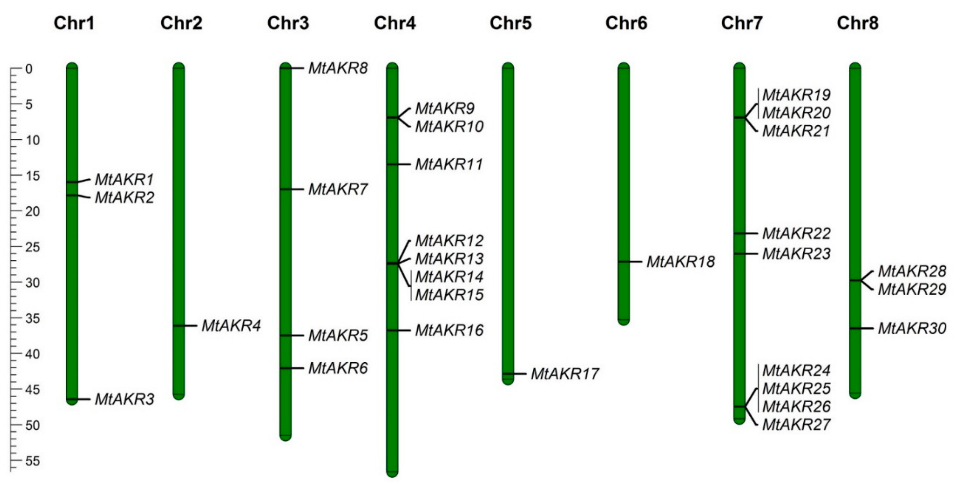
2.2. The Distribution of MtAKR Genes on the Chromosome

The chromosomal location and length information for each MtAKR from Ensembl Plant (MedtrA17_4.0) (http://plants.ensembl.org) were visualized by using Mapchart (v 2.32) (Figure 1). The MtAKRs are distributed on each of the eight chromosomes. Five groups of genes (MtAKR9/MtAKR10, MtAKR12/MtAKR13/MtAKR14/MtAKR15, MtAKR19/MtAKR20/MtAKR21, MtAKR24/MtAKR25/MtAKR26/MtAKR27, and MtAKR28/MtAKR29) on the chromosome are tightly linked on chromosome 4, chromosomes 7, and chromosomes 8, respectively. Chromosomes 2 and 6 each contain one MtAKR gene. Three, four, and three MtAKR genes are respectively distributed on chromosomes 1, 3, and 8.

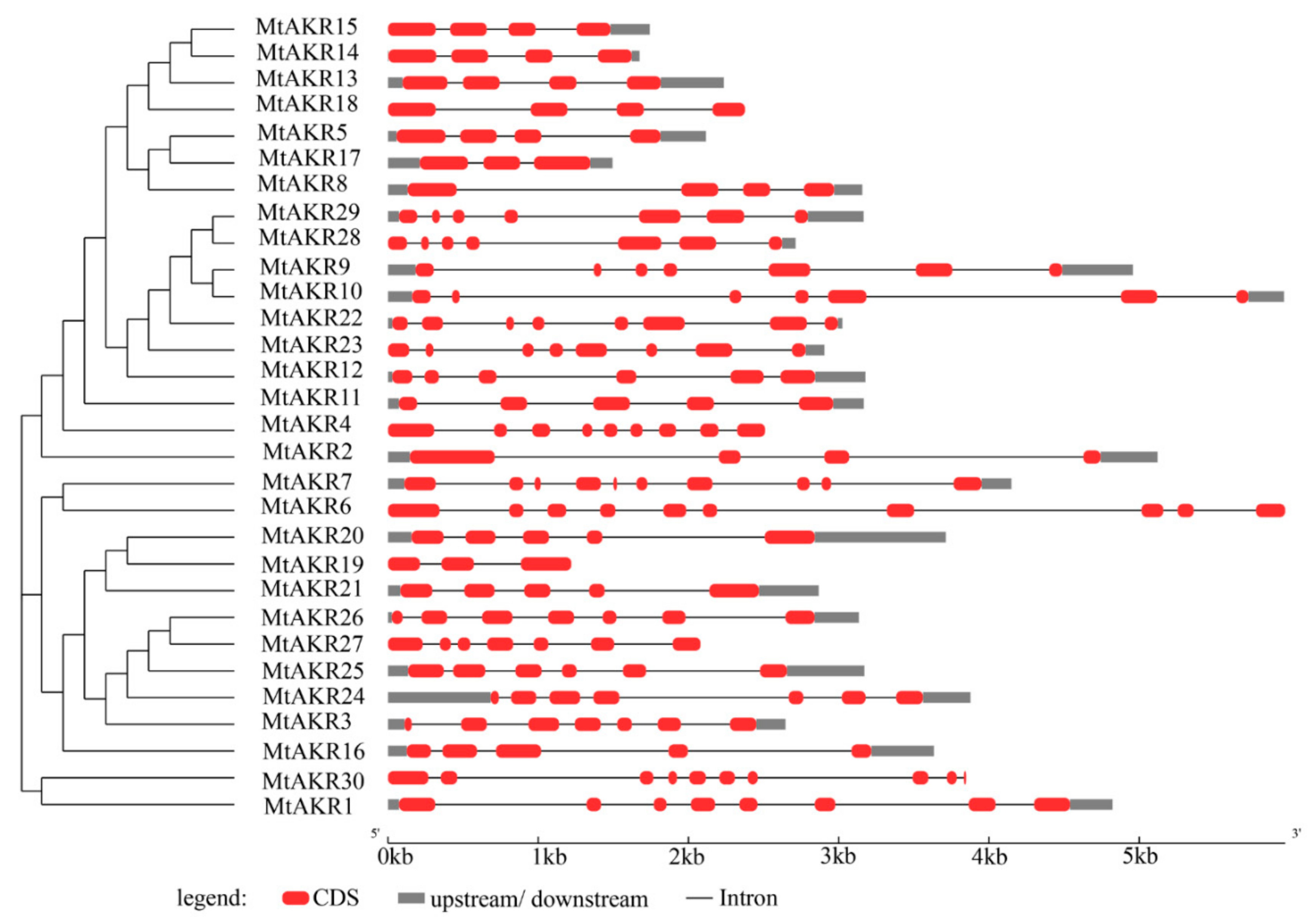
2.3. Structural Analysis of the MtAKR Genes

The sequences of the thirty MtAKRs were aligned, and a phylogenetic tree was created by using the neighbor-joining method in MEGA 7, to characterize their relationships, based on the full-length protein sequences (Figure 2). The phylogenetic tree showed that these 30 MtAKRs could be divided into three classes. The first one contained the most proteins (MtAKR15, MtAKR14, MtAKR13, MtAKR18, MtAKR5, MtAKR17, MtAKR8, MtAKR29, MtAKR28, MtAKR9, MtAKR10, MtAKR22, MtAKR23, MtAKR12, MtAKR11, MtAKR4, and MtAKR2). The second one contained eleven MtAKRs (MtAKR7, MtAKR6, MtAKR20, MtAKR19, MtAKR21, MtAKR26, MtAKR27, MtAKR25, MtAKR24, MtAKR3, and MtAKR16). The third one was consisted of only two MtAKRs (MtAKR30 and MtAKR1). All the MtAKR genes contain introns, with exon numbers ranging from 3 to 10 (Figure 2). MtAKR17 and MtAKR19 each contain three exons. MtAKR2, MtAKR5, MtAKR8, MtAKR13, MtAKR14, MtAKR15, and MtAKR18 each contain four exons. MtAKR11, MtAKR16, MtAKR20, and MtAKR21 contain five exons. MtAKR12 and MtAKR25 each contain six exons. MtAKR3, MtAKR9, MtAKR10, MtAKR24, MtAKR26, MtAKR27, MtAKR28, and MtAKR29 each contain seven exons. MtAKR1, MtAKR22, and MtAKR23 contain eight exons. MtAKR4 contains nine exons. MtAKR6, MtAKR7, and MtAKR13 each contain ten exons. The close family members typically had the same number of exons and genetic structures.

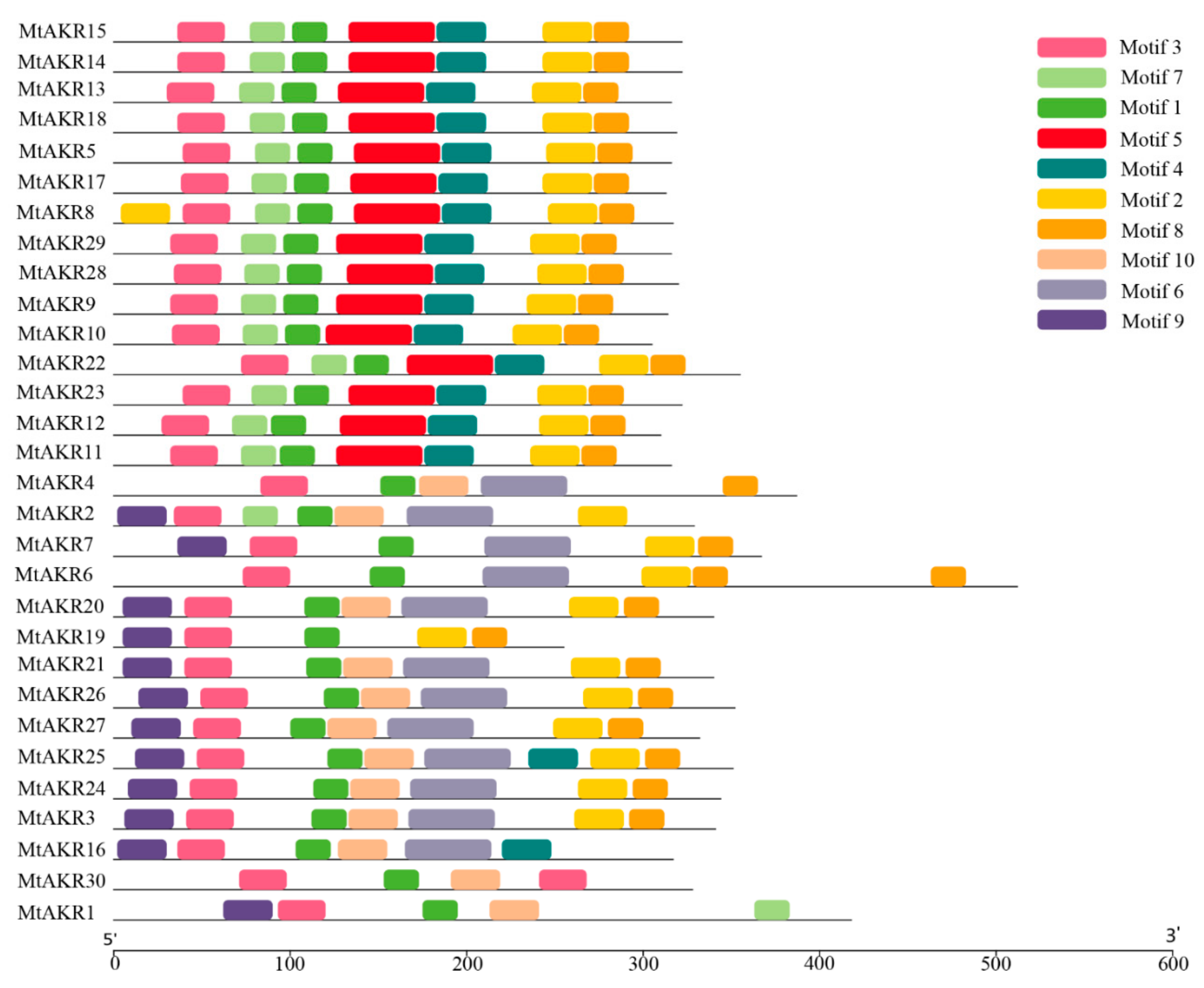
2.4. Conserved Motif in MtAKR Proteins

A total of 10 motifs ranging from 21 to 50 amino acids were identified by MEME analysis, as shown in Table 2. The obtained 10 motif sequences were submitted to Pfam search (http://pfam.xfam.org/search) for functional identification. Motif 1, motif 3, motif 5, motif 6, and motif 10 all describe aldo/keto reductase domain. Motif 1 and 3 are both present in all MtAKR proteins (Figure 3). The other motifs were designated as unknown motifs. Motif 2 is rich in Q (Gln) and was present in all MtAKR proteins except for MtAKR 1, MtAKR 4, MtAKR 16, and MtAKR 30. Similarly, motif 4 and motif 8 are both rich in Q (Gln), and motif 8 was present in all MtAKR proteins except for MtAKR1, MtAKR2, MtAKR16, and MtAKR30. Motif 7 is rich in D (Asp), and Motif 9 is rich in M (Met).
The motif information was input into the online software WebLogo (http://weblogo.berkeley.edu/ Department of Plant and Microbial Biology, University of California, Berkeley, US), and the motif logo was redrawn (Figure S1). In general, a sequence logo provides a richer and more precise description of a binding site than a consensus sequence [31,32]. For example, comparison of the logo to the consensus sequence shows a difference in the fourth amino acid in the first motif, with S or T at this site (presented here as S).

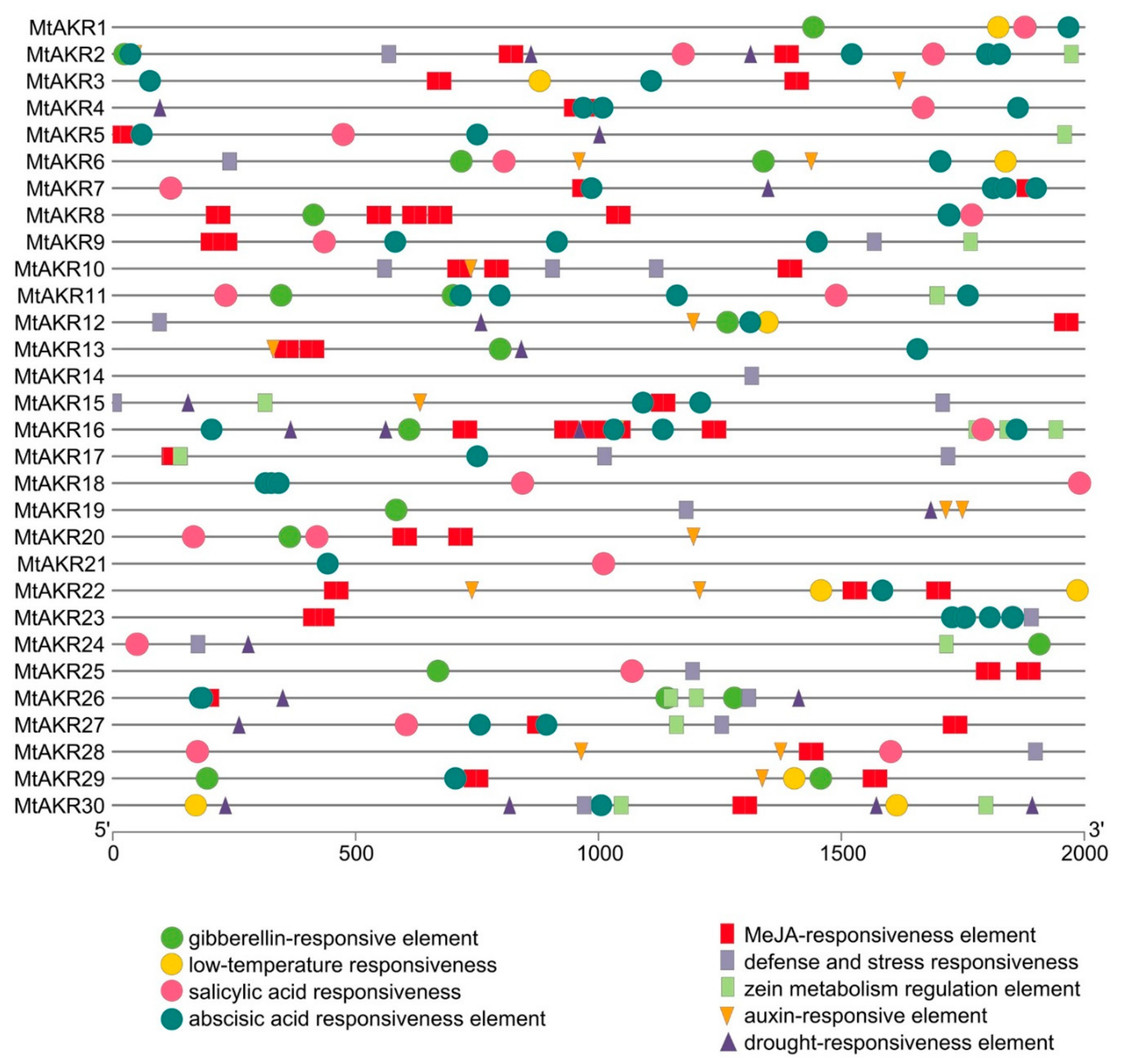
2.5. Cis-Acting Regulatory Elements in the Promoter of MtAKR Genes

Cis-acting elements in promoter is important to determine the possible regulation mechanisms of M. truncatula AKR genes in the hormone and abiotic stress responses. The promoter sequences (the 2 kb of sequence upstream of the initiation codon ATG) were analyzed to identify potential cis-regulatory elements, using the PlantCARE database (http://bioinformatics.psb.ugent.be/webtools/plantcare/html Laboratoire Associé de l’Institut National de la Recherche Agronomique (France), Universiteit Gent, Gent, Belgium). Nine types of hormone- and stress-related cis-acting regulatory elements were identified in the promoters of AKR genes in M. truncatula (Figure 4). Three stress-related cis-acting elements were identified, including TC-rich repeats (defense and stress), MBS (drought), and LTR (low-temperature responsive) elements. Six hormone-related cis-acting elements were identified, including TGA-element/AuxRR-core (auxin), O2-site (zein metabolism), TCA-element (salicylic acid), ABRE (abscisic acid), GARE-motif/P-box/TATC-box (gibberellin), and CGTCA-motif/TGACG-motif (MeJA responsive element) (Figure 4). All MtAKR genes contained one to seven cis-acting elements related to response to stresses or hormones. ABA-responsive elements were detected in 23 MtAKR genes. Among them, drought responsive elements were detected in 11 common MtAKR genes, and 13, 13, 14, 17, and 23 MtAKR genes contained responsive elements for auxin, zein metabolism, gibberellins, salicylic acid, and MeJA, respectively. results indicate the potential for significant regulation of the expression of MtAKR genes in response to various environmental factors.

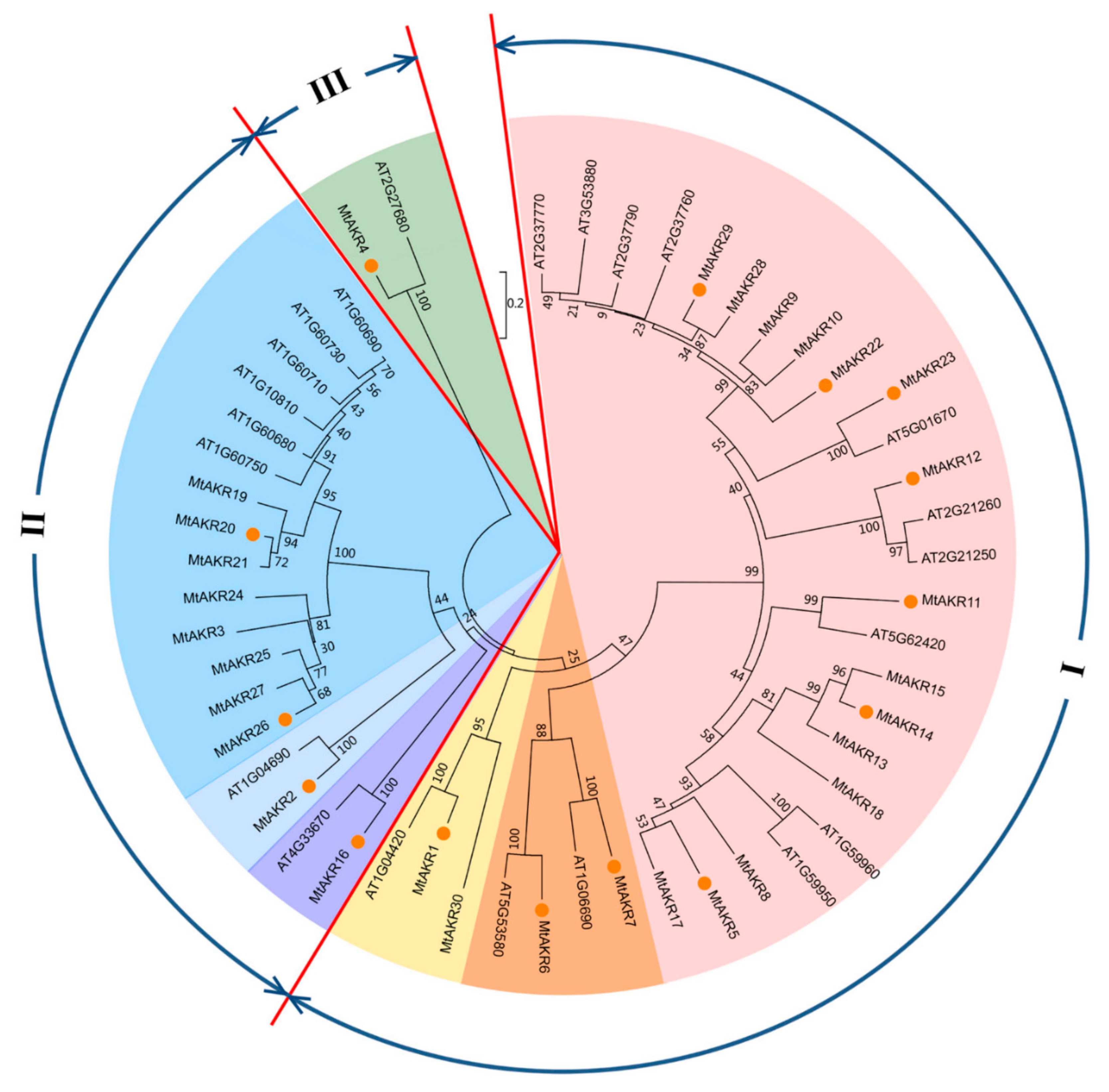
2.6. Phylogenetic Analysis of AKRs in M. Truncatula and A. Thaliana

To analyze the evolutionary relationship of AKRs between M. truncatula and A. thaliana, an unrooted neighbor-joining tree was constructed in MEGA7.0, using the protein sequences of 22 AtAKRs and 30 MtAKRs (Figure 5).
Results showed clustering of the fifty-two AKRs into three groups by genetic distance (Figure 5). Groups I and II were each further divided into three subgroups (shown in different colors). Each group or subgroup contains at least one member from A. thaliana and one member from M. truncatula. Ten AtAKRs (AT2G37770, AT3G53880, AT2G37790, AT2G37760, AT2G21260, AT2G21250, AT5G62420, AT1G59960, AT1G59950, and AT1G06690) and fifteen MtAKRs (MtAKR5, MtAKR8-MtAKR15, MtAKR17, MtAKR18, MtAKR22, MtAKR23, MtAKR28, and MtAKR29) were included in the I-1 subgroup, suggesting that these twenty-five AKRs are closely related proteins. Two AtAKRs (AT5G53580 and AT1G04420) and two MtAKRs (MtAKR6 and MtAKR7) were included in the I-2 subgroup. One AtAKR (AT4G33670) and two MtAKRs (MtAKR1 and MtAKR30) were included in the I-3 subgroup. The II-1 and II-2 subgroups each contained one AtAKR and one MtAKR, with AT4G33670 and MtAKR16 in II-1, and AT1G04690 and MtAKR2 in II-2. Six AtAKRs (AT1G60750, AT1G60680, AT1G10810, AT1G60710, AT1G60730, and AT1G60690) and eight MtAKRs (MtAKR3, MtAKR19–MtAKR21, and MtAKR24–MtAKR27) belonged to subgroup II-3. Group III contains only two members (AT2G27680 and MtAKR4), which suggests that the sequences of these two proteins are significantly different from the other proteins.

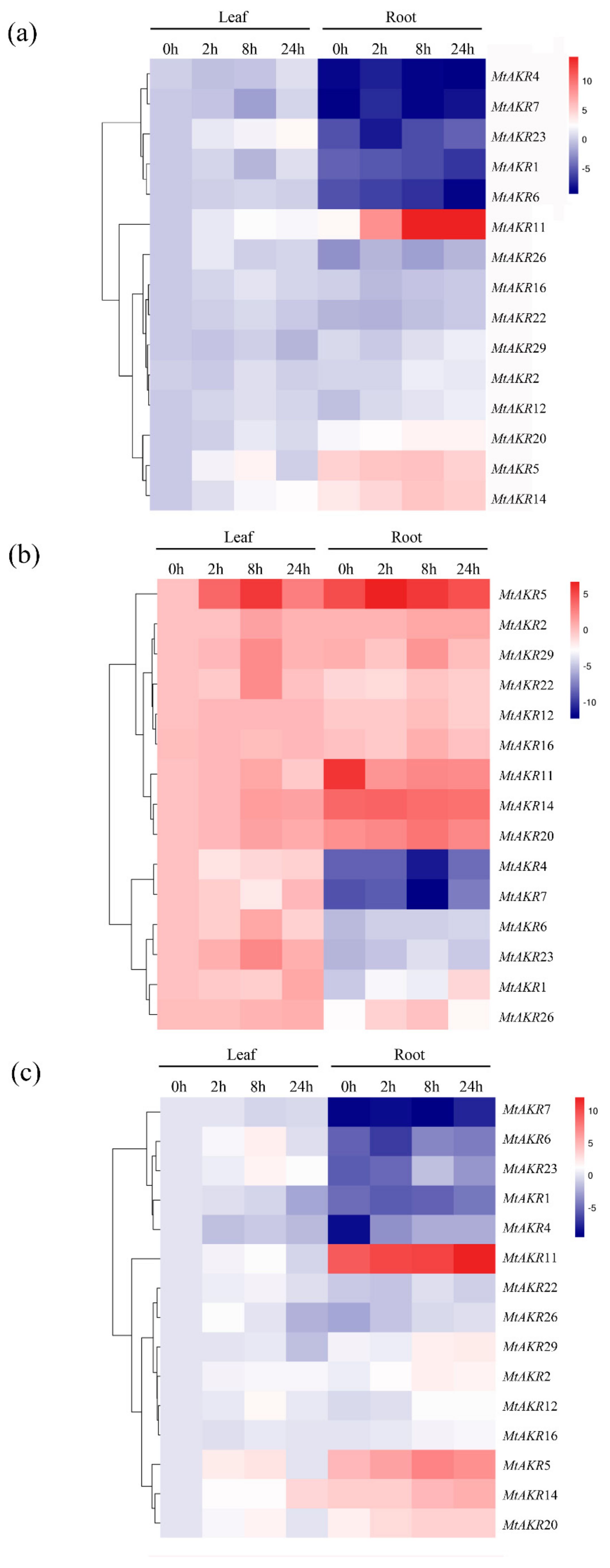
2.7. Expression Profiles of the MtAKR Genes in M. truncatula Leaves and Roots under ABA, PEG, and NaCl Treatment

The expression levels of selected AKR genes were assayed by real-time PCR, to explore the potential response of M. truncatula AKRs to ABA, PEG, and NaCl stress. A total of fifteen MtAKR genes were randomly selected from each group or subgroup for PCR analysis. Expression analysis was carried out of samples of leaves and roots of M. truncatula plants exposed to ABA, PEG, or NaCl treatment.
The heatmap representation of the overall expression patterns revealed clustering of the MtAKR genes (Figure 6). Results indicated that most of the MtAKR genes were regulated to different degrees by ABA in both leaves and roots (Figure 6a). In general, the highest expression of each gene appears at 8 or 24 h after the start of ABA stress application. The fifteen expression patterns could be divided into two groups. Five genes (MtAKR1, MtAKR4, MtAKR6, MtAKR7, and MtAKR23) exhibited lower expression levels in roots than in leaves. Compared to 0 h, the expression levels of MtAKR1 and MtAKR6 decreased in roots. The other group included MtAKR2, MtAKR5, MtAKR11, MtAKR12, MtAKR14, MtAKR16, MtAKR20, MtAKR22, MtAKR26, and MtAKR29 genes, and most of them exhibited higher expression levels in roots than those in leaves. The expression levels of MtAKR5, MtAKR11, and MtAKR14 were significantly upregulated compared to 0 h in roots. This shows that these genes are more sensitive to ABA treatment in roots.
According to the expression pattern in plants subjected to PEG stress, the 15 MtAKR genes can be divided into three groups (Figure 6b). The first one included MtAKR2, MtAKR5, MtAKR11, MtAKR12, MtAKR14, MtAKR16, MtAKR20, MtAKR22, and MtAKR29 genes. Except for MtAKR11, the other eight genes displayed upregulated transcription in both leaves and roots. The highest expression level of MtAKR11 was detected at 0 h in roots. The expression of MtAKR5, MtAKR22, and MtAKR29 was significantly upregulated in both roots and leaves. The second group included MtAKR4 and MtAKR7 genes and exhibited downregulation at the transcriptional level in both leaves and root at 8 h. The remaining genes in leaves in the third group, including MtAKR1, MtAKR6, MtAKR23, and MtAKR26, exhibited significantly higher expression level than that in roots. PEG stress increased expression levels of these genes in leaves. In roots, MtAKR1 was significantly upregulated at 24 h.
As shown in Figure 6c, the expression patterns of the fifteen genes in response to NaCl treatment can be divided into two groups. The genes in the first group, which includes MtAKR1, MtAKR4, MtAKR6, MtAKR7, and MtAKR23, exhibited higher expressions in leaves than in roots. The responses were consistent with that treated by ABA in roots. MtAKR1, MtAKR4, MtAKR6, and MtAKR7 genes were downregulated in leaves but upregulated in roots at 24 h. The expression level of MtAKR2, MtAKR5, MtAKR11, MtAKR14, MtAKR20, and MtAKR29 in another group showed significant upregulation in roots.
Overall, expression analysis indicated that MtAKR5 and MtAKR14 were largely induced both in leaves and roots, and MtAKR1, MtAKR11, MtAKR20, and MtAKR29 were significantly induced in roots when seedlings were subjected to ABA, PEG, and NaCl treatment. Results showed that these genes mentioned above may play a vital role in resisting to abiotic stresses.

3. Discussion

The AKR superfamily contains 18 families and more than 190 annotated proteins, but only few AKRs have been studied in plants [11]. The AKRs were identified and named based on sequence similarity. Subfamilies contain proteins with more than 60% amino acid sequence, and members with less than 40% shared amino acid sequence were placed in different families [11,33]. Focusing on the structure and expression patterns of AKR family members which responded to salt, drought, and ABA stress in M. truncatula was helpful in identifying candidate genes involving these abiotic stresses. The genome sequence was mined to identify gene families, analyze genetic relationships, and determine gene family distribution across the chromosomes. Phylogenetic analysis used in this study can be performed to group genes into subfamilies and will facilitate analysis of gene function [34].

3.1. Characterization and Analysis of the MtAKR Gene Family

In the past 30 years, M. truncatula has become a widely studied model organism [35,36,37]. M. truncatula is always used to address biological questions by using genome sequencing and genomics approaches similar to A. thaliana and Oryza sativa [38]. The study of M. truncatula AKRs can provide a foundation for the study of legume forage AKR genes. The MtAKRs have an average of 336 amino acids and an average protein mass of 37 kDa in MtAKRs, which is consistent with the previous published that most AKRs contain approximately 320 amino acids, with proteins 33–37 kDa in size [8,39,40]. The differences in transcriptional induction can be attributed to the quality and numbers of the cis-elements present in the promoters [41]. The presence of a response element allows regulation of the gene expression levels by the corresponding transcription factors or proteins [42]. Abscisic acid or drought or defense and stress response elements were identified in five of the six MtAKRs most strongly induced by ABA, salt, and drought stress treatments (MtAKR1, MtAKR5, MtAKR11, MtAKR14, and MtAKR29; MtAKR20 was not found) (Figure 4). Two and four ABA response cis-elements were identified in MtAKR5 and MtAKR11. MtAKR20 is abundant in salicylic acid and MeJA responsive element (Figure 4), but was clearly induced by ABA, salt, and drought stresses in this study, plotted in Figure 6. Therefore, the response of this gene to these abiotic stresses may be indirectly regulated by other mechanisms. The presence of a large number of auxin and abscisic acid response elements in the promoters of MtAKRs indicate potential functions in plant growth and responses to environmental stresses [43].

3.2. The Phylogenetic Analysis of Close Relationship between A. thaliana and M. truncatula AKR Genes and MtAKRs Expression Patterns

In this study, 22 genes in A. thaliana (Simpson et al. reported that the A. thaliana genome includes at least 21 AKR genes [34]) and 30 genes in M. truncatula that putatively encode AKR proteins were identified. A phylogenetic tree was constructed to explore the phylogenetic relationships of AKR genes from A. thaliana and M. truncatula. The expression of MtAKR genes in different stresses is helpful to identify candidate genes for subsequent detailed characterization [43]. The phylogenetic analysis revealed that five MtAKRs (MtAKR9, MtAKR10, MtAKR22, MtAKR28, and MtAKR29) and four A. thaliana AKRs (AT2G37770, AT3G53880, AT2G37790, and AT2G37760) were grouped in the same cluster in subgroup I-1 (Figure 5). These four AtAKRs are the members of AKR4C subfamily and named AKR4C8, AKR4C9, AKR4C10, and AKR4C11. Analysis of the A. thaliana AKRs suggested the function in reduction sugar-derived reactive carbonyls, as well as showed upregulation under salt and drought stress in three days [34]. In this study, the expression of MtAKR22 and MtAKR29 was detected. Results showed that the highest expression levels of MtAKR22 and MtAKR29 were present at 8 or 24 h after salt, drought, and ABA stresses, both in roots and leaves (Figure 6).
AT1G59950 (AtA6PR1) and AT1G59960 (AtA6PR2) in subgroup I-1 were previously proposed to be related to the aldose 6-phosphate reductase pathway, and they both exhibited response to cold and salt stresses [40]. The sequences of MtAKR5, MtAKR8, and MtAKR17 are in the same cluster with these genes (Figure 5). These MtAKR genes contain only three or four exons (Figure 2) and the same motif in amino acid sequence (Figure 3). These suggest the similar function of MtAKR5, MtAKR8, and MtAKR17. The expression pattern of MtAKR5 was detected in this study (Figure 6) had a similar response to that of AtA6PR1 under NaCl treatment [40].
AT1G04690 was reported as a K+ channel β subunit and is classified in the AKR6C subfamily [44]. It can be regulated by auxin, ethylene, and jasmonic acid [45]. McLoughlin and Yao [46,47] reported that AT1G04690 plays a role in salt-stress regulation. MtAKR2 is the only MtAKR clustered with AT1G04690 in subgroup II-2. The MtAKR2 promoter contains both auxin and MeJA response elements (Figure 4). Transcription of this gene can be influenced by exogenous NaCl, PEG, and ABA. MtAKR2 is therefore presumed to be a member of AKR6C subfamily and plays a role in salt-stress regulation.
Plant AKRs are regulated by many abiotic stresses, including drought, low temperature, and salt. However, a few AKRs have been studied, such as cadmium-stress-tolerance gene IbAKR [11], the drought-stress-tolerance gene MsALR [48], and the heat-stress-tolerance gene OsAKR [41]. OsAKR1 and EcAKR4-1 can detoxify glyphosate and increase herbicide resistance in plants [49,50]. PpAKR1 can be induced by ABA, salt, cold, and oxidative stresses [13]. AKR4C1 and AKR4C2 can be induced by ABA [51,52]. Therefore, AKRs are induced to protect plants from abiotic stress. Studies have been conducted to show that ABA in response to water stress is a cell-signaling process, and ABA content changes under both salt and drought stresses [53,54]. In this study, MtAKR5, MtAKR11, MtAKR14, and MtAKR20 showed significant upregulation in response to the ABA (Figure 6a) and NaCl (Figure 6c) treatments in both leaves and roots, indicating that these four MtAKRs may regulate M. truncatula response to osmotic stress in ABA-dependent manner. This conclusion is consistent with that reported by Tran [55]. MtAKR1, MtAKR6, and MtAKR7 exhibited similar responses to salt, drought, and ABA treatments (Figure 6). The gene expression of MtAKR1, MtAKR6, and MtAKR7 under ABA and PEG treatments showed upregulated with the induction of time and reached the highest value at 8 or 24 h in leaves (Figure 6a,b). The gene expression of MtAKR1 and MtAKR7 showed downregulated during the induction of time in salt stress. The highest value of these two MtAKRs in leaves occurred at 0 or 2 h (Figure 6c). The phylogenetic analyses showed that MtAKR1, MtAKR6, and MtAKR7 were in the same group (Figure 5). MtAKR1, MtAKR6, and MtAKR7 are predicted to locate in chloroplast (Table 1). The same-cluster genes in A. thaliana (AT1G04420, AT5G53580, and AT1G06690) were predicted to locate in chloroplasts, according to the UNIPROT online tool (https://www.uniprot.org/uniprot European Bioinformatics Institute, Cambridgeshire, UK). AT1G06690 and AT5G53580 genes were reported to be involved in the PLP salvage pathway and play a role in osmotic-stress resistance [56], indicating that MtAKR1, MtAKR6, and MtAKR7 may also be involved in resisting osmotic stress, as with drought and salt stress.
Some AKR genes have been considered as potential breeding targets for developing stress-tolerant varieties. The selection of candidate AKR genes is of great significance to the breeding of legume forages in the future. The characteristics identification and prediction of the selected 30 MtAKR members indicated that they are aldo–keto reductase superfamily members. The expression pattern of 15 MtAKR genes showed that MtAKR5, MtAKR11, MtAKR14, and MtAKR20 in roots under ABA and NaCl stress, and MtAKR1, MtAKR5, and MtAKR29 in both leaves and roots under PEG stress were induced significantly (Figure 6). These results suggest that the six genes can be further studied in M. truncatula as candidate genes for stress-resistance regulation.

4. Materials and Methods

4.1. Identification, Chromosomal Localization, and Preliminary Analysis of the AKR Genes in Medicago truncatula Genome

The members of the M. truncatula AKR family were identified by using the Pfam database (http://www.pfam.xfam.org/), by searching for the AKR domain (PF00248). The full-length protein sequence of each identified M. truncatula AKR family member was obtained from the phytozome database (https://phytozome.jgi.doe.gov/pz/portal). Functionally redundant sequences were removed by the online tool CD-HIT (http://weizhong-lab.ucsd.edu/cdhit_suite J. Craig Venter Institute, California, US), sequences with low similarity were removed (<40% similarity with major AKRs) by using ClustalW (v 2.1) (https://www.genome.jp/tools-bin/clustalw Institute for Chemical Research, Kyoto University, Kyoto, Japan), and sequences that lacked the AKR conserved domain were eliminated by searching the Ensembl Plant database (MedtrA17_4.0) (http://plants.ensembl.org/index.html).

4.2. Analysis of the Protein Features of the Medicago Truncatula AKRs

The CDS and genomic DNA sequence were downloaded from Ensembl Plant. General features of the AKRs, including their theoretical isoelectric point (pI) and molecular weight (mW), were predicted by using the ExPASy-Protparam tool (http://www.web.expasy.org/protparam/ The SIB Swiss Institute of Bioinformatics, Lausanne, Switzerland (appeared before)) [57]. The subcellular localization of AKRs was explored by using the TargetP (v 1.1) online tool (http://www.cbs.dtu.dk/services/TargetP/) [58].

4.3. Structural Analysis of the AKRs

The gene organization of the AKR genes was explored by the online tool GSDS (v 2.0) (Gene Structure Display Server) (http://gsds.cbi.pku.edu.cn/ Center for Bioinformatics, Peking University, Beijing, China(appeared before)) [59]. The full-length protein sequences were used to construct phylogenetic tree and performed the bootstrap neighbor-joining analysis in MEGA 7 software [60]. The parameters were taken as gap open penalty of 10.0, gap extension penalty of 0.2, and bootstrapping with 2000 replicates.

4.4. Motif Composition Analysis of MtAKR Proteins

The MEME 5.0.5 online program (http://meme-suite.org/index.html National Institutes of Health, Bethesda, US) [61] was used for motif identification of the 30 M. truncatula AKR proteins. The following optimized parameters of MEME were applied: Number of repetitions, any; maximum number of motifs, 10; and optimum width of each motif, between 6 and 50 residues. TB tools (v 0.6669) [62] and the online tool WebLogo (http://weblogo.berkeley.edu/) were used to reconstruct the motif composition and identify a logo sequence.

4.5. Prediction of Cis-Acting Elements in MtAKRs

Putative hormone-related, stress-response cis-acting elements were identified in promoter sequences by analysis of the 2kb of sequence upstream of the initiation codon ATG of each of the MtAKRs genes, using the PlantCARE database online tool (http://bioinformatics.psb.ugent.be/webtools/plantcare/html Laboratoire Associé de l’Institut National de la Recherche Agronomique (France), Universiteit Gent, Gent, Belgium) [63]. TB tools (v 0.6669) was then used for data visualization.

4.6. Phylogenetic of AKRs in Medicago truncatula and Arabidopsis thaliana

Sequences of AKRs in M. truncatula were collected from NCBI (http://www.ncbi.org/ National Center for Biotechnology Information, National Library of Medicine, Bethesda, US), using BLASTP analysis, with all of the Arabidopsis AKR protein sequences as queries. A confirmed AKRs extracted from Arabidopsis thaliana, Pfam (PF00248), was used to verify putative M. truncatula AKRs in the phytozome database. An unrooted neighbor-joining tree was constructed by MEGA (v 7.0). The parameters were taken as gap open penalty of 10.0, gap extension penalty of 0.2, and bootstrap analysis of 2000 replicates.

4.7. Plant Growth Condition and Treatments

Plump seeds of M. truncatula (wild type R108 from our lab: The Institute of Animal Science, the Chinese Academy of Agricultural Sciences) were scarified by sandpaper, surface sterilized with 75% alcohol for 10 min, washed five times with distilled water, and then germinated on plates with paper and distilled water. Seedlings (when seed cotyledons opened) were transferred to 1/2 Hoagland nutrient solution in a plant-growth chamber (GXZ-430C), with conditions of day 16 h, 24 °C, and dark 8 h, 20°C. The nutrient solution was changed every five days, and 25-day-old seedlings were then subjected to salt (200 mM of NaCl), drought (50 g·L−1 of PEG 6000), and ABA (100 µM) treatments for 0 (9:00 a.m.), 2, 8, and 24 h. Leaf and root were separately harvested and processed for RNA isolation and cDNA synthesis after the treatments. Four biological replicates (one plant per biological replicate) were used in this experiment.

4.8. RNA Preparation, cDNA Synthesis, and Expression Analysis by qRT-PCR

RNA extraction and cDNA synthesis were performed by using the Promega total RNA Kit and TaKaRa PrimeScript RT kit with gDNA Eraser, respectively. The specific primer pair for each AKR gene was designed by using Primer 5 software, and the primer sequences are listed in Table S1. The qRT-PCR was performed in an ABI 7300 instrument, with the following conditions: 95 °C for 10 min, 40 cycles of 95 °C for 15 s, and 60 °C for 60 s. All reactions were run in quadruplicate. Relative fold changes were calculated based on the comparative Ct method, and the Mt-Actin7 (MTR3g095530) gene was used as an internal standard. Heatmap representation was performed by using the normalized (2−ΔΔCt) value. The data of gene expression in qRT-PCR are listed in Tables S2 and S3. The data visualization was performed with OmicShare tools (http://www.omicshare.com/tools Gene Denovo, Guangzhou, China). Relative expression levels (FC) in roots and leaves of Medicago truncatula under ABA, PEG, and NaCl stresses are shown in Table S4.

Supplementary Materials

The following are available online at https://www.mdpi.com/1422-0067/21/3/754/s1. Figure S1: The logo of ten MtAKRs motif construction. Each logo consists of stacks of symbols, one stack for each position in the sequence. The conservation at each position is indicated by the overall height of the stack. The height of symbols within the stack is proportional to frequency of each amino acid at that position. Table S1: The qRT-PCR primers of MtAKRs. Table S2: The data of gene expression in qRT-PCR (leaf). Table S3: The data of gene expression in qRT-PCR (root). Table S4: Relative expression levels (FC) in roots and leaves of Medicago truncatula under ABA, PEG and NaCl stresses. Material S1: The sequences of MtAKR genes’ CDS and gDNA.

Author Contributions

R.L., J.Y., and Q.Y. conceived of and designed the experiments; J.Y., H.S., R.L., and J.Z. performed the experiments; J.Y., R.L., H.S., J.Z., T.Z., J.K., Z.W., and Y.H. analyzed the data; J.Y., R.L., and Q.Y. wrote the paper. All authors have read and agreed to the published version of the manuscript.

Funding

This work was supported by the National Natural Science Foundation of China (No. 31601993), the earmarked fund for China Agriculture Research System (CARS-34), and Agricultural Science and Technology Innovation Program (ASTIP-IAS14).

Conflicts of Interest

The authors declare no conflicts of interest.

References

  1. Alscher, R.G.; Erturk, N.; Heath, L.S. Role of superoxide dismutases (SODs) in controlling oxidative stress in plants. J. Exp. Bot. 2002, 53, 1331–1341. [Google Scholar] [CrossRef]
  2. Bartosz, G. Oxidative stress in plants. Acta Physiol. Plant. 1997, 19, 47–64. [Google Scholar] [CrossRef]
  3. Zeid, I.M.; Shedeed, Z.A. Response of alfalfa to putrescine treatment under drought stress. Biol. Plant. 2006, 50, 635–640. [Google Scholar] [CrossRef]
  4. Esmaeili, N.; Yang, X.; Cai, Y.; Sun, L.; Zhu, X.; Shen, G.; Payton, P.; Fang, W.; Zhang, H. Co-overexpression of AVP1 and OsSIZ1 in Arabidopsis substantially enhances plant tolerance to drought, salt, and heat stresses. Sci. Rep. 2019, 9, 7642. [Google Scholar] [CrossRef] [Green Version]
  5. Zorb, C.; Geilfus, C.M.; Dietz, K.J. Salinity and crop yield. Plant Biol. (Stuttg.) 2019, 21 (Suppl. 1), 31–38. [Google Scholar] [CrossRef]
  6. Abdelraheem, A.; Esmaeili, N.; O’Connell, M.; Zhang, J. Progress and perspective on drought and salt stress tolerance in cotton. Ind. Crop. Prod. 2019, 130, 118–129. [Google Scholar] [CrossRef]
  7. Lee, S.C.; Luan, S. ABA signal transduction at the crossroad of biotic and abiotic stress responses. Plant Cell Environ. 2012, 35, 53–60. [Google Scholar] [CrossRef]
  8. Penning, T.M. The aldo-keto reductases (AKRs): Overview. Chem. Biol. Interact. 2015, 234, 236–246. [Google Scholar] [CrossRef] [Green Version]
  9. Sengupta, D.; Naik, D.; Reddy, A.R. Plant aldo-keto reductases (AKRs) as multi-tasking soldiers involved in diverse plant metabolic processes and stress defense: A structure-function update. J. Plant Physiol. 2015, 179, 40–55. [Google Scholar] [CrossRef]
  10. Jin, Y.; Penning, T.M. Aldo-keto reductases and bioactivation/detoxication. Annu. Rev. Pharm. Toxicol. 2007, 47, 263–292. [Google Scholar] [CrossRef]
  11. Huo, J.; Bing, D.U.; Sun, S.; Shaozhen, H.E.; Zhao, N.; Liu, Q.; Zhai, H. A novel aldo-keto reductase gene, IbAKR, from sweet potato confers higher tolerance to cadmium stress in tobacco. Front. Agric. Sci. Eng. 2018, 5, 206–213. [Google Scholar] [CrossRef]
  12. Shimakawa, G.; Kohara, A.; Miyake, C. Medium-chain dehydrogenase/reductase and aldo-keto reductase scavenge reactive carbonyls in Synechocystis sp. PCC 6803. FEBS Lett. 2018, 592, 1010–1019. [Google Scholar] [CrossRef] [Green Version]
  13. Kanayama, Y.; Mizutani, R.; Yaguchi, S.; Hojo, A.; Ikeda, H.; Nishiyama, M.; Kanahama, K. Characterization of an uncharacterized aldo-keto reductase gene from peach and its role in abiotic stress tolerance. Phytochemistry 2014, 104, 30–36. [Google Scholar] [CrossRef]
  14. Kumar, D.; Singh, P.; Yusuf, M.A.; Upadhyaya, C.P.; Roy, S.D.; Hohn, T.; Sarin, N.B. The Xerophyta viscosa aldose reductase (ALDRXV4) confers enhanced drought and salinity tolerance to transgenic tobacco plants by scavenging methylglyoxal and reducing the membrane damage. Mol. Biotechnol. 2013, 54, 292–303. [Google Scholar] [CrossRef]
  15. Graham, P.H.; Vance, C.P. Legumes: Importance and constraints to greater use. Plant Physiol. 2003, 131, 872–877. [Google Scholar] [CrossRef] [Green Version]
  16. Benedito, V.A.; Torres-Jerez, I.; Murray, J.D.; Andriankaja, A.; Allen, S.; Kakar, K.; Wandrey, M.; Verdier, J.; Zuber, H.; Ott, T.; et al. A gene expression atlas of the model legume Medicago truncatula. Plant J. 2008, 55, 504–513. [Google Scholar] [CrossRef]
  17. Young, N.D.; Frédéric, D.; Oldroyd, G.E.D.; Rene, G.; Cannon, S.B.; Udvardi, M.K.; Benedito, V.A.; Mayer, K.F.X.; Jér?Me, G.; Heiko, S. The Medicago genome provides insight into the evolution of rhizobial symbioses. Nature 2011, 480, 520. [Google Scholar] [CrossRef] [Green Version]
  18. Tang, H.; Krishnakumar, V.; Bidwell, S.; Rosen, B.; Chan, A.; Zhou, S.; Gentzbittel, L.; Childs, K.L.; Yandell, M.; Gundlach, H. An improved genome release (version Mt4.0) for the model legume Medicago truncatula. BMC Genom. 2014, 15, 312. [Google Scholar] [CrossRef] [Green Version]
  19. Bonnin, I.; Prosperi, J.M.; Olivieri, I. Comparison of quantitative genetic parameters between two natural populations of a selfing plant species, Medicago truncatula Gaertn. Appl. Genet. 1997, 94, 641–651. [Google Scholar] [CrossRef]
  20. Li, L.; Cheng, H.; Gai, J.; Yu, D. Genome-wide identification and characterization of putative cytochrome P450 genes in the model legume Medicago truncatula. Planta 2007, 226, 109–123. [Google Scholar] [CrossRef]
  21. Bianco, C.; Defez, R. Medicago truncatula improves salt tolerance when nodulated by an indole-3-acetic acid-overproducing Sinorhizobium meliloti strain. J. Exp. Bot. 2009, 60, 3097–3107. [Google Scholar] [CrossRef]
  22. Lopez, M.; Herrera-Cervera, J.A.; Iribarne, C.; Tejera, N.A.; Lluch, C. Growth and nitrogen fixation in Lotus japonicus and Medicago truncatula under NaCl stress: Nodule carbon metabolism. J. Plant Physiol. 2008, 165, 641–650. [Google Scholar] [CrossRef]
  23. Filippou, P.; Antoniou, C.; Fotopoulos, V. Effect of drought and rewatering on the cellular status and antioxidant response of Medicago truncatula plants. Plant Signal Behav. 2011, 6, 270–277. [Google Scholar] [CrossRef] [Green Version]
  24. Zhao, Y.; Liu, R.; Xu, Y.; Wang, M.; Zhang, J.; Bai, M.; Han, C.; Xiang, F.; Wang, Z.Y.; Mysore, K.S.; et al. AGLF provides C-function in floral organ identity through transcriptional regulation of AGAMOUS in Medicago truncatula. Proc. Natl. Acad. Sci. USA 2019, 116, 5176–5181. [Google Scholar] [CrossRef] [Green Version]
  25. Pecrix, Y.; Staton, S.E.; Sallet, E.; Lelandais-Briere, C.; Moreau, S.; Carrere, S.; Blein, T.; Jardinaud, M.F.; Latrasse, D.; Zouine, M.; et al. Whole-genome landscape of Medicago truncatula symbiotic genes. Nat. Plants 2018, 4, 1017–1025. [Google Scholar] [CrossRef]
  26. Aubert, G.; Morin, J.; Jacquin, F.; Loridon, K.; Quillet, M.C.; Petit, A.; Rameau, C.; Lejeune-Hénaut, I.; Huguet, T.; Burstin, J. Functional mapping in pea, as an aid to the candidate gene selection and for investigating synteny with the model legume Medicago truncatula. Appl. Genet. 2006, 112, 1024–1041. [Google Scholar] [CrossRef]
  27. Zhang, C.; Zhang, H.; Zhao, Y.; Jiang, H.; Zhu, S.; Cheng, B.; Xiang, Y. Genome-wide analysis of the CCCH zinc finger gene family in Medicago truncatula. Plant Cell Rep. 2013, 32, 1543–1555. [Google Scholar] [CrossRef]
  28. Khan, Z.; Karamahmutoglu, H.; Elitas, M.; Yuce, M.; Budak, H. Through the looking glass: Real-Time Imaging in Brachypodium Roots and Osmotic Stress Analysis. Plants 2019, 8, 14. [Google Scholar] [CrossRef] [Green Version]
  29. Galvez, S.; Merida-Garcia, R.; Camino, C.; Borrill, P.; Abrouk, M.; Ramirez-Gonzalez, R.H.; Biyiklioglu, S.; Amil-Ruiz, F.; Dorado, G.; Budak, H.; et al. Hotspots in the genomic architecture of field drought responses in wheat as breeding targets. Funct. Integr. Genom. 2019, 19, 295–309. [Google Scholar] [CrossRef] [Green Version]
  30. Akpınar, B.A.; Lucas, S.J.; Budak, H. Genomics approaches for crop improvement against abiotic stress. Sci. World J. 2013, 2013, 1–9. [Google Scholar] [CrossRef] [Green Version]
  31. Crooks, G.E.; Hon, G.; Chandonia, J.M.; Brenner, S.E. WebLogo: A sequence logo generator. Genome. Res. 2004, 14, 1188–1190. [Google Scholar] [CrossRef] [PubMed] [Green Version]
  32. Schneider, T.D.; Stephens, R.M. Sequence logos: A new way to display consensus sequences. Nucleic Acids Res. 1990, 18, 6097–6100. [Google Scholar] [CrossRef] [PubMed]
  33. Jez, J.M.; Flynn, T.G.; Penning, T.M. A new nomenclature for the aldo-keto reductase superfamily. Biochem. Pharmacol. 1997, 54, 639–647. [Google Scholar] [CrossRef]
  34. Simpson, P.J.; Tantitadapitak, C.; Reed, A.M.; Mather, O.C.; Bunce, C.M.; White, S.A.; Ride, J.P. Characterization of two novel aldo-keto reductases from Arabidopsis: Expression patterns, broad substrate specificity, and an open active-site structure suggest a role in toxicant metabolism following stress. J. Mol. Biol. 2009, 392, 465–480. [Google Scholar] [CrossRef]
  35. Paven, M.C.M.; Viau, L.; Hamon, A.; Vandecasteele, C.; Pellizzaro, A.; Bourdin, C.; Laffont, C.; Lapied, B.; Lepetit, M.; Frugier, F. Characterization of a dual-affinity nitrate transporter MtNRT1.3 in the model legume Medicago truncatula. J. Exp. Bot. 2012, 62, 5595. [Google Scholar] [CrossRef] [Green Version]
  36. Cook, D.R. Medicago truncatula—A model in the making! Commentary. Curr. Opin. Plant. Biol. 1999, 2, 301–304. [Google Scholar] [CrossRef]
  37. Barker, D.G.; Bianchi, S.; Blondon, F.; Dattée, Y.; Duc, G.; Essad, S.; Flament, P.; Gallusci, P.; Génier, G.; Guy, P. Medicago truncatula, a model plant for studying the molecular genetics of theRhizobium-legume symbiosis. Plant Mol. Biol. Rep. 1990, 8, 40–49. [Google Scholar] [CrossRef]
  38. Mun, J.H.; Kim, D.J.; Choi, H.K.; Gish, J.; Debelle, F.; Mudge, J.; Denny, R.; Endre, G.; Saurat, O.; Dudez, A.M.; et al. Distribution of microsatellites in the genome of Medicago truncatula: A resource of genetic markers that integrate genetic and physical maps. Genetics 2006, 172, 2541–2555. [Google Scholar] [CrossRef] [Green Version]
  39. Hyndman, D.; Bauman, D.R.; Heredia, V.V.; Penning, T.M. The aldo-keto reductase superfamily homepage. Chem. Biol. Interact. 2003, 143, 621–631. [Google Scholar] [CrossRef]
  40. Rojas, B.; Wurman, J.; Zamudio, M.S.; Donoso, A.; Cabedo, P.; Díaz, F.; Stange, C.; Handford, M. AtA6PR1 and AtA6PR2 encode putative aldose 6-phosphate reductases that are cytosolically localized and respond differentially to cold and salt stress in Arabidopsis thaliana. J. Plant Biochem. Biotechnol. 2018. [Google Scholar] [CrossRef]
  41. Turoczy, Z.; Kis, P.; Torok, K.; Cserhati, M.; Lendvai, A.; Dudits, D.; Horvath, G.V. Overproduction of a rice aldo-keto reductase increases oxidative and heat stress tolerance by malondialdehyde and methylglyoxal detoxification. Plant Mol. Biol. 2011, 75, 399–412. [Google Scholar] [CrossRef] [PubMed]
  42. Hur, Y.S.; Shin, K.H.; Kim, S.; Nam, K.H.; Lee, M.S.; Chun, J.Y.; Cheon, C.I. Overexpression of GmAKR1, a stress-induced aldo/keto reductase from soybean, retards nodule development. Mol. Cells 2009, 27, 217–223. [Google Scholar] [CrossRef] [PubMed]
  43. Shen, C.; Yue, R.; Sun, T.; Zhang, L.; Xu, L.; Tie, S.; Wang, H.; Yang, Y. Genome-wide identification and expression analysis of auxin response factor gene family in Medicago truncatula. Front Plant Sci. 2015, 6, 73. [Google Scholar] [CrossRef] [PubMed] [Green Version]
  44. Tang, H.; Vasconcelos, A.C.; Berkowitz, G.A. Evidence that plant K+ channel proteins have two different types of subunits. Plant Physiol. 1995, 109, 327–330. [Google Scholar] [CrossRef] [PubMed] [Green Version]
  45. Ashley, M.K.; Grant, M.; Grabov, A. Plant responses to potassium deficiencies: A role for potassium transport proteins. J. Exp. Bot. 2006, 57, 425–436. [Google Scholar] [CrossRef] [PubMed] [Green Version]
  46. McLoughlin, F.; Arisz, S.A.; Dekker, H.L.; Kramer, G.; de Koster, C.G.; Haring, M.A.; Munnik, T.; Testerink, C. Identification of novel candidate phosphatidic acid-binding proteins involved in the salt-stress response of Arabidopsis thaliana roots. Biochem. J. 2013, 450, 573–581. [Google Scholar] [CrossRef] [PubMed] [Green Version]
  47. Yao, H.Y.; Xue, H.W. Phosphatidic acid plays key roles regulating plant development and stress responses. J. Integr. Plant Biol. 2018, 60, 851–863. [Google Scholar] [CrossRef]
  48. Oberschall, A.; Deak, M.K.; Sass, L.; Vass, I.; Kovacs, I.; Feher, A. A novel aldose/aldehyde reductase protects transgenic plants against lipidperoxidation under chemical and drought stresses. Plant J. 2000, 24, 437–446. [Google Scholar] [CrossRef]
  49. Vemanna, R.S.; Vennapusa, A.R.; Easwaran, M.; Chandrashekar, B.K.; Rao, H.; Ghanti, K.; Sudhakar, C.; Mysore, K.S.; Makarla, U. Aldo-keto reductase enzymes detoxify glyphosate and improve herbicide resistance in plants. Plant Biotechnol. J. 2017, 15, 794–804. [Google Scholar] [CrossRef]
  50. Pan, L.; Yu, Q.; Han, H.; Mao, L.; Nyporko, A.; Fan, L.; Bai, L.; Powles, S.B. Aldo-keto reductase metabolizes glyphosate and confers glyphosate resistance in Echinochloa colona. Plant Physiol. 2019, 181, 1519–1534. [Google Scholar] [CrossRef]
  51. Lee, S.P.; Chen, T.H. Molecular cloning of abscisic acid-responsive mRNAs expressed during the induction of freezing tolerance in bromegrass (Bromus inermis Leyss) suspension culture. Plant Physiol. 1993, 101, 1089–1096. [Google Scholar] [CrossRef] [PubMed] [Green Version]
  52. Bartels, D.; Engelhardt, K.; Roncarati, R.; Schneider, K.; Rotter, M.; Salamini, F. An ABA and GA modulated gene expressed in the barley embryo encodes an aldose reductase related protein. EMBO J. 1991, 10, 1037–1043. [Google Scholar] [CrossRef] [PubMed]
  53. Perin, E.C.; da Silva Messias, R.; Borowski, J.M.; Crizel, R.L.; Schott, I.B.; Carvalho, I.R.; Rombaldi, C.V.; Galli, V. ABA-dependent salt and drought stress improve strawberry fruit quality. Food Chem. 2019, 271, 516–526. [Google Scholar] [CrossRef] [PubMed]
  54. Zhang, J.; Jia, W.; Yang, J.; Ismail, A.M. Role of ABA in integrating plant responses to drought and salt stresses. Field Crop Res. 2006, 97, 111–119. [Google Scholar] [CrossRef]
  55. Tran, C.D.; Chu, H.D.; Nguyen, K.H.; Watanabe, Y.; La, H.V.; Tran, K.D.; Tran, L.-S.P. Genome-Wide Identification of the TCP Transcription Factor Family in Chickpea (Cicer arietinum L.) and Their Transcriptional Responses to Dehydration and Exogenous Abscisic Acid Treatments. J. Plant Growth Regul. 2018, 37, 1286–1299. [Google Scholar] [CrossRef]
  56. Herrero, S.; González, E.; Gillikin, J.W.; Vélëz, H.; Daub, M.E. Identification and characterization of a pyridoxal reductase involved in the vitamin B6 salvage pathway in Arabidopsis. Plant Mol. Biol. 2011, 76, 157. [Google Scholar] [CrossRef] [PubMed]
  57. Gasteiger, E.; Gattiker, A.; Hoogland, C.; Ivanyi, I.; Appel, R.D.; Bairoch, A. ExPASy: The proteomics server for in-depth protein knowledge and analysis. Nucleic Acids Res. 2003, 31, 3784–3788. [Google Scholar] [CrossRef] [Green Version]
  58. Emanuelsson, O.; Brunak, S.; Von Heijne, G.; Nielsen, H. Locating proteins in the cell using TargetP, SignalP and related tools. Nat. Protoc. 2007, 2, 953–971. [Google Scholar] [CrossRef]
  59. Hu, B.; Jin, J.; Guo, A.Y.; Zhang, H.; Luo, J.; Gao, G. GSDS 2.0: An upgraded gene feature visualization server. Bioinformatics 2014, 31, 1296. [Google Scholar] [CrossRef] [Green Version]
  60. Kumar, S.; Stecher, G.; Tamura, K. MEGA7: Molecular Evolutionary Genetics Analysis Version 7.0 for Bigger Datasets. Mol. Biol. Evol. 2016, 33, 1870–1874. [Google Scholar] [CrossRef] [Green Version]
  61. Bailey, T.L.; Boden, M.; Buske, F.A.; Frith, M.; Grant, C.E.; Clementi, L.; Ren, J.; Li, W.W.; Noble, W.S. MEME Suite: Tools for motif discovery and searching. Nucleic Acids Res. 2009, 37, W202–W208. [Google Scholar] [CrossRef] [PubMed]
  62. Chen, C.; Xia, R.; Chen, H.; He, Y. TBtools, a Toolkit for Biologists integrating various HTS-data handling tools with a user-friendly interface. BiorXiv 2018, 289660. [Google Scholar] [CrossRef]
  63. Magali, L.; Patrice, D.; Gert, T.; Kathleen, M.; Yves, M.; Yves, V.D.P.; Pierre, R.; Stephane, R. PlantCARE, a database of plant cis-acting regulatory elements and a portal to tools for in silico analysis of promoter sequences. Nucleic Acids Res. 2002, 30, 325–327. [Google Scholar] [CrossRef]
Figure 1. The chromosomal locations of the MtAKR genes.
Ijms 21 00754 g001
Figure 2. Structural and phylogenetic analyses of 30 identified MtAKR members. An unrooted tree of the MtAKRs constructed by neighbor-joining method, using MEGA 7. The gene organization of MtAKRs was explored by using the online tool GSDS 2.0 (http://gsds.cbi.pku.edu.cn/ Center for Bioinformatics, Peking University, Beijing, China).
Ijms 21 00754 g002
Figure 3. Distribution of the conserved motifs in different MTAKRs and the logos for each motif.
Ijms 21 00754 g003
Figure 4. Distribution of major stress- and hormone-related cis-acting regulatory elements in the promoters of putative M. truncatula AKR genes. TC-rich repeats (defense and stress), MBS (drought), and LTR (low-temperature responsive) elements, TGA- element/AuxRR-core (auxin), O2-site (zein metabolism), TCA-element (salicylic acid), ABRE (abscisic acid), GARE-motif /P-box /TATC-box (gibberellin), and CGTCA-motif /TGACG-motif (MeJA responsive element) are represented by different symbols, as indicated in figure key at the bottom.
Ijms 21 00754 g004
Figure 5. Phylogenetic tree of AKRs from M. truncatula and A. thaliana. The deduced full-length amino acid sequences were aligned by using ClustalW, and the phylogenetic tree was constructed by using the neighbor-joining (NJ) method in MEGA 7.0, with 2000 bootstrap replicates. Three groups and six subgroups are indicated. The genes marked with orange dots were subjected to expression analysis.
Ijms 21 00754 g005
Figure 6. Hierarchical clustering and heatmap representation of leaf and root MtAKR genes expression profiles in ABA (a), PEG (b), and NaCl (c) conditions. The data obtained by quantitative RT-PCR correspond to the levels of MtAKRs in total RNA samples extracted from leaves and roots (four technical replicates). The expression levels of genes were used 2−ΔΔCt, and the heatmap was presented in log2 values.
Ijms 21 00754 g006
Table 1. The fundamental characteristics of candidate gene.
No.Gene NamegDNA
Length a
cds
Length a
Gene ID bProtein Length cMass c
(kDa)
PI cGRAVY cTargetP d
1MtAKR148221254MTR1g04273041746.657.58−0.251C*
2MtAKR25122987MTR1g04725032836.487.57−0.251M
3MtAKR326461023MTR1g10275034038.025.50−0.250_
4MtAKR425101161MTR2g08512538643.448.15−0.300M
5MtAKR52116948MTR3g08313031535.165.70−0.175_
6MtAKR659711536MTR3g09214051156.969.20−0.109C*
7MtAKR741491101MTR3g44979036640.258.98−0.303C
8MtAKR83157951MTR0374s005031635.585.40−0.184C
9MtAKR94958942MTR4g02135031334.776.10−0.301_
10MtAKR105964915MTR4g02141030433.976.00−0.290_
11MtAKR113166948MTR4g03684531536.005.40−0.419M
12MtAKR123177930MTR4g07206030934.825.88−0.213_
13MtAKR132235948MTR4g07232031535.845.96−0.314_
14MtAKR141674966MTR4g07235032136.276.27−0.260_
15MtAKR151742966MTR4g07236032136.556.42−0.274_
16MtAKR163633951MTR4g09275031634.295.44−0.096_
17MtAKR171494939MTR5g09791031234.895.93−0.231_
18MtAKR182376957MTR6g07311031835.627.09−0.160_
19MtAKR191220765MTR7g02167025428.055.90−0.321_
20MtAKR2037131020MTR7g02168033937.405.77−0.285_
21MtAKR2128671020MTR7g02185033937.665.60−0.318_
22MtAKR2230241065MTR7g06358035439.516.46−0.106S*
23MtAKR232904966MTR7g07050032136.206.02−0.462_
24MtAKR2438771032MTR7g11497034337.956.02−0.181_
25MtAKR2531711053MTR7g11498035038.366.13−0.222_
26MtAKR2631341056MTR7g11499035138.355.64−0.165_
27MtAKR272080996MTR7g11501033136.335.25−0.191_
28MtAKR282713960MTR8g07009531935.695.73−0.206_
29MtAKR293165948MTR8g07011531534.926.46−0.242_
30MtAKR303848984MTR8g08816032736.836.71−0.223M*
a The length of gDNA and coding sequence were acquired from Ensembl Plant (http://plants.ensembl.org/index.html European Bioinformatics Institute, Hinxton, Cambridgeshire, UK.). b The protein IDs were obtained from phytozome (https://phytozome.jgi.doe.gov/ U.S. Department of Energy, Washington, US). c Protein length, mass (molecular weight, kDa), PI (isoelectric point), and the grand average of hydropathy value (GRAVY) were analyzed by using the ExPASy-Protparam tool (http://www.web.expasy.org/protparam/ The SIB Swiss Institute of Bioinformatics, Lausanne, Switzerland). d Subcellular localization of the MtAKRs was predicted by using TargetP (v 1.1) (http://www.cbs.dtu.dk/services/TargetP/ Department of Health Technology, Lyngby, Denmark).C:Chloroplast. S: Secretory pathway; M: Mitochondria; _: Any other location; superscript * indicates highly reliable prediction (reliability class, from 1 to 5, where 1 means the strongest prediction. RC is a measure of the size of the difference (‘diff’) between the highest (winning) and the second highest output scores. There are 5 reliability classes defined as follows: (1) diff > 0.800; (2) 0.800 > diff > 0.600; (3) 0.600 > diff > 0.400; (4) 0.400 > diff > 0.200; (5) diff < 0.200. Thus, the lower the value of RC, the safer the prediction. Superscript * denotes the value of diff is 1).
Table 2. Amino acid composition and function of different motifs.
Motif Number Length (Amino Acid)Best Possible MatchFunction
Motif 121CEASLKRLQLDYIDLYYIHWPAldo/keto reductase domain
Motif 229CTPAQIALRWGYQQGHDVCPKSFNTERMNunmatched
Motif 328ISAIHHAIKAGYRHFDTADIYGNHEENGAldo/keto reductase domain
Motif 429PAVNQVEMHPSWHQDKLREFCNQKGIHVSunmatched
Motif 550FKAEDMTPFDMKGTWKAMEECYRS
GLARAIGVSNFSIKKLQDLLEYARIP
Aldo/keto reductase domain
Motif 650IRRAHAVHPITAVQMEWSLWTRDIE
EEIIPLCRELGIGIVPYSPLGRGFF
Aldo/keto reductase domain
Motif 721NRDDLFITSKLWCTDHHPEDVunmatched
Motif 821QNIGAFDWKLTQEDMRKISQIunmatched
Motif 929PRMKLGTQGMEVSKQGFGCMGMSGFYNPPunmatched
Motif 1029DTSVPIEDTMGELKKLVEEGKIKYIGLSEAldo/keto reductase domain

