Article

Developmental Roles of FUSE Binding Protein 1 (Fubp1) in Tooth Morphogenesis

by
Yam Prasad Aryal
1,
Sanjiv Neupane
2,
Tae-Young Kim
1,
Eui-Seon Lee
1,
Nitin Kumar Pokhrel
1,
Chang-Yeol Yeon
1,
Ji-Youn Kim
3,
Chang-Hyeon An
4,
Seo-Young An
4,
Eui-Kyun Park
5,
Jung-Hong Ha
6,
Jae-Kwang Jung
7,
Hitoshi Yamamoto
8,
Sung-Won Cho
9,
Sanggyu Lee
10,
Do-Yeon Kim
11,
Tae-Yub Kwon
12,
Youngkyun Lee
1,
Wern-Joo Sohn
13 and
Jae-Young Kim
1,*
1
Department of Biochemistry, School of Dentistry, IHBR, Kyungpook National University, 2177, Dalgubeol-daero, Jung-gu, Daegu 41940, Korea
2
Department of Biochemistry and Cell Biology, Stony Brook University, Stony Brook, NY 11784, USA
3
Department of Dental Hygiene, Gachon University, Incheon 21936, Korea
4
Department of Oral and Maxillofacial Radiology, School of Dentistry, IHBR, Kyungpook National University, 2177, Dalgubeol-daero, Jung-gu, Daegu 41940, Korea
5
Department of Oral Pathology and Regenerative Medicine, School of Dentistry, IHBR, Kyungpook National University, 2177, Dalgubeol-daero, Jung-gu, Daegu 41940, Korea
6
Department of Conservative Dentistry, School of Dentistry, IHBR, Kyungpook National University, 2177, Dalgubeol-daero, Jung-gu, Daegu 41940, Korea
7
Department of Oral Medicine, School of Dentistry, IHBR, Kyungpook National University, 2177, Dalgubeol-daero, Jung-gu, Daegu 41940, Korea
8
Department of Histology and Developmental Biology, Tokyo Dental College, Tokyo 101-0061, Japan
9
Division in Anatomy and Developmental Biology, Department of Oral Biology, Yonsei University College of Dentistry, Seoul 26493, Korea
10
School of Life Science BK21 plus KNU Creative BioResearch Group, Kyungpook National University, Daegu 41566, Korea
11
Department of Pharmacology, School of Dentistry, Kyungpook National University, 2177, Dalgubeol-daero, Jung-gu, Daegu 41940, Korea
12
Department of Dental Biomaterials, School of Dentistry, Kyungpook National University, 2177, Dalgubeol-daero, Jung-gu, Daegu 41940, Korea
13
Pre-Major of Cosmetics and Pharmaceutics, Daegu Haany University, Gyeongsan 38610, Korea
*
Author to whom correspondence should be addressed.
Int. J. Mol. Sci. 2020, 21(21), 8079; https://doi.org/10.3390/ijms21218079
Submission received: 3 September 2020 / Revised: 16 October 2020 / Accepted: 27 October 2020 / Published: 29 October 2020
(This article belongs to the Special Issue Gene Networks That Control Cell Proliferation and Differentiation)

Abstract

:
FUSE binding protein 1 (Fubp1), a regulator of the c-Myc transcription factor and a DNA/RNA-binding protein, plays important roles in the regulation of gene transcription and cellular physiology. In this study, to reveal the precise developmental function of Fubp1, we examined the detailed expression pattern and developmental function of Fubp1 during tooth morphogenesis by RT-qPCR, in situ hybridization, and knock-down study using in vitro organ cultivation methods. In embryogenesis, Fubp1 is obviously expressed in the enamel organ and condensed mesenchyme, known to be important for proper tooth formation. Knocking down Fubp1 at E14 for two days, showed the altered expression patterns of tooth development related signalling molecules, including Bmps and Fgf4. In addition, transient knock-down of Fubp1 at E14 revealed changes in the localization patterns of c-Myc and cell proliferation in epithelium and mesenchyme, related with altered tooth morphogenesis. These results also showed the decreased amelogenin and dentin sialophosphoprotein expressions and disrupted enamel rod and interrod formation in one- and three-week renal transplanted teeth respectively. Thus, our results suggested that Fubp1 plays a modulating role during dentinogenesis and amelogenesis by regulating the expression pattern of signalling molecules to achieve the proper structural formation of hard tissue matrices and crown morphogenesis in mice molar development.

1. Introduction

Organogenesis is a complex phenomenon by which cells of different embryonic origin develop and associate to form organs [1]. Various organs, such as limb, mammary gland, kidney, feather, tooth, hair, lung, and pancreas, share common features, for instance, epithelial–mesenchymal interactions, during the early stages of organogenesis [2,3,4]. The interacting signalling from tissues modulates cell proliferation, differentiation, apoptosis and migration- during morphogenesis and regeneration [5]. Particularly, tooth develops through complex signalling regulations from reciprocal epithelial–mesenchymal interactions with unique and dynamic structural alterations. In mice, the thickening of the epithelium and its gradual invagination towards mesenchyme indicate the morphological commencement of odontogenesis [6]. Following epithelial invaginations, through the combined interactions of signalling molecules and transcription factors, tooth development proceeds with bud, cap, and bell stages at mouse embryonic stages E13, 14, and 16, respectively, which shows typical morphologic features of epithelial appendages with epithelial invaginations at E13, signalling centre formation with specific epithelial structural formation of enamel knot at E14, and functional differentiation of odontoblasts and ameloblasts at E16 [6,7]. There are about 500 genes involved in tooth development [8] and the spatio-temporal expression patterns that characterize the developmental function of these genes are important for proper tooth morphogenesis. The signalling pathways for proper morphogenesis during the developmental stages are strictly regulated at the transcription step which controls the paracrine factors, including Shh, Bmp, Fgf, and Wnt signalling [9]. For proper modulation of these paracrine factors, it is necessary to understand the developmental roles of transcriptional regulators, which modulate the expression of target genes and contribute to ensure proper signalling regulations during organogenesis [2,3,4].
Among the many transcription regulators, FUSE (far upstream element) binding proteins (FBPs) are regarded as important molecular tools during gene expression regulation [10]. One of these genes, Fubp1, expressed in developing tooth germ, is an evolutionary conserved gene that is present in eukaryotes and regulates c-Myc [11,12], a transcription factor involved in the regulation of gene expression [13,14,15]. As a c-Myc promoter, Fubp1 binds to FUSE, serving as a sensor of transcriptional activity [16]. In a range of tissues, Fubp1 has been reported to be involved in transcription, translation and post-translational regulation of genes [12]. Several studies reported the abnormal expression of Fubp1 in malignant cell lines with altered cellular physiology including proliferation, migration and apoptosis [17,18]. Therefore, it is necessary to emphasize the importance of Fubp1 in cellular behaviors, particularly cell cycle regulation and differentiation, the most important processes for the proper structural formation of organs. However, its precise expression pattern and developmental roles in odontogenic tissue are not yet understood.
During odontogenesis, the spatio-temporal regulation of genes at cap and bell stages play crucial roles for functional and structural formation of tooth [6]. In this study, the functional evaluation of Fubp1, a transcriptional regulator gene, is performed at cap stage through siRNA knock-down and in vitro organ cultivation system to evaluate the developmental role of Fubp1 prior to differentiation of odontoblasts and ameloblasts. Copious reports have revealed that complex interactions of signalling molecules at cap stage with precise transcriptional regulations would result in tooth morphogenesis, especially with hard tissue matrix formation at the bell stage [6,7,8,9]. In this study, as a pioneer work, we attempted to examine the detailed function of Fubp1 in tooth development at the cap stage, which shows dynamic differentiation sequences with a well-studied signalling network for tooth morphogenesis. Since the functional evaluation of transcriptional regulators, even though their importance in development, was difficult to perform in knockout animal models [3,6,9,19], this in vitro organ cultivation approach would be the plausible model system to provide the fine-tuning of transcription regulations by Fubp1.

2. Results

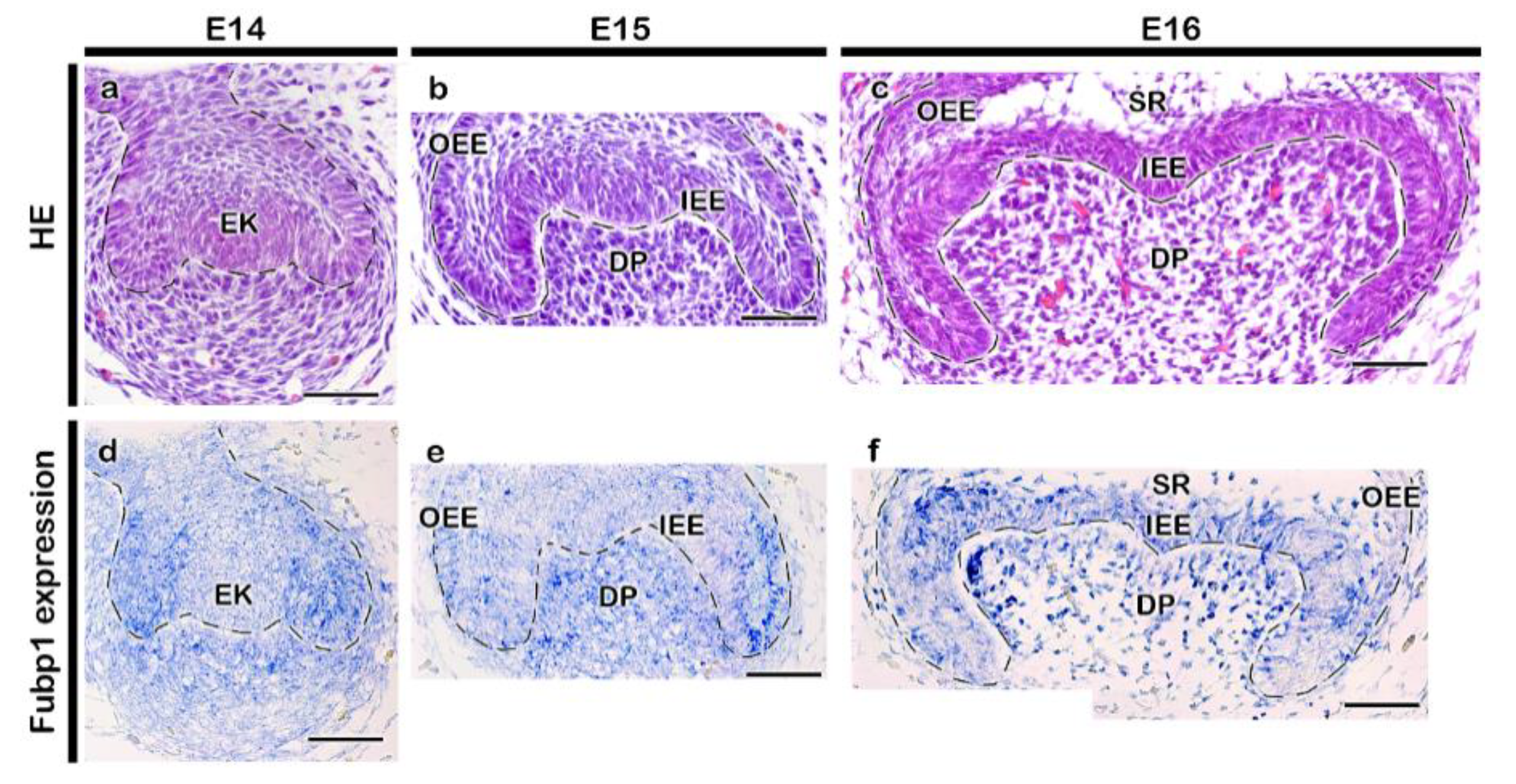
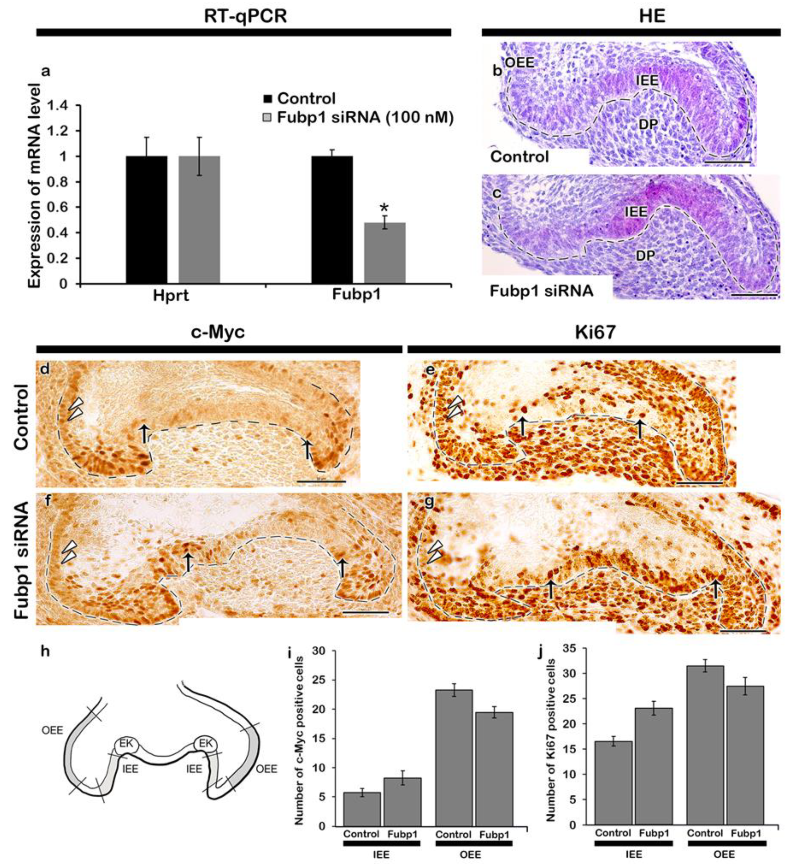
2.1. Expression Pattern and Functional Evaluation of Fubp1

The developing tooth was at cap and bell stages of tooth development (Figure 1a–c). During the cap stage (Figure 1d,e), Fubp1 was broadly expressed in the enamel epithelium and condensed mesenchyme, with intense expression in the cervical loop (Figure 1d–f). Distinct expression of Fubp1 was observed in the early bell stage (Figure 1f) along the inner enamel epithelium (IEE) and dental papilla (DP), which are presumptive ameloblast- and odontoblast-forming cells, respectively. Its expression was also observed along the outer enamel epithelium (OEE) and stratum intermedium at E16 (Figure 1f). There was 52% down regulation of Fubp1 by siRNA during in vitro organ cultivation (Figure 2a) and the developing teeth remained at bell stage after cultivation at E14 for two days (Figure 2b,c). In order to define the cellular physiology regulated by this gene, we examined the precise localization patterns of c-Myc (transcription regulator) and Ki67 (cell proliferation marker) through immunohistochemistry (Figure 2). Knocking down Fubp1 at the cap stage of tooth development resulted into a disrupted patterned arrangement of c-Myc positive cells in the IEE and OEE (Figure 2d,f). Particularly, a greater number of c-Myc positive cells are observed in the IEE, whereas less are observed in the OEE of the Fubp1 knock-down specimen (Figure 2d,f,i Supplementary Figure S1). Similar to these reactions, Ki67 reactions showed more ki67 positive cells along the IEE between secondary enamel knot-forming regions and decreased positive reactions in the OEE (Figure 2e,g; Supplementary Figure S1). The mesenchymal cells adjacent to the secondary enamel knot of control specimens showed more proliferative cells compared with the same region of Fubp1 knock-down (Figure 2e,g). In contrast, there was not much change in the c-Myc protein expression level after knock-down of Fubp1 when examined by Western blot analysis (Supplementary Figure S2). In addition, epithelial cell rearrangement, one of the important cellular events for epithelial morphogenesis, was examined using immunostaining against E-cadherin and ROCK1, and phalloidin staining, as were examined in previous study [20]. The localization of E-cadherin in epithelial cells revealed that cellular adhesion seemed weaker in the inner enamel epithelial cells of the knock-down specimen compared with the control (Figure 3a,d). Similarly, compared with the Fubp1 knock-down specimen, strong localization of ROCK1 and phalloidin staining were observed along the basement membrane of IEE and the adjacent cells of the DP in the control specimens (Figure 3b,c,e,f).

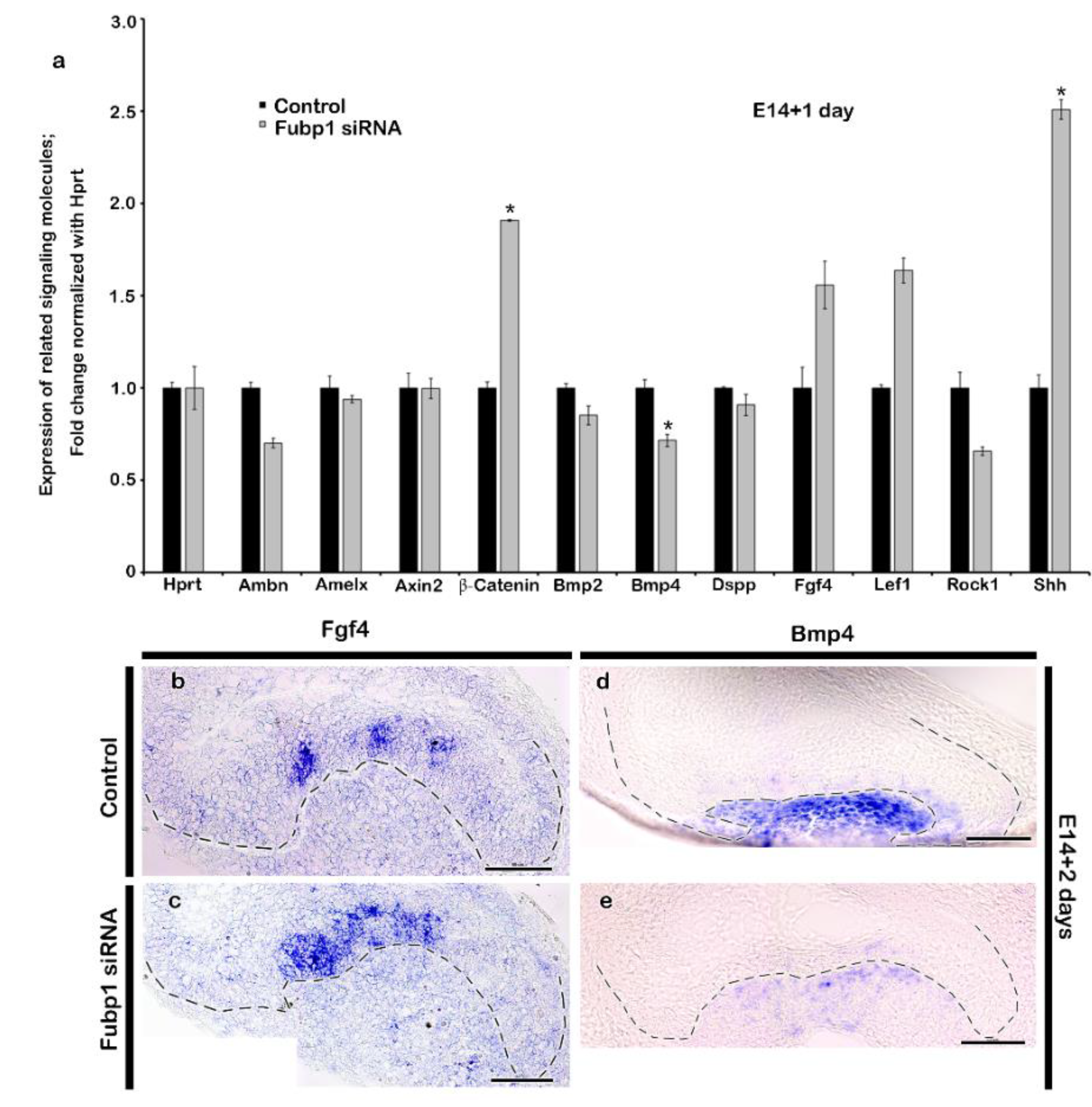
2.2. Altered Expression Patterns of Signalling Molecules by Fubp1 Knock-Down

Tooth development is regulated with the complex interactions of various signalling molecules [6,21], and therefore most of the tooth development-related signalling molecules were examined using qPCR to evaluate the knock-down effects of Fubp1. After knocking down Fubp1 expression at E14 for 24 h, signalling molecules, including Bmp2, Bmp4, and Rock1, were found to be downregulated (Figure 4a). Conversely, ß-catenin, Fgf4, Shh, and Lef1 were up-regulated, suggesting the modulating role of Fubp1 during the cap stage of tooth development. Moreover, enamel- and dentin-specific markers, such as Amelx, Ambn, Dspp, and Dmp1, were found to be down-regulated following Fubp1 knock-down (Figure 4a). After observing the expression patterns of tooth-related signalling molecules, in situ hybridization of sections was performed considering Bmp4 as a differentiation factor and Fgf4 as a proliferative factor during tooth development. The expression of Fgf4 was continuous along the enamel rope of tooth with Fubp1 knock-down (Figure 4c), whereas, its expression was restricted to the enamel knot in the control (Figure 4b). In contrast, the expression of Bmp4 was reduced in the mesenchyme of the specimen with Fubp1 knock-down (Figure 4e) compared with the control (Figure 4d).

2.3. Fubp1 in Tooth Morphogenesis

We employed renal capsule transplantation and examined the morphological alterations of the tooth to define the knock-down effect of Fubp1 at the cap stage of tooth development, as were examined in previous report [21]. The arrangement of the dentin matrix along the cusp of the Fubp1 knock-down tooth was abrogated in 1-week renal capsule calcified tooth when compared with the same region of the control specimen (Figure 5a,d). In addition, the Fubp1 knock-down tooth showed relatively weaker expressions of Dspp and Amelx mRNA along the cusp area when compared with the equivalent region of the control (Figure 5b,c,e,f). The expression pattern was also reduced to a smaller area in Fubp1 knock-down specimen compared to control (Figure 5b,c,e,f). To check whether the reduced expression pattern of Dspp and Amelx corresponds with the NESTIN localization, we performed immunostaining of NESTIN, an odontoblast differentiation marker, and the result showed the abrogated and weaker localization of NESTIN in the secreting odontoblasts and its processes relative to the control (Supplementary Figure S3). Furthermore, after observing relatively weaker expression of Amelx and Dspp in 1-week Fubp1 knock-down renal calcified tooth, and therefore, in order to evaluate the long term effect of Fubp1 knock-down, 3-week kidney capsule transplantation was employed and the thickness of dentin matrix and cuspal patterning were examined (Figure 5g–l). Although there were no significant differences in the overall dentin thickness between the control and knock-down specimen, the Fubp1 knock-down exhibited altered dentin and enamel patterned arrangements. SEM indicated an altered pattern of the enamel rod and interrod in the Fubp1 knock-down tooth compared with the control (Figure 5h,k). Moreover, the control specimen displayed well-arranged dentinal tubules (Figure 5i), but the arrangement of dentinal tubules was abrogated, especially in the cusp area of the Fubp1 knock-down tooth (Figure 5l). Furthermore, 3-week renal calcified tooth showed alteration in crown height in the Fubp1 knock-down specimen (Supplementary Figure S4). Comparatively shorter crown heights were seen in the Fubp1 knock-down specimen compared with the control. However, there was no variation in cusp height.

3. Discussion

Copious studies with experimental models, such as cell lines and animals models, have already been undertaken to identify the principle mechanisms of tooth development [22], however, there is still poor understanding of the key modulations among the candidate molecules in organogenesis processes. In this study, we firstly examined the precise expression pattern of a eukaryotic transcription regulator gene, Fubp1, along the epithelium and mesenchyme of the developing tooth (Figure 1d–f). Our results showed the co-expression pattern with Hoxc12, zfp36I1, Taf10, Cnbp and Ehmt2 [23] as candidate molecules involved in directing the tooth morphogenesis via modulating the signalling molecules during embryogenesis. Particularly, in this study, as the Fubp1 knock-out mouse is embryonically lethal [19], we tried to evaluate its crucial functional roles through transient Fubp1 knock-down and an in vitro organ cultivation system. Previous reports mention that c-Myc, a eukaryotic transcription regulator, is involved in cellular proliferation and differentiation [24,25,26]. It is also known that Fubp1 regulates the cell cycle progression through both c-Myc- dependent [27,28] and -independent [29] mechanisms. Additionally, cellular events such as proliferation and differentiation are very important during organogenesis. Our study showed that knocking down Fubp1 at E14 for 2 days increased cellular proliferation along the IEE between the secondary enamel knot-forming region (Figure 2e,h), indicating that the IEE cells are still proliferating, whereas the control cells are already set for differentiation. The rate of cellular proliferation usually decreases as cells differentiate [30], and so it seems that Fubp1 enhances the differentiation of the IEE specifically at the cusp-forming region. The region-specific cellular proliferation after Fubp1 knock-down draws particular attention during in vitro organ cultivation. Increased proliferating cells along the IEE and decreased proliferating cells along the OEE in Fubp1 knock-down tooth (Figure 2) implied that the modulating role of Fubp1 is important along the entire enamel epithelium. We hypothesized that these altered cell proliferation patterns would result from the altered localization pattern of c-Myc (Figure 2d,g). However, Fubp1 knock-down tissue did not show any obvious change in the c-Myc protein expression when examined by Western blot (Supplementary Figure S3), in agreement with a previous report [29]. Interestingly, immunohistochemistry analysis revealed a disrupted patterned arrangement of positive reactions against c-Myc in the IEE and OEE (Figure 2d,g). These results suggest that knock-down of Fubp1, one of the potential regulators of c-Myc, [27,28], would result altered tooth morphogenesis through changed cellular events.
The spatio-temporal expression of signalling molecules is critical for proper morphogenesis of the tooth and the knock-down of Fubp1 at the cap stage alters the expression of signalling molecules, including Bmp, Fgf4, Shh, Dspp, B-catenin and Rock1 (Figure 4a). The mesenchymal expression of Bmp signalling during the bell stage of tooth development is crucial for the signalling interaction between odontoblasts and ameloblasts [31]. Knock-out of Bmp2 and Bmp4 caused a low odontoblast differentiation marker expression, with reduced dentin thickness [32,33,34]. Knock-down of Fubp1 led to an observed decrease in the expression of Bmp4 mRNA, indicating its role in Bmp signalling (Figure 4d,e). At the bell stage of tooth development, inner enamel epithelial cells start to differentiate into ameloblasts and DP cells differentiate into odontoblasts [31] and the expression pattern of Fgf4 plays a crucial role in future cusp patterning [35,36]. We observed a continuous expression pattern of Fgf4 along the enamel rope in the Fubp1 knock-down tooth (Figure 4b,c). Consistent with this result, it has been reported that its expression is restricted along the enamel knot during the bell stage of mice molar development [21,35,37]. Copious studies have reported that various paracrine factors are involved in tooth morphogenesis, including Bmps, Fgfs, Shh, and Wnts [3,6,7,8,9]. Among these signalling molecules, Fgfs and Shh are well known enamel knot related signalling molecules and putatively play important roles in odontoblast differentiation and tooth morphogenesis [3,6,9]. In our study, three-week Fubp1 knock-down renal calcified teeth showed the altered crown height (Supplementary Figure S4) and based on these morphological alterations, we decided to examine expression patterns of signalling molecules which would be involved in the morphological alterations, particularly with Bmp2, Bmp4, and Fgf4 [3,6,9] (Figure 4). The knock-down effect of Fubp1 up-regulated Fgf4 and altered the crown height (Figure 4, Supplementary Figure S4). However, a similar expression pattern of Fgf4 resulted in a long cusp height in gerbils [35] suggesting that Fgf signalling not only to cuspal height morphogenesis but also with crown height. Our study showed down-regulation of Bmp2 and Bmp4 after Fubp1 knock-down during the bell stage. This result suggests that, in order to rescue weak Bmp signalling from the mesenchyme, Fgf4 would be up-regulated during reciprocal interaction between the epithelium and mesenchyme as in a previous report [38]. In addition, the increased expression of Shh in the Fubp1 knock-down could alter crown height because Shh signalling plays a crucial role during cytodifferentiation of ameloblasts and odontoblasts, and for cusp formation [39]. Meanwhile, the cellular adhesion and actin rearrangement play a crucial role during differentiation of ameloblasts [40]. In this study, the weak localization of ROCK1 and phalloidin staining along the IEE of the Fubp1 knock-down tooth implied that Fubp1 would modulate actin rearrangement during differentiation of ameloblasts before the formation of cusp morphogenesis because ROCK contributes to maintaining ameloblast polarity and enamel matrix secretion [40].
At the secretory stage of mice molar development, Fubp1 expression was observed along the secretory ameloblasts and odontoblasts (Figure 5b,c,e,f), similar to the expression pattern of Amelx and Dspp [41]. As the co-expression patterns of genes imply their shared functions in development and disease prediction [42], it showed that Fubp1, co-expressed with Amelx in ameloblasts and Dspp in odontoblasts, has a modulating role during enamel and dentin matrix secretion (Figure 5). Odontoblasts and ameloblasts initiate secretion of the extracellular matrix proteins from the late bell stage, and the knock-down of enamel- and dentin-related genes at this stage could alter the secretory function of cells. The conditional deletion of enamel- and dentin-specific genes, such as Amelx, Ambn, Enam, and Dspp, alters the entire phenotype of the tooth enamel and dentin [43,44,45]. Besides, other paracrine and transcription factors also play a role in the secretory function of cells. Our results suggest that the transient knock-down of Fubp1 at cap stage (E14) might alter the proliferation and differentiation of pre-ameloblasts and pre-odontoblasts (E16) (Figure 2) prior to differentiation of mature ameloblasts and odontoblasts at late bell stage (E18.5). This alteration in cellular events resulted comparatively reduced dentin and enamel matrix in Fubp1 knock-down renal calcified tooth (Figure 5a,d). In addition, the weak expression of Amelx and Dspp mRNA, and abrogated localization of NESTIN in 1-week renal calcified tooth implied that Fubp1 is involved in amelogenesis and dentinogenesis during tooth matrix secretion (Figure 5, Supplementary Figure S3). The synergistic role of the IEE and OEE is crucial for the Hertwig epithelial root sheath (HERS) development, and therefore, the retarded proliferation and c-Myc localization along the OEE of Fubp1 knock-down tooth (Figure 2) suggested that Fubp1 would modulate the tooth morphogenesis by controlling harmonious cellular events. To further elucidate its role, the morphology of the three-week renal calcified tooth was observed (Supplementary Figure S2) as in previous reports [39]. It showed that knocking down Fubp1 altered the enamel rod- interrod architecture and abrogated dentinal tubules (Figure 5), suggesting that Fubp1 regulates the transcription of dentin- and enamel-forming genes during the secretory stage of molar development. Moreover, the short crown height in the Fubp1 knock-down tooth might be explained by abrogation of cellular physiology, including proliferation and adhesion, during the bell stage of tooth development (Figure 2 and Figure 3). E-cadherin enhances proliferation and plays a major role in tissue morphogenesis [46,47]. However, it was weakly localized along the IEE in the Fubp1 knock-down tooth (Figure 3) which would cause altered tooth morphogenesis (Supplementary Figure S4).
Dental hard tissues, especially dentin and enamel, are required to withstand the mechanical stresses during mastication. Therefore, their precise patterned arrangement for relieving the forces is crucial for the quality and longevity of teeth. Especially during enamel and dentin structural formation, the patterned array would result from balanced cellular events of dental cells, including proliferation and differentiation, in a spatio-temporal manner. However, the knock-down of Fubp1 disrupted the patterned array of those dental tissues through alterations in signalling events including proliferation and adhesion (Figure 2 and Figure 3 and Supplementary Figure S5). To present this, only paracrine and transcription factors were paid attention during tooth development, especially for matrices formation [6,7]. Although substantial knowledge is accumulating about these signalling molecules, we still do not understand the fundamental mechanisms of tooth development. To overcome this knowledge gap, it is necessary to examine the developmental function of modulators which are known to have the integrated function of signalling molecules including transcription and paracrine factors during maintenance of homeostasis. Until now, the in vitro organ cultivation with functional analysis methods would be the only way to analyze the precise function of complicated signalling molecules, which showed lethality during the embryonic stage when genetically manipulated. In the near future, it will be vital to discover the fundamental and integrative signalling network of such genes on tooth development in order to apply these signalling pathways in regenerative medicine fields for practical purpose.

4. Material and Methods

All experiments involving animals were performed according to the guidelines of the Kyungpook National University, School of Dentistry, Intramural Animal Use and Care Committee (KNU-2020-0107).

4.1. Animals

ICR mouse embryos were obtained from time-mated pregnant mice that were maintained in an optimal environment. The day the vaginal plug was confirmed was designated embryonic day 0 (E0). All experimental protocols were approved (2020/07/23) by the Kyungpook National University, School of Dentistry, Animal Care and Use Committee (KNU-2020-0107) and followed the ARRIVE guidelines for the care and use of laboratory animal [48]. All experiments described in this study were performed 3 or more times independently.

4.2. In Situ Hybridization

In-situ hybridization of sections was performed at 68 °C using digoxigenin (DIG)-labelled RNA probes: Amelx (490 bp), Dspp (934 bp), Bmp4 (1000 bp), Fgf4 (620 bp), and Fubp1 (419 bp). Probes were hybridized overnight by following standard protocols [37].

4.3. siRNA Transfection, In Vitro Organ Cultivation and Kidney Transplantation

The embryonic mice molar tooth germs at E14 were cultivated for 1 and 2 days, and transplanted into the kidney capsule, as described by [37,49]. During cultivation, embryonic tooth specimens were transfected with scrambled siRNA (negative control) (Cat# SR30004, Origene Technologies Inc., Rockville, MD, USA), Fubp1 siRNA (Cat# SR418703, Origene Technologies Inc., Rockville, MD, USA) using siTran transfection reagent (Cat# TT30001, Origene Technologies Inc., Rockville, MD, USA), in separate culture dishes for 1 day in Opti-MEM (Cat# 31985-070, Gibco, Grand Island, NY, USA). The siRNAs were used at a final concentration of 100 nM. The naive and scrambled control did not show any differences in the expression of the gene (Data not shown).

4.4. Histology, Immunohistochemistry and Western Blotting

Histology and immunostaining were carried out, as described previously [20]. Hematoxylin and eosin (H&E) and Masson’s trichrome (MTC) staining were employed to examine the detailed morphological changes [50]. Primary antibodies were directed against Ki67 (Cat# RM-9106-s, Thermo Scientific, Waltham, MA, USA), NESTIN (Cat# ab11306, Abcam, Cambridge, MA, USA), c-Myc (Cat# ab32072, Abcam), E-cadherin (Cat# AF748, R&D Systems, Minneapolis, MN, USA) and ROCK1 (Cat# ab45171, Abcam). The secondary antibodies used in this study were biotinylated goat anti-rabbit or anti-mouse IgG. Immunocomplexes were visualized using a diaminobenzidine tetrahydrochloride (DAB) reagent kit (Cat# C09-12; C09-100; GBI Labs, Bothell, WA, USA) and goat anti-rabbit IgG Flamma 488 (Cat# RSA1241, BioActs, Incheon, Korea). Alpha-tubulin (G436) and AC-74 were used for Western blot experiments.

4.5. Quantitative PCR (qPCR)

Quantitative PCR was performed as described previously [21]. RNA was extracted from E14 + 1 day cultivated tooth germ, and qPCR was employed to examine the knock-down effect of Fubp1. The qPCR results for each sample were normalized to those of Hprt and expressed as normalized ratios. Data were represented as means ± standard deviations. The mean expression levels were compared between the experimental and control groups using the Student’s t-test at p < 0.05 level of significance. The primers used in this study are presented in Table 1.

4.6. Ground Section and Scanning Electron Microscopy (SEM)

Ground sections of teeth were prepared as described by [21,37]. Teeth were stained with Villanueva stain for 3 days, followed by dehydration, infiltration with a mixture of propylene oxide and then embedded in resin at 60 °C for 5 days. Sections were prepared by using a diamond saw (Accutom 50, Ballerup, Denmark), ground (Rotopol-35, Ballerup, Denmark), then mounted on slides in historesin mounting media (Leica, Hanau, Germany). SEM (SEM; JEOL JSM-6700 F Field emission SEM, Tokyo, Japan) was performed, as detailed by [35].

Supplementary Materials

The following are available online at https://www.mdpi.com/1422-0067/21/21/8079/s1, Figure S1. Statistical analysis of c-Myc and Ki67 positive cells. Figure S2. Protein expression level in Fubp1 knock-down tooth. Figure S3. One-week renal calcified tooth. Figure S4. Altered crown height in Fubp1 knock-down tooth. Figure S5. Schematic diagram for developmental function of Fubp1 at cap stage.

Author Contributions

Y.P.A.: Conceptualization, methodology, investigation, visualization, formal analysis, writing-original draft, funding acquisition; S.N.: Resources, investigation, visualization, writing review and editing; T.-Y.K. (Tae-Young Kim): Resources: investigation, visualization; E.-S.L.: Resources: investigation, visualization; N.K.P.: Resources, investigation, visualization; C.-Y.Y.: Resources, investigation, visualization; J.-Y.K. (Ji-Youn Kim): Validation: resources, formal analysis, writing review and editing; C.-H.A.: Validation, resources, supervision, writing review and editing; S.-Y.A.: Validation: resources, writing review and editing; E.-K.P.: Validation, resources, writing review and editing; J.-H.H.: Validation: resources, writing review and editing; J.-K.J.: Validation, resources, writing review and editing; H.Y.: Validation, resources, formal analysis, writing review and editing; S.-W.C.: Validation: resources, formal analysis, writing review and editing; S.L.: Validation: resources, formal analysis, writing review and editing; D.-Y.K.: Validation: resources, formal analysis, writing review and editing; T.-Y.K. (Tae-Yub Kwon): Validation: resources, formal analysis, writing review and editing; Y.L.: Validation, resources, supervision, writing review and editing; W.-J.S.: Validation, resources, supervision, writing review and editing; J.-Y.K. (Jae-Young Kim): Conceptualization, methodology, investigation, visualization, formal analysis, writing-original draft, supervision, project administration, funding acquisition. All authors have read and agreed to the published version of the manuscript.

Funding

This study was supported by the National Research Foundation of Korea (grant NRF-2018R1A2A3075600, 2017R1A5A2015391 and 2015R1A2A2A01008180) funded by the Ministry of Education, Science and Technology, Republic of Korea.

Conflicts of Interest

The authors declare no potential conflicts of interest with respect to the authorship and/or publication of this article.

References

  1. Gilbert, S.F.; Barresi, M.J.F. Developmental Biology, 11th Edition 2016. Am. J. Med. Genet. Part A 2017, 173, 1430. [Google Scholar] [CrossRef]
  2. Kuure, S.; Vuolteenaho, R.; Vainio, S. Kidney morphogenesis: Cellular and molecular regulation. Mech. Dev. 2000, 92, 31–45. [Google Scholar] [CrossRef]
  3. Pispa, J.; Thesleff, I. Mechanisms of ectodermal organogenesis. Dev. Biol. 2003, 262, 195–205. [Google Scholar] [CrossRef] [Green Version]
  4. Hogan, B.L.M.; Yingling, J.M. Epithelial/mesenchymal interactions and branching morphogenesis of the lung. Curr. Opin. Genet. Dev. 1998, 8, 481–486. [Google Scholar] [CrossRef]
  5. Mousavi, S.J.; Hamdy Doweidar, M. Role of mechanical cues in cell differentiation and proliferation: A 3D numerical model. PLoS ONE 2015, 10, e0124529. [Google Scholar] [CrossRef] [PubMed] [Green Version]
  6. Thesleff, I. The genetic basis of tooth development and dental defects. Am. J. Med. Genet. Part A 2006, 140A, 2530–2535. [Google Scholar] [CrossRef]
  7. Du, W.; Hu, J.K.-H.; Du, W.; Klein, O.D. Lineage tracing of epithelial cells in developing teeth reveals two strategies for building signalling centers. J. Biol. Chem. 2017, 292, 15062–15069. [Google Scholar] [CrossRef] [Green Version]
  8. Nirvani, M.; Khuu, C.; Utheim, T.P.; Hollingen, H.S.; Amundsen, S.F.; Sand, L.P.; Sehic, A. Circadian rhythms and gene expression during mouse molar tooth development. Acta Odontol. Scand. 2017, 75, 144–153. [Google Scholar] [CrossRef]
  9. Aurrekoetxea, M.; Irastorza, I.; García-Gallastegui, P.; Jiménez-Rojo, L.; Nakamura, T.; Yamada, Y.; Ibarretxe, G.; Unda, F.J. Wnt/β-Catenin Regulates the Activity of Epiprofin/Sp6, SHH, FGF, and BMP to Coordinate the Stages of Odontogenesis. Front. Cell Dev. Biol. 2016, 4, 25. [Google Scholar] [CrossRef] [PubMed] [Green Version]
  10. Chung, H.-J.; Liu, J.; Dundr, M.; Nie, Z.; Sanford, S.; Levens, D. FBPs are calibrated molecular tools to adjust gene expression. Mol. Cell. Biol. 2006, 26, 6584–6597. [Google Scholar] [CrossRef] [Green Version]
  11. He, L.; Liu, J.; Collins, I.; Sanford, S.; O’Connell, B.; Benham, C.J.; Levens, D. Loss of FBP function arrests cellular proliferation and extinguishes c-myc expression. EMBO J. 2000, 19, 1034–1044. [Google Scholar] [CrossRef] [Green Version]
  12. Zhang, J.; Chen, Q.M. Far upstream element binding protein 1: A commander of transcription, translation and beyond. Oncogene 2013, 32, 2907–2916. [Google Scholar] [CrossRef] [PubMed] [Green Version]
  13. Nie, Z.; Hu, G.; Wei, G.; Cui, K.; Yamane, A.; Resch, W.; Wang, R.; Green, D.R.; Tessarollo, L.; Casellas, R.; et al. c-Myc is a universal amplifier of expressed genes in lymphocytes and embryonic stem cells. Cell 2012, 151, 68–79. [Google Scholar] [CrossRef] [Green Version]
  14. Dang, C.V. c-Myc Target Genes Involved in Cell Growth, Apoptosis, and Metabolism. Mol. Cell. Biol. 1999, 19, 1–11. [Google Scholar] [CrossRef] [PubMed] [Green Version]
  15. Davis-Smyth, T.; Duncan, R.C.; Zheng, T.; Michelotti, G.; Levens, D. The far upstream element-binding proteins comprise an ancient family of single-strand DNA-binding transactivators. J. Biol. Chem. 1996, 271, 31679–31687. [Google Scholar] [CrossRef] [PubMed] [Green Version]
  16. Chung, H.; Levens, D. Minireview Molecules and c-myc Expression: Keep the Noise Down! Mol. Cells 2005, 20, 157–166. [Google Scholar] [PubMed]
  17. Duan, J.; Bao, X.; Ma, X.; Zhang, Y.; Ni, D.; Wang, H.; Zhang, F.; Du, Q.; Fan, Y.; Chen, J.; et al. Upregulation of far upstream element-binding protein 1 (FUBP1) promotes tumor proliferation and tumorigenesis of clear cell renal cell carcinoma. PLoS ONE 2017, 12, e0169852. [Google Scholar] [CrossRef]
  18. Hwang, I.; Cao, D.; Na, Y.; Kim, D.-Y.; Zhang, T.; Yao, J.; Oh, H.; Hu, J.; Zheng, H.; Yao, Y.; et al. Far Upstream Element-Binding Protein 1 Regulates LSD1 Alternative Splicing to Promote Terminal Differentiation of Neural Progenitors. Stem Cell Rep. 2018, 10, 1208–1221. [Google Scholar] [CrossRef] [Green Version]
  19. Zhou, W.; Chung, Y.J.; Parrilla Castellar, E.R.; Zheng, Y.; Chung, H.J.; Bandle, R.; Liu, J.; Tessarollo, L.; Batchelor, E.; Aplan, P.D.; et al. Far upstream element binding protein plays a crucial role in embryonic development, hematopoiesis, and stabilizing myc expression levels. Am. J. Pathol. 2016, 186, 701–715. [Google Scholar] [CrossRef] [Green Version]
  20. Adhikari, N.; Neupane, S.; Gwon, G.-J.; Kim, J.-Y.; An, C.-H.; Lee, S.; Sohn, W.-J.; Lee, Y.; Kim, J.-Y. Grhl3 modulates epithelial structure formation of the circumvallate papilla during mouse development. Histochem. Cell Biol. 2017, 147, 5–16. [Google Scholar] [CrossRef]
  21. Neupane, S.; Sohn, W.-J.; Rijal, G.; Lee, Y.-J.; Lee, S.; Yamamoto, H.; An, C.-H.; Cho, S.-W.; Lee, Y.; Shin, H.-I.; et al. Developmental regulations of Perp in mice molar morphogenesis. Cell Tissue Res. 2014, 358, 109–121. [Google Scholar] [CrossRef] [PubMed]
  22. Balic, A. Concise Review: Cellular and Molecular Mechanisms Regulation of Tooth Initiation. Stem Cells 2019, 37, 26–32. [Google Scholar] [CrossRef] [PubMed] [Green Version]
  23. Uchibe, K.; Shimizu, H.; Yokoyama, S.; Kuboki, T.; Asahara, H. Identification of novel transcription-regulating genes expressed during murine molar development. Dev. Dyn. 2012, 241, 1217–1226. [Google Scholar] [CrossRef] [PubMed]
  24. Wilson, A.; Murphy, M.J.; Oskarsson, T.; Kaloulis, K.; Bettess, M.D.; Oser, G.M.; Pasche, A.-C.; Knabenhans, C.; Macdonald, H.R.; Trumpp, A. c-Myc controls the balance between hematopoietic stem cell self-renewal and differentiation. Genes Dev. 2004, 18, 2747–2763. [Google Scholar] [CrossRef] [Green Version]
  25. Luo, W.; Chen, J.; Li, L.; Ren, X.; Cheng, T.; Lu, S.; Lawal, R.A.; Nie, Q.; Zhang, X.; Hanotte, O. c-Myc inhibits myoblast differentiation and promotes myoblast proliferation and muscle fibre hypertrophy by regulating the expression of its target genes, miRNAs and lincRNAs. Cell Death Differ. 2019, 26, 426–442. [Google Scholar] [CrossRef] [Green Version]
  26. Melnik, S.; Werth, N.; Boeuf, S.; Hahn, E.-M.; Gotterbarm, T.; Anton, M.; Richter, W. Impact of c-MYC expression on proliferation, differentiation, and risk of neoplastic transformation of human mesenchymal stromal cells. Stem Cell Res. Ther. 2019, 10, 73. [Google Scholar] [CrossRef]
  27. Wen, H.; Ma, H.; Li, P.; Zheng, J.; Yu, Y.; Lv, G. Expression of far upstream element-binding protein 1 correlates with c-Myc expression in sacral chordomas and is associated with tumor progression and poor prognosis. Biochem. Biophys. Res. Commun. 2017, 491, 1047–1054. [Google Scholar] [CrossRef]
  28. Wang, B.; Fan, P.; Zhao, J.; Wu, H.; Jin, X.; Wu, H. FBP1 loss contributes to BET inhibitors resistance by undermining c-Myc expression in pancreatic ductal adenocarcinoma. J. Exp. Clin. Cancer Res. 2018, 37, 224. [Google Scholar] [CrossRef] [Green Version]
  29. Rabenhorst, U.; Beinoraviciute-Kellner, R.; Brezniceanu, M.-L.; Joos, S.; Devens, F.; Lichter, P.; Rieker, R.J.; Trojan, J.; Chung, H.-J.; Levens, D.L.; et al. Overexpression of the far upstream element binding protein 1 in hepatocellular carcinoma is required for tumor growth. Hepatology 2009, 50, 1121–1129. [Google Scholar] [CrossRef] [Green Version]
  30. Ruijtenberg, S.; van den Heuvel, S. Coordinating cell proliferation and differentiation: Antagonism between cell cycle regulators and cell type-specific gene expression. Cell Cycle 2016, 15, 196–212. [Google Scholar] [CrossRef] [Green Version]
  31. Balic, A.; Thesleff, I. Tissue Interactions Regulating Tooth Development and Renewal. In Current Topics in Developmental Biology; Academic Press: Cambridge, MA, USA, 2015; pp. 157–186. [Google Scholar]
  32. Jani, P.; Liu, C.; Zhang, H.; Younes, K.; Benson, M.D.; Qin, C. The role of bone morphogenetic proteins 2 and 4 in mouse dentinogenesis. Arch. Oral Biol. 2018, 90, 33–39. [Google Scholar] [CrossRef] [PubMed]
  33. Gluhak-Heinrich, J.; Guo, D.; Yang, W.; Harris, M.A.; Lichtler, A.; Kream, B.; Zhang, J.; Feng, J.Q.; Smith, L.C.; Dechow, P.; et al. New roles and mechanism of action of BMP4 in postnatal tooth cytodifferentiation. Bone 2010, 46, 1533–1545. [Google Scholar] [CrossRef] [PubMed] [Green Version]
  34. Feng, J.; Yang, G.; Yuan, G.; Gluhak-Heinrich, J.; Yang, W.; Wang, L.; Chen, Z.; Schulze McDaniel, J.; Donly, K.J.; Harris, S.E.; et al. Abnormalities in the Enamel in Bmp2-Deficient Mice. Cells Tissues Organs 2011, 194, 216–221. [Google Scholar] [CrossRef] [PubMed] [Green Version]
  35. Li, L.; Tang, Q.; Kwon, H.-J.E.; Wu, Z.; Kim, E.-J.; Jung, H.-S. An Explanation for How FGFs Predict Species-Specific Tooth Cusp Patterns. J. Dent. Res. 2018, 97, 828–834. [Google Scholar] [CrossRef]
  36. Hamidi, K.; Darvish, J.; Matin, M.M.; Javanmard, A.S.; Kilpatrick, C.W. Tooth Morphogenesis and FGF4 Expression During Development of Molar Tooth in Three Muroid Rodents: Calomyscus elburzensis (Calomyscidae), Mesocricetus auratus (Cricetidae) and Mus musculus (Muridae). Anat. Rec. 2017, 300, 2138–2149. [Google Scholar] [CrossRef] [Green Version]
  37. Cho, S.-W.; Lee, H.-A.; Cai, J.; Lee, M.-J.; Kim, J.-Y.; Ohshima, H.; Jung, H.-S. The primary enamel knot determines the position of the first buccal cusp in developing mice molars. Differentiation 2007, 75, 441–451. [Google Scholar] [CrossRef]
  38. Shi, C.; Yuan, Y.; Guo, Y.; Jing, J.; Ho, T.V.; Han, X.; Li, J.; Feng, J.; Chai, Y. BMP Signalling in Regulating Mesenchymal Stem Cells in Incisor Homeostasis. J. Dent. Res. 2019, 98, 904–911. [Google Scholar] [CrossRef]
  39. Zhang, L.; Hua, F.; Yuan, G.-H.; Zhang, Y.-D.; Chen, Z. Sonic hedgehog signalling is critical for cytodifferentiation and cusp formation in developing mouse molars. J. Mol. Histol. 2008, 39, 87–94. [Google Scholar] [CrossRef]
  40. Otsu, K.; Harada, H. Rho GTPases in ameloblast differentiation. Jpn. Dent. Sci. Rev. 2016, 52, 32–40. [Google Scholar] [CrossRef] [Green Version]
  41. Huang, X.; Xu, X.; Bringas, P.; Hung, Y.P.; Chai, Y. Smad4-Shh-Nfic signalling cascade-mediated epithelial-mesenchymal interaction is crucial in regulating tooth root development. J. Bone Miner. Res. 2010, 25, 1167–1178. [Google Scholar] [CrossRef]
  42. van Dam, S.; Võsa, U.; van der Graaf, A.; Franke, L.; de Magalhães, J.P. Gene co-expression analysis for functional classification and gene-disease predictions. Brief. Bioinform. 2018, 19, 575–592. [Google Scholar] [CrossRef]
  43. Hatakeyama, J.; Fukumoto, S.; Nakamura, T.; Haruyama, N.; Suzuki, S.; Hatakeyama, Y.; Shum, L.; Gibson, C.W.; Yamada, Y.; Kulkarni, A.B. Synergistic roles of amelogenin and ameloblastin. J. Dent. Res. 2009, 88, 318–322. [Google Scholar] [CrossRef]
  44. Verdelis, K.; Szabo-Rogers, H.L.; Xu, Y.; Chong, R.; Kang, R.; Cusack, B.J.; Jani, P.; Boskey, A.L.; Qin, C.; Beniash, E. Accelerated enamel mineralization in Dspp mutant mice. Matrix Biol. 2016, 52–54, 246–259. [Google Scholar] [CrossRef] [PubMed] [Green Version]
  45. Zhang, H.; Xie, X.; Liu, P.; Liang, T.; Lu, Y.; Qin, C. Transgenic expression of dentin phosphoprotein (DPP) partially rescued the dentin defects of DSPP-null mice. PLoS ONE 2018, 13, e0195854. [Google Scholar] [CrossRef] [PubMed]
  46. Walker, J.L.; Menko, A.S.; Khalil, S.; Rebustini, I.; Hoffman, M.P.; Kreidberg, J.A.; Kukuruzinska, M.A. Diverse roles of E-cadherin in the morphogenesis of the submandibular gland: Insights into the formation of acinar and ductal structures. Dev. Dyn. 2008, 237, 3128–3141. [Google Scholar] [CrossRef] [PubMed] [Green Version]
  47. Park, S.Y.; Shin, J.-H.; Kee, S.-H. E-cadherin expression increases cell proliferation by regulating energy metabolism through nuclear factor-κB in AGS cells. Cancer Sci. 2017, 108, 1769–1777. [Google Scholar] [CrossRef] [PubMed]
  48. Kilkenny, C.; Browne, W.J.; Cuthill, I.C.; Emerson, M.; Altman, D.G. Improving Bioscience Research Reporting: The ARRIVE Guidelines for Reporting Animal Research. PLoS Biol. 2010, 8, e1000412. [Google Scholar] [CrossRef]
  49. Cai, J.; Cho, S.-W.; Kim, J.-Y.; Lee, M.-J.; Cha, Y.-G.; Jung, H.-S. Patterning the size and number of tooth and its cusps. Dev. Biol. 2007, 304, 499–507. [Google Scholar] [CrossRef] [Green Version]
  50. Jung, J.K.; Gwon, G.J.; Neupane, S.; Sohn, W.J.; Kim, K.R.; Kim, J.Y.; An, S.Y.; Kwon, T.Y.; An, C.H.; Lee, Y.; et al. Bortezomib Facilitates Reparative Dentin Formation after Pulp Access Cavity Preparation in Mouse Molar. J. Endod. 2017, 43, 2041–2047. [Google Scholar] [CrossRef]
Figure 1. Expression of Fubp1 in developing tooth. H&E staining of developing tooth (ac) and section in situ hybridization of Fubp1 at E14-E16 (df). At E14 and E15, Fubp1 is expressed broadly at epithelium and mesenchyme. At E16, Fubp1 is expressed in IEE and DP, however, more intense expression is observed in IEE. The dotted lines demarcate the boundary of tooth epithelium (af). EK, enamel knot; IEE, inner enamel epithelium; OEE, outer enamel epithelium; SR, stellate reticulum; DP, dental papilla. Scale bars: 50 μm (af).
Ijms 21 08079 g001
Figure 2. Altered c-Myc localization and cellular proliferation after Fubp1 knock-down. Fubp1 siRNA (100 nM) downregulates the expression of Fubp1 during in vitro organ cultivation at E14 for 1 day as determined by quantitative PCR (a). Hematoxylin and eosin staining of E14 + 2 control and Fubp1 siRNA-transfected tooth germ showing IEE and DP (b,c). Immunohistochemistry of c-Myc and Ki67 showing increased number of c-Myc and ki67 positive cells at IEE and decreased number of c-Myc and Ki67 cells at OEE in the Fubp1 knock-down specimen compared to control (dg). Schematic diagram showing region of interest for cell count (h). Statistical analysis showing increased and decreased c-Myc and Ki67 positive cell numbers along the IEE and OEE respectively in the Fubp1 knock-down specimen (i,j). Arrows and arrowheads indicate c-Myc positive cells (d,f) and Ki67 positive cells (e,g). EK; enamel knot, IEE; inner enamel epithelium, OEE; outer enamel epithelium, DP; dental Papilla. Scale bars: 50 μm (bg).
Ijms 21 08079 g002
Figure 3. Altered epithelial cell rearrangements in Fubp1 knock-down molar. Knocking down Fubp1 at E14 for 2 days shows decreased localization of E-cadherin and ROCK1 along the inner enamel epithelium (ae). Similarly, weak phalloidin staining is observed in the basement membrane of inner enamel epithelium along the secondary enamel-knot in the Fubp1 knock-down tooth compared with control (c,f). The dotted lines demarcate the boundary of tooth epithelium (af).* in control indicates comparatively increased localization of respective proteins along inner enamel epithelium when compared with the same region of Fubp1 knock-down tooth (af). Scale bars: 20 μm (af).
Ijms 21 08079 g003
Figure 4. Expression patterns of tooth related signalling molecules after Fubp1 knock-down. Knock-down of Fubp1 results altered expression of tooth development related signalling molecules including Bmp, Wnt, Shh and Fgf4 as detected by qPCR (a) and section in situ hybridization (be). Especially, Bmp2, Bmp4 and Rock1 are downregulated, whereas β-catenin, Fgf4, Lef1 and Shh are upregulated. Enamel- and dentin-specific signalling molecules: Ambn, Amelx and Dspp are downregulated in Fubp1 knock-down tooth. Similarly, section in situ hybridization shows increased expression of Fgf4 and decreased expression of Bmp4 along enamel and mesenchyme of the Fubp1 siRNA transfected tooth germ respectively compared with the control (be). * indicates p < 0.05. Scale bars: 50 μm (be).
Ijms 21 08079 g004
Figure 5. Renal capsule transplanted teeth for one-week and three-week. One-week (af) and three-week (gl) renal calcified tooth. H&E staining showing ameloblast and odontoblast layer with enamel and dentin matrix in one-week calcified tooth. (a,d). Amelx and Dspp expression is relatively weaker in the Fubp1 knock-down tooth compared with the control (b,c,e,f). Dspp and Amelx mRNA expression is comparatively weaker in knock-down tooth especially along cusp region (arrows, (ef)). Three-week renal calcified control and Fubp1 knock-down tooth (g,j). SEM showing patterned enamel rod and interrod in the control specimen (h) which is abrogated in the knock-down tooth (k). Resin section shows well organized dentinal tubules in the control specimen (i), while dentinal tubules are not well arranged (indicated by double astericks) especially along the cusp of thee Fubp1 knock-down tooth (l). Square box indicates the magnified view (il). SEM, scanning electron micrograph; HE, hematoxylin and eosin staining; R, rod; Ir, interrod; Amelx, amelogenin; Dspp, dentin sialophosphoprotein; De, dentin; Am, Ameloblast; Od, odontoblast. Arrow in ‘d’ indicates comparatively thinner dentin layer in the Fubp1 knock-down tooth. Scale bars: 50 μm (al).
Ijms 21 08079 g005
Table 1. List of primers used in the study.
GeneAccessionPrimer SequenceProduct Size (bp)Remarks
Fubp1NM_001355372.1ForwardGCATCAGCAGCAAAGCAGAT419Probe synthesis
ReverseCAGCACCAGTGTTTTGAGGC
ForwardTGCAGCAAAAATTGGGGGTG173qPCR
ReverseATCTGCTTTGCTGCTGATGC
AmbnNM_001303431.1ForwardTTCTTGCTTTCCCCAATGAC234Amelogenesis
ReverseGGTGCACTTTGTTTCCAGGT
AmelxXM_017348358.1ForwardGCAGCCGTATCCTTCCTATGGTT120Amelogenesis
ReverseGGAAGGTGGTGATGAGGCTGAA
Axin2BC057338.1ForwardTGAAGAAGAGGAGTGGACGT115Wnt signalling
ReverseAGCTGTTTCCGTGGATCTCA
β-cateninNM_007614.3ForwardTGACCTGATGGAGTTGGACA104Wnt signalling
ReverseTGGCACCAGAATGGATTCCA
Bmp2NM_007553.3ForwardAAGTGGCCCATTTAGAGGAG104Bmp signalling
ReverseCCATGGCCTTATCTGTGACC
Bmp4NM_007554.3ForwardACCTCAAGGGAGTGGAGATT113Bmp signalling
ReverseGATGCTTGGGACTACGTTTG
DsppNM_010080.3ForwardGTGGGGTTGCTACACATGAAAC169Dentinogenesis
ReverseCCATCACCAGAGCCTGTATCTTC
Fgf4NM_010202.5ForwardTCGCCTACCATGAAGGTAAC114Fgf signalling
ReverseTCTCCATCGAGAGAAAGTGC
Lef1NM_010703.4ForwardACAGCGACGAGCACTTTTCT82Shh Signalling
ReverseTGTCTGGACATGCCTTGCTT
Rock1NM_009071.2ForwardGGTATCGTCACAAGTAGCAGCATC140Wnt signalling
ReverseTAAACCAGGGCATCCAATCCA
ShhNM_009170.3ForwardCCAAAGCTCACATCCACTGT131Shh Signalling
ReverseGGGACGTAAGTCCTTCACCA
HprtNM_013556.1ForwardCCTAAGATGATCGCAAGTTG86Internal standard
ReverseCCACAGGGACTAGAACACCTGCTAA
Publisher’s Note: MDPI stays neutral with regard to jurisdictional claims in published maps and institutional affiliations.

