Review

Regulation of Epithelial-to-Mesenchymal Transition by Alternative Translation Initiation Mechanisms and Its Implications for Cancer Metastasis

1
Atlantic Cancer Research Institute, Moncton, NB E1C 8X3, Canada
2
Department of Chemistry & Biochemistry, Université de Moncton, Moncton, NB E1A 3E9, Canada
3
Beatrice Hunter Cancer Research Institute, Halifax, NS B3H 4R2, Canada
*
Author to whom correspondence should be addressed.
Int. J. Mol. Sci. 2020, 21(11), 4075; https://doi.org/10.3390/ijms21114075
Submission received: 30 April 2020 / Revised: 3 June 2020 / Accepted: 5 June 2020 / Published: 7 June 2020
(This article belongs to the Special Issue Protein Synthesis and Disease)

Abstract

:
Translation initiation plays a critical role in the regulation of gene expression for development and disease conditions. During the processes of development and disease, cells select specific mRNAs to be translated by controlling the use of diverse translation initiation mechanisms. Cells often switch translation initiation from a cap-dependent to a cap-independent mechanism during epithelial-to-mesenchymal transition (EMT), a process that plays an important role in both development and disease. EMT is involved in tumor metastasis because it leads to cancer cell migration and invasion, and is also associated with chemoresistance. In this review we will provide an overview of both the internal ribosome entry site (IRES)-dependent and N6-methyladenosine (m6A)-mediated translation initiation mechanisms and discuss how cap-independent translation enables cells from primary epithelial tumors to achieve a motile mesenchymal-like phenotype, which in turn drives tumor metastasis.

1. Introduction

Protein synthesis is a complex, energy consuming biosynthetic process that comprises three different steps: initiation, elongation and termination [1,2,3,4]. It is well-documented that for eukaryotic protein synthesis the translation initiation step is rate-limiting and is tightly controlled, involving multiple eukaryotic initiation factors (eIFs; [4,5,6]). In general, there are two classes of translation initiation: canonical cap-dependent translation and non-canonical cap-independent translation initiation.
Over the past few decades the canonical cap-dependent translation initiation mechanism has been extensively studied [4]. Cap-dependent translation is a scanning mode of translation initiation in which a trimeric protein complex, known as eIF4F and consisting of eIF4E, eIF4A, and eIF4G, recognizes and binds to the 5ʹ-m7G cap structure of mRNA [3,4,7]. Following recruitment of other eIFs and the 40S ribosome, the translation initiation complex scans along the 5ʹ untranslated region (5ʹ-UTR) until it encounters the first initiation codon (AUG) in an appropriate context [3,4,7], where the 60S ribosome joins and polypeptide synthesis begins. In contrast, cap-independent translation does not typically require 5ʹ-end recognition or scanning; instead the 40S ribosome can directly bind in close vicinity to the start codon of the respective mRNAs [4,8]. Internal ribosome entry site (IRES)-mediated cap-independent translation is a well-characterized mechanism where the 40S ribosomal subunit is recruited by a subset of canonical eIFs and/or trans-acting factors to a position in close proximity to the initiation codon to mediate protein synthesis [8,9,10,11]. N6-methyladenosine (m6A) modification is the most abundant RNA modification and it has been recently shown that mRNAs containing m6A modification within their 5ʹ-UTRs are able to initiate translation in a cap-independent manner by binding to the eIF3 complex to recruit the 40S ribosome [12,13,14,15].
Under normal growth conditions cells and tissues use the cap-dependent translation mechanism for protein synthesis; however, under various stress conditions, such as nutritional stress, oxidative stress, and hypoxia, cap-dependent protein synthesis is significantly reduced [4,6,16]. Under these conditions cells are forced to synthesize stress-responsive proteins through alternative cap-independent translation mechanisms [17,18] for survival and recovery. In addition, cells also use the cap-independent protein synthesis machinery under multiple disease conditions such as neurodegenerative disease, auto-immune disease, and cancer [19,20,21,22].
Epithelial-to-mesenchymal transition (EMT) is an important event in embryonic development; the transition of epithelial cells to mesenchymal cells permits the formation of adult tissues and organs [23,24]. Although EMT plays an important role during embryonic development, it is also aberrantly activated during cancer metastasis [24,25]. EMT allows cancer cells to acquire migratory and invasive phenotypes that lead to the dissemination of tumor cells throughout the body [26]. In addition, cancer cells that have undergone EMT have increased resistance to apoptosis, oncogene-induced senescence and exhibit increased resistance to chemotherapy [27,28]. EMT is modulated at different levels of control, such as transcriptional control, epigenetic modifications, alternative splicing, and microRNA-mediated gene silencing. A group of transcription factors (Snail1, Snail2/Slug, Twist, and Zeb1/2) that simultaneously induce the expression of genes required for mesenchymal properties and repress the expression of genes that are required for the epithelial phenotype are a key factor in the regulation of EMT [29]. The expression of these EMT-regulating transcription factors can in turn be controlled at the transcriptional level by proteins such as NF-kB and both HIF-1α and HIF-2α [30,31], at the post-transcriptional level by microRNAs (miR-200 family; [32,33]), and at the post-translational level by proteasomal degradation [34]. EMT has also been shown to be tightly controlled at the translational level [35,36,37,38,39]; for example, Evdokimova et al. [35] reported that Y-box binding protein (YB-1) is a master regulator that mediates a switch from cap-dependent to cap-independent translation during EMT, thereby inducing selective translation of EMT-associated proteins that are necessary for the mesenchymal phenotype.
Given the emerging role for translational regulation of EMT-specific protein expression it is important to understand the mechanisms that contribute to such regulation in order to better understand how dysregulation of EMT is caused by altered translation initiation, and also how such dysregulation contributes to diseases such as cancer. Herein we will briefly describe the different mechanisms of cap-independent translation initiation and discuss their role for the regulation of EMT in cancer.

2. EMT in Cancer

The process of cell de-differentiation via EMT is activated in many cancer cells and is currently accepted as one of the hallmarks of cancer [26,40,41]. Almost 80% of malignant tumors are derived from the epithelial tissues of different organs such as the lung, colon, breast, pancreas, prostate, bladder, ovary, kidney, and liver [42]. Moreover, cancer cells in early tumor states remain epithelial and have cohesive cell–cell junctions that inhibit their movements, and therefore they do not have migratory and/or invasive properties [43]. Upon overexpression of mesenchymal specific factors, including fibronectin, vimentin, or neural cadherin (N-cadherin), the epithelial tumor cells exhibit mesenchymal features, such as mobility and invasion [44,45]. The acquisition of these mesenchymal features can be explained at the mechanistic level by the activation of EMT, which is frequently dormant in early tumor cells [26]. Upon EMT activation, cancer cells dissociate from the primary tumor and undergo intravasation into blood vessels [26], where they can migrate far from the primary site to establish a distant metastatic tumor. The development of metastasis is responsible for approximately 90% of cancer mortality [46].
Investigations utilizing a number of loss-of-function and gain-of-function xenograft tumor models described the link between the activation of EMT and the degree of malignancy of a tumor [47,48,49]; however, metastasis formation may not be directly correlated with tumor size, as a recent report shows that tumor cell migration and micro-metastases can be found in early stages of cancer [50]. Activation of EMT in cancer cells leads to the activation of genes that regulate cell differentiation, proliferation, anti-apoptotic responses, epithelial and mesenchymal cell markers, proteolytic digestion of the cell–cell junctions receptors, activity of adhesion molecules that assist in cell movement, and the activation of extracellular matrix (ECM)-degrading proteases on the cell surface [51]. EMT in cancer cells follows the same pathways as normal physiological EMT, such as loss of epithelial cell polarity that leads to a disruption of cell–cell adhesion, cytoskeleton reorganization by release of mesenchymal-specific matrix metalloproteases (MMPs), and degradation of the ECM [52,53]. Moreover, stromal cells produce additional MMPs to increase the degradation of ECM and promote invasion [54]. In addition, to increase cell mobility the MMPs also generate extracellular E-cadherin fragments through proteolytic cleavage of E-cadherin [55]. Altogether, the changes induced by EMT promote the migration of cancer cells to establish metastatic tumor sites.
Current evidence suggests that activation of EMT also contributes to chemotherapy resistance in multiple cancer types and therefore EMT may serve as a potential target for overcoming chemoresistance. After chemotherapy, a significant increase in the expression of mesenchymal markers has been detected in breast, colorectal, and non-small cell lung cancers [56,57,58]. Earlier studies showed the connection between EMT and drug resistance by assessing the drug sensitivity of cancer cell lines with altered expression of EMT-specific transcription factors [27,59]. In line with these observations, using genetically-engineered mouse models it was demonstrated that inhibition of EMT-specific transcription factors and post-transcriptional regulators of EMT abrogates EMT-induced chemoresistance in breast and pancreatic cancer models [60,61]. These studies provide strong evidence linking EMT to chemoresistance and highlight the potential of targeting EMT for cancer therapy.
The cellular trans-differentiation from epithelial to mesenchymal states is regulated by many signaling pathways, of which the Ras-ERK, MAPK and TGF-β pathways are among the best characterized. These pathways trigger the activation of key transcription factors that serve as master regulators of cell–cell adhesion, cell polarity, and motility. Major EMT-inducing transcription factors such as the zinc-finger-binding transcription factors Snail1 and Snail2/Slug, the basic helix-loop-helix (bHLH) factors TWIST1 and TWIST2, and the zinc-finger E-box-binding homeobox factors ZEB1 and ZEB2, mainly repress the genes associated with the epithelial phenotype and induce the expression of mesenchymal genes, ultimately leading to the cellular hallmarks of EMT [26]. In addition to their regulation by signaling pathways, master regulators of EMT are also significantly impacted by translational regulation. Enforced expression of YB-1 in non-invasive breast epithelial cells induces EMT and promotes metastasis by directly activating the cap-independent translation of Snail1 and other transcription factors implicated in the downregulation of epithelial and growth-related proteins and activation of mesenchymal proteins [35].

3. Cap-Independent Translation and EMT

It is well-accepted that the cap-dependent translation initiation mechanism is the primary means of protein synthesis under normal growth condition for eukaryotes, and this has been extensively reviewed elsewhere [2,3,4]; however, cap-dependent translation is inhibited during cellular stress, angiogenesis, neurodegenerative disease, viral infection, and EMT that occurs during development and metastasis [5,6,19,62]. It was initially unclear how some mRNAs are able to translate under these conditions, but in 1988 it was first reported that some viral mRNAs can bypass the cap-dependent translation initiation mechanism and use an alternate translation initiation pathway, namely cap-independent translation or IRES-mediated translation [63,64]. In addition, some mRNAs that do not contain an IRES are also able to translate under the conditions that enfeeble cap-dependent translation by utilizing N6-methyladenosine (m6A)-mediated translation initiation [12,13,14,15]. Each of these cap-independent translation initiation mechanisms have been implicated in the synthesis of proteins important for the control and/or maintenance of EMT, thereby exerting a level of regulation over the EMT process and its role in diseases such as cancer.

3.1. IRES-Mediated Translation Initiation and Its Regulation of EMT

In the late 1980s, two groups reported that poliovirus and encephalomyocarditis virus (EMCV) mRNAs can be translated in a cap-independent manner where the 40S ribosome binds in close vicinity to the start codon [63,64]. The mRNA regions required for this direct recruitment of the 40S ribosomal subunit are known as IRES [63,64], and mRNAs that contain IRES within their 5ʹ-UTRs do not require eIF4E binding to the 5ʹ-cap structure to initiate translation. Moreover, since the 40S ribosome is directly recruited to the IRES, this recruitment may occur with or without the help of various canonical initiation factors [65,66]; indeed the requirement for canonical initiation factors in IRES-mediated translation initiation is a variable factor for IRES in different mRNAs. The well-characterized viral IRES possesses complex secondary and tertiary structures that aid the efficient binding of 40S ribosome [8,65] and in some cases there is no need for canonical translation factors. For many IRESs, a group of proteins known as IRES trans-acting factors (ITAFs) play an important role for IRES-mediated translation initiation [67]. These ITAFs are typically RNA-binding proteins that enhance or regulate cap-independent translation initiation and thereby ribosome recruitment, in addition to their other cellular functions.
There are four different groups of IRESs that have been classified based upon the requirement for initiation factors, the secondary structure of the IRES, and the position of the start codon relative to the IRES (Figure 1; [8,68]). Group I IRESs are highly-structured and compactly folded, which permits direct binding of the 40S ribosome without the assistance of any canonical initiation factors or Met-tRNAi (Figure 1A; [69]). Group II IRESs exhibit to some extent the structure and folding of the IRES region as seen for Group I IRES; however, the Group II IRESs require some canonical initiation factors along with Met-tRNAi to recruit the 40S ribosome (Figure 1B; [70]). Group III and IV IRESs are not properly structured and have a highly flexible configuration; along with canonical initiation factors and Met-tRNAi Group III and IV IRES also require ITAFs for proper recruitment and binding of the 40S ribosome (Figure 1C). Although the Group III and IV IRESs require the same accessory factors to mediate 40S ribosome recruitment, the main difference between these two groups is the requirement for 40S ribosome scanning to initiate translation. For Group III IRESs, 40S ribosome scanning is unnecessary, as these IRESs initiate translation at the 40S ribosome recruitment site [71]. In contrast, for Group IV IRES the 40S ribosome scans the untranslated region in a 5ʹ to 3ʹ direction to reach a downstream AUG start site to facilitate translation initiation [70,72]. In general, the highly-structured and compactly folded Group I and II IRESs require fewer canonical initiation factors and ITAFs to initiate translation than the less structured Group III and IV IRESs.
In addition to viral mRNAs, several eukaryotic mRNAs use IRES for their translation under various stress conditions, during angiogenesis, or during EMT that occurs in development and cancer [73]. Macejak and Sarnow first reported a cellular IRES in the mRNA that encodes the immunoglobulin heavy-chain binding protein (BiP; [74]). Following this discovery several cellular mRNAs that contain an IRES have been reported and current estimates suggest that almost 10–15% of cellular mRNAs could be translated through an IRES-dependent translation mechanism [73,75]. A comparative screen by Weingarten-Gabbay et al. recently reported that ~10% of human mRNAs can be translated by cap-independent IRES-mediated translation [76]. The IRESite database contains a list of the many viral and cellular IRES-containing mRNAs and the list is continuously growing [77].
Cellular IRES are typically found in mRNAs that encode proteins involved in the regulation of cellular differentiation, cell cycle progression, apoptosis, stress, and EMT [73,78,79,80,81,82,83]. Moreover, it has been shown that different cellular IRESs have varying levels of activation in response to the conditions that reduce cap-dependent translation [84]. Multiple studies have shown that the differential response of IRES activity to cellular conditions is regulated largely by ITAFs [85,86]. A striking feature of many ITAFs is that they belong to a group of heterogeneous nuclear ribonucleoproteins, known as hnRNPs (e.g., hnRNP A1, C1/C2, I, E1/E2, K and L), which are RNA-binding proteins that shuttle between the nucleus and the cytoplasm [67,85,86]. Moreover, the hnRNP proteins are also involved in the regulation of mRNA splicing, stability, and transport [87], suggesting crosstalk between translation and RNA processing. Although ITAFs are generally believed to be able to increase or decrease (in rare cases) the affinity of binding between IRES and other translation initiation factors, the exact mechanism(s) by which ITAFs regulate IRES-mediated translation is largely unknown. It has been shown that: (1) ITAFs can act as chaperones to remodel IRES spatial structures to create conformations with higher or lower affinity for components of the translation apparatus [67,85,88]; (2) ITAFs function as a adaptor proteins that create or destroy bridges between the mRNA and the ribosome, in addition to those provided by canonical initiation factors [67,85,88]; and (3) ITAFs can take the place of canonical initiation factors in bridging an interaction between the mRNA and the ribosome [67,85,88]. In vitro assays show that without the addition of ITAFs, the activity of most IRES is fairly weak; ITAF addition has been shown to increase the translation efficiency of several IRESs [89]. Hence, the efficiency of IRES-mediated translation varies in different cell types and/or physiological conditions depending on the expression level of ITAFs.
Although it is clear that the availability of ITAFs plays an important role in modulating the activity of IRES, the mechanism(s) that regulate ITAF activity have not been well-defined. Several studies have suggested that the subcellular distribution of ITAFs (nuclear or cytoplasmic) is an important determinant of IRES activity [85]. There are two hypotheses to explain the effects of ITAF localization: (1) the nuclear-localized ITAFs may sequester IRES-containing mRNAs in the nucleus by binding to the IRES, thereby preventing access to the translational machinery [90]; or (2) ITAFs themselves are sequestered in the nucleus, thereby separated from their target IRES-containing mRNAs present in the cytoplasm. Under the appropriate signals (caused by stress or other physiological conditions), either the ITAF-bound mRNAs (in the first model) or the ITAFs themselves (in the second model) translocate from the nucleus to the cytoplasm, allowing translation of the IRES-containing mRNAs to proceed [85].
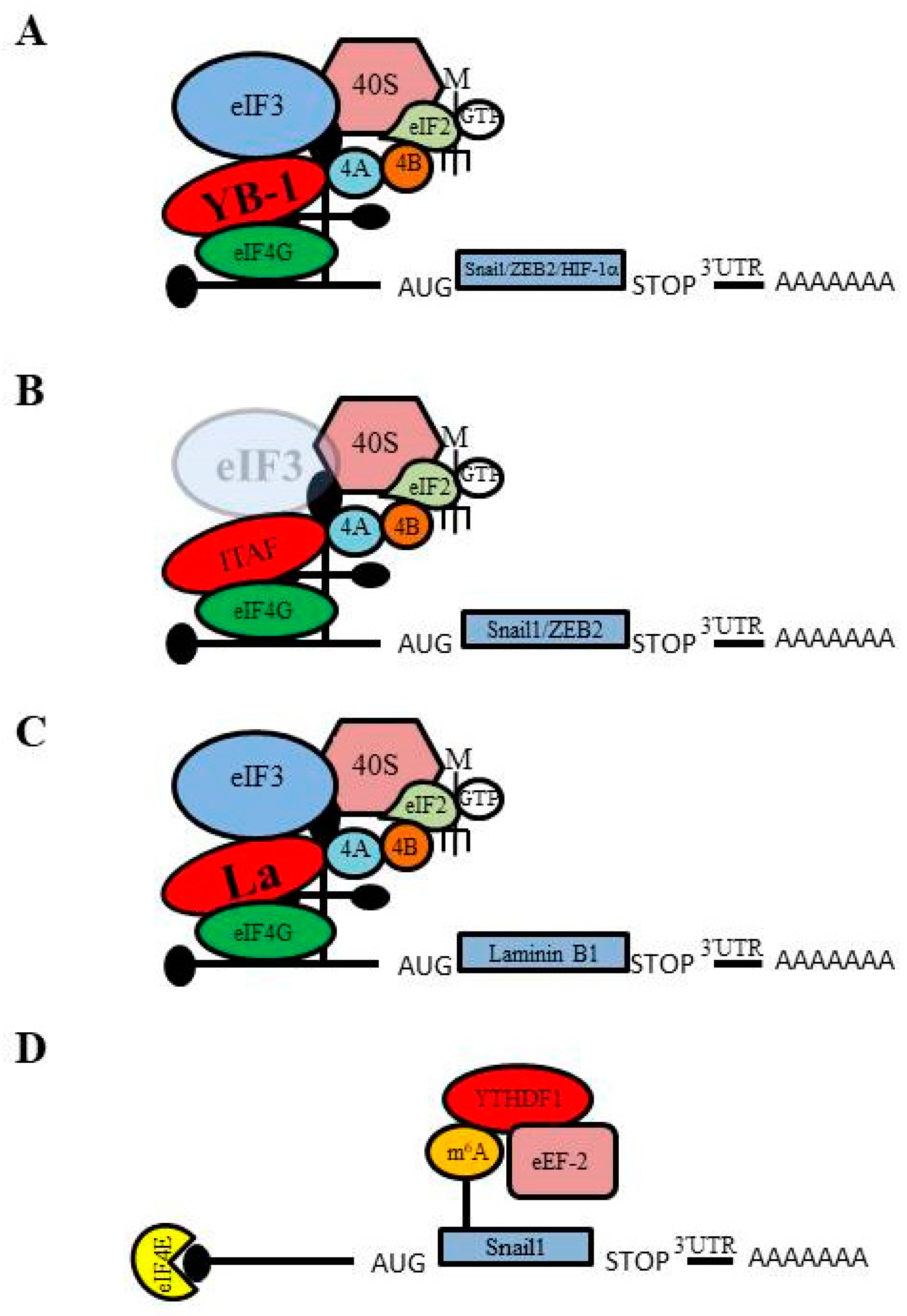
The Y-box binding protein (YB-1), a DNA/RNA-binding protein with a conserved cold-shock domain, is associated with cancer aggressiveness [91,92]; however, the exact role of YB-1 in cancer is unclear and has different effects depending on its subcellular localization. YB-1 behaves as oncogene in the nucleus, where it acts as a transcriptional activator by binding to Y-box elements in the promoter regions of pro-growth genes to induce proliferation [91]. In contrast, YB-1 acts as tumor suppressor in the cytoplasm where it can inhibit cap-dependent translation of pro-growth genes by binding at 5ʹ-UTRs to restrict binding of eIF4E to the 5ʹ-m7G cap [91]. Evdokimova et al. first reported that YB-1 regulates EMT through a novel mechanism involving the cap-independent translation of mRNAs that encode EMT regulators in Ras-transformed cells [35]. They found that YB-1 is capable of inducing an EMT-like morphological change in H-Ras-transformed human MCF-10A (MCF10-AT) and rodent EpH4 mammary epithelial cells, as well as in various carcinoma cell lines, including the cervical HeLa cell line and the prostate PC3 cell line. In addition, microarray analysis revealed that translation of >80% of mRNAs is negatively affected by YB-1 expression, but a small subset of mRNAs shows an increase in translation. These include mRNAs encoding transcriptional inducers of EMT and metastasis, such as Snail1, Zeb2/Sip1, HIF-1α, Lef-1 and TCF4. The 5ʹ-UTR of Snail1 mRNA is predicted to form a highly stable GC-rich stem-loop structure, similar to the structure of many IRESs; indeed, cap-independent translation initiation of Snail1 was found to be activated by YB-1 binding to the Snail1 5ʹ-UTR [35]. In parallel, YB-1 overexpression inhibited cap-dependent translation of proteins involved in cell proliferation, thereby inducing proliferation arrest in Ras-transformed human breast epithelial cells. These observations suggest the critical role of YB-1 in the coordinated induction of EMT through IRES-mediated Snail1 translation and proliferation arrest [93,94].
Shortly thereafter our laboratory reported that decreased expression of the translation initiation factor eIF3e induces EMT in breast epithelial cells [23]. Like YB-1, previous studies had reported that eIF3e can act as either a tumor suppressor [95,96] or an oncogene [97] in breast cells. We showed that decreased expression of eIF3e in the breast epithelial cell lines MCF-10A and MCF-12A leads to EMT, which in turn imparts invasive and migratory properties to breast epithelial cells. We found that a decrease in eIF3e expression causes a loss of expression of the epithelial markers E-cadherin and ZO-1 and an increase in the expression of the mesenchymal markers N-cadherin and vimentin. In addition, a decrease in eIF3e expression causes reduction of proliferation and enhanced migration of breast epithelial cells, phenotypes that are typical of cells that have undergone EMT [98]. Moreover, we reported reduction of eIF3e causes a decrease in global, cap-dependent translation; however, we also observed an increase in the abundance of the EMT regulators Snail1 and Zeb2. The increase in Snail1 and Zeb2 expression is due to preferential translation of Snail1 and Zeb2 mRNAs in cells that have reduced eIF3e expression. In a follow-up study we showed that reduced expression of eIF3e also induces EMT in lung epithelial cells (A549; [99]). Like breast epithelial cells, in lung epithelial cells a decrease in eIF3e expression causes loss of expression of the epithelial markers E-cadherin, increased expression of the mesenchymal markers N-cadherin and vimentin, reduced proliferation and enhanced migration.
We have found that in addition to inducing EMT, a reduction in eIF3e expression also causes a repression in global cap-dependent translation while simultaneously favoring the cap-independent translation (such as IRES-mediated translation) of mRNAs that rely on alternative translation initiation mechanisms, which are similar to the observations of Chiluiza et al. for the effects of a mutant form of eIF3e [100]. Chiluiza et al. reported that expression of a truncated version of eIF3e, similar to an isoform previously observed in breast tumor cells [100], causes a shift from cap-dependent to cap-independent IRES-mediated translation [100]. Using polysome profiling we examined the translation of mRNAs that encode proteins that regulate EMT, such as Snail1, ZEB2, Vimentin and Slug, in eIF3e-deficient A549 cells and found that ZEB2 and Vimentin translation is increased, whereas translation of Snail1 and Slug is maintained despite a decrease in global, cap-dependent translation [99]. Interestingly, by performing a β-galactosidase/CAT bicistronic reporter assay for IRES activity we found that the 5ʹ-UTRs of ZEB2, SNAI1, and vimentin exhibit IRES activity in A549 cells [99], suggesting that enhanced IRES-dependent translation of ZEB2, Snail1, and vimentin is responsible for their increased or maintained translation in eIF3e-deficient cells. Altogether, these observations indicate that upon reduced eIF3e expression in lung and breast epithelial cells ZEB2, Snail1, and vimentin rely on IRES-dependent translation for their expression and subsequent mediation of EMT [99].
Petz et al. reported that the enhanced expression of Laminin B1 (LamB1) during EMT correlates with elevated IRES activity [37]. Laminins constitute the extracellular matrix (ECM) as the main non-collagenous glycoproteins of the basement membrane and disrupt the migratory properties of cancer cells. LamB1 regulates the laminin-mediated integrin signaling that promotes cell adhesion, motility and differentiation [101] by acting as a ligand of the monomeric laminin receptor, which drives tumor cell invasion in hepatocellular carcinoma [102]. Petz et al. showed that LamB1 is translationally upregulated through IRES-mediated translation during EMT that occurs in hepatocellular carcinoma. In addition, they identified the La protein as an important ITAF that drives LamB1 translation. La is an RNA-binding protein involved in RNA processing and translation that predominantly localizes in the nucleus [102,103]. La had previously been shown to activate the IRES-dependent translation of XIAP, which supports tumorigenesis by preventing apoptosis [104]. During hepatocellular carcinoma EMT, La accumulates in the cytoplasm where it binds to the LamB1 IRES to enhance IRES-mediated translation [102]. This increased LamB1 translation results in upregulation of laminin-mediated integrin signaling through binding of LamB1 to the laminin and β1 integrin receptors, which were both found to be overexpressed in hepatocellular carcinoma [105,106], thereby leading to tumor cell invasion. Since LamB1 plays important role in the regulation of cell migration [107], increased IRES-dependent translation of LamB1 during EMT contributes to tumor cell migration and metastasis. Moreover, in a follow-up study it was shown that platelet-derived growth factor (PDGF) enhances the IRES activity of the LamB1 5ʹ-UTR by the increasing the cytoplasmic localization of La during EMT [108]. Together these findings suggest that regulated IRES-dependent translation of LamB1 is an important factor for the control of EMT in hepatocellular carcinoma.

3.2. m6A-mediated Translation and Its Regulation of EMT

Many eukaryotic mRNAs that contain N6-methyladenosine (m6A), a reversible base modification, have IRES-like activity that drives cap-independent translation initiation (Figure 2). m6A modification is the most abundant RNA modification, comprising roughly 80% of all RNA base modifications [109]. The functional effects of m6A modification on RNA are regulated by dynamic interactions among associated methyltransferases (“writers”), demethylases (“erasers”), and binding proteins (“readers”) [110,111]. Although m6A modification was first reported for the 3ʹ-UTRs of mRNAs, it can also occur within the coding region or the 5ʹ-UTR [13,112,113,114,115]. It was initially reported that m6A modification at the 3ʹ-UTR of mRNAs is responsible for the recruitment of RNA-binding proteins that affect mRNA stability [112]. In 2015, Meyer et al. reported that m6A modification at the 5ʹ-UTR of mRNAs plays a significant role for cap-independent translation initiation [13]; the m6A modification sites responsible for this cap-independent translation are referred to as ‘‘m6A-induced ribosome engagement sites’’ (MIRES). m6A-modified, uncapped mRNAs can be translated in cell-free extracts and capped, m6A-modified mRNAs can be translated in the absence of eIF4E, providing convincing evidence that m6A modification induces cap-independent translation initiation [13,116]. Moreover, m6A modification of the 5ʹ-UTR permits direct binding of the eIF3 complex, which is sufficient to recruit the 40S ribosomal subunit to initiate translation in the absence of the cap-binding protein eIF4E [13]. The m6A-mediated translation initiation mechanism was also shown to require 5ʹ-UTR scanning [13], and therefore resembles Group IV IRES-mediated translation, such as that observed for viral mRNAs that contain an eIF4G-binding IRES within their 5ʹ-UTRs [13,117,118].
It has been reported that during heat shock a single m6A modification within the 5ʹ-UTR of HSP70 mRNA enables cap-independent translation, providing a mechanism for selective mRNA translation under cellular stress [14]. Moreover, it is well-documented that HSP70 mRNA possesses an IRES within its 5ʹ-UTR [119]. At this point it remains unclear whether HSP70 utilizes both translation initiation mechanisms together or separately to increase cap-independent translation during heat shock. In addition, it is interesting to note that m6A modification of 5ʹ-UTRs is dynamic, and can be induced by UV irradiation, interferon-γ exposure, and heat shock [13,14]; however, in 2017, Ke et al. reported that m6A modification of mRNA occurs predominantly in the nucleus prior to mRNA splicing [120], suggesting that such regulation in response to stress would need to occur during mRNA biogenesis.
m6A modification of mRNA has been found to be directly associated with EMT and several reports suggest that m6A modification plays a multi-functional role in TGF-β expression and EMT regulation [121,122,123]. The functional effects of m6A on mRNA are regulated by dynamic interactions among (1) methyltransferases, such as catalytic subunit methyltransferase like 3 (METTL3) and its cofactor (METTL14) (“writers”); (2) demethylases, such as FTO and ALKBH5 (“erasers”); and (3) binding proteins, such as YTHDF family proteins, eIF3 complex members, and IGF2BP family proteins (“readers”) [110,111]. Li et al. reported that TGF-β-induced EMT is inhibited in cells that have reduced expression of METTL3 [122], a “writer” that methylates adenosines, suggesting that m6A modification of mRNA is important for the EMT process. They first demonstrated that m6A modification negatively regulates the levels of TGF-β1 protein, as METTL3 is required for the m6A modification of both TGF-β1 pre-mRNA and mature mRNA that in turn results in decreased TGF-β1 protein expression. In addition, METTL3 modulates the secretion and activation of TGF-β1 by disrupting TGF-β dimer formation [122]. These findings initially suggested that METTL3 expression, and therefore m6A modification of mRNA, has a negative effect on TGF-β1-induced EMT; however, Lin et al. further found that reduced METTL3 expression downregulates Snail1, a master regulator of EMT, which results in an inhibition of TGF-β-induced EMT [124]. This finding demonstrates that m6A modification of SNAI1 mRNA plays an important role in the regulation Snail1 expression, and that this regulation of Snail1 expression by m6A modification is a key factor in the progression of TGF-β1-induced EMT. Moreover, this observation raised the possibility that m6A modification of SNAI1 mRNA may regulate Snail1 abundance by affecting its translation, thereby controlling its activity during EMT. These findings suggest that m6A modification of mRNA is important for regulating the EMT process, and that this occurs by modulating the abundance of proteins that are important for EMT.
Overall m6A modification of mRNA was found to increase during EMT, including the m6A modification of SNAI1 mRNA [124]. Sequence analysis to detect m6A modification of mRNA on a global scale identified a ≥1.5-fold increase in the m6A modification of 128 mRNAs during EMT, several of which were found by gene ontology analysis to encode proteins related to cell migration and adherens junctions [124]. Moreover, this study found that the EMT-specific transcription factor Snail1 is affected by m6A modification during EMT. SNAI1 mRNA was found to be m6A modified within its coding sequence (CDS) and 3ʹ-UTR regions and showed a significant 2.3-fold enrichment in m6A modification during EMT progression. Gain- and loss-of-function experiments suggest that m6A modification within the SNAI1 CDS can enhance Snail1 translation elongation via interaction with YTHDF1, a “reader” that recognizes m6A-modified mRNA, which promotes the recruitment of the eEF-2 translation elongation factor and thereby enhances Snail1 translation [124]. Together, these findings demonstrate that m6A modification of mRNA can regulate the progression of EMT by affecting the translation of Snail1.
It is still early days for the exploration of the role of m6A modification of mRNA in the regulation of EMT. Given that there are at least 128 mRNAs whose m6A modification is increased during EMT [124], and that m6A modification has been shown to mediate cap-independent translation [13], it will be interesting to explore the effects m6A-mediated translation on the expression of proteins that are important for EMT. It is highly likely that the cap-independent translation of these EMT-specific proteins is mediated by m6A modification, thereby conferring a further layer of control of EMT through regulated protein synthesis. Moreover, the eIF3 complex is responsible for m6A-mediated translation initiation [13] and components of the eIF3 complex, such as eIF3e, have been shown to control EMT by affecting protein synthesis [23,99]. Together these findings raise the intriguing possibility that m6A-mediated translation initiation and changes to the eIF3 complex work in tandem to regulate translation initiation during EMT.

4. Conclusions

EMT is essential for normal embryogenesis, but also promotes cancer progression, metastasis, and chemoresistance. A complex network of transcriptional regulators, epigenetic modifications, microRNA regulators, and splicing mechanisms control the execution of EMT, and a wide variety of signaling pathways have been reported to activate EMT in various normal and cancerous tissues. Moreover, it has recently emerged that cancer cells can undergo EMT by activating alternative modes of mRNA translation initiation, such as cap-independent translation, to support the synthesis of proteins with important roles in EMT (Figure 3). Although several reports have implicated IRES-mediated translation and m6A modification of mRNA in the regulation of EMT, further investigation is necessary to obtain a detailed understanding of how these translation initiation mechanisms affect and/or control EMT. This knowledge will aid in the development of novel therapeutics that target EMT and thereby metastatic cancer and chemoresistance.

Funding

Research in the laboratory of SML is supported by a grant from the Cancer Research Society, with funding from the Beatrice Hunter Cancer Research Institute (Grant #24135).

Acknowledgments

We apologize to the many investigators whose important studies could not be cited directly here because of space limitations.

Conflicts of Interest

The authors declare no conflict of interest.

References

  1. Schmeing, T.M.; Ramakrishnan, V. What recent ribosome structures have revealed about the mechanism of translation. Nature 2009, 461, 1234–1242. [Google Scholar] [CrossRef] [PubMed]
  2. Sonenberg, N.; Hinnebusch, A.G. Regulation of translation initiation in eukaryotes: Mechanisms and biological targets. Cell 2009, 136, 731–745. [Google Scholar] [CrossRef] [PubMed] [Green Version]
  3. Hershey, J.W.B.; Sonenberg, N.; Mathews, M.B. Principles of Translational Control: An Overview. Cold Spring Harb. Perspect. Biol. 2012, 4, a011528. [Google Scholar] [CrossRef] [PubMed]
  4. Jackson, R.J.; Hellen, C.U.T.; Pestova, T.V. The mechanism of eukaryotic translation initiation and principles of its regulation. Nat. Rev. Mol. Cell Biol. 2010, 11, 113–127. [Google Scholar] [CrossRef] [Green Version]
  5. Topisirovic, I.; Sonenberg, N. mRNA translation and energy metabolism in cancer: The role of the MAPK and mTORC1 pathways. Cold Spring Harb. Symp. Quant. Biol. 2011, 76, 355–367. [Google Scholar] [CrossRef]
  6. Holcik, M.; Sonenberg, N. Translational control in stress and apoptosis. Nat. Rev. Mol. Cell Biol. 2005, 6, 318–327. [Google Scholar] [CrossRef]
  7. Kozak, M. Pushing the limits of the scanning mechanism for initiation of translation. Gene 2002, 299, 1–34. [Google Scholar] [CrossRef]
  8. Balvay, L.; Soto Rifo, R.; Ricci, E.P.; Decimo, D.; Ohlmann, T. Structural and functional diversity of viral IRESes. Biochim. Biophys. Acta 2009, 1789, 542–557. [Google Scholar] [CrossRef]
  9. Martínez-Salas, E.; Piñeiro, D.; Fernández, N. Alternative Mechanisms to Initiate Translation in Eukaryotic mRNAs. Comp. Funct. Genom. 2012, 2012, 1–12. [Google Scholar] [CrossRef] [Green Version]
  10. Jackson, R.J. The Current Status of Vertebrate Cellular mRNA IRESs. Cold Spring Harb. Perspect. Biol. 2013, 5, a011569. [Google Scholar] [CrossRef]
  11. Hellen, C.U.T.; Sarnow, P. Internal ribosome entry sites in eukaryotic mRNA molecules. Genes Dev. 2001, 15, 1593–1612. [Google Scholar] [CrossRef] [PubMed] [Green Version]
  12. Zhou, J.; Rode, K.A.; Qian, S.-B. m(6)A: A novel hallmark of translation. Cell Cycle 2016, 15, 309–310. [Google Scholar] [CrossRef] [PubMed] [Green Version]
  13. Meyer, K.D.; Patil, D.P.; Zhou, J.; Zinoviev, A.; Skabkin, M.A.; Elemento, O.; Pestova, T.V.; Qian, S.-B.; Jaffrey, S.R. 5′ UTR m(6)A Promotes Cap-Independent Translation. Cell 2015, 163, 999–1010. [Google Scholar] [CrossRef] [PubMed] [Green Version]
  14. Zhou, J.; Wan, J.; Gao, X.; Zhang, X.; Jaffrey, S.R.; Qian, S.-B. Dynamic m(6)A mRNA methylation directs translational control of heat shock response. Nature 2015, 526, 591–594. [Google Scholar] [CrossRef] [Green Version]
  15. Niu, Y.; Zhao, X.; Wu, Y.-S.; Li, M.-M.; Wang, X.-J.; Yang, Y.-G. N6-methyl-adenosine (m6A) in RNA: An old modification with a novel epigenetic function. Genom. Proteom. Bioinform. 2013, 11, 8–17. [Google Scholar] [CrossRef] [Green Version]
  16. Koumenis, C.; Naczki, C.; Koritzinsky, M.; Rastani, S.; Diehl, A.; Sonenberg, N.; Koromilas, A.; Wouters, B.G. Regulation of protein synthesis by hypoxia via activation of the endoplasmic reticulum kinase PERK and phosphorylation of the translation initiation factor eIF2alpha. Mol. Cell. Biol. 2002, 22, 7405–7416. [Google Scholar] [CrossRef] [Green Version]
  17. Liu, B.; Qian, S.-B. Translational reprogramming in cellular stress response. Wiley Interdiscip. Rev. RNA 2014, 5, 301–315. [Google Scholar] [CrossRef]
  18. Sharma, D.K.; Bressler, K.; Patel, H.; Balasingam, N.; Thakor, N. Role of Eukaryotic Initiation Factors during Cellular Stress and Cancer Progression. J. Nucleic Acids 2016, 2016, 8235121. [Google Scholar] [CrossRef] [Green Version]
  19. Silvera, D.; Formenti, S.C.; Schneider, R.J. Translational control in cancer. Nat. Rev. Cancer 2010, 10, 254–266. [Google Scholar] [CrossRef]
  20. Ruggero, D. Translational control in cancer etiology. Cold Spring Harb. Perspect. Biol. 2013, 5. [Google Scholar] [CrossRef]
  21. Lacerda, R.; Menezes, J.; Romão, L. More than just scanning: The importance of cap-independent mRNA translation initiation for cellular stress response and cancer. Cell. Mol. Life Sci. 2017, 74, 1659–1680. [Google Scholar] [CrossRef] [PubMed]
  22. Robichaud, N.; Sonenberg, N.; Ruggero, D.; Schneider, R.J. Translational Control in Cancer. Cold Spring Harb. Perspect. Biol. 2019, 11. [Google Scholar] [CrossRef] [PubMed] [Green Version]
  23. Gillis, L.D.; Lewis, S.M. Decreased eIF3e/Int6 expression causes epithelial-to-mesenchymal transition in breast epithelial cells. Oncogene 2013, 32, 3598–3605. [Google Scholar] [CrossRef] [PubMed] [Green Version]
  24. Kalluri, R.; Weinberg, R.A. The basics of epithelial-mesenchymal transition. J. Clin. Investig. 2009, 119, 1420–1428. [Google Scholar] [CrossRef] [Green Version]
  25. Nieto, M.A.; Huang, R.Y.-J.; Jackson, R.A.; Thiery, J.P. EMT: 2016. Cell 2016, 166, 21–45. [Google Scholar] [CrossRef] [Green Version]
  26. Thiery, J.P.; Acloque, H.; Huang, R.Y.J.; Nieto, M.A. Epithelial-mesenchymal transitions in development and disease. Cell 2009, 139, 871–890. [Google Scholar] [CrossRef]
  27. Kurrey, N.K.; Jalgaonkar, S.P.; Joglekar, A.V.; Ghanate, A.D.; Chaskar, P.D.; Doiphode, R.Y.; Bapat, S.A. Snail and slug mediate radioresistance and chemoresistance by antagonizing p53-mediated apoptosis and acquiring a stem-like phenotype in ovarian cancer cells. Stem Cells 2009, 27, 2059–2068. [Google Scholar] [CrossRef]
  28. Ansieau, S.; Bastid, J.; Doreau, A.; Morel, A.-P.; Bouchet, B.P.; Thomas, C.; Fauvet, F.; Puisieux, I.; Doglioni, C.; Piccinin, S.; et al. Induction of EMT by twist proteins as a collateral effect of tumor-promoting inactivation of premature senescence. Cancer Cell 2008, 14, 79–89. [Google Scholar] [CrossRef] [Green Version]
  29. Zeisberg, M.; Neilson, E.G. Biomarkers for epithelial-mesenchymal transitions. J. Clin. Investig. 2009, 119, 1429–1437. [Google Scholar] [CrossRef] [Green Version]
  30. Barberà, M.J.; Puig, I.; Domínguez, D.; Julien-Grille, S.; Guaita-Esteruelas, S.; Peiró, S.; Baulida, J.; Francí, C.; Dedhar, S.; Larue, L.; et al. Regulation of Snail transcription during epithelial to mesenchymal transition of tumor cells. Oncogene 2004, 23, 7345–7354. [Google Scholar] [CrossRef] [Green Version]
  31. Luo, D.; Wang, J.; Li, J.; Post, M. Mouse snail is a target gene for HIF. Mol. Cancer Res. 2011, 9, 234–245. [Google Scholar] [CrossRef] [PubMed] [Green Version]
  32. Gregory, P.A.; Bert, A.G.; Paterson, E.L.; Barry, S.C.; Tsykin, A.; Farshid, G.; Vadas, M.A.; Khew-Goodall, Y.; Goodall, G.J. The miR-200 family and miR-205 regulate epithelial to mesenchymal transition by targeting ZEB1 and SIP1. Nat. Cell Biol. 2008, 10, 593–601. [Google Scholar] [CrossRef] [PubMed]
  33. Korpal, M.; Lee, E.S.; Hu, G.; Kang, Y. The miR-200 family inhibits epithelial-mesenchymal transition and cancer cell migration by direct targeting of E-cadherin transcriptional repressors ZEB1 and ZEB2. J. Biol. Chem. 2008, 283, 14910–14914. [Google Scholar] [CrossRef] [PubMed] [Green Version]
  34. Zhou, B.P.; Deng, J.; Xia, W.; Xu, J.; Li, Y.M.; Gunduz, M.; Hung, M.-C. Dual regulation of Snail by GSK-3beta-mediated phosphorylation in control of epithelial-mesenchymal transition. Nat. Cell Biol. 2004, 6, 931–940. [Google Scholar] [CrossRef]
  35. Evdokimova, V.; Tognon, C.; Ng, T.; Ruzanov, P.; Melnyk, N.; Fink, D.; Sorokin, A.; Ovchinnikov, L.P.; Davicioni, E.; Triche, T.J.; et al. Translational activation of snail1 and other developmentally regulated transcription factors by YB-1 promotes an epithelial-mesenchymal transition. Cancer Cell 2009, 15, 402–415. [Google Scholar] [CrossRef] [Green Version]
  36. Waerner, T.; Alacakaptan, M.; Tamir, I.; Oberauer, R.; Gal, A.; Brabletz, T.; Schreiber, M.; Jechlinger, M.; Beug, H. ILEI: A cytokine essential for EMT, tumor formation, and late events in metastasis in epithelial cells. Cancer Cell 2006, 10, 227–239. [Google Scholar] [CrossRef] [Green Version]
  37. Petz, M.; Kozina, D.; Huber, H.; Siwiec, T.; Seipelt, J.; Sommergruber, W.; Mikulits, W. The leader region of Laminin B1 mRNA confers cap-independent translation. Nucleic Acids Res. 2007, 35, 2473–2482. [Google Scholar] [CrossRef] [Green Version]
  38. Chaudhury, A.; Hussey, G.S.; Ray, P.S.; Jin, G.; Fox, P.L.; Howe, P.H. TGF-beta-mediated phosphorylation of hnRNP E1 induces EMT via transcript-selective translational induction of Dab2 and ILEI. Nat. Cell Biol. 2010, 12, 286–293. [Google Scholar] [CrossRef] [Green Version]
  39. Chaudhury, A.; Hussey, G.S.; Howe, P.H. 3′-UTR-mediated post-transcriptional regulation of cancer metastasis: Beginning at the end. RNA Biol. 2011, 8, 595–599. [Google Scholar] [CrossRef] [Green Version]
  40. Hanahan, D.; Weinberg, R.A. Hallmarks of cancer: The next generation. Cell 2011, 144, 646–674. [Google Scholar] [CrossRef] [Green Version]
  41. Yeung, K.T.; Yang, J. Epithelial-mesenchymal transition in tumor metastasis. Mol. Oncol. 2017, 11, 28–39. [Google Scholar] [CrossRef] [PubMed] [Green Version]
  42. Sun, T.; Qin, Y.; Zhong, W. Epithelial-Mesenchymal Transition and its Regulation in Tumor Metastasis. Tumor Metastasis 2016. [Google Scholar] [CrossRef] [Green Version]
  43. Pradella, D.; Naro, C.; Sette, C.; Ghigna, C. EMT and stemness: Flexible processes tuned by alternative splicing in development and cancer progression. Mol. Cancer 2017, 16, 8. [Google Scholar] [CrossRef] [PubMed] [Green Version]
  44. Rhim, A.D.; Mirek, E.T.; Aiello, N.M.; Maitra, A.; Bailey, J.M.; McAllister, F.; Reichert, M.; Beatty, G.L.; Rustgi, A.K.; Vonderheide, R.H.; et al. EMT and dissemination precede pancreatic tumor formation. Cell 2012, 148, 349–361. [Google Scholar] [CrossRef] [PubMed] [Green Version]
  45. Iwatsuki, M.; Mimori, K.; Yokobori, T.; Ishi, H.; Beppu, T.; Nakamori, S.; Baba, H.; Mori, M. Epithelial-mesenchymal transition in cancer development and its clinical significance. Cancer Sci. 2010, 101, 293–299. [Google Scholar] [CrossRef] [PubMed]
  46. Mehlen, P.; Puisieux, A. Metastasis: A question of life or death. Nat. Rev. Cancer 2006, 6, 449–458. [Google Scholar] [CrossRef]
  47. Aleskandarany, M.A.; Negm, O.H.; Green, A.R.; Ahmed, M.A.H.; Nolan, C.C.; Tighe, P.J.; Ellis, I.O.; Rakha, E.A. Epithelial mesenchymal transition in early invasive breast cancer: An immunohistochemical and reverse phase protein array study. Breast Cancer Res. Treat. 2014, 145, 339–348. [Google Scholar] [CrossRef]
  48. Choi, Y.; Lee, H.J.; Jang, M.H.; Gwak, J.M.; Lee, K.S.; Kim, E.J.; Kim, H.J.; Lee, H.E.; Park, S.Y. Epithelial-mesenchymal transition increases during the progression of in situ to invasive basal-like breast cancer. Hum. Pathol. 2013, 44, 2581–2589. [Google Scholar] [CrossRef]
  49. Klein, C.A. Parallel progression of primary tumours and metastases. Nat. Rev. Cancer 2009, 9, 302–312. [Google Scholar] [CrossRef]
  50. Pantel, K.; Woelfle, U. Micrometastasis in breast cancer and other solid tumors. J. Biol. Regul. Homeost. Agents 2004, 18, 120–125. [Google Scholar]
  51. Heerboth, S.; Housman, G.; Leary, M.; Longacre, M.; Byler, S.; Lapinska, K.; Willbanks, A.; Sarkar, S. EMT and tumor metastasis. Clin. Transl. Med. 2015, 4, 6. [Google Scholar] [CrossRef] [PubMed]
  52. Chatterjee, S.; Seifried, L.; Feigin, M.E.; Gibbons, D.L.; Scuoppo, C.; Lin, W.; Rizvi, Z.H.; Lind, E.; Dissanayake, D.; Kurie, J.; et al. Dysregulation of cell polarity proteins synergize with oncogenes or the microenvironment to induce invasive behavior in epithelial cells. PLoS ONE 2012, 7, e34343. [Google Scholar] [CrossRef] [PubMed] [Green Version]
  53. Lee, M.; Vasioukhin, V. Cell polarity and cancer-cell and tissue polarity as a non-canonical tumor suppressor. J. Cell Sci. 2008, 121, 1141–1150. [Google Scholar] [CrossRef] [PubMed] [Green Version]
  54. Finak, G.; Bertos, N.; Pepin, F.; Sadekova, S.; Souleimanova, M.; Zhao, H.; Chen, H.; Omeroglu, G.; Meterissian, S.; Omeroglu, A.; et al. Stromal gene expression predicts clinical outcome in breast cancer. Nat. Med. 2008, 14, 518–527. [Google Scholar] [CrossRef]
  55. Noë, V.; Fingleton, B.; Jacobs, K.; Crawford, H.C.; Vermeulen, S.; Steelant, W.; Bruyneel, E.; Matrisian, L.M.; Mareel, M. Release of an invasion promoter E-cadherin fragment by matrilysin and stromelysin-1. J. Cell Sci. 2001, 114, 111–118. [Google Scholar]
  56. Creighton, C.J.; Li, X.; Landis, M.; Dixon, J.M.; Neumeister, V.M.; Sjolund, A.; Rimm, D.L.; Wong, H.; Rodriguez, A.; Herschkowitz, J.I.; et al. Residual breast cancers after conventional therapy display mesenchymal as well as tumor-initiating features. Proc. Natl. Acad. Sci. USA 2009, 106, 13820–13825. [Google Scholar] [CrossRef] [Green Version]
  57. Kawamoto, A.; Yokoe, T.; Tanaka, K.; Saigusa, S.; Toiyama, Y.; Yasuda, H.; Inoue, Y.; Miki, C.; Kusunoki, M. Radiation induces epithelial-mesenchymal transition in colorectal cancer cells. Oncol. Rep. 2012, 27, 51–57. [Google Scholar] [CrossRef] [Green Version]
  58. Shintani, Y.; Okimura, A.; Sato, K.; Nakagiri, T.; Kadota, Y.; Inoue, M.; Sawabata, N.; Minami, M.; Ikeda, N.; Kawahara, K.; et al. Epithelial to mesenchymal transition is a determinant of sensitivity to chemoradiotherapy in non-small cell lung cancer. Ann. Thorac. Surg. 2011, 92, 1794–1804. [Google Scholar] [CrossRef]
  59. Arumugam, T.; Ramachandran, V.; Fournier, K.F.; Wang, H.; Marquis, L.; Abbruzzese, J.L.; Gallick, G.E.; Logsdon, C.D.; McConkey, D.J.; Choi, W. Epithelial to mesenchymal transition contributes to drug resistance in pancreatic cancer. Cancer Res. 2009, 69, 5820–5828. [Google Scholar] [CrossRef] [Green Version]
  60. Fischer, K.R.; Durrans, A.; Lee, S.; Sheng, J.; Li, F.; Wong, S.T.C.; Choi, H.; El Rayes, T.; Ryu, S.; Troeger, J.; et al. Epithelial-to-mesenchymal transition is not required for lung metastasis but contributes to chemoresistance. Nature 2015, 527, 472–476. [Google Scholar] [CrossRef]
  61. Zheng, X.; Carstens, J.L.; Kim, J.; Scheible, M.; Kaye, J.; Sugimoto, H.; Wu, C.-C.; LeBleu, V.S.; Kalluri, R. Epithelial-to-mesenchymal transition is dispensable for metastasis but induces chemoresistance in pancreatic cancer. Nature 2015, 527, 525–530. [Google Scholar] [CrossRef] [PubMed] [Green Version]
  62. Leprivier, G.; Rotblat, B.; Khan, D.; Jan, E.; Sorensen, P.H. Stress-mediated translational control in cancer cells. Biochim. Biophys. Acta 2015, 1849, 845–860. [Google Scholar] [CrossRef] [PubMed]
  63. Jang, S.K.; Kräusslich, H.G.; Nicklin, M.J.; Duke, G.M.; Palmenberg, A.C.; Wimmer, E. A segment of the 5′ nontranslated region of encephalomyocarditis virus RNA directs internal entry of ribosomes during in vitro translation. J. Virol. 1988, 62, 2636–2643. [Google Scholar] [CrossRef] [PubMed] [Green Version]
  64. Pelletier, J.; Sonenberg, N. Internal initiation of translation of eukaryotic mRNA directed by a sequence derived from poliovirus RNA. Nature 1988, 334, 320–325. [Google Scholar] [CrossRef]
  65. Hellen, C.U.T. IRES-induced conformational changes in the ribosome and the mechanism of translation initiation by internal ribosomal entry. Biochim. Biophys. Acta 2009, 1789, 558–570. [Google Scholar] [CrossRef] [Green Version]
  66. Lozano, G.; Martínez-Salas, E. Structural insights into viral IRES-dependent translation mechanisms. Curr. Opin. Virol. 2015, 12, 113–120. [Google Scholar] [CrossRef] [Green Version]
  67. Komar, A.A.; Hatzoglou, M. Internal ribosome entry sites in cellular mRNAs: Mystery of their existence. J. Biol. Chem. 2005, 280, 23425–23428. [Google Scholar] [CrossRef] [Green Version]
  68. Kieft, J.S. Viral IRES RNA structures and ribosome interactions. Trends Biochem. Sci. 2008, 33, 274–283. [Google Scholar] [CrossRef] [Green Version]
  69. Filbin, M.E.; Kieft, J.S. Toward a structural understanding of IRES RNA function. Curr. Opin. Struct. Biol. 2009, 19, 267–276. [Google Scholar] [CrossRef] [Green Version]
  70. Kieft, J.S.; Zhou, K.; Jubin, R.; Murray, M.G.; Lau, J.Y.; Doudna, J.A. The hepatitis C virus internal ribosome entry site adopts an ion-dependent tertiary fold. J. Mol. Biol. 1999, 292, 513–529. [Google Scholar] [CrossRef] [Green Version]
  71. Pestova, T.V.; Hellen, C.U.; Shatsky, I.N. Canonical eukaryotic initiation factors determine initiation of translation by internal ribosomal entry. Mol. Cell. Biol. 1996, 16, 6859–6869. [Google Scholar] [CrossRef] [PubMed] [Green Version]
  72. Sweeney, T.R.; Abaeva, I.S.; Pestova, T.V.; Hellen, C.U.T. The mechanism of translation initiation on Type 1 picornavirus IRESs. EMBO J. 2014, 33, 76–92. [Google Scholar] [CrossRef] [PubMed] [Green Version]
  73. Spriggs, K.A.; Stoneley, M.; Bushell, M.; Willis, A.E. Re-programming of translation following cell stress allows IRES-mediated translation to predominate. Biol. Cell 2008, 100, 27–38. [Google Scholar] [CrossRef] [PubMed]
  74. Macejak, D.G.; Sarnow, P. Internal initiation of translation mediated by the 5′ leader of a cellular mRNA. Nature 1991, 353, 90–94. [Google Scholar] [CrossRef] [PubMed]
  75. Johannes, G.; Carter, M.S.; Eisen, M.B.; Brown, P.O.; Sarnow, P. Identification of eukaryotic mRNAs that are translated at reduced cap binding complex eIF4F concentrations using a cDNA microarray. Proc. Natl. Acad. Sci. USA 1999, 96, 13118–13123. [Google Scholar] [CrossRef] [Green Version]
  76. Weingarten-Gabbay, S.; Elias-Kirma, S.; Nir, R.; Gritsenko, A.A.; Stern-Ginossar, N.; Yakhini, Z.; Weinberger, A.; Segal, E. Comparative genetics. Systematic discovery of cap-independent translation sequences in human and viral genomes. Science 2016, 351. [Google Scholar] [CrossRef]
  77. Mokrejs, M.; Masek, T.; Vopálensky, V.; Hlubucek, P.; Delbos, P.; Pospísek, M. IRESite—A tool for the examination of viral and cellular internal ribosome entry sites. Nucleic Acids Res. 2010, 38, D131–D136. [Google Scholar] [CrossRef] [Green Version]
  78. Shi, Y.; Yang, Y.; Hoang, B.; Bardeleben, C.; Holmes, B.; Gera, J.; Lichtenstein, A. Therapeutic potential of targeting IRES-dependent c-myc translation in multiple myeloma cells during ER stress. Oncogene 2016, 35, 1015–1024. [Google Scholar] [CrossRef] [Green Version]
  79. Bonnal, S.; Boutonnet, C.; Prado-Lourenço, L.; Vagner, S. IRESdb: The Internal Ribosome Entry Site database. Nucleic Acids Res. 2003, 31, 427–428. [Google Scholar] [CrossRef]
  80. Philippe, C.; Dubrac, A.; Quelen, C.; Desquesnes, A.; Van Den Berghe, L.; Ségura, C.; Filleron, T.; Pyronnet, S.; Prats, H.; Brousset, P.; et al. PERK mediates the IRES-dependent translational activation of mRNAs encoding angiogenic growth factors after ischemic stress. Sci. Signal. 2016, 9, ra44. [Google Scholar] [CrossRef]
  81. Khan, D.; Katoch, A.; Das, A.; Sharathchandra, A.; Lal, R.; Roy, P.; Das, S.; Chattopadhyay, S.; Das, S. Reversible induction of translational isoforms of p53 in glucose deprivation. Cell Death Differ. 2015, 22, 1203–1218. [Google Scholar] [CrossRef] [PubMed] [Green Version]
  82. Liberman, N.; Gandin, V.; Svitkin, Y.V.; David, M.; Virgili, G.; Jaramillo, M.; Holcik, M.; Nagar, B.; Kimchi, A.; Sonenberg, N. DAP5 associates with eIF2β and eIF4AI to promote Internal Ribosome Entry Site driven translation. Nucleic Acids Res. 2015, 43, 3764–3775. [Google Scholar] [CrossRef] [PubMed] [Green Version]
  83. Vaklavas, C.; Grizzle, W.E.; Choi, H.; Meng, Z.; Zinn, K.R.; Shrestha, K.; Blume, S.W. IRES inhibition induces terminal differentiation and synchronized death in triple-negative breast cancer and glioblastoma cells. Tumour Biol. 2016, 37, 13247–13264. [Google Scholar] [CrossRef] [PubMed] [Green Version]
  84. Komar, A.A.; Hatzoglou, M. Cellular IRES-mediated translation: The war of ITAFs in pathophysiological states. Cell Cycle 2011, 10, 229–240. [Google Scholar] [CrossRef] [PubMed] [Green Version]
  85. Lewis, S.M.; Holcik, M. For IRES trans-acting factors, it is all about location. Oncogene 2008, 27, 1033–1035. [Google Scholar] [CrossRef]
  86. Spriggs, K.A.; Bushell, M.; Mitchell, S.A.; Willis, A.E. Internal ribosome entry segment-mediated translation during apoptosis: The role of IRES-trans-acting factors. Cell Death Differ. 2005, 12, 585–591. [Google Scholar] [CrossRef] [Green Version]
  87. Sawicka, K.; Bushell, M.; Spriggs, K.A.; Willis, A.E. Polypyrimidine-tract-binding protein: A multifunctional RNA-binding protein. Biochem. Soc. Trans. 2008, 36, 641–647. [Google Scholar] [CrossRef] [Green Version]
  88. King, H.A.; Cobbold, L.C.; Willis, A.E. The role of IRES trans-acting factors in regulating translation initiation. Biochem. Soc. Trans. 2010, 38, 1581–1586. [Google Scholar] [CrossRef] [Green Version]
  89. Cobbold, L.C.; Spriggs, K.A.; Haines, S.J.; Dobbyn, H.C.; Hayes, C.; de Moor, C.H.; Lilley, K.S.; Bushell, M.; Willis, A.E. Identification of internal ribosome entry segment (IRES)-trans-acting factors for the Myc family of IRESs. Mol. Cell. Biol. 2008, 28, 40–49. [Google Scholar] [CrossRef] [Green Version]
  90. Semler, B.L.; Waterman, M.L. IRES-mediated pathways to polysomes: Nuclear versus cytoplasmic routes. Trends Microbiol. 2008, 16, 1–5. [Google Scholar] [CrossRef]
  91. Evdokimova, V.; Ovchinnikov, L.P.; Sorensen, P.H.B. Y-box binding protein 1: Providing a new angle on translational regulation. Cell Cycle 2006, 5, 1143–1147. [Google Scholar] [CrossRef] [PubMed]
  92. Bader, A.G.; Vogt, P.K. Inhibition of protein synthesis by Y box-binding protein 1 blocks oncogenic cell transformation. Mol. Cell. Biol. 2005, 25, 2095–2106. [Google Scholar] [CrossRef] [PubMed] [Green Version]
  93. Vega, S.; Morales, A.V.; Ocaña, O.H.; Valdés, F.; Fabregat, I.; Nieto, M.A. Snail blocks the cell cycle and confers resistance to cell death. Genes Dev. 2004, 18, 1131–1143. [Google Scholar] [CrossRef] [PubMed] [Green Version]
  94. Barrallo-Gimeno, A.; Nieto, M.A. The Snail genes as inducers of cell movement and survival: Implications in development and cancer. Development 2005, 132, 3151–3161. [Google Scholar] [CrossRef] [PubMed] [Green Version]
  95. Suo, J.; Snider, S.J.; Mills, G.B.; Creighton, C.J.; Chen, A.C.; Schiff, R.; Lloyd, R.E.; Chang, E.C. Int6 regulates both proteasomal degradation and translation initiation and is critical for proper formation of acini by human mammary epithelium. Oncogene 2011, 30, 724–736. [Google Scholar] [CrossRef] [Green Version]
  96. Mack, D.L.; Boulanger, C.A.; Callahan, R.; Smith, G.H. Expression of truncated Int6/eIF3e in mammary alveolar epithelium leads to persistent hyperplasia and tumorigenesis. Breast Cancer Res. 2007, 9, R42. [Google Scholar] [CrossRef] [Green Version]
  97. Grzmil, M.; Rzymski, T.; Milani, M.; Harris, A.L.; Capper, R.G.; Saunders, N.J.; Salhan, A.; Ragoussis, J.; Norbury, C.J. An oncogenic role of eIF3e/INT6 in human breast cancer. Oncogene 2010, 29, 4080–4089. [Google Scholar] [CrossRef]
  98. Evdokimova, V.; Tognon, C.; Ng, T.; Sorensen, P.H.B. Reduced proliferation and enhanced migration: Two sides of the same coin? Molecular mechanisms of metastatic progression by YB-1. Cell Cycle 2009, 8, 2901–2906. [Google Scholar] [CrossRef]
  99. Desnoyers, G.; Frost, L.D.; Courteau, L.; Wall, M.L.; Lewis, S.M. Decreased eIF3e Expression Can Mediate Epithelial-to-Mesenchymal Transition through Activation of the TGFβ Signaling Pathway. Mol. Cancer Res. 2015, 13, 1421–1430. [Google Scholar] [CrossRef] [Green Version]
  100. Chiluiza, D.; Bargo, S.; Callahan, R.; Rhoads, R.E. Expression of truncated eukaryotic initiation factor 3e (eIF3e) resulting from integration of mouse mammary tumor virus (MMTV) causes a shift from cap-dependent to cap-independent translation. J. Biol. Chem. 2011, 286, 31288–31296. [Google Scholar] [CrossRef] [Green Version]
  101. Taniguchi, Y.; Ido, H.; Sanzen, N.; Hayashi, M.; Sato-Nishiuchi, R.; Futaki, S.; Sekiguchi, K. The C-terminal region of laminin beta chains modulates the integrin binding affinities of laminins. J. Biol. Chem. 2009, 284, 7820–7831. [Google Scholar] [CrossRef] [PubMed] [Green Version]
  102. Petz, M.; Them, N.; Huber, H.; Beug, H.; Mikulits, W. La enhances IRES-mediated translation of laminin B1 during malignant epithelial to mesenchymal transition. Nucleic Acids Res. 2012, 40, 290–302. [Google Scholar] [CrossRef] [PubMed] [Green Version]
  103. Wolin, S.L.; Cedervall, T. The La protein. Annu. Rev. Biochem. 2002, 71, 375–403. [Google Scholar] [CrossRef] [PubMed]
  104. Holcik, M.; Korneluk, R.G. Functional characterization of the X-linked inhibitor of apoptosis (XIAP) internal ribosome entry site element: Role of La autoantigen in XIAP translation. Mol. Cell. Biol. 2000, 20, 4648–4657. [Google Scholar] [CrossRef] [PubMed] [Green Version]
  105. Liu, L.-X.; Jiang, H.-C.; Liu, Z.-H.; Zhou, J.; Zhang, W.-H.; Zhu, A.-L.; Wang, X.-Q.; Wu, M. Integrin gene expression profiles of human hepatocellular carcinoma. World J. Gastroenterol. 2002, 8, 631–637. [Google Scholar] [CrossRef] [PubMed]
  106. Ozaki, I.; Yamamoto, K.; Mizuta, T.; Kajihara, S.; Fukushima, N.; Setoguchi, Y.; Morito, F.; Sakai, T. Differential expression of laminin receptors in human hepatocellular carcinoma. Gut 1998, 43, 837–842. [Google Scholar] [CrossRef] [Green Version]
  107. Vande Broek, I.; Vanderkerken, K.; De Greef, C.; Asosingh, K.; Straetmans, N.; Van Camp, B.; Van Riet, I. Laminin-1-induced migration of multiple myeloma cells involves the high-affinity 67 kD laminin receptor. Br. J. Cancer 2001, 85, 1387–1395. [Google Scholar] [CrossRef]
  108. Petz, M.; Them, N.C.C.; Huber, H.; Mikulits, W. PDGF enhances IRES-mediated translation of Laminin B1 by cytoplasmic accumulation of La during epithelial to mesenchymal transition. Nucleic Acids Res. 2012, 40, 9738–9749. [Google Scholar] [CrossRef]
  109. Sriram, A.; Bohlen, J.; Teleman, A.A. Translation acrobatics: How cancer cells exploit alternate modes of translational initiation. EMBO Rep. 2018, 19. [Google Scholar] [CrossRef]
  110. Roundtree, I.A.; Evans, M.E.; Pan, T.; He, C. Dynamic RNA Modifications in Gene Expression Regulation. Cell 2017, 169, 1187–1200. [Google Scholar] [CrossRef] [Green Version]
  111. Meyer, K.D.; Jaffrey, S.R. The dynamic epitranscriptome: N6-methyladenosine and gene expression control. Nat. Rev. Mol. Cell Biol. 2014, 15, 313–326. [Google Scholar] [CrossRef] [PubMed] [Green Version]
  112. Meyer, K.D.; Saletore, Y.; Zumbo, P.; Elemento, O.; Mason, C.E.; Jaffrey, S.R. Comprehensive analysis of mRNA methylation reveals enrichment in 3′ UTRs and near stop codons. Cell 2012, 149, 1635–1646. [Google Scholar] [CrossRef] [PubMed] [Green Version]
  113. Wang, Y.; Li, Y.; Toth, J.I.; Petroski, M.D.; Zhang, Z.; Zhao, J.C. N6-methyladenosine modification destabilizes developmental regulators in embryonic stem cells. Nat. Cell Biol. 2014, 16, 191–198. [Google Scholar] [CrossRef] [PubMed]
  114. Wang, X.; Lu, Z.; Gomez, A.; Hon, G.C.; Yue, Y.; Han, D.; Fu, Y.; Parisien, M.; Dai, Q.; Jia, G.; et al. N6-methyladenosine-dependent regulation of messenger RNA stability. Nature 2014, 505, 117–120. [Google Scholar] [CrossRef]
  115. Wang, X.; Zhao, B.S.; Roundtree, I.A.; Lu, Z.; Han, D.; Ma, H.; Weng, X.; Chen, K.; Shi, H.; He, C. N(6)-methyladenosine Modulates Messenger RNA Translation Efficiency. Cell 2015, 161, 1388–1399. [Google Scholar] [CrossRef] [Green Version]
  116. Mitchell, S.F.; Parker, R. Modifications on Translation Initiation. Cell 2015, 163, 796–798. [Google Scholar] [CrossRef] [Green Version]
  117. Terenin, I.M.; Andreev, D.E.; Dmitriev, S.E.; Shatsky, I.N. A novel mechanism of eukaryotic translation initiation that is neither m7G-cap-, nor IRES-dependent. Nucleic Acids Res. 2013, 41, 1807–1816. [Google Scholar] [CrossRef] [Green Version]
  118. Andreev, D.E.; Dmitriev, S.E.; Zinovkin, R.; Terenin, I.M.; Shatsky, I.N. The 5′ untranslated region of Apaf-1 mRNA directs translation under apoptosis conditions via a 5′ end-dependent scanning mechanism. FEBS Lett. 2012, 586, 4139–4143. [Google Scholar] [CrossRef] [Green Version]
  119. Sun, J.; Conn, C.S.; Han, Y.; Yeung, V.; Qian, S.-B. PI3K-mTORC1 attenuates stress response by inhibiting cap-independent Hsp70 translation. J. Biol. Chem. 2011, 286, 6791–6800. [Google Scholar] [CrossRef] [Green Version]
  120. Ke, S.; Pandya-Jones, A.; Saito, Y.; Fak, J.J.; Vågbø, C.B.; Geula, S.; Hanna, J.H.; Black, D.L.; Darnell, J.E.; Darnell, R.B. m6A mRNA modifications are deposited in nascent pre-mRNA and are not required for splicing but do specify cytoplasmic turnover. Genes Dev. 2017, 31, 990–1006. [Google Scholar] [CrossRef] [Green Version]
  121. Wanna-Udom, S.; Terashima, M.; Lyu, H.; Ishimura, A.; Takino, T.; Sakari, M.; Tsukahara, T.; Suzuki, T. The m6A methyltransferase METTL3 contributes to Transforming Growth Factor-beta-induced epithelial-mesenchymal transition of lung cancer cells through the regulation of JUNB. Biochem. Biophys. Res. Commun. 2020, 524, 150–155. [Google Scholar] [CrossRef] [PubMed]
  122. Li, J.; Chen, F.; Peng, Y.; Lv, Z.; Lin, X.; Chen, Z.; Wang, H. N6-Methyladenosine Regulates the Expression and Secretion of TGFβ1 to Affect the Epithelial-Mesenchymal Transition of Cancer Cells. Cells 2020, 9, 296. [Google Scholar] [CrossRef] [PubMed] [Green Version]
  123. Yue, B.; Song, C.; Yang, L.; Cui, R.; Cheng, X.; Zhang, Z.; Zhao, G. METTL3-mediated N6-methyladenosine modification is critical for epithelial-mesenchymal transition and metastasis of gastric cancer. Mol. Cancer 2019, 18, 142. [Google Scholar] [CrossRef] [PubMed] [Green Version]
  124. Lin, X.; Chai, G.; Wu, Y.; Li, J.; Chen, F.; Liu, J.; Luo, G.; Tauler, J.; Du, J.; Lin, S.; et al. RNA m6A methylation regulates the epithelial mesenchymal transition of cancer cells and translation of Snail. Nat. Commun. 2019, 10, 2065. [Google Scholar] [CrossRef] [PubMed]
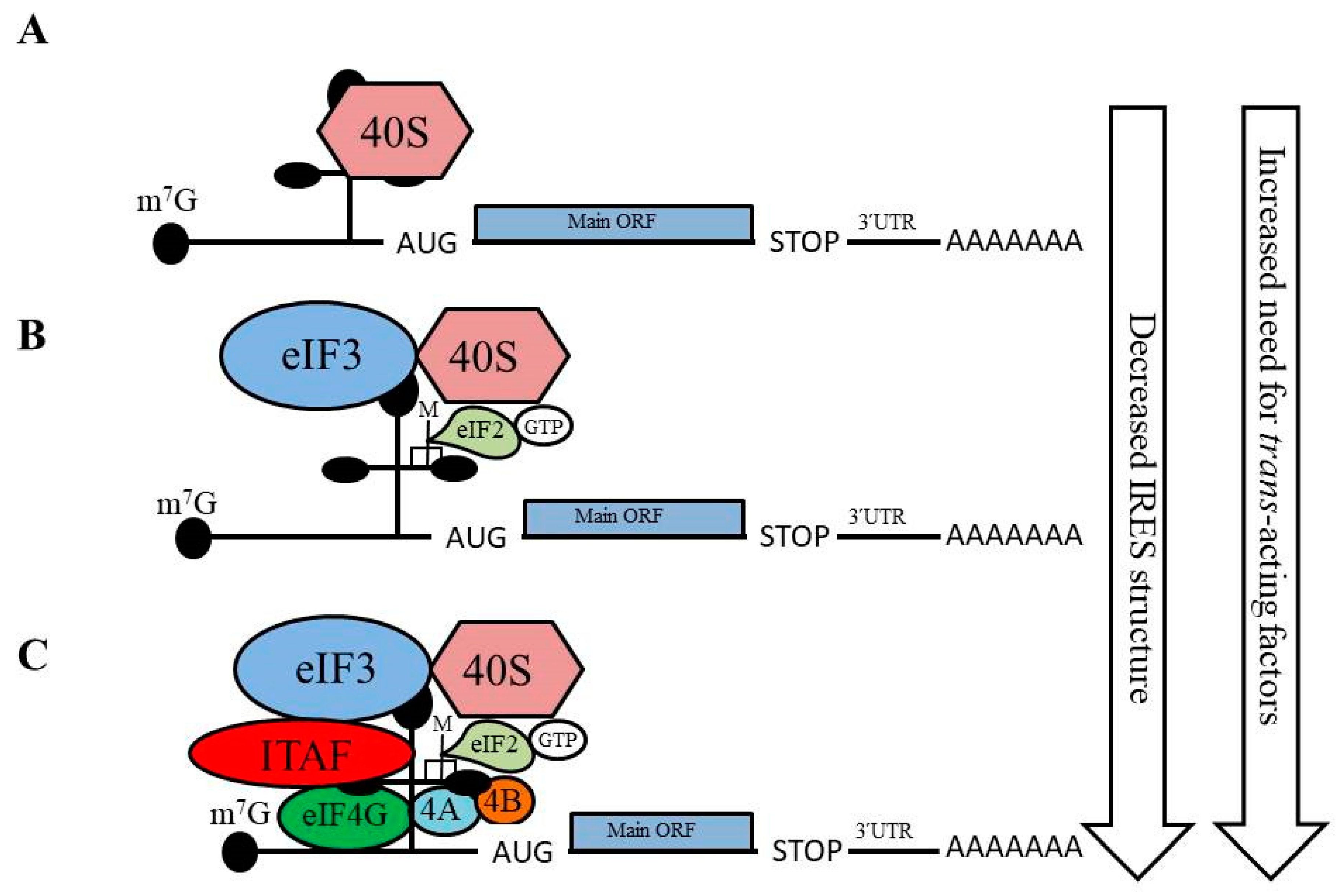
Figure 1. Overview of internal ribosome entry site (IRES)-mediated translation initiation. The requirement for canonical initiation factors and IRES trans-acting factors (ITAFs) varies among IRES-containing mRNAs. Several types of IRES-mediated translation initiation have been identified: (A) Group I: no canonical factors are required and the 40S ribosome is directly recruited to the mRNA by the IRES structure; (B) Group II: a few canonical initiation factors are required for 40S ribosome recruitment; (C) Groups III and IV: multiple canonical initiation factors, as well as ITAFs, are required for 40S ribosome recruitment and translation initiation.
Ijms 21 04075 g001
Figure 2. Overview of the role of N6-methyladenosine (m6A) modification of mRNA in protein synthesis. m6A modification of mRNA within the 5ʹ untranslated region (5ʹ-UTR) permits direct binding of the eIF3 complex to facilitate recruitment of the 43S pre-initiation complex, which mediates translation initiation. m6A modification within the coding sequence can enhance recruitment of the eEF-2 elongation factor via YDHTF1 binding to the m6A residue, which enhances translation of the mRNA.
Ijms 21 04075 g002
Figure 3. Alternative translation mechanisms are involved in the synthesis of proteins with roles in epithelial-to-mesenchymal transition (EMT). (A) The YB-1 protein recruits the translation initiation machinery to mRNAs that encode Snail1, ZEB2, and HIF-1α to support their synthesis, leading to EMT. (B) Reduced expression of the initiation factor eIF3e facilitates the translation of the Snail1 and ZEB2 proteins to mediate EMT. (C) The ITAF La mediates the translation of Laminin B1 to drive cells toward EMT. (D) m6A modifications within the Snail1 open reading frame permit enhanced recruitment of the elongation factor eEF-2 via YTHDF1 to increase Snail 1 translation and facilitate EMT.
Ijms 21 04075 g003