Article

Role of GRK6 in the Regulation of Platelet Activation through Selective G Protein-Coupled Receptor (GPCR) Desensitization

1
College of Veterinary Medicine, Chungbuk National University, Cheongju 28644, Korea
2
College of Veterinary Medicine and Veterinary Medical Research Institute, Jeju National University, Jeju 63243, Korea
*
Author to whom correspondence should be addressed.
Int. J. Mol. Sci. 2020, 21(11), 3932; https://doi.org/10.3390/ijms21113932
Submission received: 23 April 2020 / Revised: 25 May 2020 / Accepted: 28 May 2020 / Published: 30 May 2020
(This article belongs to the Section Biochemistry)

Abstract

:
Platelet G protein-coupled receptors (GPCRs) regulate platelet function by mediating the response to various agonists, including adenosine diphosphate (ADP), thromboxane A2, and thrombin. Although GPCR kinases (GRKs) are considered to have the crucial roles in most GPCR functions, little is known regarding the regulation of GPCR signaling and mechanisms of GPCR desensitization by GRKs in platelets. In this study, we investigated the functional role of GRK6 and the molecular basis for regulation of specific GPCR desensitization by GRK6 in platelets. We used GRK6 knockout mice to evaluate the functional role of GRK6 in platelet activation. Platelet aggregation, dense- and α-granule secretion, and fibrinogen receptor activation induced by 2-MeSADP, U46619, thrombin, and AYPGKF were significantly potentiated in GRK6−/− platelets compared to the wild-type (WT) platelets. However, collagen-related peptide (CRP)-induced platelet aggregation and secretion were not affected in GRK6−/− platelets. Interestingly, platelet aggregation induced by co-stimulation of serotonin and epinephrine which activate Gq-coupled 5HT2A and Gz-coupled α2A adrenergic receptors, respectively, was not affected in GRK6−/− platelets, suggesting that GRK6 was involved in specific GPCR regulation. In addition, platelet aggregation in response to the second challenge of ADP and AYPGKF was restored in GRK6−/− platelets whereas re-stimulation of the agonist failed to induce aggregation in WT platelets, indicating that GRK6 contributed to P2Y1, P2Y12, and PAR4 receptor desensitization. Furthermore, 2-MeSADP-induced Akt phosphorylation and AYPGKF-induced Akt, extracellular signal-related kinase (ERK), and protein kinase Cδ (PKCδ) phosphorylation were significantly potentiated in GRK6−/− platelets. Finally, GRK6−/− mice exhibited an enhanced and stable thrombus formation after FeCl3 injury to the carotid artery and shorter tail bleeding times, indicating that GRK6−/− mice were more susceptible to thrombosis and hemostasis. We conclude that GRK6 plays an important role in regulating platelet functional responses and thrombus formation through selective GPCR desensitization.

1. Introduction

Platelet aggregation is important to maintain hemostasis and thrombosis. Various agonists including thrombin, adenosine diphosphate (ADP), and thromboxane, mediate their cellular effects through the G protein-coupled receptors (GPCRs) to induce platelet activation, and these GPCRs have been the most common target for anti-thrombotic drug development. ADP-induced platelet aggregation requires co-activation of both the P2Y1 and P2Y12 receptors that couple to Gq and Gi, respectively, and concomitant signaling from Gq and Gi is necessary for ADP-induced platelet aggregation [1]. Thromboxane A2 (TxA2) exerts its actions via the G protein-coupled thromboxane A2 receptor (TP receptor) and requires co-activation of Gq and Gi pathways to cause platelet aggregation [2,3,4]. However, it has been shown to activate Gi pathways through the P2Y12 receptor activation by the secreted ADP. Similarly, thrombin and thrombin receptor-activating peptides mediate their effect via the G protein-coupled protease-activated receptors (PARs), by direct coupling to Gq and G12/13. PARs also cause Gi stimulation indirectly through the P2Y12 receptor activation by the secreted ADP [5]. It has also been shown that co-stimulation of serotonin and epinephrine, which acts via the Gq-coupled 5HT2A receptor and the Gz-coupled α2A adrenergic receptor, respectively, induces platelet aggregation [6]. On the other hand, collagen and collagen-related peptide (CRP) activate platelets by stimulating tyrosine phosphorylation of the Fc receptor (FcR) γ-chain that contains an immuno-receptor tyrosine-based activation motif (ITAM) through the Ig receptor, glycoprotein (GP)VI [7].
The G-protein-coupled receptor kinases (GRKs) consist of seven different genes, of which two are exclusively expressed in rod and cone cells (GRK1 and 7), respectively. One has a very limited expression in the cerebellum, testis, and kidneys (GRK4), and the remaining four are expressed ubiquitously in mammalian tissues (GRK2, GRK3, GRK5, and GRK6) [8,9,10]. All GRKs share a similar basic structure with an N-terminal, a central catalytic domain, and a distinguishing C-terminal domain [11]. The N-terminus shares considerable homology, which has been considered to be responsible for receptor recognition, rendering it a critical structure for the selective targeting of GPCRs, while the C-terminal region consists of phosphorylation sites and key binding sites to PIP2 and Gβγ [12]. GRK6 is constitutively associated with the cell membrane through the N-terminal and C-terminal basic amino acid residue motifs. While there is some evidence of substrate specificity among the different members of the GRK family, most show activity towards a wide variety of agonist-occupied receptors in vitro.
The GRKs are known to have the ability to interact with the activated conformation of seven-transmembrane receptors besides heterotrimeric G proteins [13]. GRKs are recruited concurrently to agonist bound-activated GPCRs. Upon agonist stimulation, GRKs phosphorylate agonist-activated GPCRs on serine and/or threonine residues located in the C-terminal tail region and/or the third cytoplasm loop, leading to lower active coupling to the heterotrimeric G protein and the termination of the signaling, excluding further interaction between the GPCR and G protein [12]. This process is termed as receptor desensitization. It is used by the vast majority of GPCRs to turn off receptor-mediated signal transduction pathways [11,12,14]. GRK5 has been shown to play a crucial role in the mechanism of thrombin-induced desensitization of PAR1 in endothelial cells [15], and co-expression of GRK3 (but not GRK2) with PAR1 has been shown to inhibit thrombin-activated Gq signaling in Xenopus oocytes [16]. Desensitization of P2Y12 receptor in 1321N1 cells has been shown to be mediated by GRK2 and GRK6 [17]. It has been shown that GRK6 plays a major role in oxytocin receptor desensitization in the uterine smooth muscles [18]. GRK5 and GRK6 have been shown to regulate phosphorylation and desensitization of the TPα receptor [19], while TPβ receptor internalization is regulated by GRK2 in human embryonic kindey 293 (HEK293) cells [20]. In addition, it has been demonstrated that GRK2 and GRK3 are primarily responsible for agonist-dependent receptor phosphorylation and functional uncoupling, whereas GRK5 and GRK6 make lesser contributions to this outcome [21].
Although the contribution of GRK isoforms in the regulation of specific GPCR desensitization have been reported in other cells, little is known about the role of GRKs in GPCR-mediated desensitization in platelets. It is important that the responsiveness of platelets to various agonists be tightly regulated to avoid inappropriate thrombosis or excessive bleeding. Given the crucial role of agonists such as ADP, TxA2, and thrombin in platelet activation, characterization of the role of GRKs in GPCR-mediated platelet responses may reveal novel regulatory mechanisms regulating platelet function.
This study was therefore undertaken to evaluate the functional role of GRK6 and its molecular basis for regulation of GPCR desensitization in platelets using GRK6 knockout mice. We have shown that GRK6 selectively regulates specific GPCR-mediated platelet aggregation and secretion. We have further shown that GRK6 plays an important role in ADP and PAR4 receptor desensitization and regulates both Gq- and Gi-mediated signaling in platelets. Moreover, GRK6 contributes to thrombus formation in vivo. Therefore, we conclude that GRK6 is crucial for regulating platelet functional responses through selective GPCR desensitization.

2. Results

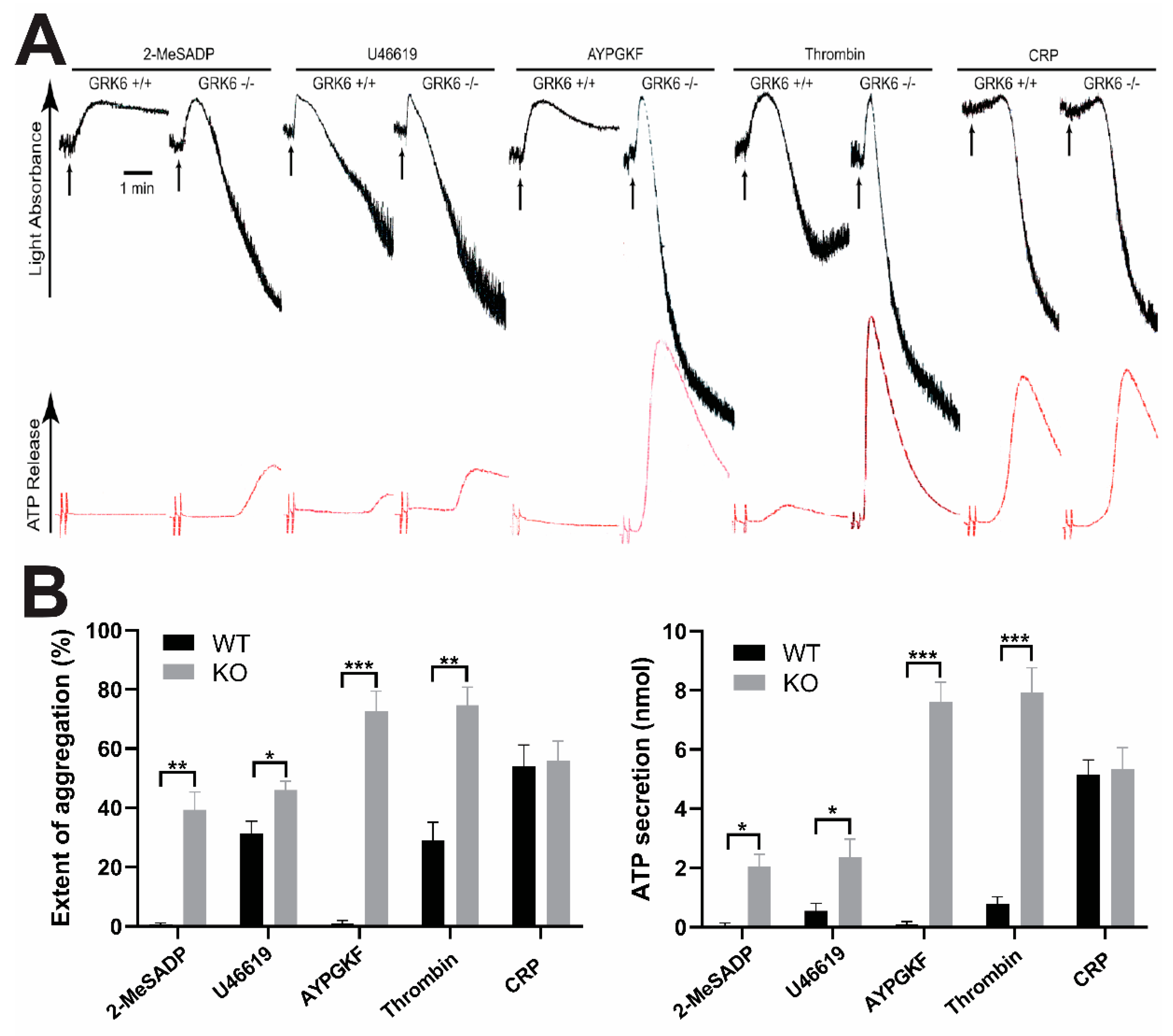
2.1. GRK6 Selectively Regulates GPCR-Mediated Platelet Functional Responses

To determine the contribution of GRK6 to platelet function, we measured the various agonists-induced platelet aggregations and dense granule secretions in the wild-type (WT) and the GRK6-deficient mouse platelets. As shown in Figure 1, platelet aggregation and dense granule secretion induced by GPCR agonists including 2-MeSADP, U46619, AYPGKF, and thrombin were significantly potentiated in the GRK6-deficient platelets compared to those in the WT platelets. We found that the dose response curves were left-shifted, and there was little difference between WT and GRK6−/− platelet in response to high dose of agonists. The extent of potentiation of the platelet function in response to U46619 was not as significant as it was for the other GPCR agonists. However, platelet aggregation and dense granule secretion in response to Glycoprotein VI (GPVI) agonist CRP were not affected in the GRK6-deficient platelets, indicating that GRK6 selectively regulated platelet aggregation and secretion in response to GPCR agonists.
A similar approach was adopted to analyze the activation of integrin αIIbβ3 by measuring the binding of the conformation-dependent antibody JON/A, and α-granule secretion by measuring the binding of an fluorescein isothiocyanate (FITC)-labeled anti-P-selectin antibody that recognizes the α-granule protein: P-selectin, by flow cytometry. Consistent with the results shown in Figure 1, we found that both JON/A binding and P-selectin expression induced by AYPGKF were significantly increased in GRK6-deficient platelets compared to WT platelets (Figure 2A,B). However, CRP-induced JON/A binding and P-selectin binding to GRK6−/− platelets were not affected compared to WT platelets (Figure 2A,B). These results show that GRK6 plays an important role in the regulation of platelet functional responses downstream of GPCR pathways.

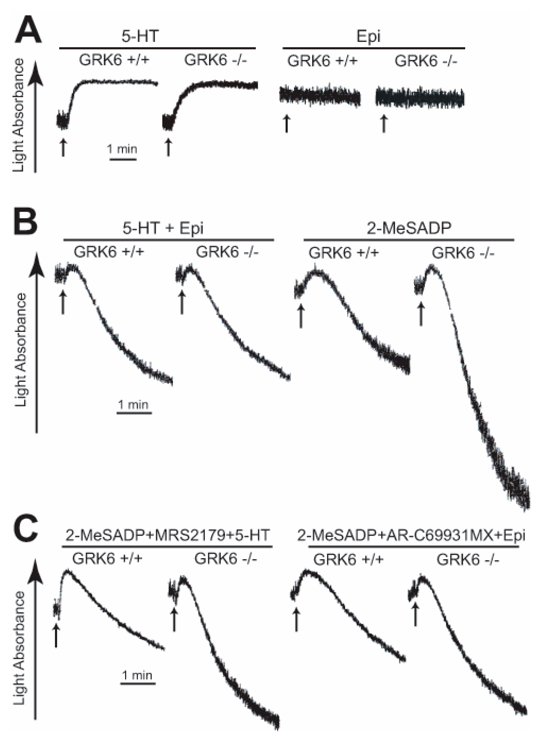
2.2. GRK6 Regulates Specific GPCR-Mediated Platelet Aggregation

It is known that ADP-induced platelet aggregation requires co-activation of the Gq-coupled P2Y1 and the Gi-coupled P2Y12 receptors while platelet aggregation induced by co-stimulation of serotonin (5HT) and epinephrine bypasses the P2Y1 and P2Y12 receptors to activate the Gq-coupled 5HT2A receptor and the Gz-coupled α2A adrenergic receptor, respectively [6]. In order to evaluate the role of GRK6 in the differential regulation of GPCRs, we measured the platelet aggregation induced by co-stimulation of serotonin and epinephrine in the WT and the GRK6-deficient platelets. Interestingly, either serotonin or epinephrine alone failed to induce platelet aggregation in both WT and GRK6-deficient platelets (Figure 3A). In addition, compared to the effect of GRK6 in 2-MeSADP-induced aggregation, platelet aggregation induced by co-stimulation of serotonin and epinephrine was not affected in the GRK6-deficient platelets (Figure 3B), suggesting that GRK6 was not involved in the regulation of 5HT2A- and α2A adrenergic receptor-induced platelet aggregation.
We further evaluated the contribution of GRK6 on P2Y1 and P2Y12 receptors. Platelet aggregation induced by co-stimulation of Gq plus Gi with serotonin and 2-MeSADP in the presence of P2Y1-receptor antagonist MRS2179 or Gq plus Gz by 2-MeSADP in the presence of P2Y12-receptor antagonist AR-C69931MX and epinephrine was significantly potentiated in GRK6-deficient platelets compared to WT platelets (Figure 3C). However, the extent of the potentiation of platelet aggregation was smaller compared to the extent of platelet aggregation induced by 2-MeSADP in GRK6-deficient platelets in Figure 3B, confirming that GRK6 selectively regulates P2Y1- and P2Y12-mediated platelet functions.

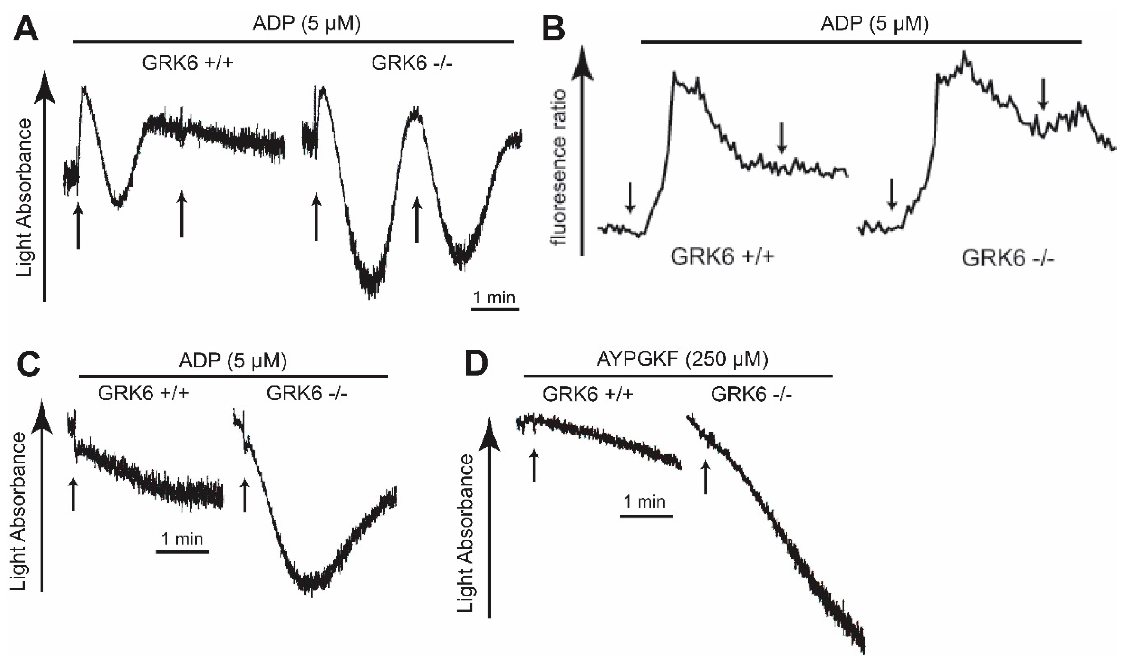
2.3. GRK6 Plays an Important Role in ADP and PAR4 Receptor Desensitization in Platelets

In order to determine the role of GRK6 in GPCR desensitization in platelets, we measured platelet aggregation in response to re-stimulation in the WT and the GRK6-deficient platelets. As shown in Figure 4A, re-stimulation with ADP immediately after disaggregation, 3 min following the first addition of ADP, did not induce shape change or aggregation in the WT platelets. In contrast, platelet aggregation in response to the second challenge of ADP was restored in the GRK6-deficient platelets. Moreover, there was an increase in ADP-induced Ca2+ release in GRK6−/− platelets compared to the WT (Figure 4B). Slight increase in Ca2+ release was also observed in response to re-stimulation of ADP in GRK6−/− platelets, indicating that GRK6 regulated ADP-induced P2Y1 receptor desensitization in platelets. Similarly, platelet aggregation was restored in response to ADP after pretreatment with ADP in the GRK6-deficient platelets compared to that in the WT platelets (Figure 4C), indicating that GRK6 played a role in the P2Y1 and P2Y12 receptor desensitization in platelets. In addition, platelet aggregation was restored in response to a second challenge of AYPGKF after pretreatment with AYPGKF in the GRK6-deficient platelets, whereas re-stimulation of AYPGKF failed to induce aggregation in the WT platelets (Figure 4D), suggesting that GRK6 contributed to PAR4 desensitization in platelets.

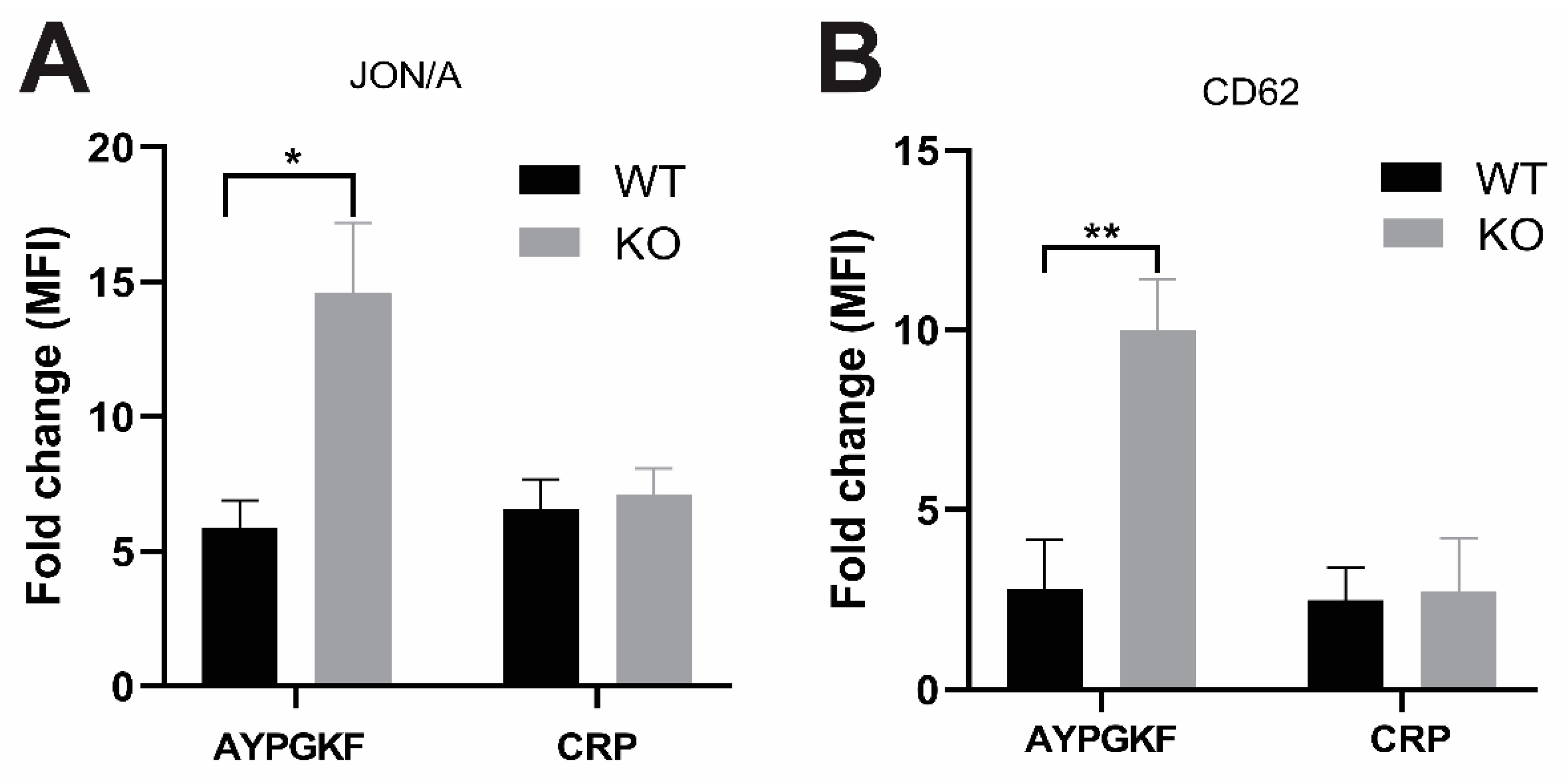
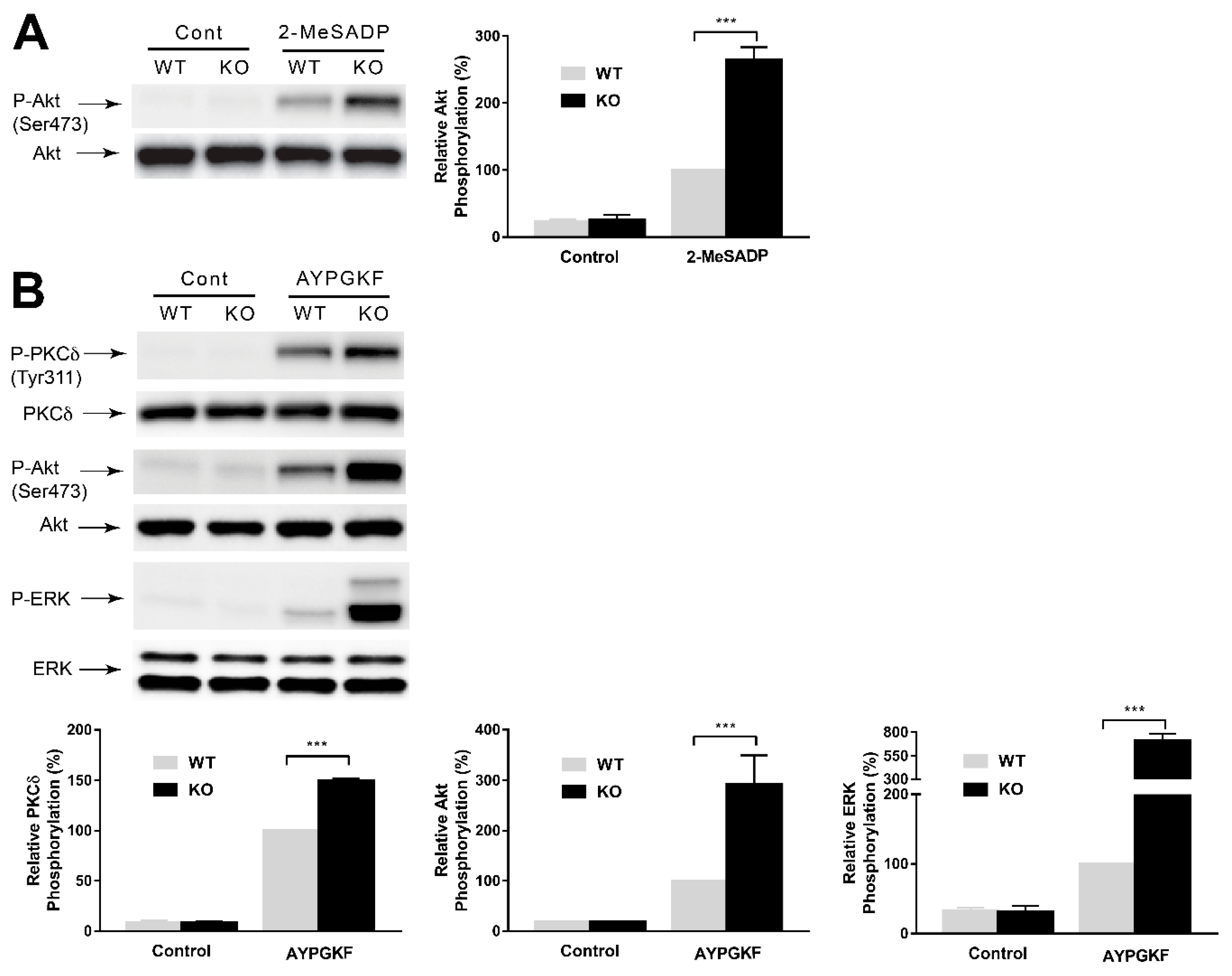
2.4. GPCR-Mediated Signaling Events are Potentiated in GRK6-Deficient Platelets

In order to evaluate the effect of GRK6 on platelet signaling, we stimulated platelets with 2-MeSADP and AYPGKF in the WT and the GRK6-deficient platelets and measured the phosphorylation of protein kinase Cδ (PKCδ), Akt, and extracellular signal-related kinase (ERK) as per a method described previously [22]. These proteins were selected for their known mechanism and effect on platelet activation [23,24,25,26,27]. Moreover, 2-MeSADP-induced Akt phosphorylation (Figure 5A) and AYPGKF-induced Akt, ERK, and PKCδ phosphorylations (Figure 5B) were potentiated in the GRK6-deficient platelets compared to WT platelets, suggesting a role of GRK6 in regulating both Gq- and Gi-mediated signaling events in platelets.

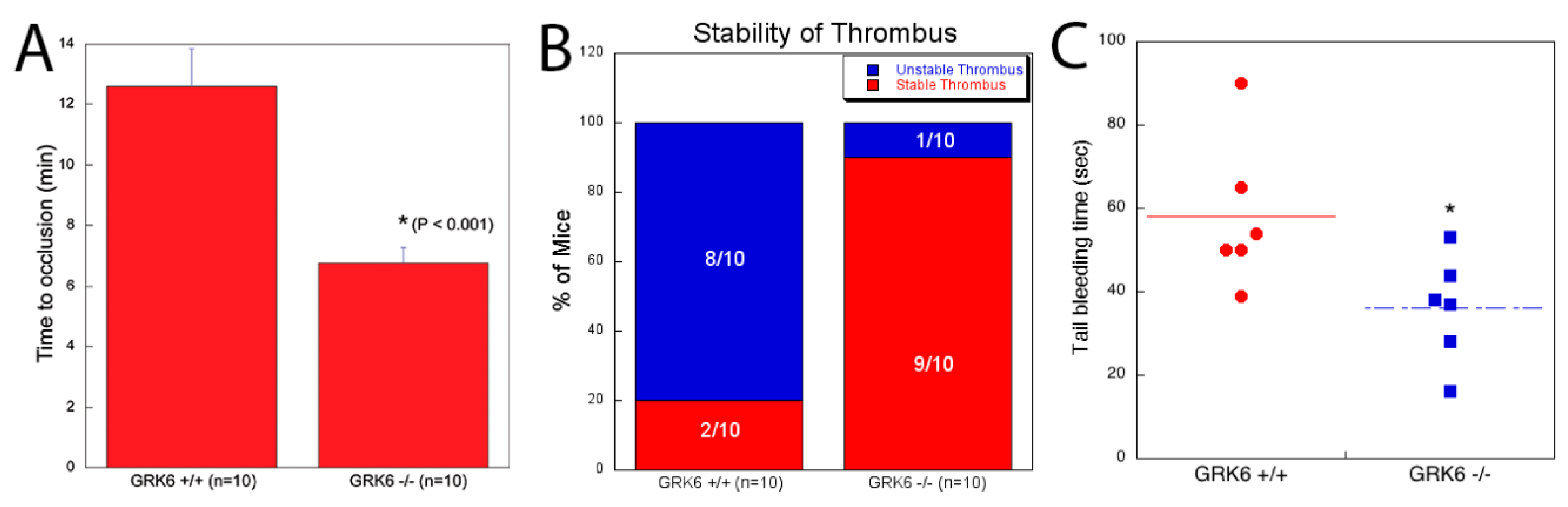
2.5. Contribution of GRK6 in the Platelet Function In Vivo

ADP, thromboxane, and thrombin are important agonists that contribute to thrombus growth and stability. To evaluate the contribution of GRK6 to the platelet function in vivo, we used in-vivo thrombosis models using FeCl3 injury of the carotid artery [28,29] in WT and GRK6−/− mice. As shown in Figure 6A, WT mice formed a thrombus 12.5 min after the vascular injury whereas the GRK6−/− mice formed a thrombus 7 min after the injury, suggesting the enhanced thrombus formation in GRK6−/− mice after the vascular injury. In addition, 90% of the GRK6−/− mice showed stable thrombus formation, while 80% of the WT mice showed unstable thrombus formation (Figure 6B). We also measured tail-bleeding time in GRK6+/+ and GRK6−/− mice, which reflects primary hemostatic function. We observed that average time taken for complete blockade of tail bleeding was 36 s in GRK6−/− compared to 58 s in GRK6+/+ mice, suggesting that GRK6 negatively regulates hemostatic function in vivo (Figure 6C). Taken together, these results indicate the critical role of GRK6 in platelet function in vivo.

3. Discussion

GRKs are ubiquitously expressed in tissues, and most of them show activity towards a wide variety of agonist-occupied receptors in vitro. Although GRKs were known to be involved in the desensitization of GPCRs in other cell types, the functional role of these signaling molecules and their mechanisms in regulating platelet physiological events have not been fully understood. It has been demonstrated that human platelets can become refractory to activation after a major surgery, possibly leading to an increased risk of post-surgical bleeding [30], suggesting that desensitization mechanisms play a critical role in regulating platelet responsiveness. The absence of a specific inhibitor of GRK6 has made it difficult to precisely determine its role in platelet function. Therefore, we used the GRK6-deficient platelets to evaluate the role of GRK6 in platelet function and characterized the novel mechanisms involved in GPCR desensitization in platelets.
It has been shown in other cells that different GRKs are involved in the regulation of specific GPCR desensitization. It has been demonstrated that the PAR1 and PAR4 receptors are internalized after desensitization following activation in platelets, but the basis of GRKs in regulation of platelet function has not been determined [31,32]. Thus, we first have investigated whether GRK6 had a role in the regulation of GPCR-mediated platelet function. We found that platelet aggregation and dense granule secretion induced by GPCR agonists including 2-MeSADP, U46619, AYPGKF, and thrombin were potentiated in the GRK6-deficient platelets compared to those in the WT platelets. We also observed that the GPCR-mediated platelet response was potentiated in the aspirin-treated GRK6-deficient platelets, indicating that the enhanced platelet aggregation in the GRK6-deficient platelet is not due to the effect of GRK6 on the positive feedback effect of generated TxA2 (data not shown). Consistently, both JON/A binding and P-selectin expression that recognizes integrin αIIbβ3 activation and α-granule secretion, respectively, were significantly enhanced upon stimulation with AYPGKF in GRK6-deficient platelets. However, GPVI agonist CRP-induced platelet aggregation and secretion were not affected in the GRK6-deficient platelets suggesting that GRK6 did not have any role in non-GPCR-mediated platelet function. Taken together, our data suggest that GRK6 plays an important role in the regulation of GPCR-mediated platelet function.
GRK6 had the least effect on the TxA2-induced platelet aggregation and secretion. TxA2 receptor exists as two alternatively spliced variants: TPα (platelets) and TPβ (endothelial cells), that differ in length and sequence of the C-terminal tail [33]. Different GRK isoforms have been shown to have different affinities toward these variants in other cells [34]. In contrast, it has been reported that the C-terminus of TPα is not capable of being phosphorylated by GRKs in other nucleated cell types [19,20]. Differences in tail sequence and number of the phosphorylation-amenable serine residues also allow for differential affinity for its agonists. It is known that TPα has a shorter C-terminus with lesser phosphorylatable serine residues, which might lead to decreased affinity towards TPα receptor than other GPCRs in platelets. Another possibility may be that receptors tend to phosphorylate and internalize rapidly following stimulation with U46619, which does not allow sufficient binding of GRK6 for phosphorylation of the residue [35]. However, further study is needed to identify the relation between serine residues and affinity, responsible for the functional regulation of GPCRs by GRK6 in platelets.
It has been shown that co-stimulation of the Gq-coupled P2Y1 and the Gi-coupled P2Y12 receptors is essential for ADP-induced platelet aggregation [1,36]. Moreover, co-stimulation of serotonin and epinephrine induces aggregation in platelets. The α2A adrenergic receptor, when stimulated with epinephrine, couples primarily to Gz, which belongs to the Gi class of the G proteins [37,38] that can mimic the P2Y12 receptor activation pathway. On the other hand, serotonin acts on the 5HT2A receptor, which bypasses the P2Y1 receptor to supplement Gq signaling [39]. Although concomitant signaling through the serotonin and epinephrine causes platelet aggregation, neither of them can cause platelet aggregation by itself [1,40,41]. Interestingly, serotonin only induced shape change while epinephrine induced neither shape change nor aggregation in both GRK6-deficient and WT platelets. Moreover, we found that in contrast to an ADP-induced aggregation, platelet aggregation induced by co-stimulation of serotonin and epinephrine was not affected in the GRK6-deficient platelets. These suggest that GRK6 is not involved in the regulation of α2A and 5HT2A receptors and plays a role in regulating specific GPCR function.
It is possible that the enhanced platelet response in the GRK6-deficient platelets is due to an altered expression of receptors in the GRK6-deficient platelets. To clarify this, we assessed the PAR4 receptor expression in both the WT and the GRK6-deficient platelets and found that the PAR4 receptor level in GRK6-deficient platelets was similar to that in the WT platelets. This confirms that the expression of PAR4 receptor is not affected by the absence of GRK6.
GRK6 has been implicated in various receptor desensitizations in vivo and in vitro [42,43]. It has been suggested that re-sensitization of the platelet aggregation response to ADP is due to desensitization of the P2Y1 receptor, while the P2Y12 receptor remains functional [44]. Another study suggested that both the P2Y1 and P2Y12 receptors undergo desensitization by different mechanisms involving PKC and GRK2 and GRK6, respectively [17]. We found that re-stimulation of platelets with ADP and AYPGKF restored platelet aggregation in GRK6-deficient platelets, suggesting that GRK6 played a role in the desensitization of ADP and PAR4 receptors. At this stage, it is not clear whether the effect of GRK6 on ADP receptors is due to the desensitization of both the P2Y1 and P2Y12 receptors or the selective desensitization of either the P2Y1 receptor or the P2Y12 receptor, because ADP requires co-activation of both the P2Y1 and P2Y12 receptors. We found that platelet aggregation was potentiated in response to serotonin or epinephrine in combination with selective activation of P2Y1 or P2Y12, respectively, in GRK6-deficient mice, while serotonin and epinephrine failed to restore aggregation. We also found the increased Ca2+ release in response to both initial and re-stimulation of platelets with ADP in GRK6-deficient platelets compared to the WT platelets, indicating the role of GRK6 in P2Y1 receptor desensitization. These data confirm that the desensitization of both P2Y1 and P2Y12 receptors is responsible for platelet desensitization to ADP, and GRK6 is involved in regulation of both the P2Y1 and P2Y12 receptors in platelets.
GPCR-mediated activation of Akt, ERK, and PKCδ is important and necessary to enhance and promote platelet aggregation. Previous studies have shown that ADP-induced Akt activation is dependent on Gi-mediated P2Y12 receptor stimulation [45] and AYPGKF-mediated Akt and ERK phosphorylation has been shown to be regulated by the Gi pathway by the secreted ADP in platelets [26]. It has been also shown that PKCδ, downstream of Gq pathway, plays a positive role in AYPGKF-induced platelet aggregation and dense granule secretion [25]. We found that the deletion of GRK6 potentiated ADP-induced Akt phosphorylation and AYPGKF-induced Akt, ERK, and PKCδ phosphorylation, suggesting that GRK6 regulates both the Gq- and Gi-mediated signaling events in platelets.
ADP, thromboxane, and thrombin are important agonists that contribute to thrombus growth and stability. Since we observed the potentiation of these agonist-induced platelet responses in GRK6-deficient platelets, we then evaluated the contribution of GRK6 to the function of platelets in vivo. We found that enhanced platelet function in GRK6−/− mice resulted in stable thrombus formation and rapid time to occlusion in in vivo thrombosis models using FeCl3 injury of the carotid artery, indicating that GRK6−/− mice are more susceptible to thrombosis. The bleeding time was also significantly reduced in GRK6−/− mice. These results confirm the critical role of GRK6 in platelet function in vivo. Since GRK6 is ubiquitously expressed, we cannot rule out the contribution of GRK6 of other cells including leukocytes, endothelial cells, and smooth muscle cells on thrombus formation.
While we were preparing this manuscript, Chen et al. [46] has published a study suggesting that GRK6 regulates the hemostatic response to injury through its rate-limiting effects on GPCR signaling in platelets. Similar to our study, they have shown that GRK6 regulates P2Y12 and PAR4 receptor signaling. Importantly, we have investigated the effect of GRK6 on 5HT2A and α2A adrenergic receptor in GRK6−/− platelets with serotonin and epinephrine and have found that these receptors are not regulated by GRK6 in platelets. This approach with the addition of either P2Y1 and P2Y12 receptor antagonists also helped us to confirm that both P2Y1 and P2Y12 receptors are regulated by GRK6 in platelets. Moreover, we have demonstrated the effect of GRK6 on desensitization of platelet aggregation in response to ADP and AYPGKF.
In conclusion, we demonstrate that GRK6 plays an important role in the regulation of GPCR-mediated platelet activation and in vivo thrombus formation by selectively regulating desensitization of specific GPCRs.

4. Materials and Methods

4.1. Materials

U46619, 2-MeSADP, ADP, thrombin, serotonin, epinephrine, apyrase (type V), prostaglandin E1 (PGE1), sodium citrate, and acetylsalicylic acid were from Sigma (St. Louis, MO, USA). CRP was obtained from Dr. Richard Farndale at the University of Cambridge. Phycoerythrin-conjugated antibody JON/A and FITC-conjugated anti-P-selectin antibody were from Emfret Analytics (Sterntalerweg, Wurzburg, Germany). Hexapeptide AYPGKF was custom synthesized by Invitrogen (Carlsbad, CA, USA). Fura-2-AM was from Millipore (Temecula, CA, USA). Anti-phospho-Akt (Ser473), anti-phospho-ERK (Thr202/Tyr204), anti-phospho-PKCδ (Tyr311), anti-Akt, anti-PKCδ, and anti-ERK antibodies were purchased from Cell Signaling Technology (Beverly, MA, USA). Horseradish peroxidase-labeled secondary antibody was bought from Santa Cruz Biotechnology (Santa Cruz, CA, USA). All other reagents were of analytical grade, and deionized water was used throughout the study.

4.2. Animals

All animal experiments were performed in accordance with the approval obtained from the Chungbuk National University Animal Ethics Committee (CBNUA-873-15-02, approved on July 1, 2019). GRK6−/− mice were obtained from Dr. Walter Koch (Temple University, Philadelphia, PA, USA). WT littermates were used as controls. The platelet count was normal in GRK6−/− mice.

4.3. Preparation of Murine Platelets

Blood was collected from equal number of both male and female mice and platelet was prepared as described [47]. Briefly, the collected citrated blood was centrifuged at 100× g for 10 min at room temperature (RT) to obtain Platelet Rich Plasma (PRP) and the obtained PRP was centrifuged at 400× g for 10 min to pellet the platelets. Platelet pellets were re-suspended in the Tyrode’s buffer (pH = 7.4) containing 0.05 units/mL of apyrase, and cells were adjusted to 2 × 10⁸ cells/mL.

4.4. Platelet Aggregation and Secretion

Agonist-induced platelet aggregation and secretion were measured using a lumi-aggregometer (Chrono-Log, Havertown, PA, USA) at 37 °C under stirring conditions (900 rpm) as per a method described previously [47]. Washed platelets (0.5 mL) were stimulated with the different agonists, and tracings of light transmission were measured. Platelet secretion was determined by measuring the release of ATP after adding the luciferin/luciferase reagent.

4.5. Flow Cytometry

Washed murine platelets were used to measure integrin αII3 activation with phycoerythrin-conjugated JON/A antibodies and the surface exposure of P-selectin with FITC-conjugated P-selectin antibodies as per a method described previously [48]. All measurements were determined using a FACSCalibur flow cytometer (BD Biosciences, San Jose, CA, USA). Appropriate isotype controls were used.

4.6. Calcium Mobilization

Intracellular Ca2+ concentration was measured in Fura-2-AM loaded platelets as previously described [49].

4.7. Western Blotting

Platelets were stimulated with different agonists for the appropriate time, and phosphorylation events were measured as described previously [22]. Briefly, platelets were stimulated with 2-MeSADP or AYPGKF and the reaction was stopped by the addition of 3× sodium dodecyl sulfate (SDS) buffer. Platelet lysates were separated by 10% SDS/PAGE and transferred onto polyvinylidene difluoride (PVDF) membranes. Membranes were incubated overnight at 4 °C with anti-phospho-Akt (Ser473), anti-phospho-ERK (Thr202/Tyr204), anti-phospho-PKCδ (Tyr311), anti-Akt, anti-PKCδ, or anti-ERK antibodies. Membranes were then probed with goat anti-rabbit antibody and immune-reactivity was detected using the Fuji-Film Luminescent Image Analyzer (LAS-3000 CH, Tokyo, Japan).

4.8. FeCl3-Induced In Vivo Thrombosis Model

Adult mice (10–12 weeks old) were anesthetized and FeCl3-induced thrombosis was assessed as per a method described previously [29]. Briefly, the carotid artery was injured by 5% FeCl3 for 90 s, and blood flow through the carotid artery was measured for 30 min using the Doppler flow probe following vascular injury. The occlusion time due to thrombus formation was recorded and plotted as time to occlusion.

4.9. Tail Bleeding Time Assay

The tail bleeding time assay was performed as described previously [29]. Briefly, a longitudinal cut was made in anesthetized mouse’s tail and tail was immediately immersed in 0.9% isotonic saline at 37 °C. The bleeding time was defined as the time required for the blockade of blood flow.

4.10. Statistical Analysis

All statistical analyses were performed using the Prism software (version 3.0). Data have been presented as mean ± standard error (SE). Statistical significance was determined by the Student’s t-test.

Author Contributions

Conceptualization, P.K.C. and S.K. (Soochong Kim); methodology, P.K.C. and S.K. (Sanggu Kim); validation, Y.J. and S.K. (Soochong Kim); investigation, P.K.C., S.-H.L., K.-M.P., and S.K. (Soochong Kim); writing―original draft, P.K.C.; writing―review and editing, Y.J., S.-H.L., K.-M.P., and S.K. (Soochong Kim); supervision, S.K. (Soochong Kim). All authors have read and agreed to the published version of the manuscript.

Funding

This work was supported by the National Research Foundation of Korea (NRF) Grant of the Korean government (NRF-2016R1D1A1B01010310), the Agriculture, Food and Rural Affairs Convergence Technologies Program for Educating Creative Global Leader (MAFRA; 320005-4), and the Global Research and Development Center (GRDC) Program through the NRF funded by the Ministry of Education, Science and Technology (2017K1A4A3014959).

Conflicts of Interest

The authors declare no conflict of interest.

Abbreviations

GPCRG protein-coupled receptor
GRKG protein-coupled receptor kinase
ADPAdenosine diphosphate
CRPCollagen-related peptide
PAR Protease-activated receptor
ERK Extracellular signal-related kinase
PKCδProtein Kinase Cδ
TPThromboxane receptor
PRPPlatelet-rich plasma

References

  1. Jin, J.; Kunapuli, S.P. Coactivation of two different G protein-coupled receptors is essential for ADP-induced platelet aggregation. Proc. Natl. Acad. Sci. USA 1998, 95, 8070–8074. [Google Scholar] [CrossRef] [PubMed] [Green Version]
  2. Paul, B.Z.; Jin, J.; Kunapuli, S.P. Molecular mechanism of thromboxane A(2)-induced platelet aggregation. Essential role for p2t(ac) and alpha(2a) receptors. J. Biol. Chem. 1999, 274, 29108–29114. [Google Scholar] [CrossRef] [PubMed] [Green Version]
  3. Offermanns, S.; Laugwitz, K.-L.; Spicher, K.; Schultz, G. G proteins of the G12 family are activated via thromboxane A2 and thrombin receptors in human platelets. Proc. Natl. Acad. Sci. USA 1994, 91, 504–508. [Google Scholar] [CrossRef] [PubMed] [Green Version]
  4. Shenker, A.; Goldsmith, P.; Unson, C.G.; Spiegel, A.M. The G protein coupled to the thromboxane A2 receptor in human platelets is a member of the novel Gq family. J. Biol. Chem. 1991, 266, 9309–9313. [Google Scholar]
  5. Kim, S.; Foster, C.; Lecchi, A.; Quinton, T.M.; Prosser, D.M.; Jin, J.; Cattaneo, M.; Kunapuli, S.P. Protease-activated receptors 1 and 4 do not stimulate Gi signaling pathways in the absence of secreted ADP and cause human platelet aggregation independently of Gisignaling. Blood 2002, 99, 3629–3636. [Google Scholar] [CrossRef] [Green Version]
  6. de Chaffoy de Courcelles, D.; Roevens, P.; van Belle, H.; de Clerck, F. The synergistic effect of serotonin and epinephrine on the human platelet at the level of signal transduction. FEBS Lett. 1987, 219, 283–288. [Google Scholar] [CrossRef] [Green Version]
  7. Nieswandt, B.; Watson, S.P. Platelet-collagen interaction: Is GPVI the central receptor? Blood 2003, 102, 449–461. [Google Scholar] [CrossRef]
  8. Benovic, J.L.; Gomez, J. Molecular cloning and expression of GRK6. A new member of the G protein-coupled receptor kinase family. J. Biol. Chem. 1993, 268, 19521–19527. [Google Scholar]
  9. Hisatomi, O.; Matsuda, S.; Satoh, T.; Kotaka, S.; Imanishi, Y.; Tokunaga, F. A novel subtype of G-protein-coupled receptor kinase, GRK7, in teleost cone photoreceptors. FEBS Lett. 1998, 424, 159–164. [Google Scholar] [CrossRef]
  10. Reiter, E.; Lefkowitz, R.J. GRKs and β-arrestins: Roles in receptor silencing, trafficking and signaling. Trends Endocrinol. Metab. 2006, 17, 159–165. [Google Scholar] [CrossRef]
  11. Willets, J.M.; Challiss, R.A.; Nahorski, S.R. Non-visual GRKs: Are we seeing the whole picture? Trends Pharm. Sci. 2003, 24, 626–633. [Google Scholar] [CrossRef] [PubMed] [Green Version]
  12. Pitcher, J.A.; Freedman, N.J.; Lefkowitz, R.J. G protein-coupled receptor kinases. Annu. Rev. Biochem. 1998, 67, 653–692. [Google Scholar] [CrossRef] [PubMed] [Green Version]
  13. Santulli, G.; Trimarco, B.; Iaccarino, G. G-protein-coupled receptor kinase 2 and hypertension. High Blood Press. Cardiovasc. Prev. 2013, 20, 5–12. [Google Scholar] [CrossRef] [PubMed] [Green Version]
  14. Freedman, N.J.; Lefkowitz, R.J. Desensitization of G protein-coupled receptors. Recent Prog. Horm. Res. 1996, 51, 319–351; discussion 352–313. [Google Scholar]
  15. Tiruppathi, C.; Yan, W.; Sandoval, R.; Naqvi, T.; Pronin, A.N.; Benovic, J.L.; Malik, A.B. G protein-coupled receptor kinase-5 regulates thrombin-activated signaling in endothelial cells. Proc. Natl. Acad. Sci. USA 2000, 97, 7440–7445. [Google Scholar] [CrossRef] [Green Version]
  16. Ishii, K.; Chen, J.; Ishii, M.; Koch, W.J.; Freedman, N.J.; Lefkowitz, R.J.; Coughlin, S.R. Inhibition of thrombin receptor signaling by a G-protein coupled receptor kinase. Functional specificity among G-protein coupled receptor kinases. J. Biol. Chem. 1994, 269, 1125–1130. [Google Scholar]
  17. Hardy, A.R.; Conley, P.B.; Luo, J.; Benovic, J.L.; Poole, A.W.; Mundell, S.J. P2Y1 and P2Y12 receptors for ADP desensitize by distinct kinase-dependent mechanisms. Blood 2005, 105, 3552–3560. [Google Scholar] [CrossRef]
  18. Willets, J.M.; Brighton, P.J.; Mistry, R.; Morris, G.E.; Konje, J.C.; Challiss, R.J. Regulation of oxytocin receptor responsiveness by G protein-coupled receptor kinase 6 in human myometrial smooth muscle. Mol. Endocrinol. 2009, 23, 1272–1280. [Google Scholar] [CrossRef] [Green Version]
  19. Zhou, H.; Yan, F.; Tai, H.-H. Phosphorylation and desensitization of the human thromboxane receptor-α by G protein-coupled receptor kinases. J. Pharmacol. Exp. Ther. 2001, 298, 1243–1251. [Google Scholar]
  20. Parent, J.-L.; Labrecque, P.; Orsini, M.J.; Benovic, J.L. Internalization of the TXA2 Receptor α and β Isoforms Role of the differentially spliced COOH terminus in agonist-promoted receptor internalization. J. Biol. Chem. 1999, 274, 8941–8948. [Google Scholar] [CrossRef] [Green Version]
  21. Kim, J.; Ahn, S.; Ren, X.-R.; Whalen, E.J.; Reiter, E.; Wei, H.; Lefkowitz, R.J. Functional antagonism of different G protein-coupled receptor kinases for β-arrestin-mediated angiotensin II receptor signaling. Proc. Natl. Acad. Sci. USA 2005, 102, 1442–1447. [Google Scholar] [CrossRef] [PubMed] [Green Version]
  22. Kim, S.; Jin, J.; Kunapuli, S.P. Relative contribution of G-protein-coupled pathways to protease-activated receptor-mediated Akt phosphorylation in platelets. Blood 2006, 107, 947–954. [Google Scholar] [CrossRef] [PubMed] [Green Version]
  23. Chen, J.; De, S.; Damron, D.S.; Chen, W.S.; Hay, N.; Byzova, T.V. Impaired platelet responses to thrombin and collagen in AKT-1–deficient mice. Blood 2004, 104, 1703–1710. [Google Scholar] [CrossRef] [PubMed] [Green Version]
  24. Kim, S.; Jin, J.; Kunapuli, S.P. Akt activation in platelets depends on Gi signaling pathways. J. Biol. Chem. 2004, 279, 4186–4195. [Google Scholar] [CrossRef] [PubMed] [Green Version]
  25. Murugappan, S.; Tuluc, F.; Dorsam, R.T.; Shankar, H.; Kunapuli, S.P. Differential role of protein kinase Cδ isoform in agonist-induced dense granule secretion in human platelets. J. Biol. Chem. 2004, 279, 2360–2367. [Google Scholar] [CrossRef] [PubMed] [Green Version]
  26. Shankar, H.; Garcia, A.; Prabhakar, J.; Kim, S.; Kunapuli, S. P2Y12 receptor-mediated potentiation of thrombin-induced thromboxane A2 generation in platelets occurs through regulation of Erk1/2 activation. J. Thromb. Haemost. 2006, 4, 638–647. [Google Scholar] [CrossRef] [PubMed]
  27. Woulfe, D.; Jiang, H.; Morgans, A.; Monks, R.; Birnbaum, M.; Brass, L.F. Defects in secretion, aggregation, and thrombus formation in platelets from mice lacking Akt2. J. Clin. Investig. 2004, 113, 441–450. [Google Scholar] [CrossRef]
  28. Nagy, B.; Bhavaraju, K.; Getz, T.; Bynagari, Y.S.; Kim, S.; Kunapuli, S.P. Impaired activation of platelets lacking protein kinase C-θ isoform. Blood 2009, 113, 2557–2567. [Google Scholar] [CrossRef] [Green Version]
  29. Bynagari-Settipalli, Y.S.; Lakhani, P.; Jin, J.; Bhavaraju, K.; Rico, M.C.; Kim, S.; Woulfe, D.; Kunapuli, S.P. Protein kinase C isoform epsilon negatively regulates ADP-induced calcium mobilization and thromboxane generation in platelets. Arterioscler. Thromb. Vasc. Biol. 2012, 32, 1211–1219. [Google Scholar] [CrossRef] [Green Version]
  30. O’Brien, J.R.; Etherington, M.; Jamieson, S. Refractory state of platelet aggregation with major operations. Lancet 1971, 2, 741–743. [Google Scholar] [CrossRef]
  31. Shapiro, M.J.; Weiss, E.J.; Faruqi, T.R.; Coughlin, S.R. Protease-activated receptors 1 and 4 are shut off with distinct kinetics after activation by thrombin. J. Biol. Chem. 2000, 275, 25216–25221. [Google Scholar] [CrossRef] [PubMed] [Green Version]
  32. Molino, M.; Bainton, D.F.; Hoxie, J.A.; Coughlin, S.R.; Brass, L.F. Thrombin receptors on human platelets initial localization and subsequent redistribution during platelet activation. J. Biol. Chem. 1997, 272, 6011–6017. [Google Scholar] [CrossRef] [PubMed] [Green Version]
  33. Raychowdhury, M.K.; Yukawa, M.; Collins, L.J.; McGrail, S.H.; Kent, K.C.; Ware, J.A. Alternative splicing produces a divergent cytoplasmic tail in the human endothelial thromboxane A2 receptor. J. Biol. Chem. 1994, 269, 19256–19261. [Google Scholar] [PubMed]
  34. Kelley-Hickie, L.P.; O’Keeffe, M.B.; Reid, H.M.; Kinsella, B.T. Homologous desensitization of signalling by the alpha (α) isoform of the human thromboxane A2 receptor: A specific role for nitric oxide signalling. Biochim. Biophys. Acta (BBA)-Mol. Cell Res. 2007, 1773, 970–989. [Google Scholar] [CrossRef] [Green Version]
  35. Habib, A.; Vezza, R.; Créminon, C.; Maclouf, J.; FitzGerald, G.A. Rapid, Agonist-dependent Phosphorylation in Vivo of Human Thromboxane Receptor Isoforms Minimal Involvement of Protein Kinase C. J. Biol. Chem. 1997, 272, 7191–7200. [Google Scholar] [CrossRef] [Green Version]
  36. Hechler, B.; Leon, C.; Vial, C.; Vigne, P.; Frelin, C.; Cazenave, J.P.; Gachet, C. The P2Y1 receptor is necessary for adenosine 5′-diphosphate-induced platelet aggregation. Blood 1998, 92, 152–159. [Google Scholar] [CrossRef]
  37. Smith, S.K.; Limbird, L.E. Solubilization of human platelet alpha-adrenergic receptors: Evidence that agonist occupancy of the receptor stabilizes receptor—Effector interactions. Proc. Natl. Acad. Sci. USA 1981, 78, 4026–4030. [Google Scholar] [CrossRef] [Green Version]
  38. Yang, J.; Wu, J.; Kowalska, M.A.; Dalvi, A.; Prevost, N.; O’Brien, P.J.; Manning, D.; Poncz, M.; Lucki, I.; Blendy, J.A. Loss of signaling through the G protein, Gz, results in abnormal platelet activation and altered responses to psychoactive drugs. Proc. Natl. Acad. Sci. USA 2000, 97, 9984–9989. [Google Scholar] [CrossRef] [Green Version]
  39. Cerrito, F.; Lazzaro, M.P.; Gaudio, E.; Arminio, P.; Aloisi, G. 5HT2-receptors and serotonin release: Their role in human platelet aggregation. Life Sci. 1993, 53, 209–215. [Google Scholar] [CrossRef]
  40. Daniel, J.L.; Dangelmaier, C.; Jin, J.; Kim, Y.B.; Kunapuli, S.P. Role of intracellular signaling events in ADP-induced platelet aggregation. Thromb. Haemost. 1999, 82, 1322–1326. [Google Scholar] [CrossRef]
  41. Kim, S.; Kunapuli, S.P. P2Y12 receptor in platelet activation. Platelets 2011, 22, 54–58. [Google Scholar] [CrossRef] [PubMed]
  42. Aiyar, N.; Disa, J.; Dang, K.; Pronin, A.N.; Benovic, J.L.; Nambi, P. Involvement of G protein-coupled receptor kinase-6 in desensitization of CGRP receptors. Eur. J. Pharmacol. 2000, 403, 1–7. [Google Scholar] [CrossRef]
  43. Gainetdinov, R.R.; Bohn, L.M.; Sotnikova, T.D.; Cyr, M.; Laakso, A.; Macrae, A.D.; Torres, G.E.; Kim, K.-M.; Lefkowitz, R.J.; Caron, M.G. Dopaminergic supersensitivity in G protein-coupled receptor kinase 6-deficient mice. Neuron 2003, 38, 291–303. [Google Scholar] [CrossRef] [Green Version]
  44. Baurand, A.; Eckly, A.; Bari, N.; Léon, C.; Hechler, B.; Cazenave, J.-P.; Gachet, C. Desensitization of the platelet aggregation response to ADP: Differential down-regulation of the P2Y1 and P2cyc receptors. Thromb. Haemost. 2000, 84, 484–491. [Google Scholar] [CrossRef] [PubMed]
  45. Kahner, B.N.; Shankar, H.; Murugappan, S.; Prasad, G.L.; Kunapuli, S.P. Nucleotide receptor signaling in platelets. J. Thromb. Haemost. 2006, 4, 2317–2326. [Google Scholar] [CrossRef] [PubMed]
  46. Chen, X.; Gupta, S.; Cooper, M.; de Helian, D.; Zhao, X.; Naik, M.U.; Wurtzel, J.G.; Stalker, T.J.; Goldfinger, L.E.; Benovic, J. GRK6 regulates the hemostatic response to injury through its rate-limiting effects on GPCR signaling in platelets. Blood Adv. 2020, 4, 76–86. [Google Scholar] [CrossRef] [PubMed]
  47. Kim, S.; Cipolla, L.; Guidetti, G.; Okigaki, M.; Jin, J.; Torti, M.; Kunapuli, S.P. Distinct role of Pyk2 in mediating thromboxane generation downstream of both G12/13 and integrin αIIbβ3 in platelets. J. Biol. Chem. 2013, 288, 18194–18203. [Google Scholar] [CrossRef] [Green Version]
  48. Daniel, J.L.; Dangelmaier, C.A.; Mada, S.; Buitrago, L.; Jin, J.; Langdon, W.Y.; Tsygankov, A.Y.; Kunapuli, S.P.; Sanjay, A. Cbl-b is a novel physiologic regulator of glycoprotein VI-dependent platelet activation. J. Biol. Chem. 2010, 285, 17282–17291. [Google Scholar] [CrossRef] [Green Version]
  49. Kim, S.; Kunapuli, S.P. Negative regulation of Gq-mediated pathways in platelets by G12/13 pathways through Fyn kinase. J. Biol. Chem. 2011, 286, 24170–24179. [Google Scholar] [CrossRef] [Green Version]
Figure 1. Agonist-induced platelet aggregation and dense granule secretion in the GPCR kinase (GRK)6-deficient platelets. Washed platelets from GRK6−/− mice and GRK6+/+ littermates were stimulated with G-protein-coupled receptor (GPCR) agonists 30 nM 2MeSADP, 50 nM U46619, 60 µM AYPGKF, 0.1 unit/mL thrombin and GPVI agonist 2.5 µg/mL collagen-related peptide (CRP) for 3.5 min under stirring conditions. (A) Platelet aggregation (top) and ATP secretion (bottom) were measured in a lumi-aggregometer. All tracings shown are representative of at least three different experiments. (B) Quantification of extent of aggregation and dense granule secretion from panel A. Data are presented as mean ± SE *, p < 0.05; **, p < 0.01; ***, p < 0.005.
Ijms 21 03932 g001
Figure 2. Integrin αIIbβ3 activation and α granule secretion in GRK6-deficient platelets. Washed platelets from GRK6−/− (knockout, KO) mice and GRK6+/+ (wild-type, WT) littermates were stimulated with 100 µM AYPGKF and 2.5 µg/mL CRP in the presence of phycoerythrin (PE)-labeled JON/A antibody to determine integrin αIIbβ3 activation (A) or FITC-labeled anti-P-selectin (CD62) antibody to evaluate α-granule secretion (B) by flow cytometry. Mean fluorescence intensities (MFI) are shown as fold change from unstimulated control. Data are representative of three independent experiments and are presented as mean ± SE. *, p < 0.05; **, p < 0.01.
Ijms 21 03932 g002
Figure 3. GRK6 selectively regulates P2Y1 and P2Y12 receptor-mediated platelet responses. Platelets from GRK6+/+ and GRK6−/− mice were stimulated with (A) 20 µM 5-HT and 100 µM epinephrine, (B) 20 µM 5-HT + 100 µM epinephrine and 30 nM 2-MeSADP, and (C) 30 nM 2-MeSADP + 100 µM MRS2179 + 20 µM 5-HT and 30 nM 2-MeSADP + 100 nM AR-C69931MX + 100 µM epinephrine, and platelet aggregation was measured. Tracings are representative of three independent experiments.
Ijms 21 03932 g003
Figure 4. Desensitization of platelet aggregation in response to adenosine diphosphate (ADP) and AYPGKF in GRK6-deficient platelets. (A) Platelets from GRK6+/+ and GRK6−/− mice were stimulated with 5 µM ADP for 3 min. Platelets were re-stimulated with 5 µM ADP for 3 min. (B) Fura-2-AM-loaded platelets from GRK6+/+ and GRK6−/− mice were stimulated with 5 µM ADP for 3 min and re-stimulated with 5 µM ADP. Intracellular Ca2+ increase was measured. (C) Platelets from GRK6+/+ and GRK6−/− mice were stimulated with 5 µM ADP after 3 min pre-incubation with 5 µM ADP. (D) Platelets from GRK6+/+ and GRK6−/− mice were stimulated with 250 µM AYPGKF after 30 min pre-incubation with 250 µM AYPGKF. Results are representative of three independent experiments.
Ijms 21 03932 g004
Figure 5. Potentiation of GPCR-mediated signaling events in GRK6-deficient platelets. Washed platelets from GRK6−/− (KO) mice and GRK6+/+ (WT) littermates were stimulated with (A) 50 nM 2-MeSADP and (B) 100 µM AYPGKF for 2 min and probed with anti-phospho-PKCδ (Tyr311), anti-phospho-Akt (Ser473), anti-phospho-extracellular signal-related kinase (ERK), anti-Akt, anti-protein kinase Cδ (PKCδ), or anti-ERK antibodies by Western blotting. Blots are representative of three independent experiments and are presented as mean ± SE. ***, p < 0.005.
Ijms 21 03932 g005
Figure 6. Enhanced in vivo platelet functional responses, thrombus growth, and stability in GRK6−/− mice in the FeCl3 injury model. The carotid arteries of WT and GRK6−/− mice were injured by 5% FeCl3 for 90 s. Flow rates through the carotid artery were measured for 30 min with a Doppler flow probe following vascular injury. (A) Time to occlusion of the carotid artery in GRK6+/+ and GRK6−/− mice was measured. Data are presented as mean ± SE. Statistical analysis was performed using a Student t test (* p < 0.001). (B) Thrombus stability was assessed by determining the percentage of mice that maintain stable thrombi for at least 10 min. (C) Tail bleeding time was measured in GRK6+/+ and GRK6−/− mice following excision of the distal 4 mm of the tail. Data are mean ± SE. *, p < 0.05.
Ijms 21 03932 g006

