Article

Dynamics of Dual Specificity Phosphatases and Their Interplay with Protein Kinases in Immune Signaling

by
Yashwanth Subbannayya
1,2,
Sneha M. Pinto
1,2,
Korbinian Bösl
1,
T. S. Keshava Prasad
2 and
Richard K. Kandasamy
1,3,*
1
Centre of Molecular Inflammation Research (CEMIR), Department of Clinical and Molecular Medicine (IKOM), Norwegian University of Science and Technology, N-7491 Trondheim, Norway
2
Center for Systems Biology and Molecular Medicine, Yenepoya (Deemed to be University), Mangalore 575018, India
3
Centre for Molecular Medicine Norway (NCMM), Nordic EMBL Partnership, University of Oslo and Oslo University Hospital, N-0349 Oslo, Norway
*
Author to whom correspondence should be addressed.
Int. J. Mol. Sci. 2019, 20(9), 2086; https://doi.org/10.3390/ijms20092086
Submission received: 5 March 2019 / Revised: 23 April 2019 / Accepted: 25 April 2019 / Published: 27 April 2019

Abstract

:
Dual specificity phosphatases (DUSPs) have a well-known role as regulators of the immune response through the modulation of mitogen-activated protein kinases (MAPKs). Yet the precise interplay between the various members of the DUSP family with protein kinases is not well understood. Recent multi-omics studies characterizing the transcriptomes and proteomes of immune cells have provided snapshots of molecular mechanisms underlying innate immune response in unprecedented detail. In this study, we focus on deciphering the interplay between members of the DUSP family with protein kinases in immune cells using publicly available omics datasets. Our analysis resulted in the identification of potential DUSP-mediated hub proteins including MAPK7, MAPK8, AURKA, and IGF1R. Furthermore, we analyzed the association of DUSP expression with TLR4 signaling and identified VEGF, FGFR, and SCF-KIT pathway modules to be regulated by the activation of TLR4 signaling. Finally, we identified several important kinases including LRRK2, MAPK8, and cyclin-dependent kinases as potential DUSP-mediated hubs in TLR4 signaling. The findings from this study have the potential to aid in the understanding of DUSP signaling in the context of innate immunity. Further, this will promote the development of therapeutic modalities for disorders with aberrant DUSP signaling.

1. Introduction

Reversible phosphorylation and dephosphorylation events serve as regulatory switches that control the structure, activity as well as the localization of the proteins in subcellular space thereby influencing vital biological processes [1,2]. A coordinated interplay between protein kinases (PKs) and protein phosphatases is crucial to regulate these intracellular signaling events as perturbation events in the basal phosphorylation levels of proteins can lead to undesirable consequences including the development of diseases such as cancers [3]. Over the years, more than 500 PKs have been reported [4], a majority of which are druggable [5]. On the contrary, protein phosphatases although being essential regulators of signaling, have drawn less attention. Among the protein phosphatases, the dual-specificity phosphatase (DUSP) family of phosphatases are the most diverse group with a wide-ranging preference for substrates. A unique feature that distinguishes DUSPs from other protein phosphatases is their ability to dephosphorylate both serine/threonine and tyrosine residues within the same substrates [6]. Recent studies have proposed that there are about 40 members of the DUSP family and nine subfamilies [7]. These DUSPs have been implicated as critical modulators of several important signaling pathways that are dysregulated in various diseases.
DUSPs include the Mitogen-activated protein kinase phosphatases (MKPs) and their role as regulators of MAPK signaling mediated cellular processes in both innate and adaptive immunity have been widely discussed [8,9,10]. For instance, DUSP1 also known as MKP-1 has been found to be a primary regulator of innate immunity [11] and was identified as an important regulator of T cell activation [12]. Additionally, it was also shown to regulate IL12-mediated Th1 immune response through enhanced expression of IRF1 [13]. Upon LPS treatment, DUSP1-deficient mouse macrophages showed increased expression and activation of p38MAPK leading to increased production of chemokines such as CCL3, CCL4, and CXCL2 thereby increasing the susceptibility to lethal LPS shock [14]. In the same study, DUSP1-deficient murine macrophages primed with LPS showed transient increase in JNK activity, elevated levels of pro-inflammatory cytokines and increased p38MAPK activation. Further, DUSP10-deficient mice induced with autoimmune encephalomyelitis showed reduced incidence and severity and prevented LPS-induced vascular damage by regulation of superoxide production in neutrophils [15] indicating its key role in innate and adaptive immune responses. Similarly, other DUSPs such as DUSP2 and DUSP5 have been found to participate in the positive regulation of inflammatory processes [16] and are required for normal T cell development and function [17].
In addition to their potential role in immune regulation, studies have discussed the association of DUSPs namely DUSP1, DUSP4 and DUSP6 in oncogenesis especially in the epithelial-to-mesenchymal transition of breast cancer cells and the maintenance of cancer stem cells [18]. Inhibition of DUSP1 and DUSP6 induces apoptosis of highly aggressive breast cancer cells through the increased activation of MAPK signaling [19]. Furthermore, DUSP1-deficient mice form rapidly growing head and neck tumors causing increased tumor-associated inflammation [20]. In addition to members of MKP subfamily, members of the Protein tyrosine phosphatase type IV subfamily (PTPIV, also known as PRLs) have also been suggested to be potential anti-tumor immunotherapy targets due to their role in carcinogenesis with antibody therapy against PRL proteins inhibiting metastasis in PRL-expressing tumors [21,22]. Additionally, PTP4A3 (PRL-3) has been reported to trigger tumor angiogenesis through the recruitment of endothelial cells [23]. Owing to their regulatory roles in cancer and immunological disorders, DUSPs have been identified as promising therapeutic targets of these diseases [24].
Although the role of certain members of DUSPs have been well characterized, the mechanism by which other members, especially atypical DUSPs, modulate immune response is still largely unknown. Furthermore, the interplay between members of the DUSP family and PKs and their reciprocal actions is minimally understood. Systems biology and integrative biology offer several approaches to identify molecular mechanisms operating behind biological processes in unprecedented detail. Integrated approaches such as Proteogenomics can provide macro-resolution snapshots to facilitate understanding of intricate molecular mechanisms in cancers [25,26] and infectious diseases [27,28]. Applying integrated approaches in the context of immunology can, therefore, offer unique insights into mechanisms of innate and adaptive immunity. In the past few years, several high-throughput datasets were published on naïve and activated immune and hematopoietic cells [29,30,31,32,33,34,35,36,37,38]. In this study, an integrated meta-analysis of high-throughput omics datasets related to innate immunity was carried out to delineate the expression dynamics of DUSPs in hematopoietic cells. In addition, the signaling crosstalk between DUSPs with the members of the protein kinase families in immune cells was deciphered. Finally, we analyzed the association of DUSP signaling pathways downstream of TLR4 signaling. Collectively, this study provides potential DUSP-mediated signaling pathways and hubs thus facilitating a better understanding of DUSP signaling in innate immunity.

2. Results

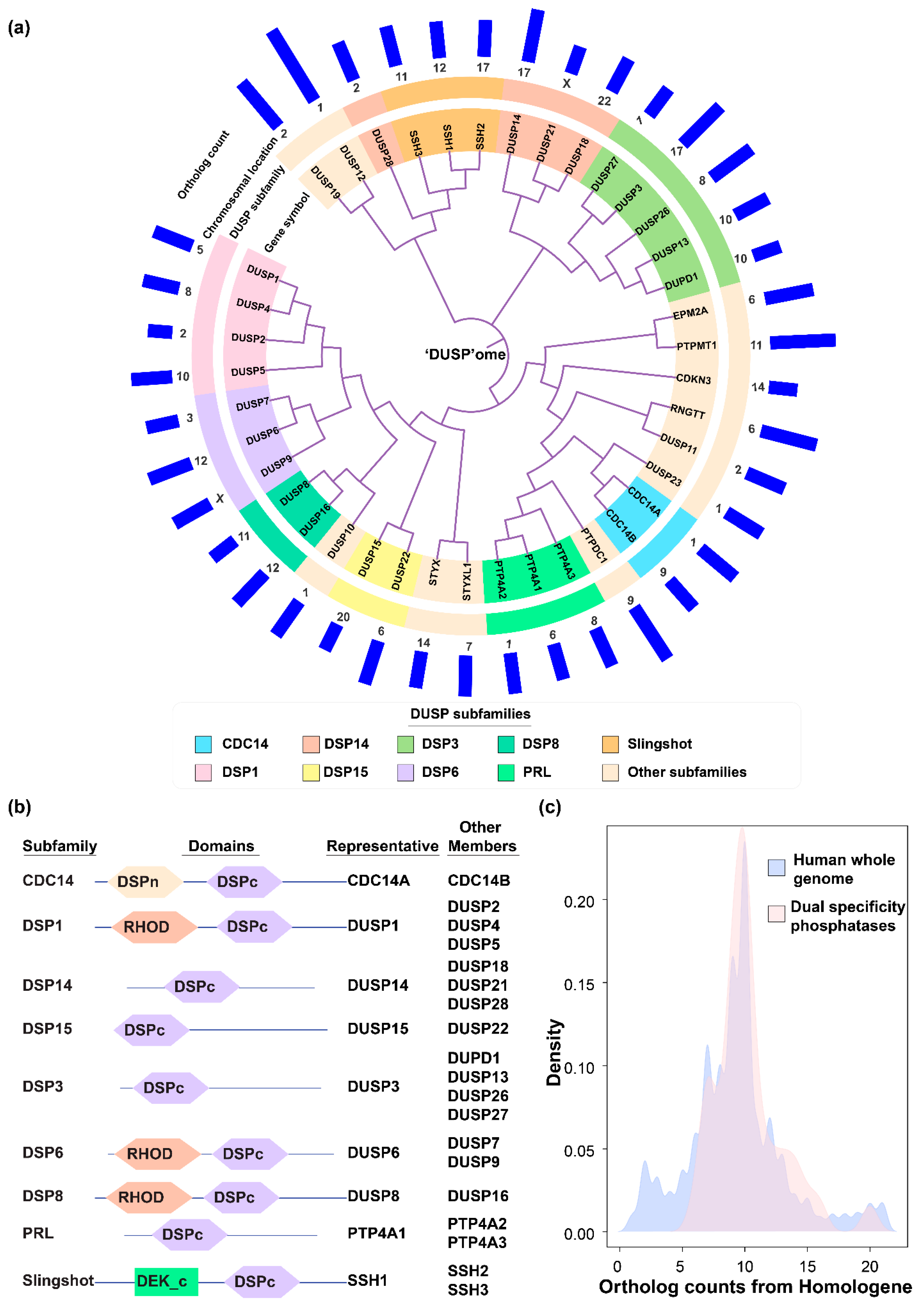
2.1. DUSP Classification into Subfamilies and Evolutionary Conservation

We compiled a list of DUSPs from previously published studies and used it to for further analysis. We also aligned protein sequences for known DUSPs, classified them into subfamilies according to their clustering patterns and validated the classification using domain analysis. The classification of DUSPs based on sequence similarity was performed by multiple sequence alignment analysis of DUSP protein sequences (Figure 1a). For the analysis, the list of DUSPs and their subfamilies was compiled from Chen et al. [7], and the results were found to be concordant with this classification system. The classification includes members belonging to CDC14, DSP1, DSP14, DSP15, DSP3, DSP6, DSP8, PRL, and Slingshot subfamilies. Protein domain analysis of all DUSP members also validated the sub-classification of DUSPs (Figure 1b). Most DUSP subfamily members exhibited similar architectures with a common DSPc domain. However, members of a few subfamilies contained additional domains besides DSPc namely CDC14 subfamily (N-terminal DSP domain), DSP1, DSP6, and DSP8 (Rhodanese-like domain) subfamilies and members of Slingshot subfamily (DEK domain at the carboxy terminus). Next, we aimed to determine the evolutionary conservation of dual specificity phosphatases across eukaryotic species by calculating the number of orthologs for all human DUSPs obtained from Homologene database [39]. Our analysis revealed the distribution of DUSPs ranging from a minimum, of six orthologs for DUSP2 to 20 for DUSP12 (Figure 1c). The distribution of the entire human proteome was similar with a minimum of one and a maximum of 21 ortholog counts. Most DUSPs were found to be conserved in mammals, and particularly among primates suggesting evolutionary conservation across eukaryotic species.

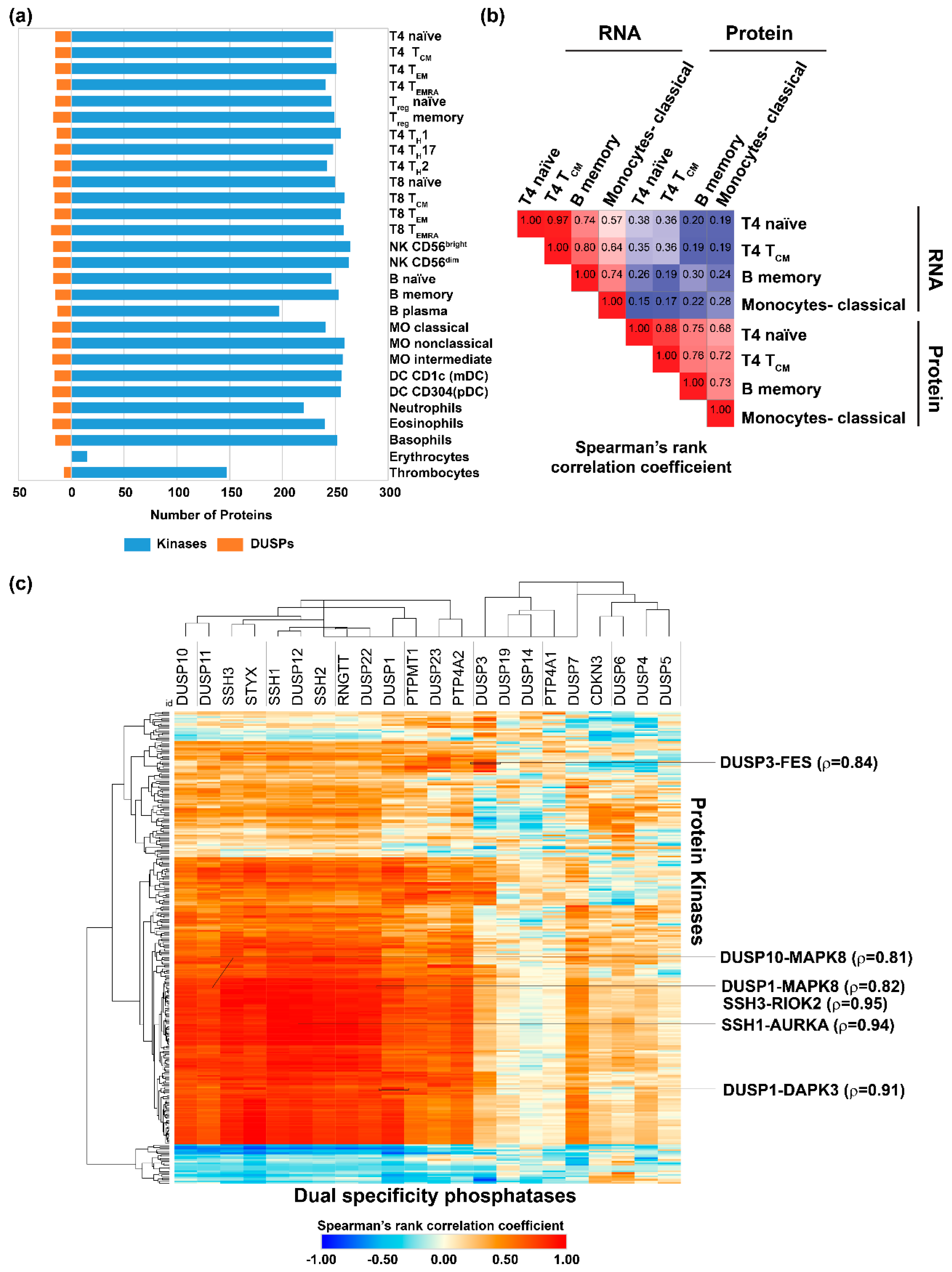
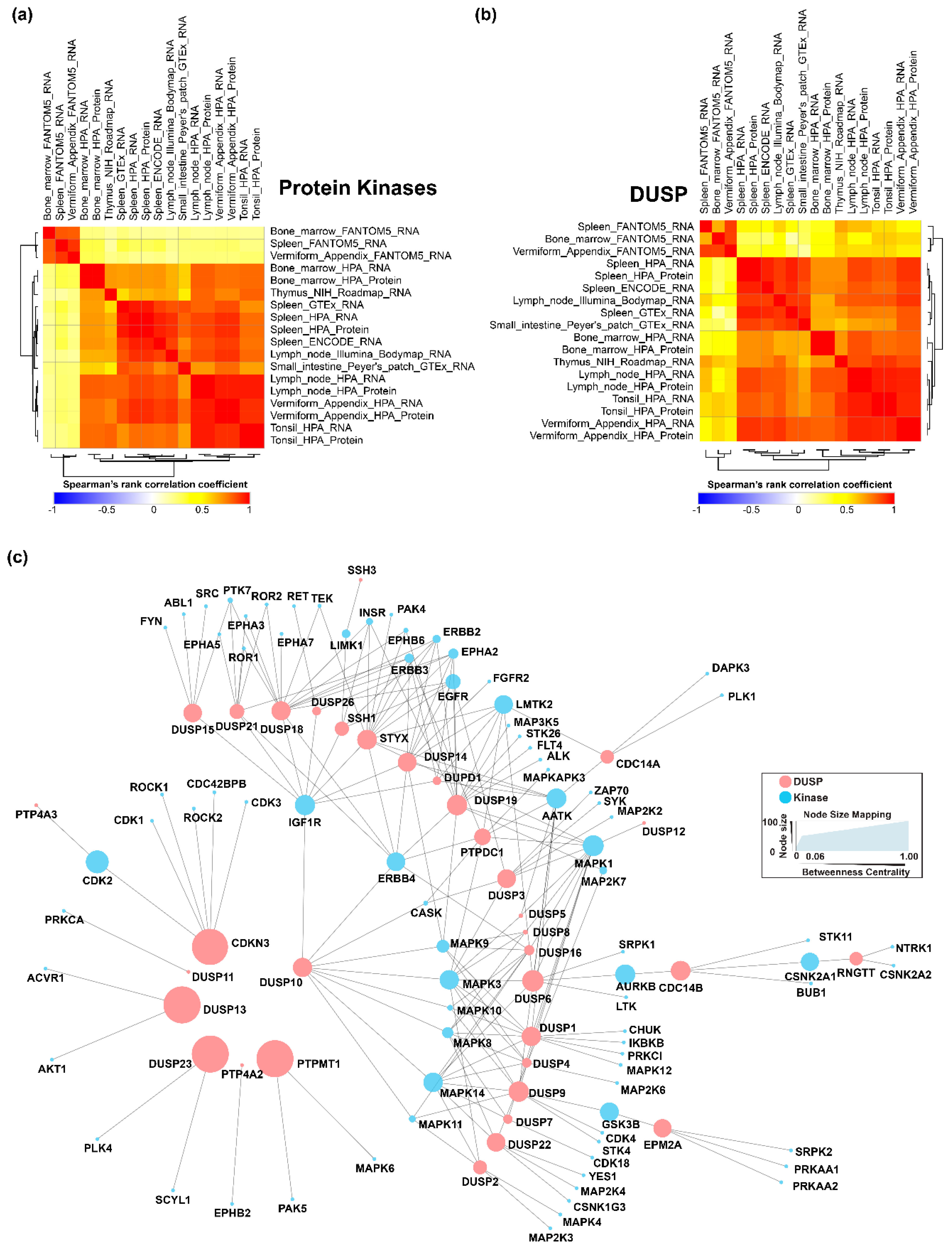
2.2. Expression of Dual Specificity Phosphatases and Protein Kinases in Hematopoietic Cells, Primary and Secondary Lymphoid Organs

Earlier reports suggest that the expression of DUSPs are regulated during development in a cell type-specific manner or upon cell activation in contrast to their ubiquitous substrate expression [8]. In addition, PKs play important roles in immunity [40,41] and are widely known to be modulated by DUSPs. In order to determine the extent of expression of DUSPs and PKs across human hematopoietic cells, we analyzed the proteomic data from Rieckemann et al. [29] as it is currently the largest high-resolution dataset containing expression data pertaining to 28 different hematopoietic cell types analyzed on a single platform (Figure S1, Table S1). On an average, 15 DUSPs and 240 PKs were found to be expressed across hematopoietic cells (Figure 2a) at the protein level. Among the various cell lineages, T8 TEMRA (terminally differentiated effector memory T cells which express CD45RA, as opposed to TEM cells which are CDC45RA-negative) cells expressed the highest number of DUSPs (19), while we did not identify expression of DUSPs from the erythrocyte data. On the contrary, 264 PKs were found to be expressed in NK CD56bright cells, whereas only 15 were found to be expressed in erythrocytes. NK CD56bright cell types have been previously described to be regulatory in nature influencing innate immunity through cytokine production as opposed to NK CD56dim cells, which have cytotoxic activity [42].
Among the DUSP family members, 11 DUSPs were found to be expressed in a large majority of the hematopoietic cells analyzed. These include DUSP12, DUSP23, DUSP3, SSH3 and PTP4A2, which were found to be expressed in 27 of 28 hematopoietic cells and DUSP11, DUSP22, PTPMT1, RNGTT, SSH1 and SSH2 found to be expressed in 26 of the 28 cells. 18 members of the DUSP family were not identified in any of the cell types. Furthermore, we did not observe restricted expression of DUSP members to any one cell type. Among the PKs, only 287 were found to be identified in at least one hematopoietic cell type with the rest not being identified in any cell type. While 13 members of the PKs including MAPK1, MAP2K2, ILK, and ROCK2 were found to have ubiquitous expression, four kinases including PRKCD, KALRN, MYLK and PTK2 were found to have restricted expression in thrombocytes. At least five PKs including MKNK2, STK33, CSNK1E, TAOK2 and BLK were found to be restricted to 2 of the 28 cell types. Of these, MKNK2, STK33 and BLK were expressed commonly in B memory cells. STK33 was expressed in B naïve and B memory cells and seemed to be linked with B cell types. We compared proteomics and transcriptomics datasets to see if changes at the transcript level could be equated with changes in protein expression (Figure 2b, Table S2).
Overall, we observed the expression of kinases and DUSPs to be relatively lower compared to housekeeping proteins such as tubulin (TUBB). The notable exceptions were PRKCB with abundant expression in B memory cells and plasmacytoid dendritic cells and PRKDC with nearly ubiquitous high expression across all hematopoietic cell types. Cell type enriched expression patterns were observed for several kinases including MAPKAPK3 (high in eosinophils), BTK (basophils), ILK (thrombocytes) and HCK with highly abundant expression in classical, non-classical and intermediate monocytes. While ILK in thrombocytes has been implicated in the regulation of integrin signaling [43], HCK has been described as a key regulator of gene expression in activated monocytes [44]. The importance of these expression patterns need to be further investigated and may be important in the regulation of cell-specific processes.
We analyzed tissue expression levels of DUSPs and PKs in immune-related tissues. We chose to study expression data pertaining to lymphoid organs that are the major components of the immune system involved in producing B- and T-cells (primary) and are responsible for the coordinating the cell-mediated immune response (secondary) [45]. The primary lymphoid organs consist of the thymus and the bone marrow while the secondary lymphoid organs consist of lymph nodes, spleen, tonsils, and the mucosa-associated lymphoid tissues such as Peyer’s patches [46]. More recently, the appendix has been deemed to be a lymphoid organ capable of carrying out immunological functions [47,48]. For the analysis, we compiled gene expression data (Table S3) from multiple projects including FANTOM5 [49], Genotype-Tissue Expression (GTEx) Project [50], The Human Protein Atlas [51,52], Illumina Body Map [53], NIH Roadmap Epigenomics Mapping Consortium [54] and the ENCODE project [55]. The PK and DUSP expression in these various tissue expression datasets largely correlated (Figure 3a,b). A majority of the 505 PKs were found to be expressed in at least one lymphoid organ and expression of only a mere 7 kinases were not observed. A significant observation was that all the members of the DUSP family were present in at least one lymphoid organ. 460 of the 505 PKs were found to be expressed in all 7 lymphoid organs while 36 of the 40 DUSPs were expressed in as many lymphoid organs. Two kinases-EPHA8 and PAK5 were found to be restricted to only lymphoid organ -spleen and thymus respectively. DUSP21 was found to be restricted in the bone marrow while DUPD1 was found to be restricted to only the thymus and the tonsil. Similarity matrices indicated a similar distribution of kinases and DUSP expression across primary and secondary lymphoid organs suggesting potential reciprocal pairs (Figure 3a,b). In conclusion, members of the DUSP family were found to be expressed in every lymphoid tissue, thereby suggesting DUSP mediated control of protein kinase activity.

2.3. Correlation of Dual Specificity Phosphatase Activity with Protein Kinase Activity in Immune Cells

Since it is widely known that DUSPs regulate protein kinases, identifying DUSPs and protein kinases pairs with reciprocal activities could identify regulatory mechanisms that can be potentially exploited to develop strategies for therapeutic interventions in conditions such as systemic inflammatory and autoimmune disorders. To identify potentially reciprocal DUSP-kinase pairs and to determine its probable role in immune cells, we correlated the expression profiles of DUSPs and PKs expressed in hematopoietic cells from Rieckemann et al. [29] (Figure 2c, Table 1, Table S4).
We identified 231 DUSP-kinase pairs with similar coexpression patterns in immune cells with a Spearman’s rank correlation coefficient of 0.9 or more indicating high confident reciprocal pairs. Similarly, 701 DUSP-kinase pairs with similar coexpression patterns in immune cells had a Spearman’s rank correlation coefficient of 0.8 or more. Some of the pairs identified include SSH1-AURKA (ρ = 0.94), DUSP1- MAPK7 (ρ = 0.79), DUSP1-MAPK8 (ρ= 0.82), DUSP10-MAPK8 (ρ = 0.81). Among these, the role of DUSP1 and DUSP10 as negative regulators of MAPK8 is well known. The significance of SSH1-AURKA pair in immune cells is currently not well described at this time.
We also used an interactome-based approach to identify co-expressed and co-interacting proteins to identify potential DUSP-kinase regulatory mechanisms. Interactome analysis to identify reciprocal DUSP-protein kinase pairs from baseline protein-protein interaction (PPI) data from the comPPI database resulted in the generation of an interaction network containing 1715 DUSP-specific interactions with 1276 nodes (Figure S2). Since the network was too complex to comprehend the interplay between DUSPs and PKs, we separated interactions between DUSPs and kinases and generated an additional kinase and DUSP-specific network. This network contained 195 protein-protein interactions between 35 DUSP members and 82 PKs (Figure 3c, Table S5). Several potential hub proteins that communicate with the dual specificity phosphatase family members were deduced from the interaction network. Among the PKs, MAPK1 and MAPK3 had 12 and 10 interactions (directed edges) respectively with DUSPs. Other PKs with a high number of interactions included MAPK14 (9), IGF1R (9), AATK (9) and ERBB4 (8). Among the DUSPs, DUSP18 and DUSP19 had the most number of directed edges -14 each. Other DUSP family members with several directed edges included STYX (13), DUSP 1 (11), DUSP14 (10), DUSP9 (9) and DUSP 10 (9). It is interesting to note that DUSP19, and STYX are some of the poorly characterized members. The PKs with the highest betweenness centrality included MAPK1, IGF1R, AURKB, and AATK. Combining the directed edge and the betweenness centrality data revealed several kinases belonging to the MAPK family and receptor tyrosine kinases such as MAPK1, MAPK3, IGF1R, AATK, MAPK14, ERBB4, LMTK2, MAPK9 and MAPK8 to be strongly associated with DUSPs.

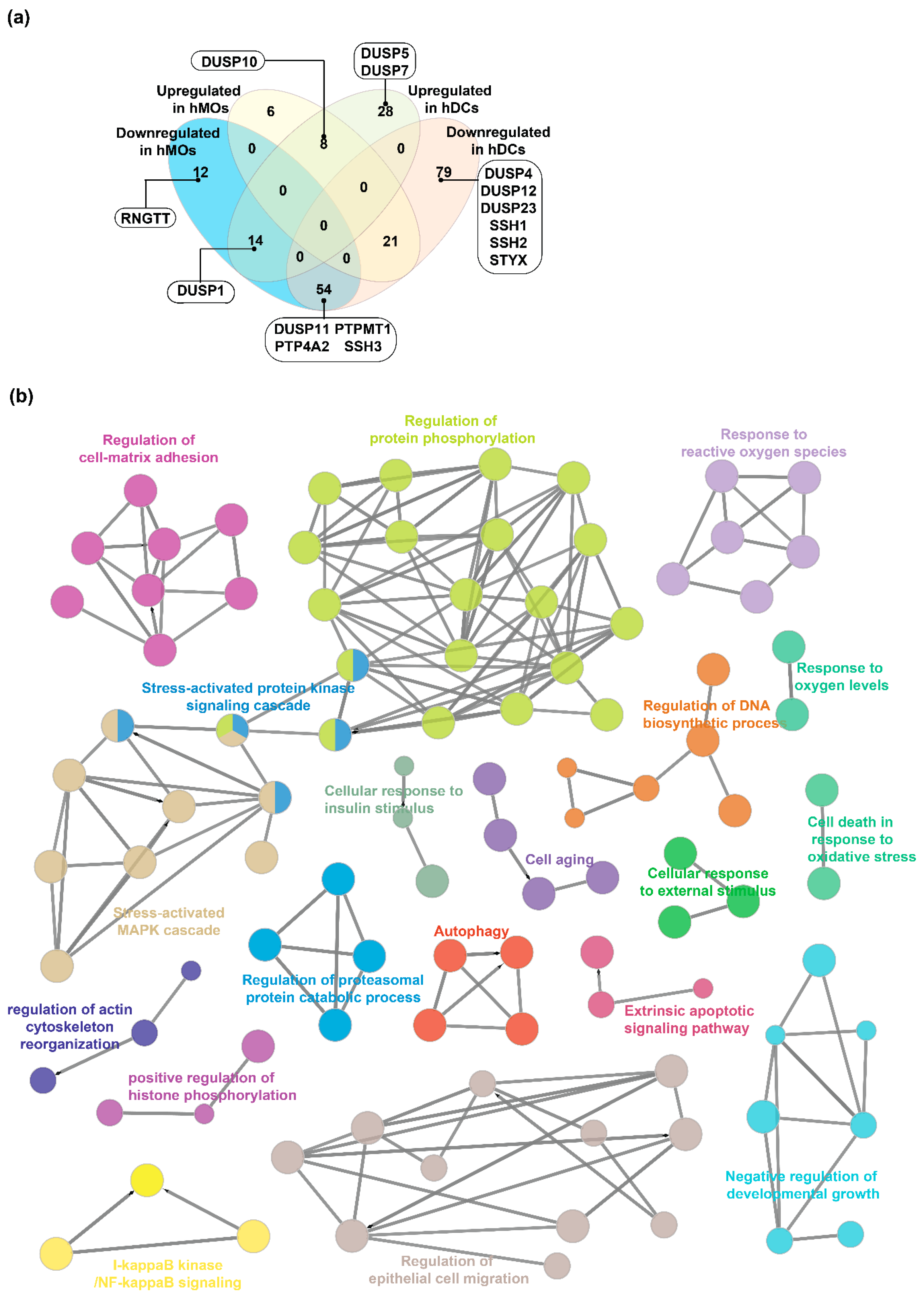
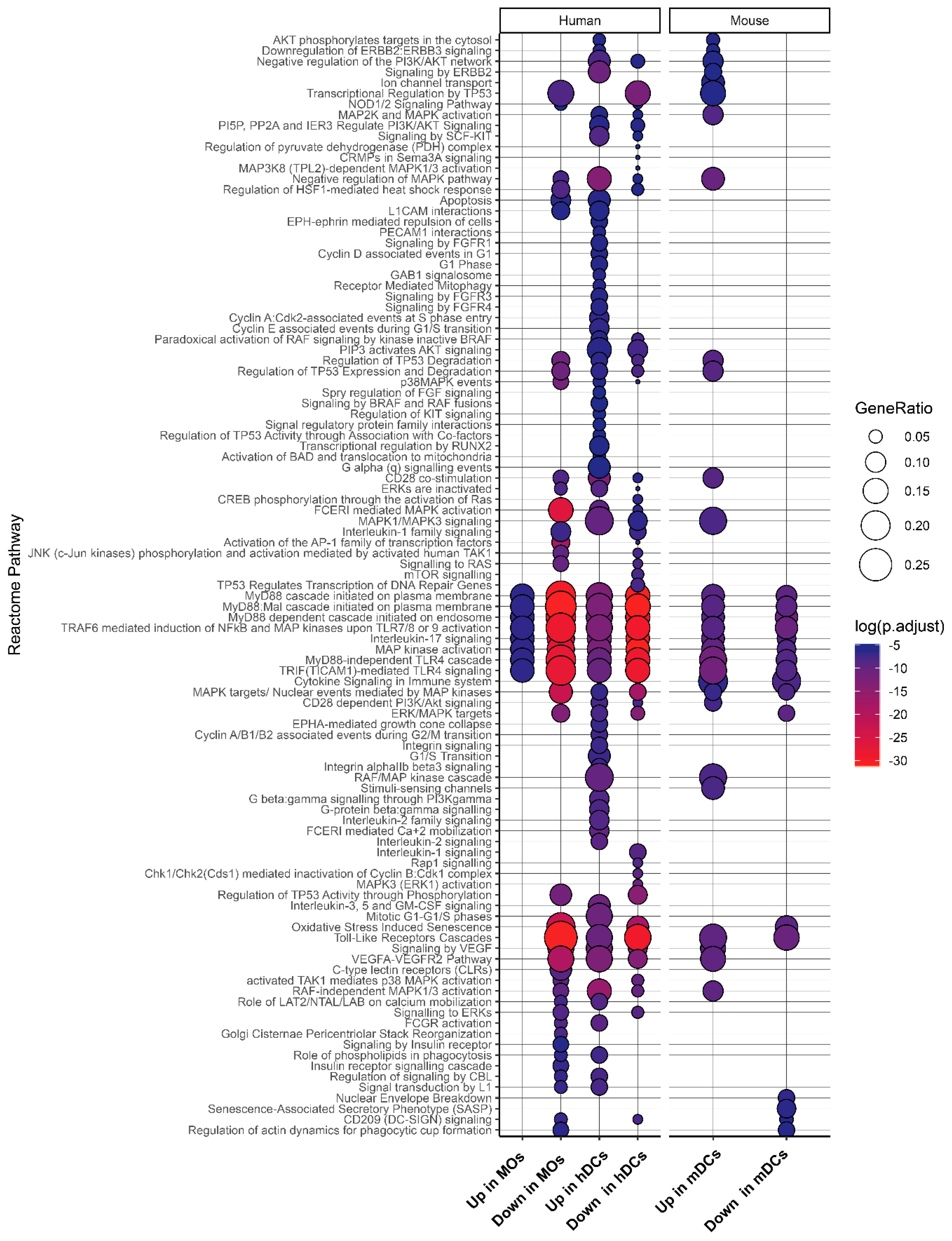
2.4. Expression Landscape and Signaling Dynamics of DUSPs and Kinases in Activated Immune Cells

The data provided by Rieckemann et al. also includes steady-state and activated protein expression profiles of 17 cell types which were analyzed to determine the effect of various activating ligands on the expression of DUSPs and PKs (Table S6). Our analysis resulted in the identification of 152 events of differential expression of 18 DUSPs across 17 cells types (Figure S3a) Of these, 57 and 95 were found to be overexpressed and downregulated respectively across the activated cell types. Similarly, we identified 2311 events of differential expression of 269 PKs across 17 cells types (Figure S3b). Of these, 1058 and 1253 were found to be overexpressed and downregulated respectively across the activated cell types. Several kinases and DUSPs were found to be differentially expressed in multiple activated cell types. The most overexpressed DUSPs included DUSP12 (9 cell types), DUSP23 (7 cell types), DUSP1, DUSP10 (6 cell types each) and STYX (4 cell types), while the most downregulated DUSPs included SSH3 (13 cell types), PTPMT1 (12 cell types), DUSP3 (10 cell types), SSH2 (9 cell types), DUSP1 (8 cell types) and SSH1 (7 cell types). Similarly, among the kinases the most overexpressed included CDK1 (14 cell types), PLK1 (14 cell types), TGFRB1 (13 cell types), RIOK3 (13 cell types) and RIOK2 (13 cell types) while the most downregulated included ATM (15 cell types), MAST3 (14 cell types), PRKACB (13 cell types), MAP3K5, LMTK2 and SYK (12 cell types each).
There have been several studies focused on the genome-wide effects of TLR ligands on hematopoietic cells such as monocytes [60]. In the current study, we aimed to determine the specific roles of DUSPs and PKs in TLR4 signaling. An integrated analysis of RNA and protein expression datasets [29,33,61] pertaining to dendritic cells (DCs) and monocytes (MOs) activated by LPS was performed (Table S17). The data were categorized into murine DCs (mDCs), human DCs (hDCs) and human MOs (hMOs) and a list of molecules differentially expressed in response to LPS was generated (Table S8). 57 proteins including 4 DUSPs and 53 PKs were found to be overexpressed in mDCs while 80 were downregulated (2 DUSPs, 78 PKs) in response to LPS. In hDCs, 50 proteins (4 DUSPs, 46 PKs) and 154 (10 DUSPs, 144 PKs) proteins were found to were found to be overexpressed and downregulated respectively. In the case of hMOs, 35 (1 DUSP, 34 PKs) were overexpressed and 80 (6 DUSPs, 74 PKs) were downregulated.
DUSPs overexpressed in dendritic cells included Dusp1/DUSP1 (mDCs and hDCs), Dusp14, Dusp16, Ptp4a2 (all in mDCs), DUSP5, DUSP7, and DUSP 10 (all in hDCs). Downregulated DUSPs included Dusp3, Dusp19 (mDCs), DUSP4, DUSP11, DUSP12, DUSP23, PTP4A2, PTPMT1, SSH1, SSH2, SSH3, and STYX (hDCs) (Figure 4a and Figure S5a–c). In hMOs stimulated with LPS, DUSP 10 was overexpressed while DUSP1, DUSP11, PTP4A2, PTPMT1, RNGTT, and SSH3 were found to be downregulated. DUSP1 (Dusp1) seems to be important in both hDCs and mDCs signaling as it was found to be upregulated in both species in response to LPS and is in concordance with previously published studies on dendritic cells stimulated with LPS [14]. However, it was found to be downregulated in hMOs. The overexpression of DUSP1 in dendritic cells identified from our analysis is in concordance with previously published studies on dendritic cells stimulated with LPS. DUSP10 was found to be upregulated in both hDCs and hMOs while in mDCs, it was not found to be differentially expressed. Among differentially expressed genes in dendritic cells, Dusp14 and Dusp16 were exclusively overexpressed in the dendritic cells from mice while DUSP5, DUSP7, and DUSP10 seemed to be exclusive to humans. Taken together, our analysis suggests probable existence of species-specific differential expression of DUSPs in TLR4 signaling. In a recent paper, human and murine macrophages were found to have varying mechanisms of immunometabolism [62]. Functional analysis of differentially expressed PKs and DUSPs in murine dendritic cells stimulated with LPS showed enrichment of several processes including MAPK cascade, response to reactive oxygen species, cellular senescence, and cell migration pathways among others (Figure 4b).
We next performed pathway enrichment analysis using pathway data from Reactome database for DUSP and PKs showing differential expression upon LPS stimulation (Figure 5, Table S9). We found several pathways including TLR signaling, MyD88 cascade, and MAPK signaling pathways to be enriched across LPS-activated MOs and DCs. Oxidative stress-induced senescence pathways were downregulated in mDCs, hDCs, and hMOs. VEGF signaling also seemed to be affected through DUSP and PKs after stimulation with LPS. While induction of TLR signaling by LPS is widely known, there are a few reports on the induction of VEGF signaling by LPS stimulation [63]. FGFR signaling and SCF-KIT signaling pathways were up in hDCs, while C-lectin receptor signaling pathway was downregulated in hMOs upon LPS stimulation. Apoptotic pathways was found to be downregulated in hMOs while being upregulated in hDCs suggesting opposing cell death phenotypes in hMOs and hDCs in response to LPS.
To identify DUSP-kinase pairs with reciprocal activities, correlation analysis between DUSP and kinase expression in mDCs and mMOs treated with LPS was carried out (Figure S5d, Table S10. Our analysis resulted in the identification of several important pairs including Ptpmt1-Mast3 (ρ = 0.97), Dusp1-Egfr (ρ = 0.73), Dusp1-Mapk8 (ρ = 0.66), Dusp16-Mapk8 (ρ = 0.83), Dusp16-Lrrk2 ρ = (0.86), Dusp10-Igf1r (ρ = 0.99).
In order to identify the interacting partners of DUSPs in TLR4 signaling, we carried out interactome analysis of members of the DUSP and protein kinase families that were found to be differentially expressed in dendritic cells and monocytes (Figures S6–S8). Analysis of network properties identified several proteins that seemed to be regulated by DUSP signaling. In the networks of differentially expressed proteins identified in activated mDCs, proteins including Akt1, Lrrk2, Pim1, Dusp1, Dusp16 (overexpressed), Mapk1/3, Pik3cg, Cdk1, Dusp3 and Dusp19 (downregulated) had a high number of edges and high betweenness centrality suggesting their importance in innate immunity. In activated hDCs, proteins including MAPK1, CDK2, PIK3CA, MAPK13, DUSP1, DUSP5, DUSP7 and DUSP10 (upregulated), PRKACA, LRRK2, CHUK and DUSP13 (downregulated) had a high betweenness centrality and were identified to be relevant in LPS-induced signaling. Similarly, in activated hMOs, PRKCZ, CDK6, DUSP10, TGFBR1 (upregulated), LRRK2, ATM, ATR, MAPK1, DUSP1, MAP3K1 (downregulated) among others had a high betweenness centrality.

3. Discussion

Dual-specificity phosphatases (DUSPs) are a family of phosphatases that can act on both serine/threonine and tyrosine residues of several protein substrates leading to wide-ranging effects on cellular signaling and biological processes. With the exact number of DUSP members still being controversial [7,64,65,66,67,68], we chose to consider the latest classification described by Chen et al. [7], consisting of 40 DUSPs with 9 subfamilies containing more than one member. We validated the subfamily-based classification of DUSPs using two approaches namely-sequence alignment and SMART-based domain analysis. Evolutionary conservation analysis of DUSP family members revealed high sequence conservation of all DUSP members, especially in higher mammals.
So as to determine the extent of expression of dual specificity phosphatases and protein kinases across the human hematopoietic cells and lymphoid organs, publicly available datasets were mined. Since hematopoietic cells are derived from lymphoid tissues, we chose to analyze expression profiles of these. To date, Rieckemann et al. have provided the largest expression dataset pertaining to 28 different hematopoietic cell types consisting of high throughput omics data acquired under a single platform containing. Our analysis of this dataset revealed new insights into the expression dynamics of several understudied DUSPs such as slingshot family of phosphatases (SSH1, SSH2 and SSH3) and STYX in various hematopoietic cell types. Additionally, our analysis also resulted in the evaluation of the expression patterns of several understudied protein kinases described by Huang et al. [69] including STK17A, SCYL3, MAST3, CSNK1G2, and the RIO family of kinases (RIOK1, RIOK2 and RIOK3). Furthermore, among the hematopoietic cells, erythrocytes and thrombocytes (platelets) expressed the least number of DUSPs and PKs The restricted expression patterns of these proteins could be potentially exploited for therapeutic modalities in erythrocyte and platelet disorders. In fact, four different PKs (PRKCD, KALRN, MYLK, and PTK2) showed restricted expression in thrombocytes. PRKCD has been previously reported to modulate collagen-induced platelet aggregation [70] while MYLK and PTK2 have been reported to be important in megakaryopoiesis [71,72]. Further, the different patterns of DUSP and PK expression in erythrocytes and thrombocytes compared to the rest of the hematopoietic cells may be attributed to their structures, diverse biological functions performed by these cells, their numbers in the human body and the differing lineages that form erythrocytes and thrombocytes during hematopoiesis.
Among the 701 DUSP-kinase pairs with potential reciprocal activities identified from the unstimulated hematopoietic cell expression data, several pairs with previously known reciprocal actions were also identified thereby proving our analysis methods. Among these, SSH1-AURKA pair was identified with a high Spearman’s rank correlation coefficient (ρ = 0.94). Aurora Kinase A (AURKA) overexpression has been previously reported to increase the expression of slingshot kinase 1 (SSH1) resulting in increased cofilin activation and migration of breast cancer cells [56]. Other notable pairs that we identified from the correlation and that were previously reported in the literature included DUSP1- MAPK7 (ρ = 0.79) and DUSP1-MAPK8 (ρ = 0.82). DUSP1 gene silencing has been shown to increase the expression of MAPK7 and MAPK8 transcripts in osteosarcoma cells suggesting reciprocal actions between them [57]. DUSP10 (MKP-5) has been well known to dephosphorylate MAPK8 (JNK) [58] and has also been implicated in immune function. Knocking down DUSP10 expression increased JNK activity and inflammation in murine mesangial cells while its overexpression led to decreased JNK activation [73]. A study by Zhang et al. further found that Dusp10-deficient murine cells exhibited increased JNK (MAPK8) activity, elevated levels of proinflammatory cytokines and increased T cell activation [74]. In our correlation analysis, the DUSP10-MAPK8 pair was identified with a Spearman’s rank correlation coefficient of 0.81.
We used a subset of omics datasets for investigating the expression of DUSPs and their interplay with PKs upon LPS stimulation in activated dendritic cells and monocytes. Though a previous study on the meta-analysis of TLR4 signaling datasets exists, its focus was on activated macrophages [75]. Biological process-based enrichment upon LPS stimulation showed the enrichment of several processes including MAPK cascade, response to reactive oxygen species, cellular senescence and cell migration, autophagy pathways among others. While these processes are widely known in the context of macrophages, the effects of LPS stimulation seem to be similar in dendritic cells as well. We also correlated DUSP and PK expression in these cells and carried out interactome analysis to identify key molecules that are potential regulators of LPS-induced signaling. Correlation analysis indicated the presence of DUSP-PK pairs with reciprocal activities in response to activation. Some of these pairs including DUSP1-MAPK8, DUSP1-MAPK8 have already been described in previous literature, thus confirming our findings. DUSP-16 (MKP-7) has previously been identified to regulate MAPK8 (JNK) in LPS-activated macrophages [59] and in activated endothelial cells [76]. Dusp16 (MKP-7) was also reported to have a critical role in the activation and functioning of T cells. Dusp16-deficient T cells had an exaggerated response to TCR activation and had enhanced proliferation properties [77]. DUSP16-deficient macrophages have also been reported to overproduce IL-12 in the context of TLR stimulation [78]. DUSP1 has been reported to play a key role in the feedback control and regulate MAPK8 during glucocorticoid-mediated repression of inflammatory gene expression [79]. DUSP1 is already known to regulate the expression of LPS-induced genes [14]. We also found DUSP 1 and DUSP16 to be important regulators of several MAP kinases from the interactome analysis.
Several novel DUSP-PK pairs were identified, and these require further characterization to confirm their role in immunity. Particularly interesting among these include pairs involving slingshot phosphatases which constitute a group of understudied phosphatases. SSH1-CDK13, SSH1-CDK19, SSH2-EPHB2 were identified with high significance. Slingshot phosphatases have been previously implicated in cancer progression [80,81] and have been known to mediate caspase-modulated actin polymerization towards bacterial clearance upon Legionella infection [82]. In our analysis, Slingshot phosphatase members SSH1, SSH2 were found to be downregulated exclusively in human DCs, while SSH3 was downregulated in both human DCs and MOs. Slingshot phosphatase members SSH1 and SSH2 were found to be downregulated exclusively in human DCs, while SSH3 was downregulated in both human DCs and MOs. DUSP10 and IGF1R expression were also found to be highly correlated. Insulin-like growth factors have been known to inhibit anti-tumoral responses of dendritic cells through the regulation of MAP kinases and have therefore been suggested to be targets for immunotherapy [83]. IGF1 has also been reported to influence activation of macrophages in response to high-fat diet or helminthic infection [84]. Additionally, we identified LRRK2 (leucine-rich repeat kinase 2) from the interactome analysis in activated DCs and MOs, to be potentially important in the context of immune signaling. LRRK2 was found to interact with DUSP1 and DUSP16. LRRK2 has been mainly associated with familial cases of Parkinson’s disease [85] and has been known to play an important in innate immunity in the peripheral and central nervous system [86], especially in microglial inflammatory processes [87]. This present finding suggests the possibility of LRRK2 being additionally important in DUSP-mediated immune signaling.

4. Materials and Methods

4.1. Datasets

Studies pertaining to immune cells acquired using high-throughput techniques were searched using PubMed. The data matrices pertaining to each dataset were downloaded from the site of the publisher of these articles. Gene expression datasets were downloaded from Gene Expression Omnibus (GEO, https://www.ncbi.nlm.nih.gov/geo/) wherever applicable. The details of all the studies used in this study are provided in Table S11.

4.2. DUSP and Kinase Lists for Analysis

The list of DUSPs used for the analysis was sourced from Chen et al. [7]. The list of human and mouse kinases was sourced from UniProt (https://www.uniprot.org/docs/pkinfam) which used data from [4,88,89]. The master lists of dual specificity phosphatases and PKs used for data analysis in the current study are provided as Tables S12 and S13.

4.3. Classification of DUSP Family Members, Domain Analysis and Species Conservation Analysis of DUSP Sequences

The similarity tree between DUSPs was drawn using iTOL with alignment performed with Clustal Omega. Briefly, RefSeq accessions of the longest protein isoforms for all dual specificity phosphatases were retrieved from NCBI gene (https://www.ncbi.nlm.nih.gov/gene). The protein sequences were obtained with Batch Entrez (https://www.ncbi.nlm.nih.gov/sites/batchentrez) and aligned using Clustal Omega (https://www.ebi.ac.uk/Tools/msa/clustalo/) using default settings. The output alignment in PHYLIP (.ph) format and was visualized with Interactive Tree of Life (https://itol.embl.de/) with custom colors and tracks.
Domain analysis was carried out for the longest isoforms of all proteins belonging to DUSP subfamilies with multiple members using the SMART domain prediction tool (http://smart.embl-heidelberg.de/) [90], using the option “PFAM domains”. Orthology data for all human genes were obtained from Homologene (Release 68, downloaded on October 4, 2018 from https://www.ncbi.nlm.nih.gov/homologene) for the analysis of sequence conservation across species. The counts for all genes in the Homologene database were obtained and the Taxonomy ID for each gene was mapped to the species type. The densities of ortholog counts for DUSP family members was plotted against the density of ortholog counts for all human genes in the background using R (v3.5.1) (https://cran.r-project.org/).

4.4. Landscape of DUSPs and Kinases in Immune Cells

Proteomic and transcriptomic data matrices were obtained from supplementary files of respective articles and accessions were converted into Entrez gene accession formats using bioDBnet:db2db (https://biodbnet-abcc.ncifcrf.gov/db/db2db.php) [91] and g:Profiler (https://biit.cs.ut.ee/gprofiler/gconvert.cgi) [92]. Gene expression data was also downloaded from Gene Expression Omnibus (GEO) wherever supplementary data was not available. Z-score-based normalization of data matrices from all studies was carried out using base R (v3.5.1). DUSP and kinase expression data were subsequently obtained from the normalized datasets. Cell type data and cell sorting information wherever available were retrieved for each of the expression datasets and appended with the expression data. Heatmaps were drawn in Morpheus (https://software.broadinstitute.org/morpheus/) with hierarchical clustering based on Euclidean distance metric, complete linkage method and clustering by rows and columns. We carried out correlation and interactome analysis to look at the interplay between DUSPs and PKs in naïve and activated immune cells. Correlation analysis between z-scores of kinase and DUSP profiles of immune cell proteomes was performed using “Spearman” method through R (v3.5.1). Heatmaps were drawn using Morpheus (https://software.broadinstitute.org/morpheus/).

4.5. Landscape of DUSPs and Kinases in Primary and Secondary Lymphoid Organs

We compiled tissue-based expression data from various datasets present in the Expression Atlas (https://www.ebi.ac.uk/gxa/home). Briefly, the pre-processed datasets from The FANTOM5 project [49], Genotype-Tissue Expression (GTEx) Project [50], The Human Protein Atlas [51,52], Illumina Body Map [53], NIH Roadmap Epigenomics Mapping Consortium [54] and the ENCODE project [55] were downloaded (Download date January 11,2019). The datasets were subjected to z-score-based normalization using R (v3.5.1). Data from primary and secondary lymphoid tissues were selected and DUSP and kinase expression data were compiled using in-house scripts. The data was plotted as heatmaps with Morpheus using the same parameters as in the previous section.

4.6. Baseline DUSP Interactome

We analyzed publicly available Protein-protein interaction (PPI) data to identify DUSP-kinase interactions. We chose the comPPI database (Compartmentalized Protein-Protein Interaction Database, v2.1.1, http://comppi.linkgroup.hu/home) to identify biologically significant high-confident interactions between proteins with similar subcellular localization patterns. ComPPI is an integrated database of protein subcellular localization and protein-protein interactions from multiple databases including BioGRID, CCSB, DIP, HPRD, IntAct and MatrixDB. [93]. The highly confident interactomes of each member of the dual specificity phosphatase family for Homo sapiens were fetched from comPPI, filtering for localization score and interaction score thresholds of 0.7 each. The accessions of interacting proteins were obtained through bioDBnet: db2db (https://biodbnet-abcc.ncifcrf.gov/db/db2db.php) and g: Profiler (https://biit.cs.ut.ee/gprofiler/gconvert.cgi) and the set of interactions were compiled and visualized in Cytoscape (version 3.7.0) to obtain an integrated DUSP interactome. The interactome was clustered into DUSP neighborhood networks using the AutoAnnotate (v1.2) package. The network statistics of the interactome were analyzed using the Network Analyzer tool in Cytoscape [94]. The network analysis identified several network parameters. For the sake of clear understanding, we define a few basic concepts of graph theory [95]. Typically, a network consists of nodes which are in this case genes/proteins and edges which describe the relationship between them. A directed graph is a triple ordered graph G = (V, E, f) where V represents vertices called node, E represents edges representing connections between nodes and f is a function that maps each element in E to an ordered pair of vertices in V. The ordered pair of vertices are known as directed edges and represented by E = (i, j). Network properties provide valuable insight into the organizational structure of a biological network and network centralities show how nodes can be ranked/prioritized according to their properties. One of the network centralities is betweenness centrality, which is a measure of how nodes that are intermediate between neighbors rank higher. This essentially means that without these nodes, there would be no other way that the two neighbors could communicate with each other. In the current study, the betweenness centralities measure were used to identify protein hubs central to the DUSP network, which could influence the flow of information triggered by DUSPs. Individual interactomes of each DUSP member were also analyzed to identify central proteins associated with DUSPs using the betweenness centrality measure.

4.7. Landscape of DUSPs and Kinases in Activated Immune Cells

Data matrices from proteomics and transcriptomics datasets containing gene/protein expression data were fetched from the supplementary tables or GEO for the following studies [29,32,33,61]. DUSP and kinase expression data were obtained from the normalized datasets. Fold-change ratios were calculated by dividing the ratio of intensity/RPKMs of activated/stimulated cells by the ratio of respective intensity/RPKM values of steady-state/unstimulated cells. The fold-change ratios were converted into log2(fold-change) by log transformation. Genes/proteins with log2(fold-change) of 1 (fold-ratio of 2) were considered to upregulated while those with −1 (fold-ratio of −1) were considered to be downregulated. Genes/proteins with ambiguous trends across studies were ignored (both up and down) and only those with overall trends of either up and down were considered for further analysis and genes. We carried out correlation and interactome analysis to investigate the interplay between DUSPs and PKs in activated immune cells. Correlation analysis between z-scores of kinase and DUSP expression profiles of activated immune cells was performed as described above.

4.8. Functional, Pathway and Network Analysis

The differentially expressed molecules in response to LPS were analyzed with Gene Ontology-based functional analysis through CLUEGO (ClueGO v2.5.2 + CluePedia v 1.5.2) [96] in Cytoscape (v 3.7.0) to identify processes affected by LPS. The parameters used included ‘ClueGO: Functions’ analysis mode, murine ‘GO: Biological Process’ (dated 18.01.2019), global network specificity, pV ≤ 0.05000 with GO Term grouping. Hypergeometric enrichment-based pathway analysis was performed using Reactome Pathways [97] in R/Bioconductor 3.5.1/3.7 [98] with clusterProfiler 3.8.1 [99] and reactome.db 1.64.0. Genesets with less than 10 genes were excluded from the analysis and p values were adjusted by Benjamini–Hochberg correction. Pathways reaching adjusted p-values ≤ 0.0075 were curated manually and plotted in R with ggplot package (v2 3.1.0) (https://cran.r-project.org/web/packages/ggplot2). Network analysis of activated DCs and MOs was performed using STRING in Cytoscape. The network properties were calculated using Network Analyzer.

5. Conclusions

Though the role of dual specificity phosphatases in innate and adaptive immunity is known, their interplay with kinases was not precisely understood. In the current study, we expanded the knowledge on the role of dual specificity phosphatase signaling in activated and steady-state cells through the analysis of high-resolution expression datasets. We confirmed the importance of several known DUSPs such as DUSP1 and DUSP10 in innate immunity. We also report potentially novel role of DUSPs such as the Slingshot phosphatases and PKs such as LRKK2 in immune signaling. These need to be further validated to confirm their roles. we also identified selective patterns of expression of a few DUSPs and PKs across hematopoietic cells which could be used as potential therapeutic targets. Furthermore, we also identified potential species-specific events of DUSP signaling which need to be further validated. Finally, we demonstrate the utility of meta-analysis of existing datasets to identify molecular mechanisms of various biological processes and fill existing gaps in understanding understudied proteins. The findings from this study will aid in the understanding of DUSP signaling in the context of innate immunity.

Supplementary Materials

Supplementary materials can be found at https://www.mdpi.com/1422-0067/20/9/2086/s1.

Author Contributions

Conceptualization, R.K.K.; methodology, R.K.K., Y.S.; formal analysis, Y.S., K.B.; data curation, S.M.P.; writing—original draft preparation, Y.S.; writing—review and editing, S.M.P., K.B., R.K.K., T.S.K.P. supervision, R.K.K. and T.S.K.P.; funding acquisition, R.K.K.

Funding

This research was funded by the Research Council of Norway (FRIMEDBIO “Young Research Talent” Grant 263168 to R.K.K.; and Centres of Excellence Funding Scheme Project 223255/F50 to CEMIR), Onsager fellowship from NTNU (to R.K.K.).

Acknowledgments

We thank the authors of the datasets used in this study for making their data publicly available.

Conflicts of Interest

The authors declare no conflict of interest

Abbreviations

DCDendritic cells
MOMonocytes
LPSLipopolysaccharides
DUSPDual Specificity Phosphatase
PKProtein Kinase
GOGene Ontology

References

  1. Humphrey, S.J.; James, D.E.; Mann, M. Protein Phosphorylation: A Major Switch Mechanism for Metabolic Regulation. Trends Endocrinol. Metab. 2015, 26, 676–687. [Google Scholar] [CrossRef] [PubMed]
  2. Cohen, P. The origins of protein phosphorylation. Nat. Cell Biol. 2002, 4, E127–E130. [Google Scholar] [CrossRef]
  3. Ardito, F.; Giuliani, M.; Perrone, D.; Troiano, G.; Lo Muzio, L. The crucial role of protein phosphorylation in cell signaling and its use as targeted therapy (Review). Int. J. Mol. Med. 2017, 40, 271–280. [Google Scholar] [CrossRef] [PubMed] [Green Version]
  4. Manning, G.; Whyte, D.B.; Martinez, R.; Hunter, T.; Sudarsanam, S. The protein kinase complement of the human genome. Science 2002, 298, 1912–1934. [Google Scholar] [CrossRef]
  5. Griffith, M.; Griffith, O.L.; Coffman, A.C.; Weible, J.V.; McMichael, J.F.; Spies, N.C.; Koval, J.; Das, I.; Callaway, M.B.; Eldred, J.M.; et al. DGIdb: Mining the druggable genome. Nat. Methods 2013, 10, 1209–1210. [Google Scholar] [CrossRef]
  6. Alonso, A.; Sasin, J.; Bottini, N.; Friedberg, I.; Friedberg, I.; Osterman, A.; Godzik, A.; Hunter, T.; Dixon, J.; Mustelin, T. Protein tyrosine phosphatases in the human genome. Cell 2004, 117, 699–711. [Google Scholar] [CrossRef] [PubMed]
  7. Chen, M.J.; Dixon, J.E.; Manning, G. Genomics and evolution of protein phosphatases. Sci. Signal. 2017, 10, 474. [Google Scholar] [CrossRef]
  8. Lang, R.; Hammer, M.; Mages, J. DUSP meet immunology: dual specificity MAPK phosphatases in control of the inflammatory response. J. Immunol. 2006, 177, 7497–7504. [Google Scholar] [CrossRef]
  9. Liu, Y.; Shepherd, E.G.; Nelin, L.D. MAPK phosphatases--regulating the immune response. Nat. Rev. Immunol. 2007, 7, 202–212. [Google Scholar] [CrossRef] [PubMed]
  10. Jeffrey, K.L.; Camps, M.; Rommel, C.; Mackay, C.R. Targeting dual-specificity phosphatases: manipulating MAP kinase signalling and immune responses. Nat. Rev. Drug Discov. 2007, 6, 391–403. [Google Scholar] [CrossRef] [PubMed]
  11. Abraham, S.M.; Clark, A.R. Dual-specificity phosphatase 1: A critical regulator of innate immune responses. Biochem. Soc. Trans. 2006, 34, 1018–1023. [Google Scholar] [CrossRef] [PubMed]
  12. Zhang, Y.; Reynolds, J.M.; Chang, S.H.; Martin-Orozco, N.; Chung, Y.; Nurieva, R.I.; Dong, C. MKP-1 is necessary for T cell activation and function. J. Biol. Chem. 2009, 284, 30815–30824. [Google Scholar] [CrossRef]
  13. Korhonen, R.; Huotari, N.; Hommo, T.; Leppanen, T.; Moilanen, E. The expression of interleukin-12 is increased by MAP kinase phosphatase-1 through a mechanism related to interferon regulatory factor 1. Mol. Immunol. 2012, 51, 219–226. [Google Scholar] [CrossRef] [PubMed]
  14. Hammer, M.; Mages, J.; Dietrich, H.; Servatius, A.; Howells, N.; Cato, A.C.; Lang, R. Dual specificity phosphatase 1 (DUSP1) regulates a subset of LPS-induced genes and protects mice from lethal endotoxin shock. J. Exp. Med. 2006, 203, 15–20. [Google Scholar] [CrossRef] [PubMed]
  15. Qian, F.; Deng, J.; Cheng, N.; Welch, E.J.; Zhang, Y.; Malik, A.B.; Flavell, R.A.; Dong, C.; Ye, R.D. A non-redundant role for MKP5 in limiting ROS production and preventing LPS-induced vascular injury. EMBO J. 2009, 28, 2896–2907. [Google Scholar] [CrossRef] [PubMed] [Green Version]
  16. Jeffrey, K.L.; Brummer, T.; Rolph, M.S.; Liu, S.M.; Callejas, N.A.; Grumont, R.J.; Gillieron, C.; Mackay, F.; Grey, S.; Camps, M.; Rommel, C.; et al. Positive regulation of immune cell function and inflammatory responses by phosphatase PAC-1. Nat. Immunol. 2006, 7, 274–283. [Google Scholar] [CrossRef] [PubMed]
  17. Kovanen, P.E.; Bernard, J.; Al-Shami, A.; Liu, C.; Bollenbacher-Reilley, J.; Young, L.; Pise-Masison, C.; Spolski, R.; Leonard, W.J. T-cell development and function are modulated by dual specificity phosphatase DUSP5. J. Biol. Chem. 2008, 283, 17362–17369. [Google Scholar] [CrossRef]
  18. Boulding, T.; Wu, F.; McCuaig, R.; Dunn, J.; Sutton, C.R.; Hardy, K.; Tu, W.; Bullman, A.; Yip, D.; Dahlstrom, J.E.; Rao, S. Differential Roles for DUSP Family Members in Epithelial-to-Mesenchymal Transition and Cancer Stem Cell Regulation in Breast Cancer. PLoS ONE 2016, 11, e0148065. [Google Scholar] [CrossRef] [PubMed]
  19. Kaltenmeier, C.T.; Vollmer, L.L.; Vernetti, L.A.; Caprio, L.; Davis, K.; Korotchenko, V.N.; Day, B.W.; Tsang, M.; Hulkower, K.I.; Lotze, M.T.; Vogt, A. A Tumor Cell-Selective Inhibitor of Mitogen-Activated Protein Kinase Phosphatases Sensitizes Breast Cancer Cells to Lymphokine-Activated Killer Cell Activity. J. Pharmacol. Exp. Ther. 2017, 361, 39–50. [Google Scholar] [CrossRef] [PubMed] [Green Version]
  20. Zhang, X.; Hyer, J.M.; Yu, H.; D’Silva, N.J.; Kirkwood, K.L. DUSP1 phosphatase regulates the proinflammatory milieu in head and neck squamous cell carcinoma. Cancer Res. 2014, 74, 7191–7197. [Google Scholar] [CrossRef] [PubMed] [Green Version]
  21. Guo, K.; Li, J.; Tang, J.P.; Tan, C.P.; Hong, C.W.; Al-Aidaroos, A.Q.; Varghese, L.; Huang, C.; Zeng, Q. Targeting intracellular oncoproteins with antibody therapy or vaccination. Sci. Transl. Med. 2011, 3, 99ra85. [Google Scholar] [CrossRef]
  22. Guo, K.; Tang, J.P.; Tan, C.P.; Wang, H.; Zeng, Q. Monoclonal antibodies target intracellular PRL phosphatases to inhibit cancer metastases in mice. Cancer Biol. Ther. 2008, 7, 750–757. [Google Scholar] [CrossRef] [PubMed] [Green Version]
  23. Guo, K.; Li, J.; Wang, H.; Osato, M.; Tang, J.P.; Quah, S.Y.; Gan, B.Q.; Zeng, Q. PRL-3 initiates tumor angiogenesis by recruiting endothelial cells in vitro and in vivo. Cancer Res. 2006, 66, 9625–9635. [Google Scholar] [CrossRef]
  24. Rios, P.; Nunes-Xavier, C.E.; Tabernero, L.; Kohn, M.; Pulido, R. Dual-specificity phosphatases as molecular targets for inhibition in human disease. Antioxid. Redox Signal. 2014, 20, 2251–2273. [Google Scholar] [CrossRef]
  25. Subbannayya, Y.; Pinto, S.M.; Gowda, H.; Prasad, T.S. Proteogenomics for understanding oncology: recent advances and future prospects. Expert. Rev. Proteomics 2016, 13, 297–308. [Google Scholar] [CrossRef] [PubMed]
  26. Lee, S.E.; Song, J.; Bosl, K.; Muller, A.C.; Vitko, D.; Bennett, K.L.; Superti-Furga, G.; Pandey, A.; Kandasamy, R.K.; Kim, M.S. Proteogenomic Analysis to Identify Missing Proteins from Haploid Cell Lines. Proteomics 2018, 18, e1700386. [Google Scholar] [CrossRef] [PubMed]
  27. Pinto, S.M.; Verma, R.; Advani, J.; Chatterjee, O.; Patil, A.H.; Kapoor, S.; Subbannayya, Y.; Raja, R.; Gandotra, S.; Prasad, T.S.K. Integrated Multi-Omic Analysis of Mycobacterium tuberculosis H37Ra Redefines Virulence Attributes. Front. Microbiol. 2018, 9, 1314. [Google Scholar] [CrossRef]
  28. Kandasamy, R.K.; Vladimer, G.I.; Snijder, B.; Muller, A.C.; Rebsamen, M.; Bigenzahn, J.W.; Moskovskich, A.; Sabler, M.; Stefanovic, A.; Scorzoni, S.; et al. A time-resolved molecular map of the macrophage response to VSV infection. NPJ Syst. Biol. Appl. 2016, 2, 16027. [Google Scholar] [CrossRef] [PubMed] [Green Version]
  29. Rieckmann, J.C.; Geiger, R.; Hornburg, D.; Wolf, T.; Kveler, K.; Jarrossay, D.; Sallusto, F.; Shen-Orr, S.S.; Lanzavecchia, A.; Mann, M.; et al. Social network architecture of human immune cells unveiled by quantitative proteomics. Nat. Immunol. 2017, 18, 583–593. [Google Scholar] [CrossRef] [PubMed]
  30. Chevrier, N.; Mertins, P.; Artyomov, M.N.; Shalek, A.K.; Iannacone, M.; Ciaccio, M.F.; Gat-Viks, I.; Tonti, E.; DeGrace, M.M.; Clauser, K.R.; et al. Systematic discovery of TLR signaling components delineates viral-sensing circuits. Cell 2011, 147, 853–867. [Google Scholar] [CrossRef]
  31. Elpek, K.G.; Cremasco, V.; Shen, H.; Harvey, C.J.; Wucherpfennig, K.W.; Goldstein, D.R.; Monach, P.A.; Turley, S.J. The tumor microenvironment shapes lineage, transcriptional, and functional diversity of infiltrating myeloid cells. Cancer Immunol. Res. 2014, 2, 655–667. [Google Scholar] [CrossRef] [Green Version]
  32. Gat-Viks, I.; Chevrier, N.; Wilentzik, R.; Eisenhaure, T.; Raychowdhury, R.; Steuerman, Y.; Shalek, A.K.; Hacohen, N.; Amit, I.; Regev, A. Deciphering molecular circuits from genetic variation underlying transcriptional responsiveness to stimuli. Nat. Biotechnol. 2013, 31, 342–349. [Google Scholar] [CrossRef] [Green Version]
  33. Jovanovic, M.; Rooney, M.S.; Mertins, P.; Przybylski, D.; Chevrier, N.; Satija, R.; Rodriguez, E.H.; Fields, A.P.; Schwartz, S.; Raychowdhury, R.; et al. Immunogenetics. Dynamic profiling of the protein life cycle in response to pathogens. Science 2015, 347, 1259038. [Google Scholar] [CrossRef] [PubMed]
  34. Kamal, A.H.M.; Fessler, M.B.; Chowdhury, S.M. Comparative and network-based proteomic analysis of low dose ethanol- and lipopolysaccharide-induced macrophages. PLoS ONE 2018, 13, e0193104. [Google Scholar] [CrossRef] [PubMed]
  35. Mertins, P.; Przybylski, D.; Yosef, N.; Qiao, J.; Clauser, K.; Raychowdhury, R.; Eisenhaure, T.M.; Maritzen, T.; Haucke, V.; Satoh, T.; et al. An Integrative Framework Reveals Signaling-to-Transcription Events in Toll-like Receptor Signaling. Cell Rep. 2017, 19, 2853–2866. [Google Scholar] [CrossRef] [PubMed]
  36. Mingueneau, M.; Kreslavsky, T.; Gray, D.; Heng, T.; Cruse, R.; Ericson, J.; Bendall, S.; Spitzer, M.H.; Nolan, G.P.; Kobayashi, K.; et al. The transcriptional landscape of alphabeta T cell differentiation. Nat. Immunol. 2013, 14, 619–632. [Google Scholar] [CrossRef] [PubMed]
  37. Muller, M.M.; Lehmann, R.; Klassert, T.E.; Reifenstein, S.; Conrad, T.; Moore, C.; Kuhn, A.; Behnert, A.; Guthke, R.; Driesch, D.; et al. Global analysis of glycoproteins identifies markers of endotoxin tolerant monocytes and GPR84 as a modulator of TNFalpha expression. Sci. Rep. 2017, 7, 838. [Google Scholar] [CrossRef] [PubMed]
  38. Shalek, A.K.; Satija, R.; Adiconis, X.; Gertner, R.S.; Gaublomme, J.T.; Raychowdhury, R.; Schwartz, S.; Yosef, N.; Malboeuf, C.; Lu, D.; et al. Single-cell transcriptomics reveals bimodality in expression and splicing in immune cells. Nature 2013, 498, 236–240. [Google Scholar] [CrossRef] [PubMed] [Green Version]
  39. Coordinators, N.R. Database resources of the National Center for Biotechnology Information. Nucleic Acids Res. 2016, 44, D7–D19. [Google Scholar]
  40. Arthur, J.S.; Ley, S.C. Mitogen-activated protein kinases in innate immunity. Nat. Rev. Immunol. 2013, 13, 679–692. [Google Scholar] [CrossRef]
  41. Yuan, X.; Wu, H.; Bu, H.; Zhou, J.; Zhang, H. Targeting the immunity protein kinases for immuno-oncology. Eur. J. Med. Chem. 2019, 163, 413–427. [Google Scholar] [CrossRef]
  42. Michel, T.; Poli, A.; Cuapio, A.; Briquemont, B.; Iserentant, G.; Ollert, M.; Zimmer, J. Human CD56bright NK Cells: An Update. J. Immunol. 2016, 196, 2923–2931. [Google Scholar] [CrossRef] [PubMed] [Green Version]
  43. Tucker, K.L.; Sage, T.; Stevens, J.M.; Jordan, P.A.; Jones, S.; Barrett, N.E.; St-Arnaud, R.; Frampton, J.; Dedhar, S.; Gibbins, J.M. A dual role for integrin-linked kinase in platelets: regulating integrin function and alpha-granule secretion. Blood 2008, 112, 4523–4531. [Google Scholar] [CrossRef]
  44. Bhattacharjee, A.; Pal, S.; Feldman, G.M.; Cathcart, M.K. Hck is a key regulator of gene expression in alternatively activated human monocytes. J. Biol. Chem. 2011, 286, 36709–36723. [Google Scholar] [CrossRef] [PubMed]
  45. Boehm, T.; Hess, I.; Swann, J.B. Evolution of lymphoid tissues. Trends Immunol. 2012, 33, 315–321. [Google Scholar] [CrossRef] [PubMed]
  46. Pabst, R. Plasticity and heterogeneity of lymphoid organs. What are the criteria to call a lymphoid organ primary, secondary or tertiary? Immunol. Lett. 2007, 112, 1–8. [Google Scholar] [CrossRef] [PubMed]
  47. Girard-Madoux, M.J.H.; Gomez de Aguero, M.; Ganal-Vonarburg, S.C.; Mooser, C.; Belz, G.T.; Macpherson, A.J.; Vivier, E. The immunological functions of the Appendix: An example of redundancy? Semin Immunol. 2018, 36, 31–44. [Google Scholar] [CrossRef]
  48. Kooij, I.A.; Sahami, S.; Meijer, S.L.; Buskens, C.J.; Te Velde, A.A. The immunology of the vermiform appendix: A review of the literature. Clin. Exp. Immunol. 2016, 186, 1–9. [Google Scholar] [CrossRef] [PubMed]
  49. Kawaji, H.; Kasukawa, T.; Forrest, A.; Carninci, P.; Hayashizaki, Y. The FANTOM5 collection, a data series underpinning mammalian transcriptome atlases in diverse cell types. Sci. Data 2017, 4, 170113. [Google Scholar] [CrossRef] [PubMed]
  50. Consortium, G.T. Human genomics. The Genotype-Tissue Expression (GTEx) pilot analysis: multitissue gene regulation in humans. Science 2015, 348, 648–660. [Google Scholar] [CrossRef] [PubMed]
  51. Uhlen, M.; Zhang, C.; Lee, S.; Sjostedt, E.; Fagerberg, L.; Bidkhori, G.; Benfeitas, R.; Arif, M.; Liu, Z.; Edfors, F.; et al. A pathology atlas of the human cancer transcriptome. Science 2017, 357, 6352. [Google Scholar] [CrossRef] [PubMed]
  52. Uhlen, M.; Fagerberg, L.; Hallstrom, B.M.; Lindskog, C.; Oksvold, P.; Mardinoglu, A.; Sivertsson, A.; Kampf, C.; Sjostedt, E.; Asplund, A.; et al. Proteomics. Tissue-based map of the human proteome. Science 2015, 347, 1260419. [Google Scholar] [CrossRef]
  53. Barbosa-Morais, N.L.; Irimia, M.; Pan, Q.; Xiong, H.Y.; Gueroussov, S.; Lee, L.J.; Slobodeniuc, V.; Kutter, C.; Watt, S.; Colak, R.; et al. The evolutionary landscape of alternative splicing in vertebrate species. Science 2012, 338, 1587–1593. [Google Scholar] [CrossRef] [PubMed]
  54. Roadmap Epigenomics Consortium; Kundaje, A.; Meuleman, W.; Ernst, J.; Bilenky, M.; Yen, A.; Heravi-Moussavi, A.; Kheradpour, P.; Zhang, Z.; Wang, J.; et al. Integrative analysis of 111 reference human epigenomes. Nature 2015, 518, 317–330. [Google Scholar] [CrossRef] [PubMed] [Green Version]
  55. Lin, S.; Lin, Y.; Nery, J.R.; Urich, M.A.; Breschi, A.; Davis, C.A.; Dobin, A.; Zaleski, C.; Beer, M.A.; Chapman, W.C.; et al. Comparison of the transcriptional landscapes between human and mouse tissues. Proc. Natl. Acad. Sci. USA 2014, 111, 17224–17229. [Google Scholar] [CrossRef] [PubMed] [Green Version]
  56. Wang, L.H.; Xiang, J.; Yan, M.; Zhang, Y.; Zhao, Y.; Yue, C.F.; Xu, J.; Zheng, F.M.; Chen, J.N.; Kang, Z.; et al. The mitotic kinase Aurora-A induces mammary cell migration and breast cancer metastasis by activating the Cofilin-F-actin pathway. Cancer Res. 2010, 70, 9118–9128. [Google Scholar] [CrossRef] [PubMed]
  57. Lopes, L.J.S.; Tesser-Gamba, F.; Petrilli, A.S.; de Seixas Alves, M.T.; Garcia-Filho, R.J.; Toledo, S.R.C. MAPK pathways regulation by DUSP1 in the development of osteosarcoma: Potential markers and therapeutic targets. Mol. Carcinog. 2017, 56, 1630–1641. [Google Scholar] [CrossRef] [PubMed]
  58. Theodosiou, A.; Smith, A.; Gillieron, C.; Arkinstall, S.; Ashworth, A. MKP5, a new member of the MAP kinase phosphatase family, which selectively dephosphorylates stress-activated kinases. Oncogene 1999, 18, 6981–6988. [Google Scholar] [CrossRef] [Green Version]
  59. Matsuguchi, T.; Musikacharoen, T.; Johnson, T.R.; Kraft, A.S.; Yoshikai, Y. A novel mitogen-activated protein kinase phosphatase is an important negative regulator of lipopolysaccharide-mediated c-Jun N-terminal kinase activation in mouse macrophage cell lines. Mol. Cell. Biol. 2001, 21, 6999–7009. [Google Scholar] [CrossRef] [PubMed]
  60. Bosl, K.; Giambelluca, M.; Haug, M.; Bugge, M.; Espevik, T.; Kandasamy, R.K.; Bergstrom, B. Coactivation of TLR2 and TLR8 in Primary Human Monocytes Triggers a Distinct Inflammatory Signaling Response. Front. Physiol. 2018, 9, 618. [Google Scholar] [CrossRef] [PubMed]
  61. Amit, I.; Garber, M.; Chevrier, N.; Leite, A.P.; Donner, Y.; Eisenhaure, T.; Guttman, M.; Grenier, J.K.; Li, W.; Zuk, O.; et al. Unbiased reconstruction of a mammalian transcriptional network mediating pathogen responses. Science 2009, 326, 257–263. [Google Scholar] [CrossRef] [PubMed]
  62. Vijayan, V.; Pradhan, P.; Braud, L.; Fuchs, H.R.; Gueler, F.; Motterlini, R.; Foresti, R.; Immenschuh, S. Human and murine macrophages exhibit differential metabolic responses to lipopolysaccharide—A divergent role for glycolysis. Redox Biol. 2019, 22, 101147. [Google Scholar] [CrossRef] [PubMed]
  63. Sakuta, T.; Matsushita, K.; Yamaguchi, N.; Oyama, T.; Motani, R.; Koga, T.; Nagaoka, S.; Abeyama, K.; Maruyama, I.; Takada, H.; et al. Enhanced production of vascular endothelial growth factor by human monocytic cells stimulated with endotoxin through transcription factor SP-1. J. Med. Microbiol. 2001, 50, 233–237. [Google Scholar] [CrossRef] [Green Version]
  64. Huang, C.Y.; Tan, T.H. DUSPs, to MAP kinases and beyond. Cell Biosci. 2012, 2, 24. [Google Scholar] [CrossRef] [PubMed]
  65. Bhore, N.; Wang, B.J.; Chen, Y.W.; Liao, Y.F. Critical Roles of Dual-Specificity Phosphatases in Neuronal Proteostasis and Neurological Diseases. Int. J. Mol. Sci. 2017, 18, 1963. [Google Scholar] [CrossRef] [PubMed]
  66. Alonso, A.; Pulido, R. The extended human PTPome: A growing tyrosine phosphatase family. FEBS J. 2016, 283, 1404–1429. [Google Scholar] [CrossRef] [PubMed]
  67. Hatzihristidis, T.; Liu, S.; Pryszcz, L.; Hutchins, A.P.; Gabaldon, T.; Tremblay, M.L.; Miranda-Saavedra, D. PTP-central: A comprehensive resource of protein tyrosine phosphatases in eukaryotic genomes. Methods 2014, 65, 156–164. [Google Scholar] [CrossRef] [PubMed] [Green Version]
  68. Rios, P.; Li, X.; Kohn, M. Molecular mechanisms of the PRL phosphatases. FEBS J. 2013, 280, 505–524. [Google Scholar] [CrossRef]
  69. Huang, L.C.; Ross, K.E.; Baffi, T.R.; Drabkin, H.; Kochut, K.J.; Ruan, Z.; D’Eustachio, P.; McSkimming, D.; Arighi, C.; Chen, C.; et al. Integrative annotation and knowledge discovery of kinase post-translational modifications and cancer-associated mutations through federated protein ontologies and resources. Sci. Rep. 2018, 8, 6518. [Google Scholar] [CrossRef]
  70. Pula, G.; Schuh, K.; Nakayama, K.; Nakayama, K.I.; Walter, U.; Poole, A.W. PKCdelta regulates collagen-induced platelet aggregation through inhibition of VASP-mediated filopodia formation. Blood 2006, 108, 4035–4044. [Google Scholar] [CrossRef]
  71. Meinders, M.; Kulu, D.I.; van de Werken, H.J.; Hoogenboezem, M.; Janssen, H.; Brouwer, R.W.; van Ijcken, W.F.; Rijkers, E.J.; Demmers, J.A.; Kruger, I.; et al. Sp1/Sp3 transcription factors regulate hallmarks of megakaryocyte maturation and platelet formation and function. Blood 2015, 125, 1957–1967. [Google Scholar] [CrossRef]
  72. Hitchcock, I.S.; Fox, N.E.; Prevost, N.; Sear, K.; Shattil, S.J.; Kaushansky, K. Roles of focal adhesion kinase (FAK) in megakaryopoiesis and platelet function: Studies using a megakaryocyte lineage specific FAK knockout. Blood 2008, 111, 596–604. [Google Scholar] [CrossRef]
  73. Wu, J.; Mei, C.; Vlassara, H.; Striker, G.E.; Zheng, F. Oxidative stress-induced JNK activation contributes to proinflammatory phenotype of aging diabetic mesangial cells. Am. J. Physiol. Renal. Physiol. 2009, 297, F1622–F1631. [Google Scholar] [CrossRef] [PubMed] [Green Version]
  74. Zhang, Y.; Blattman, J.N.; Kennedy, N.J.; Duong, J.; Nguyen, T.; Wang, Y.; Davis, R.J.; Greenberg, P.D.; Flavell, R.A.; Dong, C. Regulation of innate and adaptive immune responses by MAP kinase phosphatase 5. Nature 2004, 430, 793–797. [Google Scholar] [CrossRef]
  75. Hammer, M.; Mages, J.; Dietrich, H.; Schmitz, F.; Striebel, F.; Murray, P.J.; Wagner, H.; Lang, R. Control of dual-specificity phosphatase-1 expression in activated macrophages by IL-10. Eur. J. Immunol. 2005, 35, 2991–3001. [Google Scholar] [CrossRef] [PubMed] [Green Version]
  76. Nizamutdinova, I.T.; Kim, Y.M.; Lee, J.H.; Chang, K.C.; Kim, H.J. MKP-7, a negative regulator of JNK, regulates VCAM-1 expression through IRF-1. Cell Signal. 2012, 24, 866–872. [Google Scholar] [CrossRef] [PubMed]
  77. Zhang, Y.; Nallaparaju, K.C.; Liu, X.; Jiao, H.; Reynolds, J.M.; Wang, Z.X.; Dong, C. MAPK phosphatase 7 regulates T cell differentiation via inhibiting ERK-mediated IL-2 expression. J. Immunol. 2015, 194, 3088–3095. [Google Scholar] [CrossRef] [PubMed]
  78. Niedzielska, M.; Bodendorfer, B.; Munch, S.; Eichner, A.; Derigs, M.; da Costa, O.; Schweizer, A.; Neff, F.; Nitschke, L.; Sparwasser, T.; et al. Gene trap mice reveal an essential function of dual specificity phosphatase Dusp16/MKP-7 in perinatal survival and regulation of Toll-like receptor (TLR)-induced cytokine production. J. Biol. Chem. 2014, 289, 2112–2126. [Google Scholar] [CrossRef]
  79. Peng, H.Z.; Yun, Z.; Wang, W.; Ma, B.A. Dual specificity phosphatase 1 has a protective role in osteoarthritis fibroblastlike synoviocytes via inhibition of the MAPK signaling pathway. Mol. Med. Rep. 2017, 16, 8441–8447. [Google Scholar] [CrossRef] [PubMed]
  80. Aggelou, H.; Chadla, P.; Nikou, S.; Karteri, S.; Maroulis, I.; Kalofonos, H.P.; Papadaki, H.; Bravou, V. LIMK/cofilin pathway and Slingshot are implicated in human colorectal cancer progression and chemoresistance. Virchows Arch. 2018, 472, 727–737. [Google Scholar] [CrossRef] [PubMed]
  81. Maimaiti, Y.; Maimaitiming, M.; Li, Y.; Aibibula, S.; Ainiwaer, A.; Aili, A.; Sun, Z.; Abudureyimu, K. SSH1 expression is associated with gastric cancer progression and predicts a poor prognosis. BMC Gastroenterol. 2018, 18, 12. [Google Scholar] [CrossRef]
  82. Caution, K.; Gavrilin, M.A.; Tazi, M.; Kanneganti, A.; Layman, D.; Hoque, S.; Krause, K.; Amer, A.O. Caspase-11 and caspase-1 differentially modulate actin polymerization via RhoA and Slingshot proteins to promote bacterial clearance. Sci. Rep. 2015, 5, 18479. [Google Scholar] [CrossRef] [PubMed] [Green Version]
  83. Huang, C.T.; Chang, M.C.; Chen, Y.L.; Chen, T.C.; Chen, C.A.; Cheng, W.F. Insulin-like growth factors inhibit dendritic cell-mediated anti-tumor immunity through regulating ERK1/2 phosphorylation and p38 dephosphorylation. Cancer Lett. 2015, 359, 117–126. [Google Scholar] [CrossRef] [PubMed]
  84. Spadaro, O.; Camell, C.D.; Bosurgi, L.; Nguyen, K.Y.; Youm, Y.H.; Rothlin, C.V.; Dixit, V.D. IGF1 Shapes Macrophage Activation in Response to Immunometabolic Challenge. Cell Rep. 2017, 19, 225–234. [Google Scholar] [CrossRef] [PubMed]
  85. Cook, D.A.; Kannarkat, G.T.; Cintron, A.F.; Butkovich, L.M.; Fraser, K.B.; Chang, J.; Grigoryan, N.; Factor, S.A.; West, A.B.; Boss, J.M.; et al. LRRK2 levels in immune cells are increased in Parkinson’s disease. NPJ Parkinsons Dis. 2017, 3, 11. [Google Scholar] [CrossRef] [PubMed]
  86. Lee, H.; James, W.S.; Cowley, S.A. LRRK2 in peripheral and central nervous system innate immunity: its link to Parkinson’s disease. Biochem. Soc. Trans. 2017, 45, 131–139. [Google Scholar] [CrossRef] [PubMed]
  87. Moehle, M.S.; Webber, P.J.; Tse, T.; Sukar, N.; Standaert, D.G.; DeSilva, T.M.; Cowell, R.M.; West, A.B. LRRK2 inhibition attenuates microglial inflammatory responses. J. Neurosci. 2012, 32, 1602–1611. [Google Scholar] [CrossRef] [PubMed]
  88. Miranda-Saavedra, D.; Barton, G.J. Classification and functional annotation of eukaryotic protein kinases. Proteins 2007, 68, 893–914. [Google Scholar] [CrossRef]
  89. Hunter, T. Signaling—2000 and beyond. Cell 2000, 100, 113–127. [Google Scholar] [CrossRef]
  90. Letunic, I.; Bork, P. 20 years of the SMART protein domain annotation resource. Nucleic Acids Res. 2018, 46, D493–D496. [Google Scholar] [CrossRef] [PubMed]
  91. Mudunuri, U.; Che, A.; Yi, M.; Stephens, R.M. bioDBnet: the biological database network. Bioinformatics 2009, 25, 555–556. [Google Scholar] [CrossRef] [PubMed]
  92. Reimand, J.; Arak, T.; Adler, P.; Kolberg, L.; Reisberg, S.; Peterson, H.; Vilo, J. g:Profiler-a web server for functional interpretation of gene lists (2016 update). Nucleic Acids Res. 2016, 44, W83–W89. [Google Scholar] [CrossRef] [PubMed]
  93. Veres, D.V.; Gyurko, D.M.; Thaler, B.; Szalay, K.Z.; Fazekas, D.; Korcsmaros, T.; Csermely, P. ComPPI: A cellular compartment-specific database for protein-protein interaction network analysis. Nucleic Acids Res. 2015, 43, D485–D493. [Google Scholar] [CrossRef] [PubMed]
  94. Assenov, Y.; Ramirez, F.; Schelhorn, S.E.; Lengauer, T.; Albrecht, M. Computing topological parameters of biological networks. Bioinformatics 2008, 24, 282–284. [Google Scholar] [CrossRef] [PubMed]
  95. Pavlopoulos, G.A.; Secrier, M.; Moschopoulos, C.N.; Soldatos, T.G.; Kossida, S.; Aerts, J.; Schneider, R.; Bagos, P.G. Using graph theory to analyze biological networks. BioData Min. 2011, 4, 10. [Google Scholar] [CrossRef]
  96. Bindea, G.; Mlecnik, B.; Hackl, H.; Charoentong, P.; Tosolini, M.; Kirilovsky, A.; Fridman, W.H.; Pages, F.; Trajanoski, Z.; Galon, J. ClueGO: A Cytoscape plug-in to decipher functionally grouped gene ontology and pathway annotation networks. Bioinformatics 2009, 25, 1091–1093. [Google Scholar] [CrossRef]
  97. Fabregat, A.; Jupe, S.; Matthews, L.; Sidiropoulos, K.; Gillespie, M.; Garapati, P.; Haw, R.; Jassal, B.; Korninger, F.; May, B.; et al. The Reactome Pathway Knowledgebase. Nucleic Acids Res. 2018, 46, D649–D655. [Google Scholar] [CrossRef] [PubMed]
  98. Huber, W.; Carey, V.J.; Gentleman, R.; Anders, S.; Carlson, M.; Carvalho, B.S.; Bravo, H.C.; Davis, S.; Gatto, L.; Girke, T.; et al. Orchestrating high-throughput genomic analysis with Bioconductor. Nat. Methods 2015, 12, 115–121. [Google Scholar] [CrossRef] [PubMed] [Green Version]
  99. Yu, G.; Wang, L.G.; Han, Y.; He, Q.Y. clusterProfiler: An R package for comparing biological themes among gene clusters. OMICS 2012, 16, 284–287. [Google Scholar] [CrossRef] [PubMed]
Figure 1. (a) Dendrogram describing the sequence similarity of members of the dual specificity phosphatase (DUSP) family. Protein sequences for various members of the dual specificity family obtained from RefSeq were aligned using Clustal Omega. The DUSPs were classified into subfamilies and their chromosomal location mapped using data and classification system from Chen et al. [7]. The tree shows largely distinct clustering of distinct DUSP subfamilies. (b) Domain architecture of DUSP subfamilies. Members of the DUSP family were subjected to domain analysis using SMART domain prediction. (c) Conservation of dual specificity phosphatases across species. Ortholog counts were obtained for all human genes from Homologene and the density of ortholog counts for DUSP family members was plotted against the density of ortholog counts for all human genes in the background. The graph largely indicates conservation of DUSPs across various species.
Ijms 20 02086 g001
Figure 2. (a) Protein kinases and dual specificity phosphatase expression in hematopoietic cells. Hematopoietic cell expression data for protein kinase and dual specificity phosphatases (DUSPs) were obtained from Rieckmann et al., Nat Immunol (2017) [29]. All hematopoietic cells except erythrocytes and thrombocytes expressed similar number of kinases and DUSPs. (b) Correlation of transcriptomic and proteomic data of cells. Transcriptomic and proteomic data for T4 naïve, T4 TCM, B memory and classical monocytes from Rieckmann et al., Nat Immunol (2017) [29] showed poor correlation. (c) Correlation of protein kinase and DUSP expression patterns in hematopoietic cells. The correlation was carried out using Spearman’s rank correlation to identify kinase-DUSP pairs that may have reciprocal activities. The kinase-DUSP pairs with high correlation coefficients are shown on the right-hand side.
Ijms 20 02086 g002
Figure 3. (a) Similarity matrix for protein kinase expression in primary and secondary lymphoid organs. (b) Similarity matrix for DUSP expression in primary and secondary lymphoid organs. The expression data Bone marrow, spleen, thymus, lymph node, tonsil, appendix and Peyer’s patches were obtained from various studies including FANTOM5, HPA, GTEx, ENCODE, Illumina Bodymap and NIH Roadmap project consortia. The PK and DUSP expression in these various tissue expression datasets largely correlated (c) DUSP-Protein kinase interaction network. Protein-protein interaction data between DUSP and protein kinases obtained from Compartmentalized Protein-Protein Interaction (comPPI) Database were analyzed in Cytoscape using Network Analyzer to obtain network properties including Betweenness Centrality. Protein kinases with high Betweenness Centrality indicate primary regulatory proteins associated with DUSPs.
Ijms 20 02086 g003
Figure 4. DUSP and kinase dynamics in response to Toll-like receptor 4 ligand-LPS. (a) Venn diagram showing differentially expressed protein/transcripts in human monocytes and dendritic cells stimulated with LPS. Members of the DUSP family are indicated within insets. (b) Enriched biological processes in murine dendritic cells stimulated with LPS. Differentially expressed protein kinases and DUSPs in response to LPS were analyzed using ClueGO in Cytoscape. Different colors indicate clusters of similar processes.
Ijms 20 02086 g004
Figure 5. Pathway analysis of DUSPs and kinases differentially expressed in response to LPS. Differentially expressed genes were tested for hypergeometric enrichment of Reactome Pathways. Genesets with less than 10 genes were excluded from the analysis and p-values were adjusted by Benjamini–Hochberg (FDR) correction.
Ijms 20 02086 g005
Table 1. A table summarizing representative known and potentially novel human and murine DUSP-kinase pairs with reciprocal activities.
DUSP-Protein Kinase Pair (Spearman’s Rank Correlation Coefficient ρ)RelationshipKnown/Novel
SSH1-AURKA (0.94)Aurora Kinase A (AURKA) overexpression increased slingshot kinase 1 (SSH1) expression in breast cancer cells [56]Known
DUSP1-MAPK7 (0.79)DUSP1 gene silencing increased MAPK7 expression in osteosarcoma cells [57]Known
DUSP1-MAPK8 (0.82)DUSP1 gene silencing increased MAPK8 expression in osteosarcoma cells [57]Known
DUSP10-MAPK8 (0.81)DUSP10 (MKP-5) dephosphorylates MAPK8 (JNK) [58]Known
Dusp16-Mapk8 (0.83)DUSP-16 (MKP-7) regulates MAPK8 (JNK) in LPS-activated macrophages [59]Known
SSH1-CDK17(0.94) Novel
DUSP12-CLK3(0.97) Novel
SSH1-LMTK2 (0.97) Novel
DUSP12-AURKA (0.96) Novel
Ptpmt1-Mast3 (0.97) Novel
Dusp1-Egfr (0.73) Novel
Dusp16-Lrrk2 (0.86) Novel
Dusp10-Igf1r (0.99) Novel

