Article

Different Synergy in Amyloids and Biologically Active Forms of Proteins


1
Institute of Computer Science, Silesian University of Technology, Akademicka 16, 44-100 Gliwice, Poland
2
Department of Bioinformatics and Telemedicine, Jagiellonian University—Medical College, Łazarza 16, 31-530 Kraków, Poland
3
Faculty of Physics, Astronomy and Applied Computer Science, Jagiellonian University, Łojasiewicza 11, 30-348 Krakow, Poland
4
Chair of Medical Biochemistry, Jagiellonian University—Medical College, 31-034 Krakow, Poland
*
Author to whom correspondence should be addressed.
Int. J. Mol. Sci. 2019, 20(18), 4436; https://doi.org/10.3390/ijms20184436
Submission received: 18 August 2019 / Revised: 31 August 2019 / Accepted: 4 September 2019 / Published: 9 September 2019
(This article belongs to the Section Molecular Biology)

Abstract

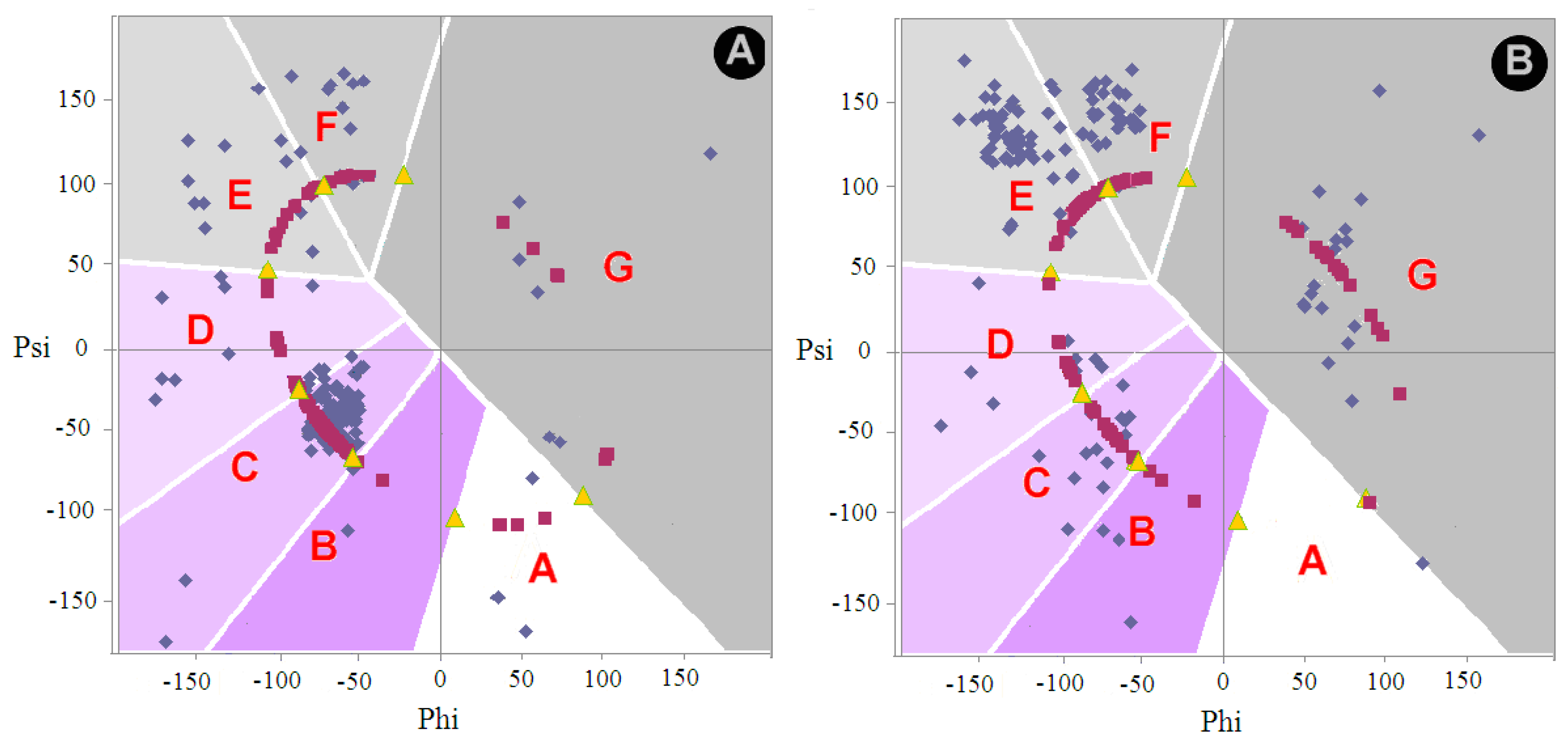
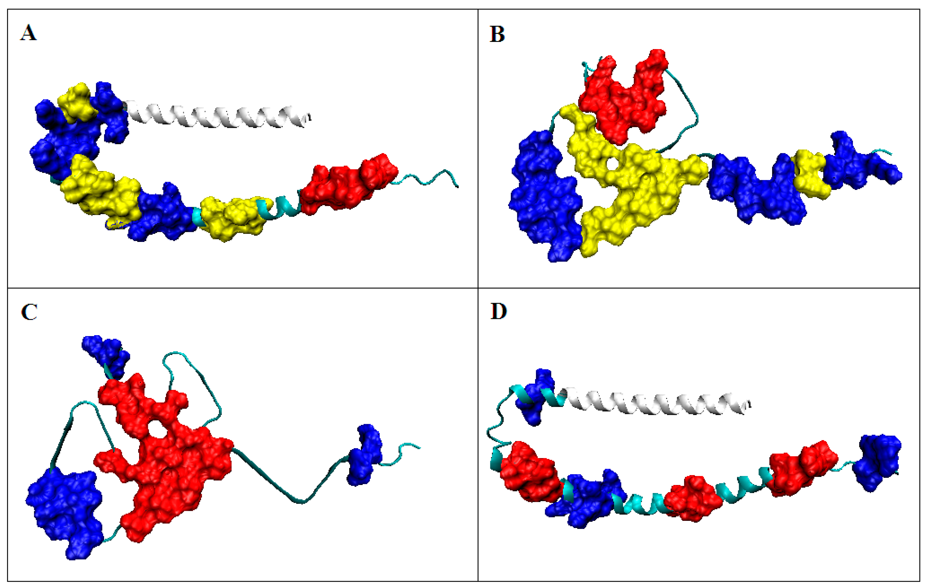
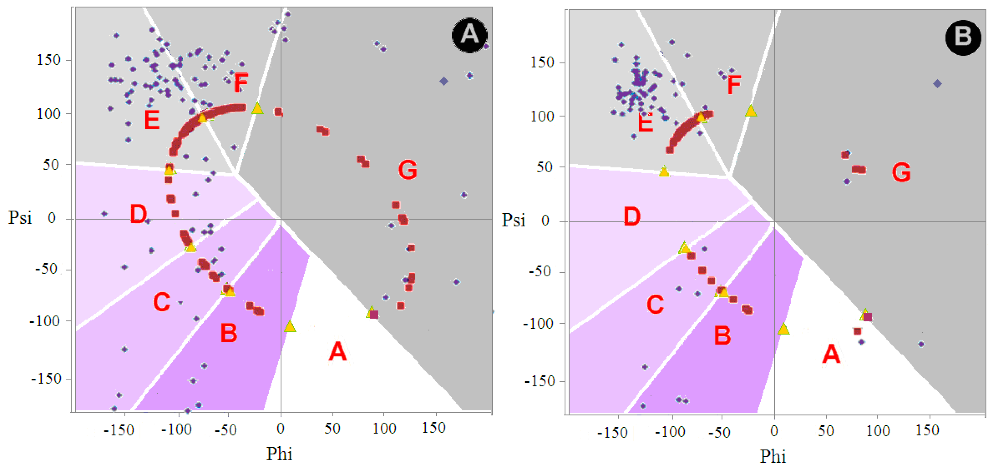
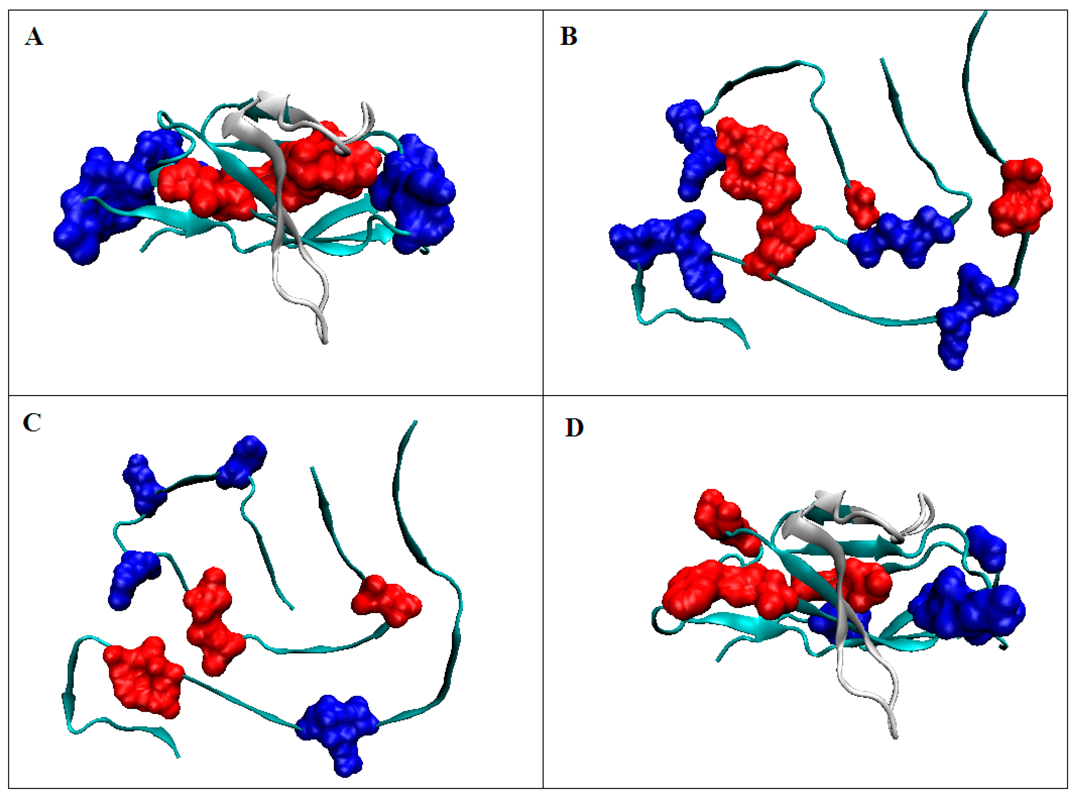
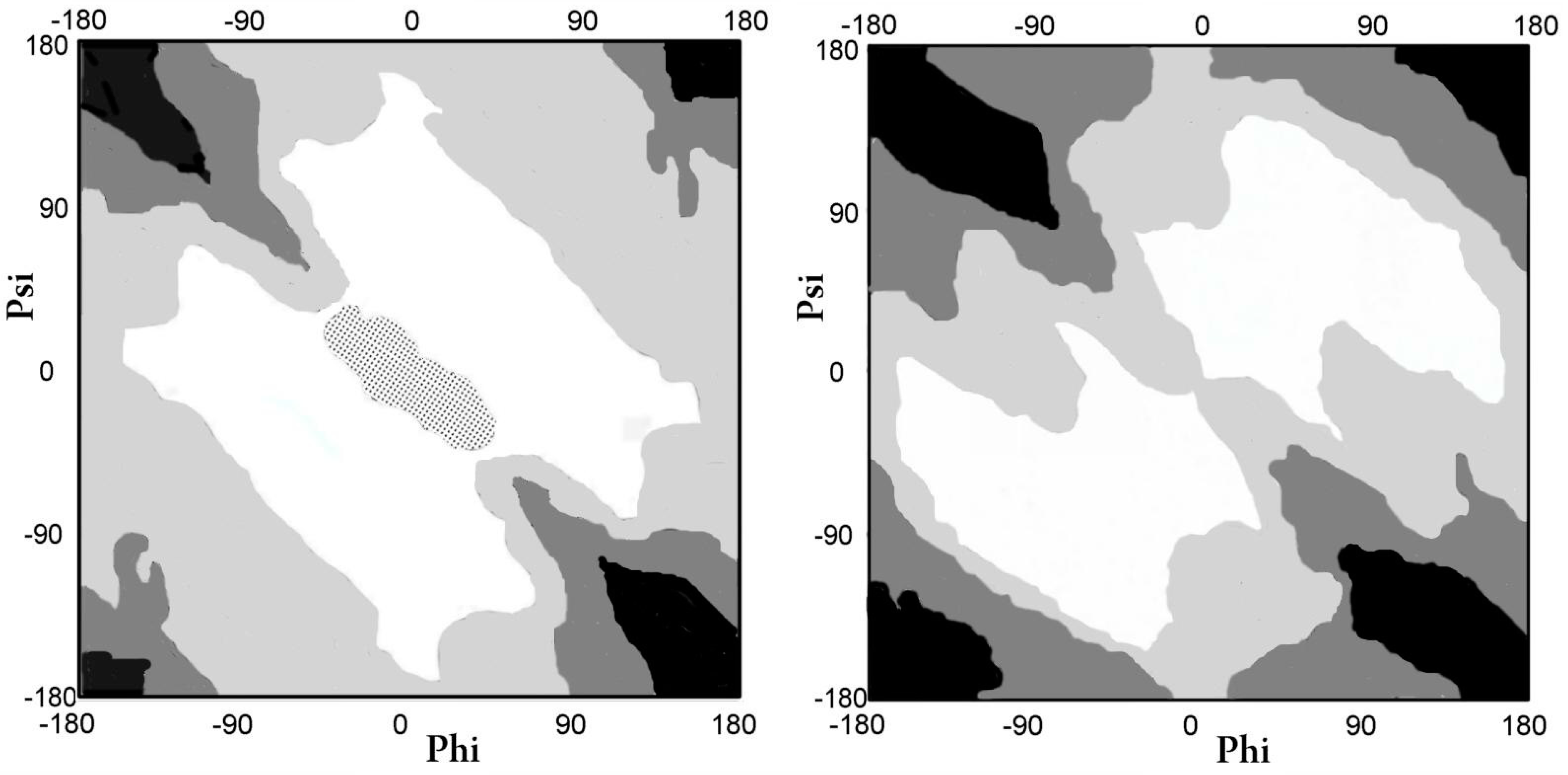
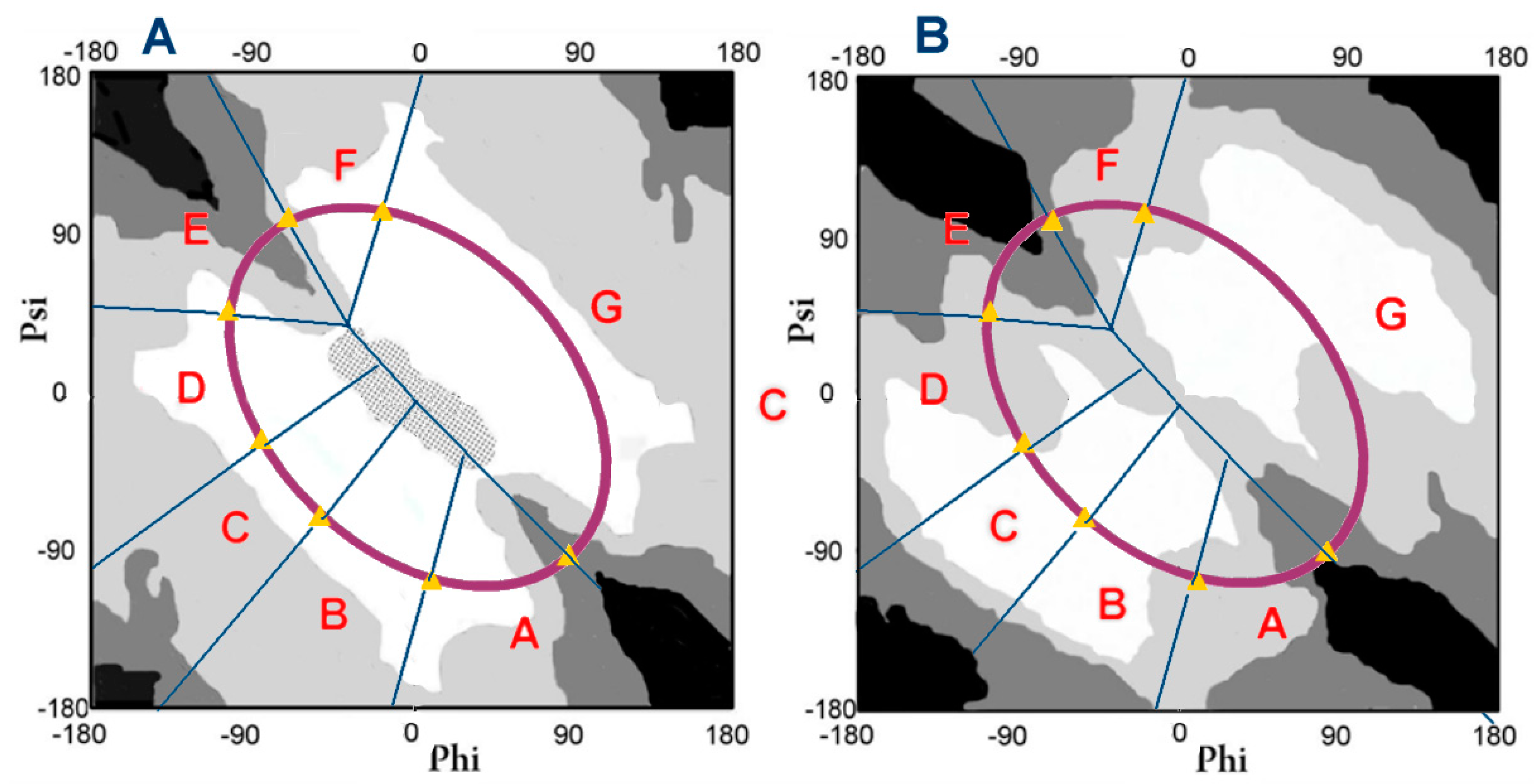
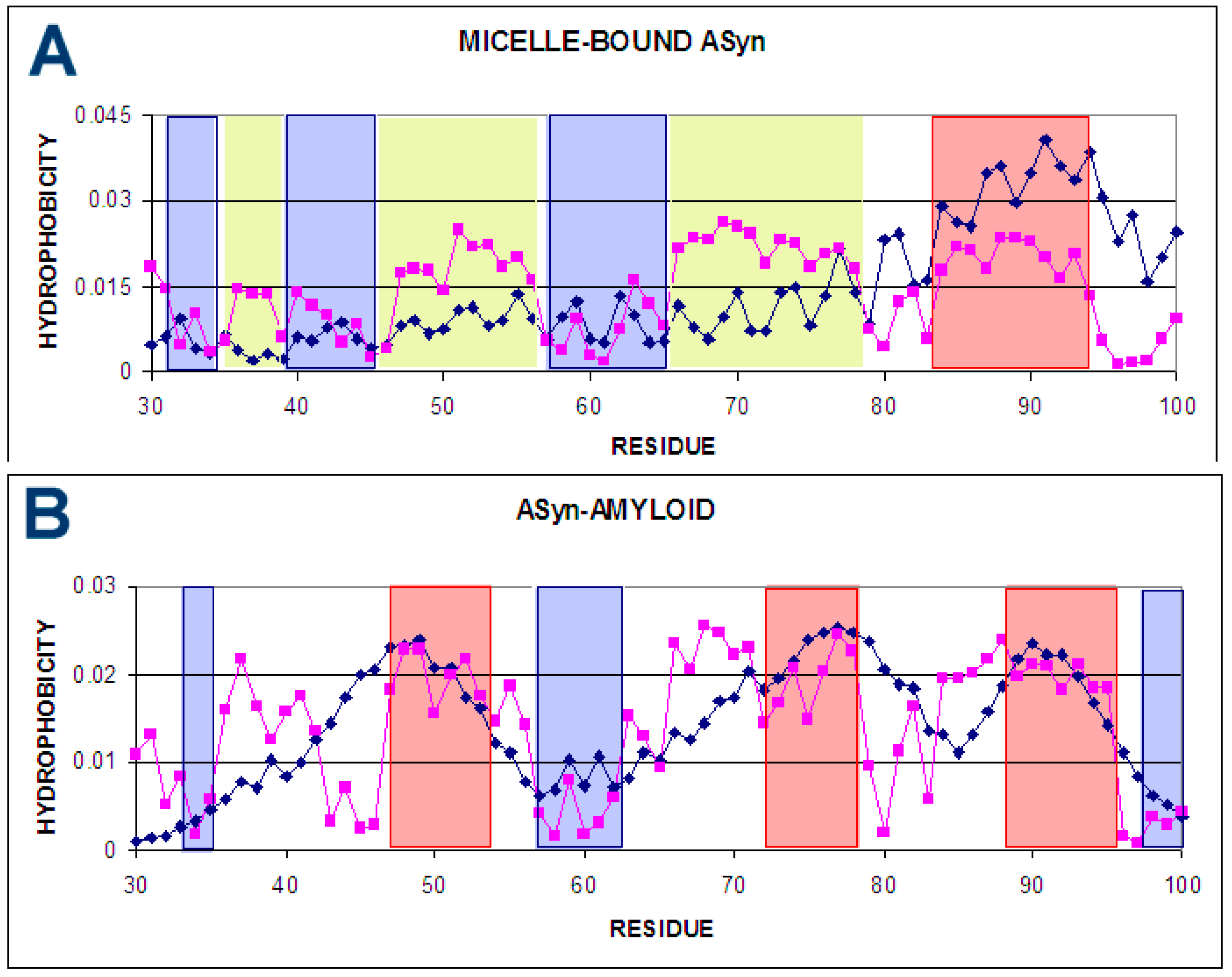
Protein structure is the result of the high synergy of all amino acids present in the protein. This synergy is the result of an overall strategy for adapting a specific protein structure. It is a compromise between two trends: The optimization of non-binding interactions and the directing of the folding process by an external force field, whose source is the water environment. The geometric parameters of the structural form of the polypeptide chain in the form of a local radius of curvature that is dependent on the orientation of adjacent peptide bond planes (result of the respective Phi and Psi rotation) allow for a comparative analysis of protein structures. Certain levels of their geometry are the criteria for comparison. In particular, they can be used to assess the differences between the structural form of biologically active proteins and their amyloid forms. On the other hand, the application of the fuzzy oil drop model allows the assessment of the role of amino acids in the construction of tertiary structure through their participation in the construction of a hydrophobic core. The combination of these two models—the geometric structure of the backbone and the determining of the participation in the construction of the tertiary structure that is applied for the comparative analysis of biologically active and amyloid forms—is presented.
Keywords: misfolding; amyloid; secondary structure; α-synuclein; V domain of the immunoglobulin G light chain; force field misfolding; amyloid; secondary structure; α-synuclein; V domain of the immunoglobulin G light chain; force field