Review

The Adaptive Mechanism of Plants to Iron Deficiency via Iron Uptake, Transport, and Homeostasis

1
State Key Laboratory of Vegetation and Environmental Change, Institute of Botany, The Chinese Academy of Sciences, Beijing 100093, China
2
College of Resources and Environment, University of Chinese Academy of Sciences, Beijing 100049, China
3
Key Laboratory of Vegetation Ecology, Ministry of Education, Institute of Grassland Science, Northeast Normal University, Changchun 130024, China
*
Author to whom correspondence should be addressed.
Current address: State key Laboratory of Protein and Plant Gene Research, Peking-Tsinghua Center for Life Sciences, School of Advanced Agricultural Sciences and School of Life Sciences, Peking University, Beijing 100871, China.
Int. J. Mol. Sci. 2019, 20(10), 2424; https://doi.org/10.3390/ijms20102424
Submission received: 16 April 2019 / Revised: 11 May 2019 / Accepted: 14 May 2019 / Published: 16 May 2019
(This article belongs to the Special Issue Mapping Abiotic Stress-Tolerance Genes in Plants)

Abstract

:
Iron is an essential element for plant growth and development. While abundant in soil, the available Fe in soil is limited. In this regard, plants have evolved a series of mechanisms for efficient iron uptake, allowing plants to better adapt to iron deficient conditions. These mechanisms include iron acquisition from soil, iron transport from roots to shoots, and iron storage in cells. The mobilization of Fe in plants often occurs via chelating with phytosiderophores, citrate, nicotianamine, mugineic acid, or in the form of free iron ions. Recent work further elucidates that these genes’ response to iron deficiency are tightly controlled at transcriptional and posttranscriptional levels to maintain iron homeostasis. Moreover, increasing evidences shed light on certain factors that are identified to be interconnected and integrated to adjust iron deficiency. In this review, we highlight the molecular and physiological bases of iron acquisition from soil to plants and transport mechanisms for tolerating iron deficiency in dicotyledonous plants and rice.

1. Introduction

Iron (Fe) is an essential micronutrient for plant growth development and plays a key role in regulating numerous cellular processes. Iron, as an important co-factor for enzymes, plays an important role in regulating plant photosynthesis, mitochondrial respiration, the synthesis and repair of nucleotides, and metal homeostasis, especially in the maintenance of structural integrity of various proteins [1]. While Fe is abundant in soil, the available Fe in soil for plants is often insufficient, particularly in calcareous soils, due to low solubility of Fe. Iron deficiency is one of the most important factors limiting crop production in the world. Plants grown in low Fe soils often exhibit chlorosis and decreased photosynthesis, leading to reduction in yield and quality of crops. To cope with this situation, plants have evolved a series of sophisticate mechanisms to adapt to iron-deficient conditions in soil. In addition, iron deficiency is a significant worldwide problem, seriously affecting over 30% of the world’s population (http://www.who.int/nutrition/topics/ida/en/). Anemia as one of the severest nutritional disorders is caused by low iron in humans. Therefore, elucidation of the molecular and physiological mechanisms by which plants sense, respond, and adapt to Fe deficiency would contribute to cultivating crop varieties with high Fe efficiency.

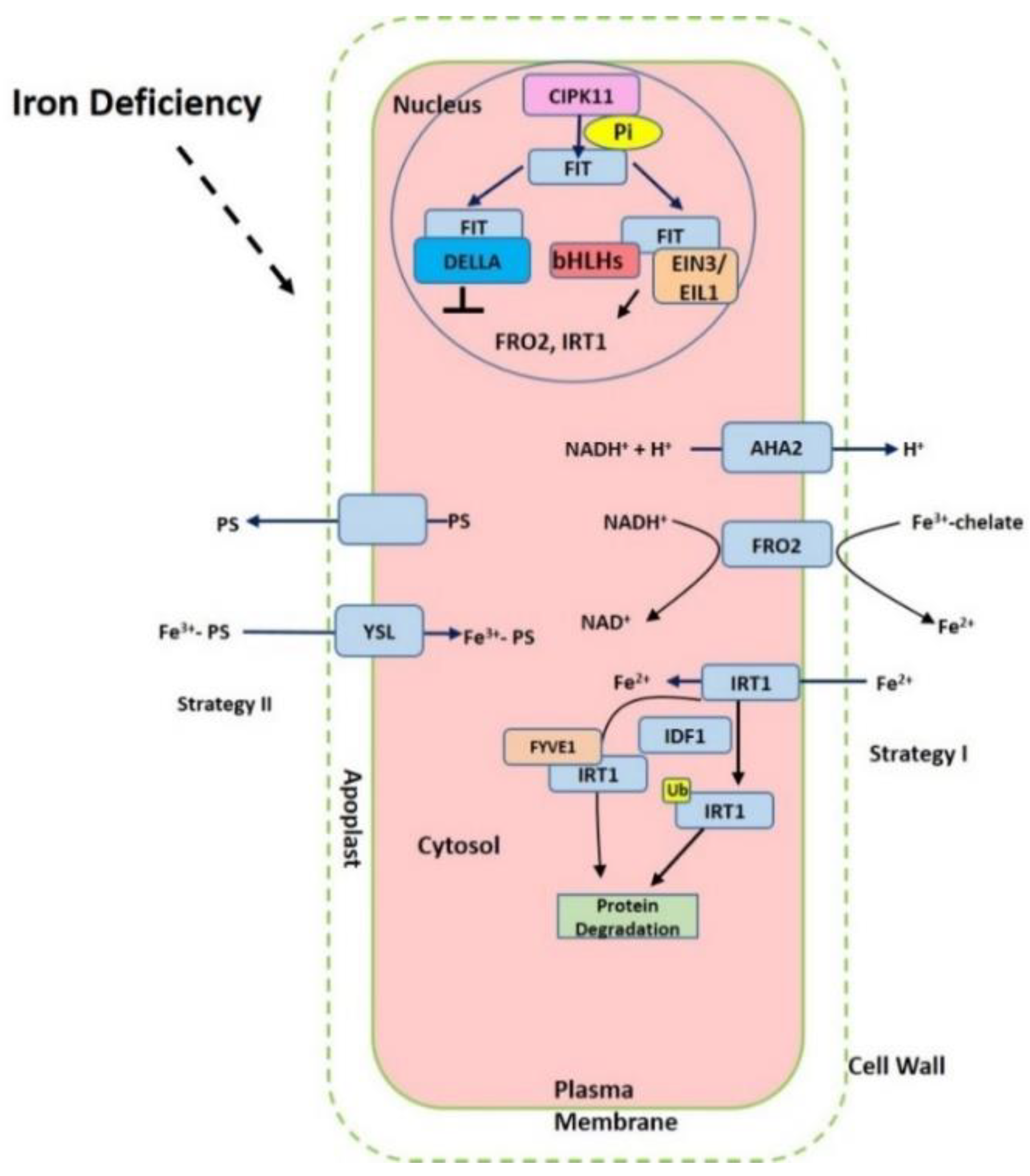
2. Iron Acquisition from Soil to Roots

Although iron is considered as the fourth most abundant element, one-third of soil on the Earth is estimated as Fe deficient [1]. The solubility and availability of iron in soil can be affected by multiple factors, including soil pH, the redox potential, microbial processes, and the amounts of organic matter and aeration in soil [2]. As a vital cofactor for enzymes, iron takes part in distinct processes, such as facilitating various chemical reactions, modulating protein stability, hormonal regulation, and nitrogen assimilation [1]. Iron deficiency could result in interveinal chlorosis in young leaves as the result of reduced chlorophyll content. The young leaves exhibit yellow color while the veins remain green. All these ultimately lead to the reduction of yield and quality [1,3]. In addition, other nutrients have antagonistic effects on iron uptake, which can significantly reduce the yield of the crops [4].
Iron in the rhizosphere is mainly present as Fe3+ which is not readily accessible to plants. Different plant species have evolved different strategies for iron acquisition from soil (Figure 1). Non-graminaceous plants, such as tomato and Arabidopsis, known as strategy-I plants, use a reduction-based strategy, in which plasma-membrane (PM)-localized H+-ATPases (AHAs) release the protons to increase rhizosphere acidification and promote Fe3+ solubility. Subsequently, the available ferric Fe3+ is reduced to the more soluble ferrous Fe (Fe(II)) by ferric reduction oxidases (FROs) at the apoplast [5]. The reduced ferrous ion (Fe2+) is imported into root cells by the Fe2+-regulated transporters such as the iron-regulated transporter (IRT1) [6,7]. Additionally, graminaceous plants, including rice, barley, and maize, known as strategy-II plants, use a chelation-based strategy to release phytosiderophores (PS). PS, as strong Fe chelators, are secreted into the rhizosphere with a high affinity for binding Fe (III) [8,9]. PS-Fe(III) is then taken up into root cells through the yellow stripe (YS) or yellow stripe-like (YSL) transporters [10].
Iron deficiency triggers the expression of many Fe uptake-associated genes. The expression of AtAHA2 and AtAHA7, for example, are at higher levels under iron-deficient conditions, but AtAHA1 is not induced by iron deficiency [11]. Twelve PM H+-ATPases AHAs are encoded in the Arabidopsis genome [11]. AtAHA2 is primarily responsible for the of rhizosphere acidification of root hairs under iron deficiency. Loss function of AtAHA2 compromised proton extrusion capacity. AHA7 is crucial for the formation of root hairs induced by iron deficiency via mediating H+ efflux in the root hair zone. The fine-tuned regulation of root tip H+ extrusion by PM H+-ATPase is required for root hair formation. H+ efflux through PM H+-ATPase causes the acidification of the cell wall apoplast, which is crucial for the root hair initiation [11]. The loss function of AtAHA7 contributed to a decreased frequency of root hairs [11]. However, the mechanism of AHAs regulation remains unknown. Recent findings indicate that cytochrome B5 reductase 1 (CBR1) is able to activate plasma membrane-localized H+-ATPases, which is achieved by facilitating the content of unsaturated fatty acids [12]. CBR1 expression is induced under iron-deficient conditions. CBR1 localizes to endoplasmic reticulum (ER) membrane and plays an important role in electron transfer from NADH to cytochrome b5. Then the cytochrome b5 mediates the electrons transfer to fatty acids desaturase 2 (FAD2) and fatty acids desaturase 3 (FAD3), allowing for double bonds into fatty acids. FAD2 is responsible for converting oleic acid (18:1) to linoleic acid (18:2), and FAD3 contributes to the conversion of 18:2 to linolenic acid (18:3). On the other side, 20 or 50 μM of the unsaturated fatty acids 18:2 or 18:3 can strongly activate H+-ATPase [12]. Other compounds such as phenolics, organic acids, flavonoids, and flavins have also been implicated in the acidification–reduction strategy to uptake iron (Strategy I) [3,13,14,15]. These small compounds significantly promote reutilization and uptake of apoplastic iron via chelation or the reduction of iron in soil. Recently it was reported that coumarins involved in iron acquisition are secreted and essential for iron uptake under iron-limited conditions [16,17]. The plants are able to secret an array of coumarin-type compounds under different iron nutrition conditions, which facilitate Fe(III) availability [18]. The synthesis of these coumarins require Feruloyl coenzyme A 6’-hydrozylase 1 (F6’H1) enzyme [19]. ATP-BINDING CASSETTE G37 (ABCG37/PDR9) transporters contribute to the exudation of coumarins [17]. Both F6’H1 and PDR9 transcript expression are upregulated by iron deficiency [19,20].
Subsequently, the soluble Fe3+ is reduced into Fe2+ in root apoplast via cellular membrane localized ferric reductase oxidase 2 (FRO2). This protein has 725 amino acids with 8 transmembrane domains, containing motif for binding hemes and NADPH [21]. The electron from NADPH in the cytoplasmic side is transferred via two hemes and Flavin to the Fe3+ in apoplast [22]. FRO2 is primarily expressed in roots [23]. In addition to expression in roots, FRO2 is largely present in flowers [24]. FRO2 transcription and post-transcription are both regulated by iron concentration, since the activity of FRO2 in FRO2 overexpression lines is highly induced under iron deficiency [24]. In addition, iron deficiency facilitates the stability of FRO2 mRNA [24]. A total of 50 FROs were identified in plants [25] and 8 FROs are encoded in the Arabidopsis genome [26]. These FROs have different tissue-specific expression patterns. AtFRO3 and AtFRO5 are predominantly expressed in roots, while AtFRO6, AtFRO7 and AtFRO8 gene expression primarily occur in shoots. AtFRO1 and AtFRO4 are present in both roots and leaves [23,27,28,29].
After Fe3+ reduced to Fe2+ in root rhizosphere, Fe2+ can be imported into cells by IRT1 with high affinity to Fe2+ (Km = 6 μM). IRT1 is the most important root transporter for ferrous Fe uptake from the soil, while the uptake of other divalent cations (manganese, zinc, cobalt, and cadmium) can also be promoted by IRT1 [6,7,30]. IRT1 is identified in Arabidopsis and can rescue the defects of the fet3fet4 mutants of yeast that are impaired in Fe uptake [6]. The expression of IRT1 is highly induced under iron-limited conditions [6,7]. IRT1 belongs to ZIP family and consists of 347 amino acids with 8 transmembrane domains. IRT1 can also promote the uptake of and Zn2+ but IRT1 can transport Zn only under low pH [30,31]. IRT1 is present in early endosomes/trans-Golgi network compartments (EE/TGN). Early studies found that IRT1 degradation and recycling between EE/TGN and the plasma membrane are modulated by ubiquitination and monoubiquitin-dependent endocytosis [32]. The IRT1 protein can transport to a vacuole for degradation [32]. IRT1 degradation factor1 (IDF1), a RING-type E3 ubiquitin ligase, is found to be responsible for IRT1 ubiquitination on plasma membrane via clathrin-mediated endocytosis. Thus, Fe-deficient induced IDF1 facilitating IRT1 degradation develops a negative feedback loop to fine tune the iron homeostasis [33]. It should be noted that recent studies point to the fact that non-iron elements (Zn, Mn, and Co) are also able to regulate this trafficking of IRT1 between EE/TGN and the plasma membrane in root epidermal cells [34]. Moreover, FYVE1, a phosphatidylinositol-3-phosphate-binding protein, is also required for the recycling of IRT1 and its polar localization to outer polar domain of plasma membrane [34]. SORTING NEXIN (SNX) protein was found to co-localize with IRT1 and is also important for recycling internalized IRT1. In the snx1 mutants, the degradation of IRT1 is enhanced [35]. Further studies reveal that there exist other transporters for iron uptake. Natural resistance associated macrophage proteins (NRAMPs) were identified as a ubiquitous family of metal efflux transporters. Quite intriguingly, NRAMP1 that acts as a transporter of manganese is also essential for low-affinity iron uptake. Pleckstrin homolog (PH) domain-containing protein AtPH1 binds phosphatidylinositol 3-phosphate (PI3P) in the late endosome, which regulates the localization of NRAMP1 to the vacuole [36].
The strategy II plants, such as rice, can secrete phytosiderophores (PS) in rhizosphere for efficiently increasing the solubility of Fe3+, ultimately facilitating the available iron for root acquisition [37]. PS-Fe3+ complexes are then imported into root epidermis cells by a specific transporter [37]. PS belong to the family of mugineic acid (MAs), such as mugineic acid (MA), 2’-deoxymugineic acid (DMA), 3-epihydroxymugineic acid (epi-HMA), and 3-epihydroxy 2’-deoxymugineic acid (epi-HDMA) [38,39]. MAs are synthesized from three S-adenosyl-methionine molecules [40]. Yellow stripe 1 (YS1) is firstly identified from maize and targeted to the plasma membrane, which is likely to responsible for transporting Fe3+-PS into root cells [10]. YS1 consists of 682 amino acid with 12 transmembrane domains [3]. The transcript expression of ZmYS1 is highly induced in both root and shoot of maize under iron-deficient condition [10,41]. Eighteen putative yellow stripe 1 (YS1)-like genes (OsYSLs) are identified in the rice genome [42].
Fe deficiency readily results in interveinal chlorosis in young leaves, ultimately reducing the yield and grain quality [43]. In order to tolerate iron deficiency, various physiological processes are induced in the root rhizosphere, including ferric reductase activity, the ratio of root and shoot, and photosynthesis. Also, root morphology is altered according to the local availability of iron and for optimizing iron uptake, such as increasing lateral root numbers, extra root hairs, and developing transfer cells to facilitate contact surface with soil [44].

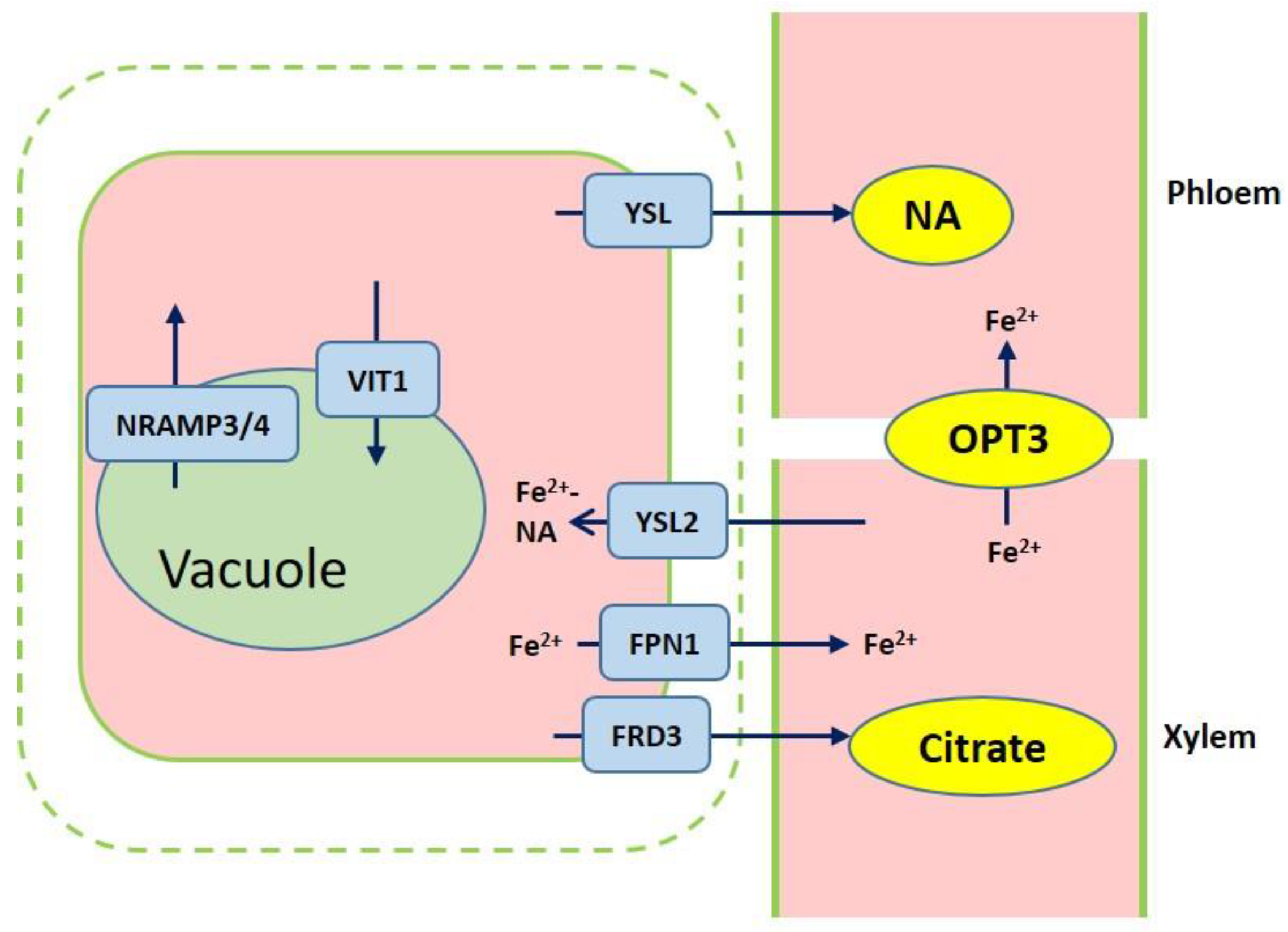
3. Iron Transport Mechanism in Plants

After iron is transported to the root endodermis from epidermis via apoplastic and symplastic pathway, it needs to be transported to the above ground parts of plants through the xylem (Figure 2). The contents of organic acids, such as citrate, malate, and succinate, are elevated in xylem under iron deficient conditions [45]. The usage of various approaches, such as the theoretical calculations, high-pressure liquid chromatography (HPLC) coupled to electrospray time-of-flight mass spectrometry (HPLC-ESI-TOFMS) and inductively coupled plasma mass spectrometry (HPLC-ICP-MS), detects the natural Fe complex and provides evidence for the transport of iron in xylem to shoots which predominantly occurs as Fe3+-citrate complex [46,47,48,49]. The transport of citrate and iron to the xylem is mediated by ferric reductase defective 3 (FRD3) in Arabidopsis and its ortholog FRDL1 in rice, which is crucial for iron translocation [50,51]. FRD3 is present only in pericycle and cells neighboring the vascular tissue [50]. frd3 mutants exhibit severe Fe-deficient phenotype even under Fe-sufficient conditions. Less citrate and less Fe are contained in xylem sap of frd3 mutants as compared to wild type [50]. Osfrdl mutants also contain reduced citrate and Fe in the xylem resembling Fe-deficiency phenotype in frd3 mutants [52]. Therefore, it is tempted to speculate that graminaceous and nongraminaceous share the similar mechanism by which Fe is transported from root to shoot although the uptake strategies for iron are very different. Ferroportin1 (FPN1) is also responsible for loading iron into the xylem [44]. The Arabidopsis genome contains three FPN which have different subcellular localizations. FPN1, for example, is targeted to the plasma membrane, FPN2 on the vacuolar membrane and FPN3 on the chloroplast envelop [44,53,54]. Fe is also capable of translocation in xylem in the form of Fe-nicotianamine (NA) and Fe-MAs. NA as a non-protein amino acid is produced from S-adenosyl methionine by nicotianamine synthase (NAS) and is also the direct biochemical precursor to PS [55,56]. In rice, NA and DMA are present in xylem exudates [57,58].
Once the iron reaches the leaves, it must be unloaded to leaf cells from the apoplastic space. NA and DMA are also required for the phloem-based transport [59]. AtYSL1, AtYSL2, and AtYSL3, as metal-NA transporters, are involved in this process, responsible for moving iron from apoplast to symplast [60,61]. These three genes are highly expressed in vascular parenchyma cells of leaves [60,61]. AtYSL2 plays a major role in regulating the lateral distribution of iron from xylem to shoot cells in Arabidopsis [54,60]. Moreover, AtYSL1 and AtYSL3 appear to transport the Fe-NA chelate from senescent leaves into the inflorescences and seeds. ysl1 and ysl3 mutants contain reduced iron content in leaves and seeds [60,62,63]. In rice, OsYSL2 is likely to be involved in the translocation of Fe(II)-NA to shoots and seeds [42,64]. OsYSL16 is expressed in the cells surrounding xylem and contributes to Fe(III)-MA allocation via the vascular bundle [65]. OsYSL18 also transports Fe(III)-DMA in reproductive organs and phloem of lamina joints [66]. Recent studies point to OsYSL9 which is involved in the Fe distribution in developing seeds via Fe(II)-NA and Fe(III)-DMA form [67]. Additionally, oligo peptide transporter 3 (OPT3) mediates the Fe transport to sink tissues via the phloem and recirculation in the roots in Arabidopsis [68]. Meanwhile, OPT3 is also found to take part in the control of iron movement out of the leaves to root or developing tissues in the form of iron ions rather than iron-ligand complexes [69,70]. Heat shock cognate protein B (HSCB) as a mitochondrial cochaperone participates in iron translocation from roots to shoots [71]. HSCB overexpression lines caused iron accumulation in roots but low iron levels in shoots; while hscb knockdown plants showed iron accumulation in shoots despite the reduced contents of iron uptake in roots [71].

4. Iron Storage in Cells

Iron mobilization in cells is essential for plant growth and development, especially under iron-deficient conditions. When transporting across cellular or intracellular membranes, ferric iron is usually reduced to ferrous iron [72]. Iron can produce cytotoxic oxygen radicals, such as hydroxyl radicals and superoxide anions [16]. Generally, the cellular iron is stored in vacuoles and is also likely to be sequestrated into ferritin, which will become available for various metabolic reactions. In Arabidopsis seeds, the vacuole is the major iron store containing about 50% of total iron, while ferritins play a minor role in iron storage including about 5% iron [16,73]. Ferritin is important for fine tuning the quantity of metal which is required for metabolic purposes [74]. In the vacuole of Arabidopsis seeds, globoids act as an important site for Fe storage [16]. However, in pea, the amount of iron-ferritin is present at about 92% of the total seed iron in embryo axis [75]. Therefore, these findings suggest that the way for iron storage in seeds may be different between different species, such as pea and Arabidopsis [73]. Plastids also act as a sink for iron in cells and appear to function in sensing and maintaining iron concentration in the plants to adapt various changes [76]. In chloroplast, ferritins represent one candidate to form the complex with Fe [76]. In Arabidopsis, three of ferritins are localized to chloroplasts. In addition, NA might also play a role in maintaining Fe soluble in plastids [76].
The changes of iron content in vacuole might trigger distinct responses. The vacuolar iron transporter 1 (VIT1), an orthologue of the yeast iron transporter Ca2+-sensitive cross-complementer 1 (CCC1), was first identified in Arabidopsis [77]. AtVIT1 was found to control iron sequestration into vacuoles. Despite there being no difference in the iron content of seeds between vit1 mutants and wild type, the iron accumulation is absent in the vacuoles of provascular cells [77]. So, what else could modulate iron mobilization efflux from vacuolar? AtNRAMP3 and AtNRAMP4 are responsible for Fe efflux from the vacuolar into the cytosol, and consequently essential for seed germination under Fe deficiency [78,79]. However, we cannot exclude other efflux transporters localized in vacuolar. In rice, the molecular mechanism underlying Fe transport in cells has also been well uncovered. OsVIT1, OsVIT2, and OsNRAMPs affect Fe translocation from the vacuole to other parts [80,81,82,83].
Ferritins, as another iron pool, are a class of universal 24-mer multi-meric, which are encoded by nuclear genes [84]. The structure of ferritins is highly conserved in eukaryotes [74]. In Arabidopsis, four ferritin genes (AtFer1–4) have been identified, among which FER1, FER3, and FER4 are proposed to exist in leaves while FER2 is present in seeds [74]. Recent studies found that ferritins are vital for protecting cells against oxidative stress [73]. Recently it was reported that ferritins are also involved in root system architecture regulation. Triple mutants of fer1 fer3 fer4 exhibited altered root architecture which was caused by the alteration in the production and balance of reactive oxygen species (ROS) [85].
In addition, mitochondrion as a crucial iron sink provides available iron for the proper respiration. In rice, FRO3 and FRO8 appear to play roles in Fe3+ reduction in the mitochondrial membrane and mitochondrial iron transporters (MITs) are responsible for the translocation of iron from cytoplasm to mitochondrial [86]. Although the total iron content of shoots is increased in mit knockdown mutants as compared to wild type, the iron concentration in mitochondria is reduced, which further suggest iron is mistransported in the mitochondria of these mutants. Additionally, mit knockdown mutants contain a significant reduction of chlorophyll content and impair plant growth [87].
Also, chloroplast represents one of the main sinks for iron in plant cells. The iron transport across the chloroplast inner envelope also depends on reduction-based strategy. AtFRO7 as a chloroplast Fe (III) chelate reductase is targeted to the chloroplast envelope and putatively function in Fe3+ reduction in chloroplast. AtFRO7 is required for the survival of young seedlings under iron-deficient conditions. Under Fe-deficient conditions, loss of function of FRO7 reduces the Fe content and hampers the reductase activity of chloroplast, leading to chlorotic appearance [29]. AtYSL6 is localized to the chloroplast envelope. Plants lacking ATYSL4 and ATYSL6 exhibit iron over-accumulated chloroplasts and the overexpression lines are characterized by decreased Fe content in chloroplast, suggesting that YSL4 and YSL6 take part in the release of iron from chloroplast [88]. In addition, PERMEASE IN CHLOROPLASTS1 (PIC1) as an ancient permease plays a role in chloroplast Fe uptake and maintaining Fe homeostasis. Interestingly, PIC1 was identified as the first protein involved in Fe uptake in plastid [89], which is localized to the inner envelope and contain four membrane-spanning α-helices [89]. The pic1 mutant exhibits altered mesophyll organization, disrupted chloroplast and thylakoid development, which is consistent with Fe-deficiency phenotype [89]. Furthermore, recent findings further confirm this function of PIC1 in plastid Fe-transport using PIC1 knockdown and overexpression lines in Nicotiana tabacum [90].

5. Transcriptional and Posttranscriptional Regulation of Fe-related Genes

Since Fe is vital for cellular process, a sophisticated regulatory mechanism to sense and adjust iron deficiency is essential for providing sufficient iron for plant growth and development. To avoid iron deficiency, various genes involved in iron acquisition and internal translocation are fine-tune regulated at the transcriptional and posttranscriptional level in adapt to iron deficient condition (Figure 1). Fe efficiency reactions (FER) was firstly identified in tomato and encoding a bHLH transcription factor. In this regard, FER controls the root physiology and morphology adapt to iron deficiency [91]. The basic helix-loop-helix (bHLH) FER-like iron deficiency-induced transcription factor (FIT) was identified in Arabidopsis and involved in iron sensing, responding, and acquisition through regulating the expression of FRO2 and IRT1 [92]. The ethylene-responsive transcription factors Ethylene Insensitive3 (EIN3) and EIN3-Like1 (EIL1) both enable interact with FIT, consequently activating FIT [93]. The activated FIT can up-regulate the transcript expression of AHA2, FRO2, and IRT1 [94,95,96]. Extensively, FIT activity is modulated via interaction with other proteins. The expressions of bHLH038, bHLH039, bHLH100, and bHLH101 have been reported to be increased under Fe starvation and interact with FIT [97,98]. These interactions result in the activation of FIT and consequently activate the expression of FIT target genes such as IRT1 and FRO2 [98,99]. However, the transcript expression of NRAMP3 is not influenced by the activated FIT [94]. What is more, the activity of FIT can be inhibited by the interaction of DELLA with FIT [100]. In addition, a bHLH transcription factor POPEYE (PYE) is identified which as part of an iron regulatory network is independent of FIT. PYE is capable of interacting with another PYE homologs-bHLH transcription factor IAA-Leu Resistant3 (ILR3), which regulates the iron deficiency response and are both required for maintaining iron homeostasis [101]. Under low iron conditions, PYE is expressed in the root vasculature, columella root cap, and also lateral root cap. Interestingly, its strongest expression occurs in the pericycle of the maturation zone [101]. A putative E3 ligase protein BRUTUS (BTS) can also interact with ILR3, but plays a negative role in response to iron deficiency [101]. Also, the transcription factors, MYB family members MYB10 and MYB72, are implicated in the regulation of NAS4 expression [102,103]. WRK46 not only regulates the expression of NAS but also enables to directly bind the promoter of VIT-LIKE1 via the W-boxes, thereby controlling the iron translocation [104]. YSL2 expression can be controlled by the transcription factors IDEF1 and IDEF2 in rice [105]. In rice, Fe-deficiency-inducible bHLH transcription factor OsIRO2, as the homologue of AtbHLH39, enhances the expression of YSL15 [106].

6. Function of Other Factors in the Iron Homeostasis

Although recent studies have demonstrated Fe-related genes are associated with plants response to Fe deficiency, the reality of signal network appears to be more complicated in adapting to iron deficiency. Various plant hormones, messenger molecules and kinases are implicated into this process. Auxin analogs for example can increase the activity of the root ferric chelate reductase (FCR) in bean [107,108,109]. In Arabidopsis, abscisic acid (ABA) and gibberellin have been suggested to facilitate the Fe deficient response, while cytokinin and jasmonic acid prevent this response [110,111,112,113]. ABA, for example, promotes the secretion of phenolics and also iron efflux from vacuole via up-regulation of AtNRAMP3. Further studies suggest that ABA enhances the Fe translocation from root to shoot [110]. Nitric oxide (NO) is also be found to act as a component of Fe signal pathway and activate root FCR activity under iron deficiency via acting downstream of auxin in Arabidopsis [114]. NO plays a role in the synthesis of cell wall. Cell wall consists of pectin, cellulose, and hemicellulose. Cell walls are full of negative charges, which provide the binding sites for metal ions. Pectin is secreted into the apoplast from the symplast. Pectin methylesterase (PME) contributes to de-methylation of pectin that can increase carboxylic groups and hence provides more negative charged sites for iron in cell wall. Fe-deficiency induced NO prevents pectin methylation of cell wall and stimulates the PME activity. These together enhance the Fe retention in root apoplast. In this regard, NO limits iron translocation from root to shoot [115]. Recent evidence points to Ca2+ direct interrelations of Fe signal. An important signaling network in deciphering Ca2+ signals is formed by specific interactions of 10 calcium B-like proteins (CBLs) and 26 CBL interacting protein kinases (CIPKs) in Arabidopsis [116,117]. CIPK23 could be as “nutritional sensors” to sense and mediate the iron homeostasis in Arabidopsis. cipk23 mutants exhibit lower activity of FCR and the regulation of FCR activity by CIPK23 is not related to the transcript expression of FRO2, FRO3, and FRO5 [118]. Additionally, it has been found that CIPKs are also involved in the regulation of H+ homeostasis. CIPK11/PKS5 suppresses the activity of the PM H+-ATPase (AHA2) via phosphorylation which prevents the interaction between AHA2 and 14-3-3 protein, and thus inhibits the extrusion of protons (H+) to the extracellular space [119]. Moreover, CIPK11 interacts with FIT and activates FIT via phosphorylation at Ser272, allowing for FIT-dependent Fe deficiency responses. Mutation at Ser272 of FIT affects seed iron content [120].

7. Conclusions

Iron acts as an essential element not only in plant physiological functions but also in the maintenance of various cell processes. Over the past decades, accumulating progresses have been achieved in understanding how the plants adapt to iron deficiency in soil. Cellular, biochemical, molecular, genetics, and genomic approaches facilitate a better understanding of iron uptake, transport, and utilization. However, how to observe the iron dynamics in plants, especially in different tissues and cells, is still a notable challenge. Despite a wealth of information pointing to the identities for many genes responsible for iron uptake from soil, transport from roots to shoots, storage in cells, and even their regulation at the transcription and post-transcription level, further research is clearly needed to uncover the further interconnection and integration of signaling pathways of iron deficiency into development and physiological networks. Finally, all of this information underlying the mechanism of iron uptake, transport, and homeostasis will be of great benefit to plants and human health.

Author Contributions

X.Z. wrote and designed the manuscript. T.W. read and approved the contents. D.Z. and W.S. edited the manuscript.

Funding

This work was funded by the National Key Research and Development Program of China (2018YFD0700202 and 2016YFC0500601), and the National Natural Science Foundation of China (31300231).

Conflicts of Interest

The authors declare no conflict of interest.

References

  1. Mahender, A.; Swamy, B.P.M.; Anandan, A.; Ali, J. Tolerance of iron-deficient and -toxic soil conditions in rice. Plants 2019, 8, 31. [Google Scholar] [CrossRef] [PubMed]
  2. Becker, M.; Asch, F. Iron toxicity in rice—Conditions and management concepts. J. Plant Nutr. Soil Sci. 2010, 168, 558–573. [Google Scholar] [CrossRef]
  3. Curie, C.; Briat, J.F. Iron transport and signaling in plants. Annu. Rev. Plant Biol. 2003, 54, 183–206. [Google Scholar] [CrossRef] [PubMed]
  4. Fageria, N.K.; Baligar, V.C.; Li, Y.C. The role of nutrient efficient plants in improving crop yields in the twenty first century. J. Plant Nutr. 2008, 31, 1121–1157. [Google Scholar] [CrossRef]
  5. Yi, Y.; Guerinot, M.L. Genetic evidence that induction of root Fe(III) chelate reductase activity is necessary for iron uptake under iron deficiency. Plant J. 1996, 10, 835–844. [Google Scholar] [CrossRef] [PubMed]
  6. Eide, D.; Broderius, M.; Fett, J.; Guerinot, M.L. A novel iron-regulated metal transporter from plants identified by functional expression in yeast. Proc. Natl. Acad. Sci. USA 1996, 93, 5624–5628. [Google Scholar] [CrossRef]
  7. Vert, G.; Grotz, N.; Dédaldéchamp, F.; Gaymard, F.; Guerinot, M.L.; Briat, J.F.; Curie, C. IRT1, an Arabidopsis transporter essential for iron uptake from the soil and for plant growth. Plant Cell 2002, 14, 1223–1233. [Google Scholar] [CrossRef]
  8. Takagi, S.-I. Naturally occurring iron-chelating compounds in oat- and rice-root washings. Soil Sci. Plant Nutr. 1976, 22, 423–433. [Google Scholar] [CrossRef]
  9. Takagi, S.I.; Nomoto, K.; Takemoto, T. Physiological aspect of mugineic acid, a possible phytosiderophore of graminaceous plants. J. Plant Nutr. 1984, 7, 469–477. [Google Scholar] [CrossRef]
  10. Curie, C.; Panaviene, Z.; Loulergue, C.; Dellaporta, S.L.; Briat, J.F.; Walker, E.L. Maize yellow stripe1 encodes a membrane protein directly involved in Fe(III) uptake. Nature 2001, 409, 346. [Google Scholar] [CrossRef] [PubMed]
  11. Yuan, W.; Zhang, D.; Song, T.; Xu, F.; Lin, S.; Xu, W.; Li, Q.; Zhu, Y.; Liang, J.; Zhang, J. Arabidopsis plasma membrane H+-ATPase genes AHA2 and AHA7 have distinct and overlapping roles in the modulation of root tip H+ efflux in response to low-phosphorus stress. J. Exp. Bot. 2017, 68, 1731–1741. [Google Scholar] [CrossRef]
  12. Oh, Y.J.; Kim, H.; Seo, S.H.; Hwang, B.G.; Chang, Y.S.; Lee, J.; Lee, D.W.; Sohn, E.J.; Lee, S.J.; Lee, Y.; et al. Cytochrome b5 reductase 1 triggers serial reactions that lead to iron uptake in plants. Mol. Plant 2016, 9, 501–513. [Google Scholar] [CrossRef] [PubMed]
  13. Abadía, J.; López Millán, A.F.; Rombolà, A.; Abadía, A. Organic acids and Fe deficiency: A review. Plant Soil 2002, 241, 75–86. [Google Scholar] [CrossRef]
  14. Jin, C.W.; You, G.Y.; He, Y.F.; Tang, C.; Wu, P.; Zheng, S.J. Iron deficiency-induced secretion of phenolics facilitates the reutilization of root apoplastic iron in red clover. Plant Physiol. 2007, 144, 278–285. [Google Scholar] [CrossRef]
  15. Connorton, J.M.; Balk, J.; Rodríguez Celma, J. Iron homeostasis in plants—A brief overview. Metallomics 2017, 9, 813–823. [Google Scholar] [CrossRef] [PubMed]
  16. Jeong, J.; Merkovich, A.; Clyne, M.; Connolly, E.L. Directing iron transport in dicots: Regulation of iron acquisition and translocation. Curr. Opin. Plant Biol. 2017, 39, 106–113. [Google Scholar] [CrossRef]
  17. Clemens, S.; Weber, M. The essential role of coumarin secretion for Fe acquisition from alkaline soil. Plant Signal. Behav. 2015, 11, e1114197. [Google Scholar] [CrossRef] [PubMed]
  18. Sisó Terraza, P.; Luis Villarroya, A.; Fourcroy, P.; Briat, J.F.; Abadía, A.; Gaymard, F.; Abadía, J.; Álvarez Fernández, A. Accumulation and secretion of coumarinolignans and other coumarins in Arabidopsis thaliana roots in response to iron deficiency at high pH. Front. Plant Sci. 2016, 7, 1711. [Google Scholar] [CrossRef] [PubMed]
  19. Fourcroy, P.; Tissot, N.; Gaymard, F.; Briat, J.F.; Dubos, C. Facilitated Fe nutrition by phenolic compounds excreted by the Arabidopsis ABCG37/PDR9 transporter requires the IRT1/FRO2 high affinity root Fe2+ transport system. Mol. Plant 2016, 9, 485–488. [Google Scholar] [CrossRef] [PubMed]
  20. Schmid, N.B.; Giehl, R.F.H.; Döll, S.; Mock, H.P.; Strehmel, N.; Scheel, D.; Kong, X.; Hider, R.C.; von Wirén, N. Feruloyl-CoA 6′-hydroxylase1-dependent coumarins mediate iron acquisition from alkaline substrates in Arabidopsis. Plant Physiol. 2014, 164, 160–172. [Google Scholar] [CrossRef]
  21. Robinson, N.J.; Procter, C.M.; Connolly, E.L.; Guerinot, M.L. A ferric-chelate reductase for iron uptake from soils. Nature 1999, 397, 694–697. [Google Scholar] [CrossRef] [PubMed]
  22. Schagerlöf, U.; Wilson, G.; Hebert, H.; Al Karadaghi, S.; Hägerhäll, C. Transmembrane topology of FRO2, a ferric chelate reductase from Arabidopsis thaliana. Plant Mol. Biol. 2006, 62, 215–221. [Google Scholar] [CrossRef] [PubMed]
  23. Mukherjee, I.; Campbell, N.H.; Ash, J.S.; Connolly, E.L. Expression profiling of the Arabidopsis ferric chelate reductase (FRO) gene family reveals differential regulation by iron and copper. Planta 2006, 223, 1178–1190. [Google Scholar] [CrossRef]
  24. Connolly, E.L.; Campbell, N.H.; Grotz, N.; Prichard, C.L.; Guerinot, M.L. Overexpression of the FRO2 ferric chelate reductase confers tolerance to growth on low iron and uncovers posttranscriptional control. Plant Physiol. 2003, 133, 1102–1110. [Google Scholar] [CrossRef] [PubMed]
  25. Chang, Y.L.; Li, W.Y.; Miao, H.; Yang, S.Q.; Li, R.; Wang, X.; Li, W.Q.; Chen, K.M. Comprehensive genomic analysis and expression profiling of the NOX gene families under abiotic stresses and hormones in plants. Genome Biol. Evol. 2016, 8, 791–810. [Google Scholar] [CrossRef]
  26. Wu, H.; Li, L.; Du, J.; Yuan, Y.; Cheng, X.; Ling, H.Q. Molecular and biochemical characterization of the Fe(III) chelate reductase gene family in Arabidopsis thaliana. Plant Cell Physiol. 2005, 46, 1505–1514. [Google Scholar] [CrossRef]
  27. Feng, H.; An, F.; Zhang, S.; Ji, Z.; Ling, H.Q.; Zuo, J. Light-regulated, tissue-specific, and cell differentiation-specific expression of the Arabidopsis Fe(III)-chelate reductase gene AtFRO6. Plant Physiol. 2006, 140, 1345–1354. [Google Scholar] [CrossRef]
  28. Li, L.-Y.; Cai, Q.-Y.; Yu, D.-S.; Guo, C.-H. Overexpression of AtFRO6 in transgenic tobacco enhances ferric chelate reductase activity in leaves and increases tolerance to iron-deficiency chlorosis. Mol. Biol. Rep. 2011, 38, 3605–3613. [Google Scholar] [CrossRef] [PubMed]
  29. Jeong, J.; Cohu, C.; Kerkeb, L.; Pilon, M.; Connolly, E.L.; Guerinot, M.L. Chloroplast Fe(III) chelate reductase activity is essential for seedling viability under iron limiting conditions. Proc. Natl. Acad. Sci. USA 2008, 105, 10619–10624. [Google Scholar] [CrossRef]
  30. Korshunova, Y.O.; Eide, D.; Clark, W.G.; Guerinot, M.L.; Pakrasi, H.B. The IRT1 protein from Arabidopsis thaliana is a metal transporter with a broad substrate range. Plant Mol. Biol. 1999, 40, 37–44. [Google Scholar] [CrossRef] [PubMed]
  31. Rogers, E.E.; Eide, D.J.; Guerinot, M.L. Altered selectivity in an Arabidopsis metal transporter. Proc. Natl. Acad. Sci. USA 2000, 97, 12356–12360. [Google Scholar] [CrossRef]
  32. Barberon, M.; Zelazny, E.; Robert, S.; Conéjéro, G.; Curie, C.; Friml, J.; Vert, G. Monoubiquitin-dependent endocytosis of the IRON-REGULATED TRANSPORTER 1 (IRT1) transporter controls iron uptake in plants. Proc. Natl. Acad. Sci. USA 2011, 108, E450–E458. [Google Scholar] [CrossRef] [PubMed]
  33. Shin, L.J.; Lo, J.C.; Chen, G.H.; Callis, J.; Fu, H.; Yeh, K.C. IRT1 degradation factor1, a RING E3 ubiquitin ligase, regulates the degradation of iron-regulated transporter1 in Arabidopsis. Plant Cell 2013, 25, 3039–3051. [Google Scholar] [CrossRef]
  34. Barberon, M.; Dubeaux, G.; Kolb, C.; Isono, E.; Zelazny, E.; Vert, G. Polarization of IRON-REGULATED TRANSPORTER 1 (IRT1) to the plant-soil interface plays crucial role in metal homeostasis. Proc. Natl. Acad. Sci. USA 2014, 111, 8293–8298. [Google Scholar] [CrossRef]
  35. Ivanov, R.; Brumbarova, T.; Blum, A.; Jantke, A.M.; Fink Straube, C.; Bauer, P. SORTING NEXIN1 is required for modulating the trafficking and stability of the Arabidopsis IRON-REGULATED TRANSPORTER1. Plant Cell 2014, 26, 1294–1307. [Google Scholar] [CrossRef] [PubMed]
  36. Agorio, A.; Giraudat, J.; Bianchi, M.W.; Marion, J.; Espagne, C.; Castaings, L.; Lelièvre, F.; Curie, C.; Thomine, S.; Merlot, S. Phosphatidylinositol 3-phosphate-binding protein AtPH1 controls the localization of the metal transporter NRAMP1 in Arabidopsis. Proc. Natl. Acad. Sci. USA 2017, 114, E3354–E3363. [Google Scholar] [CrossRef] [PubMed]
  37. Takemoto, T.; Nomoto, K.; Fushiya, S.; Ouchi, R.; Kusano, G.; Hikino, H.; Takagi, S.I.; Matsuura, Y.; Kakudo, M. Structure of mugineic acid, a new amino acid possessing an iron-chelating activity from roots washings of water-cultured Hordeum vulgare L. Proc. Jpn. Acad. 1978, 54, 469–473. [Google Scholar] [CrossRef]
  38. Mori, S. Iron acquisition by plants. Curr. Opin. Plant Biol. 1999, 2, 250–253. [Google Scholar] [CrossRef]
  39. Negishi, T.; Nakanishi, H.; Yazaki, J.; Kishimoto, N.; Fujii, F.; Shimbo, K.; Yamamoto, K.; Sakata, K.; Sasaki, T.; Kikuchi, S.; et al. cDNA microarray analysis of gene expression during Fe-deficiency stress in barley suggests that polar transport of vesicles is implicated in phytosiderophore secretion in Fe-deficient barley roots. Plant J. 2002, 30, 83–94. [Google Scholar] [CrossRef] [PubMed]
  40. Conte, S.S.; Walker, E.L. Transporters contributing to iron trafficking in plants. Mol. Plant 2011, 4, 464–476. [Google Scholar] [CrossRef] [PubMed]
  41. Roberts, L.A.; Pierson, A.J.; Panaviene, Z.; Walker, E.L. Yellow Stripe1 expanded roles for the maize iron-phytosiderophore transporter. Plant Physiol. 2004, 135, 112–120. [Google Scholar] [CrossRef]
  42. Koike, S.; Inoue, H.; Mizuno, D.; Takahashi, M.; Nakanishi, H.; Mori, S.; Nishizawa, N.K. OsYSL2 is a rice metal-nicotianamine transporter that is regulated by iron and expressed in the phloem. Plant J. 2004, 39, 415–424. [Google Scholar] [CrossRef]
  43. Karim, M.R. Responses of Aerobic rice (Oryza sativa L.) to iron deficiency. J. Integr. Agric. 2012, 11, 938–945. [Google Scholar]
  44. Morrissey, J.; Baxter, I.R.; Lee, J.; Li, L.; Lahner, B.; Grotz, N.; Kaplan, J.; Salt, D.E.; Guerinot, M.L. The ferroportin metal efflux proteins function in iron and cobalt homeostasis in Arabidopsis. Plant Cell 2009, 21, 3326–3338. [Google Scholar] [CrossRef] [PubMed]
  45. López-Millán, A.F.; Morales, F.; Abadía, A.; Abadía, J. Effects of iron deficiency on the composition of the leaf apoplastic fluid and xylem sap in sugar beet. Implications for iron and carbon transport. Plant Physiol. 2000, 124, 873–884. [Google Scholar] [CrossRef] [PubMed]
  46. Durrett, T.P.; Gassmann, W.; Rogers, E.E. The FRD3-mediated efflux of citrate into the root vasculature is necessary for efficient iron translocation. Plant Physiol. 2007, 144, 197–205. [Google Scholar] [CrossRef] [PubMed]
  47. Rellán Álvarez, R.; Giner Martínez Sierra, J.; Orduna, J.; Orera, I.; Rodríguez Castrillón, J.Á.; García Alonso, J.I.; Abadía, J.; Álvarez Fernández, A. Identification of a tri-iron(III), tri-citrate complex in the xylem sap of iron-deficient tomato resupplied with iron: New insights into plant iron long-distance transport. Plant Cell Physiol. 2009, 51, 91–102. [Google Scholar] [CrossRef] [PubMed]
  48. Rellán-Álvarez, R.; Abadía, J.; Álvarez-Fernández, A. Formation of metal-nicotianamine complexes as affected by pH, ligand exchange with citrate and metal exchange. A study by electrospray ionization time-of-flight mass spectrometry. Rapid Commun. Mass Sp. 2008, 22, 1553–1562. [Google Scholar]
  49. Von Wiren, N.; Klair, S.; Bansal, S.; Briat, J.F.; Khodr, H.; Shioiri, T.; Leigh, R.A.; Hider, R.C. Nicotianamine chelates both FeIII and FeII. Implications for metal transport in plants. Plant Physiol. 1999, 119, 1107–1114. [Google Scholar] [CrossRef]
  50. Green, L.S.; Rogers, E.E. FRD3 controls iron localization in Arabidopsis. Plant Physiol. 2004, 136, 2523–2531. [Google Scholar] [CrossRef]
  51. Yokosho, K.; Yamaji, N.; Ma, J.F. OsFRDL1 expressed in nodes is required for distribution of iron to grains in rice. J. Exp. Bot. 2016, 67, 5485–5494. [Google Scholar] [CrossRef] [PubMed]
  52. Yokosho, K.; Yamaji, N.; Ueno, D.; Mitani, N.; Ma, J.F. OsFRDL1 is a citrate transporter required for efficient translocation of iron in rice. Plant Physiol. 2009, 149, 297–305. [Google Scholar] [CrossRef] [PubMed]
  53. Conte, S.; Stevenson, D.; Furner, I.; Lloyd, A. Multiple antibiotic resistance in Arabidopsis is conferred by mutations in a chloroplast-localized transport protein. Plant Physiol. 2009, 151, 559–573. [Google Scholar] [CrossRef]
  54. Schaaf, G.; Häberle, J.; von Wirén, N.; Schikora, A.; Curie, C.; Vert, G.; Briat, J.-F.; Ludewig, U. A Putative function for the Arabidopsis Fe–phytosiderophore transporter homolog AtYSL2 in Fe and Zn homeostasis. Plant Cell Physiol. 2005, 46, 762–774. [Google Scholar] [CrossRef]
  55. Bonneau, J.; Baumann, U.; Beasley, J.; Li, Y.; Johnson, A.A.T. Identification and molecular characterization of the nicotianamine synthase gene family in bread wheat. Plant Biotechnol. J. 2016, 14, 2228–2239. [Google Scholar] [CrossRef]
  56. Inoue, H.; Higuchi, K.; Takahashi, M.; Nakanishi, H.; Mori, S.; Nishizawa, N.K. Three rice nicotianamine synthase genes, OsNAS1, OsNAS2, and OsNAS3 are expressed in cells involved in long-distance transport of iron and differentially regulated by iron. Plant J. 2003, 36, 366–381. [Google Scholar] [CrossRef]
  57. Kawai, S.; Kamei, S.; Matsuda, Y.; Ando, R.; Kondo, S.; Ishizawa, A.; Alam, S. Concentrations of iron and phytosiderophores in xylem sap of iron-deficient barley plants. J. Soil Sci. Plant Nutr. 2001, 47, 265–272. [Google Scholar] [CrossRef]
  58. Mori, S.; Nishizawa, N.; Hayashi, H.; Chino, M.; Yoshimura, E.; Ishihara, J. Why are young rice plants highly susceptible to iron deficiency? Plant Soil 1991, 130, 143–156. [Google Scholar] [CrossRef]
  59. Curie, C.; Cassin, G.; Couch, D.; Divol, F.; Higuchi, K.; Le Jean, M.; Misson, J.; Schikora, A.; Czernic, P.; Mari, S. Metal movement within the plant: Contribution of nicotianamine and yellow stripe 1-like transporters. Ann. Bot. 2009, 103, 1–11. [Google Scholar] [CrossRef]
  60. DiDonato, R.J., Jr.; Roberts, L.A.; Sanderson, T.; Eisley, R.B.; Walker, E.L. Arabidopsis Yellow Stripe-Like2 (YSL2): A metal-regulated gene encoding a plasma membrane transporter of nicotianamine–metal complexes. Plant J. 2004, 39, 403–414. [Google Scholar] [CrossRef]
  61. Waters, B.M.; Chu, H.H.; DiDonato, R.J.; Roberts, L.A.; Eisley, R.B.; Lahner, B.; Salt, D.E.; Walker, E.L. Mutations in Arabidopsis Yellow Stripe-Like1 and Yellow Stripe-Like3 reveal their roles in metal ion homeostasis and loading of metal ions in seeds. Plant Physiol. 2006, 141, 1446–1458. [Google Scholar] [CrossRef]
  62. Jean, M.L.; Schikora, A.; Mari, S.; Briat, J.F.; Curie, C. A loss-of-function mutation in AtYSL1 reveals its role in iron and nicotianamine seed loading. Plant J. 2005, 44, 769–782. [Google Scholar] [CrossRef] [PubMed]
  63. Chu, H.-H.; Chiecko, J.; Punshon, T.; Lanzirotti, A.; Lahner, B.; Salt, D.E.; Walker, E.L. Successful reproduction requires the function of Arabidopsis Yellow Stripe-Like1 and Yellow Stripe-Like3 metal-nicotianamine transporters in both vegetative and reproductive structures. Plant Physiol. 2010, 154, 197–210. [Google Scholar] [CrossRef]
  64. Ishimaru, Y.; Masuda, H.; Bashir, K.; Inoue, H.; Tsukamoto, T.; Takahashi, M.; Nakanishi, H.; Aoki, N.; Hirose, T.; Ohsugi, R.; et al. Rice metal-nicotianamine transporter, OsYSL2, is required for the long-distance transport of iron and manganese. Plant J. 2010, 62, 379–390. [Google Scholar] [CrossRef]
  65. Kakei, Y.; Ishimaru, Y.; Kobayashi, T.; Yamakawa, T.; Nakanishi, H.; Nishizawa, N.K. OsYSL16 plays a role in the allocation of iron. Plant Mol. Biol. 2012, 79, 583–594. [Google Scholar] [CrossRef] [PubMed]
  66. Aoyama, T.; Kobayashi, T.; Takahashi, M.; Nagasaka, S.; Usuda, K.; Kakei, Y.; Ishimaru, Y.; Nakanishi, H.; Mori, S.; Nishizawa, N.K. OsYSL18 is a rice iron(III)-deoxymugineic acid transporter specifically expressed in reproductive organs and phloem of lamina joints. Plant Mol. Biol. 2009, 70, 681–692. [Google Scholar] [CrossRef] [PubMed]
  67. Senoura, T.; Sakashita, E.; Kobayashi, T.; Takahashi, M.; Aung, M.; Masuda, H.; Nakanishi, H.; Nishizawa, N.K. The iron-chelate transporter OsYSL9 plays a role in iron distribution in developing rice grains. Plant Mol. Biol. 2017, 95, 375–387. [Google Scholar] [CrossRef] [PubMed]
  68. Zhai, Z.; Gayomba, S.R.; Jung, H.I.; Vimalakumari, N.K.; Piñeros, M.; Craft, E.; Rutzke, M.A.; Danku, J.; Lahner, B.; Punshon, T.; et al. OPT3 is a phloem-specific iron transporter that is essential for systemic iron signaling and redistribution of iron and cadmium in Arabidopsis. Plant Cell 2014, 26, 2249–2264. [Google Scholar] [CrossRef]
  69. Mendoza-Cózatl, D.G.; Xie, Q.; Akmakjian, G.Z.; Jobe, T.O.; Patel, A.; Stacey, M.G.; Song, L.; Demoin, D.W.; Jurisson, S.S.; Stacey, G.; et al. OPT3 is a component of the iron-signaling network between leaves and roots and misregulation of OPT3 leads to an over-accumulation of cadmium in seeds. Mol. Plant 2014, 7, 1455–1469. [Google Scholar] [CrossRef] [PubMed]
  70. Wintz, H.; Fox, T.; Wu, Y.-Y.; Feng, V.; Chen, W.; Chang, H.-S.; Zhu, T.; Vulpe, C. Expression profiles of Arabidopsis thaliana in mineral deficiencies reveal novel transporters involved in metal homeostasis. J. Biol. Chem. 2003, 278, 47644–47653. [Google Scholar] [CrossRef] [PubMed]
  71. Leaden, L.; Pagani, M.A.; Balparda, M.; Busi, M.V.; Gomez-Casati, D.F. Altered levels of AtHSCB disrupts iron translocation from roots to shoots. Plant Mol. Biol. 2016, 92, 613–628. [Google Scholar] [CrossRef] [PubMed]
  72. Jain, A.; Wilson, G.T.; Connolly, E.L. The diverse roles of FRO family metalloreductases in iron and copper homeostasis. Front. Plant Sci. 2014, 5, 100. [Google Scholar] [CrossRef] [PubMed]
  73. Ravet, K.; Touraine, B.; Boucherez, J.; Briat, J.-F.; Gaymard, F.; Cellier, F. Ferritins control interaction between iron homeostasis and oxidative stress in Arabidopsis. Plant J. 2009, 57, 400–412. [Google Scholar] [CrossRef] [PubMed]
  74. Briat, J.-F.; Duc, C.; Ravet, K.; Gaymard, F. Ferritins and iron storage in plants. BBA-Gen. Subj. 2010, 1800, 806–814. [Google Scholar] [CrossRef]
  75. Marentes, E.; Grusak, M.A. Iron transport and storage within the seed coat and embryo of developing seeds of pea (Pisum sativum L.). Seed Sci. Res. 1998, 8, 367–375. [Google Scholar] [CrossRef]
  76. López-Millán, A.F.; Duy, D.; Philippar, K. Chloroplast iron transport proteins—Function and impact on plant physiology. Front. Plant Sci. 2016, 7, 178. [Google Scholar] [CrossRef] [PubMed]
  77. Kim, S.A.; Punshon, T.; Lanzirotti, A.; Li, L.; Alonso, J.M.; Ecker, J.R.; Kaplan, J.; Guerinot, M.L. Localization of iron in Arabidopsis seed requires the vacuolar membrane transporter VIT1. Science 2006, 314, 1295–1298. [Google Scholar] [CrossRef] [PubMed]
  78. Lanquar, V.; Lelièvre, F.; Bolte, S.; Hamès, C.; Alcon, C.; Neumann, D.; Vansuyt, G.; Curie, C.; Schröder, A.; Krämer, U.; et al. Mobilization of vacuolar iron by AtNRAMP3 and AtNRAMP4 is essential for seed germination on low iron. EMBO J. 2005, 24, 4041–4051. [Google Scholar] [CrossRef] [PubMed]
  79. Thomine, S.; Lelièvre, F.; Debarbieux, E.; Schroeder, J.I.; Barbier-Brygoo, H. AtNRAMP3, a multispecific vacuolar metal transporter involved in plant responses to iron deficiency. Plant J. 2003, 34, 685–695. [Google Scholar] [CrossRef] [PubMed]
  80. Bashir, K.; Takahashi, R.; Akhtar, S.; Ishimaru, Y.; Nakanishi, H.; Nishizawa, N.K. The knockdown of OsVIT2 and MIT affects iron localization in rice seed. Rice 2013, 6, 31. [Google Scholar] [CrossRef] [PubMed]
  81. Bashir, K.; Hanada, K.; Shimizu, M.; Seki, M.; Nakanishi, H.; Nishizawa, N.K. Transcriptomic analysis of rice in response to iron deficiency and excess. Rice 2014, 7, 18. [Google Scholar] [CrossRef] [PubMed]
  82. Zhang, Y.; Xu, Y.H.; Yi, H.Y.; Gong, J.M. Vacuolar membrane transporters OsVIT1 and OsVIT2 modulate iron translocation between flag leaves and seeds in rice. Plant J. 2012, 72, 400–410. [Google Scholar] [CrossRef]
  83. Nevo, Y.; Nelson, N. The NRAMP family of metal-ion transporters. Biochim. Biophys. Acta 2006, 1763, 609–620. [Google Scholar] [CrossRef] [PubMed]
  84. Harrison, P.M.; Arosio, P. The ferritins: Molecular properties, iron storage function and cellular regulation. Biochim. Biophys. Acta 1996, 1275, 161–203. [Google Scholar] [CrossRef]
  85. Reyt, G.; Boudouf, S.; Boucherez, J.; Gaymard, F.; Briat, J.F. Iron-and ferritin-dependent reactive oxygen species distribution: Impact on Arabidopsis root system architecture. Mol. Plant 2015, 8, 439–453. [Google Scholar] [CrossRef] [PubMed]
  86. Jain, A.; Connolly, E.L. Mitochondrial iron transport and homeostasis in plants. Front. Plant Sci. 2013, 4, 348. [Google Scholar] [CrossRef] [PubMed]
  87. Bashir, K.; Ishimaru, Y.; Shimo, H.; Nagasaka, S.; Fujimoto, M.; Takanashi, H.; Tsutsumi, N.; An, G.; Nakanishi, H.; Nishizawa, N.K. The rice mitochondrial iron transporter is essential for plant growth. Nat. Commun. 2011, 2, 322. [Google Scholar] [CrossRef]
  88. Conte, S.S.; Chu, H.H.; Rodriguez, D.C.; Punshon, T.; Vasques, K.A.; Salt, D.E.; Walker, E.L. Arabidopsis thaliana Yellow Stripe1-Like4 and Yellow Stripe1-Like6 localize to internal cellular membranes and are involved in metal ion homeostasis. Front. Plant Sci. 2013, 4, 283. [Google Scholar] [CrossRef] [PubMed]
  89. Duy, D.; Wanner, G.; Meda, A.R.; von Wirén, N.; Soll, J.; Philippar, K. PIC1, an ancient permease in Arabidopsis chloroplasts, mediates iron transport. Plant Cell 2007, 19, 986–1006. [Google Scholar] [CrossRef] [PubMed]
  90. Gong, X.; Guo, C.; Terachi, T.; Cai, H.; Yu, D. Tobacco PIC1 mediates iron transport and regulates chloroplast development. Plant Mol. Biol. Rep. 2015, 33, 401–413. [Google Scholar] [CrossRef]
  91. Ling, H.Q.; Pich, A.; Scholz, G.; Ganal, M.W. Genetic analysis of two tomato mutants affected in the regulation of iron metabolism. Mol. Gen. Genet. 1996, 252, 87–92. [Google Scholar] [CrossRef] [PubMed]
  92. Bauer, P.; Ling, H.Q.; Guerinot, M.L. FIT, the FER-LIKE IRON DEFICIENCY INDUCED TRANSCRIPTION FACTOR in Arabidopsis. Plant Physiol. Biochem. 2007, 45, 260–261. [Google Scholar] [CrossRef]
  93. Lingam, S.; Mohrbacher, J.; Brumbarova, T.; Potuschak, T.; Fink-Straube, C.; Blondet, E.; Genschik, P.; Bauer, P. Interaction between the bHLH transcription factor FIT and ETHYLENE INSENSITIVE3/ETHYLENE INSENSITIVE3-LIKE1 reveals molecular linkage between the regulation of iron acquisition and ethylene signaling in Arabidopsis. Plant Cell 2011, 23, 1815–1829. [Google Scholar] [CrossRef]
  94. Colangelo, E.P.; Guerinot, M.L. The essential basic Helix-Loop-Helix protein FIT1 is required for the iron deficiency response. Plant Cell 2004, 16, 3400–3412. [Google Scholar] [CrossRef]
  95. Jakoby, M.; Wang, H.Y.; Reidt, W.; Weisshaar, B.; Bauer, P. FRU (BHLH029) is required for induction of iron mobilization genes in Arabidopsis thaliana. FEBS Lett. 2004, 577, 528–534. [Google Scholar] [CrossRef] [PubMed]
  96. Yuan, Y.X.; Zhang, J.; Wang, D.W.; Ling, H.Q. AtbHLH29 of Arabidopsis thaliana is a functional ortholog of tomato FER involved in controlling iron acquisition in strategy I plants. Cell Res. 2005, 15, 613. [Google Scholar] [CrossRef]
  97. Wang, H.-Y.; Klatte, M.; Jakoby, M.; Bäumlein, H.; Weisshaar, B.; Bauer, P. Iron deficiency-mediated stress regulation of four subgroup Ib BHLH genes in Arabidopsis thaliana. Planta 2007, 226, 897–908. [Google Scholar] [CrossRef] [PubMed]
  98. Wang, N.; Cui, Y.; Liu, Y.; Fan, H.; Du, J.; Huang, Z.; Yuan, Y.; Wu, H.; Ling, H.Q. Requirement and functional redundancy of Ib subgroup bHLH proteins for iron deficiency responses and uptake in Arabidopsis thaliana. Mol. Plant 2013, 6, 503–513. [Google Scholar] [CrossRef] [PubMed]
  99. Yuan, Y.; Wu, H.; Wang, N.; Li, J.; Zhao, W.; Du, J.; Wang, D.; Ling, H.Q. FIT interacts with AtbHLH38 and AtbHLH39 in regulating iron uptake gene expression for iron homeostasis in Arabidopsis. Cell Res. 2008, 18, 385. [Google Scholar] [CrossRef] [PubMed]
  100. Wild, M.; Davière, J.M.; Regnault, T.; Sakvarelidze Achard, L.; Carrera, E.; Lopez Diaz, I.; Cayrel, A.; Dubeaux, G.; Vert, G.; Achard, P. Tissue-specific regulation of gibberellin signaling fine-tunes Arabidopsis iron-deficiency responses. Dev. Cell 2016, 37, 190–200. [Google Scholar] [CrossRef] [PubMed]
  101. Long, T.A.; Tsukagoshi, H.; Busch, W.; Lahner, B.; Salt, D.E.; Benfey, P.N. The bHLH transcription factor POPEYE regulates response to iron deficiency in Arabidopsis roots. Plant Cell 2010, 22, 2219–2236. [Google Scholar] [CrossRef] [PubMed]
  102. Palmer, C.M.; Hindt, M.N.; Schmidt, H.; Clemens, S.; Guerinot, M.L. MYB10 and MYB72 are required for growth under iron-limiting conditions. PLoS Genet. 2013, 9, e1003953. [Google Scholar] [CrossRef] [PubMed]
  103. Zamioudis, C.; Hanson, J.; Pieterse, C.M.J. β-Glucosidase BGLU42 is a MYB72-dependent key regulator of rhizobacteria-induced systemic resistance and modulates iron deficiency responses in Arabidopsis roots. New Phytol. 2014, 204, 368–379. [Google Scholar] [CrossRef] [PubMed]
  104. Yan, J.Y.; Li, C.X.; Sun, L.; Ren, J.Y.; Li, G.X.; Ding, Z.J.; Zheng, S.J. A WRKY transcription factor regulates fe translocation under Fe deficiency. Plant Physiol. 2016, 171, 2017–2027. [Google Scholar] [CrossRef]
  105. Kobayashi, T.; Ogo, Y.; Aung, M.S.; Nozoye, T.; Itai, R.N.; Nakanishi, H.; Yamakawa, T.; Nishizawa, N.K. The spatial expression and regulation of transcription factors IDEF1 and IDEF2. Ann. Bot. 2010, 105, 1109–1117. [Google Scholar] [CrossRef] [PubMed]
  106. Ogo, Y.; Nakanishi Itai, R.; Nakanishi, H.; Kobayashi, T.; Takahashi, M.; Mori, S.; Nishizawa, N.K. The rice bHLH protein OsIRO2 is an essential regulator of the genes involved in Fe uptake under Fe-deficient conditions. Plant J. 2007, 51, 366–377. [Google Scholar] [CrossRef] [PubMed]
  107. Li, C.; Zhu, X.; Zhang, F.S. Role of shoot in regulation of iron deficiency responses in cucumber and bean plants. J. Plant Nutr. 2000, 23, 1809–1818. [Google Scholar] [CrossRef]
  108. Li, X.; Li, C. Is ethylene involved in regulation of root ferric reductase activity of dicotyledonous species under iron deficiency? Plant Soil 2004, 261, 147–153. [Google Scholar] [CrossRef]
  109. Shao, J.Z.; Tang, C.; Arakawa, Y.; Masaoka, Y. The responses of red clover (Trifolium pratense L.) to iron deficiency: A root Fe(III) chelate reductase. Plant Sci. 2003, 164, 679–687. [Google Scholar]
  110. Lei, G.J.; Zhu, X.F.; Wang, Z.W.; Dong, F.; Dong, N.Y.; Zheng, S.J. Abscisic acid alleviates iron deficiency by promoting root iron reutilization and transport from root to shoot in Arabidopsis. Plant Cell Environ. 2014, 37, 852–863. [Google Scholar] [CrossRef]
  111. Matsuoka, K.; Bidadi, H.; Furukawa, J.; Satoh, S.; Asahina, M.; Yamaguchi, S. Gibberellin-induced expression of Fe uptake-related genes in Arabidopsis. Plant Cell Physiol. 2013, 55, 87–98. [Google Scholar] [CrossRef]
  112. Séguéla, M.; Briat, J.-F.; Vert, G.; Curie, C. Cytokinins negatively regulate the root iron uptake machinery in Arabidopsis through a growth-dependent pathway. Plant J. 2008, 55, 289–300. [Google Scholar] [CrossRef]
  113. Maurer, F.; Müller, S.; Bauer, P. Suppression of Fe deficiency gene expression by jasmonate. Plant Physiol. Biochem. 2011, 49, 530–536. [Google Scholar] [CrossRef] [PubMed]
  114. Chen, W.W.; Yang, J.L.; Qin, C.; Jin, C.W.; Mo, J.H.; Ye, T.; Zheng, S.J. Nitric oxide acts downstream of auxin to trigger root ferric-chelate reductase activity in response to iron deficiency in Arabidopsis. Plant Physiol. 2010, 154, 810–819. [Google Scholar] [CrossRef]
  115. Ye, Y.Q.; Jin, C.W.; Fan, S.K.; Mao, Q.Q.; Sun, C.L.; Yu, Y.; Lin, X.Y. Elevation of NO production increases Fe immobilization in the Fe-deficiency roots apoplast by decreasing pectin methylation of cell wall. Sci. Rep. 2015, 5, 10746. [Google Scholar] [CrossRef]
  116. Kudla, J.; Xu, Q.; Harter, K.; Gruissem, W.; Luan, S. Genes for calcineurin B-like proteins in Arabidopsis are differentially regulated by stress signals. Proc. Natl. Acad. Sci. USA 1999, 96, 4718–4723. [Google Scholar] [CrossRef] [PubMed]
  117. Weinl, S.; Kudla, J. The CBL–CIPK Ca2+-decoding signaling network: Function and perspectives. New Phytol. 2009, 184, 517–528. [Google Scholar] [CrossRef] [PubMed]
  118. Tian, Q.; Zhang, X.; Yang, A.; Wang, T.; Zhang, W.-H. CIPK23 is involved in iron acquisition of Arabidopsis by affecting ferric chelate reductase activity. Plant Sci. 2016, 246, 70–79. [Google Scholar] [CrossRef]
  119. Fuglsang, A.T.; Guo, Y.; Cuin, T.A.; Qiu, Q.; Song, C.; Kristiansen, K.A.; Bych, K.; Schulz, A.; Shabala, S.; Schumaker, K.S. Arabidopsis protein kinase PKS5 inhibits the plasma membrane H+-ATPase by preventing interaction with 14-3-3 protein. Plant Cell 2007, 19, 1617–1634. [Google Scholar] [CrossRef] [PubMed]
  120. Gratz, R.; Manishankar, P.; Ivanov, R.; Köster, P.; Mohr, I.; Trofimov, K.; Steinhorst, L.; Meiser, J.; Mai, H.-J.; Drerup, M.; et al. CIPK11-dependent phosphorylation modulates FIT activity to promote Arabidopsis iron acquisition in response to calcium signaling. Dev. Cell 2019, 48, 726–740. [Google Scholar] [CrossRef] [PubMed]
Figure 1. Summary of the iron-deficient response in plant cells. The proton ATPase AHA2, Ferric chelate reductase FRO2 (ferric reduction oxidase), Fe2+-regulated transporters iron-regulated transporter (IRT1) and FER-like iron deficiency-induced transcription factor (FIT) are activated under iron starvation, respectively. AHA2 (H+-ATPase) increases the acidification of rhizosphere to facilitate iron solubilization. FRO2 reduces ferric iron to ferrous iron that is imported into the cell via IRT1. The expression of FRO2, IRT1 can be induced via FIT interaction with other transcription factors such as bHLHs and EIN3/EIL1 but prevented with DELLA.
Ijms 20 02424 g001
Figure 2. Overview of iron transport from roots to shoots. Ferric reductase defective 3 (FRD3) and ferroportin1 (FPN1) are responsible for importing citrate and iron into the xylem. Iron chelation with citrate or NA are translocated to shoots. Yellow stripe-like 2 (YSL2) contributes to the Fe2+-NA distribution from the xylem to neighboring cells. Iron is loaded into vacuole through VIT1, while iron efflux of vacuolar occurs via NRAMP3 and NRAMP4. OPT3 mediates the Fe transport to sink tissues via the phloem.
Ijms 20 02424 g002

