Article

Royal Jelly Abrogates Cadmium-Induced Oxidative Challenge in Mouse Testes: Involvement of the Nrf2 Pathway

1
Department of Zoology, College of Science, King Saud University, Riyadh 11495, Saudi Arabia
2
Department of Zoology and Entomology, Faculty of Science, Helwan University, Cairo 11795, Egypt
3
Parasitology Department, Faculty of Veterinary Medicine, Zagazig University, Zagazig 44519, Egypt
*
Author to whom correspondence should be addressed.
These authors contributed equally to this work.
These authors also contributed equally to this work.
Int. J. Mol. Sci. 2018, 19(12), 3979; https://doi.org/10.3390/ijms19123979
Submission received: 16 November 2018 / Revised: 30 November 2018 / Accepted: 6 December 2018 / Published: 10 December 2018
(This article belongs to the Section Bioactives and Nutraceuticals)

Abstract

:
The current study examined the efficacy of royal jelly (RJ) against cadmium chloride (CdCl2)-induced testicular dysfunction. A total of 28 Swiss male mice were allocated into four groups (n = 7), and are listed as follows: (1) the control group, who was intraperitoneally injected with physiological saline (0.9% NaCl) for 7 days; (2) the RJ group, who was orally supplemented with RJ (85 mg/kg daily equivalent to 250 mg crude RJ) for 7 days; (3) the CdCl2 group, who was intraperitoneally injected with 6.5 mg/kg for 7 days; and (4) the fourth group, who was supplemented with RJ 1 h before CdCl2 injection for 7 days. Cd-intoxicated mice exhibited a decrease in serum testosterone, luteinizing hormone (LH), and follicle stimulating hormone (FSH) levels. A disturbance in the redox status in the testicular tissue was recorded, as presented by the increase in lipid peroxidation and nitrate/nitrite levels and glutathione (GSH) depletion. Moreover, the activities of glutathione peroxidase (GPx), glutathione reductase (GR), superoxide dismutase (SOD), catalase (CAT), and nuclear factor (erythroid-derived 2)-like-2 factor (Nrf2) and their gene expression were inhibited. In addition, interleukin-1ß (IL-1β) and tumor necrosis factor-α (TNF-α) levels were elevated. Furthermore, Cd triggered an apoptotic cascade via upregulation of caspase-3 and Bax and downregulation of Bcl-2. Histopathological examination showed degenerative changes in spermatogenic cells, detachment of the spermatogenic epithelium from the basement membrane, and vacuolated seminiferous tubules. Decreased cell proliferation was reflected by a decrease in proliferating cell nuclear antigen (PCNA) expression. Interestingly, RJ supplementation markedly minimized the biochemical and molecular histopathological changes in testes tissue in response to Cd exposure. The beneficial effects of RJ could be attributed to its antioxidative properties.

Graphical Abstract

1. Introduction

Cadmium (Cd) is a heavy metal, and is placed between the most ubiquitous environmental toxicants that cause deleterious side effects to the living organisms at low or high levels [1]. Humans are mainly exposed to Cd through inhalation and ingestion from different sources, including contaminated water, food, and smoking, but also from several industrial products such as paints, batteries, fertilizers, and plastics [2]. Cd salts are characterized by great stability, low excretion rate, and long biological half-life, which may extend upwards of 20 years [3]. Once absorbed, Cd accumulates in various organs, especially brain, liver, bones, kidney, and testes [4].
Cd is associated with the development of several health problems, including reproductive dysfunction, renal failure, cancer, cardiovascular diseases, and neurological disorders [2]. Moreover, numerous studies have reported that Cd produces reproductive dysfunctions by disturbing the redox balance, blood-testis barrier, sex hormones homeostasis, sperm count, and enhancing germ cells lose [5,6,7]. Although the precise mechanisms of adverse reactions following Cd exposure are not fully explained, oxidative stress has been reported to play a fundamental role in Cd-induced tissue injuries [8]. Cd has been tightly linked with the overproduction of reactive oxygen species (ROS), which interact with proteins, lipids, carbohydrates, and DNA, and subsequently produce many pathological conditions [2]. In addition, Cd was found to potentiate apoptotic cascades and alter the ratio between the pro- and anti-apoptotic proteins [9]. Furthermore, Cd is able to enhance a massive cellular inflammatory status through the excessive release of pro-inflammatory mediators, namely interleukin-1 β (IL-1β), tumor necrosis factor-α (TNF-α), and nitric oxide (NO) (Elmallah et al. 2017).
Treatment of Cd intoxication using antioxidants produced with the chemical chelating agents has been suggested as an accepted strategy, considering their efficiency and low side effects [1]. Natural products and their polyphenolic active constituents are well known to protect the cellular macromolecules by scavenging ROS and increasing the activity for cellular antioxidant molecules. For thousands of years, honey bees and their products have not only been used as a food source, but for the treatment of many disorders, due to their rich nutraceutical and pharmacological capacity [10].
Royal Jelly (RJ) is produced from the hypopharyngeal and mandibular glands of nurse bees (Apis mellifera L.) as a white viscous secretion with a pH between 3.6 and 4.2. RJ is used as a food that directs the development of larva into the queen bees [11]. RJ is a mixture of water, carbohydrates (mainly fructose, glucose, and sucrose), proteins (mainly the Major RJ Proteins [MRJPs]), fatty acids (mainly 10-hydroxy-2-decenoic [10-HDA]), vitamins (mainly vitamin B5, thiamin, niacin, and riboflavin), and mineral salts (mainly iron, zinc, and calcium) [12,13]. RJ has been associated with many biological and pharmacological functions, such as an anti-inflammatory [14], antioxidant [9], anti-tumor [15], anti-bacterial [14], anti-diabetic [16], anti-hypertensive [17], and immuno-modulator [18].
Our previous findings showed the protective efficiency of RJ supplementation in Cd-induced hepato- and neurotoxicity. Our interest has further risen to assess the potential protective role of RJ against Cd-induced testicular dysfunction in rats by evaluating the levels of sex hormones [testosterone, luteinizing hormone (LH), and follicle-stimulating hormone (FSH)], oxidants [lipid peroxidation (LPO) and nitrate/nitrite], antioxidants [glutathione (GSH), glutathione peroxidase (GPx), glutathione reductase (GR), superoxide dismutase (SOD), catalase (CAT), and nuclear factor (erythroid-derived 2)-like-2 factor (Nrf2) and their gene expression], pro-inflammatory cytokines [Interleukin-1ß (IL-1β) and Tumor Necrosis Factor-α (TNF-α)], apoptotic proteins [caspase-3, Bcl-2-associated X (Bax), and B-cell lymphoma 2 (Bcl-2)], and proliferating cell nuclear antigen (PCNA) expression.

2. Results

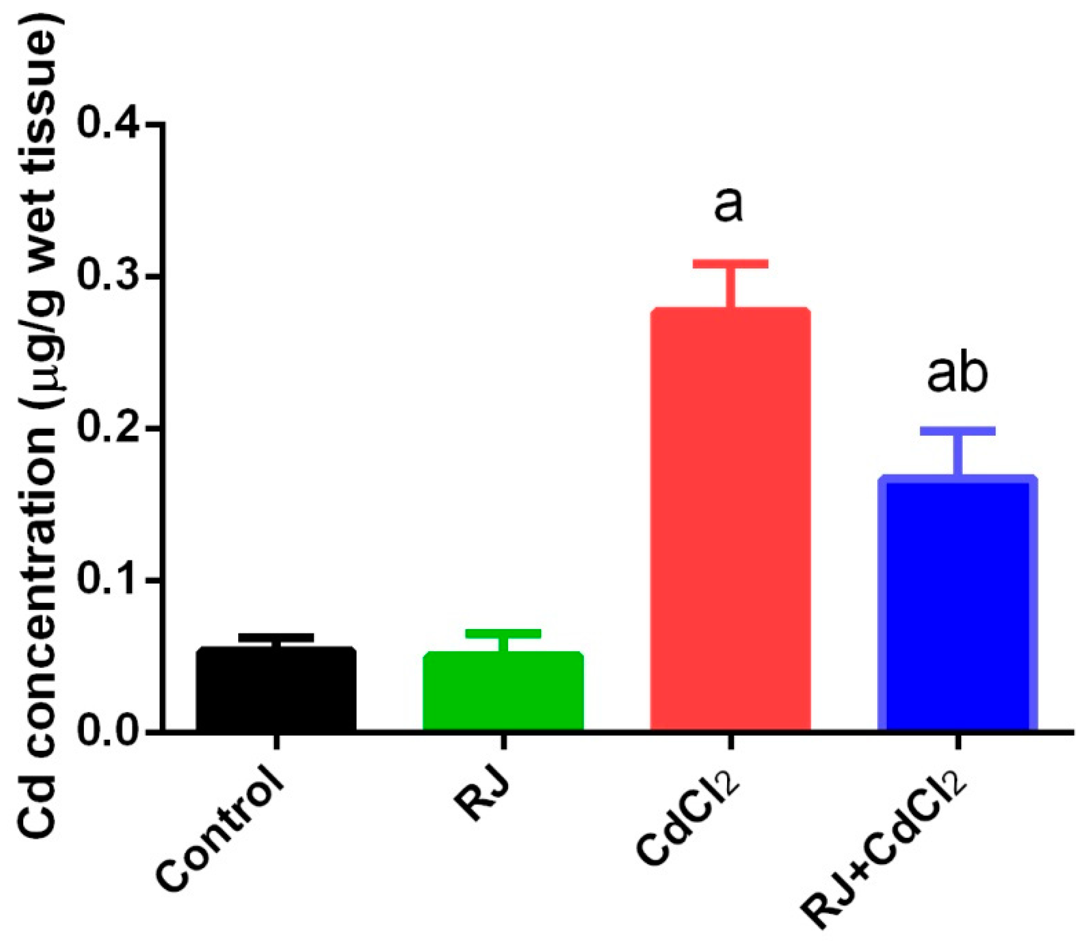
2.1. Effect of RJ on Cd Accumulation in the Testicular Tissue

Cd concentration was observed to have increased significantly (p < 0.05) in testicular homogenates of mice injected intraperitoneally with CdCl2 for 7 days at a dose of 6.5 mg/kg when compared with the control group. The level of this heavy metal was non-significantly changed in the RJ treated group at a dose of 85 mg/kg. Moreover, RJ supplementation 1 h before CdCl2 treatment significantly attenuated Cd accumulation in testicular tissue compared to mice treated with CdCl2 alone (Figure 1).

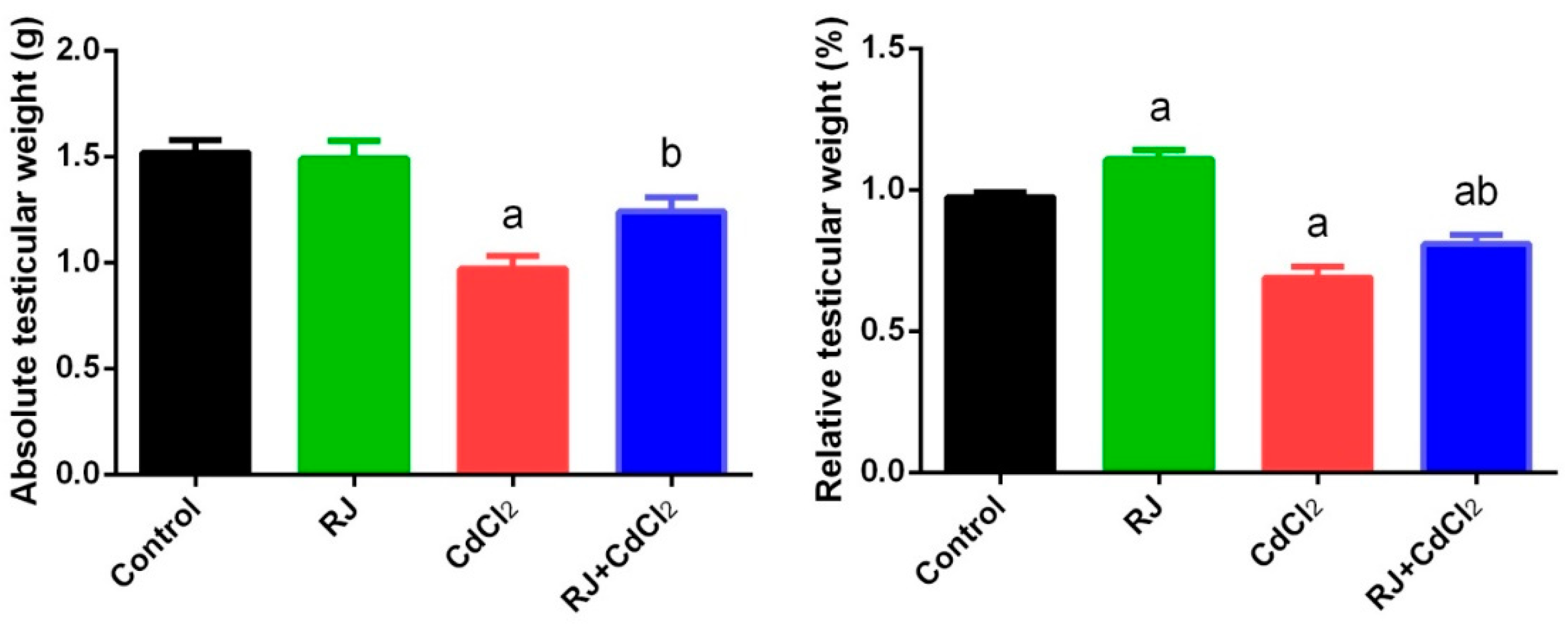
2.2. Effect of RJ and Cd on the Absolute and Relative Weight of Testes

The effect of Cd on testicle absolute and relative weight has been illustrated in Figure 2. In comparison to the control group, Cd-intoxicated mice also showed a significant decrease (p < 0.05) in the testicular absolute and relative weight. Interestingly, oral administration of RJ significantly increased the testicular relative weight, but not the absolute weight, as compared against the control mice. Furthermore, RJ pretreatment significantly suppressed the decrease in the testes weight caused by CdCl2 injection.

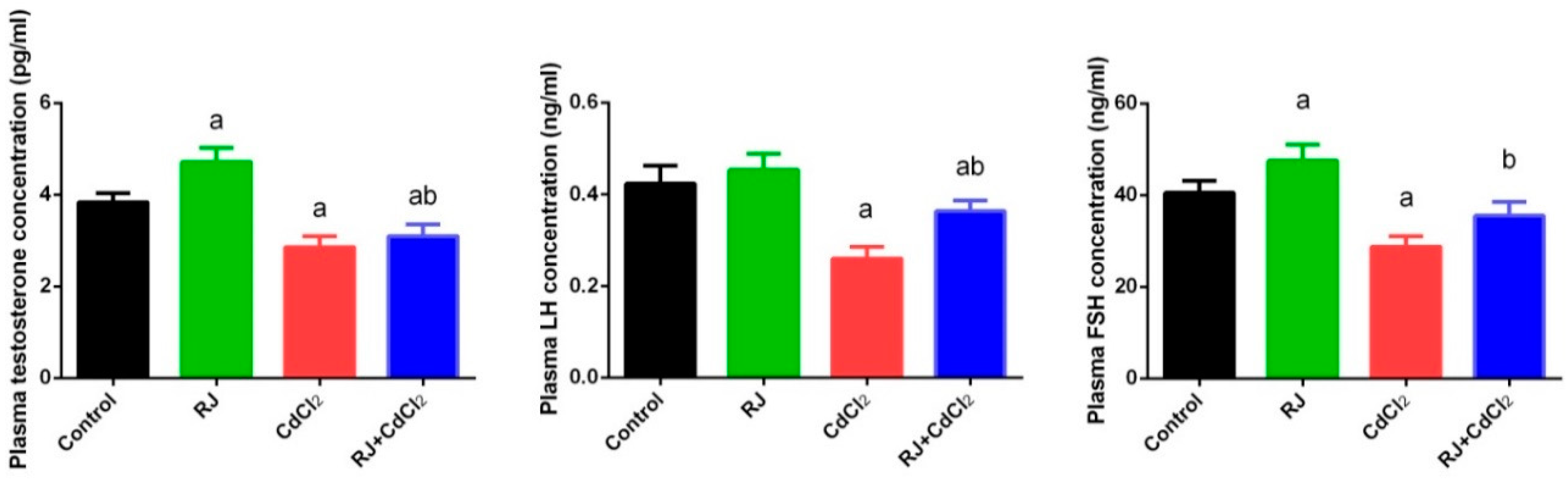
2.3. Effect of RJ and Cd on Serum Levels of Testosterone, LH, and FSH

In the current study, we assessed male sex hormone alteration following Cd exposure. Serological testosterone, LH, and FSH concentrations were found to be significantly declined (p < 0.05) in Cd-intoxicated mice when compared to their corresponding levels in control mice. In contrast, RJ supplemented mice exhibited an increase in the level of testosterone and FSH, suggesting that RJ may have a role in testosterone and FSH synthesis. RJ and CdCl2 treated mice showed a marked increase in testosterone, LH, and FSH concentration, compared to CdCl2-only treated mice. However, the hormonal level was still significantly lower than the control group (Figure 3).

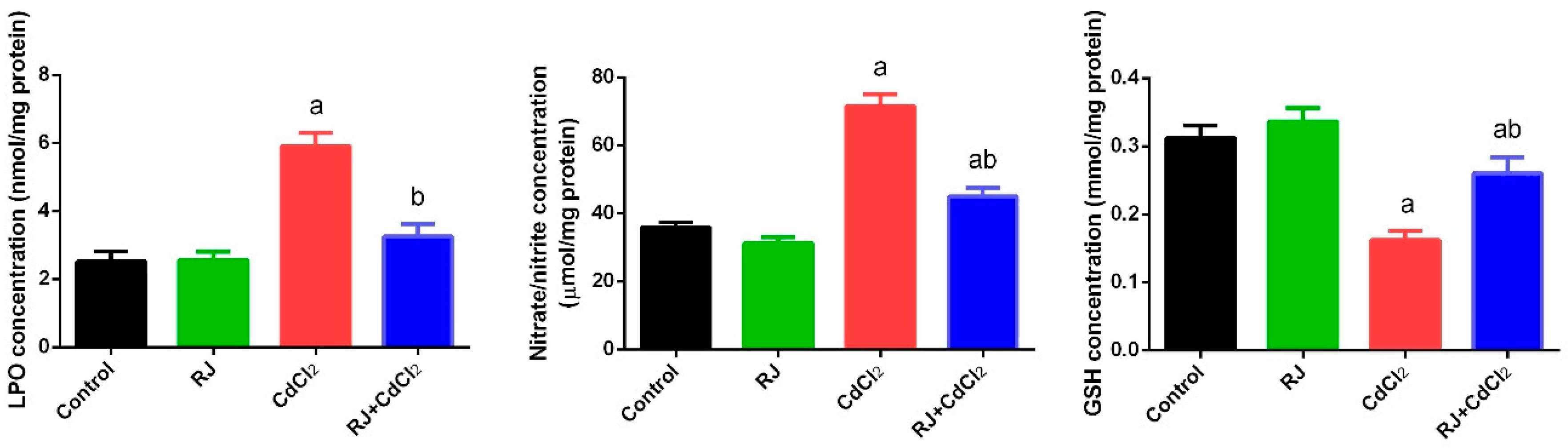
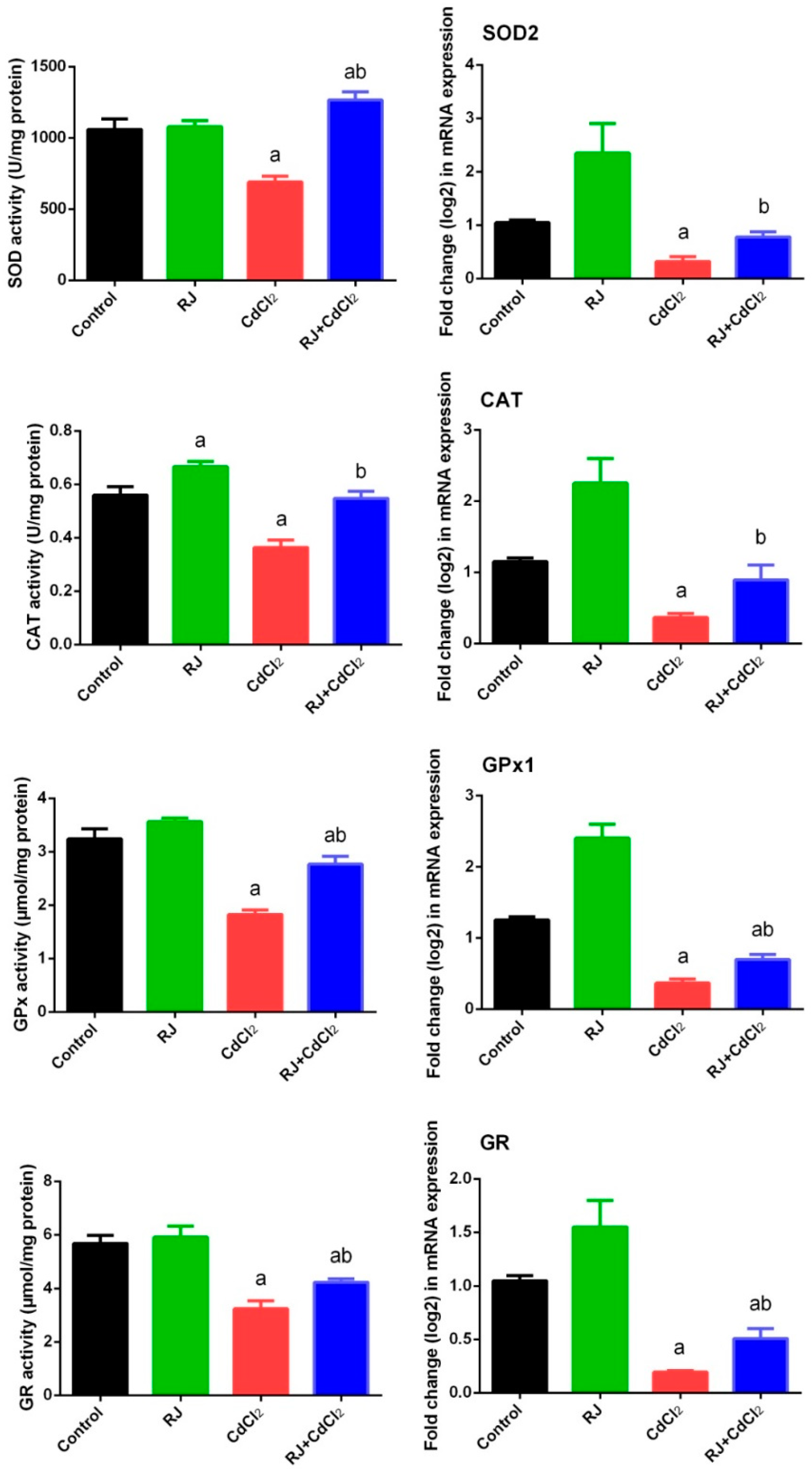
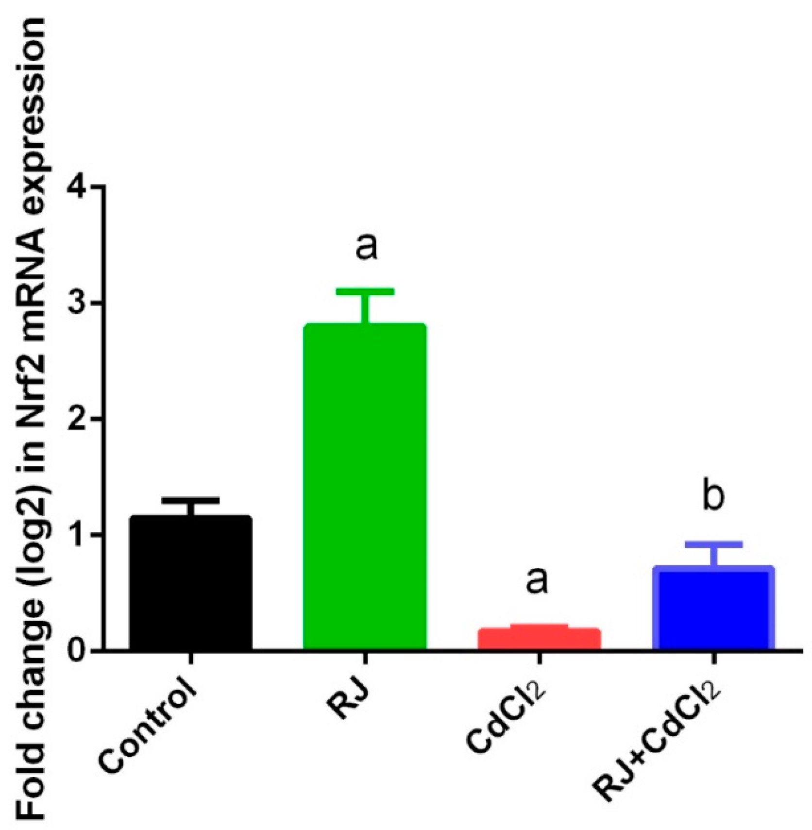
2.4. Effect of RJ on Cd-Induced Oxidative Stress in the Testicular Tissue

Mice exposed to CdCl2 showed a perturbation in the redox status in the testicular homogenates as indicated by the marked elevation (p < 0.05) of LP and NO. The increment of these oxidants was associated with a decrease in GSH content and the activities of GPx, GR, SOD, and CAT, as compared with their levels in the control mice. RJ-gavaged mice recorded a non-significant change in the levels of the aforementioned oxidative stress markers after 7 days. Meanwhile, RJ pretreatment restored the oxidant/antioxidant balance in the testicular homogenates of Cd-intoxicated mice (Figure 4 and Figure 5). Consistent with these results, RT-qPCR data showed that mRNA expression of GPx1, GR, SOD2, and CAT was significantly downregulated (p < 0.05) following Cd exposure as compared to their corresponding expression levels in control mice. No marked change in the expression of these antioxidant enzymes was recorded in the RJ orally treated group. Meanwhile, RJ treatment prior to CdCl2 clarified the ability of RJ to significantly increase the mRNA expression of these endogenous antioxidants in the testicular tissue as compared with the CdCl2-injected group. In order to understand the molecular antioxidant properties of RJ, Nrf2 expression—which enhances the expression of cellular antioxidants and detoxifies the system—has been estimated using RT-qPCR. In comparison to the control group, our findings revealed a significant downregulation (p < 0.05) of Nrf2 in CdCl2-exposed mice (Figure 6). Conversely, Nrf2 expression was upregulated significantly following RJ supplementation as compared to the control values. In addition, RJ also was recorded to normalize the Nrf2 expression in RJ and CdCl2 treated mice, reflecting its potent antioxidant activity against Cd-induced oxidative stress in rat testes.

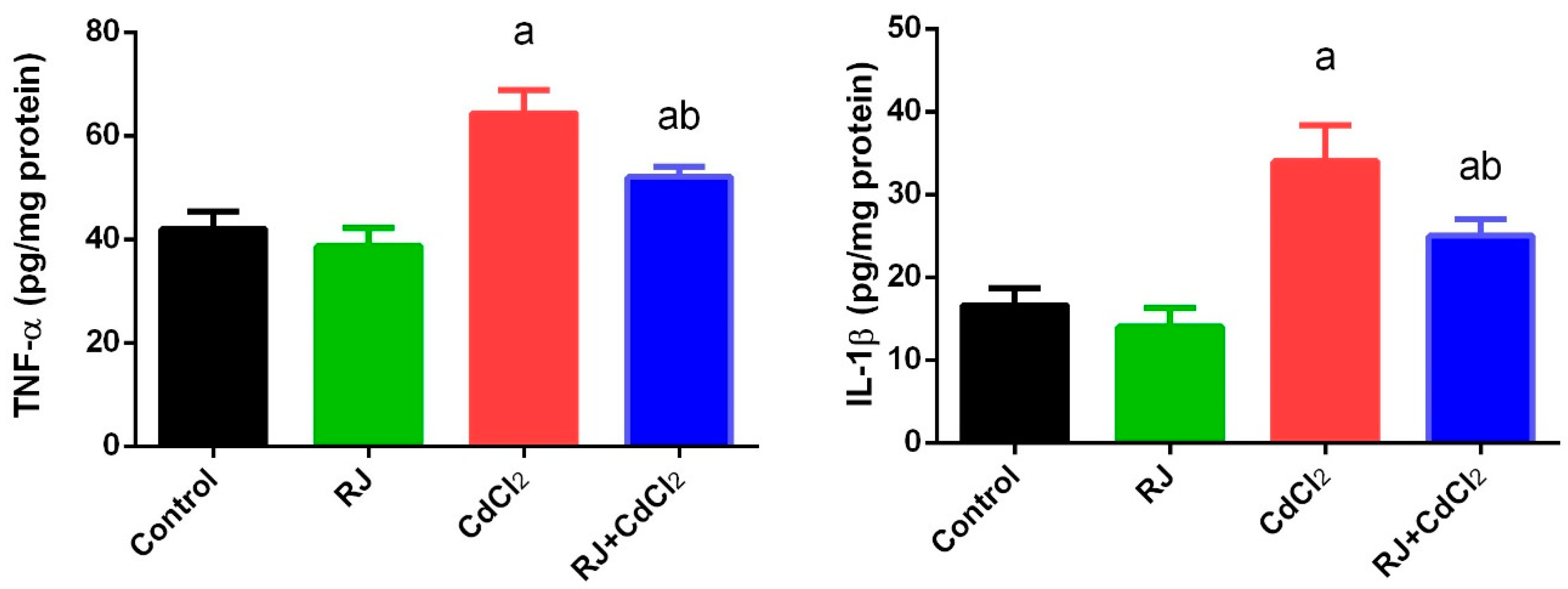
2.5. Effect of RJ on Cd induced-Inflammatory Status in the Testicular Tissue

To evaluate the potential anti-inflammatory activity of RJ following CdCl2 intoxication, the levels of IL-1β and TNF-α were estimated in the testicular tissue. ELISA results showed a significant increase (p < 0.05) in IL-1β and TNF-α levels in Cd-exposed mice as compared with the control values. No change in the concentration of these chemical messengers was observed following RJ treatment. Meanwhile, IL-1β and TNF-α were decreased in the RJ and CdCl2 treated group (Figure 7).

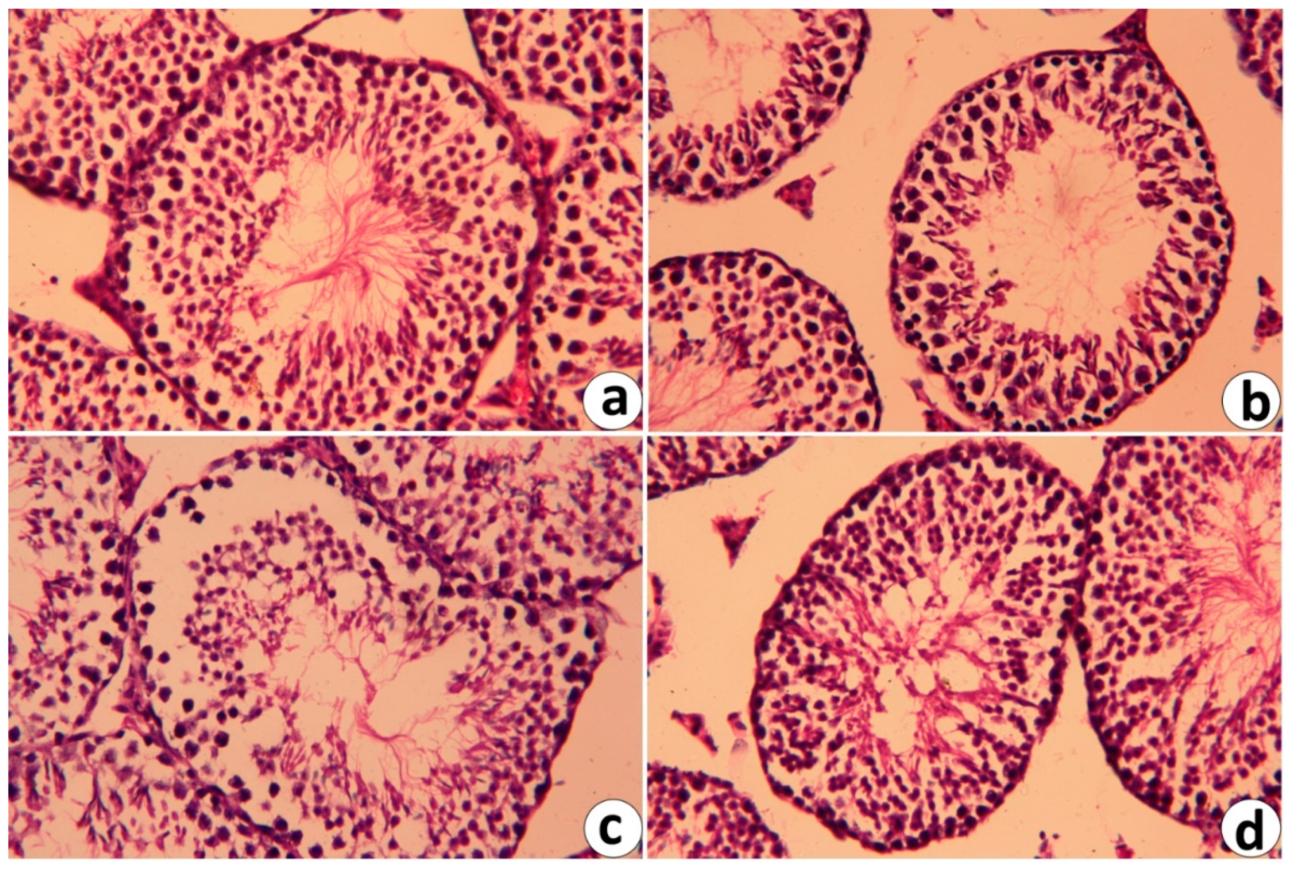
2.6. Histopathological Changes in Testicular Tissue Following RJ and/or CdCl2

Histopathological investigation with light microscopy (Figure 7) of the testes of the control and RJ-treated groups (Figure 8a,b, respectively) exhibited a typical testicular histology with normal and functional seminiferous tubules with all stages of the spermatogenic cells and the Leydig cells filling the space between the seminiferous tubules. The testes from Cd-treated animals displayed many histopathological alterations, including degenerative changes in spermatogenic cells, detachment of the spermatogenic epithelium from the basement membrane, and appearance of vacuolated area in the seminiferous tubules (Figure 8c), whereas RJ-pre-administration significantly alleviated these abnormalities (Figure 8d).

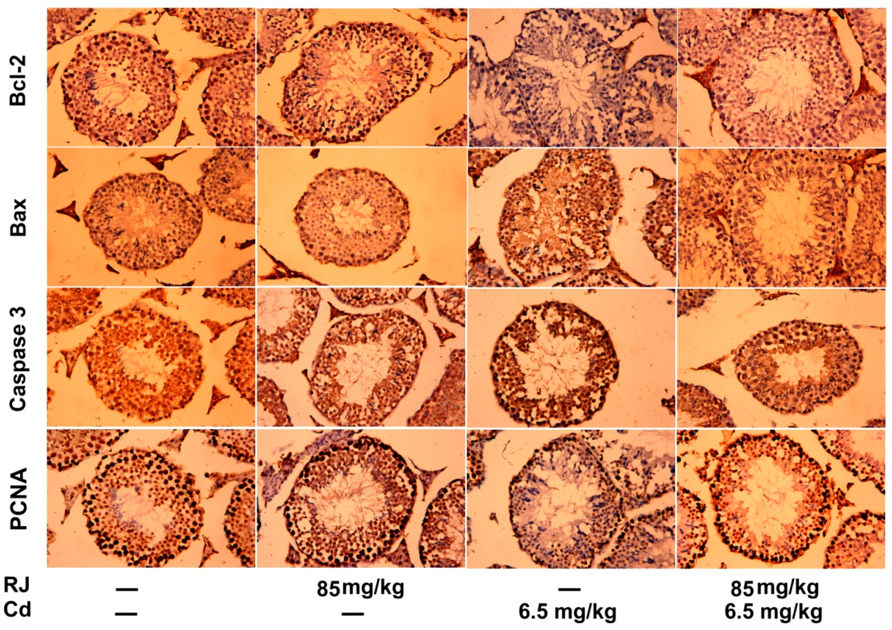
2.7. Effect of RJ on Cd-Triggered Apoptosis and Cytotoxicity in the Testicular Tissue

Immunohistochemical analysis clearly showed that CdCl2 potentiates the apoptotic cascade in the testicular tissue as presented by increasing the immunostaining intensity signal for the anti-apoptotic proteins, including caspase-3 and Bax, and decreasing the immunoreactivity of the anti-apoptotic, Bcl-2. RJ exhibited no significant immunoreactivity to the tested apoptotic markers. Meanwhile, supplementation of RJ before CdCl2 significantly decreased the number of the positively stained spermatogenic cells for caspase-3 and Bax, as well as increased Bcl-2 immunoreactivity. Moreover, the immunohistochemical examination displayed a marked depletion in PCNA immunostained cells in germinal cells of the Cd-injected group. Furthermore, mice treated with RJ and CdCl2 showed an increase in PCNA immunostaining intensity as compared to its corresponding expression in CdCl2-exposed mice (Figure 9).

3. Discussion

Environmental and occupational exposure to heavy metals, including Cd, has been tightly associated with the progression of several pathological impairments by creating oxidative reactions. Testes have been classified among the most negatively affected organs following Cd intoxication. Here, we tried to investigate the potential protective efficiency of RJ against CdCl2-induced testicular dysfunction in mice.
Organ weight has been widely used as an important toxicological marker [19]. Our results recorded a decrease in the absolute and relative weight of the testes after 7 days of treatment with CdCl2. This weight decrease may be due to a decrease in food intake, which has been reported to decrease germ cell numbers that cause testicular dysfunction [20,21]. In the same context, the decreased testes weight was accompanied by a decrease in the levels of serological testosterone, LH, and FSH in response to Cd exposure. According to previous studies, the decrease in the assessed sex hormones is due to the inactivation of steroidogenic enzymes, including 3β-and 17β-hydroxysteroid dehydrogenase, which disturbs the synthesis of androgen and suppresses testosterone production [22]. Additionally, Cd impairs the hormonal receptors and affecting hypothalamic-pituitary-gonadal axis, which inhibit steroidogenesis and further spermatogenesis [23]. Meanwhile, RJ pretreatment was able to protect and minimize Cd-induced testicular weight loss; this may be due to its high and rich nutraceutical constituents. RJ protection against Cd toxicity is extended to restore the levels of testosterone, LH, and FSH to be close to the normal values.
Morita et al. [24] explained the increase of testosterone in volunteers who ingested RJ for 6 months as the ability of RJ to convert dehydroepiandrosterone sulfate (DHEA-S) into testosterone through androstenedione by 3β-and 17β-hydroxysteroid dehydrogenase found in the adrenal and testes; this effect could be due to RJ antioxidant enzyme activity. Moreover, 10-hydroxy-2-decenoic acid and MRJPs, the most active ingredients, in RJ were shown to have estrogenic-like effects in different animal models [25,26]. The increased testosterone concentration might be due to the presence of zinc in RJ. Zinc has been found to play a crucial role in spermatogenesis and its deficiency is associated with a decrease in testosterone concentration [27]. Furthermore, the amino acid content in RJ was found to have a role in androgen homeostasis [28]. An increase in Cd concentration was observed in the current study, suggesting its slow metabolism and excretion. Previous studies revealed that Cd exposure is associated with renal dysfunction, which further affects Cd elimination rate [29]. On the other hand, RJ was found to decrease Cd accumulation in the testicular homogenate. This behavior may be due to the metal chelating properties of the polyphenolic ingredients in RJ [30].
The obtained data further confirmed the involvement of oxidative challenge in Cd-induced testicular dysfunction, as indicated by the elevation of LPO and nitrate/nitrite levels and the deactivation of the cellular antioxidant and detoxifying molecules, including GSH, GPx, GR, SOD, CAT, and Nrf2 at the biochemical and molecular levels. Cd exposition has been linked to oxidant/antioxidant imbalance and ROS production in different body tissues. The produced free radicals enhance DNA oxidation, lipid peroxidation, and protein degradation leading to severe cytotoxic effects [31]. It is well known that Cd triggers fatty acids oxidation as a consequence of nitric oxide, hydroxyl radicals, superoxide anions, and hydrogen peroxide formation [32]. According to our previous study, the increased nitrate/nitrite level is due to iNOS upregulation which is responsible for nitric oxide production [9]. In addition, Cd interacts with sulfhydryl groups leading to GSH pool depletion. Meanwhile, our findings showed that the deactivation of endogenous antioxidant enzymes is due to the inhibition of their gene transcription in the testicular tissue. The suppression of GPx after Cd exposure has been attributed to the depletion of selenium, which plays a crucial role in GPx function [33]. Furthermore, the decrease in SOD could be due to the interaction between Cd and SOD which leads to changes in the enzyme structure, thus affecting its catalytic activity [34].
Jurczuk et al. [35] attributed CAT deactivation to the Fe deficiency, which is a necessary element in its active site. It has been suggested that the antioxidant agents may mitigate Cd-induced oxidative challenges due to their ability to scavenge free radicals and increase the activity of endogenous antioxidant defense systems [1]. Interestingly, RJ, in the current study, showed potent antioxidant activity against Cd-induced oxidative stress in the testicular tissue. RJ peptide content showed strong antioxidant properties and provided cellular protection through scavenging hydroxyl radicals and inhibiting lipid peroxidation in vivo and in vitro [36]. RJ supplementation downregulated the gene expression of the cytochrome P450 4A14 (CYP4A14) enzymes, which is responsible for the production of intracellular free radicals and inhibition enzymes which activate lipid peroxidation. Additionally, it upregulated gene expression of glutathione S-transferase and glutathione peroxidase in fumonisin-treated rats [37]. RJ was found to decrease malondialdehyde levels and increase the activity of GPx, SOD, and CAT in rats treated with cisplatin [38]. The antioxidant activity of RJ has been attributed to its free amino acids content [39]. Furthermore, RJ enhanced the activities of SOD and GPx and decreased malondialdehyde concentration in female diabetic rats [40].
In the present study, mice exposed to Cd showed a marked decrease in Nrf2 expression in the testicular tissue. Nrf2 is known to provide cellular protection against ROS via different mechanisms, including increasing GSH synthesis, upregulating antioxidant and detoxifying enzymes, and degrading superoxide and peroxide radicals by GPx and SOD [41]. In contrast, RJ administered mice minimize the Cd-induced oxidative stress through the upregulation of Nrf2. In our recent work, Nrf2 expression was also found to be upregulated in hepatic tissue of RJ treated mice following Cd intoxication [9]. Moreover, RJ protected lymphocytes against doxorubicin-induced oxidative stress by enhancing Nrf2 and homoxygenase-1 [42]. Additionally, 4-hydroperoxy-2-decenoic acid ethyl ester (HPO-DAEE), a lipid component in RJ, enhanced Nrf2 translocation to the nucleus, activating antioxidant elements, which in turn enhanced the gene expression of the antioxidant and detoxifying enzymes in 6-hydroxydopamine-induced cell death [43].
An inflammatory response in the testicular homogenate was recorded in our experiment following Cd-intoxication, as indicated by the increase of IL-1β and TNF-α. The overproduction of TNF-α in response to pathogens stimulates the release of other pro-inflammatory cytokines, including IL-β, which enhances gene expression associated with inflammation [44]. According to previous studies, this increase is due to ROS production, which activates NF-κB (nuclear factor-kappa B) translocation then upregulates IL-1β and TNF-α mRNA expression [45,46]. RJ treatment reversed the alterations of these pro-inflammatory cytokines. Several reports recorded the anti-inflammatory activity of RJ and its ingredients in different experimental models. RJ supplementation for 14 days at a dose of 300 mg/kg elicited a decrease in TNF-α levels in cyclophosphamide-treated rats [47]. Similarly, rats pre-administered with RJ (50 and 100 mg/kg/7 day) showed a marked decline in the concentration of TNF-α in response to a single dose of bleomycin [48]. Moreover, 10-hydroxy-2-decenoic acid, the major lipid constituent in RJ, was found to exert anti-inflammatory effects in human colon cancer cells via inhibiting NF-κB, which further suppressed the release of IL-1β and TNF-α [14].
In the current study, immunohistochemical investigation showed that Cd is potentiating apoptotic effect in the testes tissue by upregulating the pro-apoptotic proteins (caspase-3 and Bax) and downregulating Bcl-2, the anti-apoptotic protein in the testicular tissue. It has been proposed that Cd may mediate germ cell apoptosis with oxidative stress [49]. Oxidative stress is known to impair calcium ion channels and alter the mitochondrial membrane potential, leading to cytochrome C release, which enhances caspase cascade and fragmentation of DNA. RJ counteracted the apoptotic cascade produced by Cd [49]. On the other hand, RJ supplementation arrested the apoptotic progression and protected the testicular tissue. Rafat et al. [50] demonstrated that RJ consumption protected the human peripheral blood leukocytes against radiation-induced apoptosis. Moreover, RJ inhibited pro-apoptotic (caspase-3) and enhanced expression of the anti-apoptotic (Bcl-2) related proteins in liver and kidney tissues of cisplatin treated rats [51]. Bax, the pro-apoptotic protein, was found to be downregulated after RJ treatment in rats treated with cyclophosphamide [47]. The anti-apoptotic effect of RJ might be due to its antioxidant capacity [52].
PCNA is known to regulate cell cycle and DNA replication. In testes, PCNA has been used as a proliferative parameter to evaluate the spermatogenesis status. Cd-treated mice showed few positive immunostained germ cells for PCNA; this effect is suggested to be due to free radicals production, which enhances oxidation of DNA in spermatogenic cells [6]. Interestingly, RJ upregulated the expression of PCNA in the spermatogenic cells, which may be in part due to its antioxidant activity.

4. Materials and Methods

4.1. Chemicals

CdCl2 was supplied by Sigma Chemical Co. (St. Louis, MO, USA), whereas capsulated pure royal jelly was purchased from Pharco pharmaceuticals Co. Egypt and contains 6% 10-HDA. All other chemicals used for the experiments were of analytical grade.

4.2. Animals and Experimental Design

In total, 28 adult Swiss male mice, weighing 22–27 g, were attained from the Egyptian Organization for Biological Products and Vaccine. Each cage housed seven mice; the animals were given free access to water and a commercial pelleted rodent feed ad libitum. The mice were kept in the animal facility of the Zoology Department at Helwan University, Cairo, Egypt under standard conditions of laboratory with a temperature of 22–25 °C and a 12 h artificial light/dark cycle.
The animals were treated per criteria of the Investigations and Ethics for Laboratory Animal Care at Zoology department, Faculty of Science, Helwan University (approval no, HU2017/Z/03). To investigate the prophylactic effect of RJ on Cd-induced oxidative damage in mice’s testes, the mice were randomly distributed into four equal groups (n = 7, each) after one week of acclimatization. Group I was the control; the animals in this group were intraperitoneally (i.p.) injected with physiological saline (0.9% NaCl) daily for 7 days. Group II were administered with RJ (85 mg/kg daily equivalent to 250 mg crude RJ dissolved in saline) for 7 days by oral gavage. Group III were i.p. treated with 6.5 mg/kg CdCl2 dissolved in physiological saline daily for 7 days. Group IV was supplemented with an oral administration of 85 mg/kg RJ 1 h before the i.p. injection of 6.5 mg/kg CdCl2 daily for 7 days. After 24 h of the last treatment, the mice were sacrificed using ether, then decapitated; the testes were rapidly excised, weighed, and then half of them directly homogenized in ice cold 10 mM phosphate buffer (pH 7.4) to prepare a 10% (w/v) homogenate, which was centrifuged at 4 °C for 10 min (10,000 rpm). The supernatant was obtained for further analyses, and the other half was fixed immediately for the histopathological and immunohistochemical examinations. For the determination of plasma testosterone, LH, and FSH, blood samples were also collected.

4.3. Estimation of Cadmium Concentration in Testes

Cd concentration in testes was measured by atomic absorption spectrophotometer (Perkin Elmer 3100), as previously described [53]. Briefly, an appropriate weight of testes was digested with 3 mL of nitric acid (HNO3) in a tightly capped 30 mL acid-washed polyethylene bottle. This bottle was left for 30 min at room temperature followed by an incubation at 70 °C in a water bath for 3 h. Sample volume was completed into 5 mL with HNO3 and then cured with an equal volume of 10% v/v H2O2. Finally, the solutions were incubated at room temperature for 10 min. Then, an appropriate sample volume was injected into a graphite furnace at 228 nm. Samples were analyzed in duplicate and Cd values were calculated from the standard curve on wet testes tissue basis in µg/g.

4.4. Testicular Index

The testicular weight was measured using a sensitive weighing balance (Radwag, Model AS220/C/2, Clarkson laboratory and supply Inc., Chula Vista, CA, USA), whereas the relative testicular weight was calculated using the following formula:
R e l a t i v e   T e s t i c u l a r   W e i g h t   ( R T W ) = L e f t   T e s t i s   ( L T ) B o d y   w e i g h t × 100

4.5. Biochemical Analyses

4.5.1. Hormones Measurements

Plasma FSH and LH were measured by double antibody radioimmunoassay, as previously described. Plasma testosterone was assayed according to the instructions in the kit’s manual (Elecsys Testosterone Assay Kits, Roche Diagnostics, Mannheim, Germany), by a microplate reader (Chromate awareness 4300, Palm City, FL, USA). Samples were calculated at 620 nm.

4.5.2. Lipid Peroxidation (LPO)

The LPO index in 10% testicular homogenate was accomplished by estimating the concentration of malondialdehyde (MDA) according to the method described by Ohkawa et al. [54]. The developed color was determined as thiobarbituric acid reactive substances (TBARS) at 532 nm excitation and 555 nm emission. Accordingly, 100 mg of the testicular homogenate in phosphate buffer (pH 7.4) was mixed with 100 µL 100% trichloroacetic acid (TCA), 100 µL of sodium thioglycolate (1%), and 250 µL of 1 N HCl. The mixture was incubated for 20 min at 100 °C, and then centrifuged for 10 min (4000 rpm). Spectrophotometric determination of TBA-MDA complex was measured in the supernatant.

4.5.3. Nitrate/Nitrite Level

Valuation of NO in testicular homogenate was carried out according to the method of Sastry et al. through quantification of its stable products represented in both nitrite (NO2) nitrate (NO3) levels [55]. Briefly, 100 µL of the homogenate was added to 400 µL carbonate buffer and a trace amount of activated copper-cadmium alloy. The mixture was incubated at room temperature with continuous shaking. To stop the reaction, the alloy was removed and 100 µL of 0.35 M NaOH and 120 Mm ZnSO4 was added. The mixture was exposed to vigorous vortex and then permitted to stand for 10 min. Afterward, the mixture was centrifuged at room temperature for 10 min (4000 rpm). Griess reagent (50 µL) was added to 10 µL of the supernatant, incubated for 10 min, and lastly, the absorbance was determined at 545 nm using a microplate ELISA reader.

4.5.4. Reduced Glutathione (GSH)

Testicular glutathione content in the testicular homogenate was detected by the method of Sedlak and Lindsay [56]. As such, 250 µL of 10% homogenate was added to 250 µL distilled water and 50 µL of 50% TCA. The mixture was exposed to successive shaking intervals for 15 min, then centrifuged at room temperature at 3000 rpm for 10 min. The supernatant (10 µL) was mixed with 400 µL 0.4 M Tris buffer (pH 8.9) and 10 µL of 5,5-dithio-bis-2-nitrobenzoic acid (DTNB) with continuous shaking. The color developed was evaluated at 512 nm by UV-VIS spectrophotometer (V-630; Jasco, Japan).

4.6. Catalase (CAT) Activity

The activity of CAT was mainly determined based on the degradation rate of H2O2 per minute. The activity unit of CAT was showed as U/mg protein [57]. The total volume (1 mL) of the enzymatic reaction mixture was mainly consisted of 50 mM potassium phosphate (pH 7.0), 19 mM H2O2, and 50 µL of homogenate supernatant. The molar extinction coefficient of H2O2 was examined by UV-VIS spectrophotometer at 240 nm. One unit of enzyme activity was known as the amount of H2O2 (µmol) consumed per min per milligram of tissue protein (U/mg protein).

4.7. Superoxide Dismutase (SOD) Activity

Testicular SOD activity was determined based on the method described by Misra and Fridovich [58]. The method depends on the susceptibility of epinephrine toward oxidation at higher pH (10.2) and generation of adrenochrome and superoxide radicals (O2). The testicular SOD activity is then calculated by the degree of inhibition of this reaction by decreasing the absorbance at 480 nm.

4.8. Glutathione Peroxidase (GPx) Activity

The activity of GPx in testicular homogenate was examined as previously described [59]. The reaction volume was adjusted to 1.5 mL. Briefly, 200 µL of testicular supernatant was mixed with 1 mL of 75 mM phosphate buffer (pH 7.0), 10 mL of 150 mM glutathione, 10 mL of 340 U/mL glutathione reductase, 30 mL of 25 mM EDTA, 30 mL of 5 mM NADPH, 10 mL of 20% Triton X-100, and 50 µL of 7.5 mM H2O2. The oxidation of GSH is linked to the conversion of NADPH (extinction coefficient = 6.22 3 × 10³ M−1 cm−1) to NADP+ that monitored at 340 nm for 3 min. One unit of GPx activity was known as the amount of GSH (nanomoles) oxidized per minute per milligram of protein (U/mg protein).

4.9. Glutathione Reductase (GR) Activity

The activity of GR enzyme was carried out by mixing the testicular supernatant (20 µL) with 0.44 mM oxidized glutathione (GSSG), 0.30 M EDTA, in 0.1 M phosphate buffer (pH 7.0). To begin the enzymatic reaction, 0.036 M NADPH was added. The rate of NADPH oxidation was observed by decreasing the absorbance at 340 nm as a function of time. One unit of enzyme was known as the amount of enzyme required to oxidize 1 µmol of NADPH per minute [60].

4.10. Determination of Testicular Levels of IL-1ß and TNF-α Levels

Quantitative measurements of IL-1ß (IL-1β; Cat. no. EM2IL1B, ThermoFisher Scientific, Waltham, MA, USA) and TNF-α (TNF-α; Cat. no. EZMTNFA, Millipore) levels were performed using enzyme-linked immunosorbent assay (ELISA) kits specified for mice according to the protocol provided with each kit.

4.11. Real Time-PCR

Isolation of total RNA from testes tissue was accomplished with a Trizol reagent and then converted to complementary DNA (cDNA) using cDNA Synthesis Kit (Bio-Rad, Hercules, CA, USA), according to the manufacturer’s instructions. For gene expression analysis, cDNA of the oxidative stress enzyme markers (CAT, SOD, GPx, and GR) were used as a template for quantitative Real-Time PCR. QuantiFast SYBR Green RT-PCR kit (Qiagen, Hilden, Germany) and the corresponding forward and reverse primers shown in Table 1 were implemented. Primers were acquired from (Jena Bioscience GmbH, Jena, Germany). All reactions were accomplished in triplicate using Applied Biosystems 7500 Instrument (ThermoFisher Scientific, CA, USA). The PCR cycling thermal conditions were established as follows: preliminary denaturation at 95 °C for 12 min, then by 40 cycles of denaturation at 94 °C for 60 s and annealing at 55 °C for 60 s, extension at 72 °C for 90 s, and afterwards held for a final extension at 72 °C for 10 min. The relative differences in gene expression between different groups were measured by delta-delta cycle threshold (Ct) method [61]. Glyceraldehyde-3-phosphate dehydrogenase (GAPDH) was utilized as a reference housekeeping gene.

4.12. Histopathological Investigation

The testes were removed from the sacrificed animals and fixed for 24 h at room temperature in 10% neutral-buffered formalin. The tissues were dehydrated in ascending series of alcohol, cleared in xylene, embedded in paraffin wax, and then sectioned at 5-μm thickness. The paraffin sections were washed with water, stained with hematoxylin and eosin [62], and examined using a light microscope. Images were obtained at an original magnification of 400× (Nikon Eclipse E200-LED, Tokyo, Japan).

4.13. Immunohistochemical Investigations

For immunohistochemistry investigations, the paraffin sections were mounted on glass slides and dewaxed. The antigen sites were revealed by washing the sections with boiled water after treatment for 10 min with 0.03% H2O2 in absolute methanol to stop endogenous peroxidase activity. Sections were incubated at 4 °C overnight with (1:50) polyclonal rabbit anti- Bcl-2 antibody, anti-Bax antibody, anti-caspases-3 antibody and anti-PCNA antibody (Santa Cruz, CA, USA). To get rid of the unbound primary antibodies, sections were washed with phosphate buffer saline (PBS). Afterward, sections were incubated for 30 min with goat-derived secondary anti-rabbit antibody conjugated to horseradish peroxidase at 37 °C. Antigen-antibody interactions were finally detected by incubating the sections for 10 min at room temperature with the chromogen 3,3′-diaminobenzidine tetrachloride (DAB-H2O2) as substrate. Testicular sections were visualized using 400× magnification lens (Nikon Eclipse E200-LED, Tokyo, Japan).

4.14. Statistical Analysis

Results are presented as the mean ± standard error of the mean values (SEM) of seven mice. Data from various evaluations were analyzed by one-way analysis of variance (ANOVA). Post hoc Duncan multiple tests were done. p values < 0.05 indicated statistical significance.

5. Conclusions

Overall, RJ supplementation was found to provide protection against Cd-induced testicular dysfunction. RJ ameliorated the hormonal alterations, oxidative status, inflammatory response, and the apoptotic cascade produced following Cd-exposure. These effects may be due to its potent antioxidant activity.

Author Contributions

A.E.A.M. and R.S.A. conceived and designed the experiments; A.E.A.M., R.B.K. and D.S. performed the experiments, analyzed the data, and wrote the paper; G.I.A., S.A. (Saud Alarifi), S.A. (Saad Alkahtani), D.A. and D.M. contributed reagents/materials/analysis tools.

Funding

This research was funded by the Deanship of Scientific Research at King Saud University grant number RGP-180.

Acknowledgments

The authors would like to extend their sincere appreciation to the Deanship of Scientific Research at King Saud University for its funding of this research through the research Group Project no. RGP-180.

Conflicts of Interest

The authors declare no conflicts of interest.

Abbreviations

RJ, royal jelly; MRJP, major royal jelly protein; Cd, cadmium; 10-HAD: 10-hydroxy-2-decenoic; IL-1β: interleukin-1β; TNF-α: tumor necrosis factor-α; NO: nitric oxide; ROS: reactive oxygen species; LH: luteinizing hormone; FSH: follicle-stimulating hormone; LPO: lipid peroxidation; GSH: glutathione; GPx: glutathione peroxidase; GR: glutathione reductase; SOD: superoxide dismutase; CAT: catalase; Nrf2: nuclear factor (erythroid-derived 2)-like-2 factor; Bax: Bcl-2-associated X; Bcl-2: B-cell lymphoma 2; PCNA: proliferating cell nuclear antigen.

References

  1. Elkhadragy, M.F.; Kassab, R.B.; Metwally, D.M.; Almeer, R.; Abdel-Gaber, R.; Al-Olayan, E.M.; Essawy, E.A.; Amin, H.K.; Abdel Moneim, A.E. Protective effects of fragaria ananassa methanolic extract in a rat model of cadmium chloride-induced neurotoxicity. Biosci. Rep. 2018, 38, BSR20180861. [Google Scholar] [CrossRef] [PubMed]
  2. Bernhoft, R.A. Cadmium toxicity and treatment. Sci. World J. 2013, 2013, 394652. [Google Scholar] [CrossRef] [PubMed]
  3. Wang, B.; Du, Y. Cadmium and its neurotoxic effects. Oxidat. Med. Cell. Longev. 2013, 2013, 898034. [Google Scholar] [CrossRef] [PubMed]
  4. Bernard, A. Cadmium & its adverse effects on human health. Indian J. Med. Res. 2008, 128, 557–564. [Google Scholar] [PubMed]
  5. Ahmed, M.M.; El-Shazly, S.A.; Alkafafy, M.E.; Mohamed, A.A.; Mousa, A.A. Protective potential of royal jelly against cadmium-induced infertility in male rats. Andrologia 2018, 50, e12996. [Google Scholar] [CrossRef]
  6. Elmallah, M.I.Y.; Elkhadragy, M.F.; Al-Olayan, E.M.; Abdel Moneim, A.E. Protective effect of fragaria ananassa crude extract on cadmium-induced lipid peroxidation, antioxidant enzymes suppression, and apoptosis in rat testes. Int. J. Mol. Sci. 2017, 18, 975. [Google Scholar] [CrossRef]
  7. Yang, S.H.; Yu, L.H.; Li, L.; Guo, Y.; Zhang, Y.; Long, M.; Li, P.; He, J.B. Protective mechanism of sulforaphane on cadmium-induced sertoli cell injury in mice testis via nrf2/are signaling pathway. Molecules 2018, 23, 1774. [Google Scholar] [CrossRef]
  8. Al Omairi, N.E.; Radwan, O.K.; Alzahrani, Y.A.; Kassab, R.B. Neuroprotective efficiency of mangifera indica leaves extract on cadmium-induced cortical damage in rats. Metab. Brain Dis. 2018, 33, 1121–1130. [Google Scholar] [CrossRef]
  9. Almeer, R.S.; Alarifi, S.; Alkahtani, S.; Ibrahim, S.R.; Ali, D.; Moneim, A. The potential hepatoprotective effect of royal jelly against cadmium chloride-induced hepatotoxicity in mice is mediated by suppression of oxidative stress and upregulation of nrf2 expression. Biomed. Pharmacother. 2018, 106, 1490–1498. [Google Scholar] [CrossRef]
  10. Ramanathan, A.N.K.G.; Nair, A.J.; Sugunan, V.S. A review on royal jelly proteins and peptides. J. Funct. Foods 2018, 44, 255–264. [Google Scholar] [CrossRef]
  11. Fratini, F.; Cilia, G.; Mancini, S.; Felicioli, A. Royal jelly: An ancient remedy with remarkable antibacterial properties. Microbiol. Res. 2016, 192, 130–141. [Google Scholar] [CrossRef]
  12. Okamoto, I.; Taniguchi, Y.; Kunikata, T.; Kohno, K.; Iwaki, K.; Ikeda, M.; Kurimoto, M. Major royal jelly protein 3 modulates immune responses in vitro and in vivo. Life Sci. 2003, 73, 2029–2045. [Google Scholar] [CrossRef]
  13. Melliou, E.; Chinou, I. Chemistry and bioactivity of royal jelly from greece. J. Agric. Food Chem. 2005, 53, 8987–8992. [Google Scholar] [CrossRef] [PubMed]
  14. Yang, Y.C.; Chou, W.M.; Widowati, D.A.; Lin, I.P.; Peng, C.C. 10-hydroxy-2-decenoic acid of royal jelly exhibits bactericide and anti-inflammatory activity in human colon cancer cells. BMC Complement. Altern. Med. 2018, 18, 202. [Google Scholar] [CrossRef]
  15. Zhang, S.; Shao, Q.; Geng, H.; Su, S. The effect of royal jelly on the growth of breast cancer in mice. Oncol. Lett. 2017, 14, 7615–7621. [Google Scholar] [CrossRef] [PubMed]
  16. Ghanbari, E.; Nejati, V.; Khazaei, M. Antioxidant and protective effects of royal jelly on histopathological changes in testis of diabetic rats. Int. J. Reprod. Biomed. 2016, 14, 519–526. [Google Scholar] [CrossRef]
  17. Fan, P.; Han, B.; Feng, M.; Fang, Y.; Zhang, L.; Hu, H.; Hao, Y.; Qi, Y.; Zhang, X.; Li, J. Functional and proteomic investigations reveal major royal jelly protein 1 associated with anti-hypertension activity in mouse vascular smooth muscle cells. Sci. Rep. 2016, 6, 30230. [Google Scholar] [CrossRef] [PubMed]
  18. Guendouz, M.; Haddi, A.; Grar, H.; Kheroua, O.; Saidi, D.; Kaddouri, H. Preventive effects of royal jelly against anaphylactic response in a murine model of cow’s milk allergy. Pharm. Biol. 2017, 55, 2145–2152. [Google Scholar] [CrossRef] [PubMed] [Green Version]
  19. Satarug, S.; Baker, J.R.; Urbenjapol, S.; Haswell-Elkins, M.; Reilly, P.E.; Williams, D.J.; Moore, M.R. A global perspective on cadmium pollution and toxicity in non-occupationally exposed population. Toxicol. Lett. 2003, 137, 65–83. [Google Scholar] [CrossRef]
  20. Fan, R.; Hu, P.C.; Wang, Y.; Lin, H.Y.; Su, K.; Feng, X.S.; Wei, L.; Yang, F. Betulinic acid protects mice from cadmium chloride-induced toxicity by inhibiting cadmium-induced apoptosis in kidney and liver. Toxicol. Lett. 2018, 299, 56–66. [Google Scholar] [CrossRef] [PubMed]
  21. Takahashi, O.; Oishi, S. Testicular toxicity of dietary 2,2-bis(4-hydroxyphenyl)propane (bisphenol a) in f344 rats. Arch. Toxicol. 2001, 75, 42–51. [Google Scholar] [CrossRef] [PubMed]
  22. Hachfi, L.; Sakly, R. Effect of cd transferred via food product on spermatogenesis in the rat. Andrologia 2010, 42, 62–64. [Google Scholar] [CrossRef] [PubMed]
  23. Lafuente, A. The hypothalamic-pituitary-gonadal axis is target of cadmium toxicity. An update of recent studies and potential therapeutic approaches. Food Chem. Toxicol. 2013, 59, 395–404. [Google Scholar] [CrossRef] [PubMed]
  24. Morita, H.; Ikeda, T.; Kajita, K.; Fujioka, K.; Mori, I.; Okada, H.; Uno, Y.; Ishizuka, T. Effect of royal jelly ingestion for six months on healthy volunteers. Nutr. J. 2012, 11, 77. [Google Scholar] [CrossRef] [Green Version]
  25. Moutsatsou, P.; Papoutsi, Z.; Kassi, E.; Heldring, N.; Zhao, C.; Tsiapara, A.; Melliou, E.; Chrousos, G.P.; Chinou, I.; Karshikoff, A.; et al. Fatty acids derived from royal jelly are modulators of estrogen receptor functions. PLoS ONE 2010, 5, e15594. [Google Scholar] [CrossRef] [PubMed]
  26. Mishima, S.; Suzuki, K.M.; Isohama, Y.; Kuratsu, N.; Araki, Y.; Inoue, M.; Miyata, T. Royal jelly has estrogenic effects in vitro and in vivo. J. Ethnopharmacol. 2005, 101, 215–220. [Google Scholar] [CrossRef]
  27. Fallah, A.; Mohammad-Hasani, A.; Colagar, A.H. Zinc is an essential element for male fertility: A review of zn roles in men’s health, germination, sperm quality, and fertilization. J. Reprod. Infertil. 2018, 19, 69–81. [Google Scholar]
  28. Tamler, R.; Mechanick, J.I. Dietary supplements and nutraceuticals in the management of andrologic disorders. Endocrinol. Metab. Clin. N. Am. 2007, 36, 533–552. [Google Scholar] [CrossRef]
  29. Abarikwu, S.O.; Adebayo, O.L.; Otuechere, C.A.; Iserhienrhien, B.O.; Badejo, T.A. Selenium and rutin alone or in combination do not have stronger protective effects than their separate effects against cadmium-induced renal damage. Pharm. Biol. 2016, 54, 896–904. [Google Scholar] [CrossRef]
  30. Tang, B.; Zhang, L.; Geng, Y. Determination of the antioxidant capacity of different food natural products with a new developed flow injection spectrofluorimetry detecting hydroxyl radicals. Talanta 2005, 65, 769–775. [Google Scholar] [CrossRef]
  31. Cuypers, A.; Plusquin, M.; Remans, T.; Jozefczak, M.; Keunen, E.; Gielen, H.; Opdenakker, K.; Nair, A.R.; Munters, E.; Artois, T.J.; et al. Cadmium stress: An oxidative challenge. Biometals 2010, 23, 927–940. [Google Scholar] [CrossRef] [PubMed]
  32. Liu, J.; Qu, W.; Kadiiska, M.B. Role of oxidative stress in cadmium toxicity and carcinogenesis. Toxicol. Appl. Pharmacol. 2009, 238, 209–214. [Google Scholar] [CrossRef] [PubMed] [Green Version]
  33. Jihen el, H.; Imed, M.; Fatima, H.; Abdelhamid, K. Protective effects of selenium (se) and zinc (zn) on cadmium (cd) toxicity in the liver of the rat: Effects on the oxidative stress. Ecotoxicol. Environ. Saf. 2009, 72, 1559–1564. [Google Scholar] [CrossRef] [PubMed]
  34. Casalino, E.; Calzaretti, G.; Sblano, C.; Landriscina, C. Molecular inhibitory mechanisms of antioxidant enzymes in rat liver and kidney by cadmium. Toxicology 2002, 179, 37–50. [Google Scholar] [CrossRef]
  35. Jurczuk, M.; Brzoska, M.M.; Moniuszko-Jakoniuk, J.; Galazyn-Sidorczuk, M.; Kulikowska-Karpinska, E. Antioxidant enzymes activity and lipid peroxidation in liver and kidney of rats exposed to cadmium and ethanol. Food Chem. Toxicol. 2004, 42, 429–438. [Google Scholar] [CrossRef] [PubMed]
  36. Guo, H.; Ekusa, A.; Iwai, K.; Yonekura, M.; Takahata, Y.; Morimatsu, F. Royal jelly peptides inhibit lipid peroxidation in vitro and in vivo. J. Nutr. Sci. Vitaminol. 2008, 54, 191–195. [Google Scholar] [CrossRef] [PubMed]
  37. Kamakura, M.; Maebuchi, M.; Ozasa, S.; Komori, M.; Ogawa, T.; Sakaki, T.; Moriyama, T. Influence of royal jelly on mouse hepatic gene expression and safety assessment with a DNA microarray. J. Nutr. Sci. Vitaminol. 2005, 51, 148–155. [Google Scholar] [CrossRef]
  38. Silici, S.; Ekmekcioglu, O.; Eraslan, G.; Demirtas, A. Antioxidative effect of royal jelly in cisplatin-induced testes damage. Urology 2009, 74, 545–551. [Google Scholar] [CrossRef]
  39. Tamura, S.; Kono, T.; Harada, C.; Yamaguchi, K.; Moriyama, T. Estimation and characterisation of major royal jelly proteins obtained from the honeybee apis merifera. Food Chem. 2009, 114, 1491–1497. [Google Scholar] [CrossRef]
  40. Pourmoradian, S.; Mahdavi, R.; Mobasseri, M.; Faramarzi, E.; Mobasseri, M. Effects of royal jelly supplementation on glycemic control and oxidative stress factors in type 2 diabetic female: A randomized clinical trial. Chin. J. Integr. Med. 2014, 20, 347–352. [Google Scholar] [CrossRef]
  41. Ma, Q. Role of nrf2 in oxidative stress and toxicity. Annu. Rev. Pharmacol. Toxicol. 2013, 53, 401–426. [Google Scholar] [CrossRef] [PubMed]
  42. Jenkhetkana, W.; Thitiorulb, S.; Jansomc, C.; Ratanavalachai, T. Genoprotective effects of thai royal jelly against doxorubicin in human lymphocytes in vitro. Nat. Prod. Commun. 2018, 13, 79–84. [Google Scholar]
  43. Inoue, Y.; Hara, H.; Mitsugi, Y.; Yamaguchi, E.; Kamiya, T.; Itoh, A.; Adachi, T. 4-hydroperoxy-2-decenoic acid ethyl ester protects against 6-hydroxydopamine-induced cell death via activation of nrf2-are and eif2alpha-atf4 pathways. Neurochem. Int. 2018, 112, 288–296. [Google Scholar] [CrossRef] [PubMed]
  44. Garlanda, C.; Dinarello, C.A.; Mantovani, A. The interleukin-1 family: Back to the future. Immunity 2013, 39, 1003–1018. [Google Scholar] [CrossRef] [PubMed]
  45. Lee, J.; Lim, K.T. Preventive effect of phytoglycoprotein (27 kda) on inflammatory factors at liver injury in cadmium chloride-exposed icr mice. J. Cell. Biochem. 2011, 112, 694–703. [Google Scholar] [CrossRef]
  46. Freitas, M.; Fernandes, E. Zinc, cadmium and nickel increase the activation of nf-kappab and the release of cytokines from thp-1 monocytic cells. Metallomics: Integr. Biometal Sci. 2011, 3, 1238–1243. [Google Scholar] [CrossRef]
  47. Abdel-Hafez, S.M.N.; Rifaai, R.A.; Abdelzaher, W.Y. Possible protective effect of royal jelly against cyclophosphamide induced prostatic damage in male albino rats; a biochemical, histological and immuno-histo-chemical study. Biomed. Pharmacother. 2017, 90, 15–23. [Google Scholar] [CrossRef]
  48. Zargar, H.R.; Hemmati, A.A.; Ghafourian, M.; Arzi, A.; Rezaie, A.; Javad-Moosavi, S.A. Long-term treatment with royal jelly improves bleomycin-induced pulmonary fibrosis in rats. Can. J. Physiol. Pharmacol. 2017, 95, 23–31. [Google Scholar] [CrossRef]
  49. Turner, T.T.; Lysiak, J.J. Oxidative stress: A common factor in testicular dysfunction. J. Androl. 2008, 29, 488–498. [Google Scholar] [CrossRef]
  50. Rafat, N.; Monfared, A.S.; Shahidi, M.; Pourfallah, T.A. The modulating effect of royal jelly consumption against radiation-induced apoptosis in human peripheral blood leukocytes. J. Med. Phys. 2016, 41, 52–57. [Google Scholar]
  51. Karadeniz, A.; Simsek, N.; Karakus, E.; Yildirim, S.; Kara, A.; Can, I.; Kisa, F.; Emre, H.; Turkeli, M. Royal jelly modulates oxidative stress and apoptosis in liver and kidneys of rats treated with cisplatin. Oxidat. Med. Cell. Longev. 2011, 2011, 981793. [Google Scholar] [CrossRef] [PubMed]
  52. Valiollahpoor Amiri, M.; Deldar, H.; Ansari Pirsaraei, Z. Impact of supplementary royal jelly on in vitro maturation of sheep oocytes: Genes involved in apoptosis and embryonic development. Syst. Biol. Reprod. Med. 2016, 62, 31–38. [Google Scholar] [CrossRef] [PubMed]
  53. Sharma, R.P.; Street, J.C.; Shupe, J.L.; Bourcier, D.R. Accumulation and depletion of cadmium and lead in tissues and milk of lactating cows fed small amounts of these metals. J. Dairy Sci. 1982, 65, 972–979. [Google Scholar] [CrossRef]
  54. Ohkawa, H.; Ohishi, N.; Yagi, K. Assay for lipid peroxides in animal tissues by thiobarbituric acid reaction. Anal. Biochem. 1979, 95, 351–358. [Google Scholar] [CrossRef]
  55. Sastry, K.V.; Moudgal, R.P.; Mohan, J.; Tyagi, J.S.; Rao, G.S. Spectrophotometric determination of serum nitrite and nitrate by copper-cadmium alloy. Anal. Biochem. 2002, 306, 79–82. [Google Scholar] [CrossRef] [PubMed]
  56. Sedlak, J.; Lindsay, R.H. Estimation of total, protein-bound, and nonprotein sulfhydryl groups in tissue with ellman’s reagent. Anal. Biochem. 1968, 25, 192–205. [Google Scholar] [CrossRef]
  57. Aebi, H. Catalase in vitro. Methods Enzymol. 1984, 105, 121–126. [Google Scholar]
  58. Misra, H.P.; Fridovich, I. The role of superoxide anion in the autoxidation of epinephrine and a simple assay for superoxide dismutase. J. Biol. Chem. 1972, 247, 3170–3175. [Google Scholar]
  59. Lawrence, R.A.; Burk, R.F. Glutathione peroxidase activity in selenium-deficient rat liver. 1976. Biochem. Biophys. Res. Commun. 2012, 425, 503–509. [Google Scholar] [CrossRef]
  60. Farias, J.G.; Puebla, M.; Acevedo, A.; Tapia, P.J.; Gutierrez, E.; Zepeda, A.; Calaf, G.; Juantok, C.; Reyes, J.G. Oxidative stress in rat testis and epididymis under intermittent hypobaric hypoxia: Protective role of ascorbate supplementation. J. Androl. 2010, 31, 314–321. [Google Scholar] [CrossRef]
  61. Livak, K.J.; Schmittgen, T.D. Analysis of relative gene expression data using real-time quantitative pcr and the 2(-delta delta c(t)) method. Methods 2001, 25, 402–408. [Google Scholar] [CrossRef] [PubMed]
  62. Drury, R.A.B.; Wallington, E.A. Preparation and fixation of tissues. In Carleton’s Histological Technique; Oxford University Press: Oxford, UK, 1980; pp. 41–54. [Google Scholar]
Figure 1. Effects of Royal jelly (RJ) administration on concentration of cadmium in testes of mice treated with cadmium chloride (CdCl2). All values are expressed as mean ± SEM (n = 7). a refers to a significant change from the control mice at p < 0.05; b refers to a significant change from the CdCl2-treated mice at p < 0.05, using Duncan’s post hoc test.
Ijms 19 03979 g001
Figure 2. Effects of Royal jelly (RJ) administration on absolute and relative testicular weight of mice treated with cadmium chloride (CdCl2) toxicity. All values are expressed as mean ± SEM (n = 7). a refers to a significant change from the control mice at p < 0.05; b refers to a significant change from the CdCl2-treated mice at p < 0.05, using Duncan’s post hoc test.
Ijms 19 03979 g002
Figure 3. Effects of royal jelly (RJ) on serological testosterone, LH, and FSH levels of mice exposed to cadmium chloride (CdCl2). All values are expressed as mean ± SEM (n = 7). a refers to a significant change from the control mice at p < 0.05; b refers to a significant change from the CdCl2-treated mice at p < 0.05, using Duncan’s post hoc test.
Ijms 19 03979 g003
Figure 4. Effects of royal jelly (RJ) on lipid peroxidation (LPO), nitrate/nitrite, and reduced glutathione (GSH) levels in testes of mice exposed to cadmium chloride (CdCl2). All values are expressed as mean ± SEM (n = 7). a refers to a significant change from the control mice at p < 0.05; b refers to a significant change from the CdCl2-treated mice at p < 0.05, using Duncan’s post hoc test.
Ijms 19 03979 g004
Figure 5. Effects of royal jelly (RJ) on the levels of superoxide dismutase (SOD), catalase (CAT), glutathione peroxidase (GPx), glutathione reductase (GR), and their corresponding mRNA expression in testes of mice exposed to cadmium chloride (CdCl2). Values of antioxidant enzyme activities are expressed as the mean ± SEM (n = 7), whereas data on mRNA expression levels (mean ± SEM of triplicate assays) were normalized to the GAPDH mRNA levels and are shown as the fold induction (in log2 scale) relative to the mRNA levels in the controls. a refers to a significant change from the control mice at p < 0.05; b refers to a significant change from the CdCl2-treated mice at p < 0.05, using Duncan’s post hoc test.
Ijms 19 03979 g005
Figure 6. Effects of royal jelly (RJ) on the mRNA level of nuclear factor (erythroid-derived 2)-like-2 factor (Nrf2) in testes of mice exposed to cadmium chloride (CdCl2). All values are expressed as mean ± SEM of triplicate assays and were normalized to the GAPDH mRNA levels and are shown as the fold induction (in log2 scale) relative to the mRNA levels in the controls. a refers to a significant change from the control mice at p < 0.05; b refers to a significant change from the CdCl2-treated mice at p < 0.05, using Duncan’s post hoc test.
Ijms 19 03979 g006
Figure 7. Effects of royal jelly (RJ) on the levels of tumor necrosis factor-α (TNF-α) and interleukin 1β (IL-1β) in testes of mice exposed to cadmium chloride (CdCl2). All values are expressed as mean ± SEM (n = 7). a refers to a significant change from the control mice at p < 0.05; b refers to a significant change from the CdCl2-treated mice at p < 0.05, using Duncan’s post hoc test.
Ijms 19 03979 g007
Figure 8. Photomicrographs of light microscope of testes of mice treated with RJ and CdCl2 for 7 days. Cross sections of testes were stained with hematoxylin and eosin (400×). (a,b) testes from control and royal jelly (RJ) groups, respectively exhibiting typical features of seminiferous epithelium and Leydig cells (LC). (c) testes from the CdCl2-treated mice showing degenerative seminiferous tubules. (d) testis from the pretreated group with RJ against CdCl2 showing an obvious preservation of spermatogenic epithelium in most seminiferous tubules.
Ijms 19 03979 g008
Figure 9. Photomicrographs showing changes in Bcl-2, Bax, caspase-3, and PCNA expression in the testicular tissue of mice following treatment with royal jelly (RJ) and cadmium chloride (CdCl2). (magnification, 400×).
Ijms 19 03979 g009
Table 1. Primer sequences of genes analyzed in real time PCR.
NameAccession NumberForward Primer (5’---3’)Reverse Primer (5’---3’)
GAPDHNM_001289726.1TCACCACCATGGAGAAGGCGCTAAGCAGTTGGTGGTGCA
SOD2NM_013671.3GCCCAAACCTATCGTGTCCAAGGGAACCCTAAATGCTGCC
CATNM_009804.2CCGACCAGGGCATCAAAAGAGGCCATAATCCGGATCTTC
GSH-Px1NM_001329527.1CAGCCGGAAAGAAAGCGATGTTGCCATTCTGGTGTCCGAA
GSH-RNM_010344.4TGGCACTTGCGTGAATGTTGCGAATGTTGCATAGCCGTGG
Nrf2NM_010902.4CCTCTGTCACCAGCTCAAGGTTCTGGGCGGCGACTTTATT
iNOSNM_001313922.1CGAAACGCTTCACTTCCAATGAGCCTATATTGCTGTGGCT
IL-1βNM_008361.4TGCCACCTTTTGACAGTGATGTTCTTGTGACCCTGAGCGAC
TNF-αNM_013693.3AGAGGCACTCCCCCAAAAGACGATCACCCCGAAGTTCAGT
The abbreviations of the genes are as follows: GAPDH, glyceraldehyde-3-phosphate dehydrogenase; SOD2, superoxide dismutase 2 mitochondrial (MnSOD); CAT, catalase; GSH-Px1, glutathione peroxidase 1; GSH-R, glutathione reductase; Nrf2, nuclear factor erythroid 2-related factor 2; iNOS, inducible nitric oxide synthase; IL-1β: Interleukin 1 beta; TNF-α: Tumor necrosis factor.

