Review

Metabolic and Homeostatic Changes in Seizures and Acquired Epilepsy—Mitochondria, Calcium Dynamics and Reactive Oxygen Species

1
Department of Neurology, University of Münster, 48149 Münster, Germany
2
Division of Cancer Research, School of Medicine, Jacqui Wood Cancer Centre, Ninewells Hospital and Medical School, University of Dundee, Dundee DD1 9SY, UK
3
Departments of Medicine and Pharmacology and Molecular Sciences, Johns Hopkins University School of Medicine, Baltimore, MD 21205, USA
4
Shefa Neuroscience Research Center, Khatam Alanbia Hospital, Tehran 1996836111, Iran
5
Department of Neuroscience, Mashhad University of Medical Sciences, Mashhad 9177948564, Iran
6
Department of Neurosurgery, University of Münster, 48149 Münster, Germany
7
Epilepsy Research Center, University of Münster, 48149 Münster, Germany
*
Author to whom correspondence should be addressed.
Int. J. Mol. Sci. 2017, 18(9), 1935; https://doi.org/10.3390/ijms18091935
Submission received: 13 August 2017 / Revised: 2 September 2017 / Accepted: 5 September 2017 / Published: 8 September 2017

Abstract

:
Acquired epilepsies can arise as a consequence of brain injury and result in unprovoked seizures that emerge after a latent period of epileptogenesis. These epilepsies pose a major challenge to clinicians as they are present in the majority of patients seen in a common outpatient epilepsy clinic and are prone to pharmacoresistance, highlighting an unmet need for new treatment strategies. Metabolic and homeostatic changes are closely linked to seizures and epilepsy, although, surprisingly, no potential treatment targets to date have been translated into clinical practice. We summarize here the current knowledge about metabolic and homeostatic changes in seizures and acquired epilepsy, maintaining a particular focus on mitochondria, calcium dynamics, reactive oxygen species and key regulators of cellular metabolism such as the Nrf2 pathway. Finally, we highlight research gaps that will need to be addressed in the future which may help to translate these findings into clinical practice.

1. Introduction

Epilepsy, a devastating disease, affects over 50 million people worldwide [1] and is defined by the occurrence of unprovoked seizures. Most of these epilepsies are acquired as a consequence of a brain injury and are followed by a latent period of epileptogenesis [2]. Once unprovoked seizures occur, the patient is diagnosed with epilepsy and anticonvulsive treatment is initiated. Many patients become seizure free with antiepileptic drugs, although approximately one third of patients develop pharmacoresistant epilepsy [3], highlighting the unmet need for new treatment strategies. Current anticonvulsants mainly act on neuronal voltage gated ion channels, whereas downstream signaling cascades and non-neuronal cells are not targeted directly. However, the latter may be instrumental in mediating pharmacoresistance and also epilepsy comorbidities. It is likely that downstream signaling cascades such as metabolic and homeostatic cellular mechanisms contribute to epileptogenesis, fully established epilepsy and pharmacoresistant epilepsy, although the precise mechanisms remain unclear.
There are clinical hints pointing to a strong involvement of mitochondria and bioenergetics in epileptogenesis, seizures and epilepsy. For example, patients with mitochondrial mutations often present with epilepsy as a phenotypic manifestation of the disease [4], highlighting the involvement of mitochondrial dysfunction in epileptogenesis. In addition, seizure activity and epilepsy have been linked to energy failure, which has been hypothesized to lead to neuronal injury responsible for the clinical sequelae associated with epilepsy patients. The brain, which makes up only 2% of the total bodyweight, contributes up to 20% to the resting whole body metabolism. This large metabolic turnover is mainly due to synaptic transmission [5,6] where vesicle cycling consumes the majority of presynaptic adenosine triphosphate (ATP) to mediate neuronal function [7]. Epileptiform activity also induces large ionic conductances and depletes vesicular stores. Restoration of these changes, i.e., restoration of cellular homeostasis, is an energy demanding process [8]. Thus, it is not surprising that ATP demand and production during seizures, epilepsy and particularly prolonged seizures, such as those seen in status epilepticus, the maximum expression of epilepsy, is critical. It should be noted though that mitochondria are not the only source of dysfunction in epileptic seizures, and there are also other targets of homeostatic imbalances that occur during seizure activity. The most prominent homeostatic changes during seizure activity include the accumulation of intracellular calcium and the increased production of reactive oxygen species (ROS). Neuronal compromise during seizure activity is dependent on intracellular Ca2+ entry [9]. There is accumulating evidence that N-methyl-d-aspartate (NMDA) receptors play a pivotal role in intracellular Ca2+ accumulation during seizure activity. This is supported by robust evidence showing that blocking of NMDA receptor activity abolishes cell death both in vitro and in vivo [10,11,12]. NMDA receptor opening has also been shown to promote ROS production via nicotinamide adenine dinucleotide phosphate (NADPH) oxidase, an enzyme which has recently been drawn into the spotlight of seizure induced neuronal damage and neuronal autophagy [13,14]. Excess ROS and Ca2+ are potent triggers of the mitochondrial permeability transition pore opening, which is a key event and point of no return leading to mitochondrial swelling and cytochrome c release from mitochondria, subsequently triggering the cell death cascade [15,16,17].
In this review, we summarize current knowledge about metabolic and homeostatic changes in seizures and acquired epilepsy with a particular focus on mitochondria, Ca2+ dynamics, ROS and key regulators of cellular metabolism such as the nuclear factor erythroid 2–related factor 2 (Nrf2)pathway. It is not in the scope of this review to cover the extensive literature on genetic syndromes with mutations in genes coding for mitochondrial proteins or key enzymes of metabolism, which has been the subject of a number of other extensive reviews (e.g., [4,18]). Instead, we wish to focus on patients suffering from acquired epilepsies (e.g., such as epilepsy due to hippocampal sclerosis) who are frequently encountered in clinical practice and pose a challenge to practitioners given that 30% present with pharmacoresistant seizures [3,19]. Finally, we highlight research gaps that will need to be addressed in the future which may help to translate findings into clinical practice.
References for this review were identified through searches of PubMed with combinations of the terms from keywords from the subsection titles (e.g., mitochondria) and “epilepsy” or “seizures” from 1950 until July 2017. In addition, manuscripts were also identified through searches of the authors’ own files and from reference lists of the articles pointed out by the PubMed searches. Due to space restrictions, the final reference list was compiled from a selection of the articles identified which were prioritized according to their originality and their ability to fit into the narrative style of the current review.

2. Mitochondria and Epilepsy—Adenosine Triphosphate (ATP), Ca2+ and Cell Death

Mitochondria, initially coined bioblasts, were first described by Richard Altmann in 1890 and were recognized to function as elementary organisms within the cell [20]. A further advance in their functional characterization came through the introduction of new methods to study electron transport and metabolic states of the respiratory chain [21,22,23,24]. Peter Mitchell introduced the chemiosmotic hypothesis of oxidative phosphorylation in 1961, linking energy metabolism to hydrogen transport across the mitochondrial membrane [25]. Oxidative phosphorylation is the main source of ATP generation in neurons since they lack powerful enzymes for glycolytic ATP production such as those that are available in astrocytes [26].
The mitochondria are organelles that are amongst others essential in three different homeostatic mechanisms. Firstly, their most prominent and obvious function is ATP production. Secondly, they are involved in Ca2+ homeostasis through buffering of intracellular Ca2+, and thus have been referred to as the “hub of Ca2+ signaling” [27]. Lastly, mitochondria are instrumental in apoptotic cell death amongst others through the release of intramitochondrial cytochrome c [28]. Links to seizures and epilepsy have been established for all three mechanisms.

2.1. ATP during Seizures and Epilepsy

Early pioneering experimental studies showed that glucose, ATP and other energetic substrates decrease during seizure activity, particularly if this is prolonged [29,30,31]. This has been confirmed on a cellular level, where ATP decrease during seizure activity has been shown in neurons [12]. Mitochondrial dysfunction and seizures have been closely linked to each other not only in epilepsies, i.e., mitochondrial epilepsies, but are also increasingly recognized as a target in acquired epilepsy [18]. Anaplerosis, a strategy that aims to restore mitochondrial function through providing tricarboxylic acid cycle substrates, has been recently advocated for use in acquired epilepsy [32]. The ketogenic diet, a high fat diet used for the treatment of epilepsies, has been shown amongst other effects to upregulate the neuronal expression of genes involved in the tricarboxylic acid cycle, oxidative phosphorylation and glycolysis, along with improving mitochondria complex activities and boosting mitochondrial biogenesis [33,34,35,36]. The ketogenic diet is particularly the treatment of choice in patients suffering from seizures due to glucose transporter 1 (GLUT-1) deficiency syndrome and pyruvate dehydrogenase complex deficiency, given that it circumvents the metabolic deficiencies in these syndromes [37]. However, the ketogenic diet has also been found to be an effective treatment in syndromes which are not directly linked to mutations in the metabolic pathway, such as Dravet syndrome or Doose syndrome [37,38] and has been advocated for the treatment of epilepsies due to mitochonridal diseases [39]. The exact mechanisms of the seizure suppressive effect of the ketogenic remain elusive. Beside its anaplerotic properties and its effect on mitochondria, direct anti-seizure effects of ketone bodies, neurotransmitter and ion channel regulation as well as regulation of ROS amongst others have been shown to play a role [36].

2.2. Mitochondria and Ca2+ Buffering in Seizures and Epilepsy

Besides ATP production, the most important function of mitochondria is the buffering of excess Ca2+. Excess Ca2+ entry into neurons has been observed in rats that underwent status epilepticus induced by bicuculline and l-allylglycine [9]. In these studies, mitochondrial Ca2+ overload has been shown to lead to cell death and was particularly pronounced in CA1 and CA3 regions, which are areas of the central nervous system (CNS) that are very susceptible to seizure induced cell death [9,40]. It is interesting that these very early studies already identified mitochondria as the main site of Ca2+ accumulation during prolonged seizures.
Different pathways for mitochondrial Ca2+ buffering have been described in the literature. The mitochondrial uniporter (MCU), whose structure has been unraveled only recently [41], is the main site of Ca2+ entry into the mitochondria and is thus instrumental in Ca2+ buffering during excess Ca2+ overload. The uptake and extrusion of Ca2+ in mitochondria is coupled to H+ and Na+ cycling that is maintained by the electron transport chain, which creates a potential gradient across the mitochondrial membrane [27,42] (Figure 1). Within the mitochondrial membrane, Ca2+ precipitates to insoluble Ca2+ phosphate complexes, a process which is dependent on the availability of phosphate. This in turn is also dependent on the proton gradient and thus more phosphate enters the mitochondria if the gradient is higher, i.e., when the mitochondria are hyperpolarized. Ca2+ homeostasis through the MCU has been shown to play a role during seizure activity. Inhibition of the MCU significantly attenuated neuronal death after pilocarpine-induced status epilepticus and reduced levels of intracellular ROS [43].
Mitochondrial permeability transition pore (MPTP) opening is another route for Ca2+ trafficking across the mitochondrial membrane (Figure 2). The MPTP is a protein complex in the inner mitochondrial membrane which has long been a mystery, but was recently found to be formed of two ATP synthase (Complex V) dimers [45,46]. The importance of MPTP opening was soon recognized in the triggering of apoptotic cell death through an increase in permeability of Ca2+ ions [16,46]. Our group has shown that seizure-induced cell death is partly mediated via MPTP opening as treatment with cyclosporine A, a potent inhibitor of MPTP opening, was able to reduce mitochondrial depolarization, the initial event leading to cell death during seizure activity [12]. More recently, an interesting link between the MPTP and epilepsy treatment was made. Ketone bodies, which are the main effectors of anti-seizure effects in the ketogenic diet and are a very effective anti-seizure treatment, have been shown to mediate anti-seizure effects via MPTP modulation [47]. In this study, mitochondria isolated from hippocampi of Kcna1-null mice with a genetic susceptibility to seizures had a higher threshold for Ca2+ induced mitochondrial permeability transition if they were pre-treated with ketone bodies.

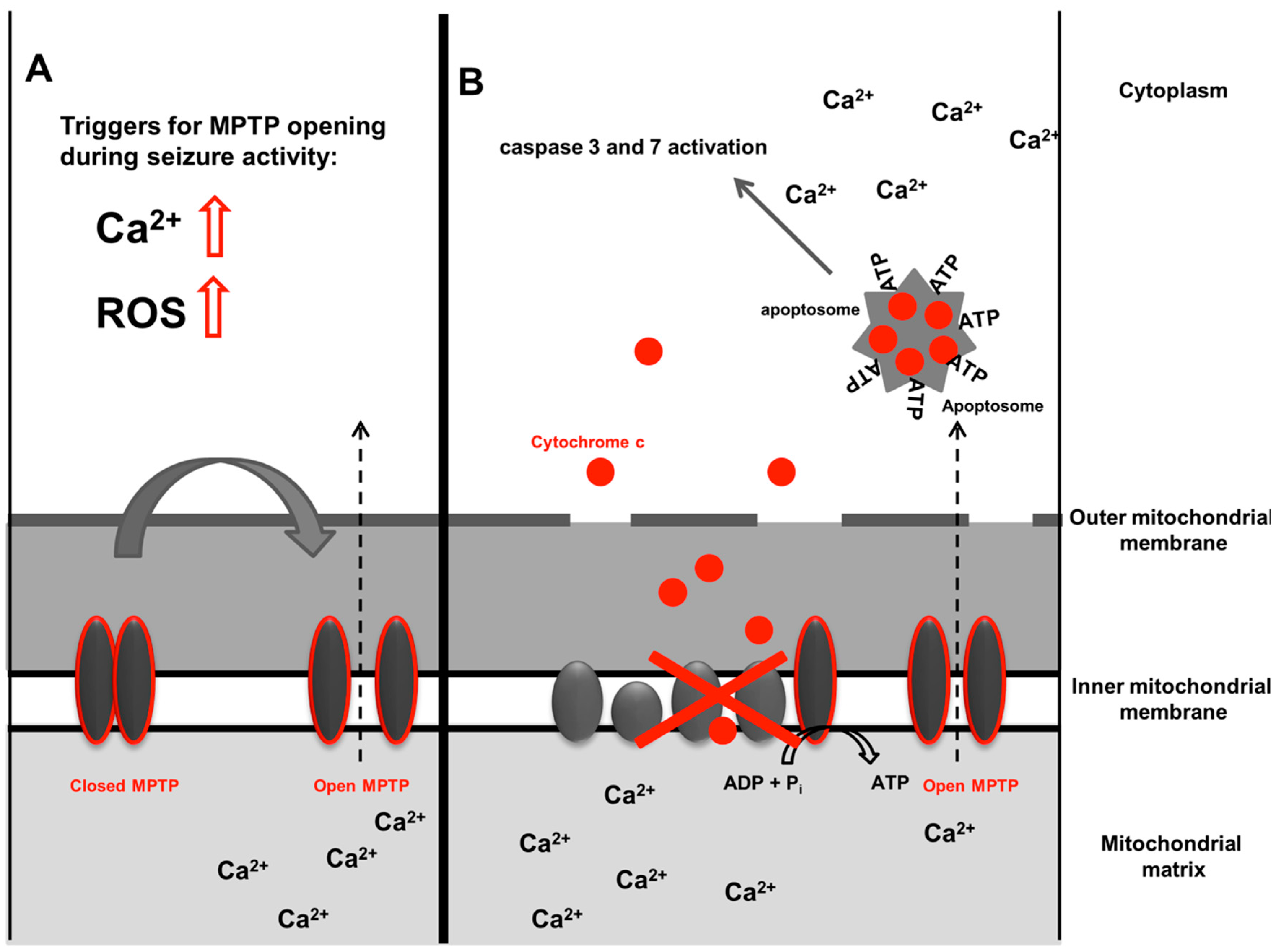
2.3. Cell Death, Mitochondria and Epilepsy

Early pioneering experiments by Meldrum and colleagues in baboons showed that brain seizure activity can cause neuronal damage even in the absence of a systemic convulsion, showing that brain damage is not only a secondary phenomenon which occurs due to excessive muscle activation and subsequent lactate acidosis [48]. That seizure activity itself can cause neuronal injury is now widely accepted and it is appreciated that even non-convulsive status epilepticus can be a life threatening emergency that is often encountered in certain clinical settings such as the intensive care unit [49]. Mitochondria have now been put into the spotlight of seizure induced cell death. Continuous seizure activity leads to ATP depletion and subsequent cell death [12,50]. Excess Ca2+ accumulation results in mitochondrial swelling, permeability transition pore opening, activation of mitochondrial proteins that trigger cell death (Bcl-2 family proteins Bax and Bok proteins [51]), release of cytochrome c, activation of caspases which lead to mitochondrial permeability transition pore opening and loss of mitochondrial outer membrane integrity [16,52,53,54,55,56]. Abnormal mitochondrial distribution, altered mitochondrial motility, decreased mitochondrial membrane potential, and diminished mitochondrial respiration was observed in fibroblasts derived from two lethal encephalopathic patients with a loss of function TRAK1 (trafficking of kinesin proteins 1) variant that was also shown to be associated with seizures [57].

3. Excess Intracellular Ca2+ during Seizure Activity—The Endoplasmic Reticulum

Excess intracellular Ca2+ accumulation, which is one of the leading causes for seizure induced sequelae, is an event that is not only governed by the interplay of Ca2+ entry through the plasma membrane, but also by intracellular Ca2+ stores. An intracellular site of Ca2+ accumulation resides, as mentioned above, within the mitochondria. However, the endoplasmic reticulum (ER) has a larger capability to store intracellular Ca2+ and thus is pivotal in Ca2+ homeostasis.
The ER, which was first identified as the site for intracellular Ca2+ release for muscle contraction [58,59], was soon accepted to regulate Ca2+ homeostasis in cells other than muscle cells [60]. Inositol triphosphate (IP3) mediated mechanisms of Ca2+ release have been shown to play a role in many neurological diseases [61].
With regards to epilepsy, surprisingly very few studies have examined the role of ER Ca2+ stores. Preincubation of neuronal hippocampal cultures with thapsigargin, a drug that depletes intracellular ER Ca2+ stores, resulted in a decrease in the amount of neuronal excitation produced by bicuculline [62]. Ictal discharges induced by pilocarpine or (RS)-3,5-dihydroxyphenylglycine in hippocampal slices were dependent on internal Ca2+ stores as they were blocked by thapsigargin or dantrolene, which both affect ER-Ca2+ stores [63]. A recent study showed that genetic silencing of nitric oxide-induced activation of the ryanodine receptor, a receptor triggering Ca2+ release from the ER, provides protection against cell death produced by kainate-induced status epilepticus [64]. This study highlights the importance of ER Ca2+ stores in mediating seizure-induced cell death.
In contrast to Ca2+ release from intracellular stores, extracellular Ca2+ entry has been identified as the most important route of Ca2+ entry during seizure activity, which is supported by a plethora of studies [11,12].

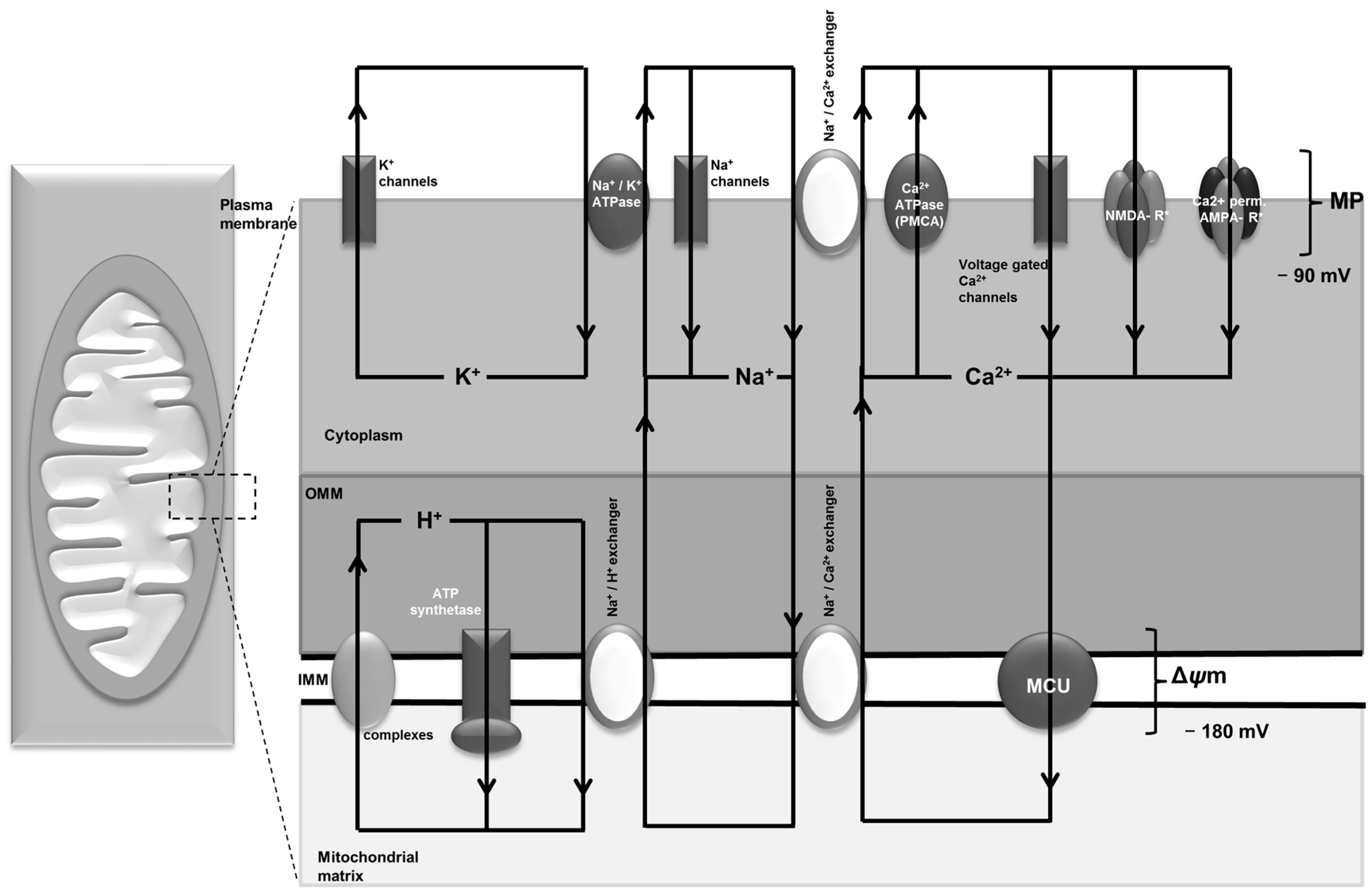
4. Ca2+ Channels and Transporters in the Plasma Cell Membrane and Their Role in Epilepsy—NMDA Receptor, AMPA Receptor, VGCC and PMCA

The NMDA receptor (NMDA-R) forms a heterotetramer comprising two GluN1 and two GluN2 subunits and is mainly permeable by Na+ and Ca2+ ions. Depolarization of the cell opens the channel by dislodging Mg2+ and Zn2+ ions from the pore [65]. The NMDA receptor has been shown to be instrumental in seizure-induced neuronal death both in vitro [11,12] and in vivo [66,67]. The importance of NMDA receptors in epilepsy is underpinned by the fact that NMDA receptor hyperactivation and upregulation of NMDA regulatory subunits is found in focal cortical dysplasia, a highly epileptogenic lesion [68,69]. Ca2+ ions entering the cell upon NMDA receptor stimulation have been shown to be responsible for subsequent cell death promoted by NMDA receptor activation, since omission of Ca2+ ions from the extracellular solution mitigated the harmful NMDA receptor mediated effects [11].
However, clinical evidence points to a more complicated role of NMDA receptors in epilepsy. Antibodies against the extracellular N-terminal domain of the GluN1 subunit have been identified to be responsible for NMDA receptor encephalitis [70,71], an antibody mediated disease which commonly presents with seizures. It remains an unresolved paradox though, why reduction of surface NMDA receptors as seen in anti-NMDA-R encephalitis leads to seizures. In this context, it is interesting that, despite a dramatic clinical presentation of anti-NMDA-receptor encephalitis, including status epilepticus, only a paucity of inflammatory markers and neuronal cell death have been observed, which has repeatedly been confirmed in pathological brain specimens of affected patients [72,73,74].
Interestingly, mutations in subunits of the NMDA receptor, e.g., GRIN1 and GRIN2B, have recently been identified as a cause of epileptic encephalopathy presenting with seizures [75,76]. In addition, the NR1neo/neo mouse model of NMDA receptor hypofunction showed a dramatic sensitivity to kainate induced seizures [77], highlighting the proconvulsant effect of NMDA receptor dysfunction. It has been suggested that NMDA hypofunction may play a role in neurotoxicity during seizure events [78].
Besides NMDA receptors, Ca2+ permeable AMPA receptors have been shown to play a role in status epilepticus [79]. How substantial their contribution is to Ca2+ mediated injury in seizures remains to be determined.
A review of the role of voltage gated Ca2+ channels (VGCC) channels is beyond the scope of this review. It should just be mentioned though that T-type and P/Q-type channels contribute to epileptogenesis, modulation of network activity, and genetic seizure susceptibility [80]. These channels have been linked to genetic forms of epilepsy and idiopathic epilepsy such as childhood absence epilepsy. Rodent genetic models of absence epilepsy have revealed that CaV3.1 and CaV3.2 T-type channel isoforms of VGCC are essential in the pathogenesis of absence epilepsy [81]. In addition, subsequently these channels have also been shown to play a role in acquired epilepsy [82,83].
While NMDA receptors, Ca2+ permeable AMPA receptors and voltage gated Ca2+ channels mediate the influx of Ca2+ into the cell, the plasma membrane ATPase (PMCA) together with the sodium Ca2+ exchanger (NCX) removes Ca2+ from the cell against its concentration gradient (Figure 1). A decrease of Ca2+-ATPase activities was found in pentylentetrazole treated rats [84]. The expression of the Ca2+ extrusion proteins (PMCA and NCX) has been studied in a rat model of kainate-induced status epilepticus [85]. This study found differences of PMCA and NCX isoform expression in neurons and astrocytes in hippocampal formation during epileptogenesis. Decreased PMCA expression levels have also been observed after status epilepticus [86]. The net effect on Ca2+ homeostasis in these regions during epileptogenesis, however, was not assessed in these studies.

5. Epilepsy and Reactive Oxygen Species

ROS contribute to neuronal damage in a wide range of neurological diseases [87,88,89], including seizures and epilepsy [90,91]. ROS include oxygen radicals such as superoxide, hydroxyl radicals, and hydrogen peroxide (H2O2) molecules that are by-products of many biological reactions [92]. ROS in excess cause cellular damage due to oxidation induced protein dysfunction and oxidation of DNA and lipids. In an attempt to repair the cell’s DNA, repair enzymes such as the poly(ADP-ribose) polymerase (PARP) excessively consumes ATP and thus stimulates cell death cascades through ATP depletion [93]. It is interesting in this context that PARP activation has recently been shown to contribute to status epilepticus induced mitochondrial function [94]. Whether ROS or sources of ROS are active in the upstream mechanisms of PARP activation has not been determined. ROS are also powerful enhancers of mitochondrial permeability transition pore opening. By stimulation of IP3/ryanodine receptors, sarco/endoplasmic reticulum Ca2+-ATPase pump inhibition and inhibition of plasma membrane Ca2+ channels, ROS increase intracellular Ca2+ levels, which together with ROS, then trigger mitochondrial permeability transition [46] (Figure 2). ROS can also directly interact with membrane lipids triggering lipid peroxidation. This process affects polyunsaturated lipids and increases the instability of the cell membrane [95]. The brain with its high content of polyunsaturated fatty acids is particularly prone to such damage [89].
There is overwhelming evidence supporting a role for ROS in epilepsy [96]. Early studies have investigated brain homogenates [97], whereas with advances in ROS imaging techniques, more sophisticated real time experiments could be performed allowing more detailed cellular analyses [13,98]. Despite this prominent role of ROS in cell homeostasis and in the triggering of mitochondrial dysfunction, previous results of antioxidant therapy in neurologic disease have been mixed. This is most likely due to a lack of characterization of the sources of free radical production and insight into mechanisms of antioxidant protection of the compounds used in these trials [99].

5.1. Mitochondria and ROS in Seizures and Epilepsy

Traditionally, mitochondria have been assumed to be the main site of ROS production during seizure activity. Some of this has been initially concluded by the coincidence of mitochondrial membrane depolarization, cellular ROS increases and cellular damage [98]. Complex III has been proposed to be a site of ROS production during seizures, which is based on findings from isolated mitochondria [90]. Using a mitochondria specific ROS probe, we were unable to demonstrate that ROS originate from the mitochondria during seizure like activity at least in the first few minutes of seizure activity [13]. Thus, the exact contribution of mitochondrial ROS to the overall ROS burden during seizures remains to be determined.
A more recent study analyzed hippocampal and parahippocampal tissue samples from 74 patients with drug-refractory temporal lobe epilepsy and found that neuropathological signs of inflammation in patients suffering from hippocampal sclerosis correlated with mitochondrial DNA (mtDNA) mutations [100]. This finding supports the hypothesis that chronic inflammation leads to mitochondrial dysfunction by ROS-mediated mtDNA mutagenesis, which promotes epileptogenesis and neuronal cell loss in patients suffering from mesial temporal lobe epilepsy due to hippocampal sclerosis. This study is interesting since it draws attention to inflammatory ROS. In this model, mitochondria are the targets of ROS induced mutagenesis, with mitochondrial dysfunction occurring as a secondary effect.

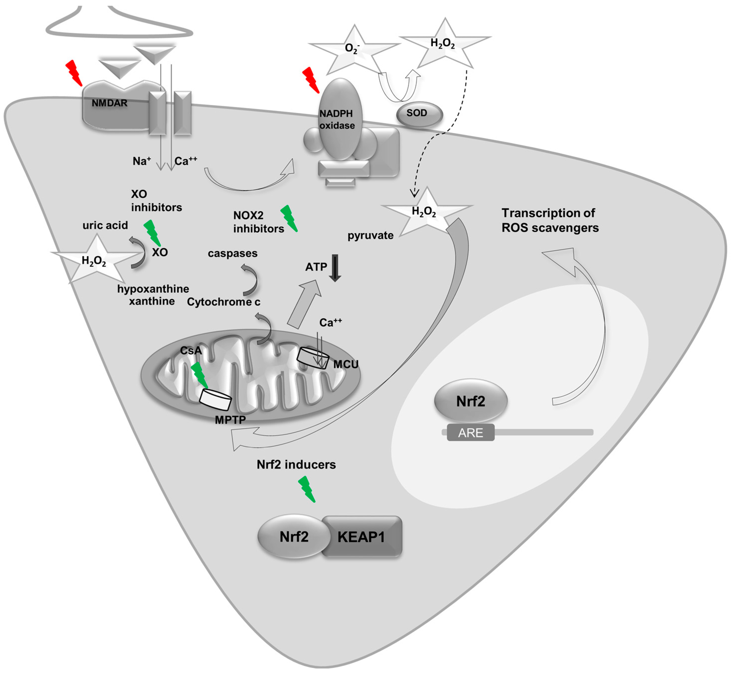
5.2. NADPH Oxidase Derived ROS and Epilepsy

NADPH oxidase was discovered by studying the respiratory burst in phagocytes and granulocytes [101]. Subsequently, NADPH oxidase expression has been documented in many tissues. There is accumulating evidence highlighting the importance of NADPH oxidase, particularly the isoform NOX2 and NOX4, in brain disease [13,102]. In mammals, seven NADPH isoforms known as NOX1, NOX2, NOX3, NOX4, NOX5, dual oxidase (DUOX1) and DUOX2 have been documented [103,104]. NOX1, NOX2, NOX3 and NOX4 expression has been shown in neurons. NOX4 was demonstrated to play a major role in astrocytes, although NOX2 and NOX1 expression has also been documented [105]. Phagocytes were the cell type that fuelled the discovery of the NADPH oxidase [106]. Thus, besides neurons and astrocytes, microglia, the resident phagocytes of the CNS, unsurprisingly represent a prominent cell type that shows NADPH oxidase activity upon activation [107]. Oligodendrocytes are the only neural cells that do not express NADPH oxidase [104]. Amongst non-neural cells in brain tissue, endothelial cells or pericytes show high expression of NOX4 [108]. In fact, endothelial cells have been shown to be the main site of NOX4 expression [109].
NOX2 has been highlighted to play a role in seizures and epilepsy. This is not surprising since NMDA receptor activation, which has a leading role in epilepsy, has been found to trigger NOX2 assembly and activity [110,111]. We and others have found significant activation of NOX2 during seizure activity and suppression of this enzyme was effective in reducing seizure induced cell death in various epilepsy models [13,112,113,114]. Moreover, NOX2 seems to be involved in status epilepticus induced hypotension since NOX2 was found to be upregulated in the rostral ventrolateral medulla, a key nucleus of the baroreflex loop, which mediated status epilepticus-induced hypotension [115]. In addition, NADPH oxidase was also found to be involved in the vasogenic edema formation during status epilepticus [116]. Analyses of human tissue showed that NOX2 was upregulated in surgical hippocampal specimens from a patient suffering from pharmacoresistant seizures, highlighting that NADPH oxidase plays a role in acquired epilepsy [117]. Table 1 summarizes studies that have looked at the role of NADPH oxidase in epilepsy. It should be noted that some of the studies on the involvement of NADPH oxidase in epilepsy used non-subtype specific NADPH oxidase inhibitors such as apocynin and AEBSF (4-(2-aminoethyl)benzenesulfonyl fluoride hydrochloride) and thus, no conclusion on the isoforms of NADPH oxidase activation can be drawn [14,118]. In addition, there is a lack of studies on knock out animals, which would definitely add to the current evidence.
With regards to the other NADPH oxidase isoforms, besides NOX2, NOX4 has been highlighted as a major source of ROS in acute brain diseases such as stroke [102]. NOX4 is mainly located within intracellular organelles and produces superoxide that is rapidly dismutated into H2O2 [103]. We have previously shown that NOX2 produces ROS during seizure activity [13], yet NOX2 is particularly active in the early stages of seizure activity. It remains unclear whether other sources of ROS production are involved particularly in later stages of seizure activity, i.e., beyond the first few minutes. We have previously shown that energy depletion and impaired mitochondrial function occurs particularly in prolonged seizure activity [12]. In this context, NOX4-derived ROS are particularly interesting because of the interaction of NOX4 with mitochondrial function and its expression within mitochondrial membranes [119,120].

5.3. Other Sources of ROS in Epilepsy

Other sources of ROS have been described in seizures and epilepsy, including xanthine oxidase, cyclooxygenase and lipoxygenase [13,96,121,122]. Xanthine oxidase, an enzyme involved in the catabolism of purines, may be an important source of ROS production during prolonged seizures since breakdown of ATP enhances xanthine oxidase activity. The exact contribution of these sources to ROS production during seizures and epilepsy remains unknown, but it is likely that they are not the main ROS producers in these conditions.
One strategy to decrease the ROS burden during seizure activity is to reduce ROS production by blocking key enzymes; another strategy is to boost ROS scavengers. With regards to the latter, the Nrf2 pathway represents an ideal target.

6. Key Regulators of Energy Metabolism and ROS—A Focus on Nrf2 in Seizures and Epilepsy

Nrf2 is a transcription factor that has been shown to regulate both antioxidant defense and intermediary metabolism and thus combines some of the mechanisms outlined above [123,124,125,126]. One of its main negative cytoplasmic regulators is Kelch-like ECH associated protein 1 (KEAP1). KEAP1 is responsible for ubiquitination and proteosomal degradation of Nrf2 and thus tightly controls its activity [127,128,129]. Nrf2 is a potential drug target and there are powerful small molecule inducers that activate Nrf2 by chemically modifying cysteine sensors of KEAP1 or disrupt KEAP1 binding [130,131,132,133]. Some of these small molecule Nrf2 inducers, such as RTA408, are currently being investigated in clinical trials for the treatment of mitochondrial myopathy and Friedreich’s ataxia (Avaliable online: https://clinicaltrials.gov). Moreover, dimethyl fumarate, a drug licensed for use in multiple sclerosis, is an Nrf2 inducer [134], and thus this drug would lend itself to repurposing. Binding of these small molecules to KEAP1 leads to Nrf2 stabilization and translocation to the nucleus where it binds as a heterodimer with a small Maf transcription factor to the antioxidant response element, a specific DNA sequence in the promoter of Nrf2 target genes. This then stimulates transcription of antioxidant proteins such as glutathione-S-transferases (GSTs), NAD(P)H:quinone oxidoreductase 1 (NQO1) as well as enzymes involved in glutathione biosynthesis and regeneration [135,136]. Interestingly Nrf2 also controls mitochondrial function by enhancing substrate availability [124]. In addition, it has been reported that treatment of rats with the pharmacological Nrf2 activator sulforaphane promotes resistance of liver mitochondria to redox-regulated MPTP opening [137]. More recently, we have also highlighted an important interaction between NADPH oxidase expression and the Nrf2 pathway where different expression patterns of NOX4 were seen in neuronal cultures with constitutively active Nrf2 in comparison to Nrf2-knockout cells and controls [138]. All these mechanisms of action render Nrf2 an attractive target to treat seizures.
Nrf2 has been highlighted as a target in the treatment of seizures and epilepsy [96,139]. One of the first descriptions of a protective effect of Nrf2 on seizures and epilepsy came from Mazzuferi and colleagues [140]. They screened biosets from epilepsy-related studies and identified Nrf2 as an important transcription factor in epilepsy. They then showed that Nrf2 mRNA expression is increased in human epileptic tissue and in murine brain tissue after status epilepticus. Adeno-associated virus mediated-overexpression of Nrf2 reduced the frequency and duration of seizures induced by pilocarpine in mice. Several studies with small molecule enhancers, i.e., Nrf2 inducers, have been performed. Daily injections of sulforaphane for five days elevated the seizure thresholds to 6 Hz stimulation and fluorothyl-induced seizures. In addition, it protected mice against pilocarpine-induced status epilepticus, demonstrating its efficacy in various epilepsy models [141]. Sulforaphane also suppressed the progression of amygdala kindling, and also ameliorated the cognitive impairment induced by epileptic seizure [142]. An interesting approach was chosen in a recent study by Pauletti and colleagues [143] where they combined the Nrf2 inducer sulforaphane with N-acetylcysteine treatment with the rationale that these mechanisms are complementary in increasing glutathione levels, as glutathione is one of the main intracellular antioxidants and thus one of the most potent ROS scavengers within the brain. Sulforaphane at high doses (>100 mg/kg) was shown to lead to sedation, hypothermia, impairment of motor coordination, decrease in skeletal muscle strength, and deaths in addition to offsite effects such as leucopenia [144]. It should be noted, however, that such high concentrations are not necessary for Nrf2 activation and were not administered in the studies that showed a protective effect of sulforaphane. These studies largely used a dose of 5 mg/kg. In addition, efforts are underway to develop highly potent blood–brain barrier permeable Nrf2 inducers and some of the currently available inducers as omaveloxolone (RTA408) have been developed in an attempt to increase blood–brain barrier permeability [145].
Figure 3 summarizes metabolic and homeostatic changes during seizures and epilepsy and the pathways that can be targeted to ameliorate these changes.

7. Conclusions and Unmet Research Needs

We have highlighted some exciting activity in the field of metabolic and homeostatic changes during seizure activity and in epilepsy. These studies show that mitochondria are both the source and target of metabolic and homeostatic dysfunction during seizures and epilepsy. Ca2+ excess is another key candidate for these changes, but the involvement of intracellular calcium stores and particularly the ER in Ca2+ excess during seizures and epilepsy remains understudied. Moreover, mitochondrial and ER-Ca2+ stores are intimately linked with each other. How this interconnection is in seizures and epilepsy remains an open question. It is known that Ca2+ together with ROS induce cell death during seizure activity. We have outlined some ideas about the sources of ROS involved in this process. However, the precise sources of ROS involved remain a matter of debate. It is likely that different sources are active at different time points during seizures and epilepsy, such as has been shown in other diseases, e.g., stroke [146]. With regards to this, we think that investigations into the role of the NADPH oxidase with a focus on the different subtypes is pressing, and such investigations are ideally performed in knock out animals, since current evidence on the role of NADPH oxidase in seizures and epilepsy relies on pharmacological manipulation. This is difficult since NADPH oxidase inhibitors are rarely isoform selective and often not even NADPH oxidase specific [147]. As outlined above, NADPH oxidase isoform expression varies between different brain resident cells, thus another pertinent question is to what extent different cell types contribute to ROS formation during seizure activity. Finally, the Nrf2 pathway has been highlighted as an important pathway that is at the interface of redox and intermediary metabolism within the cell. Nrf2 activation boosts ROS scavengers, but was more recently found to have an effect on ROS producing enzymes such as the NADPH oxidase. Further characterization of this interaction will help to design ideal drug targets and allow for combinations of approaches such as have been recently advocated [143] to combat seizure induced ROS. These are one of the main events leading to cell death and continuous seizures during seizure activity and thus contribute to epilepsy and epilepsy comorbidities.

Acknowledgments

This work is supported by the Innovative Medical Research Fund (IMF, University of Münster; KO111715); the medical Faculty of the University of Münster (17-003 fellowship to SK); Cancer Research UK (C20953/A18644); the BBSRC (BB/L01923X/1); the German Research Foundation (DFG; ME 3283/5-1; GO 2505/1-1); the Iran National Science Foundation (INSF) and the German Academic Exchange Service (DAAD; 57348208). In such a short review, which is meant to highlight certain aspects in the field to outline unmet research needs, it is not possible to describe all the activity in this field and so we also apologize to those whose work is not cited here.

Author Contributions

Stjepana Kovac conceptualized and wrote the first draft for the manuscript. Ali Gorji discussed and extensively revised the manuscript. Albena T. Dinkova Kostova revised the whole manuscript with a particular focus on the section on Nrf2 and epilepsy. Alexander M. Herrmann, Nico Melzer and Sven G. Meuth all revised and contributed to the manuscript. All authors approved the final version of the manuscript.

Conflicts of Interest

The authors declare no conflict of interest.

References

  1. Brodie, M.J.; Shorvon, S.D.; Canger, R.; Halász, P.; Johannessen, S.; Thompson, P.; Wieser, H.G.; Wolf, P. Commission on European Affairs: Appropriate standards of epilepsy care across Europe. Epilepsia 1997, 38, 1245–1250. [Google Scholar] [CrossRef] [PubMed]
  2. Pitkänen, A.; Lukasiuk, K. Mechanisms of epileptogenesis and potential treatment targets. Lancet Neurol. 2011, 10, 173–186. [Google Scholar] [CrossRef]
  3. Duncan, J.S.; Sander, J.W.; Sisodiya, S.M.; Walker, M.C. Adult epilepsy. Lancet 2006, 367, 1087–1100. [Google Scholar] [CrossRef]
  4. Schapira, A.H.V. Mitochondrial diseases. Lancet 2012, 379, 1825–1834. [Google Scholar] [CrossRef]
  5. Attwell, D.; Laughlin, S.B. An energy budget for signaling in the grey matter of the brain. J. Cereb. Blood Flow Metab. 2001, 21, 1133–1145. [Google Scholar] [CrossRef] [PubMed]
  6. Liotta, A.; Rösner, J.; Huchzermeyer, C.; Wojtowicz, A.; Kann, O.; Schmitz, D.; Heinemann, U.; Kovács, R. Energy demand of synaptic transmission at the hippocampal Schaffer-collateral synapse. J. Cereb. Blood Flow Metab. 2012, 32, 2076–2083. [Google Scholar] [CrossRef] [PubMed]
  7. Rangaraju, V.; Calloway, N.; Ryan, T.A. Activity-driven local ATP synthesis is required for synaptic function. Cell 2014, 156, 825–835. [Google Scholar] [CrossRef] [PubMed]
  8. Lux, H.D.; Heinemann, U.; Dietzel, I. Ionic changes and alterations in the size of the extracellular space during epileptic activity. Adv. Neurol. 1986, 44, 619–639. [Google Scholar] [PubMed]
  9. Griffiths, T.; Evans, M.C.; Meldrum, B.S. Intracellular calcium accumulation in rat hippocampus during seizures induced by bicuculline or l-allylglycine. Neuroscience 1983, 10, 385–395. [Google Scholar] [CrossRef]
  10. Gilbert, M.E. The NMDA-receptor antagonist, MK-801, suppresses limbic kindling and kindled seizures. Brain Res. 1988, 463, 90–99. [Google Scholar] [CrossRef]
  11. Deshpande, L.S.; Lou, J.K.; Mian, A.; Blair, R.E.; Sombati, S.; Attkisson, E.; DeLorenzo, R.J. Time course and mechanism of hippocampal neuronal death in an in vitro model of status epilepticus: Role of NMDA receptor activation and NMDA dependent calcium entry. Eur. J. Pharmacol. 2008, 583, 73–83. [Google Scholar] [CrossRef] [PubMed]
  12. Kovac, S.; Domijan, A.-M.; Walker, M.C.; Abramov, A.Y. Prolonged seizure activity impairs mitochondrial bioenergetics and induces cell death. J. Cell Sci. 2012, 125, 1796–1806. [Google Scholar] [CrossRef] [PubMed]
  13. Kovac, S.; Domijan, A.-M.; Walker, M.C.; Abramov, A.Y. Seizure activity results in calcium- and mitochondria-independent ROS production via NADPH and xanthine oxidase activation. Cell Death Dis. 2014, 5, e1442. [Google Scholar] [CrossRef] [PubMed]
  14. Zhu, X.; Shen, K.; Bai, Y.; Zhang, A.; Xia, Z.; Chao, J.; Yao, H. NADPH oxidase activation is required for pentylenetetrazole kindling-induced hippocampal autophagy. Free Radic. Biol. Med. 2016, 94, 230–242. [Google Scholar] [CrossRef] [PubMed]
  15. Bernardi, P. Modulation of the mitochondrial cyclosporin A-sensitive permeability transition pore by the proton electrochemical gradient. Evidence that the pore can be opened by membrane depolarization. J. Biol. Chem. 1992, 267, 8834–8839. [Google Scholar] [PubMed]
  16. Bernardi, P.; Petronilli, V.; Di Lisa, F.; Forte, M. A mitochondrial perspective on cell death. Trends Biochem. Sci. 2001, 26, 112–117. [Google Scholar] [CrossRef]
  17. Bernardi, P.; Krauskopf, A.; Basso, E.; Petronilli, V.; Blachly-Dyson, E.; Blalchy-Dyson, E.; Di Lisa, F.; Forte, M.A. The mitochondrial permeability transition from in vitro artifact to disease target. FEBS J. 2006, 273, 2077–2099. [Google Scholar] [CrossRef] [PubMed]
  18. Zsurka, G.; Kunz, W.S. Mitochondrial dysfunction and seizures: The neuronal energy crisis. Lancet Neurol. 2015, 14, 956–966. [Google Scholar] [CrossRef]
  19. Sander, J.W. Some aspects of prognosis in the epilepsies: A review. Epilepsia 1993, 34, 1007–1016. [Google Scholar] [CrossRef] [PubMed]
  20. Altmann, R. Die Elementarorganismen Und Ihre Beziehungen Zu Den Zellen; Veit & Comp.: Leipzig, Germany, 1890. [Google Scholar]
  21. Chance, B. Spectra and reaction kinetics of respiratory pigments of homogenized and intact cells. Nature 1952, 169, 215–221. [Google Scholar] [CrossRef] [PubMed]
  22. Chance, B.; Williams, G.R. Respiratory enzymes in oxidative phosphorylation. IV. The respiratory chain. J. Biol. Chem. 1955, 217, 429–438. [Google Scholar] [PubMed]
  23. Chance, B.; Williams, G.R. Respiratory enzymes in oxidative phosphorylation. II. Difference spectra. J. Biol. Chem. 1955, 217, 395–407. [Google Scholar] [PubMed]
  24. Ernster, L.; Schatz, G. Mitochondria: A historical review. J. Cell Biol. 1981, 91, 227s–255s. [Google Scholar] [CrossRef] [PubMed]
  25. Mitchell, P. Coupling of phosphorylation to electron and hydrogen transfer by a chemi-osmotic type of mechanism. Nature 1961, 191, 144–148. [Google Scholar] [CrossRef] [PubMed]
  26. Almeida, A.; Almeida, J.; Bolaños, J.P.; Moncada, S. Different responses of astrocytes and neurons to nitric oxide: The role of glycolytically generated ATP in astrocyte protection. Proc. Natl. Acad. Sci. USA 2001, 98, 15294–15299. [Google Scholar] [CrossRef] [PubMed]
  27. Szabadkai, G.; Duchen, M.R. Mitochondria: The hub of cellular Ca2+ signaling. Physiology 2008, 23, 84–94. [Google Scholar] [CrossRef] [PubMed]
  28. Ugarte-Uribe, B.; García-Sáez, A.J. Apoptotic foci at mitochondria: In and around Bax pores. Philos. Trans. R. Soc. Lond. Ser. B Biol. Sci. 2017, 372, 20160217. [Google Scholar] [CrossRef] [PubMed]
  29. King, L.J.; Lowry, O.H.; Passonneau, J.V.; Venson, V. Effects of convulsants on energy reserves in the cerebral cortex. J. Neurochem. 1967, 14, 599–611. [Google Scholar] [CrossRef] [PubMed]
  30. Sacktor, B.; Wilson, J.E.; Tiekert, C.G. Regulation of glycolysis in brain, in situ, during convulsions. J. Biol. Chem. 1966, 241, 5071–5075. [Google Scholar] [PubMed]
  31. Sanders, A.P.; Kramer, R.S.; Woodhall, B.; Currie, W.D. Brain adenosine triphosphate: Decreased concentration precedes convulsions. Science 1970, 169, 206–208. [Google Scholar] [CrossRef] [PubMed]
  32. Kovac, S.; Abramov, A.Y.; Walker, M.C. Energy depletion in seizures: Anaplerosis as a strategy for future therapies. Neuropharmacology 2013, 69, 96–104. [Google Scholar] [CrossRef] [PubMed]
  33. Bough, K.J.; Wetherington, J.; Hassel, B.; Pare, J.F.; Gawryluk, J.W.; Greene, J.G.; Shaw, R.; Smith, Y.; Geiger, J.D.; Dingledine, R.J. Mitochondrial biogenesis in the anticonvulsant mechanism of the ketogenic diet. Ann. Neurol. 2006, 60, 223–235. [Google Scholar] [CrossRef] [PubMed]
  34. Bough, K. Energy metabolism as part of the anticonvulsant mechanism of the ketogenic diet. Epilepsia 2008, 49 (Suppl. S8), 91–93. [Google Scholar] [CrossRef] [PubMed]
  35. Greco, T.; Glenn, T.C.; Hovda, D.A.; Prins, M.L. Ketogenic diet decreases oxidative stress and improves mitochondrial respiratory complex activity. J. Cereb. Blood Flow Metab. 2016, 36, 1603–1613. [Google Scholar] [CrossRef] [PubMed]
  36. Rho, J.M. How does the ketogenic diet induce anti-seizure effects? Neurosci. Lett. 2017, 637, 4–10. [Google Scholar] [CrossRef] [PubMed]
  37. Pasca, L.; De Giorgis, V.; Macasaet, J.A.; Trentani, C.; Tagliabue, A.; Veggiotti, P. The changing face of dietary therapy for epilepsy. Eur. J. Pediatr. 2016, 175, 1267–1276. [Google Scholar] [CrossRef] [PubMed]
  38. Dressler, A.; Trimmel-Schwahofer, P.; Reithofer, E.; Mühlebner, A.; Gröppel, G.; Reiter-Fink, E.; Benninger, F.; Grassl, R.; Feucht, M. Efficacy and tolerability of the ketogenic diet in Dravet syndrome—Comparison with various standard antiepileptic drug regimen. Epilepsy Res. 2015, 109, 81–89. [Google Scholar] [CrossRef] [PubMed]
  39. Paleologou, E.; Ismayilova, N.; Kinali, M. Use of the Ketogenic Diet to Treat Intractable Epilepsy in Mitochondrial Disorders. J. Clin. Med. 2017, 6, E56. [Google Scholar] [CrossRef] [PubMed]
  40. Griffiths, T.; Evans, M.C.; Meldrum, B.S. Intracellular sites of early calcium accumulation in the rat hippocampus during status epilepticus. Neurosci. Lett. 1982, 30, 329–334. [Google Scholar] [CrossRef]
  41. De Stefani, D.; Raffaello, A.; Teardo, E.; Szabò, I.; Rizzuto, R. A forty-kilodalton protein of the inner membrane is the mitochondrial calcium uniporter. Nature 2011, 476, 336–340. [Google Scholar] [CrossRef] [PubMed]
  42. Nicholls, D.G.; Chalmers, S. The integration of mitochondrial calcium transport and storage. J. Bioenerg. Biomembr. 2004, 36, 277–281. [Google Scholar] [CrossRef] [PubMed]
  43. Wang, C.; Xie, N.; Wang, Y.; Li, Y.; Ge, X.; Wang, M. Role of the Mitochondrial Calcium Uniporter in Rat Hippocampal Neuronal Death after Pilocarpine-Induced Status Epilepticus. Neurochem. Res. 2015, 40, 1739–1746. [Google Scholar] [CrossRef] [PubMed]
  44. Nicholls, D.G.; Budd, S.L. Mitochondria and neuronal survival. Physiol. Rev. 2000, 80, 315–360. [Google Scholar] [PubMed]
  45. Giorgio, V.; von Stockum, S.; Antoniel, M.; Fabbro, A.; Fogolari, F.; Forte, M.; Glick, G.D.; Petronilli, V.; Zoratti, M.; Szabó, I.; et al. Dimers of mitochondrial ATP synthase form the permeability transition pore. Proc. Natl. Acad. Sci. USA 2013, 110, 5887–5892. [Google Scholar] [CrossRef] [PubMed] [Green Version]
  46. Bernardi, P.; Rasola, A.; Forte, M.; Lippe, G. The Mitochondrial Permeability Transition Pore: Channel Formation by F-ATP Synthase, Integration in Signal Transduction, and Role in Pathophysiology. Physiol. Rev. 2015, 95, 1111–1155. [Google Scholar] [CrossRef] [PubMed]
  47. Kim, D.Y.; Simeone, K.A.; Simeone, T.A.; Pandya, J.D.; Wilke, J.C.; Ahn, Y.; Geddes, J.W.; Sullivan, P.G.; Rho, J.M. Ketone bodies mediate antiseizure effects through mitochondrial permeability transition. Ann. Neurol. 2015, 78, 77–87. [Google Scholar] [CrossRef] [PubMed]
  48. Meldrum, B.S.; Vigouroux, R.A.; Brierley, J.B. Systemic factors and epileptic brain damage. Prolonged seizures in paralyzed, artificially ventilated baboons. Arch. Neurol. 1973, 29, 82–87. [Google Scholar] [CrossRef] [PubMed]
  49. Schmutzhard, E.; Pfausler, B. Complications of the management of status epilepticus in the intensive care unit. Epilepsia 2011, 52 (Suppl. S8), 39–41. [Google Scholar] [CrossRef] [PubMed]
  50. Norwood, B.A.; Bauer, S.; Wegner, S.; Hamer, H.M.; Oertel, W.H.; Sloviter, R.S.; Rosenow, F. Electrical stimulation-induced seizures in rats: A “dose-response” study on resultant neurodegeneration. Epilepsia 2011, 52, e109–e112. [Google Scholar] [CrossRef] [PubMed]
  51. D’Orsi, B.; Mateyka, J.; Prehn, J.H.M. Control of mitochondrial physiology and cell death by the Bcl-2 family proteins Bax and Bok. Neurochem. Int. 2017, in press. [Google Scholar]
  52. Nobili, P.; Colciaghi, F.; Finardi, A.; Zambon, S.; Locatelli, D.; Battaglia, G.S. Continuous neurodegeneration and death pathway activation in neurons and glia in an experimental model of severe chronic epilepsy. Neurobiol. Dis. 2015, 83, 54–66. [Google Scholar] [CrossRef] [PubMed]
  53. Shalini, S.; Dorstyn, L.; Dawar, S.; Kumar, S. Old, new and emerging functions of caspases. Cell Death Differ. 2015, 22, 526–539. [Google Scholar] [CrossRef] [PubMed]
  54. Henshall, D.C.; Murphy, B.M. Modulators of neuronal cell death in epilepsy. Curr. Opin. Pharmacol. 2008, 8, 75–81. [Google Scholar] [CrossRef] [PubMed]
  55. Henshall, D.C.; Araki, T.; Schindler, C.K.; Lan, J.-Q.; Tiekoter, K.L.; Taki, W.; Simon, R.P. Activation of Bcl-2-Associated Death Protein and Counter-Response of Akt within Cell Populations during Seizure-Induced Neuronal Death. J. Neurosci. 2002, 22, 8458–8465. [Google Scholar] [PubMed]
  56. Henshall, D.C.; Chen, J.; Simon, R.P. Involvement of caspase-3-like protease in the mechanism of cell death following focally evoked limbic seizures. J. Neurochem. 2000, 74, 1215–1223. [Google Scholar] [CrossRef] [PubMed]
  57. Barel, O.; Christine V Malicdan, M.; Ben-Zeev, B.; Kandel, J.; Pri-Chen, H.; Stephen, J.; Castro, I.G.; Metz, J.; Atawa, O.; Moshkovitz, S.; et al. Deleterious variants in TRAK1 disrupt mitochondrial movement and cause fatal encephalopathy. Brain 2017, 140, 568–581. [Google Scholar] [CrossRef] [PubMed]
  58. Heilbrunn, L.V. An Outline of General Physiology, 3rd ed.; W. B. Saunders: Philadelphia, PA, USA, 1952. [Google Scholar]
  59. Heilbrunn, L.V.; Wiercinski, F.J. The action of various cations on muscle protoplasm. J. Cell. Physiol. 1947, 29, 15–32. [Google Scholar] [CrossRef]
  60. Streb, H.; Irvine, R.F.; Berridge, M.J.; Schulz, I. Release of Ca2+ from a nonmitochondrial intracellular store in pancreatic acinar cells by inositol-1,4,5-trisphosphate. Nature 1983, 306, 67–69. [Google Scholar] [CrossRef] [PubMed]
  61. Verkhratsky, A. Physiology and Pathophysiology of the Calcium Store in the Endoplasmic Reticulum of Neurons. Physiol. Rev. 2005, 85, 201–279. [Google Scholar] [CrossRef] [PubMed]
  62. Sokal, D.M.; Mason, R.; Parker, T.L. Multi-neuronal recordings reveal a differential effect of thapsigargin on bicuculline- or gabazine-induced epileptiform excitability in rat hippocampal neuronal networks. Neuropharmacology 2000, 39, 2408–2417. [Google Scholar] [CrossRef]
  63. Rutecki, P.A.; Sayin, U.; Yang, Y.; Hadar, E. Determinants of ictal epileptiform patterns in the hippocampal slice. Epilepsia 2002, 43 (Suppl. S5), 179–183. [Google Scholar] [CrossRef] [PubMed]
  64. Mikami, Y.; Kanemaru, K.; Okubo, Y.; Nakaune, T.; Suzuki, J.; Shibata, K.; Sugiyama, H.; Koyama, R.; Murayama, T.; Ito, A.; et al. Nitric Oxide-induced Activation of the Type 1 Ryanodine Receptor Is Critical for Epileptic Seizure-induced Neuronal Cell Death. EBioMedicine 2016, 11, 253–261. [Google Scholar] [CrossRef] [PubMed]
  65. Dingledine, R.; Borges, K.; Bowie, D.; Traynelis, S.F. The glutamate receptor ion channels. Pharmacol. Rev. 1999, 51, 7–61. [Google Scholar] [PubMed]
  66. Ormandy, G.C.; Jope, R.S.; Snead, O.C. Anticonvulsant actions of MK-801 on the lithium-pilocarpine model of status epilepticus in rats. Exp. Neurol. 1989, 106, 172–180. [Google Scholar] [CrossRef]
  67. Fujikawa, D.G.; Daniels, A.H.; Kim, J.S. The competitive NMDA receptor antagonist CGP 40116 protects against status epilepticus-induced neuronal damage. Epilepsy Res. 1994, 17, 207–219. [Google Scholar] [CrossRef]
  68. Finardi, A.; Colciaghi, F.; Castana, L.; Locatelli, D.; Marras, C.E.; Nobili, P.; Fratelli, M.; Bramerio, M.A.; Lorusso, G.; Battaglia, G.S. Long-duration epilepsy affects cell morphology and glutamatergic synapses in type IIB focal cortical dysplasia. Acta Neuropathol. 2013, 126, 219–235. [Google Scholar] [CrossRef] [PubMed]
  69. Battaglia, G.; Colciaghi, F.; Finardi, A.; Nobili, P. Intrinsic epileptogenicity of dysplastic cortex: Converging data from experimental models and human patients. Epilepsia 2013, 54 (Suppl. S6), 33–36. [Google Scholar] [CrossRef] [PubMed]
  70. Dalmau, J.; Tüzün, E.; Wu, H.; Masjuan, J.; Rossi, J.E.; Voloschin, A.; Baehring, J.M.; Shimazaki, H.; Koide, R.; King, D.; et al. Paraneoplastic anti-N-methyl-d-aspartate receptor encephalitis associated with ovarian teratoma. Ann. Neurol. 2007, 61, 25–36. [Google Scholar] [CrossRef] [PubMed]
  71. Dalmau, J.; Gleichman, A.J.; Hughes, E.G.; Rossi, J.E.; Peng, X.; Lai, M.; Dessain, S.K.; Rosenfeld, M.R.; Balice-Gordon, R.; Lynch, D.R. Anti-NMDA-receptor encephalitis: Case series and analysis of the effects of antibodies. Lancet Neurol. 2008, 7, 1091–1098. [Google Scholar] [CrossRef]
  72. Bien, C.G.; Vincent, A.; Barnett, M.H.; Becker, A.J.; Blümcke, I.; Graus, F.; Jellinger, K.A.; Reuss, D.E.; Ribalta, T.; Schlegel, J.; et al. Immunopathology of autoantibody-associated encephalitides: Clues for pathogenesis. Brain 2012, 135, 1622–1638. [Google Scholar] [CrossRef] [PubMed]
  73. Camdessanché, J.-P.; Streichenberger, N.; Cavillon, G.; Rogemond, V.; Jousserand, G.; Honnorat, J.; Convers, P.; Antoine, J.-C. Brain immunohistopathological study in a patient with anti-NMDAR encephalitis. Eur. J. Neurol. 2011, 18, 929–931. [Google Scholar] [CrossRef] [PubMed]
  74. Tüzün, E.; Zhou, L.; Baehring, J.M.; Bannykh, S.; Rosenfeld, M.R.; Dalmau, J. Evidence for antibody-mediated pathogenesis in anti-NMDAR encephalitis associated with ovarian teratoma. Acta Neuropathol. 2009, 118, 737–743. [Google Scholar] [CrossRef] [PubMed]
  75. Epi4K Consortium; Epilepsy Phenome/Genome Project; Allen, A.S.; Berkovic, S.F.; Cossette, P.; Delanty, N.; Dlugos, D.; Eichler, E.E.; Epstein, M.P.; Glauser, T.; et al. De novo mutations in epileptic encephalopathies. Nature 2013, 501, 217–221. [Google Scholar] [CrossRef] [PubMed]
  76. Ohba, C.; Shiina, M.; Tohyama, J.; Haginoya, K.; Lerman-Sagie, T.; Okamoto, N.; Blumkin, L.; Lev, D.; Mukaida, S.; Nozaki, F.; et al. GRIN1 mutations cause encephalopathy with infantile-onset epilepsy, and hyperkinetic and stereotyped movement disorders. Epilepsia 2015, 56, 841–848. [Google Scholar] [CrossRef] [PubMed]
  77. Duncan, G.E.; Inada, K.; Koller, B.H.; Moy, S.S. Increased sensitivity to kainic acid in a genetic model of reduced NMDA receptor function. Brain Res. 2010, 1307, 166–176. [Google Scholar] [CrossRef] [PubMed]
  78. Gorji, A.; Speckmann, E.J. Low concentration of dl-2-amino-5-phosphonovalerate induces epileptiform activity in guinea pig hippocampal slices. Epilepsia 2001, 42, 1228–1230. [Google Scholar] [CrossRef] [PubMed]
  79. Rajasekaran, K.; Todorovic, M.; Kapur, J. Calcium-permeable AMPA receptors are expressed in a rodent model of status epilepticus. Ann. Neurol. 2012, 72, 91–102. [Google Scholar] [CrossRef] [PubMed]
  80. Khosravani, H.; Zamponi, G.W. Voltage-Gated Calcium Channels and Idiopathic Generalized Epilepsies. Physiol. Rev. 2006, 86, 941–966. [Google Scholar] [CrossRef] [PubMed]
  81. Cain, S.M.; Snutch, T.P. Voltage-Gated Calcium Channels in Epilepsy. In Jasper’s Basic Mechanisms of the Epilepsies; Noebels, J.L., Avoli, M., Rogawski, M.A., Olsen, R.W., Delgado-Escueta, A.V., Eds.; National Center for Biotechnology Information (US): Bethesda, MD, USA, 2012. [Google Scholar]
  82. Van Loo, K.M.J.; Schaub, C.; Pitsch, J.; Kulbida, R.; Opitz, T.; Ekstein, D.; Dalal, A.; Urbach, H.; Beck, H.; Yaari, Y.; et al. Zinc regulates a key transcriptional pathway for epileptogenesis via metal-regulatory transcription factor 1. Nat. Commun. 2015, 6, 8688. [Google Scholar] [CrossRef] [PubMed]
  83. Becker, A.J.; Pitsch, J.; Sochivko, D.; Opitz, T.; Staniek, M.; Chen, C.-C.; Campbell, K.P.; Schoch, S.; Yaari, Y.; Beck, H. Transcriptional upregulation of Cav3.2 mediates epileptogenesis in the pilocarpine model of epilepsy. J. Neurosci. 2008, 28, 13341–13353. [Google Scholar] [CrossRef] [PubMed]
  84. Naziroğlu, M.; Kutluhan, S.; Yilmaz, M. Selenium and topiramate modulates brain microsomal oxidative stress values, Ca2+-ATPase activity, and EEG records in pentylentetrazol-induced seizures in rats. J. Membr. Biol. 2008, 225, 39–49. [Google Scholar] [CrossRef] [PubMed]
  85. Ketelaars, S.O.M.; Gorter, J.A.; Aronica, E.; Wadman, W.J. Calcium extrusion protein expression in the hippocampal formation of chronic epileptic rats after kainate-induced status epilepticus. Epilepsia 2004, 45, 1189–1201. [Google Scholar] [CrossRef] [PubMed]
  86. Garcia, M.L.; Murray, K.D.; Garcia, V.B.; Strehler, E.E.; Isackson, P.J. Seizure-induced alterations of plasma membrane calcium ATPase isoforms 1, 2 and 3 mRNA and protein in rat hippocampus. Brain Res. Mol. Brain Res. 1997, 45, 230–238. [Google Scholar] [CrossRef]
  87. Abramov, A.Y.; Canevari, L.; Duchen, M.R. Beta-amyloid peptides induce mitochondrial dysfunction and oxidative stress in astrocytes and death of neurons through activation of NADPH oxidase. J. Neurosci. 2004, 24, 565–575. [Google Scholar] [CrossRef] [PubMed]
  88. Gandhi, S.; Wood-Kaczmar, A.; Yao, Z.; Plun-Favreau, H.; Deas, E.; Klupsch, K.; Downward, J.; Latchman, D.S.; Tabrizi, S.J.; Wood, N.W.; et al. PINK1-associated Parkinson’s disease is caused by neuronal vulnerability to calcium-induced cell death. Mol. Cell 2009, 33, 627–638. [Google Scholar] [CrossRef] [PubMed]
  89. Halliwell, B. Oxidative stress and neurodegeneration: Where are we now? J. Neurochem. 2006, 97, 1634–1658. [Google Scholar] [CrossRef] [PubMed]
  90. Malinska, D.; Kulawiak, B.; Kudin, A.P.; Kovacs, R.; Huchzermeyer, C.; Kann, O.; Szewczyk, A.; Kunz, W.S. Complex III-dependent superoxide production of brain mitochondria contributes to seizure-related ROS formation. Biochim. Biophys. Acta 2010, 1797, 1163–1170. [Google Scholar] [CrossRef] [PubMed]
  91. Pestana, R.R.F.; Kinjo, E.R.; Hernandes, M.S.; Britto, L.R.G. Reactive oxygen species generated by NADPH oxidase are involved in neurodegeneration in the pilocarpine model of temporal lobe epilepsy. Neurosci. Lett. 2010, 484, 187–191. [Google Scholar] [CrossRef] [PubMed]
  92. Dröge, W. Free Radicals in the Physiological Control of Cell Function. Physiol. Rev. 2002, 82, 47–95. [Google Scholar] [CrossRef] [PubMed]
  93. Heeres, J.T.; Hergenrother, P.J. Poly(ADP-ribose) makes a date with death. Curr. Opin. Chem. Biol. 2007, 11, 644–653. [Google Scholar] [CrossRef] [PubMed]
  94. Lai, Y.-C.; Baker, J.S.; Donti, T.; Graham, B.H.; Craigen, W.J.; Anderson, A.E. Mitochondrial Dysfunction Mediated by Poly(ADP-Ribose) Polymerase-1 Activation Contributes to Hippocampal Neuronal Damage Following Status Epilepticus. Int. J. Mol. Sci. 2017, 18, E1502. [Google Scholar] [CrossRef] [PubMed]
  95. Gutteridge, J.M.C.; Halliwell, B. The measurement and mechanism of lipid peroxidation in biological systems. Trends Biochem. Sci. 1990, 15, 129–135. [Google Scholar] [CrossRef]
  96. Kovac, S.; Dinkova-Kostova, A.T.; Abramov, A.Y. The Role of Reactive Oxygen Species in Epilepsy. React. Oxyg. Species 2016, 1, 38–52. [Google Scholar] [CrossRef]
  97. Bruce, A.J.; Baudry, M. Oxygen free radicals in rat limbic structures after kainate-induced seizures. Free Radic. Biol. Med. 1995, 18, 993–1002. [Google Scholar] [CrossRef]
  98. Kovács, R.; Schuchmann, S.; Gabriel, S.; Kann, O.; Kardos, J.; Heinemann, U. Free radical-mediated cell damage after experimental status epilepticus in hippocampal slice cultures. J. Neurophysiol. 2002, 88, 2909–2918. [Google Scholar] [CrossRef] [PubMed]
  99. Delanty, N.; Dichter, D.M. Antioxidant therapy in neurologic disease. Arch. Neurol. 2000, 57, 1265–1270. [Google Scholar] [CrossRef] [PubMed]
  100. Volmering, E.; Niehusmann, P.; Peeva, V.; Grote, A.; Zsurka, G.; Altmüller, J.; Nürnberg, P.; Becker, A.J.; Schoch, S.; Elger, C.E.; et al. Neuropathological signs of inflammation correlate with mitochondrial DNA deletions in mesial temporal lobe epilepsy. Acta Neuropathol. 2016, 132, 277–288. [Google Scholar] [CrossRef] [PubMed]
  101. Royer-Pokora, B.; Kunkel, L.M.; Monaco, A.P.; Goff, S.C.; Newburger, P.E.; Baehner, R.L.; Cole, F.S.; Curnutte, J.T.; Orkin, S.H. Cloning the gene for an inherited human disorder—Chronic granulomatous disease—On the basis of its chromosomal location. Nature 1986, 322, 32–38. [Google Scholar] [CrossRef] [PubMed]
  102. Kleinschnitz, C.; Grund, H.; Wingler, K.; Armitage, M.E.; Jones, E.; Mittal, M.; Barit, D.; Schwarz, T.; Geis, C.; Kraft, P.; et al. Post-stroke inhibition of induced NADPH oxidase type 4 prevents oxidative stress and neurodegeneration. PLoS Biol. 2010, 8, e1000479. [Google Scholar] [CrossRef] [PubMed] [Green Version]
  103. Bedard, K.; Krause, K.-H. The NOX family of ROS-generating NADPH oxidases: Physiology and pathophysiology. Physiol. Rev. 2007, 87, 245–313. [Google Scholar] [CrossRef] [PubMed]
  104. Nayernia, Z.; Jaquet, V.; Krause, K.-H. New insights on NOX enzymes in the central nervous system. Antioxid. Redox Signal. 2014, 20, 2815–2837. [Google Scholar] [CrossRef] [PubMed]
  105. Olguín-Albuerne, M.; Domínguez, G.; Morán, J. Effect of staurosporine in the morphology and viability of cerebellar astrocytes: Role of reactive oxygen species and NADPH oxidase. Oxidative Med. Cell. Longev. 2014, 2014, 678371. [Google Scholar] [CrossRef] [PubMed]
  106. Hohn, D.C.; Lehrer, R.I. NADPH oxidase deficiency in X-linked chronic granulomatous disease. J. Clin. Investig. 1975, 55, 707–713. [Google Scholar] [CrossRef] [PubMed]
  107. Fischer, M.T.; Sharma, R.; Lim, J.L.; Haider, L.; Frischer, J.M.; Drexhage, J.; Mahad, D.; Bradl, M.; van Horssen, J.; Lassmann, H. NADPH oxidase expression in active multiple sclerosis lesions in relation to oxidative tissue damage and mitochondrial injury. Brain 2012, 135, 886–899. [Google Scholar] [CrossRef] [PubMed]
  108. Kuroda, J.; Ago, T.; Nishimura, A.; Nakamura, K.; Matsuo, R.; Wakisaka, Y.; Kamouchi, M.; Kitazono, T. Nox4 is a major source of superoxide production in human brain pericytes. J. Vasc. Res. 2014, 51, 429–438. [Google Scholar] [CrossRef] [PubMed]
  109. Sirokmány, G.; Donkó, Á.; Geiszt, M. Nox/Duox Family of NADPH Oxidases: Lessons from Knockout Mouse Models. Trends Pharmacol. Sci. 2016, 37, 318–327. [Google Scholar] [CrossRef] [PubMed] [Green Version]
  110. Brennan, A.M.; Suh, S.W.; Won, S.J.; Narasimhan, P.; Kauppinen, T.M.; Lee, H.; Edling, Y.; Chan, P.H.; Swanson, R.A. NADPH oxidase is the primary source of superoxide induced by NMDA receptor activation. Nat. Neurosci. 2009, 12, 857–863. [Google Scholar] [CrossRef] [PubMed]
  111. Girouard, H.; Wang, G.; Gallo, E.F.; Anrather, J.; Zhou, P.; Pickel, V.M.; Iadecola, C. NMDA receptor activation increases free radical production through nitric oxide and NOX2. J. Neurosci. 2009, 29, 2545–2552. [Google Scholar] [CrossRef] [PubMed]
  112. Kim, J.H.; Jang, B.G.; Choi, B.Y.; Kim, H.S.; Sohn, M.; Chung, T.N.; Choi, H.C.; Song, H.K.; Suh, S.W. Post-treatment of an NADPH oxidase inhibitor prevents seizure-induced neuronal death. Brain Res. 2013, 1499, 163–172. [Google Scholar] [CrossRef] [PubMed]
  113. Patel, M.; Li, Q.-Y.; Chang, L.-Y.; Crapo, J.; Liang, L.-P. Activation of NADPH oxidase and extracellular superoxide production in seizure-induced hippocampal damage. J. Neurochem. 2005, 92, 123–131. [Google Scholar] [CrossRef] [PubMed]
  114. Di Maio, R.; Mastroberardino, P.G.; Hu, X.; Montero, L.; Greenamyre, J.T. Pilocapine alters NMDA receptor expression and function in hippocampal neurons: NADPH oxidase and ERK1/2 mechanisms. Neurobiol. Dis. 2011, 42, 482–495. [Google Scholar] [CrossRef] [PubMed]
  115. Tsai, C.-Y.; Chan, J.Y.H.; Hsu, K.; Chang, A.Y.W.; Chan, S.H.H. Brain-Derived Neurotrophic Factor Ameliorates Brain Stem Cardiovascular Dysregulation during Experimental Temporal Lobe Status Epilepticus. PLoS ONE 2012, 7, e33527. [Google Scholar] [CrossRef] [PubMed]
  116. Kim, J.-E.; Ryu, H.J.; Kang, T.-C. Status epilepticus induces vasogenic edema via tumor necrosis factor-α/ endothelin-1-mediated two different pathways. PLoS ONE 2013, 8, e74458. [Google Scholar] [CrossRef] [PubMed]
  117. Pecorelli, A.; Natrella, F.; Belmonte, G.; Miracco, C.; Cervellati, F.; Ciccoli, L.; Mariottini, A.; Rocchi, R.; Vatti, G.; Bua, A.; et al. NADPH oxidase activation and 4-hydroxy-2-nonenal/aquaporin-4 adducts as possible new players in oxidative neuronal damage presents in drug-resistant epilepsy. Biochim. Biophys. Acta 2015, 1852, 507–519. [Google Scholar] [CrossRef] [PubMed]
  118. Williams, S.; Hamil, N.; Abramov, A.Y.; Walker, M.C.; Kovac, S. Status epilepticus results in persistent overproduction of reactive oxygen species, inhibition of which is neuroprotective. Neuroscience 2015, 303, 160–165. [Google Scholar] [CrossRef] [PubMed]
  119. Case, A.J.; Li, S.; Basu, U.; Tian, J.; Zimmerman, M.C. Mitochondrial-localized NADPH oxidase 4 is a source of superoxide in angiotensin II-stimulated neurons. Am. J. Physiol. Heart Circ. Physiol. 2013, 305, H19–H28. [Google Scholar] [CrossRef] [PubMed]
  120. Kozieł, R.; Pircher, H.; Kratochwil, M.; Lener, B.; Hermann, M.; Dencher, N.A.; Jansen-Dürr, P. Mitochondrial respiratory chain complex I is inactivated by NADPH oxidase Nox4. Biochem. J. 2013, 452, 231–239. [Google Scholar] [CrossRef] [PubMed]
  121. Ueda, Y.; Yokoyama, H.; Niwa, R.; Konaka, R.; Ohya-Nishiguchi, H.; Kamada, H. Generation of lipid radicals in the hippocampal extracellular space during kainic acid-induced seizures in rats. Epilepsy Res. 1997, 26, 329–333. [Google Scholar] [CrossRef]
  122. Baran, H.; Vass, K.; Lassmann, H.; Hornykiewicz, O. The cyclooxygenase and lipoxygenase inhibitor BW755C protects rats against kainic acid-induced seizures and neurotoxicity. Brain Res. 1994, 646, 201–206. [Google Scholar] [CrossRef]
  123. Hayes, J.D.; Dinkova-Kostova, A.T. The Nrf2 regulatory network provides an interface between redox and intermediary metabolism. Trends Biochem. Sci. 2014, 39, 199–218. [Google Scholar] [CrossRef] [PubMed]
  124. Holmström, K.M.; Baird, L.; Zhang, Y.; Hargreaves, I.; Chalasani, A.; Land, J.M.; Stanyer, L.; Yamamoto, M.; Dinkova-Kostova, A.T.; Abramov, A.Y. Nrf2 impacts cellular bioenergetics by controlling substrate availability for mitochondrial respiration. Biol. Open 2013, 2, 761–770. [Google Scholar] [CrossRef] [PubMed]
  125. Holmström, K.M.; Kostov, R.V.; Dinkova-Kostova, A.T. The multifaceted role of Nrf2 in mitochondrial function. Curr. Opin. Toxicol. 2016, 1, 80–91. [Google Scholar] [CrossRef] [PubMed]
  126. Dinkova-Kostova, A.T.; Baird, L.; Holmström, K.M.; Meyer, C.J.; Abramov, A.Y. The spatiotemporal regulation of the Keap1-Nrf2 pathway and its importance in cellular bioenergetics. Biochem. Soc. Trans. 2015, 43, 602–610. [Google Scholar] [CrossRef] [PubMed]
  127. Cullinan, S.B.; Gordan, J.D.; Jin, J.; Harper, J.W.; Diehl, J.A. The Keap1-BTB protein is an adaptor that bridges Nrf2 to a Cul3-based E3 ligase: Oxidative stress sensing by a Cul3-Keap1 ligase. Mol. Cell. Biol. 2004, 24, 8477–8486. [Google Scholar] [CrossRef] [PubMed]
  128. Kobayashi, A.; Kang, M.-I.; Okawa, H.; Ohtsuji, M.; Zenke, Y.; Chiba, T.; Igarashi, K.; Yamamoto, M. Oxidative stress sensor Keap1 functions as an adaptor for Cul3-based E3 ligase to regulate proteasomal degradation of Nrf2. Mol. Cell. Biol. 2004, 24, 7130–7139. [Google Scholar] [CrossRef] [PubMed]
  129. Zhang, D.D.; Lo, S.-C.; Cross, J.V.; Templeton, D.J.; Hannink, M. Keap1 is a redox-regulated substrate adaptor protein for a Cul3-dependent ubiquitin ligase complex. Mol. Cell. Biol. 2004, 24, 10941–10953. [Google Scholar] [CrossRef] [PubMed]
  130. Dinkova-Kostova, A.T.; Holtzclaw, W.D.; Cole, R.N.; Itoh, K.; Wakabayashi, N.; Katoh, Y.; Yamamoto, M.; Talalay, P. Direct evidence that sulfhydryl groups of Keap1 are the sensors regulating induction of phase 2 enzymes that protect against carcinogens and oxidants. Proc. Natl. Acad. Sci. USA 2002, 99, 11908–11913. [Google Scholar] [CrossRef] [PubMed]
  131. McMahon, M.; Lamont, D.J.; Beattie, K.A.; Hayes, J.D. Keap1 perceives stress via three sensors for the endogenous signaling molecules nitric oxide, zinc, and alkenals. Proc. Natl. Acad. Sci. USA 2010, 107, 18838–18843. [Google Scholar] [CrossRef] [PubMed]
  132. Hu, L.; Magesh, S.; Chen, L.; Wang, L.; Lewis, T.A.; Chen, Y.; Khodier, C.; Inoyama, D.; Beamer, L.J.; Emge, T.J.; et al. Discovery of a small-molecule inhibitor and cellular probe of Keap1-Nrf2 protein-protein interaction. Bioorg. Med. Chem. Lett. 2013, 23, 3039–3043. [Google Scholar] [CrossRef] [PubMed]
  133. Marcotte, D.; Zeng, W.; Hus, J.-C.; McKenzie, A.; Hession, C.; Jin, P.; Bergeron, C.; Lugovskoy, A.; Enyedy, I.; Cuervo, H.; et al. Small molecules inhibit the interaction of Nrf2 and the Keap1 Kelch domain through a non-covalent mechanism. Bioorg. Med. Chem. 2013, 21, 4011–4019. [Google Scholar] [CrossRef] [PubMed]
  134. Fox, R.J.; Kita, M.; Cohan, S.L.; Henson, L.J.; Zambrano, J.; Scannevin, R.H.; O’Gorman, J.; Novas, M.; Dawson, K.T.; Phillips, J.T. BG-12 (dimethyl fumarate): A review of mechanism of action, efficacy, and safety. Curr. Med. Res. Opin. 2014, 30, 251–262. [Google Scholar] [CrossRef] [PubMed]
  135. Wild, A.C.; Moinova, H.R.; Mulcahy, R.T. Regulation of γ-glutamylcysteine synthetase subunit gene expression by the transcription factor Nrf2. J. Biol. Chem. 1999, 274, 33627–33636. [Google Scholar] [CrossRef] [PubMed]
  136. Sasaki, H.; Sato, H.; Kuriyama-Matsumura, K.; Sato, K.; Maebara, K.; Wang, H.; Tamba, M.; Itoh, K.; Yamamoto, M.; Bannai, S. Electrophile response element-mediated induction of the cystine/glutamate exchange transporter gene expression. J. Biol. Chem. 2002, 277, 44765–44771. [Google Scholar] [CrossRef] [PubMed]
  137. Greco, T.; Shafer, J.; Fiskum, G. Sulforaphane inhibits mitochondrial permeability transition and oxidative stress. Free Radic. Biol. Med. 2011, 51, 2164–2171. [Google Scholar] [CrossRef] [PubMed]
  138. Kovac, S.; Angelova, P.R.; Holmström, K.M.; Zhang, Y.; Dinkova-Kostova, A.T.; Abramov, A.Y. Nrf2 regulates ROS production by mitochondria and NADPH oxidase. Biochim. Biophys. Acta 2015, 1850, 794–801. [Google Scholar] [CrossRef] [PubMed]
  139. Carmona-Aparicio, L.; Pérez-Cruz, C.; Zavala-Tecuapetla, C.; Granados-Rojas, L.; Rivera-Espinosa, L.; Montesinos-Correa, H.; Hernández-Damián, J.; Pedraza-Chaverri, J.; Sampieri, A.; Coballase-Urrutia, E.; et al. Overview of Nrf2 as Therapeutic Target in Epilepsy. Int. J. Mol. Sci. 2015, 16, 18348–18367. [Google Scholar] [CrossRef] [PubMed]
  140. Mazzuferi, M.; Kumar, G.; van Eyll, J.; Danis, B.; Foerch, P.; Kaminski, R.M. Nrf2 defense pathway: Experimental evidence for its protective role in epilepsy. Ann. Neurol. 2013, 74, 560–568. [Google Scholar] [CrossRef] [PubMed]
  141. Carrasco-Pozo, C.; Tan, K.N.; Borges, K. Sulforaphane is anticonvulsant and improves mitochondrial function. J. Neurochem. 2015, 135, 932–942. [Google Scholar] [CrossRef] [PubMed]
  142. Wang, W.; Wu, Y.; Zhang, G.; Fang, H.; Wang, H.; Zang, H.; Xie, T.; Wang, W. Activation of Nrf2-ARE signal pathway protects the brain from damage induced by epileptic seizure. Brain Res. 2014, 1544, 54–61. [Google Scholar] [CrossRef] [PubMed]
  143. Pauletti, A.; Terrone, G.; Shekh-Ahmad, T.; Salamone, A.; Ravizza, T.; Rizzi, M.; Pastore, A.; Pascente, R.; Liang, L.-P.; Villa, B.R.; et al. Targeting oxidative stress improves disease outcomes in a rat model of acquired epilepsy. Brain 2017, 140, 1885–1899. [Google Scholar] [CrossRef] [PubMed]
  144. Socała, K.; Nieoczym, D.; Kowalczuk-Vasilev, E.; Wyska, E.; Wlaź, P. Increased seizure susceptibility and other toxicity symptoms following acute sulforaphane treatment in mice. Toxicol. Appl. Pharmacol. 2017, 326, 43–53. [Google Scholar] [CrossRef] [PubMed]
  145. Williamson, T.P.; Amirahmadi, S.; Joshi, G.; Kaludov, N.K.; Martinov, M.N.; Johnson, D.A.; Johnson, J.A. Discovery of potent, novel Nrf2 inducers via quantum modeling, virtual screening and in vitro experimental validation. Chem. Biol. Drug Des. 2012, 80, 810–820. [Google Scholar] [CrossRef] [PubMed]
  146. Abramov, A.Y.; Scorziello, A.; Duchen, M.R. Three distinct mechanisms generate oxygen free radicals in neurons and contribute to cell death during anoxia and reoxygenation. J. Neurosci. 2007, 27, 1129–1138. [Google Scholar] [CrossRef] [PubMed]
  147. Altenhöfer, S.; Radermacher, K.A.; Kleikers, P.W.M.; Wingler, K.; Schmidt, H.H.H.W. Evolution of NADPH Oxidase Inhibitors: Selectivity and Mechanisms for Target Engagement. Antioxid. Redox Signal. 2015, 23, 406–427. [Google Scholar] [CrossRef] [PubMed]
Figure 1. Ion circuits across mitochondrial and plasma membranes of a neuron. The figure shows a schematic drawing of ion circuits across mitochondrial and plasma membranes of a neuron. The respiratory chain is the driving force of these circuits either directly or indirectly by providing adenosine triphosphate (ATP) for all ATP-dependent processes [44]. MCU: mitochondrial Ca2+ uniporter; Δψm: mitochondrial membrane potential; MP: membrane potential; NMDA-R: N-methyl-d-aspartate receptor, AMPA-R: α-amino-3-hydroxy-5-methyl-4-isoxazolepropionic acid receptor; OMM: outer mitochondrial membrane; IMM: inner mitochondrial membrane; PMCA: plasma membrane Ca2+ ATPase.
Ijms 18 01935 g001
Figure 2. Mitochondrial permeability transition pore (MPTP) opening. Simplified model of the mitochondrial permeability transition pore. Permanent opening of the permeability transition pore leads to mitochondrial outer membrane permeabilization (MOMP). ADP: adenosine diphosphate; Pi: phosphate group; ROS: reactive oxygen species. Reduced function of the respiratory chain, as indicated with the red arrow (in B) subsequently leads to mitochondrial disintegration and release of cytochrome c.
Ijms 18 01935 g002
Figure 3. Metabolic and homeostatic changes during seizures and epilepsy. Metabolic and homeostatic changes during seizures and epilepsy and pathways that can be targeted to ameliorate these [96]; NMDA-R: NMDA-Receptor; SOD: superoxide dismutase; ARE: Antioxidant Response element; MCU: mitochondrial Ca2+ uniporter; MPTP: mitochondrial permeability transition pore, O2-: superoxide. KEAP1: Kelch-like ECH-associated protein 1; XO: xanthine oxidase. The red flashes indicate processes which lead to cell death during seizure activity whereas the green flashes represent interventions and targets which reduce cell death during seizure activity.
Ijms 18 01935 g003
Table 1. Studies on the role of nicotinamide adenine dinucleotide phosphate (NADPH) oxidase in seizures and epilepsy.
StudySpeciesEpilepsy Model (In Vivo/Ex Vivo/In Vitro)NADPH Oxidase Subtype StudiedNADPH Oxidase InhibitionMain Findings
Zhu et al., 2016 [14]MousePentylenetetrazol (PTZ) model (in vivo)No Pharmacological (Apocynin)
-
Kindling induces NADPH dependent ROS production accompanied by mitochondrial ultrastructural damage
-
Pharmacological inhibition of NADPH oxidase by apocynin suppressed hippocampal autophagy in the PTZ model
Williams et al., 2015, [118]RatPerforant path stimulation (PPS) model (ex vivo and in vivo)NoPharmacological (AEBSF)
-
ROS are upregulated and glutathione levels are downregulated in chronic epilepsy
-
ROS induced cell death in epilepsy can be blocked with NADPH oxidase inhibition with AEBSF
Pecorelli et al., 2015, [117] HumanTissue from Patients with drug resistant epilepsy (ex vivo)Yes (NOX2)N/A
-
p47(phox) and p67(phox) (NOX2) expression in epileptic hippocampus
Kovac et al., 2015 [13]RatLow magnesium model (in vitro)Yes (NOX2)Pharmacological (AEBSF, gp-91-tat)
-
ROS were generated primarily by NADPH oxidase and later Xanthine oxidase
-
Inhibition of NADPH or xanthine oxidase reduced seizure-like activity-induced neuronal apoptosis
Kim et al., 2013, [116]RatPilocarpine induced SE (in vivo)Yes (NOX2)Pharmacological (Apocynin)
-
Vasogenic edema in SE is mediated via tumor necrosis factor-α (TNF-α) stimulated endothelin-1 (ET-1) release and subsequent endothelial nitric oxide synthase and NADPH oxidase activation
-
Inhibition of NADPH oxidase attenuated SE induced vasogenic edema
Kim et al., 2013, [112]RatPilocarpine induced epilepsy (ex vivo and in vivo)Yes (NOX2)Pharmacological (Apocynin)
-
Pilocarpine-induced seizure increased NOX2 expression in the plasma membrane of hippocampal neurons at 12 h post-insult
-
Apocynin treatment prevented this increase
Tsai et al., 2012, [115]RatSE due to focal temporal injection of kainic acid (TLSE; ex vivo and in vivo)Yes (NOX2)Pharmacological (Apocynin)
-
p47phox (NOX2) is upregulated in the rostral ventrolateral medulla, a key nucleus of the baroreflex loop, which mediated SE induced hypotension
-
Pretreatment with apocynin by microinjection reduced baroreflex-mediated sympathetic vasomotor tone in an experimental model of temporal lobe status epilepticus
Di Maio et al., 2011, [114]RatPilocarpine induced seizures (in vitro and ex vivo)Yes (NOX2)Pharmacological (Apocynin, 6-amino-nicotidamid)
-
Apocynin and 6-aminonicotidamid were able to prevent thiol oxidation in vitro
-
p47phox (NOX2) redistribution to the neuronal cell membrane was seen after pilocarpine treatment (ex vivo)
Pestana et al., 2010, [91]RatPilocarpine induced SE (in vivo)NoPharmacological (Apocynin)
-
Apocynin inhibited ROS production and cell death in CA1 and CA3 areas
Patel et al., 2005, [113]RatKainate model of epilepsy (ex vivo)Yes (NOX2)N/A
-
Kainate-induced seizures result in the translocation of gp91phox (NOX2) and increased NADPH-driven superoxide production in hippocampal membranes
ROS: reactive oxygen species; NOX2: NADPH Oxidase Subtype 2; CA1 and CA3: hippocampal regions: cornu ammonis 1 and cornu ammonis 3.

