Review

Molecular Selection, Modification and Development of Therapeutic Oligonucleotide Aptamers

1
Institute for Advancing Translational Medicine in Bone & Joint Diseases, School of Chinese Medicine, Hong Kong Baptist University, Hong Kong, China
2
Zhengzhou Hospital of Traditional Chinese Medicine, Zhengzhou 450007, China
*
Authors to whom correspondence should be addressed.
Int. J. Mol. Sci. 2016, 17(3), 358; https://doi.org/10.3390/ijms17030358
Submission received: 22 December 2015 / Revised: 1 February 2016 / Accepted: 29 February 2016 / Published: 11 March 2016

Abstract

:
Monoclonal antibodies are the dominant agents used in inhibition of biological target molecules for disease therapeutics, but there are concerns of immunogenicity, production, cost and stability. Oligonucleotide aptamers have comparable affinity and specificity to targets with monoclonal antibodies whilst they have minimal immunogenicity, high production, low cost and high stability, thus are promising inhibitors to rival antibodies for disease therapy. In this review, we will compare the detailed advantages and disadvantages of antibodies and aptamers in therapeutic applications and summarize recent progress in aptamer selection and modification approaches. We will present therapeutic oligonucleotide aptamers in preclinical studies for skeletal diseases and further discuss oligonucleotide aptamers in different stages of clinical evaluation for various disease therapies including macular degeneration, cancer, inflammation and coagulation to highlight the bright commercial future and potential challenges of therapeutic oligonucleotide aptamers.

Graphical Abstract

1. Introduction

Monoclonal antibodies have been the dominant agents in the biomedical field for detection and inhibition of target molecules in biomedical research since they were introduced in 1975 [1]. Highly sensitive antibody-based diagnostics and therapeutics have been aggressively applied in industries without any intellectual property restriction [2]. However, the main issues of monoclonal antibodies are the high immunogenicity, low production, high cost and low stability. Recently oligonucleotide aptamers have become the most promising agents to compete with antibodies not only in diagnostics but also in therapeutics.
Aptamers are short (20–70 bases) single stranded oligonucleotides (ssRNA/ssDNA) which bind to their targets through 3D conformational complementarities with high affinity and specificity. The term aptamer is derived from a Latin word “aptus” with the meaning of “to fix”, indicating the lock and key relationship of aptamers for their targets [3,4]. Aptamers can be tailored selectively against various targets including nucleotides, amino acids, proteins, small molecules, virus and live cells [5]; proteins are the major targets in aptamer research.
Aptamers can be selected through an in vitro process called Systematic Evolution of Ligands by EXponential enrichment (SELEX), which was first developed by three groups independently in 1990 [3,4,6]. Compared to monoclonal antibodies, aptamers possess similar affinity and specificity, but have minimal immunogenicity, high production, low cost and high stability, making them the most advanced reagents for detection and inhibition of target molecules beyond monoclonal antibodies. Until now, there have been over 900 aptamers developed against various targets for diagnostic and therapeutic purposes [7]. For therapeutic applications, aptamers have been developed against a broad spectrum of diseases, including AIDS, cancer, diabetes, skeletal diseases. There are 11 aptamers under different stages of clinical trials for treatment of macular degeneration, cancer, coagulation and inflammation. Pegaptanib, an aptamer against vascular endothelial growth factor (VEGF), the first therapeutic aptamer approved by the FDA for the treatment of wet age-related macular degeneration (wet AMD), has been successfully used in market [8,9,10,11]. It opens a wide window for the following development of more therapeutic oligonucleotide aptamers.
In this review, we will first explain the advantages and limitations of oligonucleotide aptamers from the aspects of immunogenicity, production, cost and stability, and then talk about recent progress in optimization of aptamer selection process and downstream aptamer modifications. We will summarize therapeutic oligonucleotide aptamers in preclinical studies for skeletal diseases and further discuss oligonucleotide aptamers in different stages of clinical evaluation for various disease therapies including macular degeneration, cancer, inflammation and coagulation, to highlight the bright commercial future and potential challenges of therapeutic oligonucleotide aptamers. At the end, we will discuss the potential targets for developing therapeutic oligonucleotide aptamers based on the known targets of approved monoclonal antibodies, which will provide a clear direction for development of therapeutic oligonucleotide aptamers.

2. Monoclonal Antibodies versus Oligonucleotide Aptamers

2.1. Advantages of Oligonucleotide Aptamers

Aptamers possess similar affinity and specificity as monoclonal antibodies, but have some important advantages over antibodies. It is difficult to develop monoclonal antibodies with no immunogenicity, but aptamers are not recognized by the immune system as foreign and do not stimulate a negative immune response because of the small size (around 30 kDa) [12]. On the other hand, special modifications such as substitution of C or G with 2′-O-methylribonucleotide could avoid stimulating immune response [13,14,15]. There is no aptamer with high immunogenicity reported till now. Pegaptanib, the first aptamer approved by FDA for treating wet AMD showed no immunogenicity in either preclinical evaluation in animals or clinical trials in patients. For production and cost, identification of antibodies starts in mice and requires screening a series of cells, which is rather laborious and expensive. Aptamers are identified in vitro so the selection conditions can be controlled and adjusted on demand, and nonphysiological buffers or nonphysiological temperatures could be used if necessary. Aptamers can be easily but accurately synthesized by chemical methods, so production of large quantities of aptamers is less expensive and less risky [16]. More importantly, there is no batch to batch variation in aptamer production. For stability, antibodies are proteins, which are very sensitive to temperature and would be denatured or degraded easily under wrong storage or transport conditions. So antibodies have limited shelf life and require a continuous cold chain during transportation to avoid denaturation [5]. Aptamers have an indefinite shelf life as they are temperature resistant and can tolerate transportation without any special requirements for cooling. This eliminates the need for a continuous cold chain in long-term storage or transportation [5]. The function of aptamers could be regenerated easily even if they are denatured, as the denaturation could be easily reversed. Thus, aptamers display distinct advantages over monoclonal antibodies in both diagnostic and therapeutic applications.

2.2. Limitations of Oligonucleotide Aptamers

There are also some barriers for aptamer identification and application. Aptamers can be degraded by nuclease in serum and have short half-lives and can be cleared rapidly in the circulation due to their small size. Therefore, downstream modifications are needed before use in vivo. Aptamer modifications are rather sequence dependent and have a high risk of failing as modifications may affect folding structures of aptamers and lead to loss of function. Aptamers identified from SELEX that have high specificity in vitro may fail to inhibit their targets in vivo as expected. The successful rate of effective aptamer identification by conventional in vitro aptamer selection methods is lower than 30% [17]. Optimization of selection strategies or conjugation of specific aptamers to an effective therapeutic payload such as microRNAs/siRNAs/small molecule compounds/monoclonal antibodies to form nanocomplex (aptamer guided target delivery) for desired therapeutic aim can help to overcome this barrier [18].

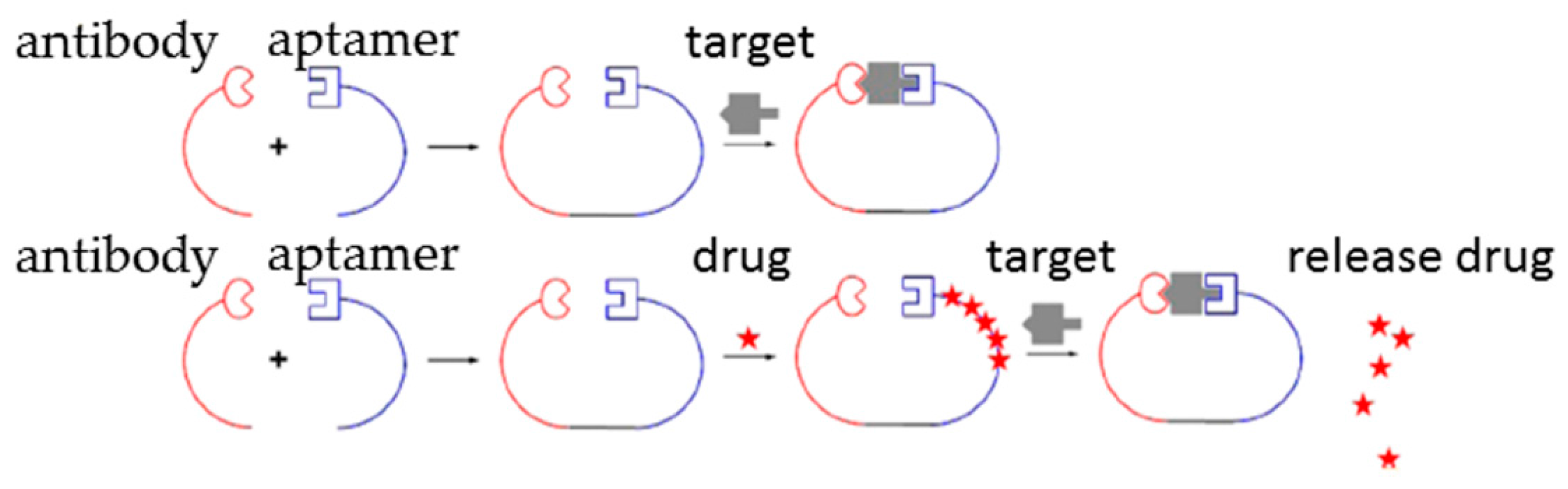
2.3. Aptamer-Antibody Conjugation

Combination use of aptamers and antibodies is a novel therapeutic strategy that shows higher potency than using aptamers or antibodies individually in some cases [18,19,20]. Antibody-aptamer pincers (AAPs) have been developed to increase binding affinity and inhibition potency of antibodies or aptamers to their targets (Figure 1). Anti-thrombin aptamer and antibody conjugation that binds to different epitopes of thrombin have been designed. The AAP has significant lower dissociation constant value (Kd = 567 pM) than the antibody alone (Kd = 50 nM) or aptamer alone (Kd = 3.5 nM) to thrombin [21]. In a breast cancer study, the AAP system was combined with human epidermal growth factor receptor 2 (HER2) drug targeted delivery system. Anti-HER2 aptamer loaded with doxorubicin is conjugated with anti-HER2 antibody to form an AAP-HER2-Dox drug targeted delivery system. This system has much higher cytotoxicity (IC50 = 15.5 nM) to tumors than drug only (IC50 = 43.9 nM) or aptamer loaded with Dox (IC50 = 38.6 nM). Therefore, this AAP system would be helpful to improve affinity and specificity of antibody or aptamer to their targets in new drug development and existing highly toxic drug targeted delivery systems, especially for therapeutic development for many malignancies [21].
Generally, aptamers are used in combination with antibodies without chemical conjugation. For example, anti-platelet-derived growth factor (PDGF) aptamer E10010 combined with anti-VEGF antibody ranibizumab shows higher therapeutic potency than antibody alone for wet AMD treatment, and have passed phase II clinical evaluation and are waiting for phase III clinical trial now (clinical trial IDs NCT01944839 and NCT01940900) [18,19]. On the other hand, combination therapeutics of anti-PDGF antibody with anti-VEGF aptamer also has promising therapeutic effects.

3. Aptamer Selection and Modifications

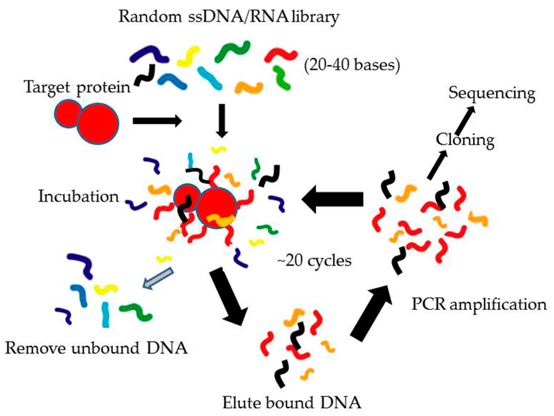
3.1. Systematic Evolution of Ligands by EXponential Enrichment (SELEX)

3.1.1. Conventional SELEX

The method used to develop aptamers is a process called SELEX, which was originally performed and described by Gold and Ellington individually in the 1990s [3,4].
There are several steps in SELEX to find and develop specific aptamers (Figure 2). The first step is synthesis of a screening library, which contains a large number of randomly combinatorial ssDNA and/or ssRNA. All random ssDNA/RNAs have one conserved sequence at each end used for primer binding and amplification and a central random region. The length of the random sequence is normally 20–40 bases so the number of sequences in the whole library would be 1012–1015, which is enough for library diversity. The second step is to incubate target proteins with the random library under proper conditions. Then through a partition step, the sequences that bind to target proteins are separated from those that do not bind. In the third step, the binding sequences are eluted and amplified using the PCR method (for ssDNA) or RT-PCR (for ssRNA) based on the conserved primer sequences. After these steps, a single cycle of SELEX is completed, which would obtain only a small number of binding sequences. Then in the last step, the selection process is repeated for about 7–20 rounds of incubation, partitioning and amplification, resulting in identification of a small number of binding sequences with high affinity and specificity for further processing and optimization. Generally, the binding sequences are then transformed into bacteria (E. coli) for further sequencing as well as characterization. In the post-SELEX process, the specific aptamers can be chemically modified to stabilize and protect them against nucleases in vivo. This is the general process of conventional SELEX.

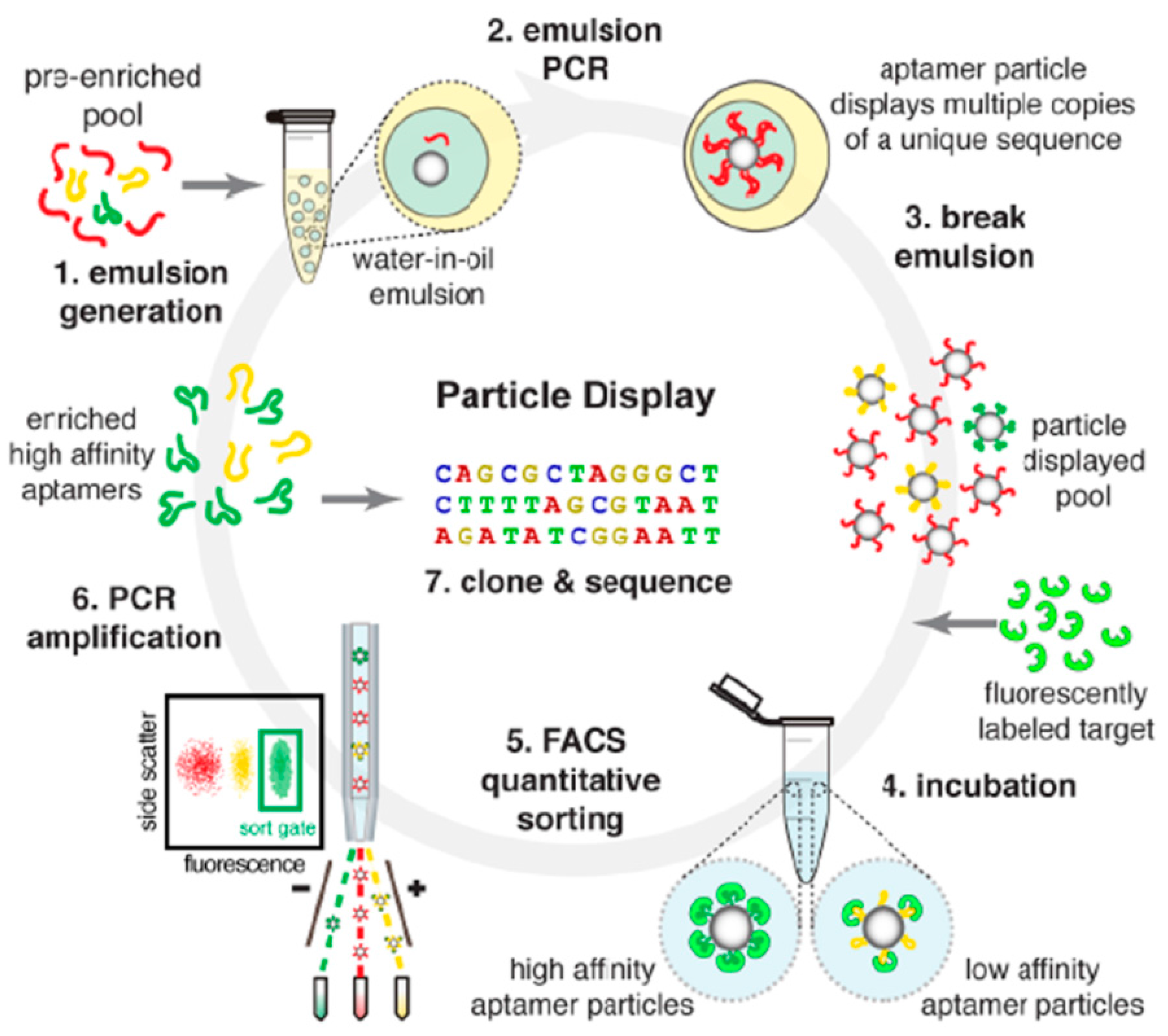
3.1.2. Modified SELEX

As mentioned above, aptamers identified from conventional SELEX process which have high specificity may fail to stimulate or inhibit their targets as expected. The successful rate of effective aptamer identification by conventional SELEX is lower than 30% [21]. Therefore, optimization or variations based on the conventional SELEX may be required in most cases. There are several modifications which are useful and used in research widely (Table 1) [22]. Counter (negative) SELEX is usually performed after positive SELEX to exclude aptamers which bind to negative targets to discriminate highly similar structures [23,24]. Using conventional SELEX in combination with counter SELEX strategy, aptamers that specifically recognized osteoblasts but did not enter hepatocytes and peripheral blood cells were successfully developed for targeted delivery of therapeutic siRNA [24]. This counter SELEX strategy could effectively decrease adverse effects caused by off-target phenomenon in vivo. Toggle SELEX is a selection method in which multiple positive targets are used to select aptamers that bind to all targets [25]. For example, drug resistance was frequently found in breast cancer patients when treating with monoclonal antibodies against HER2. Mutation of HER2 on antibody binding sites is one of the major reasons for drug resistance. Aptamers that bind to two different sites on HER2 could still be effective with one of the binding sites mutated (data not shown). Cell SELEX is using live cells to select aptamers for targets which are difficult to be identified or isolated [24,26]. If a genome sequence is used as a library for selection to obtain natural sequences in genomes that can bind to particular targets, it is called genomic SELEX [27,28].
It is worthwhile to mention that a recently developed aptamer particle display technique can yield aptamers with rather high affinity just in few rounds of SELEX (Figure 3) [29]. In this method, random ssDNA/RNA library is first pre-enriched with several rounds of conventional SELEX and then transformed to particle phase by emulsion PCR with modified primers. Each particle displays multiple copies of one identical sequence on its surface which could be called monoclonal particles. Monoclonal particles are then incubated with fluorescently labeled targets for binding. After removal of non-specific binding targets, fluorescence-activated cell sorting (FACS) could be employed to separate monoclonal particles according to the intensity of fluorescence signal. Using FACS, affinities of over 108 aptamer particles could be measured each time during SELEX which is rather high-throughput. High affinity aptamers are then cloned and sequenced. This technique can dramatically increase the successful rate of aptamer selection, especially for targets which failed to develop aptamer through conventional SELEX [29]. It offers an efficient and economic approach to generate aptamers with high quality, which is helpful for therapeutic aptamer development.

3.2. Modifications of Aptamers for Preclinical Studies

For therapeutic purposes, as oligonucleotide aptamers could be degraded easily in serum, modifications after SELEX are required for stabilization. Various chemical modifications can significantly improve the stability of aptamers (Figure 4) [30].

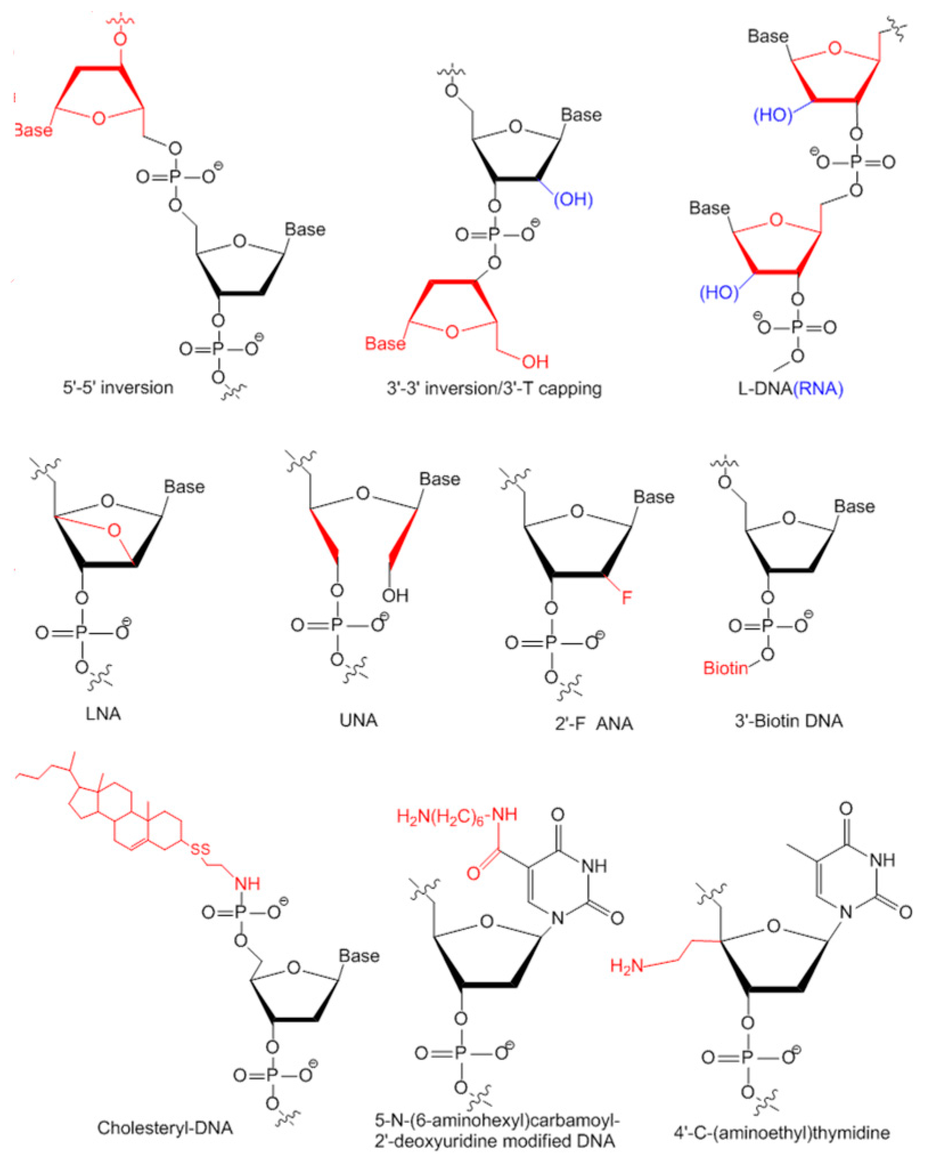
3.2.1. Modifications on Linkage

The 15-mer guanine rich (G-rich) thrombin aptamer d(GGTTGGTGTGGTTGG) is a typical and popular model used for developing and illustrating of novel characterization or modification methods for aptamers [31]. It has a G-quadruplex structure formed by two stacking G-tetrads and a central loop TGT which is optimal for stability. It is interesting to find that adding an extra guanine at the 5′ end caused decreased stability while adding an extra guanine to the 3′ end caused increased stability, indicating that the themostability of an aptamer is sequence dependent [32]. It was found that the G-quadruplex is a rather common structure for aptamers, especially for DNA aptamers. DNA aptamers against various targets have a G-quadruplex structure with high sequence identity with high diversity. Another group tried to invert 5′–5′ of a polarity site to form a folded aptamer with a non-common structure d(GGTTGGTGTGGTTGG). This structure has higher stability and affinity to thrombin, although lower inhibiting activity compared to unmodified aptamers [33,34]. Terminal 3′–3′ and 3′–5′ internucleotide linkage was first tried in 1991. A sense deoxyoligonucleotide capping of both ends of the aptamer with inverted thymidine could not only increase stability significantly but also slowed nuclease degradation from 30 min (unmodified) down to 90 min (modified) in snake venom phosphodiesterase digestion [35]. 3′-capping with inverted thymidine modification is a commonly used approach to block 3′-exonuclease degradation by nucleases and prolong an aptamer’s half-life time in serum [23,36]. Most aptamers in clinical trials are modified with this method (Table 2).
Besides inverted thymidine modification, 3′-biotin-streptavidin conjugation is also designed to fight with 3′-exonuclease digestion in serum. It is found that 3′-biotin-streptavidin conjugating aptamers which have bigger size is not only protected from nucleases degradation, but also protected from rapid clearance by circulation system in vivo [37]. 3′-biotin modification is also used for aptamers against other targets. Anti-SARS coronavirus helicase aptamer can remain intact for up to 16 h in 10% fetal bovine serum compared to 6 h for unmodified aptamers [23].
Cholesterol can be added to the 5′-end of an aptamer to form a cholesteryl-oligonucleotide (cholODN) and further linked to low density lipoprotein (LDL) to form a compact cholODN-LDL complex. This complex has high stability and is highly resistant to nucleases degradation in serum which has a 10-fold longer half-life than unmodified aptamers [38]. Substitute phosphodiester linkage of DNA with methylphosphonate or phosphorothioate is also commonly used for aptamer stabilization [39,40,41].

3.2.2. Modifications on Sugar Ring or Bases

The natural oligonucleotides are all in d-form. l-form oligonucleotides (Spiegelmer) are chiral inversions of natural d-forms. In identification of l-form aptamers, d-form aptamers are firstly selected against synthesized l-form protein targets from a general single strand random oligonucleotide library. After SELEX, l-form aptamers are synthesized according to the mirror image of corresponding d-form aptamers [43]. l-form aptamers are much more stable than d-form with high resistance to nuclease degradation in vivo and do not hybridize to or affect the original nucleic acids in the cells [44,45,46,47]. Clinical evaluated aptamers NOX-A12, NOX-H94 and NOX-E36 are all l-form aptamers.
Locked nucleic acid (LNA) is a modification on a sugar ring with a methylene linkage between 2′-O and 4′-C, which can generate the most stable pairs to dramatically increase the themostability and nucleases resistance of aptamers [48,49]. The LNA/DNA chimera aptamer against HIV-1 trans-activating response target could retain an intact structure without degradation for up to 20 h in serum [48].
Unlocked nucleic acid (UNA) is an opposite modification to LNA. There is a bond between C2′ and C3′ missing in UNA which makes aptamer more flexible. Different from LNA which can stabilize structure, UNA has an uncertain affect to themostability of aptamers. It is found that UNA replacement on a loop region of an anti-thrombin aptamer increased its themostability while replacement on G-tetrads disrupted the structure formation [50]. It is uncertain whether UNA modification has any effect on protecting aptamers from nucleases degradation [50].
Other positions of the sugar ring could be amended for chemical modifications, such as 2′-F [39,51,52], 4′-C-(aminoethyl) thymidine [51], 5-N-(6-aminohexyl)carbamoyl-2′-deoxyuridine [39,51] and so on. For more modifications in detail, please read reviews written by Wang et al. [30]. More studies are required to characterize and discuss the stabilizing effects of these modifications and design more modification strategies in the future.

4. Aptamers for Skeletal Diseases Therapy in Preclinical Studies

The number of bone marrow mesenchymal stem cells (BMSCs) is decreased through aging while increased through adipocyte differentiation. It is found that miR-188 level is much higher in BMSCs in old than in young mice and human. Animals lacking miR-188 can be protected from age-related bone loss and fat accumulation in bone marrow. An aptamer that specifically recognizes BMSCs is developed and conjugated with miR-188 to form a nanocomplex. This targeted delivery nanocomplex could promote bone formation and reduce fat accumulation in bone marrow with high efficacy in aged mice, indicating a potential approach for age-related bone loss therapy [53].
Furthermore, BMSCs are important in bone marrow but there is no specific marker on their surface, making it difficult to be isolated from bone marrow directly. Aptamers with high binding affinity against porcine BMSCs are developed by SELEX. Using the high affinity and specificity aptamers, BMSCs could be fished out from cell solution and bone marrow, which is a novel method for BMSCs isolation and provide a foundation for aptamer applications in tissue engineering and regenerative medicine for skeletal diseases therapy [54].
In another preliminary study, a specific aptamer against human jaw periosteal cells (JPCs) is developed for tissue engineering in oral and maxillofacial surgery. This aptamer has high affinity to human osteogenically induced JPCs and BMSCs from bone marrow while it does not bind to any other cell lines or undifferentiated JPCs or JPCs induced from other sources. It can be used to purify osteogenic progenitor cells from undifferentiated JPCs or stem cells of other sources. The mineralization capacity is higher in the aptamer positive fraction, which is a promising technique for tissue engineering [55].
There are other aptamers or other aptamer-ligand complexes developed and studied in preclinical research at the moment. However, there is no aptamer evaluated in clinical trials. More efforts and studies are needed for this therapeutic field in the future.
Adult mesenchymal stem cells (aMSCs) are stem cells.

5. Aptamers in On-Going or Completed Clinical Trials for Therapeutics

At the moment, there are 11 aptamers evaluating in clinical trials for the treatment of macular degeneration, cancer, inflammation and coagulation, and one of them has been approved by FDA for the treatment of AMD. In this section, we will review the research and clinical evaluation progress of these aptamers.

5.1. Aptamers against Macular Degeneration

Wet (neovascular) and dry (atrophic) AMD are two major causes of vision loss in the elderly due to retinal damage, and affect around eight million people in America. There are three aptamers being evaluated in clinical trials now for wet or dry AMD therapy.

5.1.1. Pegaptanib

Pegaptanib (Macugen; Pfizer and Eyetech, New York, NY, USA), a 27-mer RNA aptamer specifically binds to and inhibits VEGF against AMD, is the only aptamer approved by the FDA for disease treatment on the market [56]. Pegaptanib is selected directly against VEGF165, the VEGF isoform primarily responsible for pathological ocular neovasculariztion and vascular permeability [8]. After in vitro selection and characterization, the aptamer which inhibits VEGF165 with high affinity and efficacy is chosen and modified with 2′-fluoro pyrimidines and 2′-O-methyl purines and further capped with 3′–3′-linked deoxythymidine to avoid nuclease degradation and increase stability. In a preclinical animal study, a 40 kDa polyethylene glycol (PEG) is conjugated to the 5′- end of the RNA aptamer to increase half life of the aptamer for better bioavailability. After around 10 years preclinical studies to optimize and evaluate its therapeutic potency, pegaptanib which shows high efficacy in inhibiting VEGF in different models was approved by the FDA for the treatment of AMD in 2004 with dosage of 0.3 mg per eye every 6 weeks administered intravitreally [56]. This is extraordinary progress as it is the first aptamer approved for use in human and it opens a wide window for therapeutic aptamers in disease treatment.
However, pegaptanib failed to compete with anti-VEGF monoclonal antibody ranibizumab (Lucentis; Genentech, South San Francisco, California, USA) as it only inhibits VEGF165 isoforms, while antibodies inhibit all isoforms of VEGF [57], which has negative affects on the pharmacy investment to therapeutic aptamers in these years. Fortunately, it was found recently that blocking all activity of VEGF may cause high risk of hypertension and other adverse effects [58]. Therefore, aptamer pegaptanib is better than antibody ranibizumab in long term maintenance therapy, especially in patients with systemic comorbidities [18,59].

5.1.2. ARC1905

ARC1905 (Ophthotech Corp, New York, NY, USA) is a 39 bases RNA aptamer specifically against complement component 5 (C5) for the treatment of both wet and dry AMDs [60]. C5 is a downstream pro-inflammatory protein in the complement system associated with AMD pathogenesis. Inhibition of C5 can prevent the key terminal fragments formation which is critical for tissue pathology [61]. A PEG is also conjugated to the 3′- end with an inverted thymidine. A phase I clinical trial using ARC1905 in combination with ranibizumab for the treatment of wet AMD (NCT00709527) was finished in 2011 and a phase I clinical for dry AMD treatment (NCT00950638) has recently been completed (data not shown).

5.1.3. E10030

It may not be sufficient to inhibit VEGF only for wet AMD treatment to prevent angiogenesis due to the limit of new vessels regression associated with vision loss. PDGF plays an important role in pericyte recruitment and maturation and new vessels may resist to anti-VEGF drugs due to the role of PDGF [19,62]. Combination treatment using pegaptanib and anti-PDGF antibody can not only prevent new vessel formation but also promote vessel regression [19]. On the other hand, combination treatment using anti-PDGF aptamer and anti-VEGF antibody also has promising therapeutic effect. A 29 bases RNA aptamer E10030 (Fovista; Ophthotech Corp) targeting PDGF is developed and modified with 2′-fluoro pyrimidine and 2′-O-methyl purines. In preclinical studies, PEG-conjugated E10030 was able to facilitate neovascular regression when combined with anti-VEGF agents [19]. In clinical trials, patients treated with E10030 in combination with anti-VEGF antibody raninizumab (administered once a month) showed significant neovascular regression and 59% of them have increased visual acuity after three months of treatment without any side effects observed. In phase II clinical trials of this combination therapy, patients treated with combination therapy gained 62% higher vision than the patients treated with anti-VEGF antibody ranizumab only. The combination therapy is waiting for phase III clinical evaluation for wet AMD treatment at the moment (clinical trial IDs NCT01944839 and NCT01940900). The promising results suggest that combination therapy targeting two different antigens at the same time using aptamer-aptamer combination or aptamer-antibody combination could be a new therapeutic direction in future study.

5.2. Aptamers against Cancer

Cancer treatment requires more effective and precise therapies, especially specific therapies, for discriminating normal cells and tumor cells to avoid toxicity. The highly disorganized vessel architecture inside the tumors and the surrounding extracellular matrixes as well as stromal cells are both barriers for drug delivery [63]. Monoclonal antibodies can recognize targeting tumor cells with high specificity but are difficult to penetrate into the deep sites of tumor cells due to the large size (around 150 kDa). Aptamers with much smaller size (around 30 kDa) are able to cross the barriers and penetrate into tumor cells and therefore are ideal therapeutic reagents for cancer. Generally, aptamers could diffuse into tumors in 10 min after injection. For example, a fluorescent aptamer against extracellular matrix protein tenascin-C was able to diffuse into tumors rapidly with perivascular fluorescence signal detected in tumor only 10 min after intravenous injection [64]. Furthermore, another group has compared the tumor penetration times between aptamer and antibody, and found that they were detected in tumors after 10 min and 3 h after intravenous injection, respectively [65]. Immuno-therapy of cancer by aptamers is a recent novel research focus. There are two individual aptamers for cancer therapy now being evaluated in clinical trials.

5.2.1. AS1411

AS1411 (Antisoma, London, UK) is a guanine-rich aptamer with G-quadruplex structure identified from a guanine-rich ssRNA library by antiproliferation selection [66]. It has a G-quadruplex structure which is highly stable and resistant to nuclease degradation. It can penetrate into tumor cells easily. AS1411 binds to the external domain of nucleolin, which is a protein over-expressed on the surface of cancer cells and responsible for survival, growth, and proliferation of cells [66]. AS411 can inhibit over 80 types of cancer cells in in vitro studies and it is the first oligonucleotide aptamer approved for clinical trial for human cancer therapy. In preclinical studies, AS1411 has inhibition efficacy in multiple cancer models including non-small cell lung, renal cells and breast cancers. In phase I clinical trial (NCT00881244), AS1411 is well tolerated by patients with advanced cancers and has no side effects. AS1411 shows promising therapeutic efficacy especially for patients with renal cell carcinoma after six months of therapy. Phase II clinical trial shows therapeutic efficacy to acute myeloid leukemia patients without toxicity and adverse effects. However, a following phase II evaluation for renal cell carcinoma (clinical trial ID NCT00740441) found AS1411 only has therapeutic effect in 2.9% of patients, and shows minimal activity in unselected patients with metastatic renal cell carcinoma [67], indicating more research is required to optimize the therapeutic potency of AS1411 in the future.

5.2.2. NOX-A12

NOX-A12 (Olaptesed pegol; Noxxon, Berlin, Germany) is a 45-mer l-RNA aptamer developed for use in autologous hematopoietic stem cell transplants [66]. NOX-A12 targets to stroma cell-derived factor-1 The chemokine (C–X–C motif) ligand 12 (CXCL-12), which plays important roles in stem cell migration towards the bone marrow and controls tumor growth, metastasis and vasculogenesis. Binding to CXCL-12 can block its receptor binding and prevent CXCL-12 tissue gradients and decrease the possibility of tumor metastasis and drug resistance caused by cancer cell homing [68]. As l-form aptamers can not be recognized by nucleases, it does not require any chemical modifications for in vivo studies. It showed efficacy against non- Hodgkin’s lymphoma and myelomas in preclinical studies. Phase I clinical trial for safety and tolerability evaluation confirms NOX-A12 can be well tolerated without serious adverse effects. NOX-A12 has a 37 h long half-life and patients with NOX-A12 treatment are more susceptible to chemotherapy due to its particular blockage of stromal cell-derived factor-1 (SDF-1), making it a promising and successful drug in cancer therapy [69]. Two phase II clinical trials are in progress, one for treatment of Chromin Lymphocytic Leukemia (clinical trial ID NCT01486797) and the other one for evaluating and comparing the therapeutic efficacy of NOX-A12 alone and combination therapy with chemotherapy for patients with multiple myeloma (clinical trial ID NCT01521533).

5.3. Aptamers against Coagulation

More and more aptamers for coagulation therapy have been developed independently by different research groups. Till now, there are four aptamers being evaluated in different stages of clinical trials.

5.3.1. REG1

REG1 (Regado Biosciences, Basking Ridge, NJ, USA) is an aptamer system consisting of a 37-mer RNA aptamer RB006 (Pegnivacogin) and a 17-mer antidote RB007 with sequence complementary to RB006 [70]. RB006 is an antagonist of factor IXa for preventing the downstream conversion of factor X and avoidance of clotting. RB007 is the antidote of RB006, which can specifically reverse the inhibition function of RB006 to control sheath removal time after percutaneous coronary intervention. Phase I clinical trials have shown REG1 is well tolerated in patients and no significant adverse effects or major bleeding are found [71,72,73]. More importantly, therapeutic effects of REG1 can be controlled by dosage of RB007 and it is less toxic than heparin/protamine, which is also an anticoagulation/antidote pair currently available [74]. Phase II clinical trials for using REG1 in percutaneous coronary intervention for patients with coronary artery have just been completed (clinical trial ID NCT00715455). However, a later randomized clinical trial had to be terminated before it finished as patients treated with REG1 showed severe allergic reactions and major bleeding with no significant efficacy after percutaneous coronary intervention [75]. Therefore, more studies are required to evaluate the safety and efficacy of the REG1 system.

5.3.2. ARC1779

ARC1779 (Archemix Corp, Cambridge, MA, USA) is a 39-mer PEGylated DNA aptamer that binds to the A1 domain of von Willebrand factor (vWF) [76], which is a key factor in the coagulation cascade related to platelet recruitment, to block interaction between A1 domain and platelet receptor glycoprotein 1B [12,77]. Therefore, it is a potential therapeutic target for treatment of vWF-related platelet disorders, von Willebrand disease as well as acute coronary syndromes [78]. A phase II pilot study showed that ARC1779 can inhibit platelet depletion induced by a vWF agonist desmopressin in patients [79]. Phase II clinical trial for VWF 2D treatment is still on-going.

5.3.3. NU172

Aptamer NU172 (ARCA Biopharma, London, UK) is an unmodified DNA aptamer for short-term anticoagulation and is distinct from the long-term anticoagulation aptamer REG1 and ARC1779. In preclinical studies, NU172 showed efficacy to prolong clotting time and the anticoagulation effect would be stopped rapidly due to nuclease degradation. A phase II clinical trial for evaluating the therapeutic effects in coronary artery bypass graft surgery is in progress (clinical trial ID NCT00808964).

5.3.4. BAX499

The above three anticoagulation aptamers all target to proteins in the intrinsic coagulation pathway, while BAX499 (Baxter, Deerfield, IL, USA) targets to the negative regulator of factor VIIa in the extrinsic tissue factor pathway. In a preclinical hemophilia pathology mimicking monkey model, BAX499 is able to recover the clotting caused by anti-factor VIII antibody [80]. The phase I clinical trial was started from 2010 (clinical trial ID NCT01191372) and the evaluation results have not been published yet.

5.4. Aptamers against Inflammation

Two anti-inflammation aptamers in clinical trials are both l-form aptamers from Noxxon.

5.4.1. NOX-H94

Hepcidin is over-expressed in patients with chronic inflammation induced by cancer or dialysis, which may lead to anemia due to the hepcidin-induced ferroportin degradation [81]. NOX-H94 (Lexaptepid pegol; Noxxon) targets to hepcidin, a peptide hormone regulator for iron homeostasis with the role to inhibit the interaction between hepcidin and ferroportin and reduce the anemia symptom in patients [81,82]. In a preclinical study, cynomolgus monkey could be prevented from interleukin-6 induced iron concentration decrease in serum when treated with NOX-H94. Phase IIa clinical trial for anemia therapeutic evaluation for patients with cancers have been completed. Clinical trials for treatment of erythropoiesis agent-induced anemia in patients with dialysis are still in progress (clinical trial ID NCT02079896).

5.4.2. NOX-E36

NOX-E36 is another l-form anti-inflammation aptamer from Noxxon [83]. The target of NOX-E36 is chemokine ligand 2 (also called monocyte chemoattractant protein 1) which can mediate inflammation by recruiting leukocytes from intravascular to extravascular environments. Upon binding, NOX-E36 can inhibit chemokine ligands 2-induced inflammation to reduce the recruitment of leukocytes, which is quite effective for anti-inflammation treatment for lupus nephritis in mouse models. It is also useful for preventing type-2 diabetic glomerulosclerosis in mice [84]. Phase II clinical trials for treatment of type-2 diabetes are in progress (clinical trial IDs NCT01085292 and NCT01547897).

6. Conclusions

Oligonucleotide aptamers are more and more popular in recent years, especially from 2005 after the first aptamer Pegaptanib was approved for wet AMD therapy by FDA. There are over 900 aptamers developed by SELEX for a broad spectrum of both diagnostic and therapeutic applications. You can find over 5000 reports on aptamer research in PubMed, and this number currently generally increases by two to five every day. There are 11 aptamers in clinical trials, which have advantages especially for safety issues and have significant improvements in efficacy for therapeutic application. In addition to these aptamers, there are nearly 100 aptamers waiting for approval for evaluation in clinical trials.
With promising advantages compared to monoclonal antibodies, oligonucleotide aptamers may become the predominant agent for therapeutic application. With monoclonal antibody technology commonly developed, and used in therapeutic application, the targets of antibodies are well characterized [85] (Table 3). Therefore in the future, based on the promising therapeutic effects of inhibiting particular targets with monoclonal antibodies, oligonucleotide aptamers can be identified against the same targets to develop the second generation of therapy. This would save considerable research effort and allow more rapid progress in developing more economic and efficient therapeutic approaches for various diseases with oligonucleotide aptamers.

Acknowledgments

We thank academic staff members in Baoqin Liu’s group at Zhengzhou Hospital of Traditional Chinese Medicine, and Aiping Lu and Ge Zhang’s group at Hong Kong Baptist University. We also thank Hong Kong Baptist University for providing critical comments and technical support. This study was supported by the Hong Kong General Research Fund (HKBU262913 to Ge Zhang).

Author Contributions

Yuanyuan Yu wrote the manuscript; Chao Liang, Quanxia Lv, Defang Li and Xuegong Xu contributed the manuscript for literature research; Baoqin Liu, Aiping Lu and Ge Zhang revised and approved the manuscript.

Conflicts of Interest

The authors declare no conflict of interest.

Abbreviations

The following abbreviations are used in this manuscript:
SELEX
Systematic Evolution of Ligands by EXponential enrichment
AMD
Age-related Macular Degeneration
AAPs
Antibody-aptamer pincers
FACS
Fluorescence-Activated Cell Sorting
cholODN
cholesteryl-oligonucleotide
LDL
low density lipoprotein
VEGF
vascular endothelial growth factor
PDGF
Platelet-Derived Growth Factor
vWF
von Willebrand factor
CD
Cluster of Differentiation
IL
InterLeukin
EGFR
Epidermal Growth Factor Receptor
sulfo-SMCC
sulfosuccinimidyl 4-(N-maleimidomethyl) cyclohexane-1-carboxylate
SATA
N-succinimidyl-S-acetylthioacetate
CXCL-12
The chemokine (C–X–C motif) ligand 12

References

  1. Kohler, G.; Milstein, C. Continuous cultures of fused cells secreting antibody of predefined specificity. Nature 1975, 256, 495–497. [Google Scholar] [CrossRef] [PubMed]
  2. Bradbury, A.R.; Sidhu, S.; Dubel, S.; McCafferty, J. Beyond natural antibodies: The power of in vitro display technologies. Nat. Biotechnol. 2011, 29, 245–254. [Google Scholar] [CrossRef] [PubMed]
  3. Ellington, A.D.; Szostak, J.W. In vitro selection of RNA molecules that bind specific ligands. Nature 1990, 346, 818–822. [Google Scholar] [CrossRef] [PubMed]
  4. Tuerk, C.; Gold, L. Systematic evolution of ligands by exponential enrichment: RNA ligands to bacteriophage t4 DNA polymerase. Science 1990, 249, 505–510. [Google Scholar] [CrossRef] [PubMed]
  5. Jayasena, S.D. Aptamers: An emerging class of molecules that rival antibodies in diagnostics. Clin. Chem. 1999, 45, 1628–1650. [Google Scholar] [PubMed]
  6. Robertson, D.L.; Joyce, G.F. Selection in vitro of an RNA enzyme that specifically cleaves single-stranded DNA. Nature 1990, 344, 467–468. [Google Scholar] [CrossRef] [PubMed]
  7. Cruz-Toledo, J.; McKeague, M.; Zhang, X.; Giamberardino, A.; McConnell, E.; Francis, T.; DeRosa, M.C.; Dumontier, M. Aptamer base: A collaborative knowledge base to describe aptamers and SELEX experiments. Database 2012, 2012. [Google Scholar] [CrossRef] [PubMed]
  8. Jellinek, D.; Green, L.S.; Bell, C.; Janjic, N. Inhibition of receptor binding by high-affinity RNA ligands to vascular endothelial growth factor. Biochemistry 1994, 33, 10450–10456. [Google Scholar] [CrossRef] [PubMed]
  9. Ambati, J.; Ambati, B.K.; Yoo, S.H.; Ianchulev, S.; Adamis, A.P. Age-related macular degeneration: Etiology, pathogenesis, and therapeutic strategies. Surv. Ophthalmol. 2003, 48, 257–293. [Google Scholar] [CrossRef]
  10. Que-Gewirth, N.S.; Sullenger, B.A. Gene therapy progress and prospects: RNA aptamers. Gene Ther. 2007, 14, 283–291. [Google Scholar] [CrossRef] [PubMed]
  11. Ruckman, J.; Green, L.S.; Beeson, J.; Waugh, S.; Gillette, W.L.; Henninger, D.D.; Claesson-Welsh, L.; Janjic, N. 2′-Fluoropyrimidine RNA-based aptamers to the 165-amino acid form of vascular endothelial growth factor (VEGF165). J. Biol. Chem. 1998, 273, 20556–20567. [Google Scholar] [CrossRef] [PubMed]
  12. Keefe, A.D.; Pai, S.; Ellington, A. Aptamers as therapeutics. Nat. Rev. Drug Discov. 2010, 9, 537–550. [Google Scholar] [CrossRef] [PubMed]
  13. Latz, E.; Verma, A.; Visintin, A.; Gong, M.; Sirois, C.M.; Klein, D.C.; Monks, B.G.; McKnight, C.J.; Lamphier, M.S.; Duprex, W.P.; et al. Ligand-induced conformational changes allosterically activate Toll-like receptor 9. Nat. Immunol. 2007, 8, 772–779. [Google Scholar] [CrossRef] [PubMed]
  14. Zhao, Q.; Temsamani, J.; Iadarola, P.L.; Jiang, Z.; Agrawal, S. Effect of different chemically modified oligodeoxynucleotides on immune stimulation. Biochem. Pharmacol. 1996, 51, 173–182. [Google Scholar] [CrossRef]
  15. Yu, D.; Wang, D.; Zhu, F.G.; Bhagat, L.; Dai, M.; Kandimalla, E.R.; Agrawal, S. Modifications incorporated in CpG motifs of oligodeoxynucleotides lead to antagonist activity of Toll-like receptors 7 and 9. J. Med. Chem. 2009, 52, 5108–5114. [Google Scholar] [CrossRef] [PubMed]
  16. Banerjee, J. Antibodies are challenged. Indian J. Med. Sci. 2010, 64, 144–147. [Google Scholar] [PubMed]
  17. Gold, L.; Ayers, D.; Bertino, J.; Bock, C.; Bock, A.; Brody, E.N.; Carter, J.; Dalby, A.B.; Eaton, B.E.; Fitzwater, T.; et al. Aptamer-based multiplexed proteomic technology for biomarker discovery. PLoS ONE 2010, 5, e15004. [Google Scholar] [CrossRef] [PubMed]
  18. Lao, Y.H.; Phua, K.K.; Leong, K.W. Aptamer nanomedicine for cancer therapeutics: Barriers and potential for translation. ACS Nano 2015, 9, 2235–2254. [Google Scholar] [CrossRef] [PubMed]
  19. Jo, N.; Mailhos, C.; Ju, M.; Cheung, E.; Bradley, J.; Nishijima, K.; Robinson, G.S.; Adamis, A.P.; Shima, D.T. Inhibition of platelet-derived growth factor B signaling enhances the efficacy of anti-vascular endothelial growth factor therapy in multiple models of ocular neovascularization. Am. J. Pathol. 2006, 168, 2036–2053. [Google Scholar] [CrossRef] [PubMed]
  20. Zhang, P.; Zhao, N.; Zeng, Z.; Chang, C.C.; Zu, Y. Combination of an aptamer probe to CD4 and antibodies for multicolored cell phenotyping. Am. J. Clin. Pathol. 2010, 134, 586–593. [Google Scholar] [CrossRef] [PubMed]
  21. Kang, S.; Hah, S.S. Improved ligand binding by antibody-aptamer pincers. Bioconjug. Chem. 2014, 25, 1421–1427. [Google Scholar] [CrossRef] [PubMed]
  22. Darmostuk, M.; Rimpelova, S.; Gbelcova, H.; Ruml, T. Current approaches in SELEX: An update to aptamer selection technology. Biotechnol. Adv. 2015, 33, 1141–1161. [Google Scholar] [CrossRef] [PubMed]
  23. Shum, K.T.; Chan, C.; Leung, C.M.; Tanner, J.A. Identification of a DNA aptamer that inhibits sclerostin’s antagonistic effect on Wnt signalling. Biochem. J. 2011, 434, 493–501. [Google Scholar] [CrossRef] [PubMed]
  24. Liang, C.; Guo, B.; Wu, H.; Shao, N.; Li, D.; Liu, J.; Dang, L.; Wang, C.; Li, H.; Li, S.; et al. Aptamer-functionalized lipid nanoparticles targeting osteoblasts as a novel RNA interference-based bone anabolic strategy. Nat. Med. 2015, 21, 288–294. [Google Scholar] [CrossRef] [PubMed]
  25. White, R.; Rusconi, C.; Scardino, E.; Wolberg, A.; Lawson, J.; Hoffman, M.; Sullenger, B. Generation of species cross-reactive aptamers using “toggle“ SELEX. Mol. Ther. 2001, 4, 567–573. [Google Scholar] [CrossRef] [PubMed]
  26. Thiel, T.; Graner, A.; Waugh, R.; Grosse, I.; Close, T.J.; Stein, N. Evidence and evolutionary analysis of ancient whole-genome duplication in barley predating the divergence from rice. BMC Evol. Biol. 2009, 9. [Google Scholar] [CrossRef] [PubMed]
  27. Gold, L.; Brown, D.; He, Y.; Shtatland, T.; Singer, B.S.; Wu, Y. From oligonucleotide shapes to genomic SELEX: Novel biological regulatory loops. Proc. Natl. Acad. Sci. USA 1997, 94, 59–64. [Google Scholar] [CrossRef] [PubMed]
  28. Shtatland, T.; Gill, S.C.; Javornik, B.E.; Johansson, H.E.; Singer, B.S.; Uhlenbeck, O.C.; Zichi, D.A.; Gold, L. Interactions of Escherichia coli RNA with bacteriophage MS2 coat protein: Genomic SELEX. Nucleic Acids Res. 2000, 28. [Google Scholar] [CrossRef]
  29. Wang, J.; Gong, Q.; Maheshwari, N.; Eisenstein, M.; Arcila, M.L.; Kosik, K.S.; Soh, H.T. Particle display: A quantitative screening method for generating high-affinity aptamers. Angew. Chem. Int. Ed. 2014, 53, 4796–4801. [Google Scholar] [CrossRef] [PubMed]
  30. Wang, R.E.; Wu, H.; Niu, Y.; Cai, J. Improving the stability of aptamers by chemical modification. Curr. Med. Chem. 2011, 18, 4126–4138. [Google Scholar] [CrossRef] [PubMed]
  31. Bock, L.C.; Griffin, L.C.; Latham, J.A.; Vermaas, E.H.; Toole, J.J. Selection of single-stranded DNA molecules that bind and inhibit human thrombin. Nature 1992, 355, 564–566. [Google Scholar] [CrossRef] [PubMed]
  32. Smirnov, I.; Shafer, R.H. Effect of loop sequence and size on DNA aptamer stability. Biochemistry 2000, 39, 1462–1468. [Google Scholar] [CrossRef] [PubMed]
  33. Martino, L.; Virno, A.; Randazzo, A.; Virgilio, A.; Esposito, V.; Giancola, C.; Bucci, M.; Cirino, G.; Mayol, L. A new modified thrombin binding aptamer containing a 5′–5′ inversion of polarity site. Nucleic Acids Res. 2006, 34, 6653–6662. [Google Scholar] [CrossRef] [PubMed]
  34. Pagano, B.; Martino, L.; Randazzo, A.; Giancola, C. Stability and binding properties of a modified thrombin binding aptamer. Biophys. J. 2008, 94, 562–569. [Google Scholar] [CrossRef] [PubMed]
  35. Seliger, H.; Frohlich, A.; Groger, G.; Krist, B.; Montenarh, M.; Rosch, H.; Rosch, R.; Ortigao, F.R. Synthetic oligonucleotides for biomedical applications. Nucleic Acids Symp. Ser. 1991, 193–196. [Google Scholar]
  36. Shaw, J.P.; Kent, K.; Bird, J.; Fishback, J.; Froehler, B. Modified deoxyoligonucleotides stable to exonuclease degradation in serum. Nucleic Acids Res. 1991, 19, 747–750. [Google Scholar] [CrossRef] [PubMed]
  37. Dougan, H.; Lyster, D.M.; Vo, C.V.; Stafford, A.; Weitz, J.I.; Hobbs, J.B. Extending the lifetime of anticoagulant oligodeoxynucleotide aptamers in blood. Nucl. Med. Biol. 2000, 27, 289–297. [Google Scholar] [CrossRef]
  38. De Smidt, P.C.; Le Doan, T.; de Falco, S.; van Berkel, T.J. Association of antisense oligonucleotides with lipoproteins prolongs the plasma half-life and modifies the tissue distribution. Nucleic Acids Res. 1991, 19, 4695–4700. [Google Scholar] [CrossRef] [PubMed]
  39. Sacca, B.; Lacroix, L.; Mergny, J.L. The effect of chemical modifications on the thermal stability of different G-quadruplex-forming oligonucleotides. Nucleic Acids Res. 2005, 33, 1182–1192. [Google Scholar] [CrossRef] [PubMed]
  40. Zaitseva, M.; Kaluzhny, D.; Shchyolkina, A.; Borisova, O.; Smirnov, I.; Pozmogova, G. Conformation and thermostability of oligonucleotide d(GGTTGGTGTGGTTGG) containing thiophosphoryl internucleotide bonds at different positions. Biophys. Chem. 2010, 146, 1–6. [Google Scholar] [CrossRef] [PubMed]
  41. Pozmogova, G.E.; Zaitseva, M.A.; Smirnov, I.P.; Shvachko, A.G.; Murina, M.A.; Sergeenko, V.I. Anticoagulant effects of thioanalogs of thrombin-binding DNA-aptamer and their stability in the plasma. Bull. Exp. Biol. Med. 2010, 150, 180–184. [Google Scholar] [CrossRef] [PubMed]
  42. Sundaram, P.; Kurniawan, H.; Byrne, M.E.; Wower, J. Therapeutic RNA aptamers in clinical trials. Eur. J. Pharm. Sci. 2013, 48, 259–271. [Google Scholar] [CrossRef] [PubMed]
  43. Hoellenriegel, J.; Zboralski, D.; Maasch, C.; Rosin, N.Y.; Wierda, W.G.; Keating, M.J.; Kruschinski, A.; Burger, J.A. The spiegelmer NOX-A12, a novel CXCL12 inhibitor, interferes with chronic lymphocytic leukemia cell motility and causes chemosensitization. Blood 2014, 123, 1032–1039. [Google Scholar] [CrossRef] [PubMed]
  44. Purschke, W.G.; Radtke, F.; Kleinjung, F.; Klussmann, S. A DNA spiegelmer to staphylococcal enterotoxin B. Nucleic Acids Res. 2003, 31, 3027–3032. [Google Scholar] [CrossRef] [PubMed]
  45. Wlotzka, B.; Leva, S.; Eschgfaller, B.; Burmeister, J.; Kleinjung, F.; Kaduk, C.; Muhn, P.; Hess-Stumpp, H.; Klussmann, S. In vivo properties of an anti-GnRH spiegelmer: An example of an oligonucleotide-based therapeutic substance class. Proc. Natl. Acad. Sci. USA 2002, 99, 8898–8902. [Google Scholar] [CrossRef] [PubMed]
  46. Leva, S.; Lichte, A.; Burmeister, J.; Muhn, P.; Jahnke, B.; Fesser, D.; Erfurth, J.; Burgstaller, P.; Klussmann, S. Gnrh binding RNA and DNA spiegelmers: A novel approach toward GnRH antagonism. Chem. Biol. 2002, 9, 351–359. [Google Scholar] [CrossRef]
  47. Eulberg, D.; Klussmann, S. Spiegelmers: Biostable aptamers. Chembiochem 2003, 4, 979–983. [Google Scholar] [CrossRef] [PubMed]
  48. Darfeuille, F.; Hansen, J.B.; Orum, H.; Di Primo, C.; Toulme, J.J. LNA/DNA chimeric oligomers mimic RNA aptamers targeted to the TAR RNA element of HIV-1. Nucleic Acids Res. 2004, 32, 3101–3107. [Google Scholar] [CrossRef] [PubMed]
  49. Schmidt, K.S.; Borkowski, S.; Kurreck, J.; Stephens, A.W.; Bald, R.; Hecht, M.; Friebe, M.; Dinkelborg, L.; Erdmann, V.A. Application of locked nucleic acids to improve aptamer in vivo stability and targeting function. Nucleic Acids Res. 2004, 32, 5757–5765. [Google Scholar] [CrossRef] [PubMed]
  50. Pasternak, A.; Hernandez, F.J.; Rasmussen, L.M.; Vester, B.; Wengel, J. Improved thrombin binding aptamer by incorporation of a single unlocked nucleic acid monomer. Nucleic Acids Res. 2011, 39, 1155–1164. [Google Scholar] [CrossRef] [PubMed]
  51. Matsuda, A. Development of highly nuclease-resistant chemically-modified oligonucleotides. Yakugaku Zasshi 2011, 131, 285–298. [Google Scholar] [CrossRef] [PubMed]
  52. Floege, J.; Ostendorf, T.; Janssen, U.; Burg, M.; Radeke, H.H.; Vargeese, C.; Gill, S.C.; Green, L.S.; Janjic, N. Novel approach to specific growth factor inhibition in vivo: Antagonism of platelet-derived growth factor in glomerulonephritis by aptamers. Am. J. Pathol. 1999, 154, 169–179. [Google Scholar] [CrossRef]
  53. Li, C.J.; Cheng, P.; Liang, M.K.; Chen, Y.S.; Lu, Q.; Wang, J.Y.; Xia, Z.Y.; Zhou, H.D.; Cao, X.; Xie, H.; et al. MicroRNA-188 regulates age-related switch between osteoblast and adipocyte differentiation. J. Clin. Invest. 2015, 125, 1509–1522. [Google Scholar] [CrossRef] [PubMed]
  54. Guo, K.T.; SchAfer, R.; Paul, A.; Gerber, A.; Ziemer, G.; Wendel, H.P. A new technique for the isolation and surface immobilization of mesenchymal stem cells from whole bone marrow using high-specific DNA aptamers. Stem Cells 2006, 24, 2220–2231. [Google Scholar] [CrossRef] [PubMed]
  55. Ardjomandi, N.; Niederlaender, J.; Aicher, W.K.; Reinert, S.; Schweizer, E.; Wendel, H.P.; Alexander, D. Identification of an aptamer binding to human osteogenic-induced progenitor cells. Nucleic Acid Ther. 2013, 23, 44–61. [Google Scholar] [CrossRef] [PubMed]
  56. Ng, E.W.; Adamis, A.P. Anti-VEGF aptamer (pegaptanib) therapy for ocular vascular diseases. Ann. NY Acad. Sci. 2006, 1082, 151–171. [Google Scholar] [CrossRef] [PubMed]
  57. Ferrara, N.; Damico, L.; Shams, N.; Lowman, H.; Kim, R. Development of ranibizumab, an anti-vascular endothelial growth factor antigen binding fragment, as therapy for neovascular age-related macular degeneration. Retina 2006, 26, 859–870. [Google Scholar] [CrossRef] [PubMed]
  58. Tunon, J.; Ruiz-Moreno, J.M.; Martin-Ventura, J.L.; Blanco-Colio, L.M.; Lorenzo, O.; Egido, J. Cardiovascular risk and antiangiogenic therapy for age-related macular degeneration. Surv. Ophthalmol. 2009, 54, 339–348. [Google Scholar] [CrossRef] [PubMed]
  59. Friberg, T.R.; Tolentino, M.; Weber, P.; Patel, S.; Campbell, S.; Goldbaum, M. Pegaptanib sodium as maintenance therapy in neovascular age-related macular degeneration: The level study. Br. J. Ophthalmol. 2010, 94, 1611–1617. [Google Scholar] [CrossRef] [PubMed]
  60. Biesecker, G.; Dihel, L.; Enney, K.; Bendele, R.A. Derivation of RNA aptamer inhibitors of human complement C5. Immunopharmacology 1999, 42, 219–230. [Google Scholar] [CrossRef]
  61. Anderson, D.H.; Radeke, M.J.; Gallo, N.B.; Chapin, E.A.; Johnson, P.T.; Curletti, C.R.; Hancox, L.S.; Hu, J.; Ebright, J.N.; Malek, G.; et al. The pivotal role of the complement system in aging and age-related macular degeneration: Hypothesis re-visited. Prog. Retin. Eye Res. 2010, 29, 95–112. [Google Scholar] [CrossRef] [PubMed]
  62. Alvarez, R.H.; Kantarjian, H.M.; Cortes, J.E. Biology of platelet-derived growth factor and its involvement in disease. Mayo Clin. Proc. 2006, 81, 1241–1257. [Google Scholar] [CrossRef] [PubMed]
  63. Jain, R.K.; Stylianopoulos, T. Delivering nanomedicine to solid tumors. Nat. Rev. Clin. Oncol. 2010, 7, 653–664. [Google Scholar] [CrossRef] [PubMed]
  64. Hicke, B.J.; Stephens, A.W.; Gould, T.; Chang, Y.F.; Lynott, C.K.; Heil, J.; Borkowski, S.; Hilger, C.S.; Cook, G.; Warren, S.; et al. Tumor targeting by an aptamer. J. Nucl. Med. 2006, 47, 668–678. [Google Scholar] [PubMed]
  65. Xiang, D.; Zheng, C.; Zhou, S.F.; Qiao, S.; Tran, P.H.; Pu, C.; Li, Y.; Kong, L.; Kouzani, A.Z.; Lin, J.; et al. Superior performance of aptamer in tumor penetration over antibody: Implication of aptamer-based theranostics in solid tumors. Theranostics 2015, 5, 1083–1097. [Google Scholar] [CrossRef] [PubMed]
  66. Bates, P.J.; Laber, D.A.; Miller, D.M.; Thomas, S.D.; Trent, J.O. Discovery and development of the G-rich oligonucleotide AS1411 as a novel treatment for cancer. Exp. Mol. Pathol. 2009, 86, 151–164. [Google Scholar] [CrossRef] [PubMed]
  67. Rosenberg, M.J.; Vaske, D.; Killoran, C.E.; Ning, Y.; Wargowski, D.; Hudgins, L.; Tifft, C.J.; Meck, J.; Blancato, J.K.; Rosenbaum, K.; et al. Detection of chromosomal aberrations by a whole-genome microsatellite screen. Am. J. Hum. Genet. 2000, 66, 419–427. [Google Scholar] [CrossRef] [PubMed]
  68. Vater, A.; Klussmann, S. Toward third-generation aptamers: Spiegelmers and their therapeutic prospects. Curr. Opin. Drug Discov. Devel. 2003, 6, 253–261. [Google Scholar] [PubMed]
  69. Marasca, R.; Maffei, R. NOX-A12: Mobilizing CLL away from home. Blood 2014, 123, 952–953. [Google Scholar] [CrossRef] [PubMed]
  70. Rusconi, C.P.; Scardino, E.; Layzer, J.; Pitoc, G.A.; Ortel, T.L.; Monroe, D.; Sullenger, B.A. RNA aptamers as reversible antagonists of coagulation factor IXa. Nature 2002, 419, 90–94. [Google Scholar] [CrossRef] [PubMed]
  71. Dyke, C.K.; Steinhubl, S.R.; Kleiman, N.S.; Cannon, R.O.; Aberle, L.G.; Lin, M.; Myles, S.K.; Melloni, C.; Harrington, R.A.; Alexander, J.H.; et al. First-in-human experience of an antidote-controlled anticoagulant using RNA aptamer technology: A phase 1a pharmacodynamic evaluation of a drug-antidote pair for the controlled regulation of factor IXa activity. Circulation 2006, 114, 2490–2497. [Google Scholar] [CrossRef] [PubMed]
  72. Chan, M.Y.; Cohen, M.G.; Dyke, C.K.; Myles, S.K.; Aberle, L.G.; Lin, M.; Walder, J.; Steinhubl, S.R.; Gilchrist, I.C.; Kleiman, N.S.; et al. Phase 1b randomized study of antidote-controlled modulation of factor IXa activity in patients with stable coronary artery disease. Circulation 2008, 117, 2865–2874. [Google Scholar] [CrossRef] [PubMed]
  73. Chan, M.Y.; Rusconi, C.P.; Alexander, J.H.; Tonkens, R.M.; Harrington, R.A.; Becker, R.C. A randomized, repeat-dose, pharmacodynamic and safety study of an antidote-controlled factor IXa inhibitor. J. Thromb. Haemost. 2008, 6, 789–796. [Google Scholar] [CrossRef] [PubMed]
  74. Cohen, M.G.; Purdy, D.A.; Rossi, J.S.; Grinfeld, L.R.; Myles, S.K.; Aberle, L.H.; Greenbaum, A.B.; Fry, E.; Chan, M.Y.; Tonkens, R.M.; et al. First clinical application of an actively reversible direct factor IXa inhibitor as an anticoagulation strategy in patients undergoing percutaneous coronary intervention. Circulation 2010, 122, 614–622. [Google Scholar] [CrossRef] [PubMed]
  75. Lincoff, A.M.; Mehran, R.; Povsic, T.J.; Zelenkofske, S.L.; Huang, Z.; Armstrong, P.W.; Steg, P.G.; Bode, C.; Cohen, M.G.; Buller, C.; et al. Effect of the REG1 anticoagulation system versus bivalirudin on outcomes after percutaneous coronary intervention (REGULATE-PCI): A randomised clinical trial. Lancet 2015, 387, 349–356. [Google Scholar] [CrossRef]
  76. Diener, J.L.; Daniel Lagasse, H.A.; Duerschmied, D.; Merhi, Y.; Tanguay, J.F.; Hutabarat, R.; Gilbert, J.; Wagner, D.D.; Schaub, R. Inhibition of von willebrand factor—mediated platelet activation and thrombosis by the anti-von willebrand factor A1—domain aptamer ARC1779. J. Thromb. Haemost. 2009, 7, 1155–1162. [Google Scholar] [CrossRef] [PubMed]
  77. Lillicrap, D. Genotype/phenotype association in von willebrand disease: Is the glass half full or empty? J. Thromb. Haemost. 2009, 7, 65–70. [Google Scholar] [CrossRef] [PubMed]
  78. Sadler, J.E.; Budde, U.; Eikenboom, J.C.; Favaloro, E.J.; Hill, F.G.; Holmberg, L.; Ingerslev, J.; Lee, C.A.; Lillicrap, D.; Mannucci, P.M.; et al. Update on the pathophysiology and classification of von willebrand disease: A report of the subcommittee on von willebrand factor. J. Thromb. Haemost. 2006, 4, 2103–2114. [Google Scholar] [CrossRef] [PubMed]
  79. Jilma, B.; Paulinska, P.; Jilma-Stohlawetz, P.; Gilbert, J.C.; Hutabarat, R.; Knobl, P. A randomised pilot trial of the anti-von willebrand factor aptamer ARC1779 in patients with type 2b von willebrand disease. Thromb. Haemost. 2010, 104, 563–570. [Google Scholar] [CrossRef] [PubMed]
  80. Waters, E.K.; Genga, R.M.; Schwartz, M.C.; Nelson, J.A.; Schaub, R.G.; Olson, K.A.; Kurz, J.C.; McGinness, K.E. Aptamer ARC19499 mediates a procoagulant hemostatic effect by inhibiting tissue factor pathway inhibitor. Blood 2011, 117, 5514–5522. [Google Scholar] [CrossRef] [PubMed]
  81. Schwoebel, F.; van Eijk, L.T.; Zboralski, D.; Sell, S.; Buchner, K.; Maasch, C.; Purschke, W.G.; Humphrey, M.; Zollner, S.; Eulberg, D.; et al. The effects of the anti-hepcidin spiegelmer NOX-H94 on inflammation-induced anemia in cynomolgus monkeys. Blood 2013, 121, 2311–2315. [Google Scholar] [CrossRef] [PubMed]
  82. Ganz, T. Hepcidin, a key regulator of iron metabolism and mediator of anemia of inflammation. Blood 2003, 102, 783–788. [Google Scholar] [CrossRef] [PubMed]
  83. Kulkarni, O.; Pawar, R.D.; Purschke, W.; Eulberg, D.; Selve, N.; Buchner, K.; Ninichuk, V.; Segerer, S.; Vielhauer, V.; Klussmann, S.; et al. Spiegelmer inhibition of CCL2/MCP-1 ameliorates lupus nephritis in MRL-(Fas)lpr mice. J. Am. Soc. Nephrol. 2007, 18, 2350–2358. [Google Scholar] [CrossRef] [PubMed]
  84. Ninichuk, V.; Clauss, S.; Kulkarni, O.; Schmid, H.; Segerer, S.; Radomska, E.; Eulberg, D.; Buchner, K.; Selve, N.; Klussmann, S.; et al. Late onset of Ccl2 blockade with the spiegelmer mNOX-E36-3′PEG prevents glomerulosclerosis and improves glomerular filtration rate in db/db mice. Am. J. Pathol. 2008, 172, 628–637. [Google Scholar] [CrossRef] [PubMed]
  85. Chames, P.; van Regenmortel, M.; Weiss, E.; Baty, D. Therapeutic antibodies: Successes, limitations and hopes for the future. Br. J. Pharmacol. 2009, 157, 220–233. [Google Scholar] [CrossRef] [PubMed]
Figure 1. Aptamer-antibody conjugation can be used directly against the same target or for drug targeted delivery (Reproduced with permission from Reference [21]). Upper: Anti-thrombin antibody and anti-thrombin aptamer bind to different sites to thrombin. Conjugation of antibody and aptamer (AAP) has 100 and 35 fold higher affinity to thrombin than antibody and aptamer alone, respectively. For conjugation, amine-functionalized aptamer was maleimide activated by sulfosuccinimidyl 4-(N-maleimidomethyl) cyclohexane-1-carboxylate (sulfo-SMCC), and thio-functionalized antibody was conjugated to N-succinimidyl-S-acetylthioacetate (SATA). Then aptamer and antibody were mixed and incubated for covalent conjugation. Lower: Anti-human epidermal growth factor receptor 2 (HER2) aptamer was conjugated with anti-HER2 antibody by same conjugation method (AAP) and then loaded with doxorubicin (AAP-Dox). Folding of the aptamer which loaded Dox changes when aptamer binds to HER2, then Dox will be released from AAP-Dox. AAP-Dox has approximately three- and six-fold higher cytotoxicity than Dox alone and antibody alone, respectively [21].
Ijms 17 00358 g001
Figure 2. Process of conventional Systematic Evolution of Ligands by EXponential enrichment (SELEX). Different sequences of ssDNA/RNA are shown in different color.
Ijms 17 00358 g002
Figure 3. Mechanism of the aptamer particle display system (Reproduced with permission from Reference [29]). Different sequences of ssDNA/RNA are shown in different color.
Ijms 17 00358 g003
Figure 4. Various chemical modifications to stabilize aptamers (Adapted from Reference [30]). Modification sites are shown in red. The hydroxyl group of RNA is shown in blue to distinguish DNA and RNA.
Ijms 17 00358 g004
Table 1. List of modified Systematic Evolution of Ligands by EXponential Enrichment (SELEX) methods which are commonly used.
NamePropertyAdvantages
Counter SELEXIntroduce negative selection to exclude aptamers bind to negative targetCould discriminate highly similar structure to increase specificity
Toggle SELEXMultiple positive selection targetsCould select aptamers bind to multiple targets
Capillary electrophoresis-SELEXSeparate aptamer–target complexes from free aptamers according to their electrophoretic mobility with capillary electrophoresisCould effectively identify high affinity aptamers in four rounds
Cell SELEXSelect against whole cellsNo prior target knowledge required
In vivo SELEXgenerate aptamers in living organismsNo prior target knowledge required Suitable for cancer therapy as tumors have high varieties and in vitro selection may not work
In silico SELEXEmploy computational dockingCould be used to predict aptamer affinity, specificity, 3D structure and aptamer-target interaction by computer prior to experimental characterization
SELEX with high-throughput sequencingCould use high-through sequencing after each round of selection.Could be used for selection of a large number of aptamers. Could identify aptamers in two to three rounds of SELEX and could perform comprehensive characterization of identified aptamers.
Table 2. Progress of aptamers for diseases therapy in on-going or completed clinical trials [42].
Therapeutic PurposeNameTargetFormModificationStatusSection
Macular degenerationPegaptanibVascular endothelial growth factor (VEGF)RNA2′-fluoro pyrimidines, 2′-O-methyl purines, 3′-inverted dT, PEGylatedApproved for age-related macular degeneration (wet AMD)5.1.1
ARC1905Complement component 5RNA3′-inverted dT, PEGylatedPhase I completed5.1.2
E10030Platelet-derived growth factor (PDGF)DNA2′-fluoro pyrimidines, 2′-O-methyl purines 3′-inverted dTPhase III await5.1.3
CancerAS1411NucleolinRNAG-rich, PEGylatedPhase II on-going5.2.1
NOX-A12The chemokine (C–X–C motif) ligand 12 (CXCL-12)l-RNAl-form, PEGylatedPhase II on-going5.2.2
CoagulationREG1Coagulation factor IXaRNA3′-inverted dT, PEGylatedPhase III await5.3.1
ARC1779von Willebrand factor (vWF) A1 domainDNA3′-inverted dT, PEGylatedPhase II on-going5.3.2
NU172ThrombinDNAUnmodified DNAPhase II on-going5.3.3
BAX499Tissue factor pathwayRNA3′-inverted dT, PEGylatedPhase I on-going5.3.4
InflammationNOX-H94Hepcidlinl-RNAl-form, PEGylatedPhase II on-going5.4.1
NOX-E36The chemokine (C–C motif) ligand 2 (CCL2)l-RNAl-form, PEGylatedPhase II on-going5.4.2
Table 3. Monoclonal antibodies approved by FDA for therapeutic use.
AntibodyTrade NameTargetApproved Indication
MuromomabOrthocloneCD3Allograft rejection in allogeneic renal transplantation
AbciximabReoProGlycoprotein IIb/IIIaPercutaneous coronary intervention
RituximabRituxanCD20RA, Wegner granulomatosis, microscopic polyangiitis
DaclizumabZenapaxCD25 (II2r)Allograft rejection
BasiliximabSimulectCD25 (II2r)Allograft rejection
PalivizumabSynagisProtein FRespiratory syncytial virus (RSV inhibitor) in children
InfliximabRemicadeTNFαCrohn’s disease and rheumatoid arthritis
TrastuzumabHerceptinHER2/NeuMetastatic breast cancer
EtanerceptEnbrelTNFα and βAutoimmune diseases such as ankylosing spondylitis
GemtuzumabMylotargCD33CD33-positive acute myeloid leukemia
AlemtuzumabMabcampathCD52B-cell chronic lymphocytic leukemia
IbritomomabZevalin 90YCD20B-cell non-Hodgkin’s lymphoma
AdalimumabTrudexaTNFαCrohn’s disease and rheumatoid arthritis
AlefaceptAmeviveCD2Chronic plaque psoriasis
OmalizumabXolairIgEasthema
TositumomabBexxarCD20CD20-positive B-cell non-Hodgkin’s lymphoma
EfalizumabRaptivaCD11aModerate to severe plaque psoriasis
CetuximabErbitusEGFRMetastatic colorectal and head and neck carcinoma
BevacizumabAvastinVEGF-AMetastatic colorectal and non-small cell lung carcinoma
NatalizumabTysabriIntegrin-α4Multiple sclerosis
RanibizumabLucentisVEGF-AWet type age-related macular degeneration
PanitumumabVectibidEGFRMetastatic colorectal carcinoma
EculizumabSolirisC5Paroxysmal nocturnal haemoglobinuria
CertolizumabCimziaTNFαCrohn’s disease
DaratumumabDarzalexCD38Multiple myeloma
ElotuzumabEMPLICITICS1In combination with lenalidomide and dexamethasone for Multiple myeloma
MepolizumabNucalaIL-5Asthma
DenosumabProlia/XgevaNuclear factor kappa B ligandBone matastases, osteoporosis, giant cell tumor of bone
SecukinumabCosentyxIL-17Psoriasis
Sirukumab(CNTO 136)IL-6Rheumatoid arthritis (soon)

