Article

Evaluation of Phytochemical Constituents, Antioxidant Potential, and Toxicological Profile of Selected Medicinal Plants from Romania’s Spontaneous Flora

1
Faculty of Medicine, “Lucian Blaga” University of Sibiu, Lucian Blaga Str. 2A, 550169 Sibiu, Romania
2
Faculty of Agriculture Sciences, Food Industry and Environmental Protection, “Lucian Blaga” University of Sibiu, Dr. Ion Rațiu Str. 7-9, 550012 Sibiu, Romania
3
Institute of Nutrition Science, Faculty of Agricultural and Food Sciences and Environmental Management, University of Debrecen, Böszörményi Str. 128, 4032 Debrecen, Hungary
4
Department of Life Sciences, Faculty of Medicine, Vasile Goldis Western University of Arad, L. Rebreanu Str. 86, 310414 Arad, Romania
5
Faculty of Sciences, “Lucian Blaga” University of Sibiu, Dr. Ion Rațiu Str. 5-7, 550012 Sibiu, Romania
*
Authors to whom correspondence should be addressed.
Molecules 2026, 31(9), 1527; https://doi.org/10.3390/molecules31091527
Submission received: 24 March 2026 / Revised: 1 May 2026 / Accepted: 2 May 2026 / Published: 4 May 2026

Abstract

The aim of this study was to analyze the composition and dual beneficial and toxic effects of Achillea millefolium L., Mentha longifolia L., and Thymus serpyllum L. extracts. The phenolic profile, total phenolic content (TPC), antioxidant activity, and drosopterin eye content (DEC) were determined by modern methods. The viability and developmental time of D. melanogaster were assessed by a diet-dependent viability test. The results show that the phenolic profile varied depending on the extract type and plant species. The TPC ranged between 5.32 and 29.32 mg GAE/g dry weight. All the plant extracts exert antioxidant effect in the applied in vitro tests. In the case of D. melanogaster fed with a normal diet supplement with different concentrations of the plant A. millefolium L. extract, a biphasic effect was observed. A more complex effect was recorded for the M. longifolia L. and T. serpyllum L. extracts. On a high-sugar diet, all the extracts were toxic. All the plant extracts in tested concentrations influenced the DEC, suggesting an impact on gene expression. This study contributes to the expanding knowledge about the beneficial and toxic effects of local medicinal plants, suggesting the need for future studies to elucidate the appropriate use of natural products in therapy.

1. Introduction

Medicinal plants have been used since ancient times for their healing properties [1]. Nowadays, the world’s population is still interested in the use of traditional medicine to prevent or alleviate diseases [2]. Research in the field of medicinal plants is constantly developing, as the transition from traditional use to the integration of plant-based therapies into modern health care requires knowledge of the composition of herbal remedies and evidence of the efficacy and safety of these products [3].
A variety of phytochemicals with antioxidant potential are found in fruits, vegetables, grains, and medicinal plants. They are known to protect against chronic diseases due to their ability to neutralize oxidants [4]. Oxidative stress is a body condition characterized by an imbalance between free radicals and antioxidants. Free radicals are highly reactive chemical species with unpaired electrons. Above a limit, they can induce damage to other structures, such as proteins, lipids, and nucleic acids, leading to neurodegenerative and cardiovascular disorders, inflammation, diabetes mellitus, and cancer [5].
Achillea millefolium L. (yarrow) is a member of the Asteraceae family, commonly known for its applications in folk medicine [6]. In West Azerbaijan, a dry flower infusion is used against dyspepsia, anorexia, gastralgia, and gynecological, among other complaints [7]. A. millefolium L. has been known since the time of the Dacians, who used this plant for its antiviral, anti-inflammatory, analgesic, sedative, cicatrizing, anti-asthmatic, antitussive, appetite stimulant, and detoxifying effect. This plant was considered a remedy for hemorrhoids, digestive, gallbladder, and liver diseases [8]. In Lithuanian traditional medicine, the leaves of A. millefolium L. are used externally, fresh or crushed, for wound healing [9]. In Polish folk medicine, the plant is also mentioned for the treatment of open cut wounds [10]. Yarrow aerial parts are used in Europe and Asia to alleviate skin disorders. Lotions and ointments are recommended for external use [6]. This plant contain phenolic compounds, such as caffeic [11,12,13,14], chlorogenic [11,13,14,15,16], ferulic [13,14], and gallic [13] acids, as well as quercetin [11,12,13,15] and rutin [11,12,13,15]. Recent studies have demonstrated that A. millefolium L. extracts are potential antioxidant agents [14,17,18,19,20,21]. Anti-inflammatory [22], antimicrobial [23,24], and anticancer [25] effects were also reported for yarrow extracts.
Mentha longifolia (L.) Huds. (wild mint) is a medicinal plant belonging to the Lamiaceae family [26]. The leaves of the plant are used in Jordanian traditional medicine as an infusion against constipation, common cold, fever, and general weakness [27]. In India and Iran, M. longifolia L. infusions and decoctions from leaves are used for their antiseptic and carminative effect. The wild mint leaf infusion and decoction are also used in Turkey for their carminative effect. Infusion or decoction prepared from wild mint leaves is a popular remedy for gastrointestinal disorders in the folk medicine of India, Iran, Iraq, and Pakistan. The plant is also known by many cultures for alleviating respiratory disorders, such as sore throat (Iraq, Turkey, and Nepal), nasal decongestant (Iraq and Turkey), antitussive, and expectorant (China) [28]. Wild mint contains phenolic compounds, including caffeic [29,30,31,32,33], chlorogenic [29,33], cinnamic [32], ferulic [29,30,32], gallic [30], and syringic [30,31] acids, as well as rutin [29,30,31,32,34] and quercetin [30,31,34]. Some of the plant beneficial health properties have been demonstrated, such as antioxidant [29,30,35,36], antimicrobial [30,35,37], antithrombotic [38], antiproliferative [37], hepatoprotective, and renoprotective [32] effects.
Thymus serpyllum L. (wild thyme) is also a member of the Lamiaceae family, being widely used in traditional medicine against respiratory and digestive complaints [39]. The plant aerial part is used in Bulgarian traditional medicine as a decoction to alleviate cold, stimulate appetite, and for its tranquilizer effect [40]. T. serpyllum L. aerial parts are used as a tea in Albania and Kosovo for sedative, neurorelaxant, immunostimulant, anti-asthmatic, carminative, and spasmolytic effects. It is recommended in traditional medicine to alleviate influenza, respiratory inflammations, bronchitis, gastrointestinal disorders, and to improve blood circulation [41]. In Bosnia and Herzegovina, infusion of wild thyme aerial parts is also used to treat digestive and respiratory ailments [42]. Phenolic compounds, such as caffeic [43], chlorogenic [43,44], gallic [44,45], and ferulic [43] acids, as well as catechin [43], rutin [43,45], and quercetin [43], are among the most important secondary metabolites. T. serpyllum L. extracts exert various pharmacological effects, including antioxidant [45,46,47,48,49], antimicrobial [46,48,49], and spasmolytic [49].
Polyphenols are generally considered to be safe when they are consumed by healthy individuals as a component of a normal diet. However, some polyphenols have been incriminated for their adverse effects and harmful interactions, especially when they are consumed in high doses [50]. For example, gallic acid has been reported to induce cerebral hemorrhage and muscular hemorrhagic liposis [51], as well as renal, cardiac, and lung injuries [52]. Caffeic acid exerted reproductive and development toxicity in mice by affecting the implantation of the embryos and fetal weight gain [53]. Rutin dose-dependently increased the cardiac toxicity induced by isoprenaline in rats, the toxic effect being attributed to the pro-oxidative effect [54]. Resveratrol has been reported to induced renal toxicity in rats [55]. Cinnamic acid has relatively low toxicity at conventional doses. However, it can produce allergic reactions [56]. Chlorogenic acid can cause hepatotoxicity when used in high doses [57]. Regarding interactions, polyphenols interact with iron supplements used in the treatment of anemia. They act as inhibitors of cytochrome P450 enzymes, causing harmful effects, especially when administered simultaneously with drugs with a narrow therapeutic index, including digoxin, warfarin, and cyclosporine A [50]. Flavonoids interact with anticoagulant drugs resulting in an increased risk of bleeding [58]. Therefore, assessing the toxicity of plant extracts is a mandatory step which involves establishing safe doses, detecting possible interactions and contraindications before introducing plant products into clinical use [59].
Drosophila melanogaster (fruit fly) is a translational model organism used for a long time in developmental and genetic studies. Compared to mammalians, it has a short lifespan and high reproductive rate, offering the possibility to generate large populations in a short time, which make the fruit fly a valuable candidate for longevity and lifespan studies [60]. Fruit flies are small, easy to handle, cost-effective, and they can be used in studies with fewer ethical issues [61]. Moreover, changes generated by exposure to foods and natural products can be easily observed [62]. Also, the biological, physiological, and metabolic processes of D. melanogaster are similar to those of mammalians [60].
With this background, the aim of this study is to assess extracts from A. millefolium L., M. longifolia L., and T. serpyllum L., three plant species growing wild in the central Romanian mountainous region, regarding their dual beneficial and toxic potential. In addition, their phenolic profile, total phenolic content, antioxidant activity, and the impact on D. melanogaster viability and gene expression by position-effect variegation (PEV) studies were evaluated. To the best of our knowledge this is the first study investigating the effect on viability and PEV in the D. melanogaster model system for the extracts from A. millefolium L., M. longifolia L., and T. serpyllum collected from Romania.

2. Results and Discussions

2.1. Phenolic Compounds Identification and Quantification

The identification and quantification of phenolic compounds in plant extracts was of interest in order to compare which composition impacts their biological activities. Table 1 presents the phenolic profile of the extracts analyzed in this study. The results show that rutin was the main compound found in two of the three hydro-methanolic extracts (MLmet-1 and TSmet-1), while it was the second most abundant in AMmet-1, after (+)-catechin. (+)-Catechin was only detected in AMmet-1. TSmet-1 had a higher rutin content (3183.42 μg/g dw) compared to MLmet-1 (2725.58 μg/g dw) and AMmet-1 (1304.45 μg/g dw). For each plant species, the hydro-methanolic extracts contained higher amounts of rutin than the hydro-ethanolic extracts. AMmet-1 contained about 2.80-fold greater rutin content than AMeth-2. A similar pattern was observed in the case of MLmet-1 and MLeth-2. TSmet-1 had about 1.72-fold higher rutin content than TSeth-2. The most abundant compounds in the hydro-ethanolic extracts were resveratrol in the AMeth-2 (1493.86 μg/g dw), quercetin in MLeth-2 (2077.26 μg/g dw), and cinnamic acid in TSeth-2 (4912.66 μg/g dw). The second most abundant compounds were quercetin in MLmet-1 (883.11 μg/g dw) and TSmet-1 (56.37 μg/g dw), rutin in AMeth-2 (466.88 μg/g dw) and TSeth-2 (1847.72 μg/g dw), and chlorogenic acid in MLeth-2 (1619.75 μg/g dw). High quantities of rutin were also detected in MLeth-2 (973.14 μg/g dw). Some phenolic compounds were detected in concentrations over 100 μg/g dw, such as cinnamic acid in AMeth-2, MLmet-1, and MLeth-2, caffeic acid in TSeth-2, chlorogenic acid in AMmet-1 and AMeth-2, ferulic acid in AMeth-2 and MLeth-2, quercetin and resveratrol in AMmet-1 and TSeth-2. Other phenolic compounds were detected in lower quantities.
Generally, using 70% ethanol as a solvent led to a better recovery of individual phenolic compounds compared to 70% methanol. However, the total quantity of phenolic compounds was higher in the hydro-methanolic extracts because some of them were extracted more efficiently with 70% methanol, such as gallic acid from A. millefolium L., syringic acid from M. longifolia L. and T. serpyllum L., caffeic acid from A. millefolium L., rutin from all investigated plant species, and quercetin from A. millefolium L. For most of the plant species, if a compound was not detected in the hydro-methanolic extract, it was not detected in the hydro-ethanolic extract either, such as gallic acid in M. longifolia L. and T. serpyllum L., syringic acid in A. millefolium L., chlorogenic acid in T. serpyllum L., and (+)-catechin in M. longifolia L. and T. serpyllum L. Some phenolic compounds were identified only in the hydro-methanolic extracts, such as syringic acid from M. longifolia L. and T. serpyllum L., caffeic acid and (+)-catechin from A. millefolium L. However, in the case of chlorogenic acid extracted from M. longifolium L., an opposite pattern was observed. Specifically, it was only detected in the hydro-ethanolic extract and it was not identified in the hydro-methanolic extract.
The level of antioxidants, expressed in terms of the TPC, depends on the plant species and botanical family. The Lamiaceae family generally exhibit a higher TPC and stronger antioxidant activity compared to many, though not all, members of the Asteraceae family [63,64].
Comparing the flavonoid profiles of the Asteraceae and Lamiaceae families reveals distinct differences in their chemical composition, structural diversity, and total content, although both are rich in bioactive phenolic compounds. The Asteraceae family is characterized by high structural diversity, with a predominance of flavonols, such as quercetin [65].
Resveratrol is a stilbenoid typically associated with grapes and berries, not commonly found in high concentrations in the Asteraceae or Lamiaceae families [66,67,68].
The results of this study are consistent with those of other studies which have reported various amounts of chlorogenic acid [15,23,69], rutin [15,23], and quercetin [15] in the extracts of A. millefolium L. collected from Romania. Table S1 (Supplementary Materials) presents a summary of the results of other studies on selected phenolic compounds from the analyzed plant species.
The results of the present study are in agreement with those of the other studies from the specialized literature (presented in Table S1—Supplementary Materials), which have reported that the A. millefolium L. extracts contained between 17.46 and 460 μg caffeic acid/g dw [11,16,18,23], 10 and 28,123.17 μg chlorogenic acid/g dw [15,16,18,23,69], and 29 and 2634.46 μg rutin/g dw [15,16,18,23]. Compared to the results obtained in this study, lower quantities of quercetin (14–27.61 μg/g dw) were reported by other researchers [15,16,18]. In contrast with the present study, a total extract obtained from a 70% ethanolic extract and a hydrolyzed extract of A. millefolium L. (whole plant) collected from Transylvania did not contain ferulic acid [23]. However, ferulic acid was quantified in a 70% methanolic extract of Millefolii herba collected from Poland [16].
Regarding the M. longifolia L. extracts, previous studies have reported various quantities of individual phenolic compounds (Table S1—Supplementary Materials). The results of the present study are consistent with those of the other studies that found caffeic acid between less than 2 (or even not detected) and 1540 μg/g dw [29,31,34], rutin between 8.22 and 7571 μg/g μg/g dw [29,31,34], and quercetin between 134.3 and 3180 μg/g dw [31,34]. The M. longifolia L. extracts analyzed in the present study contained higher ferulic acid concentrations than that obtained in some other studies, which have reported between less than 2 (or even not detected) and 10.45 μg/g dry [29,31,34]. The results of the present study are in accordance with those in which gallic acid was not detected in the M. longifolia L. extracts [31,34]. However, this compound was quantified in the M. longifolia L. extract obtained by another extraction method [31]. Similar to our results, some of the other investigated extracts of M. longifolia L. contained low (30 μg/g dw) [31] or lacked syringic acid [31,34]. In another extract of M. longifolia L., syringic acid was found in a higher concentration (220 μg/g dw) [31].
The phenolic profile of the T. serpyllum L. extracts also varied in the studies identified in the specialized literature (Table S1—Supplementary Materials). The content of caffeic acid from the T. serpyllum L. hydro-ethanolic extract analyzed in the present study was similar with the results obtained in the other studies [43,45]. The T. serpyllum L. extracts assessed in the present study contained higher quantities of rutin and quercetin as compared to the extract of T. serpyllum L. collected from Târgu Mureș, Romania [43]. Our findings are in accordance with the results of the other studies which indicated that the T. serpyllum L. extracts contain low amounts of ferulic acid [43]. In contrast with the findings of the present study, in which catechin and chlorogenic acid were not detected, other wild thyme extracts contained low quantities of these compounds [43,44]. Other studies found in the literature show a concentration ranging from 50 to 1402 μg/g dw gallic acid [44,45], but this compound was not identified in the T. serpyllum extracts analyzed in the present study. The differences in the phytochemical composition or biological activity of the analyzed extracts compared to the data reported in the scientific literature can vary due to several factors, such as the plant part analyzed, its geographical origin, pedoclimatic conditions and altitude, developmental stage, time of harvest, post-harvest processing and storage conditions, exposure to stress, specific extraction parameters (method, solvent, time, temperature, particle size, etc.), and analytical methods used [11,70,71,72,73,74]. Therefore, the observed differences cannot be attributed to a single contributing factor, and further targeted studies are necessary to determine the exact causes.
Many parameters, such as plant part, polarity of the solvents used for extraction, extraction method, identification and quantification techniques, and environmental conditions influence the composition of plant extracts [30].
The biological effects previously reported for the extracts obtained from these three plant species (A. millefolium L., M. longifolia L., and T. serpyllum L.), as well as their therapeutic applications, including those in traditional medicine, might be supported by the pharmacological effects of some of their most abundant bioactive compounds already described in the literature alongside other less abundant ones. Rutin and resveratrol, the most abundant compounds from the A. millefolium L. extracts investigated in this study, were previously found to possess antiviral [75,76,77,78,79,80], anti-asthmatic [81,82], anti-inflammatory [80,83,84], and wound healing [85,86] properties. Other studies revealed the beneficial effects of rutin and resveratrol in alleviating digestive [87,88] and respiratory disorders [89,90,91]. Rutin was the most abundant compound also identified in the MLmet-1 and TSmet-1 extracts, and the above mentioned effects support the applications of these plant species in traditional medicine. Quercetin, the predominant compound from MLeth-2, was reported to exert antiviral [92] and prokinetic [93] effects. Sedative [94] and immunomodulatory [95] properties were previously reported for cinnamic acid, the most abundant compound from TSeth-2.
Therefore, the pharmacological activities of these extracts, driven by their specific phytochemical profiles, can provide a scientific basis for their traditional medicinal use.

2.2. Total Phenolic Content and Antioxidant Activity

The results demonstrated that the hydro-methanolic extracts contained a significantly higher total phenolic content (TPC) than the hydro-ethanolic extracts. In both types of extracts, M. longifolia L. had the highest TPC, followed by T. serpyllum L. and A. millefolium L. A comparable TPC was obtained for TSmet-1 and MLeth-2 (Table 2). For both types of extracts, M. longifolia L. demonstrated the strongest DPPH radical scavenging activity, followed by A. millefolium L. and T. serpyllum L. However, only AMeth-2 demonstrated a significantly greater DPPH inhibition compared to AMmet-1, whereas no significant difference was obtained between the hydro-methanolic and hydro-ethanolic extracts of M. longifolia L. and T. serpyllum L., respectively. Similar rankings of the antioxidant activities were observed for the investigated extracts, both in the ABTS and FRAP assays. Ascorbic acid exerted a higher antioxidant activity compared to the analyzed plant extracts in all applied tests.
A summary of the results of the previous studies regarding the TPC and antioxidant activity of the A. millefolium L., M. longifolia L., and T. serpyllum L. extracts is presented in Table S2 (Supplementary Materials). The results of the present study are in accordance with those previously reported in the specialized literature regarding the TPC and antioxidant activity of the A. millefolium L. extracts. Specifically, the studies selected from the specialized literature (Table S2—Supplementary Materials) show that the A. millefolium L. extracts have a variable TPC between 0.761 mg and 194.59 mg GAE/g dw [15,16,19,23,24,25,96,97,98], this range includes the values obtained for AMmet-1 (15.62 ± 0.82 mg GAE/g dw) and AMeth-2 (5.32 ± 0.58 mg GAE/g dw). High variations of the TPC of the M. longifolia L. extracts were found in the literature (Table S2—Supplementary Materials) ranging from 5.97 to 219.20 mg GAE/g dw [29,35,36,99]. Therefore, the TPC of the MLmet-1 extract (29.32 ± 1.31 mg GAE/g dw) and the MLeth-2 extract (17.65 ± 0.17 mg GAE/g dw) corroborate the earlier studies. Regarding the T. serpyllum L. extracts, the values of the TPC found in the other studies (Table S2—Supplementary Materials) varied between 2.45 and 119.52 mg GAE/g dw [45,48,96,100,101,102]. The results obtained for TSmet-1 (19.09 ± 0.61 mg GAE/g dw) and TSeth-2 (11.29 ± 0.37 mg GAE/g dw) are within this range.
Similar to our results, other researchers have reported a higher DPPH radical scavenging activity of the A. millefolium L. extract as compared to the T. serpyllum L. extract [96]. The DPPH scavenging activity of AMmet-1 and AMeth-2 are similar with those reported by the other studies (Table S2—Supplementary Materials), which demonstrated that A. millefolium L. extracts had a DPPH inhibition capacity between 70.36% and 91% [17,20,97,103]. Comparable ABTS scavenging activity and higher FRAP activity were previously reported for A. millefolium L. extracts [18]. The M. longifolia L. extracts analyzed in the present study had a better DPPH radical scavenging capacity than that found by the other studies [29,104]. Comparable DPPH radical scavenging activity and higher FRAP and ABTS values was previously reported for T. serpyllum L. extracts [47,101].
The TPC and antioxidant activity of plant extracts are highly variable and they are influenced by many factors, including the geographic region of the plant species, plant part, solvent used for extraction, plant/solvent ratio, particle size, and time of extraction [36,71,100,102].

2.3. Pearson Correlation Analysis Between Total Phenolic Content and Antioxidant Activity

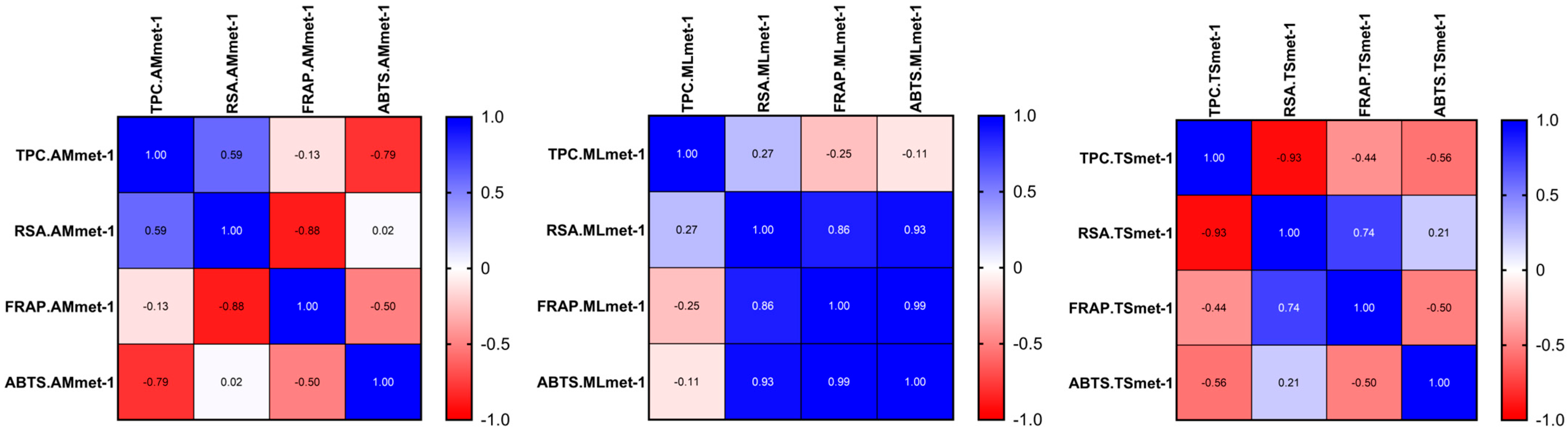
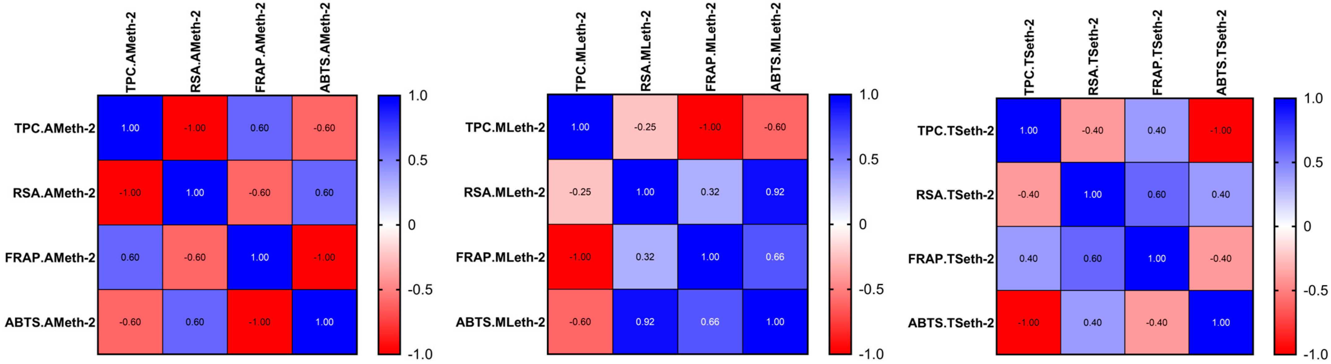
A Pearson correlation analysis was performed to evaluate the relationships between the TPC and antioxidant activities assessed by RSA, FRAP, and ABTS assays across the investigated plant extracts (A. millefolium L., M. longifolia L., and T. serpyllum L.), obtained using hydro-methanolic and hydro-ethanolic solvents (Figure 1 and Figure 2).
The analysis revealed highly variable and extract-dependent correlation patterns, with both positive and negative associations observed between the TPC and antioxidant activity. The Pearson correlation coefficients ranged from −1.00 to 0.60, indicating substantial variability in both the magnitude and direction of these relationships depending on the plant species and the extraction solvent. However, none of these correlations reached statistical significance (p > 0.05), indicating the absence of a consistent linear relationship between the TPC and antioxidant capacity.
A more detailed evaluation showed that the relationship between the TPC and RSA was inconsistent across the extracts. AMmet-1 exhibited a moderate positive correlation (r = 0.59), whereas TSmet-1 showed a strong negative correlation (r = −0.93), and AMeth-2 presented an extreme negative association (r = −1.00). Similarly, the TPC–FRAP and TPC–ABTS relationships varied markedly across the samples, with both positive and negative coefficients observed (TPC–ABTS in AMmet-1: r = −0.79; TPC–FRAP in MLeth-2: r = −1.00; TPC–ABTS in TSeth-2: r = −1.00). These findings clearly demonstrate that the relationship between the TPC and antioxidant activity is not only weak but inconsistent in direction, indicating that the TPC alone is not a reliable predictor of antioxidant capacity in the analyzed extracts. By contrast, strong positive correlations were consistently observed among the antioxidant assays, particularly between ABTS and FRAP (r = 0.99) and between RSA and ABTS (r = 0.93), especially in MLmet-1 and MLeth-2. This suggests a high level of agreement between these methods in evaluating antioxidant potential, despite their different underlying mechanisms.
The lack of significant and consistent correlations between the TPC and antioxidant activity may be explained by several factors. First, the TPC represents a global quantitative parameter and does not reflect the qualitative composition of phenolic compounds, which may differ substantially in antioxidant efficiency [105]. Second, other bioactive constituents, such as flavonoids, terpenoids, or vitamins, may contribute significantly to the overall antioxidant activity [106]. Third, the different mechanisms underlying the assays—radical scavenging (RSA, ABTS) and reducing power (FRAP)—may result in distinct responses depending on the phytochemical profile of each extract [107].
Overall, the results indicate that antioxidant activity is governed by a complex interplay of phytochemical constituents and cannot be reliably predicted based solely on the TPC.

2.4. The Impact of Plant Extracts on Drosophila Melanogaster Viability

2.4.1. Neutral Diet

Drosophila melanogaster is one of the most suitable model organisms used for a developmental toxicity evaluation of plants [62]. For this purpose, we investigated the influence of the concentration of the AMmet-3, MLmet-3, and TSmet-3 extracts on the viability and developmental time of the fruit fly reared on three culture media (0M, ND, and HSD). It is worth noting that all individuals tested had the same age, genotypes, and epigenomes, and all the differences between the control and tested groups could be attributed to the plant extracts [62].
The life cycle of D. melanogaster was previously monitored. The 0–2-hour-old embryos transform into first-instar larvae after 24 h. If there are sufficient nutrients, the first-instar larvae develop into the second and third instar larvae, each stage lasting 24 h. After the completion of the third-instar larval stage (three days), pupation occurs. Metamorphosis takes place in the next four to five days, which ends with the hatching of adult flies. On the other hand, if nutrients are insufficient to support a stage of development, the life cycle will be interrupted and the individuals will die without reaching the next stage of life [108].
Considering day 0 the day of seeding embryos into the medium, for all the extracts at the concentration of 100 μL of extract/2 mL of 0M, larvae were observed on day 2. Some of them died by day 10 and the majority were dead by day 15. All of the larvae were dead by day 19. At the concentration of 500 μL/2 mL of 0M, larvae emerged on day 3. By day 13, most of them were dead. For the concentration of 1 mL of extract/2 mL of 0M, some small larvae were seen on day 6. No pupae or flies were observed. Since 0M was free of nutrients, these results suggest that AMmet-3, MLmet-3, and TSmet-3 did not contain enough nutrients to support the completion of the larval stage of D. melanogaster.

2.4.2. Normal Diet

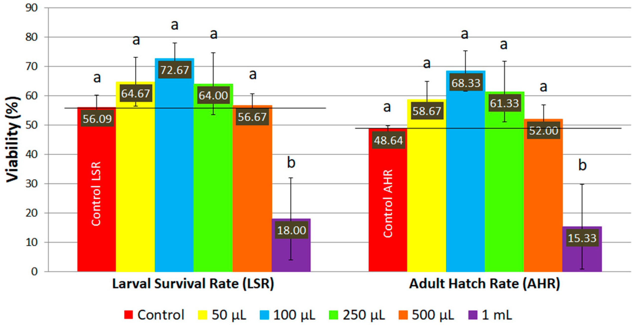
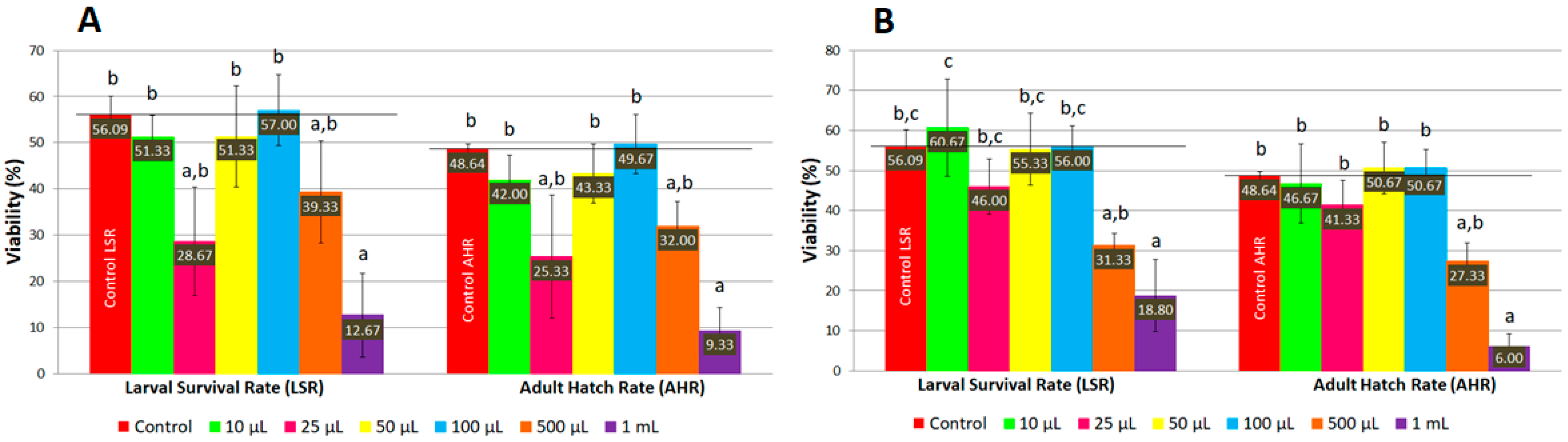
The viability of D. melanogaster fed with ND supplemented with AMmet-3 increased as the concentration of plant extract increased up to 100 μL per 2 mL of ND, where maximum survival rates were reached both for larvae (72.67%) and adult individuals (68.33%) (Figure 3). For higher concentrations (0.25, 0.50, and 1 mL of AMmet-3 per 2 mL of ND), the viability of D. melanogaster decreased as the concentration of the extract increased. However, at concentrations of 0.05, 0.10, 0.25, and 0.50 mL of extract per 2 mL of ND, higher survival rates of larvae and adult flies were recorded compared to the control group, but the mean values were not significantly different. At 1 mL of AMmet-3 per 2 mL of ND, a significant decrease of the viability was recorded compared to the control group.
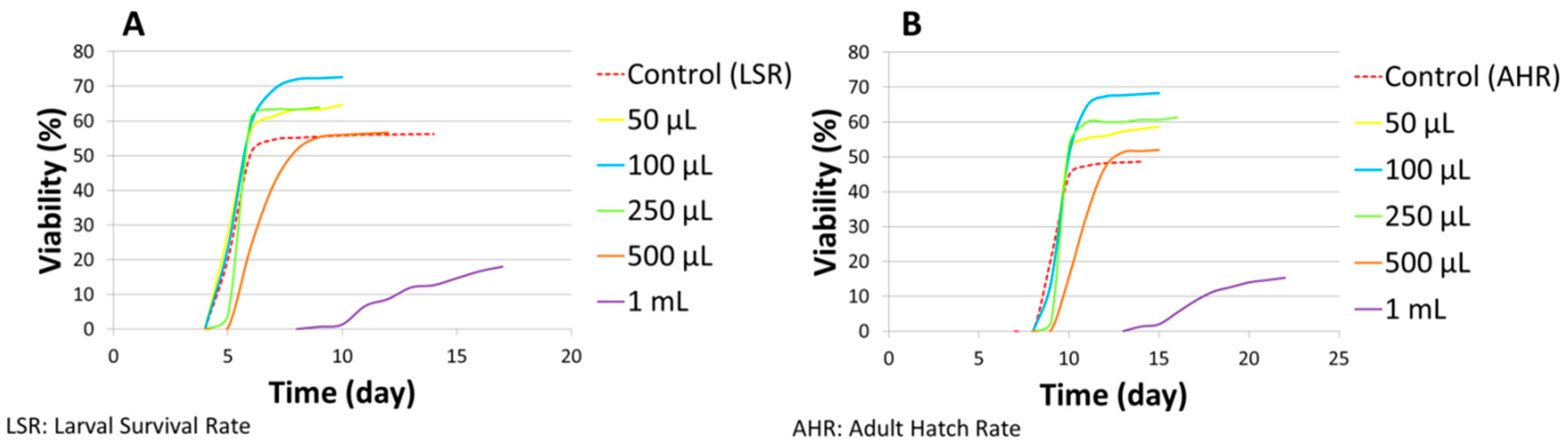
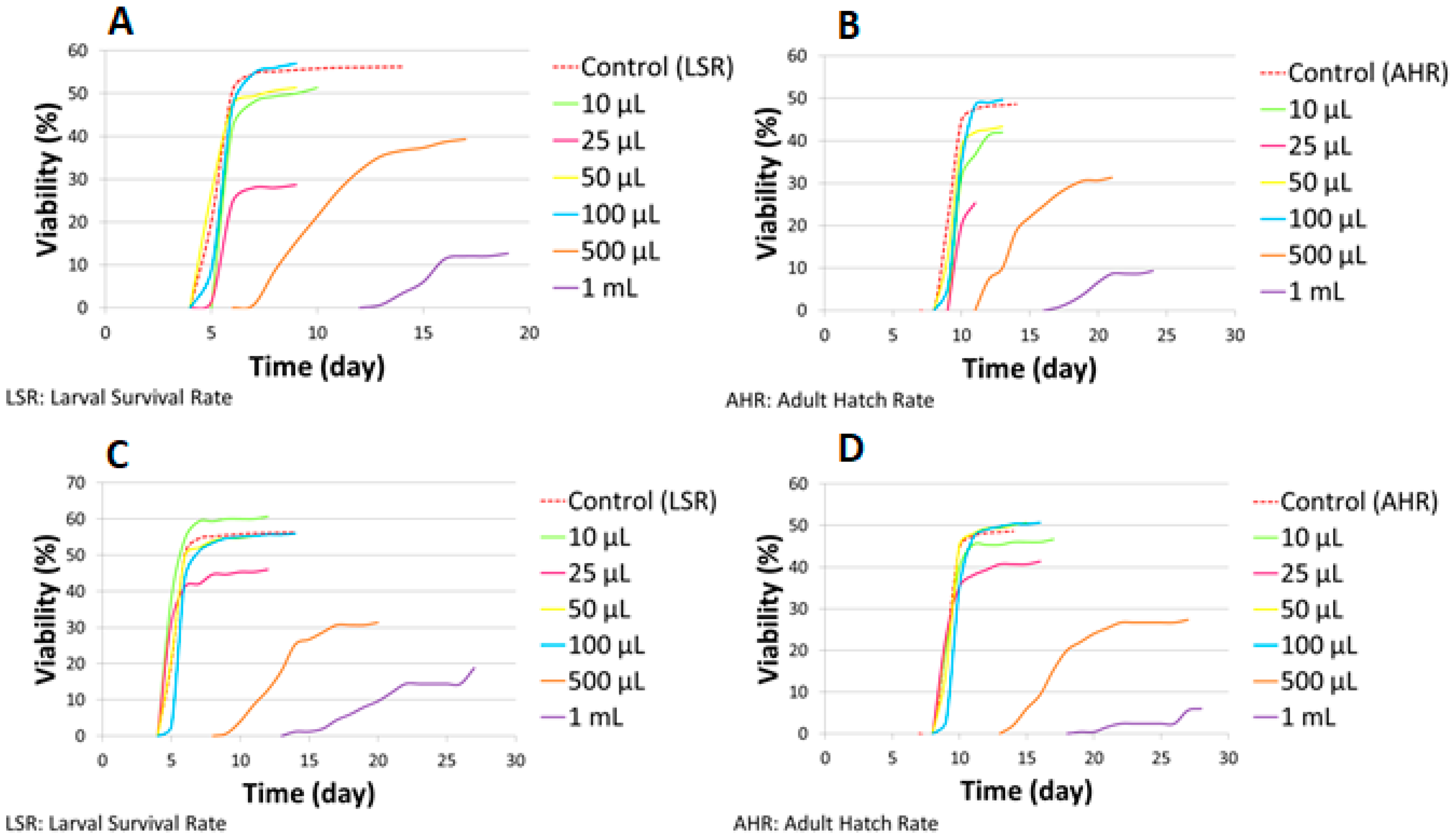
Simultaneously, the developmental time of D. melanogaster raised on ND supplemented with AMmet-3 was recorded (Figure 4). No differences were observed in the developmental time of larvae between the control and the groups treated with AMmet-3 at concentrations from 50 to 500 μL of AMmet-3 per 2 mL of ND (Figure 4A). Compared to the control, at 1 mL of AMmet-3 per 2 mL of ND, the larval period was extended by four days, with a significant decrease of the larval survival rate. A one-day delay was observed in the development of pupae to adult individuals in the groups treated with AMmet-3 at 50–500 μL/2 mL concentration range as compared to the control group, without affecting the viability (Figure 4B). At the concentration of 1 mL AMmet-3 per 2 mL ND, the development of pupae to adult individuals was extended by five days, with a significant decrease of the adult hatch rate.
Hormesis is a biphasic dose response, with beneficial or stimulatory effects at low doses and inhibition or toxicity at high doses. Some phytocompounds in low doses can exert a hormetic effect, which is an adaptive response of an organism to moderate stress [109]. The in vivo D. melanogaster-based viability assay revealed that the AMmet-3 extract tends to exert a hormetic effect with increasing the survival rates at low doses and inducing toxicity at high doses.
Previous studies have reported the hormetic effect induced by phytocompounds in D. melanogaster. Rutin, one of the most abundant compound detected in the AMmet-3 extract, extended the lifespan of fruit flies through a hormetic effect at a 100–800 μM concentration range. Furthermore, it significantly increased the percentage of pupation and adult hatching at 200 and 400 μM [110].
In another study, diet supplementation with resveratrol at 200 μM significantly increased the viability of pre-adult individuals according to the control, while at lower (25–100 μM) and higher (800 μM) concentrations no significant differences were reported compared to the control. Resveratrol did not significantly impact the developmental time of the pre-adults. However, supplementation of the larval diet with resveratrol extended the lifespan of adult individuals by stimulating the activity of the antioxidant enzymes which are involved in reactive oxygen species scavenging [111]. A previous study reported that resveratrol at low and moderate doses (7.5–60 mg/kg diet) increased the lifespan of fruit flies in a dose-dependent manner by 41.9% as compared to the control. However, at higher doses (120 mg/kg) no extinction of the lifespan was observed, suggesting a hormetic-like effect [112]. Previous research has highlighted the influence of the sex and the nutrient content of the diet. A standard diet supplemented with resveratrol up to 200 μM did not influence the lifespan of D. melanogaster males and females. Unlike males, an increase in lifespan was observed in females in the group fed a low-sugar, high-protein diet and 200 μM resveratrol and in the group fed a high-fat diet supplemented with 400 μM resveratrol [113].
Another study demonstrated that a normal diet supplemented with 50 and 100 μM gallic acid could reduce the oxidative stress in a D. melanogaster model of Alzheimer’s disease. Specifically, it decreased the reactive oxygen species and malondialdehyde levels and increased the catalase activity and total thiol level [114]. Quercetin, another bioactive compound identified in the investigated extracts, could also influence the antioxidant parameters in fruit flies. A previous study reported that the supplementation of the flies’ diet with quercetin (100 and 200 mg/g) increased the catalase activity, and a higher concentration of quercetin (300 mg/g) decreased the malondialdehyde level [115].
Comparable results were reported by Uysal et al. (2007) [116], who investigated the toxicity of A. millefolium L. (flowers and leaves) water extract at 1, 5, and 10 mL/100 mL standard food medium against D. melanogaster. They observed that the plant extract did not impact the duration of metamorphosis, since adult individuals emerged on the 9th day and lasted until the 17th day, both for control and test groups. Similar to our results, they reported that the viability of the flies increased in a concentration-dependent manner, without being significantly different according to the control. They mentioned that the observed effect could be attributed to the antioxidant effect of the extract [116]. A similar biphasic effect with stimulation at low doses and inhibition at high doses was observed in our previous study that assessed the impact of Lythrum salicaria L. extract on the viability of fruit flies [108]. Similar to our results, santol flesh extract exhibited a biphasic effect regarding adult emergence rate, with a maximum at 60 mg/mL and lower values at 20, 40, 80, and 100 mg/mL [117].
In the case of ND supplemented with MLmet-3 and TSmet-3, a similar pattern was observed regarding the viability of D. melanogaster (Figure 5). Specifically, for the concentrations of 10 and 25 μL of extract per 2 mL of ND, the viability of D. melanogaster decreased as the extract concentration increased. At these two concentrations, a higher viability compared to the control was only observed for the larvae in the group treated with 10 μL of TSmet-3 per 2 mL of ND. However, no significant difference was detected between the control and the tested groups at the above-mentioned concentrations. The viability increased in a dose-dependent manner at a concentration range of 25–100 μL of extract per 2 mL of ND, followed by a decrease of the viability at 0.1–1 mL concentration range.
Regarding larval developmental time, no differences were recorded with respect to the control in the groups treated with MLmet-3 and TSmet-3 at 10–100 μL/2 mL ND, except the group treated with 10 μL of MLmet-3 per 2 mL of ND, in which one day extinction was observed (Figure 6A,C). The larval stage was prolonged by two and eight days at 0.5 and 1 mL of MLmet-3 per 2 mL of ND. For 0.5 and 1 mL of TSmet-3 per 2 mL of ND, the larval period was extended by four and nine days compared to the control. The pupal stage extension varied depending on the extract concentration, between 1 and 9 days in the case of MLmet-3 and 1 and 11 days in the case of TSmet-3 (Figure 6B,D).
In the present study, MLmet-3 and TSmet-3 exerted a triphasic dose–response, with inhibition at ultra-low concentrations, stimulation at low concentrations, and inhibition at high concentrations [118]. The triphasic dose–response relationship implies a reparative response that occurs after the threshold for detecting damage is exceeded. Specifically, very low concentrations of a toxicant can induce moderate damage without being detected by the biological system. At low doses, more damage can occur, exceeding the threshold for detecting damage. At moderate concentrations, damage can be detected, and the system will initiate a reparative response (the hormetic domain of the triphasic dose–response curve). At higher concentrations, more damage occurs, and the reparative process will be overloaded, resulting in massive damage [119].

2.4.3. High-Sugar Diet

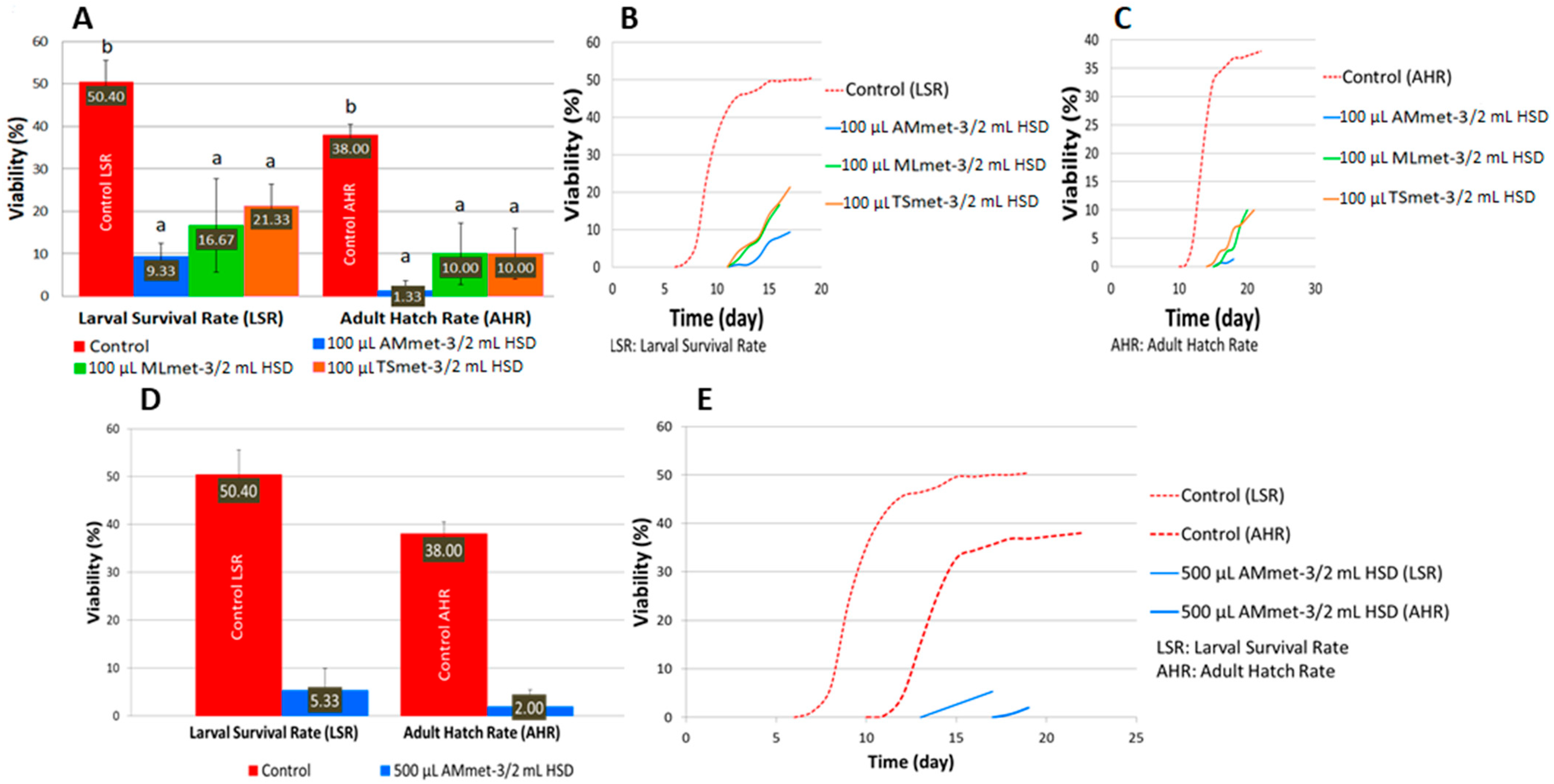
Compared to the ND, the HSD decreased the larval and adult hatch rates of the controls simultaneously with a developmental shift. Supplementation of the HSD with the investigated plant extracts (AMmet-3, MLmet-3, and TSmet-3) increased the toxicity compared to the supplemented ND. At the concentration of 100 μL of extract/2 mL of HSD, all the extracts significantly decreased the viability of the larvae and pupae compared to the control; AMmet-3 was the most toxic, followed by MLmet-3 and TSmet-3 (Figure 7A). At 500 μL of extract/2 mL of HSD, MLmet-3 and TSmet-3 were completely toxic, while AMmet-3 reduced the larval survival and adult hatch rates at 5.33% and 2.00%, respectively (Figure 7D). At 1 mL of extract/2 mL of HSD, no larvae or adult individuals were detected. Overall, the toxicity of the HSD supplemented with the tested extract concentrations increased in a dose-dependent manner. Moreover, the decrease in viability is accompanied by a change in the development time, with a delay of approximately 5 days at 100 μL of extract/2 mL of HSD (Figure 7B,C) and 7 days at 500 μL of AMmet-3 per 2 mL of HSD (Figure 7E).
This study is in agreement with previous reports where a HSD also decreased the survival rate of D. melanogaster compared to a normal diet [120,121]. Furthermore, other researchers have also observed a developmental delay in larval or pupal stages after the exposure of D. melanogaster to a HSD [121,122,123]. Feeding D. melanogaster on a HSD has been previously reported to induce a type 2 diabetes mellitus-like phenotype by increasing glucose and triglycerides levels [120,121,122,123], stimulating the activity of carbohydrate hydrolyzing enzymes, such as α-amylase and α-glucosidase [120,122], modulating the expression of genes implicated in the carbohydrate metabolism [122,123,124], and reducing the body size, body weight, and movement capacity of the larvae [124]. It was also associated with insulin resistance and obesity [123]. Moreover, a HSD was reported to generate oxidative stress [120,121] by increasing the level of reactive oxygen species and thiobarbituric acid reactive species, and by decreasing the level of thiol, glutathione-S-transferase, superoxide dismutase, and catalase. Cognitive decline and brain damage could be the consequences of a HSD through increased acetylcholinesterase and monoamine oxidase activity [120].
Other researchers have demonstrated the antidiabetic potential of plant products in Drosophila melanogaster model, such as bayberry leaves [122], Flos Chrysanthemi Indici extract [125], Cordia myxa [124], Syzygium cumini [121], and moringa leaves [120]. On the other hand, some plant extracts were not effective in ameliorating the harmful effects induced by a HSD, some of them even exacerbating the toxicity. Similar to our results, Syzygium malaccense extract has shown a dose-dependent toxic effect. At concentrations of 0.625–2.5%, the survival rate of larvae and pupae decreased (significantly, except for the viability of pupae at the concentration of 0.625%). At the concentration of 0.625%, a low toxicity was reported without major developmental issues; but, at higher concentrations (1.25-2.5%), mortality increased and the development was delayed [124]. Similar results were observed for Lythrum salicaria L. extract, which exacerbated the toxic effect of the HSD simultaneously with a developmental shift [108]. Overall, these results suggest that an investigation of toxicity is crucial for plant extracts before introducing them into therapy as various interactions could occur, resulting in protective or toxic effects, depending on the type of extract and diet.

2.5. The Impact of Plant Extracts on Position-Effect Variegation

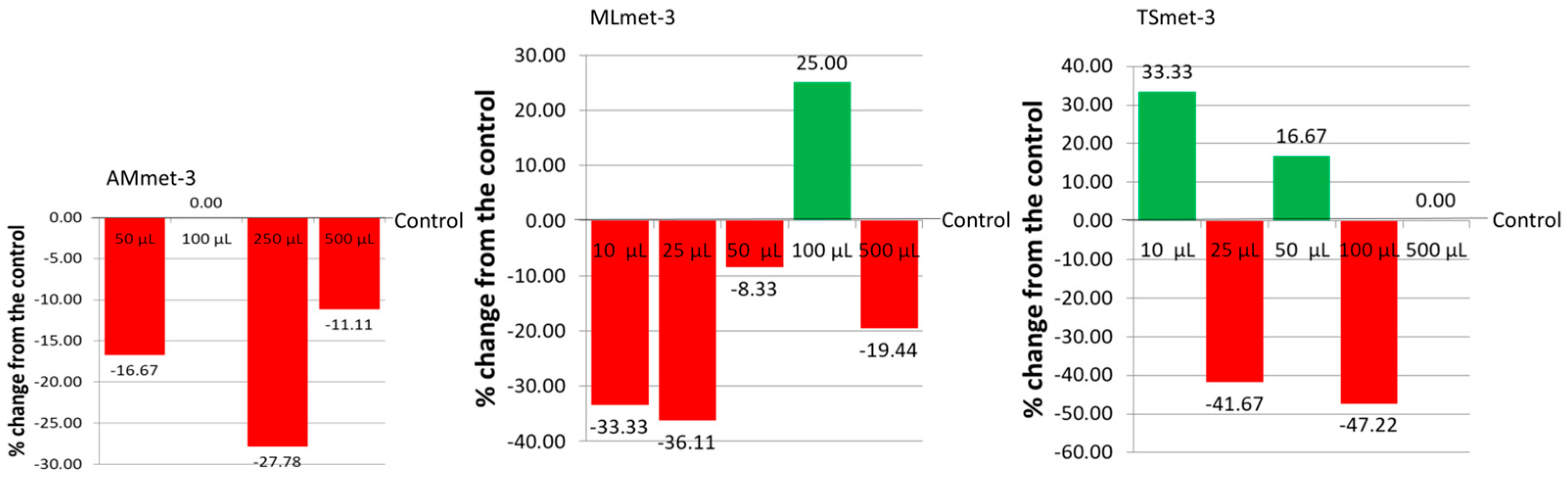
In order to study the impact of the investigated extracts on PEV, we quantified the red pigment, drosopterin, from the eyes of the D. melanogaster wm4h strain. The results revealed that all three extracts influenced the expression of genes, leading to different drosopterin content in the eyes of adult male individuals (Figure 8). Compared to the control, three of four tested concentrations (50, 250, and 500 μL per 2 mL of ND) decreased the drosopterin eye content in D. melanogaster wm4h. No difference compared to the control was recorded at the concentration of 100 μL of AMmet-3 per 2 mL of ND. The concentration of 100 μL of MLmet-3 per 2 mL of ND increased the drosopterin eye content, while the other tested concentrations decreased the drosopterin levels compared to the untreated control. Drosopterin content was elevated in the eyes of male individuals fed with 10 and 50 μL of TSmet-3 per 2 mL of ND, while it was lower than the control at 25 and 100 μL/2 mL of ND. Compared to the control, 500 μL of TSmet-3 per 2 mL of ND had no influence on drosopterin eye content.
PEV is a phenomenon that occurs when a gene located in the euchromatin region is relocated to the heterochromatin region resulting in a variegated phenotype. The genes involved could be stochastically silenced by the spread of heterochromatin across the euchromatin/heterochromatin border [126]. The white gene encodes a transporter responsible for red eye pigmentation in wild fruit flies [127]. The D. melanogaster wm4h strain is an intensively studied model of PEV, in which the white gene is relocated in the pericentric heterochromatin zone of the X chromosome [128]. This rearrangement leads to expression of the white gene in some ocular cells and inhibition of its expression in other cells, resulting in individuals with white-mottled eyes [126]. The results of this study are consistent with the findings of our previous study, according to which Lythrum salicaria L. extract influenced the drosopterin concentration in adult male individuals of D. melanogaster wm4h compared to the untreated control [108].

3. Materials and Methods

3.1. Chemical and Reagents

Chloroform, methanol, glycerol, acetic acid, 2,2-diphenyl-1-picrylhydrazyl (DPPH), 2,4,6-tripyridyl-s-triazine (TPTZ), and ammonium hydroxide were acquired from the Sigma-Aldrich Company, St. Louis, MO, USA. Gallic acid, chlorogenic acid, caffeic acid, syringic acid, ferulic acid, cinnamic acid, (+)-catechin, rutin, quercetin, and resveratrol were purchased from Dr. Ehrenstorfer GmbH, Augsburg, Germany. Phosphomolybdotungstic reagent, potassium persulfate, charcoal, saccharose, and agar powder were obtained from VWR Chemicals, Leuven, Belgium. Sodium carbonate, ascorbic acid, sodium acetate, FeCl3, and HCl were acquired from Chemical Company S.A., Iași, Romania. 2,2′-azinobis [3-ethylbenzothiazoline-6-sulfonic acid]-diammonium salt (ABTS), trolox, and nipagin were purchased from Thermo Fisher Scientific, Kandel, Germany. Ethanol was obtained from Chimreactiv S.R.L., Bucharest, Romania.

3.2. Plant Collection

The aerial parts of the studied medicinal plants were collected from the mountainous region in Sibiu County, Romania, in July 2021 (Achillea millefolium L.—45°41′20” N, 23°59′9” E, 1130 m; Mentha longifolia L.—45°41′22” N, 23°59′29” E, 1100 m; Thymus serpyllum L.—45°42′34” N, 24°1′33” E, 630 m). They were authenticated by the botanist Dr. Oana Danci, according to the “Vascular Plants of Romania, illustrated field guide” by Sârbu et al. (2013) [129]. A voucher specimen of each vegetal product is held at the Faculty of Medicine (Pharmacy specialization), “Lucian Blaga” University of Sibiu, Sibiu, Romania (A. millefolium L. no. 101/2, M. longifolia L. no. 101/3, and T. serpyllum L. 101/4). After harvesting, the raw materials were chopped and shade dried at room temperature. After drying, the vegetal products were kept in labeled paper bags at room temperature, away from light until extraction.

3.3. Extraction Procedure

Two types of extracts were prepared from each plant species. The vegetal aerial parts were milled using a domestic coffee mill until a powder was obtained. For the first extraction, the powder/solvent ratio was 0.5:10 w/v. The powder and the solvent (70% methanol) were introduced in an Erlenmeyer flask with a ground-glass stopper. The extraction was performed in an ultrasonic water bath at 40 °C for 30 min. The selection of the solvent mixture (methanol/distilled water, 70:30, v/v) was based on a previous study that showed that mixing methanol and water in this ratio extracts a greater amount of phenolic compounds than when using methanol alone [70]. Subsequently, the mixture was cooled, filtered in a 10 mL volumetric flask, and brought to mark with the same solvent. Three distinct extracts, named as A. millefolium L. hydro-methanolic extract (AMmet-1), M. longifolia L. hydro-methanolic extract (MLmet-1), and T. serpyllum L. hydro-methanolic extract (TSmet-1) were obtained. For the second extraction, the powder/solvent (70% ethanol) ratio was 1:10, w/v. Three different extracts, named as A. millefolium L. hydro-ethanolic extract (AMeth-2), M. longifolia L. hydro-ethanolic extract (MLeth-2), and T. serpyllum L. hydro-ethanolic extract (TSeth-2) were obtained. As soon as the extracts were prepared, their chemical composition and antioxidant capacity were assessed. To avoid the toxicity of methanol in the testes performed on D. melanogaster, the hydro-methanolic extracts were further processed by evaporating the methanol. The hydro-methanolic extracts were chosen due to the expectation of a grater phenolic content and a stronger antioxidant capacity compared to the hydro-ethanolic extracts. The products obtained after methanol evaporation were diluted with glycerol in a 2:1, v/v ratio, resulting the final mixtures labeled as AMmet-3, MLmet-3, and TSmet-3 [108].

3.4. HPLC-DAD Analysis

The phenolic profile of the extracts was assessed by an HPLC method adapted after different methods from the specialized literature [34,108,130], using an SCL-40 HPLC system (Shimadzu, Kyoto, Japan) provided with a quaternary pump, autosampler, thermostatted column oven, and photodiode array detector. A Nucleosil C18 column, set to 40 °C, 250 mm × 4.6 mm, and i.d. 5 μm, was used. The injection volume of the sample was 5 μL. The elution was performed in gradient, at a flow rate of 0.8 mL/min, using a mobile phase with two components: (1) purified water/acetic acid 96:4, v/v and (2) methanol. The change in the composition of the mobile phase over time was set as follows: 85% (1) and 15% (2) for the first 15 min, 75% (1) and 25% (2) for the next 5 min, 15% (1) and 85% (2) for 10 min, 85% (1) and 15% (2) for 5 min, and 85% (1) and 15% (2) for the last min. The detection was performed at 280 nm for gallic acid, (+)-catechin, syringic acid, and cinnamic acid, at 306 nm for resveratrol, at 330 nm for chlorogenic acid, caffeic acid, and ferulic acid, and at 360 nm for rutin and quercetin. Concentrations between 0.2 and 50 μg/mL were tested. All the standards used for the identification and quantification of the phenolic compounds presented purity greater than 94%: purity > 99% for gallic acid, ferulic acid, cinnamic acid, caffeic acid, and resveratrol; > 98% for (+)-catechin; > 95% for syringic acid, chlorogenic acid, and quercetin; > 94% for rutin. They were analyzed due to their potential beneficial properties. All the analyses were performed in triplicate. The results were expressed as μg of compound/g of dry weight.

3.5. Spectrophotometric Analysis

3.5.1. Total Phenolic Content

The total phenolic content (TPC) of the extracts was estimated by the Folin–Ciocâlteu method. In a test tube, the extract was mixed with Phosphomolybdotungstic reagent, distilled water, and 290 g/L of sodium carbonate solution in a volume ratio of 0.4:1:15:2. The mixture was shaken for 10 min and kept in a water bath at 40 °C for 20 min. Subsequently, the mixture was cooled at room temperature and its absorbance at 760 nm was measured against a blank using a UV-1900 UV-VIS spectrophotometer (Shimadzu, Kyoto, Japan) [131].
For the calibration curve, solutions with concentrations ranging from 0.86 to 8.57 μg gallic acid/mL were prepared. All the analyses were performed in triplicate. The results were expressed as mg gallic acid equivalents (GAE)/g dry weight.

3.5.2. DPPH Radical Scavenging Assay

The DPPH radical scavenging assay was performed to analyze the antioxidant activity of the extracts. Prior to the analysis, DPPH was dissolved in methanol to prepare a 25 μg/mL DPPH stock solution. It was kept in a dark place for 2 h before analysis. A mixture consisting of 1.94 mL DPPH stock solution and 60 μL extract was prepared and kept away from light at room temperature for 15 min, until absorbance stabilization. Subsequently, the absorbance at 515 nm was reached against a blank using a UV-1900 UV-VIS spectrophotometer (Shimadzu, Kyoto, Japan) [71]. A solution of 1 mg ascorbic acid/mL was used as a positive control.
For the calibration curve, solutions with concentrations ranging from 0.5 to 25 μg DPPH/mL were prepared. All the analyses were performed in triplicate. The results were expressed as DPPH inhibition percentages ( I % ), which were calculated by formula (1):
I % = C s t C s a m C s t ,
C s t is the concentration of the stock solution and C s a m is the concentration of the sample, both in μg DPPH/mL.
The concentration of DPPH in the sample ( C s a m ), expressed as μg DPPH/mL, was calculated with Formula (2):
C s a m = A s a m i n t e r c e p t s l o p e ,
A s a m is the absorbance of the sample, i n t e r c e p t is the intercept of the calibration curve, and s l o p e is the slope of the calibration curve.

3.5.3. ABTS Radical Scavenging Assay

Due to the fact that a single test is insufficient to evaluate the antioxidant capacity of an extract, in addition to the DPPH assay, the ABTS radical scavenging assay was conducted. ABTS and potassium persulfate were dissolved in water to form a 7 mM ABTS and 2.45 mM potassium persulfate stock solution. It was left at room temperature away from light for 16 h prior to analysis. When the time had passed, the stock solution was diluted until the absorbance at 734 nm reached 0.7 ± 0.02. A mixture consisting of 2.5 mL of the stock solution and 25 μL of the extract was prepared in a test tube and vortexed for 30 s. Its absorbance at 734 nm was measured after 1 min, using a UV-1900 UV-VIS spectrophotometer (Shimadzu, Kyoto, Japan) [132]. A solution of 1 mg ascorbic acid/mL was used as a positive control.
For the calibration curve, solutions with concentrations ranging from 0.125 to 2 mmol trolox/L were prepared. All the analyses were performed in triplicate. The results were expressed as mmol trolox equivalents (TE)/g dry weight.

3.5.4. Ferric-Reducing Antioxidant Power (FRAP) Assay

In order to evaluate the antioxidant activity of the extracts, their capacity to reduce Fe3+ to Fe2+ was investigated by the FRAP assay. Three stock solutions were prepared: 300 mM acetate buffer (pH = 3.6), 20 mM FeCl3, and 10 mM TPTZ acidified with 150 μL HCl. The FRAP working solution was obtained just before the analysis by mixing 50 mL of the acetate buffer solution with 5 mL of the FeCl3 solution and 5 mL of the TPTZ solution. A mixture consisting of 0.1 mL of the plant extract, 1.5 mL of the FRAP reagent, and 2 mL of distilled water was prepared in a test tube and kept at room temperature protected from light. The absorbance at 595 nm against a blank was recorded using a UV-1900 UV-VIS spectrophotometer (Shimadzu, Kyoto, Japan) [132]. A solution of 1 mg ascorbic acid/mL was used as a positive control.
For the calibration curve, solutions with concentrations ranging from 0.15 to 0.50 μmol trolox/mL were prepared. All the analyses were performed in triplicate. The results were expressed as μmol TE/g dry weight.

3.6. In Vivo Drosophila melanogaster Viability Assay

This study investigated the toxicity of the AMmet-3, MLmet-3, and TSmet-3 extracts prepared as presented in the Section 2.3 by using an in vivo test on the D. melanogaster model system. The protocol consisted of assessing the viability of D. melanogaster fed with three types of diets supplemented with plant extracts at various concentrations. A culture medium with no nutrients (0 M) was prepared by mixing 1 g of charcoal with 100 mL of distilled water and 1 g of agar powder. The distilled water was previously boiled to prevent mold contamination. Subsequently, the charcoal and agar powder were added under continuous stirring, boiled for 30 s, and cooled to 45 °C. The second medium simulated a normal diet (ND) and was obtained by introducing 70 g of yeast paste into 1200 mL of distilled water under continuous stirring, followed by addition of 52 g of saccharose and 30 g of wheat flour. After boiling, 10 g of agar powder were added and the mixture was boiled for 30 min. After cooling to 50 °C, 1 g of nipagin was incorporated to preserve the microbiological stability. The third medium simulated a high sugar diet (HSD). It was prepared similarly to the ND, but with a concentration of saccharose of 0.75M [133].
Three extract concentrations were tested on 0M and HSD: 4.76%, 20%, and 33.33%. They were obtained by introducing 0.1, 0.5, and 1 mL of extract in 2 mL of medium dispensed in culture vials. These concentrations were selected based on a previous study that assessed the influence of plant extracts on the viability of D. melanogaster, which tested concentrations between 11% and 50% [133]. Based on our observations of 0M, for a better understanding of the dose–effect response, we decided to test additional extract concentrations on ND. For AMmet-3, a total of five concentrations were tested: 2.44%, 4.76%, 11.11%, 20%, and 33.33% (prepared by mixing 0.05, 0.1, 0.25, 0.5, and 1 mL of extract with 2 mL of culture medium). For MLmet-3 and TSmet-3, a total of six extract concentrations were tested on ND: 0.5%, 1.23%, 2.44%, 4.76%, 20%, and 33.33%, (obtained by mixing 0.01, 0.025, 0.05, 0.1, 0.5, and 1 mL of extract with 2 mL of culture medium).
An embryo collector containing about 200 of 5-day old male and female D. melanogaster wm4h individuals was placed over a Petri dish filled with 0M supplemented with yeast paste. On the next day, 50 embryos of D. melanogaster wm4h aged between 0 and 2 hours were introduced into each of the vials containing the media supplemented with plant extracts under the microscope (VWR VisiScope SZB260 stereo microscope, VWR, Milano, Italy) by using a fine forceps. The negative control was prepared by introducing 50 embryos into a vial with 2 mL of culture medium. The number of newly emerged pupae and flies were monitored daily. All the experiments were performed in triplicate, under specific conditions at 25 °C and constant humidity. The results were expressed as percentage of viability [133].

3.7. Position Effect Variegation Study

This study evaluated the position-effect variegation by quantifying the drosopterin pigment in the eyes of D. melanogaster adult male individuals. For each concentration tested on ND, ten males were randomly selected and their heads were removed with a scalpel under a stereo microscope (VWR VisiScope SZB260 stereo microscope, VWR, Milano, Italy). The pigments were extracted by mixing the heads with 300 μL of chloroform and 300 μL of 0.1% ammonium hydroxide solution, followed by homogenization and centrifugation at 4000 revolutions per min for 4 min. A volume of 200 μL of supernatant was added over 500 μL of 0.1% ammonium hydroxide solution, followed by homogenization. The absorbances were read against ethanol at 485 nm using a UV/Vis Ultrospec 2100 pro spectrophotometer (Biochrom Ltd., Cambridge, UK) [134].

3.8. Statistical Analysis

The results of the tests performed in triplicate were expressed as mean values and standard deviation. The significant difference between the mean was calculated by one-way ANOVA (Tukey’s post hoc test) using IMB SPSS statistics for Windows, version 26 (IBM Corp., Armonk, NY, USA). A Pearson correlation analysis was performed using GraphPad Prism 8 (GraphPad Software, San Diego, CA, USA) to evaluate the relationships between total phenolic content and antioxidant activities. A p-value < 0.05 was considered statistically significant.

4. Conclusions

This study investigated the phenolic profile, antioxidant activity, and the impact on D. melanogaster viability and gene expression, of natural extracts obtained from A. millefolium L., M. longifolia (L.) Huds., and T. serpyllum L. Rutin was the main phenolic compound in the M. longifolia (L.) Huds. and T. serpyllum L. hydro-methanolic extracts, while (+)-catechin predominated in the A. millefolium L. hydro-methanolic extract. Resveratrol, quercetin, and cinnamic acid were the most abundant phenolic compounds in the hydro-ethanolic extracts of A. millefolium L., M. longifolia (L.) Huds., and T. serpyllum L., respectively. Due to the different amounts of polyphenols in the extracts, they demonstrated variable antioxidant activity.
Regarding the toxicity, low concentrations (50–500 μL/2 mL normal medium) of the A. millefolium L. extract did not affect the viability of D. melanogaster, while the concentration of 1 μL/2 mL of the normal medium was toxic. A more complex response was recorded in the case of normal medium supplemented with the M. longifolia (L.) Huds. and T. serpyllum L. extracts. A high sugar diet increased the toxicity of the investigated extracts.
The drosopterin content was affected by the tested extracts, suggesting the presence of some phytochemicals that could interfere with gene expression. Future studies are needed to clearly define the conditions for using these extracts in therapy.

Supplementary Materials

The following supporting information can be downloaded at: https://www.mdpi.com/article/10.3390/molecules31091527/s1, Table S1: Summary of the results of other studies on selected phenolic compounds from the analyzed plant species; Table S2: Summary of the results of other studies on total phenolic content and antioxidant activity from the analyzed plant species. Reference [135] is cited in Supplementary Materials.

Author Contributions

Conceptualization, C.G., A.F., E.M., and F.-G.G.; methodology, L.-I.V., C.G., A.F., B.P., R.N., O.V.D., M.M., E.M., M.T., M.-L.M., and F.-G.G.; software, L.-I.V., A.F., B.P., and R.N.; validation, L.-I.V., C.G., M.M., A.F., E.M., O.V.D., M.T., M.-L.M., and F.-G.G.; formal analysis, L.-I.V., A.F., B.P., R.N., O.V.D., and M.-L.M.; investigation, L.-I.V., C.G., A.F., B.P., R.N., O.V.D., M.M., E.M., M.T., M.-L.M., and F.-G.G.; resources, C.G., A.F., E.M., and F.-G.G.; data curation, L.-I.V., A.F., B.P., R.N., O.V.D., M.M., M.T., and M.-L.M.; writing—original draft preparation, L.-I.V., C.G., A.F., E.M., and F.-G.G.; writing—review and editing, L.-I.V., C.G., A.F., B.P., R.N., O.V.D., M.M., E.M., M.T., M.-L.M., and F.-G.G.; visualization, L.-I.V., A.F., O.V.D., and M.T.; supervision, C.G., A.F., E.M., and F.-G.G.; project administration, C.G., A.F., E.M., and F.-G.G.; funding acquisition, C.G. All authors have read and agreed to the published version of the manuscript.

Funding

Project financed by Lucian Blaga University of Sibiu through the research grant LBUS-IRG-2024.

Institutional Review Board Statement

All animal experimental procedures in this study were approved by the Ethical Committee for the Welfare of Laboratory Animals, the University of Debrecen (Ethics review number DEMAB4/2021). Date of approval: 22 July 2021.

Informed Consent Statement

Not applicable.

Data Availability Statement

Data are contained within the article.

Conflicts of Interest

The authors declare no conflicts of interest.

References

  1. Jamshidi-Kia, F.; Lorigooini, Z.; Amini-Khoei, H. Medicinal Plants: Past History and Future Perspective. J. Herbmed Pharmacol. 2018, 7, 1–7. [Google Scholar] [CrossRef]
  2. World Health Organization. WHO Global Report on Traditional, Complementary and Integrative Medicine 2024; World Health Organization: Geneva, Switzerland, 2025; ISBN 978-92-4-011138-7. [Google Scholar]
  3. Etaware, P.M.; America, O.; Egara, O.W.; Ekun, V.S. Herbal Medicine: Scientific Validation and Future Prospects. Int. J. Pharm. Chem. 2025, 11, 67–75. [Google Scholar] [CrossRef]
  4. Muscolo, A.; Mariateresa, O.; Giulio, T.; Mariateresa, R. Oxidative Stress: The Role of Antioxidant Phytochemicals in the Prevention and Treatment of Diseases. Int. J. Mol. Sci. 2024, 25, 3264. [Google Scholar] [CrossRef]
  5. Chandimali, N.; Bak, S.G.; Park, E.H.; Lim, H.-J.; Won, Y.-S.; Kim, E.-K.; Park, S.-I.; Lee, S.J. Free Radicals and Their Impact on Health and Antioxidant Defenses: A Review. Cell Death Discov. 2025, 11, 19. [Google Scholar] [CrossRef]
  6. Ali, S.I.; Gopalakrishnan, B.; Venkatesalu, V. Pharmacognosy, Phytochemistry and Pharmacological Properties of Achillea Millefolium L.: A Review. Phytother. Res. 2017, 31, 1140–1161. [Google Scholar] [CrossRef] [PubMed]
  7. Miraldi, E.; Ferri, S.; Mostaghimi, V. Botanical Drugs and Preparations in the Traditional Medicine of West Azerbaijan (Iran). J. Ethnopharmacol. 2001, 75, 77–87. [Google Scholar] [CrossRef]
  8. Segneanu, A.-E.; Cepan, C.; Grozescu, I.; Cziple, F.; Olariu, S.; Ratiu, S.; Lazar, V.; Murariu, S.M.; Velciov, S.M.; Marti, T.D. Therapeutic Use of Some Romanian Medicinal Plants. In Pharmacognosy—Medicinal Plants; Perveen, S., Al-Taweel, A., Eds.; IntechOpen: Rijeka, Croatia, 2019; ISBN 978-1-83880-610-1. [Google Scholar]
  9. Karpavičienė, B. Traditional Uses of Medicinal Plants in South-Western Part of Lithuania. Plants 2022, 11, 2093. [Google Scholar] [CrossRef]
  10. Kozlowska, W.; Wagner, C.; Moore, E.M.; Matkowski, A.; Komarnytsky, S. Botanical Provenance of Traditional Medicines From Carpathian Mountains at the Ukrainian-Polish Border. Front. Pharmacol. 2018, 9, 295. [Google Scholar] [CrossRef]
  11. Radušienė, J.; Karpavičienė, B.; Raudone, L.; Vilkickyte, G.; Çırak, C.; Seyis, F.; Yayla, F.; Marksa, M.; Rimkienė, L.; Ivanauskas, L. Trends in Phenolic Profiles of Achillea millefolium from Different Geographical Gradients. Plants 2023, 12, 746. [Google Scholar] [CrossRef]
  12. Turker, A.U.; Yildirim, A.B.; Tas, I.; Ozkan, E.; Turker, H. Evaluation of Some Traditional Medicinal Plants: Phytochemical Profile, Antibacterial and Antioxidant Potentials. Rom. Biotechnol. Lett. 2021, 26, 2499–2510. [Google Scholar] [CrossRef]
  13. Afshari, M.; Rahimmalek, M.; Miroliaei, M. Variation in Polyphenolic Profiles, Antioxidant and Antimicrobial Activity of Different Achillea Species as Natural Sources of Antiglycative Compounds. Chem. Biodivers. 2018, 15, e1800075. [Google Scholar] [CrossRef]
  14. Sowa, P.; Marcinčáková, D.; Miłek, M.; Sidor, E.; Legáth, J.; Dżugan, M. Analysis of Cytotoxicity of Selected Asteraceae Plant Extracts in Real Time, Their Antioxidant Properties and Polyphenolic Profile. Molecules 2020, 25, 5517. [Google Scholar] [CrossRef]
  15. Benedec, D.; Popica, I.-E.; Oniga, I.; Hanganu, D.; Duma, M.; Silaghi-Dumitrescu, R.; Bischin, C.; Vlase, L. Comparative HPLC-MS Analysis of Phenolics from Achillea Distans and Achillea millefolium and Their Bioactivity. Stud. UBB Chem. 2015, 60, 257–266. [Google Scholar]
  16. Konarska, A.; Weryszko-Chmielewska, E.; Materska, M.; Sulborska-Różycka, A.; Dmitruk, M.; Chilczuk, B. Phenolic Compounds in Flowers and Herb of Achillea millefolium L.: Histochemical and Phytochemical Studies. Molecules 2025, 30, 2084. [Google Scholar] [CrossRef]
  17. Devadharshini, R.; Kavya, S.V.; Gowrishankar, M.; Renuka, K.; Muthukumaran, P. In Vitro Evaluation of Total Phenol and Antioxidant Activity of Achillea millefolium and Stevia Rebaudiana. J. Chem. Pharm. Res. 2021, 13, 1–8. [Google Scholar]
  18. Raudone, L.; Vilkickyte, G.; Marksa, M.; Radusiene, J. Comparative Phytoprofiling of Achillea millefolium Morphotypes: Assessing Antioxidant Activity, Phenolic and Triterpenic Compounds Variation across Different Plant Parts. Plants 2024, 13, 1043. [Google Scholar] [CrossRef] [PubMed]
  19. Georgieva, L.; Gadjalova, A.; Mihaylova, D.; Pavlov, A. Achillea millefolium L.—Phytochemical Profile and In Vitro Antioxidant Activity. Int. Food Res. J. 2015, 22, 1347–1352. [Google Scholar]
  20. Adil, M.; Dastagir, G.; Quddoos, A.; Naseer, M.; Filimban, F.Z. HPLC Analysis, Genotoxic and Antioxidant Potential of Achillea millefolium L. and Chaerophyllum villosum Wall ex. Dc. BMC Complement. Med. Ther. 2024, 24, 91. [Google Scholar] [CrossRef]
  21. Bljajić, K.; Brajković, A.; Čačić, A.; Vujić, L.; Jablan, J.; Saraiva De Carvalho, I.; Zovko Končić, M. Chemical Composition, Antioxidant, and α-Glucosidase-Inhibiting Activity of Aqueous and Hydroethanolic Extracts of Traditional Antidiabetics from Croatian Ethnomedicine. Horticulturae 2021, 7, 15. [Google Scholar] [CrossRef]
  22. Mohamed, D.A.; Hanfy, E.A.; Fouda, K. Evaluation of Antioxidant, Anti-Inflammatory and Anti-Arthritic Activities of Yarrow (Achillea millefolium). J. Biol. Sci. 2018, 18, 317–328. [Google Scholar] [CrossRef]
  23. Bobis, O.; Dezmirean, D.S.; Tomos, L.; Chirila, F.; Marghitas, L.A. Influence of Phytochemical Profile on Antibacterial Activity of Different Medicinal Plants against Gram-Positive and Gram-Negative Bacteria. Appl. Biochem. Microbiol. 2015, 51, 113–118. [Google Scholar] [CrossRef]
  24. Faiku, F.; Haziri, A.; Faiku, F.; Faiku, B. Composition of Some Macro and Micro Elements, Polyphenol Content, Antimicrobial and Antioxidant Activity of Achillea millefolium (L.) Grown in Kosovo. Chemija 2025, 36, 69–77. [Google Scholar] [CrossRef]
  25. Ayhan, N.K.; Tunc, M.G.K.; Noma, S.A.A.; Kurucay, A.; Ates, B. Characterization of the Antioxidant Activity, Total Phenolic Content, Enzyme Inhibition, and Anticancer Properties of Achillea millefolium L. (Yarrow). Instrum. Sci. Technol. 2022, 50, 654–667. [Google Scholar] [CrossRef]
  26. Farzaei, M.H.; Bahramsoltani, R.; Ghobadi, A.; Farzaei, F.; Najafi, F. Pharmacological Activity of Mentha longifolia and Its Phytoconstituents. J. Tradit. Chin. Med. 2017, 37, 710–720. [Google Scholar] [CrossRef]
  27. Darwish, R.M.; Aburjai, T.A. Effect of Ethnomedicinal Plants Used in Folklore Medicine in Jordan as Antibiotic Resistant Inhibitors on Escherichia Coli. BMC Complement. Altern. Med. 2010, 10, 9. [Google Scholar] [CrossRef]
  28. Shahar, B.; Chongtham, N. Traditional Uses and Advances in Recent Research on Wild Aromatic Plant Mentha longifolia and Its Pharmacological Importance. Phytochem. Rev. 2024, 23, 529–550. [Google Scholar] [CrossRef]
  29. Benedec, D.; Vlase, L.; Oniga, I.; Mot, A.C.; Silaghi-Dumitrescu, R.; Hanganu, D.; Tiperciuc, B.; Crişan, G. LC-MS Analysis and Antioxidantactivity of Phenolic Compounds from Two Indigenous Species of Mentha. Note I. Farmacia 2013, 61, 262–267. [Google Scholar]
  30. Tourabi, M.; El Abdali, Y.; Faiz, K.; Mssillou, I.; Agour, A.; Louasté, B.; Merzouki, M.; Salamatullah, A.M.; Bourhia, M.; Younous, Y.A.; et al. Exploration of Bioactive Components, Nutritional Properties, Antioxidant, Antimicrobial and Antihemolytic Potential of Different Parts of Mentha longifolia L. Food Sci. Nutr. 2025, 13, e70634. [Google Scholar] [CrossRef] [PubMed]
  31. Tourabi, M.; Faiz, K.; Ezzouggari, R.; Louasté, B.; Merzouki, M.; Dauelbait, M.; Bourhia, M.; Almaary, K.S.; Siddique, F.; Lyoussi, B.; et al. Optimization of Extraction Process and Solvent Polarities to Enhance the Recovery of Phytochemical Compounds, Nutritional Content, and Biofunctional Properties of Mentha longifolia L. Extracts. Bioresour. Bioprocess. 2025, 12, 24. [Google Scholar] [CrossRef] [PubMed]
  32. Tourabi, M.; Ghouizi, A.E.; Loukili, E.H.; Aboulghazi, A.; Zouirech, O.; Siddique, F.; Elidrissi, A.E.; Qamar, M.U.; Salamatullah, A.M.; Alsahli, A.A.; et al. Protective Effects of Hydroethanolic Extract of Mentha longifolia L. Leaves against Hepato-Renal Toxicity Induced by Furan Exposure: In Vivo and In Silico Studies. Biocatal. Agric. Biotechnol. 2025, 67, 103670. [Google Scholar] [CrossRef]
  33. Raeisi, H.; Azimirad, M.; Abdemohamadi, E.; Pezzani, R.; Zali, M.R.; Yadegar, A. Pleiotropic Effects of Mentha longifolia L. Extract on the Regulation of Genes Involved in Inflammation and Apoptosis Induced by Clostridioides Difficile Ribotype 001. Front. Microbiol. 2023, 14, 1273094. [Google Scholar] [CrossRef]
  34. Patonay, K.; Korózs, M.; Murányi, Z.; Pénzesné Kónya, E. Polyphenols in Northern Hungarian Mentha longifolia (L.) L. Treated with Ultrasonic Extraction for Potential Oenological Uses. Turk. J. Agric. For. 2017, 41, 208–217. [Google Scholar] [CrossRef]
  35. Tourabi, M.; Metouekel, A.; Ghouizi, A.E.L.; Jeddi, M.; Nouioura, G.; Laaroussi, H.; Hosen, M.E.; Benbrahim, K.F.; Bourhia, M.; Salamatullah, A.M.; et al. Efficacy of Various Extracting Solvents on Phytochemical Composition, and Biological Properties of Mentha longifolia L. Leaf Extracts. Sci. Rep. 2023, 13, 18028. [Google Scholar] [CrossRef]
  36. Patonay, K.; Szalontai, H.; Csugány, J.; Szabó-Hudák, O.; Kónya, E.P.; Németh, É.Z. Comparison of Extraction Methods for the Assessment of Total Polyphenol Content and in Vitro Antioxidant Capacity of Horsemint (Mentha longifolia (L.) L.). J. Appl. Res. Med. Aromat. Plants 2019, 15, 100220. [Google Scholar] [CrossRef]
  37. Mohammed, F.S.; Uysal, I.; Sevindik, M.; Eraslan, E.C.; Akgul, H. Analysis of Phenolic Contents and Biological Activities of Wild Mint, Mentha longifolia (L.) L. IJEB 2024, 62, 192–198. [Google Scholar] [CrossRef]
  38. Alamgeer; ul Ain, Q.; Hasan, U.H.; Asif, H. Antithrombotic Activity of Mentha longifolia in Animal Model. Bangladesh J. Pharmacol. 2018, 13, 67–73. [Google Scholar] [CrossRef]
  39. Jalil, B.; Pischel, I.; Feistel, B.; Suarez, C.; Blainski, A.; Spreemann, R.; Roth-Ehrang, R.; Heinrich, M. Wild Thyme (Thymus serpyllum L.): A Review of the Current Evidence of Nutritional and Preventive Health Benefits. Front. Nutr. 2024, 11, 1380962. [Google Scholar] [CrossRef]
  40. Kozuharova, E.; Lebanova, H.; Getov, I.; Benbassat, N.; Napier, J. Descriptive Study of Contemporary Status of the Traditional Knowledge on Medicinal Plants in Bulgaria. Afr. J. Pharm. Pharmacol. 2013, 7, 185–198. [Google Scholar] [CrossRef]
  41. Icka, P.; Damo, R. Traditional Tea Uses of Wild and Cultivated Plants in Albania and Kosovo, an Ethnobotanical Review. Int. Sci. J. Sci. Bus. Soc. 2023, 8, 10–19. [Google Scholar]
  42. Ginko, E.; Alajmovic Demirović, E.; Šarić-Kundalić, B. Ethnobotanical Study of Traditionally Used Plants in the Municipality of Zavidovići, BiH. J. Ethnopharmacol. 2023, 302, 115888. [Google Scholar] [CrossRef]
  43. Varga, E.; Bardocz, A.; Belák, Á.; Maráz, A.; Boros, B.; Felinger, A.; Böszörményi, A.; Horváth, G. Antimicrobial Activity and Chemical Composition of Thyme Essential Oils and the Polyphenolic Content of Different Thymus Extracts. Farmacia 2015, 63, 357–361. [Google Scholar]
  44. Ibragic, S.; Djukic, A.; Murga, M.; Tuka, P.; Radoncic, I.; Bozur, I.; Dizdar, M.; Culum, D.; Topcagic, A. Phenolic Composition, Cholinesterase Inhibition, and in Silico Study of Traditional Medicinal Plants from Bosnia And Herzegovina. Nat. Prod. Res. 2025, 1–10. [Google Scholar] [CrossRef]
  45. Shahar, B.; Indira, A.; Santosh, O.; Dolma, N.; Chongtham, N. Nutritional Composition, Antioxidant Activity and Characterization of Bioactive Compounds from Thymus serpyllum L.: An Underexploited Wild Aromatic Plant. Meas. Food 2023, 10, 100092. [Google Scholar] [CrossRef]
  46. Jovanović, A.A.; Djordjević, V.B.; Petrović, P.M.; Pljevljakušić, D.S.; Zdunić, G.M.; Šavikin, K.P.; Bugarski, B.M. The Influence of Different Extraction Conditions on Polyphenol Content, Antioxidant and Antimicrobial Activities of Wild Thyme. J. Appl. Res. Med. Aromat. Plants 2021, 25, 100328. [Google Scholar] [CrossRef]
  47. Papp, N.; Sali, N.; Csepregi, R.; Tóth, M.; Gyergyák, K.; Dénes, T.; Bartha, S.G.; Varga, E.; Kaszás, A.; Kőszegi, T. Antioxidant Potential of Some Plants Used in Folk Medicine in Romania. Farmacia 2019, 67, 323–330. [Google Scholar] [CrossRef]
  48. Predescu, C.; Papuc, C.; Ștefan, G.; Petcu, C. Phenolics Content, Antioxidant and Antimicrobial Activities of Some Extracts Obtained from Romanian Summer Savory and Lebanon Wild Thyme. Sci. Works Ser. C Vet. Med. 2020, 66, 17–22. [Google Scholar]
  49. Jovanović, A.A.; Petrović, P.M.; Zdunić, G.M.; Šavikin, K.P.; Kitić, D.; Đorđević, V.B.; Bugarski, B.M.; Branković, S. Influence of Lyophilized Thymus serpyllum L. Extracts on the Gastrointestinal System: Spasmolytic, Antimicrobial and Antioxidant Properties. S. Afr. J. Bot. 2021, 142, 274–283. [Google Scholar] [CrossRef]
  50. Duda-Chodak, A.; Tarko, T. Possible Side Effects of Polyphenols and Their Interactions with Medicines. Molecules 2023, 28, 2536. [Google Scholar] [CrossRef]
  51. Hsieh, C.-L.; Lin, C.-H.; Wang, H.-E.; Peng, C.-C.; Peng, R.Y. Gallic Acid Exhibits Risks of Inducing Muscular Hemorrhagic Liposis and Cerebral Hemorrhage-Its Action Mechanism and Preventive Strategy. Phytother. Res. 2015, 29, 267–280. [Google Scholar] [CrossRef] [PubMed]
  52. Hurtado-Nuñez, G.-E.; Cortés-Rojo, C.; Sánchez-Ceja, S.-G.; Martínez-Flores, H.-E.; Salgado-Garciglia, R.; Bartolomé-Camacho, M.-C.; García-Pérez, M.-E. Gallic, Ellagic Acids and Their Oral Combined Administration Induce Kidney, Lung, and Heart Injury after Acute Exposure in Wistar Rats. Food Chem. Toxicol. 2022, 170, 113492. [Google Scholar] [CrossRef]
  53. Liu, Y.; Qiu, S.; Wang, L.; Zhang, N.; Shi, Y.; Zhou, H.; Liu, X.; Shao, L.; Liu, X.; Chen, J.; et al. Reproductive and Developmental Toxicity Study of Caffeic Acid in Mice. Food Chem. Toxicol. 2019, 123, 106–112. [Google Scholar] [CrossRef]
  54. Filipský, T.; Říha, M.; Hašková, P.; Pilařová, V.; Nováková, L.; Semecký, V.; Vávrová, J.; Holečková, M.; Palicka, V.; Šimůnek, T.; et al. Intravenous Rutin in Rat Exacerbates Isoprenaline-Induced Cardiotoxicity Likely Due to Intracellular Oxidative Stress. Redox Rep. 2017, 22, 78–90. [Google Scholar] [CrossRef]
  55. Crowell, J.A.; Korytko, P.J.; Morrissey, R.L.; Booth, T.D.; Levine, B.S. Resveratrol-Associated Renal Toxicity. Toxicol. Sci. 2004, 82, 614–619. [Google Scholar] [CrossRef]
  56. Tian, Y.; Jiang, X.; Guo, J.; Lu, H.; Xie, J.; Zhang, F.; Yao, C.; Hao, E. Pharmacological Potential of Cinnamic Acid and Derivatives: A Comprehensive Review. Pharmaceuticals 2025, 18, 1141. [Google Scholar] [CrossRef] [PubMed]
  57. Pang, Y.; Jiang, M.; Ge, B.; Huang, L.; Rao, X.; Wang, X.; Zhou, H.; Lyu, J.; Wang, Z.; Tan, G. Safe Dosage and Potential Risks of Chlorogenic Acid: Insights from in Vitro and in Vivo Studies. Front. Pharmacol. 2026, 17, 1740609. [Google Scholar] [CrossRef] [PubMed]
  58. Prisa, D.; Jamal, A. Achillea millefolium L.: A Comprehensive Review of Its Phytochemistry and Pharmacological Properties. Adv. Hortic. Sci. 2025, 39, 245–257. [Google Scholar] [CrossRef]
  59. Dar, R.A.; Shahnawaz, M.; Ahanger, M.A.; Majid, I.U. Exploring the Diverse Bioactive Compounds from Medicinal Plants: A Review. J. Phytopharm. 2023, 12, 189–195. [Google Scholar] [CrossRef]
  60. Lopez-Ortiz, C.; Gracia-Rodriguez, C.; Belcher, S.; Flores-Iga, G.; Das, A.; Nimmakayala, P.; Balagurusamy, N.; Reddy, U.K. Drosophila melanogaster as a Translational Model System to Explore the Impact of Phytochemicals on Human Health. Int. J. Mol. Sci. 2023, 24, 13365. [Google Scholar] [CrossRef]
  61. Jennings, B.H. Drosophila—A Versatile Model in Biology & Medicine. Mater. Today 2011, 14, 190–195. [Google Scholar] [CrossRef]
  62. Panchal, K.; Tiwari, A.K. Drosophila melanogaster “a Potential Model Organism” for Identification of Pharmacological Properties of Plants/Plant-Derived Components. Biomed. Pharmacother. 2017, 89, 1331–1345. [Google Scholar] [CrossRef]
  63. Ulewicz-Magulska, B.; Wesolowski, M. Total Phenolic Contents and Antioxidant Potential of Herbs Used for Medical and Culinary Purposes. Plant Foods Hum. Nutr. 2019, 74, 61–67. [Google Scholar] [CrossRef]
  64. Sytar, O.; Hemmerich, I.; Zivcak, M.; Rauh, C.; Brestic, M. Comparative Analysis of Bioactive Phenolic Compounds Composition from 26 Medicinal Plants. Saudi J. Biol. Sci. 2018, 25, 631–641. [Google Scholar] [CrossRef] [PubMed]
  65. Emerenciano, V.P.; Militão, J.S.L.T.; Campos, C.C.; Romoff, P.; Kaplan, M.A.C.; Zambon, M.; Brant, A.J.C. Flavonoids as Chemotaxonomic Markers for Asteraceae. Biochem. Syst. Ecol. 2001, 29, 947–957. [Google Scholar] [CrossRef]
  66. Koushki, M.; Amiri-Dashatan, N.; Ahmadi, N.; Abbaszadeh, H.; Rezaei-Tavirani, M. Resveratrol: A Miraculous Natural Compound for Diseases Treatment. Food Sci. Nutr. 2018, 6, 2473–2490. [Google Scholar] [CrossRef] [PubMed]
  67. Vojvodić, S.; Božović, D.; Aćimović, M.; Gašić, U.; Zeković, Z.; Bebek Markovinović, A.; Bursać Kovačević, D.; Zlatković, B.; Pavlić, B. A Preliminary Insight into Under-Researched Plants from the Asteraceae Family in the Balkan Peninsula: Bioactive Compound Diversity and Antioxidant Potential. Plants 2025, 14, 2904. [Google Scholar] [CrossRef]
  68. Ulewicz-Magulska, B.; Wesolowski, M. Antioxidant Activity of Medicinal Herbs and Spices from Plants of the Lamiaceae, Apiaceae and Asteraceae Families: Chemometric Interpretation of the Data. Antioxidants 2023, 12, 2039. [Google Scholar] [CrossRef]
  69. Popovici, M.; Coldea, G.; Coste, A.; Pop, C.; Tămaș, M. Comparative Phytochemical Evaluation in Several Achillea Species from Romania. Farmacia 2021, 69, 928–933. [Google Scholar] [CrossRef]
  70. Frum, A.; Georgescu, C.; Gligor, F.G.; Lengyel, E.; Stegarus, D.I.; Dobrea, C.M.; Tita, O. Identification and Quantification of Phenolic Compounds from Red Grape Pomace. Sci. Study Res. Chem. Chem. Eng. Biotechnol. Food Ind. 2018, 19, 45–52. [Google Scholar]
  71. Georgescu, C.; Frum, A.; Virchea, L.-I.; Sumacheva, A.; Shamtsyan, M.; Gligor, F.-G.; Olah, N.K.; Mathe, E.; Mironescu, M. Geographic Variability of Berry Phytochemicals with Antioxidant and Antimicrobial Properties. Molecules 2022, 27, 4986. [Google Scholar] [CrossRef]
  72. Dhami, N.; Mishra, A.D. Phytochemical Variation: How to Resolve the Quality Controversies of Herbal Medicinal Products? J. Herb. Med. 2015, 5, 118–127. [Google Scholar] [CrossRef]
  73. Mwamatope, B.; Tembo, D.; Kampira, E.; Maliwichi-Nyirenda, C.; Ndolo, V. Seasonal Variation of Phytochemicals in Four Selected Medicinal Plants. Pharmacogn. Res. 2021, 13, 218–226. [Google Scholar] [CrossRef]
  74. Bhandari, S.R.; Kwak, J.-H. Seasonal Variation in Phytochemicals and Antioxidant Activities in Different Tissues of Various Broccoli Cultivars. Afr. J. Biotechnol. 2014, 13, 604–615. [Google Scholar] [CrossRef]
  75. Rizzuti, B.; Grande, F.; Conforti, F.; Jimenez-Alesanco, A.; Ceballos-Laita, L.; Ortega-Alarcon, D.; Vega, S.; Reyburn, H.T.; Abian, O.; Velazquez-Campoy, A. Rutin Is a Low Micromolar Inhibitor of SARS-CoV-2 Main Protease 3CLpro: Implications for Drug Design of Quercetin Analogs. Biomedicines 2021, 9, 375. [Google Scholar] [CrossRef]
  76. Agrawal, P.K.; Agrawal, C.; Blunden, G. Rutin: A Potential Antiviral for Repurposing as a SARS-CoV-2 Main Protease (Mpro ) Inhibitor. Nat. Prod. Commun. 2021, 16, 1–12. [Google Scholar] [CrossRef]
  77. Abba, Y.; Hassim, H.; Hamzah, H.; Noordin, M.M. Antiviral Activity of Resveratrol against Human and Animal Viruses. Adv. Virol. 2015, 2015, 184241. [Google Scholar] [CrossRef]
  78. Leonardi, R.; Lo Bianco, M.; Spinello, S.; Betta, P.; Gagliano, C.; Calabrese, V.; Polizzi, A.; Malaguarnera, G. Resveratrol as an Adjunct Antiviral Agent in Pediatric Viral Infections: A Review on Mechanistic Insights and Gut Microbiota Modulation. Int. J. Mol. Sci. 2025, 26, 11341. [Google Scholar] [CrossRef] [PubMed]
  79. van Brummelen, R.; van Brummelen, A.C. The Potential Role of Resveratrol as Supportive Antiviral in Treating Conditions Such as COVID-19—A Formulator’s Perspective. Biomed. Pharmacother. 2022, 148, 112767. [Google Scholar] [CrossRef]
  80. Chen, X.; Song, X.; Zhao, X.; Zhang, Y.; Wang, Y.; Jia, R.; Zou, Y.; Li, L.; Yin, Z. Insights into the Anti-Inflammatory and Antiviral Mechanisms of Resveratrol. Mediat. Inflamm. 2022, 2022, 138756. [Google Scholar] [CrossRef]
  81. Jung, C.H.; Lee, J.Y.; Cho, C.H.; Kim, C.J. Anti-Asthmatic Action of Quercetin and Rutin in Conscious Guinea-Pigs Challenged with Aerosolized Ovalbumin. Arch. Pharmacal Res. 2007, 30, 1599–1607. [Google Scholar] [CrossRef]
  82. Leis, K.; Gałązka, P.; Kazik, J.; Jamrożek, T.; Bereźnicka, W.; Czajkowski, R. Resveratrol in the Treatment of Asthma Based on an Animal Model. Postępy Dermatol. Alergol. 2022, 39, 433–438. [Google Scholar] [CrossRef] [PubMed]
  83. Muvhulawa, N.; Dludla, P.V.; Ziqubu, K.; Mthembu, S.X.H.; Mthiyane, F.; Nkambule, B.B.; Mazibuko-Mbeje, S.E. Rutin Ameliorates Inflammation and Improves Metabolic Function: A Comprehensive Analysis of Scientific Literature. Pharmacol. Res. 2022, 178, 106163. [Google Scholar] [CrossRef] [PubMed]
  84. Lee, G.B.; Kim, Y.; Lee, K.E.; Vinayagam, R.; Singh, M.; Kang, S.G. Anti-Inflammatory Effects of Quercetin, Rutin, and Troxerutin Result From the Inhibition of NO Production and the Reduction of COX-2 Levels in RAW 264.7 Cells Treated with LPS. Appl. Biochem. Biotechnol. 2024, 196, 8431–8452. [Google Scholar] [CrossRef]
  85. Naseeb, M.; Albajri, E.; Almasaudi, A.; Alamri, T.; Niyazi, H.A.; Aljaouni, S.; Mohamed, A.B.O.; Niyazi, H.A.; Ali, A.S.; Shaker Ali, S.; et al. Rutin Promotes Wound Healing by Inhibiting Oxidative Stress and Inflammation in Metformin-Controlled Diabetes in Rats. ACS Omega 2024, 9, 32394–32406. [Google Scholar] [CrossRef]
  86. Hecker, A.; Schellnegger, M.; Hofmann, E.; Luze, H.; Nischwitz, S.P.; Kamolz, L.-P.; Kotzbeck, P. The Impact of Resveratrol on Skin Wound Healing, Scarring, and Aging. Int. Wound J. 2022, 19, 9–28. [Google Scholar] [CrossRef]
  87. Zou, Z.; Zhang, F.; Pan, Z.; Xu, J.; Li, X.; Sun, X.; Wang, X.; Qin, F.; Abulizi, A.; Chen, Q.; et al. Assessing the Therapeutic Potential of Rutin in Alleviating Symptoms of Inflammatory Bowel Disease: A Meta-Analysis. Front. Pharmacol. 2025, 16, 1539469. [Google Scholar] [CrossRef]
  88. Yasmin, T.; Menon, S.N.; Pandey, A.; Siddiqua, S.; Kuddus, S.A.; Rahman, M.M.; Khan, F.; Hoque, N.; Rana, M.S.; Subhan, N.; et al. Resveratrol Attenuates Hepatic Oxidative Stress and Preserves Gut Mucosal Integrity in High-Fat Diet-Fed Rats by Modulating Antioxidant and Anti-Inflammatory Pathways. Sci. Rep. 2025, 15, 25162. [Google Scholar] [CrossRef]
  89. Moravkar, K.K.; Laddha, U.D.; Patil, M.D.; Kale, S.S.; Girase, N.; Bhairav, B.A.; Bhaumik, J.; Chalikwar, S.S. Extraction of Rutin from Tagetes Erecta (Marigold) and Preparation of Peroral Nano-Suspension for Effective Antitussive/Expectorant Therapy. Carbohydr. Polym. Technol. Appl. 2023, 5, 100320. [Google Scholar] [CrossRef]
  90. Zhu, X.-D.; Lei, X.-P.; Dong, W.-B. Resveratrol as a Potential Therapeutic Drug for Respiratory System Diseases. Drug Des. Dev. Ther. 2017, 11, 3591–3598. [Google Scholar] [CrossRef] [PubMed]
  91. Filardo, S.; Di Pietro, M.; Mastromarino, P.; Sessa, R. Therapeutic Potential of Resveratrol against Emerging Respiratory Viral Infections. Pharmacol. Ther. 2020, 214, 107613. [Google Scholar] [CrossRef] [PubMed]
  92. Ganesan, S.; Faris, A.N.; Comstock, A.T.; Wang, Q.; Nanua, S.; Hershenson, M.B.; Sajjan, U.S. Quercetin Inhibits Rhinovirus Replication In Vitro and In Vivo. Antivir. Res. 2012, 94, 258–271. [Google Scholar] [CrossRef]
  93. Kim, J.E.; Lee, M.R.; Park, J.J.; Choi, J.Y.; Song, B.R.; Son, H.J.; Choi, Y.W.; Kim, K.M.; Hong, J.T.; Hwang, D.Y. Quercetin Promotes Gastrointestinal Motility and Mucin Secretion in Loperamide-Induced Constipation of SD Rats through Regulation of the mAChRs Downstream Signal. Pharm. Biol. 2018, 56, 309–317. [Google Scholar] [CrossRef]
  94. Ye, X.; Sun, C.; Zhao, Y.; Wang, W.; Li, Z.; Liu, L.; Han, X. Promotion of Sleep by Cinnamic Acid in Parachlorophenylalanine-Induced Insomnia in Rats. Int. Immunopharmacol. 2025, 158, 114852. [Google Scholar] [CrossRef]
  95. Conti, B.J.; Búfalo, M.C.; Golim, M.d.A.; Bankova, V.; Sforcin, J.M. Cinnamic Acid Is Partially Involved in Propolis Immunomodulatory Action on Human Monocytes. Evid.-Based Complement. Altern. Med. 2013, 2013, 109864. [Google Scholar] [CrossRef]
  96. Ivanišová, E.; Horňák, M.; Čech, M.; Harangozo, Ľ.; Kačániová, M.; Grygorieva, O.; Kowalczewski, P.Ł. Polyphenol Content, Mineral Compounds Composition, Antimicrobial and Antioxidant Activities of Selected Medicinal Herbs from Slovak Republic. Appl. Sci. 2023, 13, 1918. [Google Scholar] [CrossRef]
  97. Fierascu, I.; Ungureanu, C.; Avramescu, S.M.; Fierascu, R.C.; Ortan, A.; Soare, L.C.; Paunescu, A. In Vitro Antioxidant and Antifungal Properties of Achillea millefolium L. Rom. Biotechnol. Lett. 2015, 20, 10626–10636. [Google Scholar]
  98. Gavan, A.; Colobatiu, L.; Hanganu, D.; Bogdan, C.; Olah, N.K.; Achim, M.; Mirel, S. Development and Evaluation of Hydrogel Wound Dressings Loaded with Herbal Extracts. Processes 2022, 10, 242. [Google Scholar] [CrossRef]
  99. Tabar, F.M.; Fathi, S.; Shameh, S.; Alirezalu, A. Phytochemicals and Antioxidant Activity of Mentha longifolia Ecotypes. Russ. J. Plant Physiol. 2025, 72, 80. [Google Scholar] [CrossRef]
  100. Horablaga, N.M.; Cozma, A.; Alexa, E.; Obistioiu, D.; Cocan, I.; Poiana, M.-A.; Lalescu, D.; Pop, G.; Imbrea, I.M.; Buzna, C. Influence of Sample Preparation/Extraction Method on the Phytochemical Profile and Antimicrobial Activities of 12 Commonly Consumed Medicinal Plants in Romania. Appl. Sci. 2023, 13, 2530. [Google Scholar] [CrossRef]
  101. Yang, R.; Dong, Y.; Gao, F.; Li, J.; Stevanovic, Z.D.; Li, H.; Shi, L. Comprehensive Analysis of Secondary Metabolites of Four Medicinal Thyme Species Used in Folk Medicine and Their Antioxidant Activities In Vitro. Molecules 2023, 28, 2582. [Google Scholar] [CrossRef] [PubMed]
  102. Jovanović, A.; Skrt, M.; Petrović, P.; Častvan, I.; Zdunić, G.; Šavikin, K.; Bugarski, B. Ethanol Thymus serpyllum Extracts: Evaluation of Extraction Conditions via Total Polyphenol Content and Radical Scavenging Activity. Lek. Sirov. 2019, 39, 23–29. [Google Scholar] [CrossRef]
  103. Alexandru, V.; Balan, M.; Gaspar, A.; Craciunescu, O.; Moldovan, L. Studies on the Antioxidant Activity, Phenol and Flavonoid Contents of Some Selected Romanian Medicinal Plants Used for Wound Healing. Rom. Biotechnol. Lett. 2007, 12, 3467–3472. [Google Scholar]
  104. Fahmideh, L.; Mazaraie, A.; Tavakoli, M. Total Phenol/Flavonoid Content, Antibacterial and DPPH Free Radical Scavenging Activities of Medicinal Plants. J. Agric. Sci. Technol. 2019, 21, 1459–1471. [Google Scholar]
  105. Sepahpour, S.; Selamat, J.; Abdul Manap, M.; Khatib, A.; Abdull Razis, A. Comparative Analysis of Chemical Composition, Antioxidant Activity and Quantitative Characterization of Some Phenolic Compounds in Selected Herbs and Spices in Different Solvent Extraction Systems. Molecules 2018, 23, 402. [Google Scholar] [CrossRef]
  106. Saini, R.K.; Ranjit, A.; Sharma, K.; Prasad, P.; Shang, X.; Gowda, K.G.M.; Keum, Y.-S. Bioactive Compounds of Citrus Fruits: A Review of Composition and Health Benefits of Carotenoids, Flavonoids, Limonoids, and Terpenes. Antioxidants 2022, 11, 239. [Google Scholar] [CrossRef] [PubMed]
  107. Müller, L.; Fröhlich, K.; Böhm, V. Comparative Antioxidant Activities of Carotenoids Measured by Ferric Reducing Antioxidant Power (FRAP), ABTS Bleaching Assay (αTEAC), DPPH Assay and Peroxyl Radical Scavenging Assay. Food Chem. 2011, 129, 139–148. [Google Scholar] [CrossRef]
  108. Virchea, L.-I.; Georgescu, C.; Máthé, E.; Frum, A.; Mironescu, M.; Pecsenye, B.; Nagy, R.; Danci, O.; Mureșan, M.-L.; Totan, M.; et al. Assessment of Purple Loosestrife (Lythrum salicaria L.) Extracts from Wild Flora of Transylvania: Phenolic Profile, Antioxidant Activity, In Vivo Toxicity, and Gene Expression Variegation Studies. Pharmaceutics 2025, 17, 1097. [Google Scholar] [CrossRef] [PubMed]
  109. Mattson, M.P. Hormesis Defined. Ageing Res. Rev. 2008, 7, 1–7. [Google Scholar] [CrossRef]
  110. Chattopadhyay, D.; Chitnis, A.; Talekar, A.; Mulay, P.; Makkar, M.; James, J.; Thirumurugan, K. Hormetic Efficacy of Rutin to Promote Longevity in Drosophila melanogaster. Biogerontology 2017, 18, 397–411. [Google Scholar] [CrossRef]
  111. Chandrashekara, K.T.; Shakarad, M.N. Aloe Vera or Resveratrol Supplementation in Larval Diet Delays Adult Aging in the Fruit Fly, Drosophila melanogaster. J. Gerontol. Ser. A Biol. Sci. Med. Sci. 2011, 66A, 965–971. [Google Scholar] [CrossRef]
  112. Abolaji, A.O.; Adedara, A.O.; Adie, M.A.; Vicente-Crespo, M.; Farombi, E.O. Resveratrol Prolongs Lifespan and Improves 1-Methyl-4-Phenyl-1,2,3,6-Tetrahydropyridine-Induced Oxidative Damage and Behavioural Deficits in Drosophila melanogaster. Biochem. Biophys. Res. Commun. 2018, 503, 1042–1048. [Google Scholar] [CrossRef]
  113. Wang, C.; Wheeler, C.T.; Alberico, T.; Sun, X.; Seeberger, J.; Laslo, M.; Spangler, E.; Kern, B.; de Cabo, R.; Zou, S. The Effect of Resveratrol on Lifespan Depends on Both Gender and Dietary Nutrient Composition in Drosophila melanogaster. Age 2013, 35, 69–81. [Google Scholar] [CrossRef]
  114. Ogunsuyi, O.B.; Oboh, G.; Oluokun, O.O.; Ademiluyi, A.O.; Ogunruku, O.O. Gallic Acid Protects against Neurochemical Alterations in Transgenic Drosophila Model of Alzheimer’s Disease. Adv. Tradit. Med. 2020, 20, 89–98. [Google Scholar] [CrossRef]
  115. Olanrewaju, J.A.; Bayo-Olugbami, A.A.; Enya, J.I.; Etuh, M.A.; Soyinka, O.O.; Akinnawo, W.; Oyebanjo, O.; Okwute, P.; Omotoso, D.; Afolabi, T.O.; et al. Modulatory Role of Dose-Dependent Quercetin Supplemented Diet on Behavioral and Anti-Oxidant System in Drosophila melanogaster Model. J. Afr. Assoc. Physiol. Sci. 2023, 11, 45–54. [Google Scholar] [CrossRef]
  116. Uysal, H.; Kara, A.A.; Algur, O.F.; Dumlupynar, R.; Aydogan, M.N. Recovering Effects of Aqueous Extracts of Some Selected Medical Plants on the Teratogenic Effects During the Development of D. Melanogaster. Pak. J. Biol. Sci. 2007, 10, 1708–1712. [Google Scholar] [CrossRef]
  117. Aphichartphankawee, S.; Poeaim, S.; Pedklang, N. Nutritional Evaluation of Santol (Sandoricum Koetjape) and the Effects of Santol Flesh Extract on Drosophila melanogaster. Int. J. Agric. Tech. 2025, 21, 803–816. [Google Scholar] [CrossRef]
  118. Hooker, A.M.; Bhat, M.; Day, T.K.; Lane, J.M.; Swinburne, S.J.; Morley, A.A.; Sykes, P.J. The Linear No-Threshold Model Does Not Hold for Low-Dose Ionizing Radiation. Radiat. Res. 2004, 162, 447–452. [Google Scholar] [CrossRef] [PubMed]
  119. Calabrese, E.; Iavicoli, I.; Calabrese, V. Hormesis: Its Impact on Medicine and Health. Hum. Exp. Toxicol. 2012, 32, 120–152. [Google Scholar] [CrossRef]
  120. Oyeniran, O.H.; Ademiluyi, A.O.; Oboh, G. Modulatory Effects of Moringa ( Moringa oleifera L.) Leaves Infested with African Mistletoe ( Tapinanthus bangwensis L.) on the Antioxidant, Antidiabetic, and Neurochemical Indices in High Sucrose Diet-induced Diabetic-like Phenotype in Fruit Flies (Drosophila melanogaster M.). J. Food Biochem. 2021, 45, e13318. [Google Scholar] [CrossRef] [PubMed]
  121. Ecker, A.; Gonzaga, T.K.S.D.N.; Seeger, R.L.; Santos, M.M.D.; Loreto, J.S.; Boligon, A.A.; Meinerz, D.F.; Lugokenski, T.H.; Rocha, J.B.T.D.; Barbosa, N.V. High-Sucrose Diet Induces Diabetic-like Phenotypes and Oxidative Stress in Drosophila melanogaster: Protective Role of Syzygium Cumini and Bauhinia Forficata. Biomed. Pharmacother. 2017, 89, 605–616. [Google Scholar] [CrossRef]
  122. Wang, M.; Mao, H.; Chen, J.; Qi, L.; Wang, J. Ameliorative Effect of Bayberry Leaves Proanthocyanidins on High Sugar Diet Induced Drosophila melanogaster. Front. Pharmacol. 2022, 13, 1008580. [Google Scholar] [CrossRef] [PubMed]
  123. Palanker Musselman, L.; Fink, J.L.; Narzinski, K.; Ramachandran, P.V.; Sukumar Hathiramani, S.; Cagan, R.L.; Baranski, T.J. A High-Sugar Diet Produces Obesity and Insulin Resistance in Wild-Type Drosophila. Dis. Models Mech. 2011, 4, 842–849. [Google Scholar] [CrossRef]
  124. Nainu, F.; Bahar, M.A.; Habibie, H.; Najib, A.; Zubair, M.S.; Arba, M.; Asbah, A.; Mudjahid, M.; Latada, N.P.; Filmaharani, F.; et al. Exploring the Antidiabetic Potential of Sulawesi Ethnomedicines: A Study of Cordia Myxa and Syzygium Malaccense in a Drosophila Model of Hyperglycemia. Narra J. 2025, 5, e1712. [Google Scholar] [CrossRef] [PubMed]
  125. Bai, Y.; Li, K.; Shao, J.; Luo, Q.; Jin, L.H. Flos Chrysanthemi Indici Extract Improves a High-Sucrose Diet-Induced Metabolic Disorder in Drosophila. Exp. Ther. Med. 2018, 16, 2564–2572. [Google Scholar] [CrossRef] [PubMed]
  126. Elgin, S.C.; Reuter, G. Position-Effect Variegation, Heterochromatin Formation, and Gene Silencing in Drosophila. Cold Spring Harb. Perspect. Biol. 2013, 5, a017780. [Google Scholar] [CrossRef]
  127. Hoyer-Fender, S. Transgenerational Effect of Drug-Mediated Inhibition of LSD1 on Eye Pigment Expression in Drosophila. BMC Ecol. 2020, 20, 62. [Google Scholar] [CrossRef]
  128. Örkenby, L.; Skog, S.; Ekman, H.; Gozzo, A.; Kugelberg, U.; Ramesh, R.; Magadi, S.; Zambanini, G.; Nordin, A.; Cantú, C.; et al. Stress-sensitive Dynamics of miRNAs and Elba1 in Drosophila Embryogenesis. Mol. Syst. Biol. 2023, 19, e11148. [Google Scholar] [CrossRef] [PubMed]
  129. Sârbu, I.; Ştefan, N.; Oprea, A. Plante Vasculare din Romania: Determinator Ilustrat de Teren; Editura Victor B Victor: Bucureşti, Romania, 2013; pp. 670–671+676+802. [Google Scholar]
  130. Moisei, A.; Totan, M.; Gligor, F.G.; Crăciun, I.; Todoran, N.; Chiş, A.A.; Popa, D.E. The Simultaneous Determination of Candesartan, Amlodipine and Hydrochlorothiazide Byhigh-Performance Liquid Chromatography, from A Mixture and Pharmaceutical Formulations. Farmacia 2016, 64, 612–618. [Google Scholar]
  131. Frum, A.; Dobrea, C.M.; Rus, L.L.; Virchea, L.-I.; Morgovan, C.; Chis, A.A.; Arseniu, A.M.; Butuca, A.; Gligor, F.G.; Vicas, L.G.; et al. Valorization of Grape Pomace and Berries as a New and Sustainable Dietary Supplement: Development, Characterization, and Antioxidant Activity Testing. Nutrients 2022, 14, 3065. [Google Scholar] [CrossRef]
  132. Vicaș, L.; Teușdea, A.; Vicaș, S.; Marian, E.; Tunde, J.; Mureșan, M.; Gligor, F. Assessment of Antioxidant Capacity of Some Extracts for Further Use in Therapy. Farmacia 2015, 63, 267–274. [Google Scholar]
  133. Aleya, A.; Mihok, E.; Pecsenye, B.; Jolji, M.; Kertész, A.; Bársony, P.; Vígh, S.; Cziaky, Z.; Máthé, A.-B.; Burtescu, R.F.; et al. Phytoconstituent Profiles Associated with Relevant Antioxidant Potential and Variable Nutritive Effects of the Olive, Sweet Almond, and Black Mulberry Gemmotherapy Extracts. Antioxidants 2023, 12, 1717. [Google Scholar] [CrossRef]
  134. Evans, B.A.; Howells, A.J. Control of Drosopterin Synthesis in Drosophila melanogaster: Mutants Showing an Altered Pattern of GTP Cyclohydrolase Activity During Development. Biochem. Genet. 1978, 16, 13–26. [Google Scholar] [CrossRef] [PubMed]
  135. Alexandru, V.; Gaspar, A.; Savin, S.; Toma, A.; Tatia, R.; Gille, E. Phenolic Content, Antioxidant Activity and Effect on Collagen Synthesis of a Traditional Wound Healing Polyherbal Formula. Stud. Univ. Vasile Goldiş Ser. Ştiinţ. Vieţii 2015, 25, 41–46. [Google Scholar]
Figure 1. Pearson correlation between the total phenolic content (TPC) and antioxidant activities (RSA, FRAP, and ABTS) in the hydro-methanolic extracts (AMmet-1: Achillea millefolium L. hydro-methanolic extract; MLmet-1: Mentha longifolia L. hydro-methanolic extract; TSmet-1: Thymus serpyllum L. hydro-methanolic extract).
Molecules 31 01527 g001
Figure 2. Pearson correlation between the total phenolic content (TPC) and antioxidant activities (RSA, FRAP, and ABTS) in the hydro-ethanolic extracts (AMeth-2: Achillea millefolium L. hydro-ethanolic extract; MLeth-2: Mentha longifolia L. hydro-ethanolic extract; TSeth-2: Thymus serpyllum L. hydro-ethanolic extract).
Molecules 31 01527 g002
Figure 3. The viability of D. melanogaster on ND supplemented with AMmet-3. Different letters (a, b) indicate a significant difference between the mean values (p < 0.05).
Molecules 31 01527 g003
Figure 4. The developmental time of D. melanogaster on ND supplemented with AMmet-3.
Molecules 31 01527 g004
Figure 5. The viability of D. melanogaster on ND supplemented with MLmet-3 (A) and TSmet-3 (B). Different letters (a, b, c) indicate a significant difference between the mean values (p < 0.05).
Molecules 31 01527 g005
Figure 6. The developmental time of D. melanogaster on ND supplemented with MLmet-3 (A,B) and TSmet-3 (C,D).
Molecules 31 01527 g006
Figure 7. The viability (A,D) and developmental time (B,C,E) of D. melanogaster on a HSD supplemented with plant extracts. Different letters (a, b) indicate a significant difference between the mean values (p < 0.05).
Molecules 31 01527 g007
Figure 8. Drosopterin % change from the control in the eyes of D. melanogaster fed with ND supplemented with plant extracts.
Molecules 31 01527 g008
Table 1. Concentration of phenolic compounds in A. millefolium L., M. longifolia L., and T. serpyllum L. extracts.
Compound (μg/g dw)Extract Type
AMmet-1AMeth-2MLmet-1MLeth-2TSmet-1TSeth-2
Phenolic acidsGallic acid64.75 ± 0.73 b53.56 ± 2.65 cn.d. an.d. an.d. an.d. a
Cinnamic acid53.79 ± 0.18 b224.57 ± 0.29 d152.73 ± 1.30 c349.03 ± 0.23 e9.51 ± 0.30 a4912.66 ± 8.11 f
Syringic acidn.d. an.d. a18.35 ± 0.19 bn.d. a28.02 ± 0.30 cn.d. a
Caffeic acid18.10 ± 0.04 bn.d. a36.19 ± 0.35 c39.35 ± 0.15 d42.59 ± 0.21 e132.11 ± 0.68 f
Chlorogenic acid199.40 ± 0.80 b364.67 ± 1.29 cn.d. a1619.75 ± 1.41 dn.d. an.d. a
Ferulic acid36.37 ± 0.90 c223.42 ± 2.22 f51.67 ± 1.06 d174.56 ± 0.43 e3.29 ± 0.36 a (n.q.)28.40 ± 0.61 b
Flavonoids(+)-Catechin2058.49 ± 61.19 bn.d. an.d. an.d. an.d. an.d. a
Rutin1304.45 ± 2.51 c466.88 ± 1.37 a2725.58 ± 28.38 e973.14 ± 0.35 a3183.42 ± 16.12 f1847.72 ± 3.13 d
Quercetin186.13 ± 0.27 c64.05 ± 0.29 b883.11 ± 2.94 e2077.26 ± 0.80 f56.37 ± 0.55 a391.98 ± 0.36 d
StilbenesResveratrol336.31 ± 0.84 e1493.86 ± 3.53 f4.21 ± 0.33 a (n.q.)60.65 ± 1.18 c23.27 ± 4.03 b230.47 ± 11.34 d
AMmet-1: Achillea millefolium L. hydro-methanolic extract; MLmet-1: Mentha longifolia L. hydro-methanolic extract; TSmet-1: Thymus serpyllum L. hydro-methanolic extract; AMeth-2: Achillea millefolium L. hydro-ethanolic extract; MLeth-2: Mentha longifolia L. hydro-ethanolic extract; TSeth-2: Thymus serpyllum L. hydro-ethanolic extract; dw: dry weight; n.d.: not detected; n.q.: not quantified. Values from the same row followed by different letters are significantly different (p < 0.05).
Table 2. Total phenolic content and antioxidant activity of A. millefolium L., M. longifolia L., and T. serpyllum L. extracts.
ExtractTPC
(mg GAE/g dw)
RSA
(%)
ABTS
(mmol TE/g dw)
FRAP
(μmol TE/g dw)
AMmet-115.62 ± 0.82 c70.77 ± 0.95 a0.14 ± 0.03 a32.20 ± 0.72 a
MLmet-129.32 ± 1.31 e75.07 ± 0.94 b,c0.13 ± 0.04 a29.90 ± 0.76 a
TSmet-119.09 ± 0.61 d70.51 ± 0.26 a0.06 ± 0.01 a29.87 ± 0.63 a
AMeth-25.32 ± 0.58 a74.32 ± 0.86 b0.10 ± 0.02 a21.00 ± 0.31 a
MLeth-217.65 ± 0.17 d76.65 ± 0.69 c0.33 ± 0.01 a10.36 ± 0.39 a
TSeth-211.29 ± 0.37 b69.94 ± 0.90 a0.19 ± 0.01 a17.58 ± 0.49 a
Ascorbic acidn.d.100.00 ± 0.42 d,*21.45 ± 1.53 b mmol TE/g ascorbic acid20.07 ± 1.09 b mmol TE/g ascorbic acid
AMmet-1: Achillea millefolium L. hydro-methanolic extract; MLmet-1: Mentha longifolia L. hydro-methanolic extract; TSmet-1: Thymus serpyllum L. hydro-methanolic extract; AMeth-2: Achillea millefolium L. hydro-ethanolic extract; MLeth-2: Mentha longifolia L. hydro-ethanolic extract; TSeth-2: Thymus serpyllum L. hydro-ethanolic extract; TPC: total phenolic content; RSA: inhibition of DPPH free radical; ABTS: ABTS free radical scavenging test; FRAP: ferric-reducing antioxidant power; GAE: gallic acid equivalents; TE: trolox equivalents; dw: dry weight; n.d.: not determined; *: ascorbic acid solution 1 mg/mL. Mean values and standard deviations from the same column followed by different letters are significantly different (p < 0.05).
Disclaimer/Publisher’s Note: The statements, opinions and data contained in all publications are solely those of the individual author(s) and contributor(s) and not of MDPI and/or the editor(s). MDPI and/or the editor(s) disclaim responsibility for any injury to people or property resulting from any ideas, methods, instructions or products referred to in the content.