Traditional Uses, Phytochemicals, Biological Activities, and Biotechnological Applications of Serjania Species: A Review of Current Knowledge and Future Prospects

 , , , , ,
  , , and
Abstract
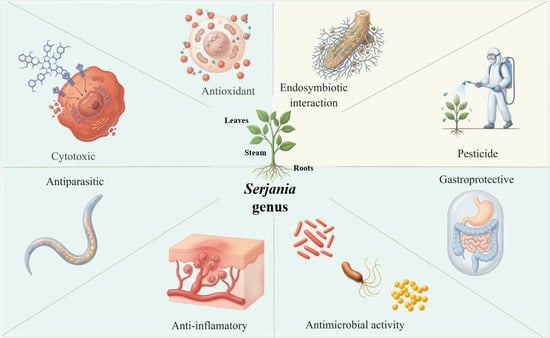
1. Introduction
2. Literature Search, Selection, and Data Analysis
Literature Search
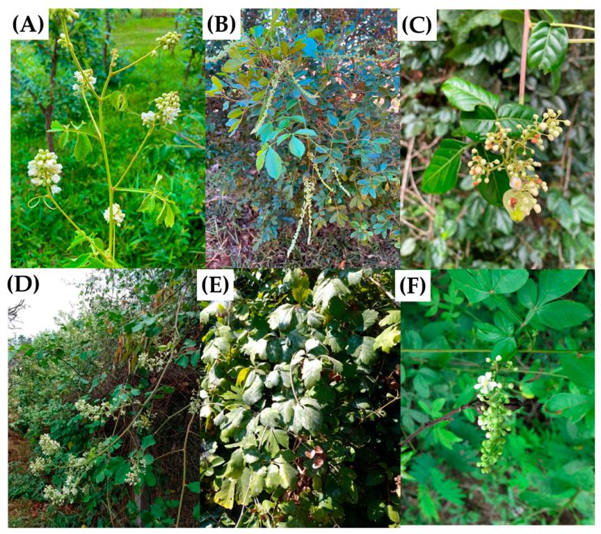
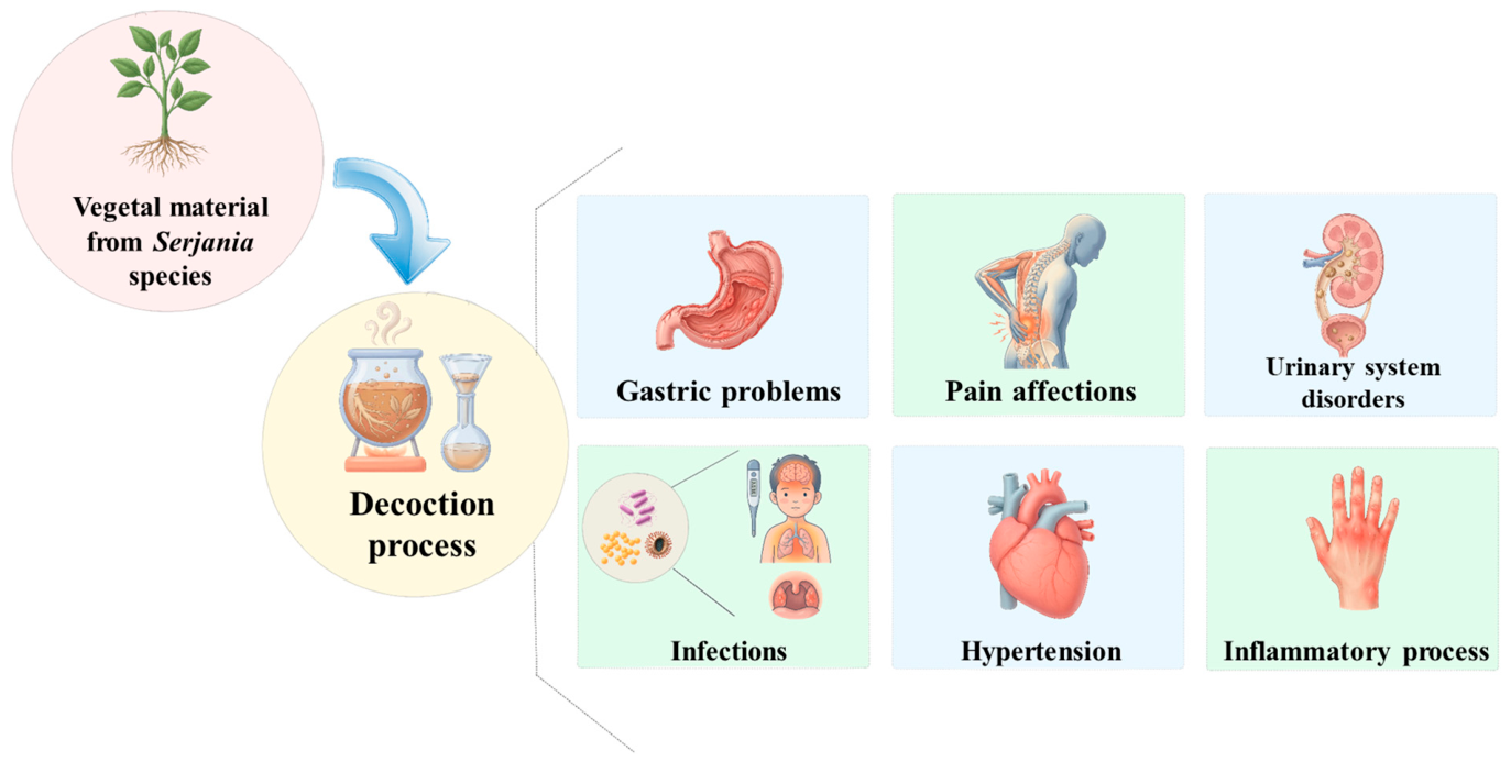
3. Traditional Uses of Serjania Species
4. Phytochemicals Reported in Serjania Species
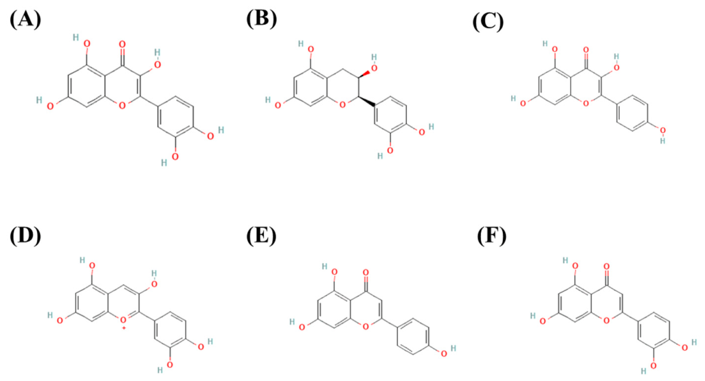
4.1. Phenolic Compounds
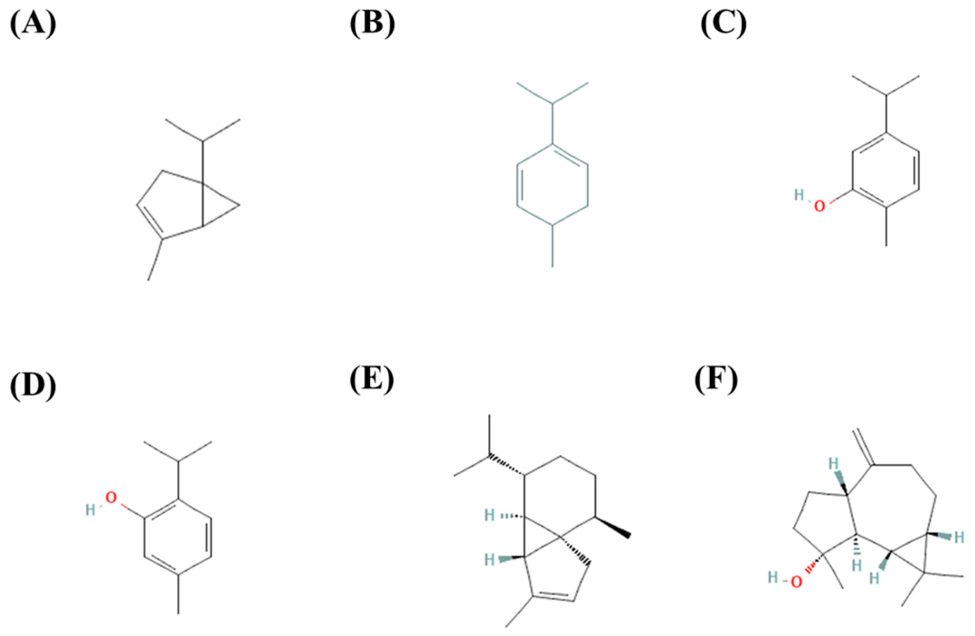
4.2. Terpenoids
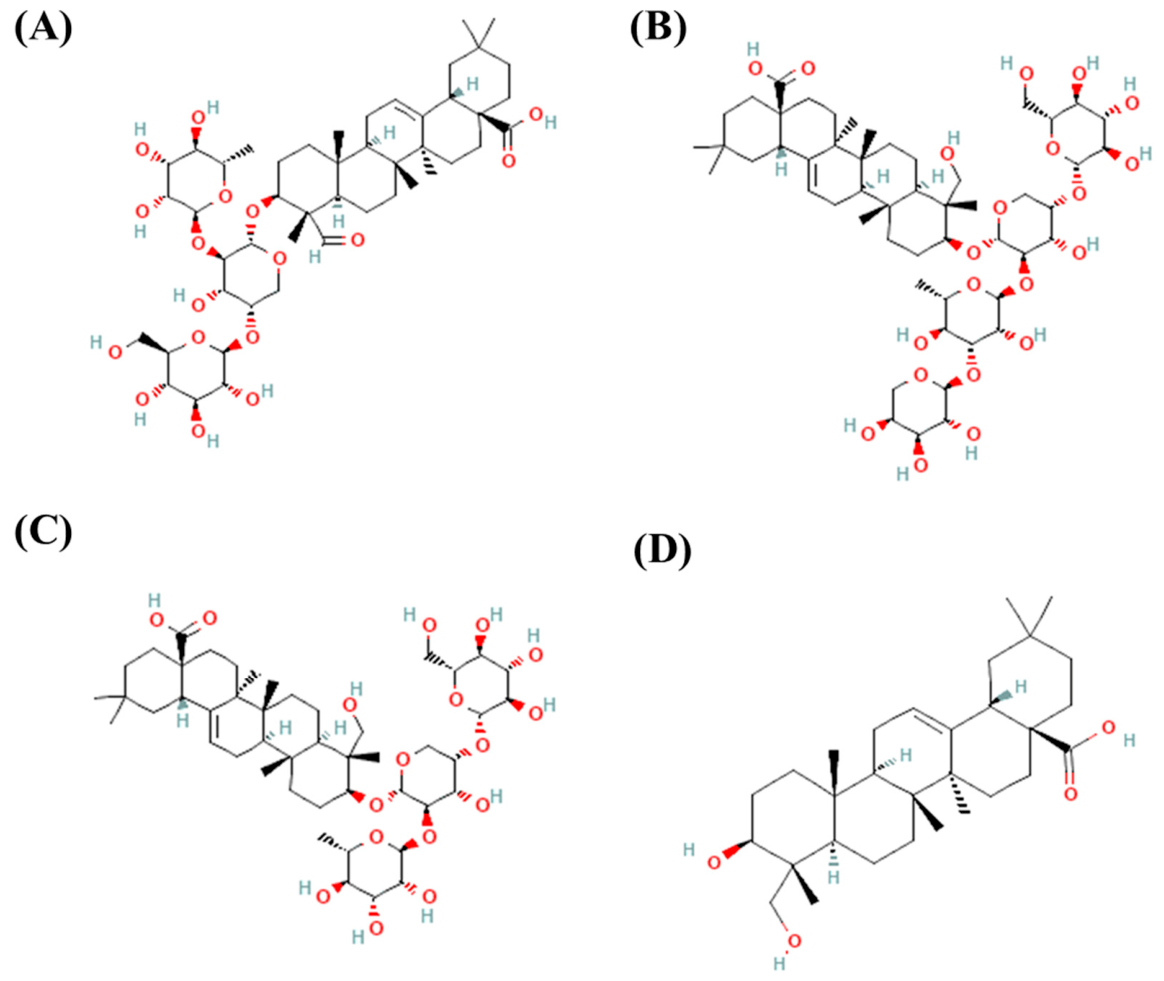
4.3. Saponins
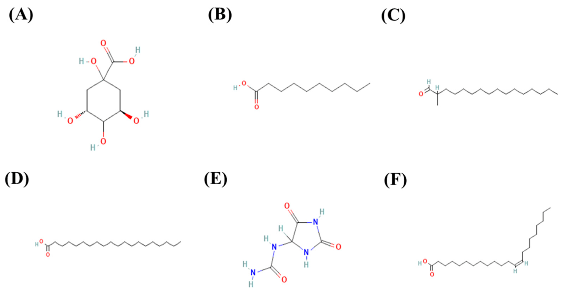
4.4. Other Compounds
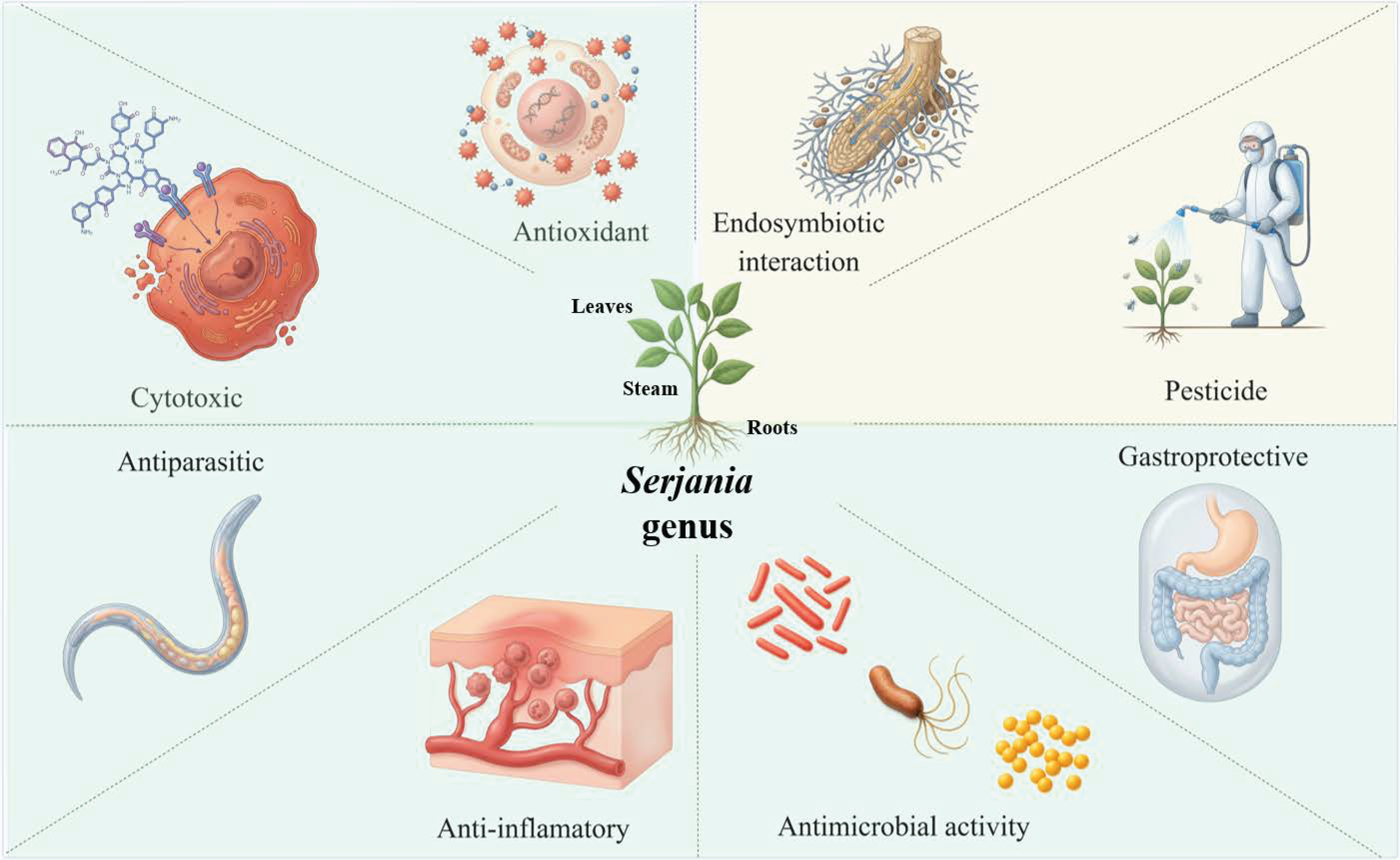
5. Biological Activities and Biotechnological Applications of Serjania Species
5.1. Serjania marginata
5.2. Serjania erecta
5.3. Serjania lethalis
5.4. Serjania salszmaniana
5.5. Serjania yucatanensis
5.6. Serjania caracasana
5.7. Serjania goniocarpa
5.8. Serjania laruotteana
5.9. Serjania schiedeana
5.10. Serjania triquetra
5.11. Serjania racemosa
6. Challenges and Prospects of Serjania Species
7. Conclusions
Supplementary Materials
Author Contributions
Funding
Institutional Review Board Statement
Informed Consent Statement
Data Availability Statement
Acknowledgments
Conflicts of Interest
Abbreviations
| DNA | Deoxyribonucleic acid |
| TLC | Thin-layer chromatography |
| HPLC | High-Performance Liquid Chromatography |
| FIA-ESI-IT-MS | Flow Injection Analysis–Electrospray Ionization–Ion Trap–Mass Spectrometry |
| UHPLC-(ESI)-HRMS | Ultra-High Performance Liquid Chromatography-Electrospray Ionization–High-Resolution Mass Spectrometry |
| NMR | Nuclear Magnetic Resonance |
| GS-MS | Gas Chromatography–Mass Spectrometry |
| GS-FID | Gas Chromatography Flame Ionization Detection |
| HPLC-PDA | High-Performance Liquid Chromatography with Photodiode Array Detection |
| IR | Infrared spectroscopy |
| OCC | Open-Column Chromatography |
| UPLC-MS | Ultra-Performance Liquid Chromatography-Mass Spectrometry |
| FTIR | Fourier Transform Infrared |
| EI-MS | Electron Ionization Mass Spectrometry |
| COX | Cyclooxygenase |
| LOX | Lipoxygenase |
| DMSO | Dimethyl sulfoxide |
| HPLC-DAD | High-Performance Liquid Chromatography with Diode Array Detection |
| DPPH | 2,2-diphenyl-1-picrylhydrazyl |
| ABTS | 2,2′-azino-bis(3-ethylbenzothiazoline-6-sulfonic acid) |
| MIC | Minimum Inhibitory Concentration |
| MTT | 3-[4,5-dimethylthiazol-2-yl]-2,5 diphenyl tetrazolium bromide |
References
- Pattanayak, S. Plants in Healthcare: Past, Present and Future. Explor. Anim. Med. Res. 2021, 11, 140–144. [Google Scholar] [CrossRef] [Scilit]
- Sengar, M.; Sachan, A. Traditional Medicinal System: Significance and Future. In Medicinal Biotechnology: Methods and Applications; Elsevier: Amsterdam, The Netherlands, 2024; pp. 39–59. [Google Scholar]
- Chebabe, M.; Elkhoudri, N.; Chahboune, M.; Laamiri, F.Z.; Marc, I.; Mochhoury, L. Study of Chemical Characterization of Medicinal Plants Used for Traditional Medicine: A Review. Biomed. Pharmacol. J. 2025, 18, 535–546. [Google Scholar] [CrossRef] [Scilit]
- Bernal, L.P.T.; Leitão, M.M.; Radai, J.A.S.; Cardoso, C.A.L.; Lencina, J.d.S.; Fraga, T.L.; Arena, A.C.; Silva-Filho, S.E.; Kassuya, C.A.L. Analgesic and Anti-Inflammatory Potential of Ethanolic Extract from Serjania erecta Leaves. J. Ethnopharmacol. 2023, 303, 116019. [Google Scholar] [CrossRef] [Scilit]
- Falcão, R.J.R.; Cardoso, C.A.L.; Matias, R.; Castro, T.L.A.; Vieira, S.C.H. Biotic and Abiotic Factors Influence the Chemical Composition, Toxicity, and Biological Potential of Serjania marginata Casar. Ciênc. Nat. 2024, 46, e85493. [Google Scholar] [CrossRef] [Scilit]
- Aguilar-Guadarrama, A.B.; Yáñez-Ibarra, G.; Cancino-Marentes, M.E.; González-Ibarra, P.; Ortiz-Andrade, R.; Sánchez-Recillas, A.; Rodríguez-Carpena, J.G.; Aguirre-Vidal, Y.; Medina-Diaz, I.M.; Ávila-Villarreal, G. Chromatographic Techniques and Pharmacological Analysis as a Quality Control Strategy for Serjania triquetra a Traditional Medicinal Plant. Pharmaceuticals 2022, 15, 1289. [Google Scholar] [CrossRef] [Scilit] [PubMed]
- Chávez, M.I.; Delgado, G. Isolation and Relay Synthesis of 11α-Hydroperoxy Diacetyl Hederagenin, a Novel Triterpenoid Derivative from Serjania triquetra (Sapindaceae). Biogenetic Implications. Tetrahedron 1994, 50, 3869–3878. [Google Scholar] [CrossRef] [Scilit]
- Serpeloni, J.M.; Specian, A.F.; Ribeiro, D.L.; Cólus, I.M.; Vilegas, W.; Aparecida-Varanda, E.; Ozden, S. Serjania marginata Casar Extract Has No Mutagenic Effects but Induce Necrosis in Human Gastric Cancer Cells. Toxicol. Lett. 2014, 229, S40–S252. [Google Scholar] [CrossRef] [Scilit]
- Heredia-Vieira, S.C.; Simonet, A.M.; Vilegas, W.; MacÍas, F.A. Unusual C,O-Fused Glycosylapigenins from Serjania marginata Leaves. J. Nat. Prod. 2015, 78, 77–84. [Google Scholar] [CrossRef] [Scilit]
- Périco, L.L.; Heredia-Vieira, S.C.; Beserra, F.P.; De Cássia Dos Santos, R.; Weiss, M.B.; Resende, F.A.; Dos Santos Ramos, M.A.; Bonifácio, B.V.; Bauab, T.M.; Varanda, E.A.; et al. Does the Gastroprotective Action of a Medicinal Plant Ensure Healing Effects? An Integrative Study of the Biological Effects of Serjania marginata Casar. (Sapindaceae) in Rats. J. Ethnopharmacol. 2015, 172, 312–324. [Google Scholar] [CrossRef] [Scilit]
- Matos-Leitão, M.; Silva-Filho, S.E.; Arena, A.C.; Heredia-Vieira, S.C.; Cardoso, C.A.L.; Kassuya, C.A.L. Antinociceptive and Anti-Inflammatory Properties of Aqueous Extract Obtained from Serjania marginata Casar Leaves. J. Ethnopharmacol. 2023, 304, 116018. [Google Scholar] [CrossRef] [Scilit]
- Sánchez-Aguirre, O.A.; Salazar-Vela, D.; Guevara-Valencia, M.; Juárez-Aguilar, E.; Malagón-Avilés, O.G.; Cano-Asseleih, L.M. Antioxidant, Antimicrobial and Antiproliferative Effects of Serjania racemosa Schumach Leaves. Pharmacogn. Res. 2026, 18, 616–624. [Google Scholar] [CrossRef] [Scilit]
- Broggini, L.S.C.; Fernandes, R.S.; Nogueira, T.; Suzano, F.R.; Caetano, A.L.; Buck, H.S.; Couto, L.B.; França, S.C.; Pereira, P.S. Behavioral and Enzymatic Bioassays with Serjania erecta Radlk., Sapindaceae, Correlated with Cognitive Dysfunctions. Rev. Bras. Farmacogn. 2010, 20, 519–528. [Google Scholar] [CrossRef] [Scilit]
- Guimarães, C.C.; Oliveira, D.D.; Valdevite, M.; Saltoratto, A.L.F.; Pereira, S.I.V.; de Castro França, S.; Pereira, A.M.S.; Pereira, P.S. The Glycosylated Flavonoids Vitexin, Isovitexin, and Quercetrin Isolated from Serjania erecta Radlk (Sapindaceae) Leaves Protect PC12 Cells against Amyloid-Β25-35 Peptide-Induced Toxicity. Food Chem. Toxicol. 2015, 86, 88–94. [Google Scholar] [CrossRef] [Scilit]
- Maschieto, C.; Bruder Nascimento, T.; Roberta, G.C.; Arnildo, P.; Sandra, C.; Juliana, I.F.D.G. Serjania erecta Radlk. Improves Endothelial Function in Isolated Aortic Rings. J. Med. Plants Res. 2015, 9, 771–778. [Google Scholar] [CrossRef] [Scilit]
- Gomig, F.; Pietrovski, E.F.; Guedes, A.; Dalmarco, E.M.; Calderari, M.T.; Guimarães, C.L.; Pinheiro, R.M.; Cabrini, D.A.; Otuki, M.F. Topical Anti-Inflammatory Activity of Serjania erecta Radlk (Sapindaceae) Extracts. J. Ethnopharmacol. 2008, 118, 220–224. [Google Scholar] [CrossRef] [Scilit]
- Andréa, C.; Cardoso, L.; Gomes Coelho, R.; Kika Honda, N.; Pott, A.; Pavan, F.R.; Queico, C.; Leite, F. Phenolic Compounds and Antioxidant, Antimicrobial and Antimycobacterial Activities of Serjania erecta Radlk. (Sapindaceae). Braz. J. Pharm. Sci. 2013, 49, 775–782. [Google Scholar] [CrossRef] [Scilit]
- Laundry De Mesquita, M.; Desrivot, J.; Bories, C.; Fournet, A.; Elias De Paula, J.; Grellier, P.; Salmen Espindola, L. Antileishmanial and Trypanocidal Activity of Brazilian Cerrado Plants. Mem. Inst. Oswaldo Cruz 2005, 100, 783–787. [Google Scholar] [CrossRef] [Scilit]
- Grisi, P.U.; Imatomi, M.; de Cassia Pereira, V.; Anese, S.; Gualtieri, S.C.J. Influence of Serjania lethalis A. St.-Hil. (Sapindaceae) Leaf and Stem Crude Extracts on Diaspores and Seedlings of Different Cultivated Species. S. Afr. J. Bot. 2016, 105, 97–105. [Google Scholar] [CrossRef] [Scilit]
- Cavalcante, S.F.; Almeida, A.M.S.; Moraes, M.M.; da Camara, C.A.G.; de Melo, J.P.R.; Badji, C.A. Chemical Composition, Antioxidant Activity and Determination of the Total Phenolics Content of Essential Oil from the Leaves of Serjania lethalis. Chem. Nat. Compd. 2025, 61, 574–576. [Google Scholar] [CrossRef] [Scilit]
- Quintal-Novelo, C.; Torres-Tapia, L.W.; Moo-Puc, R.; Peraza-Sanchez, S.R. Bio-Guided Isolation of a New Sesterterpene from Serjania goniocarpa. Nat. Prod. Commun. 2015, 10, 1513–1516. [Google Scholar] [CrossRef] [Scilit] [PubMed]
- Polanco-Hernández, G.; Escalante-Erosa, F.; García-Sosa, K.; Chan-Bacab, M.J.; Sagua-Franco, H.; González, J.; Osorio-Rodríguez, L.; Peña-Rodríguez, L.M. Metabolites from the Leaf Extract of Serjania yucatanensis with Trypanocidal Activity against Trypanosoma cruzi. Parasitol. Res. 2012, 111, 451–455. [Google Scholar] [CrossRef] [Scilit] [PubMed]
- Navarro, V.; Villarreal, M.L.; Rojas, G.; Lozoya, X. Antimicrobial Evaluation of Some Plants Used in Mexican Traditional Medicine for the Treatment of Infectious Diseases. J. Ethnopharmacol. 1996, 53, 143–147. [Google Scholar] [CrossRef] [Scilit]
- Zanatta, A.C.; Vilegas, W.; Edrada-Ebel, R.A. UHPLC-(ESI)-HRMS and NMR-Based Metabolomics Approach to Access the Seasonality of Byrsonima intermedia and Serjania marginata from Brazilian Cerrado Flora Diversity. Front. Chem. 2021, 9, 710025. [Google Scholar]
- Zanatta, A.C.; Borges, M.S.; Mannochio-Russo, H.; Heredia-Vieira, S.C.; Campaner dos Santos, L.; Rinaldo, D.; Vilegas, W. Green Chromatography as a Novel Alternative for the Quality Control of Serjania marginata Casar. Leaves. Microchem. J. 2022, 181, 107671. [Google Scholar] [CrossRef] [Scilit]
- Serpeloni, J.M.; Specian, A.F.L.; Ribeiro, D.L.; Tuttis, K.; Heredia-Vieira, S.C.; Vilegas, W.; Martínez-López, W.; Varanda, E.A.; de Syllos Cólus, I.M. Selective Anticancer Effects of Serjania marginata Casar. Extract in Gastric Cells Are Mediated by Antioxidant Response. Environ. Toxicol. 2021, 36, 1544–1556. [Google Scholar] [CrossRef] [Scilit]
- do Carmo-Ota, E.; Honorato, C.A.; Heredia-Vieira, S.C.; Flores-Quintana, C.I.; de Castro Silva, T.S.; Inoue, L.A.K.A.; Cardoso, C.A.L. Hepatic and Gastroprotective Activity of Serjania marginata Leaf Aqueous Extract in Nile Tilapia (Oreochromis niloticus). Fish Physiol. Biochem. 2019, 45, 1051–1065. [Google Scholar] [CrossRef] [Scilit]
- Faca, E.C.; Ferreira, E.A.; Pereira, H.C.; Rodrigues, A.; Da Silva, R.M.; Fioratti, C.A.G.; Pereira, F.F.; Mussury, R. Selectivity of Water-Based Extracts of Serjania spp. on Tetrastichus howardi (Hymenoptera: Eulophidae), an Endoparasitoid of Plutella xylostella (Lepidoptera: Plutellidae). Acta Paul. Enferm. 2025, 85, e284440. [Google Scholar] [CrossRef] [Scilit] [PubMed]
- Marcussi, S.; Pereira, P.S. Neutralization of Pharmacological and Toxic Activities of Bothrops jararacussu Snake Venom and Isolated Myotoxins by Serjania erecta Methanolic Extract and Its Fractions. J. Venom. Anim. Toxins Incl. Trop. Dis. 2011, 17, 85–93. [Google Scholar] [CrossRef] [Scilit]
- Passos, A.; Rodríguez, R.; Ferreira, C.; Soares, C.; Somner, V.; Hamerski, L.; da Cunha Pinto, A.; Rezende, M.; Saraiva, E.M. Anti-Leishmania Amazonensis Activity of Serjania lethalis A. St.-Hil. Parasitol. Int. 2017, 66, 940–947. [Google Scholar] [CrossRef] [Scilit]
- de Freitas, S.T.F.; Silva, F.G.; Bessa, L.A.; de Souza, U.J.B.; Augusto, D.S.S.; de Faria, G.S.; Vitorino, L.C. Low Microbial Diversity, Yeast Prevalence, and Nematode-Trapping Fungal Presence in Fungal Colonization and Leaf Microbiome of Serjania erecta. Sci. Rep. 2024, 14, 15456. [Google Scholar] [CrossRef] [Scilit]
- Faca, E.C.; Ferreira, E.A.; da Silva, R.M.; Fioratti, C.A.G.; Silva, R.M.M.F. Toxicity of Aqueous extracts of Serjania spp. on Eggs, Larvae and Pupae of Plutella xylostella (Lepidoptera: Plutellidae). Obs. Econ. Latinoam. 2024, 22, e3762. [Google Scholar]
- da Silva Ribeiro, A.; Polonio, J.C.; dos Santos Oliveira, J.A.; Ferreira, A.P.; Alves, L.H.; Mateus, N.J.; Mangolin, C.A.; de Azevedo, J.L.; Pamphile, J.A. Retrotransposons and Multilocus Sequence Analysis Reveals Diversity and Genetic Variability in Endophytic Fungi-Associated with Serjania laruotteana Cambess. Braz. J. Microbiol. 2021, 52, 2179–2192. [Google Scholar] [CrossRef] [Scilit] [PubMed]
- dos Santos Almeida, C.; Somner, G.V.; de Sá-Haiad, B. Floral Anatomy in Serjania clematidifolia (Paullinieae, Sapindaceae): Insights into a Monoecious Sexual System with Multicyclic Dichogamy. Flora 2024, 320, 152614. [Google Scholar] [CrossRef] [Scilit]
- Quintanilla, R.H.R.; Pinto, A.C. Chemical Constituents and Biological Properties of Species of the Genus Serjania. Rev. Virtual Quim. 2014, 6, 1583–1606. [Google Scholar] [CrossRef] [Scilit]
- Sotelo-Leyva, C.; Salinas-Sánchez, D.O.; Rivas-González, J.M.; Dorado, O.; Arias, D.M.; González-Cortazar, M.; Zamilpa, A. Aphidicidal Activity of an Aqueous Fraction of Serjania schiedeana Sapindaceae against Melanaphis sacchari Hemiptera: Aphididae. Southwest. Entomol. 2019, 44, 585–594. [Google Scholar] [CrossRef] [Scilit]
- Salinas-Sánchez, D.O.; Avilés-Montes, D.; Aldana-Llanos, L.; Gutiérrez-Ochoa, M.; Figueroa-Brito, R.; Sotelo-Leyva, C. Insecticide Effect of Serjania schiedeana for the Control of Spodoptera frugiperda Under Laboratory and Greenhouse Conditions. Southwest. Entomol. 2020, 45, 521–530. [Google Scholar] [CrossRef] [Scilit]
- De Pereira, V.C.; Anese, S.; Imatomi, M.; Grisi, P.U.; Monte Canedo, E.; Juliano Gualtieri, S.C.; Rodrigues-Filho, E. Allelopathic Potential of Serjania lethalis: Evidence from Sesamum indicum. Acta Biolo. Colomb. 2015, 20, 31–37. [Google Scholar] [CrossRef] [Scilit]
- Silva, F.L.; Da Silva, J.L.V.; Silva, J.M.; Marcolin, L.S.A.; Nouailhetas, V.L.A.; Yoshida, M.; Vendramini, P.H.; Eberlin, M.N.; Barbosa-Filho, J.M.; Moreno, P.R.H. Antispasmodic Activity from Serjania caracasana Fractions and Their Safety. Rev. Bras. Farmacogn. 2017, 27, 346–352. [Google Scholar] [CrossRef] [Scilit]
- Salinas-Sánchez, D.O.; Jiménez-Ferrer, E.; Sánchez-Sánchez, V.; Zamilpa, A.; González-Cortazar, M.; Tortoriello, J.; Herrera-Ruiz, M. Anti-Inflammatory Activity of a Polymeric Proanthocyanidin from Serjania schiedeana. Molecules 2017, 22, 863. [Google Scholar] [CrossRef] [Scilit] [PubMed]
- Twaij, B.M.; Hasan, M.N. Bioactive Secondary Metabolites from Plant Sources: Types, Synthesis, and Their Therapeutic Uses. Int. J. Plant Biol. 2022, 13, 4–14. [Google Scholar] [CrossRef] [Scilit]
- Intharuksa, A.; Kuljarusnont, S.; Sasaki, Y.; Tungmunnithum, D. Flavonoids and Other Polyphenols: Bioactive Molecules from Traditional Medicine Recipes/Medicinal Plants and Their Potential for Phytopharmaceutical and Medical Application. Molecules 2024, 29, 5760. [Google Scholar] [CrossRef] [Scilit]
- Ventura, A.S.; Filho, R.A.C.C.; Spica, L.N.; Silva, A.C.F.; De Araújo, A.M.; Cardoso, C.A.L.; Jerônimo, G.T.; Povh, J.A. Toxicological, Biochemical and Morphophysiological Effects of Serjania erecta Leaf Aqueous Extract on Piaractus mesopotamicus. An. Acad. Bras. Cienc. 2021, 93, e20190479. [Google Scholar] [CrossRef] [Scilit] [PubMed]
- da Silva Moreira, S.; Tamashiro, L.K.; Jorge, B.C.; da Silva Balin, P.; Heredia-Vieira, S.C.; de Almeida, G.L.; Cardoso, C.A.L.; Kassuya, C.A.L.; Arena, A.C. Toxicological Safety Evaluation in Acute and 28-Day Studies of Aqueous Extract from Serjania marginata Casar. (Sapindaceae) Leaves in Rats. J. Ethnopharmacol. 2019, 231, 197–204. [Google Scholar] [CrossRef] [Scilit] [PubMed]
- Corrêa, A.P.; Branco, C.; Arruda, N.; Gomes Coelho, R.; Honda, N.K.; Ferrazoli, C.; Pott, A.; Hiruma-Lima, C.A. Gastroprotective Effect of Serjania erecta Radlk (Sapindaceae): Involvement of Sensory Neurons, Endogenous Nonprotein Sulfhydryls, and Nitric Oxide. J. Med. Food 2009, 12, 1411–141545. [Google Scholar] [CrossRef] [Scilit]
- Mayworm, M.A.S.; Salatino, A. Fatty acid composition of ‘cerrado’ seed oils. J. Sci. Food Agric. 1996, 72, 226–230. [Google Scholar] [CrossRef]
- Filho, O.C.; Costa, F.M.; Andrade, M.O.; Pimenta Cruz, J.; Duarte, E.R.; de Oliveira Vasconcelos, V. Phytochemical Characterization of Aqueous Extract of Leaves of Cerrado Plant Species and Pharmacological Implications. Rev. Unimontes Cient. 2024, 26, 1–11. [Google Scholar] [CrossRef] [Scilit]
- Pinho, R.S.; Oliveira, A.F.M.; Silva, S.I. Potential Oilseed Crops from the Semiarid Region of Northeastern Brazil. Bioresour. Technol. 2009, 100, 6114–6117. [Google Scholar] [CrossRef] [Scilit]
- Coutinho, D.J.G.; Barbosa, M.O.; de Souza, R.J.C.; da Silva, A.S.; da Silva, S.I.; de Oliveira, A.F.M. Comparative Study of the Physicochemical Properties of FAME from Seed Oils of Some Native Species of Brazilian Atlantic Forest. J. Am. Oil Chem. Soc. 2016, 93, 1519–1528. [Google Scholar] [CrossRef] [Scilit]
- Ekabo, O.A.; Farnsworth, N.R.; Henderson, T.O.; Mao, G.; Mukherjee, R. Antifungal and Molluscicidal Saponins from Serjania salzmanniana. J. Nat. Prod. 1996, 59, 431–435. [Google Scholar] [CrossRef] [Scilit]
- Salinas-Sánchez, D.O.; Peña-Chora, G.; Gonzalez-Cortazar, M.; Zamilpa-Alvarez, A.; Hernández-Velázquez, V.M.; Tagle-Emigdio, L.J.; Sotelo-Leyva, C. Insecticidal Effect of a Fraction of Ethyl Acetate of Serjania schiedeana Schltdl (Sapindaceae) against Melanaphis sacchari Zehntner (Hemiptera: Aphididae). Acta Agríc. Pecu. 2020, 6, e0061013. [Google Scholar]
- Goel, F.; Chaudhary, H.; Singh, P. Biochemical Characterization, Production, and Medicinal Properties of Phytochemicals. In Phytochemicals and Gut Health: Evidence and Opportunities for Gastrointestinal Management; Academic Press: Cambridge, MA, USA, 2025; pp. 83–105. [Google Scholar]
- Kim, S.; Chen, J.; Cheng, T.; Gindulyte, A.; He, J.; He, S.; Li, Q.; Shoemaker, B.A.; Thiessen, P.A.; Yu, B.; et al. PubChem 2025 Update. Nucleic Acids Res. 2025, 53, D1516–D1525. [Google Scholar] [CrossRef] [Scilit] [PubMed]
- Wang, Q.; Zhao, X.; Jiang, Y.; Jin, B.; Wang, L. Functions of Representative Terpenoids and Their Biosynthesis Mechanisms in Medicinal Plants. Biomolecules 2023, 13, 1725. [Google Scholar] [CrossRef] [Scilit] [PubMed]
- Singh, L.J.; Challam, D.A.; Senjam, B.D. Medicinal Plants as a Sources of Terpenoids and Their Impact on Central Nervous System Disorders: A Review. J. Phytopharmacol. 2023, 12, 104–110. [Google Scholar] [CrossRef] [Scilit]
- Huang, W.; Wang, Y.; Tian, W.; Cui, X.; Tu, P.; Li, J.; Shi, S.; Liu, X. Biosynthesis Investigations of Terpenoid, Alkaloid, and Flavonoid Antimicrobial Agents Derived from Medicinal Plants. Antibiotics 2022, 11, 1380. [Google Scholar] [CrossRef] [Scilit] [PubMed]
- Shakeel, A.; Noor, J.J.; Jan, U.; Gul, A.; Handoo, Z.; Ashraf, N. Saponins, the Unexplored Secondary Metabolites in Plant Defense: Opportunities in Integrated Pest Management. Plants 2025, 14, 861. [Google Scholar] [CrossRef] [Scilit]
- El Aziz, M.M.A.; Ashour, A.S.; Melad, A.S.G. A Review on Saponins from Medicinal Plants: Chemistry, Isolation, and Determination. J. Nanomed. Res. 2019, 25, 6–12. [Google Scholar]
- Zhong, J.; Tan, L.; Chen, M.; He, C. Pharmacological activities and molecular mechanisms of Pulsatilla saponins. Chin. Med. 2022, 17, 59. [Google Scholar] [CrossRef] [Scilit]
- Gerkens, P.C.; Dobson, R.; Tabatadze, N.; Mshviladzade, V.; Elias, R.; Peulen, O.J.; Jolois, O.M.; De Pauw-Gillet, M.C.A. Apoptosis and cytolysis induced by giganteosides and hederacolchisides in HL-60 cells. Anticancer Res. 2007, 27, 2529–2534. [Google Scholar] [PubMed]
- Lima, M.C.; Dias, K.S.; Fonseca, G.G.; Leite, R.S.R.; Batistote, M.; Paz, M.F. Use of Cerrado Plants as an Alternative in the Control of Bacterial Contamination in the Alcoholic Fermentation Process. Braz. J. Biol. 2024, 84, e281448. [Google Scholar] [CrossRef] [Scilit]
- Leitão, M.M.; Radai, J.A.S.; Macorini, L.F.B.; Fraga, T.L.; Heredia-Vieira, S.C.; Cardoso, C.A.L.; Arena, A.C.; Kassuya, C.A.L. Serjania marginata Casar. Hydroalcoholic Extract Reduced Cytokine and Inflammatory Parameters in Experimental Models of Inflammation and Infection in Mice. Pharmacog. Res. 2022, 14, 127–134. [Google Scholar] [CrossRef] [Scilit]
- Couto, I.F.S.; da Silva, S.V.; Valente, F.I.; de Araújo, B.S.; de Souza, S.A.; Mauad, M.; de Paula Quintão Scalon, S.; Mussury, R.M. Botanical Extracts of the Brazilian Savannah Affect Feeding and Oviposition of Plutella xylostella (Linnaeus, 1758) (Lepidoptera: Plutellidae). J. Agric. Sci. 2019, 11, 322–333. [Google Scholar] [CrossRef] [Scilit]
- De Rossi, L.; Rocchetti, G.; Lucini, L.; Rebecchi, A. Antimicrobial Potential of Polyphenols: Mechanisms of Action and Microbial Responses—A Narrative Review. Antioxidants 2025, 14, 200. [Google Scholar] [CrossRef] [Scilit] [PubMed]
- Mendes de Cordova, S.; Simões Benfatti, C.; Alberton Magina, M.; Guedes, A.; Mendes de Cordova, C.M. Evaluation of the Antibacterial Activity of Extracts Isolated from Native Plants of the Brazilian against Mycoplasma arginini, M. Hominis and Ureaplasma urealyticum. RBAC 2010, 42, 241–244. [Google Scholar]
- Nossa, P.M.; Guenka, L.C.; Couto, L.B.; da-Cruz-Perez, D.E. Effects of the Serjania erecta and Zeyheria montana Ethanol Extracts in Experimental Pulpitis in Rats: A Histological Study. Med. Oral Patol. Oral Cir. Bucal 2012, 18, e337–e342. [Google Scholar] [CrossRef] [Scilit]
- de Freitas, S.T.F.; dos Santos Rodrigues, A.R.; Ataídes, A.C.C.; de Oliveira Menino, G.C.; de Faria, G.S.; Vitorino, L.C.; Silva, F.G.; Dyszy, F.H. Inhibitory Effects of Serjania erecta on the Development of Chrysodeixis includens. Sci. Rep. 2022, 12, 14702. [Google Scholar] [CrossRef] [Scilit] [PubMed]
- Rodriguez, A.M.S.; De Paula, J.E.; Dedallier, N.; Molez, J.F.; Espíndola, L.S. Larvicidal Activity of Some Cerrado Plant Extracts Against Aedes aegypti. J. Am. Mosq. Control Assoc. 2006, 22, 314–317. [Google Scholar] [CrossRef] [Scilit]
- Vasconcelos, V.O.; Costa, E.G.L.; Moreira, V.R.; Morais-Costa, F.; Duarte, E.R. Efficacy of Plants Extracts from the Cerrado against Adult Female of Dermacentor nitens (Acari: Ixodidae). Exp. Appl. Acarol. 2018, 75, 419–427. [Google Scholar] [CrossRef] [Scilit]
- Grisi, P.U.; Gualtieri, S.C.J.; Anese, S.; Pereira, V.C.; Forim, M.R. Effect of Serjania lethalis Ethanolic Extract on Weed Control. Planta Daninha 2013, 31, 239–248. [Google Scholar] [CrossRef] [Scilit]
- Pereira, V.D.C.; Grisi, P.U.; Dodonov, P.; Anese, S.; Toffano, L.; Gualtieri, S.C.J. Atividade Fitotóxica de Serjania lethalis Sobre a Germinação e Crescimento de Panicum maximum. Biotemas 2013, 27, 29–35. [Google Scholar] [CrossRef] [Scilit]
- Cassia-Pereira, V.; Monte-Canedo, E.; Rodrigues-Filho, E.; Alves dos Santos, A.C.; Umeda-Grisi, P.; Imatomi, M.; Anese, S.; Habermann, E.; Juliano-Gualtieri, C.J. Potencial Fitotóxico de Folhas, Raízes e Caules de Serjania lethalis A. St.-Hil. Iheringia Sér. Bot. 2016, 71, 93–98. [Google Scholar]
- Polanco-Hernández, G.; Escalante-Erosa, F.; García-Sosa, K.; Acosta-Viana, K.; Chan-Bacab, M.J.; Sagua-Franco, H.; González, J.; Osorio-Rodríguez, L.; Moo-Puc, R.E.; Peña-Rodríguez, L.M. In Vitro and In Vivo Trypanocidal Activity of Native Plants from the Yucatan Peninsula. Parasitol. Res. 2012, 110, 31–35. [Google Scholar] [CrossRef] [Scilit]
- Silva, J.L.V.; Carvalho, V.S.; Silva, F.L.; Barbosa-Filho, J.M.; Rigoni, V.L.S.; Nouaihetas, V.L.A. Gastrointestinal Property of Serjania caracasana (Jacq.) Willd. (Sapindaceae) on Rats. PharmacologyOnline 2012, 1, 22–26. [Google Scholar]
- Yañez-Ibarra, G.; Aguilar-Guadarrama, B.; Rodríguez-Carpena, J.G.; Ávila-Villarreal, G. Safety Assesment of Serjania triquetra. In Proceedings of the 71 St International Congress and Annual Meeting of the Society for Medicinal Plant and Natural Product Research (GA), Dublin, Ireland, 2–5 July 2023; Georg Thieme Verlag KG: Berlin, Germany, 2023; Volume 89, pp. 1274–1275. [Google Scholar]
- Lopes, W.A.L.; de Souza, L.A. Morphoanatomy of Serjania communis Cambess. Seedling (Sapindaceae). Acta Sci. Biol. Sci. 2015, 37, 377–383. [Google Scholar] [CrossRef] [Scilit]
- Ferrucci, M.S.; Coulleri, J.P. Serjania lucianoi (Sapindaceae: Paullinieae), a New Species from Northern Bahia, Brazil. Syst. Bot. 2013, 38, 172–177. [Google Scholar] [CrossRef] [Scilit]
- López-Muraira, I.G.; Flores-Martínez, H.; Gómez-Leyva, J.F.; Iruegas-Buentello, H.R. First record in México of Caloptilia stigmatella (Fabricius, 1781) (Lepidoptera:Gracillariidae) and first report on Serjania recemosa Schumach (Sapindaceae). Rev. Bio Cienc. 2023, 10, e1423. [Google Scholar]
- Ferrucci, M.S. Urvillea paucidentata y Serjania adenophylla (Sapindaceae), Dos Especies Nuevas de Mato Grosso, Brasil. Bonplandia 1996, 9, 19–23. [Google Scholar] [CrossRef] [Scilit]
- de Oliveira-Barbosa, J.R.; de Carvalho, S.H.D.S.; de Almeida, S.S. Morphometry of Seeds of the Species Serjania comata Radlk in a Remnant of Atlantic Forest, Alagoinhas, Bahia. J. Bioeng. Technol. Health 2024, 7, 15–19. [Google Scholar]
- Araújo, G.U.C.; Costa, C.G. Cambial Variant in the Stem or Serjania corrugata (Sapindaceae). IAWA J. 2006, 27, 269–280. [Google Scholar] [CrossRef] [Scilit]
- Souza, M.; Milaneze-Gutierre, M.; Souza, L. Flower Structure of Serjania fuscifolia (Sapindaceae) and Its Relationship with Visiting Insect. Int. J. Life Sci. Res. Arch. 2020, 3, 62–74. [Google Scholar]
- Somner, G.V.; Ferrucci, M.S. Serjania littoralis (Sapindaceae), a New Species from Brazil. Ann. Bot. Fenn. 2009, 46, 479–483. [Google Scholar] [CrossRef] [Scilit]
- Solís, S.M.; Ferrucci, M.S. Embryological Studies in Thinouia mucronata and Serjania meridionalis (Paullinieae, Sapindaceae): Development of Gametophytes in Both Floral Morphs and Its Phylogenetic Implications. Braz. J. Bot. 2022, 45, 399–413. [Google Scholar] [CrossRef] [Scilit]
- Rajput, K.S.; Gondaliya, A.D.; Moya, R. Structure of the Secondary Xylem and Development of a Cambial Variant in Serjania mexicana (Sapindaceae). IAWA J. 2021, 51, 103–115. [Google Scholar] [CrossRef] [Scilit]
- Ferrucci, M.S.; Medina-Lemos, R. New Combination for a Species of the Genus Serjania (Sapindaceae-Paullinieae) Endemic in Mexico. Act. Bot. Mex. 2013, 104, 19–29. [Google Scholar] [CrossRef] [Scilit]
- Ferrucci, M.S.; Steinmann, V.W. Serjania rzedowskiana (Sapindaceae-Paullinieae): A New Species from Michoacán, Mexico. Syst. Bot. 2016, 41, 775–780. [Google Scholar] [CrossRef] [Scilit]
- Coulleri, J.P.; Ferrucci, M.S. Serjania setigera (Sapindaceae), Nuevo Registro Para La flora de Bolivia. Bol. Soc. Argent. Bot. 2015, 50, 601–605. [Google Scholar] [CrossRef] [Scilit]
- Ferrucci, M.S.; Steinmann, V.W. Two New Species of Serjania (Sapindaceae) from Michoacán, Mexico, with Notes on S. biternata. Syst. Bot. 2019, 44, 670–680. [Google Scholar] [CrossRef] [Scilit]




| Serjania Species | Common Name | Country or Region | Claimed Therapeutic Use | Plant Part | Preparation/Application | Ref. |
|---|---|---|---|---|---|---|
| S. marginata | Cipó-uva | Brazil Argentina Paraguay Bolivia | Gastrointestinal disorders, ulcers, cancer, and infections | Leaves | Decoction/Oral | [8,9,10,11] |
| S. erecta | Cipó-cinco-folhas, cinco-folhas | Brazil | Inflammation, stomachache, ulcerative diseases, hypertension, gastritis, pain management, and back pain | Stems Leaves Roots | Decoction/Oral | [4,13,14,15,16,17,29] |
| S. lethalis | Timbo vine cipó-timbó timbó | Brazil | Inflammation, infections, skin diseases, ulcers, diarrhea, fever, malaria, kidney pain, narcotic | Stems Leaves | Decoction/Oral | [18,19,20,38] |
| S. caracasana | Tingui-da-mata | Brazil | Gastric problems | No information | No information | [39] |
| S. goniocarpa | But aak | Mexico | Leg sores, abscesses | No information | No information | [21] |
| S. schiedeana | Costilla de vieja, Cuapalachtle | Mexico | Kidney inflammation, burning feet, back pain, healing wounds, and bruises | No information | No information | [37,40] |
| S. yucatenensis | Chéen peek Chac uayam | Mexico | Abscesses, infections, vomiting, headache, diarrhea | Stems Leaves | Decoction/Oral | [22] |
| S. triquetra | Palo de tres costillas, bejuco de tres costillas, tres equis | Mexico | Kidney pain, kidney stones, diuretic agents, hepatitis, urinary infections, and kidney inflammation | Stems Leaves | Decoction/Oral | [6,7,23] |
| S. racemosa | Seven-hearted bejuco | Mexico | Treatment of diabetes, diuretic, kidney problems, kidney inflammation, urinary problems, and prostate disorders | No information | No information | [12] |
| Serjania Species | Plant Part | Extraction Method | Solvent/Liquid-to-Solid Ratio | Detection/Quantification Method | Reported Compound | Classification | Identified/ Content | Ref. |
|---|---|---|---|---|---|---|---|---|
| S. marginata | Leaves | Percolation | Ethanol, then the extract was fractionated | NMR | 3-O-D-β-glucopyranosylsitosterol | Phytosterol glycoside | Identified | [9] |
| Pulsatilla saponin D | Saponin | Identified | ||||||
| Hederacolchiside A | Saponin | Identified | ||||||
| Salzmannianoside B | Saponin | Identified | ||||||
| Quercetin 3-O-α-L-rhamnopyranoside | Flavonoid glycoside | Identified | ||||||
| Epicatechin | Flavanol | Identified | ||||||
| Cassiaoccidentalin A | Flavonoid glycoside | Identified | ||||||
| Tetrastigma B | Flavonoid glycoside | Identified | ||||||
| Apigenin 6-C-β- boivinopyranosyl-7-O-β-D-glucopyranoside | Flavone glycoside | Identified | ||||||
| apigenin 6-C-[2- O-α-L-rhamnopyranosyl(1→2)]-β-D-xylopyranoside | Flavone glycoside | Identified | ||||||
| proanthocyanidins A-1 | Anthocyanin | Identified | ||||||
| proanthocyanidins A-2 | Anthocyanin | Identified | ||||||
| Cinnamtannin B-1 | Protoanthocyanidin | Identified | ||||||
| Leaves | Percolation | 70% ethanol solution | TLC HPLC | Saponins | Terpene | Identified | [10] | |
| Flavonoid glycosides | Phenolic compound | Identified | ||||||
| Tannins | Polyphenol | Identified | ||||||
| Leaves | Maceration | Distilled water (10:1 mL/g) | FIA-ESI-IT-MS | Soluble phenols | Phenolic compound | Identified | [11] | |
| Flavonoids | Phenolic compound | Identified | ||||||
| Tannins | Polyphenol | Identified | ||||||
| Rutin | Flavonol glycoside | Identified | ||||||
| Leaves | Ultrasound | 70% ethanol solution (10:1000 mL/mg) | HPLC-PDA | Protocatechuic acid | Phenolic acid | Identified | [25] | |
| (epi)catechin-(epi)catechin | Flavanol | Identified | ||||||
| (epi)catechin-A-(epi)catechin-(epi)catechin | Flavanol | Identified | ||||||
| (epi)catechin | Flavanol | Identified | ||||||
| (epi)catechin-(epi)catechin-(epi)catechin | Flavanol | Identified | ||||||
| (epi)catechin-A-(epi)catechin | Flavanol | Identified | ||||||
| Quercetin-O- hexoside | Flavonol glycoside | Identified | ||||||
| Apigenin-C-[2″-O-(deoxyhexosyl)- pentoside] | Flavone glycoside | Identified | ||||||
| Quercetin-O-deoxyhexoside | Flavonol glycoside | Identified | ||||||
| Luteolin-6-C-[2″-O-(deoxyhexosyl)-hexos-3-uloside] (cassiaoccidentalin B) | Flavone glycoside | Identified | ||||||
| Luteolin-8-C-[2″-O-(deoxyhexosyl)-hexos-3-uloside] | Flavone glycoside | Identified | ||||||
| Apigenin-6-C-[2″-O-(deoxyhexosyl)-hexos-3-uloside] (cassiaoccidentalin A) | Flavone glycoside | Identified | ||||||
| Apigenin-8-C-[2″-O-(deoxyhexosyl)-hexos-3-uloside] (tetrastigma B) | Flavone glycoside | Identified | ||||||
| Apigenin-C-hexos-3-uloside-2″-O-deoxyhexosyl | Flavone glycoside | Identified | ||||||
| O-deoxyhexosyl-C-glycosyde apigenin derivative | Flavone glycoside | Identified | ||||||
| Methyl-luteolin-6-C-[2″-O-(deoxyhexosyl)-hexos-3-uloside] (cassiaoccidentalin C) | Flavone glycoside | Identified | ||||||
| O-deoxyhexosyl apigenin derivative | Flavone glycoside | Identified | ||||||
| Methyl-luteolin-8-C-[2″-O-(deoxyhexosyl)-hexos-3-uloside] | Flavone glycoside | Identified | ||||||
| Leaves | Maceration | Water (20 mL/2 g) | FIA-ESI-IT-MS | Quinic acid | Organic acid | Identified | [27] | |
| Quercitrin | Flavonol glycoside | Identified | ||||||
| Isoquercitrin | Flavonol glycoside | Identified | ||||||
| Proanthocyanidin trimer—A-type | Tannin | Identified | ||||||
| Leaves | Maceration | Water: (50:2 mL/g) | Mass spectrometry | Soluble phenols | Phenolic compound | 266.70 mg/g DM | [44] | |
| Flavonoids | Phenolic compound | 189.10 mg/g DM | ||||||
| Tannins | Phenolic compound | 56.3 mg/g DM | ||||||
| Quinic acid | Carboxylic acid | Identified | ||||||
| Quercetin-3- O-rhamnoside (quercitrin | Flavonol glycoside | Identified | ||||||
| Quercetin-3-O-glucose (isoquercetin) | Flavonol glycoside | Identified | ||||||
| Proanthocyanidin trimer-A.type | Anthocyanin | Identified | ||||||
| Leaves | Ultrasound | 70% ethanol solution (1:100 mL/mg) | UHPLC-(ESI)-HRMS NMR | Phenolic acids | Phenolic compound | Identified | [24] | |
| Cinnamic acids | Phenolic acid | Identified | ||||||
| Triterpene | Terpene | Identified | ||||||
| B-type proanthoccyanidins | Anthocyanin | Identified | ||||||
| B-type proanthoccyanidins trimer | Anthocyanin | Identified | ||||||
| B-type proanthoccyanidins tretamer | Anthocyanin | Identified | ||||||
| A-type proanthoccyanidins dimer | Anthocyanin | Identified | ||||||
| A-type proanthoccyanidins trimer | Anthocyanin | Identified | ||||||
| A-type proanthoccyanidins tretamer | Anthocyanin | Identified | ||||||
| A-type proanthoccyanidins pentamer | Anthocyanin | Identified | ||||||
| Flavonoids glycosylated | Phenolic compound | Identified | ||||||
| C-glycosylated flavone | Flavone glycoside | Identified | ||||||
| C,O-glycosylated flavone | Flavone glycoside | Identified | ||||||
| O-glycosylated flavone | Flavone glycoside | Identified | ||||||
| Leaves and stems | Maceration | Distilled water (200:20 mL/g) | Spectrometric techniques | Phenolic compounds | Phenolic compound | 195–300 mg/g DM | [5] | |
| Flavonoids | Phenolic compound | 7–255 mg/g DM | ||||||
| Tannins | Polyphenol | 248–265 mg/g DM | ||||||
| S.erecta | Roots | Decoction | Water | Colorimetric test | Flavonoids | Phenolic compound | Identified | [15] |
| Saponins | Terpene | Identified | ||||||
| Tannis | Polyphenol | Identified | ||||||
| Catechins | Flavanol | Identified | ||||||
| Cardiac glycosides | Glycoside compound | Identified | ||||||
| Steams and leaves | Maceration | Methanol | Column chromatographic | Saponins | Terpene | Identified | [29] | |
| Terpenes | --- | Identified | ||||||
| Flavonoids | Polyphenol | Identified | ||||||
| Tannins | Polyphenol | Identified | ||||||
| Leaves | Foam test | Saponins | Terpene | Identified | [43] | |||
| Spectrophotometric techniques | Flavonoids | Flavonoids | 212 mg/g DM | |||||
| Phenolic compounds | Phenolic compounds | 386 mg/g DM | ||||||
| Tannins | Tannins | 89 mg/g DM | ||||||
| Leaves | Polyisoprenoids | Terpene | Identified | [45] | ||||
| Leaves | Sequential extraction | N- hexane, ethyl acetate, ethanol | NMR FTIR TLC | (−)-epicatechin | Flavanol | Identified | [4] | |
| kaempferol | Flavonol | Identified | ||||||
| Isovitexin | Flavone glycoside | Identified | ||||||
| apigenin-8-C-β-D-glucopyranoside | Flavone glycoside | Identified | ||||||
| kaempferol-3,7-di-O-α-L-rhamnopyranoside | Flavonol glycoside | Identified | ||||||
| kaempferol-3-O-α-L-rhamnopyranoside | Flavonol glycoside | Identified | ||||||
| Leaves | Maceration | Ethanol | NMR | Kaempferol | Flavonol | Identified | [17] | |
| Kaempferol-3,7-di-O-a-L-rhamnopyranoside | Flavonol glycoside | Identified | ||||||
| (-)-epicatechin | Flavanol | Identified | ||||||
| Apigenin-6-C-b-D-glucopyranoside (isovitexin) | Flavone glycoside | Identified | ||||||
| apigenin-8-C-b-D-glucopyranoside (vitexin) | Flavone glycoside | Identified | ||||||
| kaempferol-3-O-a-l-rhamnopyranoside | Flavonol glycoside | Identified | ||||||
| kaempferol-3-O-a-l-rhamnopyranosyl-(1→6)-b-d- glucopyranoside | Flavonol glycoside | Identified | ||||||
| Leaves | Decoction | Distilled boiling water (90 g/900 mL) | TLC HPLC | Saponins | Terpene | Identified | [13] | |
| Tannins | Polyphenol | Identified | ||||||
| Glycosidic flavonoids | Phenolic compound | Identified | ||||||
| Leaves/stems | Maceration | 70% ethanol solution | No information | Saponins | Terpene | Identified | [16] | |
| Flavonoids | Polyphenol | Identified | ||||||
| Triterpenoids | Terpenes | Identified | ||||||
| Steroids | Terpene | Identified | ||||||
| Tannins | Polyphenol | Identified | ||||||
| Catechins | Flavanol | Identified | ||||||
| Shrub | Homogenization | 2:1 v/v Chloroform:methanol solution | Gas chromatography | Capric acid | Fatty acid | 3.0 g/100 g DM | [46] | |
| Palmitoleic acid | Fatty acid | 0.19 g/100 g DM | ||||||
| Oleic acid | Fatty acid | 1.33 g/100 g DM | ||||||
| Linoleic acid | Fatty acid | 0.20 g/100 g DM | ||||||
| α-Linoleic acid | Fatty acid | 0.11 g/100 g DM | ||||||
| Arachidonic acid | Fatty acid | 0.98 g/100g DM | ||||||
| Eicosadienoic acid | Fatty acid | 6.23 g/100 g DM | ||||||
| Leaves | Maceration | Methanol | TLC HPLC | Isovitexin | Flavone glycoside | Identified | [14] | |
| Vitexin | Flavone glycoside | Identified | ||||||
| Quercetin | Flavonol | Identified | ||||||
| S. lethalis | Leaves | Static extraction | Distilled water | HPLC | Flavonoids | Polyphenol | Identified | [47] |
| Saponins | Terpene | Identified | ||||||
| Terpenoids | Terpene | Identified | ||||||
| Leaves | Maceration | 90:10 v/v ethanol-water | GC-MS GC-FID | Benzoic acid | Phenolic acid | Identified | [30] | |
| α-Cubene | Monoterpene | Identified | ||||||
| 4-epi-cubedol | Monoterpene | Identified | ||||||
| (-)-Spathulenol | Monoterpene | Identified | ||||||
| Caryophyllene oxide | Sesquiterpene | Identified | ||||||
| Conifery alcohol | Monolignol | Identified | ||||||
| Hexadecanal | Fatty aldehyde | Identified | ||||||
| 6,10,14-Trymethyl-2-pentadecacone | Ketone | Identified | ||||||
| Phytol | Diterpene alcohol | Identified | ||||||
| Methyl hexadecanoate | Fatty acid ester | Identified | ||||||
| Hexadecanoic acid | Fatty acid | Identified | ||||||
| Ethyl hexadecanoate | Fatty acid ester | Identified | ||||||
| (E)-Phytol | Diterpene | Identified | ||||||
| Methyl octadecanoate | Fatty acid ester | Identified | ||||||
| Octadecanoic acid | Fatty acid | Identified | ||||||
| Phytol acetate | Diterpenes | Identified | ||||||
| Ethyl octadecenoate | Fatty acid ester | Identified | ||||||
| 4,8,12,16-Tretamethylheptadecan-4-olide | Fatty alcohol | Identified | ||||||
| γ-Tocopherol | Monoterpene | Identified | ||||||
| β-Amyrone | Triterpene | Identified | ||||||
| β-Amyrin | Triterpene | Identified | ||||||
| Lup-20(29)-en-3-one | Triterpene | Identified | ||||||
| Lup-20(29)-en-3-ol | Triterpene | Identified | ||||||
| Glutinone | Triterpene | Identified | ||||||
| β-Amyrin acetate | Triterpene | Identified | ||||||
| Leaves | Hydrodistillation | Water | GC-MS GC-FID | α-Thujene | Monoterpene | 4.7% * | [20] | |
| δ-Terpinene | Monoterpene | 3.9% * | ||||||
| Methyl eter thymol | Monoterpene | 3.8% * | ||||||
| Thymol | Monoterpene | 5.2% * | ||||||
| Carvacrol | Monoterpene | 74.1% * | ||||||
| β-Caryophylene | Sesquiterpene | 5.1% * | ||||||
| Seeds | Soxhlet extraction | n-hexane | GC-MS | Palmitic acid | Fatty acid | 3.2% * | [48] | |
| Gadoleic acid | Fatty acid | 27.5% * | ||||||
| Oleic acid | Fatty acid | 9.7% * | ||||||
| Linoleic acid | Fatty acid | 1.7% * | ||||||
| Arachidonic acid | Fatty acid | 15.8% * | ||||||
| Eicosenoic acid | Fatty acid | 69.6% * | ||||||
| Saturated fatty acids | Fatty acid | 19.0% * | ||||||
| Unsaturated fatty acids | Fatty acid | 81.0% * | ||||||
| S. salzmanniana | Seeds | Soxhlet extraction | Hexane | GS-MS | Palmitic acid | Fatty acid | 1.0% * | [49] |
| Arachidic acid | Fatty acid | 3.4% * | ||||||
| Behemic acid | Fatty acid | 3.4 * | ||||||
| Oleic acid | Fatty acid | 7.6% * | ||||||
| Eicosanoid acid | Fatty acid | 64.7% * | ||||||
| Erucic acid | Fatty acid | 19.0% * | ||||||
| Stems | No information | Methanol | TLC | Salzmannianoside A | Saponin | Identified | [50] | |
| Salzmannianoside B | Saponin | Identified | ||||||
| Pulsatilla saponin D | Saponin | Identified | ||||||
| 3-O-[[β-D-glucopyranosyl-(1→4)]-[α-L-rhamnopyranosyl-(1→2)]-α-L-arabinopyranosyl]oleanolic acid | Saponin glycoside | Identified | ||||||
| S. caracasana | Seeds | Soxhlet method | Hexane | GC-MS | Palmitic acid | Fatty acid | 2.2% * | [49] |
| Stearic acid | Fatty acid | 1.6% * | ||||||
| Arachidic acid | Fatty acid | 9.6 * | ||||||
| oleic acid | Fatty acid | 8.8% * | ||||||
| Eicosanoid acid | Fatty acid | 69.4% * | ||||||
| Erucic acid | Fatty acid | 5.0% * | ||||||
| Linoleic acid | Fatty acid | 1.4% * | ||||||
| Aerial parts | Maceration | 96% methanol solution | GC–MS NMR | Spathulenol | Sesquiterpene | 4.2% * | [39] | |
| 6,10,14-Trimethyl-2-pentadecanone | Terpenoid | 7.3% * | ||||||
| Methyl palmitate | Fatty acid ester | 7.7% * | ||||||
| β-Sitosterol | Phytosterol | 21.4 mg DM ** | ||||||
| β-Amyrin | Triterpene | 1698. mg DM ** | ||||||
| Friedelin | Triterpene | 5.6 mg DM ** | ||||||
| Stigmasterol | Phytosterol | 37.8 mg DM ** | ||||||
| β-Sitosterol glucoside | Phytosterol glycoside | 3.2 mg DM ** | ||||||
| Allantoin | Diureide | 6.5 mg DM ** | ||||||
| Quercitrin | Flavonol Glycoside | 30.9 mg DM ** | ||||||
| S. goniocarpa | Leaves | Maceration | Methanol | IR GC-MS NMR | Goniocarpic acid | Sesterpene | Identified | [21] |
| Phytol | Diterpene alcohol | Identified | ||||||
| S. schiedeana | Stems | Maceration | Methanol (5:1 mL/g) | Colorimetric test | Alkaloids | Alkaloids | Identified | [36] |
| Flavonoids | Phenolic compounds | Identified | ||||||
| Tannins | Polyphenols | Identified | ||||||
| Stems | Maceration | Methanol (5:1 mL/g) | Colorimetric test | Alkaloids | Alkaloids | Identified | [37] | |
| Flavonoids | Phenolic compounds | Identified | ||||||
| Tannins | Polyphenols | Identified | ||||||
| Stem | Maceration | Methanol (2.5:1 mL/g) | TLC HPLC | epicatechin– (4β → 8)–epicatechin–(4β → 8, 2β →O→ 7) epicatechin | Flavanol | Identified | [40] | |
| Stem | Maceration | Methanol | GC/MS | Phytol | Diterpene alcohol | 1.18% * | [51] | |
| Phytone | Sesquiterpene | 1.13 * | ||||||
| Methyl palmitate | Fatty acid ester | 38.66 * | ||||||
| Methyl arachidate | Fatty acid ester | 1.05 * | ||||||
| 4,8,12,16-tetramethylpentadecan-4-olide | Macrolide lactone | 15.99 * | ||||||
| Methyl linoleate | Fatty acid ester | 17.17 * | ||||||
| Methyl stearate | Fatty acid ester | 1.23 * | ||||||
| Methyl behenate | Fatty acid ester | 2.07 * | ||||||
| Methyl tetradecanoate | Fatty acid ester | 12.97 * | ||||||
| Tert-butyl (4-(2,6-di-tert-butyl-4-methoxyphenoxy)-3-nitro-4-oxobutyl)prolinate | Phenolic acid | 4.13 * | ||||||
| S. yucatanensis | Leaves | Maceration | Ethanol for extraction and fractionation with hexane and ethyl acetate | TLC GC-MS | lup-20(29)-en-3-one | Triterpene | Identified | [22] |
| β-caryophyllene oxide | Sesquiterpene | Identified | ||||||
| S. triquetra | Aereal parts | Sequential extractions: n-hexane, methanol and ethyl acetate, evaporated to dryness | No information | IR 1H/13C NMR EIMS | Stigmasterol | Phytosterol | Identified | [7] |
| Oleanolic acid | Fatty acid | Identified | ||||||
| Morolic acid | Triterpene | Identified | ||||||
| Hederagenin | Saponin | Identified | ||||||
| 11α-hydroperoxy-hederagenin | Saponin | Identified | ||||||
| Stems | Maceration (collected extract was concentrated by rotatory evaporator) | 85% ethanol solution | OCC TLC UPLC-MS RMN | Ethyl palmitate | Fatty acid ester | 25.77% ** | [6] | |
| Stigmasta-3,5-dien-7-one | Steroid | 13.92% ** | ||||||
| Methyl pentacosanoate | Fatty acid ester | 10.75% ** | ||||||
| Ethyl docosanoate | Fatty acid ester | 8.64% ** | ||||||
| Ethyl oleate | Fatty acid ester | 4.97% ** | ||||||
| Stigmasta-5,22-dien-3-ol | Phytosterol | 9.96% ** | ||||||
| Erucic acid | Fatty acid | 1.43% ** | ||||||
| S. racemosa | Leaves | Maceration (collected extract was filtered and concentrated by rotatory evaporator) | Hexane Ethyl acetate Methanol (50 g/300 mL) | NMR | Saponins | Terpenes | Identified | [12] |
| Flavonoids | Glycosylated flavonoids | Identified |
| Activity | Plant Part/Conditioning Sample | Extraction Method/Extract Conditioning | Solvent/Liquid-to-Solid Ratio | Resuspension/ Fractionation | Dose/ Concentration | In Vitro/In Vivo Model | Model Assay/ Control | Relevant Results | Ref. |
|---|---|---|---|---|---|---|---|---|---|
| Antimicrobial | Leaves (Dried at 60 °C/24 h, concentrate, and lyophilized) | Maceration (Extract was concentrated via evaporation) | Water (1:10 mL/mg) | Resuspension with distilled water | 15–1000 µg/mL | Burkholderia cepacia, Enterococcus faecalis, Escherichia coli, Pseudomonas aeruginosa, Staphylococcus. epidermidis, Staphylococcus aureus, Staphylococcus saprophyticus | Microdilution Control: Tetracycline | All aqueous extracts exhibited antibacterial properties | [5] |
| Antimicrobial | Leaves | Maceration | Ethanol: water 7:3 v/v | Resuspended in 0.9% saline solution | 7.81–1000 µg/mL | Escherichia coli, Staphylococcus aureus, Salmonella setubal, Helicobacter pylori, Candida albicans | Microdilution Controls: ampicillin and amoxicillin | The extract showed antimicrobial activity in a concentration- and strain-dependent response | [10] |
| Antimicrobial | Leaves (Dried at 60 °C) | Maceration (Concentrated via evaporation) | 95% ethanol solution | Hexane fraction (rich in essential oils) | 0.1–1000 µg/mL | Bacillus toyonensis, Bacillus thuringiensis, Bacillus cereus, Bacillus proteolyticus | Disk diffusion/No information | The extract did not show antimicrobial activity against Bacillus species | [61] |
| Antimicrobial | Leaves (Dried at room temperature) | Percolation (Extract was lyophilized) | 70% ethanol solution | Resuspended in 5% DMSO solution | 0.98–1000 µg/mL | Mycobacterium tuberculosis, Klebsiella pneumoniae, Pseudomonas aeruginosa, Staphylococcus epidermidis | Microdilution/No information | The extract exhibited antimicrobial effects in a concentration- and strain-dependent response | [62] |
| Anti-inflammatory | Leaves | Maceration | Hydroalcoholic | Resuspended in saline solution (0.9%) | 30, 100, and 300 mg/kg | Murine model | Carrageenan-induced paw edema | The extract showed anti-inflammatory properties | [11] |
| Anti-inflammatory | Leaves (Dried at room temperature) | Percolation (Extract was lyophilized) | 70% ethanol solution | Resuspended in saline solution (0.9%) | 30, 100, and 300 mg/kg | Murine model | Carrageenan-induced paw edema/Dexamethasone | The extract showed anti-inflammatory properties | [62] |
| Antinociceptive | Leaves | Maceration | Hydroalcoholic (10 g/100 mL) | Resuspended in saline solution (0.9%) | 30, 100, and 300 mg/kg | Murine model | Acid formalin-induced nociception | The extracts reduce nociception in a dose-dependent response | [11] |
| Antihyperalgesic | Leaves | Percolation | Ethanol | Resuspended in saline solution (0.9%) | 30, 100, and 300 mg/kg | Swiss murine model | Von Frey acetone test Control: dexamethasone | The extract effectively reduced mechanical hyperalgesia | [11] |
| Antioxidant | Leaves Steams | Maceration | Water | Lyophilized extract was resuspended in distilled water | 50–1000 µg/mL | DPPH | Inhibition of radical | The stems showed higher antioxidant properties than the leaves | [5] |
| Cytotoxic | Leaves (Dried at room temperature) | Percolation (vacuum concentrated to dryness at 40 °C and lyophilized) | Ethanol (70:30 v/v) | The dried extract was fractionated with water: butanol solution (30:70 v/v) | 50–500 mg/mL | Non-tumor gastric epithelium cells and gastric adenocarcinoma cells | MTT Negative control: PBS | The extract showed cytotoxicity against cells in a concentration-dependent manner | [26] |
| Cytotoxic | Leaves | No information | Ethanol | No information | 150–300 µg/mL | Human normal and cancer gastric cells | MTT assay Cell proliferation curves AO/EB staining | The extract showed cytotoxicity against cancer (150 µg/mL) and normal cells (µg/mL) in a concentration-dependent manner | [8] |
| Antimutagenic | Leaves | No information | Ethanol | No information | 150–300 µg/mL | Human normal and cancer gastric cells | Cytokinesis-block micronucleus cytome assay | The extract did not show mutagenic effects | [8] |
| Antimutagenic | Leaves | Maceration | Ethanol: water 7:3 v/v | Extract diluted in DMSO | 2.5–20 mg/plate | Salmonella typhimurium | Ames test | It was reported that the absence of mutagenic effects | [10] |
| Antimutagenic | Leaves (dried at room temperature) | Percolation (vacuum concentrated to dryness at 40 °C and lyophilized) | Ethanol (70:30 v/v) | The dried extract was fractionated with water: butanol solution (30:70 v/v) | 50–500 mg/mL | Non-tumor gastric epithelium cells and gastric adenocarcinoma cells | MTT:CBMN-cyt Positive control: DXR (0.2 µg/mL) | The extract did not show mutagenic effects | [26] |
| Hepatoprotective | Leaves (dried at 37 °C for 48 h) | Maceration (lyophilized) | Water | 5 mg of dry extract solubilized in minimal methanol | 224.3 mg/kg/day | Nile tilapia | Feeding trial/Control: commercial food | The extract showed hepatoprotective effects | [27] |
| Gastroprotective | Leaves | Maceration | Ethanol: water 7:3 v/v | Resuspended in saline solution (0.9%) | 125–500 mg/kg | Wistar rat | Ethanol-induced ulcers, ischemia–reperfusion Controls: carbenoxolone, lansoprazole | The extract reduces the gastric lesions by 60-90% at 500 mg/kg | [10] |
| Gastroprotective | Leaves (dried at 37 °C for 48 h) | Maceration (lyophilized) | Water | 5 mg of dry extract solubilized in minimal methanol | 224.3 mg/kg/day | Nile tilapia | Feeding trial/Control: commercial food | The extract exhibited gastroprotective effects and stimulated intestinal digestion | [27] |
| Anti-diarrhea | Leaves | Maceration | Ethanol: water 7:3 v/v | Resuspended in saline solution (0.9%) | 250 mg/kg | Wistar rat | Castor oil-induced diarrhea Control: loperamide and saline solution | The extract did not show a decrease or an increase in the severity of diarrhea | [10] |
| Photoprotective | Leave Steams | Maceration (vacuum concentrated to dryness at 40 °C and lyophilized | Water | Resuspended in water | 200–1000 µg/mL | Spectrophotometer | SPF spectrophotometric method | The leaf extract demonstrated a superior sun protective factor than the steam extract | [5] |
| Acute oral toxicity | Leaves | Maceration | Ethanol: water 7:3 v/v | Resuspended in saline solution (0.9%) | 5000 mg/kg | Wistar rat | Acute toxicity assay Control: saline solution | No sign and symptoms of toxicity were reported | [10] |
| Acute and subacute toxicity | Leaves (dried at 37 °C for 48 h) | Maceration (Lyophilized) | Water | Resuspended in saline solution (0.9%) | 30–2000 mg/kg | Wistar rats | Subchronic toxicity assay/ Saline solution (0.9%) | No toxicity was observed. However, after 14 days of daily administration, alterations in kidney histology and an increase in abnormal sperm were reported. | [44] |
| Toxicity | Leaves Steams | Maceration | Water | Resuspended in distilled water | 50–1000 µg/mL | Artemia salina | Acute toxicity assay Negative control: saline solution | The extract did not show toxicity against A. salina | [5] |
| Insecticide | Leaves (Dried at 40 °C) | Maceration (Evaporation) | Ethanol Water | Resuspended in distilled water | 1000–10,000 µg/mL | Plutella xylostella | Food preference and oviposition | The extracts were effective as oviposition suppressants for this insect | [63] |
| Insecticide | Leaves (dried at 40 °C in an air oven) | Maceration | Water | Direct use of filtered extract | 5 and 10% w/v | Plutella xylostella | Direct contact toxicity assay on a model | Both concentrations were toxic to eggs and pupae of P. xylostella. The 10% concentration was the most effective in terms of larval mortality | [32] |
| Antiparasitic | Leaves (dried at 40 °C in an air oven) | Maceration | Water | Direct use of filtered extract | 5 and 10% w/v | Tetrastichus howardi | Observational assay on morphological changes in the model | The extracts did not show antiparasitic properties against T. howardi | [28] |
| Activity | Plant Part/Conditioning Sample | Extraction Method/Extract Conditioning | Solvent/Liquid-to-Solid Ratio | Resuspension/ Fractionation | Dose/ Concentration | In Vitro/In Vivo Model | Model Assay/ Controls | Relevant Results | Ref. |
|---|---|---|---|---|---|---|---|---|---|
| Antimicrobial | Leaves (Dried at 40 °C) | Maceration (Evaporation) | 70% ethanol solution | Resuspended in 10% DMSO solution | 6.25–50 µg/mL | Mycoplasma hominis, Ureaplasma urealyticum, Mycoplasma arginini | Microdilution Control: DMSO (10%) | The extracts showed antimicrobial activity in concentration- and strain-dependent response | [65] |
| Antimicrobial | Leaves Roots | Maceration at room temperature | Ethanol | Fractionation: (water, methanol, acetone) | 10–400 µg/mL | Staphylococcus aureus, Pseudomonas aeruginosa, Escherichia coli, Salmonella setubal, Saccharomyces cerevisiae, Candida albicans | Rezasurin Microtiter Assay/Control: isoniazis | Leaves and roots inhibited the growth of all tested microorganisms | [17] |
| Antimicrobial | Leaves (Dried at 60 °C) | Maceration (Evaporation) | Ethanol (95%): water 1:1 | Hexane fraction (rich in essential oils) | 1–100 µg/mL | Bacillus toyonensis, Bacillus thuringiensis, Bacillus cereus, Bacillus proteolyticus | Disk diffusion | The extract showed antimicrobial activity against the tested microorganisms | [61] |
| Antiparasitic | Leaves (dried at 40 °C in an air oven) | Maceration | Water | Direct use of filtered extract | 5 and 10% w/v | Tetrastichus howardi | Direct contact assay Negative control: water Positive control: acephate | The aqueous extract did not interfere with the parasitism of T. howardi on 4th instar larvae of P. xylostella. | [28] |
| Anti-inflammatory | Leaves | Sequential extraction | Ethanol, N-hexane, ethyl acetate, ethanol | Fractionated with water, methanol, and acetone | 30, 100, and 300 mg/kg | Murine model | Carrageenan pleurisy Zymosan peritonitis | The extract exhibited anti-inflammatory properties in a concentration dependence | [4] |
| Anti-inflammatory | Steam Leaves | Maceration | Ethanol | 0.003–4 mg/ear | Murine model | Ear edema | Topical application of the extract and its fractions caused a dose-dependent reduction in ear edema and tissue myeloperoxidase activity | [16] | |
| Anti-inflammatory | Leaves (Dried) | Maceration (lyophilized) | 95% Ethanol | Saline solution | 300 mg/kg | Murine model | Induction of inflammatory pulp tissue/Saline solution | The extract did not show anti-inflammatory effects on pulp tissue in the analyzed periods. | [66] |
| Antioxidant | Leaves Steam Roots | Decoction 24 h in the dark | Distilled boiling water (90 g/900 mL) | Crude extract | 2.5–10 mg/mL | DPPH | Inhibition of radical/Rutin as a positive control | The extracts exhibited antioxidant properties | [13] |
| Antioxidant | Leaves Roots | Maceration at room temperature | Ethanol | Fractionation: with water, methanol, acetone | 10–400 µg/mL | DPPH β-carotene–linoleic acid assay | Inhibition of radical/Quercetin β-carotene-linoleic acid assay/BHT | The extracts showed low antioxidant properties | [17] |
| Antioxidant | Leaves | Maceration (Evaporation) | Methanol Chloroform | No information | 40–640 mg/mL | DPPH β-carotene–linoleic acid assay | DPPH test Positive control: quercetin β-carotene–linoleic acid assay Positive control: quercetin | The extract showed antioxidant properties | [45] |
| Analgesic | Leaves | Direct extraction Sequential extraction (concentrated under vacuum) | Ethanol N- hexane, ethyl acetate, ethanol | Fractionation with water, methanol, and acetone | 30, 100, and 300 mg/kg | Mice Leukocytes Neutrophils | Formalin test control/ Control indomethacin, morphine | The extract showed analgesic properties in a dose-dependent manner | [4] |
| Neuroprotective | Leaves | Maceration for three days | Methanol (concentrated under vacuum at 50 °C and further lyophilized) | The crude extract was dissolved in methanol | 25–200 µg/mL | Rat adrenal pheochromocytoma (PC12) cell line | MTT assay (cell viability), controls: untreated cells and Aβ-treated cells | Isolated compounds from leaves of S. erecta exhibited neuroprotective effects in a concentration-dependent response | [14] |
| Anti-hypertensive | Roots | Decoction | Water | Administered with condensed milk (oral intake) | 10 mL of 5% w/v solution | Murine model | Direct measurement of blood pressure | The treatment improved endothelial function and promoted nitric oxide production by the endothelium | [15] |
| Anti-ulcer | Leaves | Maceration in chloroform and methanol Three extractions, 48 h each: concentrated by rotary evaporation at 38 °C | Extract in chloroform: (4 L/Kg) Extract in methanol: (4 L/Kg) | No information | 125–5000 mg/kg | In vivo Murine model | Ethanol-induced gastric ulcer model/Control positive: carbenoxolone: 100 mg/kg | The extract showed gastroprotective properties | [45] |
| Antivenom | Leaves (dried at 60 °C in an air oven) | Maceration for 72 h (concentrated via rotary evaporation at 50 °C) | Methanol | Extract was fractionated | Venom/toxin with the extracts at a 1:30 (w/w) | Enzymatic assay | Indirect hemolytic assay for phospholipase A2 activity. Positive control: Bothrops jararacussu snake venom Negative control: PBS | The crude extract and the fractions neutralized the toxic activities of the Bothrops jararacussu snake venom and the isolated myotoxins. | [29] |
| Toxicity | Leaves Steam Roots | Decoction 24 h in the dark | Distilled boiling water (90 g/900 mL) | Crude extract | 50–1250 mg/kg | Murine model | Screening, biochemical and hematological analysis | The extract did not show toxicity | [13] |
| Toxicity | Leaves | Maceration in chloroform and methanol Three extractions, 48 h each: concentrated by rotary evaporation at 38 °C | Extract in chloroform: (4:1 L/Kg) Extract in methanol: (4L/Kg) | No information | 125–5000 mg/kg | Murine model | Acute oral toxicity assay Control group: saline solution | The extract did not promote acute oral toxicity | [45] |
| Toxicity | Leaves | Maceration: for 24 h at room temperature, followed by filtration and lyophilization | Water 1:1000 mL/µg | Water at different concentrations 2.5–150 µg/mL | 2.5–150 µg/mL | Piaractus mesopotamicus | Toxicity bioassay: Blood plasma biochemical analysis and electrolyte assay Histopathological analysis: Light microscopy-based histopathology/untreated fish Morphometric analysis: organ morphometric/untreated fish | The extract caused morphofunctional and histological alterations in the gills and liver. Mortality occurred at extract levels above 50 μg/mL | [43] |
| Insecticide | Leaves (dried at 40 °C in an air oven) | Maceration | Water | Direct use of filtered extract | 5 and 10% w/v | Plutella xylostella | Direct contact | Both concentrations were toxic to eggs, larvae, and pupae of P. xylostella | [28] |
| Pesticide | Leaves (dried at 40 °C/120 h) | Maceration (Evaporation) | Methanol | Resuspended in distilled water containing 2.5% (v/v) methyl alcohol | 0.0078–20 µg/mL | Chrysodeixis includens | Inhibition of growth and development Control: distilled water containing 2.5% (v/v) methyl alcohol | Extract treatments increased the duration of the larval, pupal, and total development | [67] |
Disclaimer/Publisher’s Note: The statements, opinions and data contained in all publications are solely those of the individual author(s) and contributor(s) and not of MDPI and/or the editor(s). MDPI and/or the editor(s) disclaim responsibility for any injury to people or property resulting from any ideas, methods, instructions or products referred to in the content. |
© 2026 by the authors. Licensee MDPI, Basel, Switzerland. This article is an open access article distributed under the terms and conditions of the Creative Commons Attribution (CC BY) license.
Share and Cite
Rubio-García, A.B.; de Loza-García, C.G.; Silva-Jara, J.M.; González-Silva, N.; Ramirez-Contreras, L.A.; Villagran, Z.; Graciano-Machuca, O.; Ramírez-Anaya, J.d.P.; Martínez-Esquivias, F.; Anaya-Esparza, L.M. Traditional Uses, Phytochemicals, Biological Activities, and Biotechnological Applications of Serjania Species: A Review of Current Knowledge and Future Prospects. Molecules 2026, 31, 1477. https://doi.org/10.3390/molecules31091477
Rubio-García AB, de Loza-García CG, Silva-Jara JM, González-Silva N, Ramirez-Contreras LA, Villagran Z, Graciano-Machuca O, Ramírez-Anaya JdP, Martínez-Esquivias F, Anaya-Esparza LM. Traditional Uses, Phytochemicals, Biological Activities, and Biotechnological Applications of Serjania Species: A Review of Current Knowledge and Future Prospects. Molecules. 2026; 31(9):1477. https://doi.org/10.3390/molecules31091477
Chicago/Turabian StyleRubio-García, Ana Belem, Cecilia Guadalupe de Loza-García, Jorge Manuel Silva-Jara, Napoleón González-Silva, Luis Antonio Ramirez-Contreras, Zuamí Villagran, Omar Graciano-Machuca, Jessica del Pilar Ramírez-Anaya, Fernando Martínez-Esquivias, and Luis Miguel Anaya-Esparza. 2026. "Traditional Uses, Phytochemicals, Biological Activities, and Biotechnological Applications of Serjania Species: A Review of Current Knowledge and Future Prospects" Molecules 31, no. 9: 1477. https://doi.org/10.3390/molecules31091477
APA StyleRubio-García, A. B., de Loza-García, C. G., Silva-Jara, J. M., González-Silva, N., Ramirez-Contreras, L. A., Villagran, Z., Graciano-Machuca, O., Ramírez-Anaya, J. d. P., Martínez-Esquivias, F., & Anaya-Esparza, L. M. (2026). Traditional Uses, Phytochemicals, Biological Activities, and Biotechnological Applications of Serjania Species: A Review of Current Knowledge and Future Prospects. Molecules, 31(9), 1477. https://doi.org/10.3390/molecules31091477