Diagnostic Potential of Exosomes in Colorectal Cancer: Current Advances and Future Perspectives



Abstract
1. Introduction
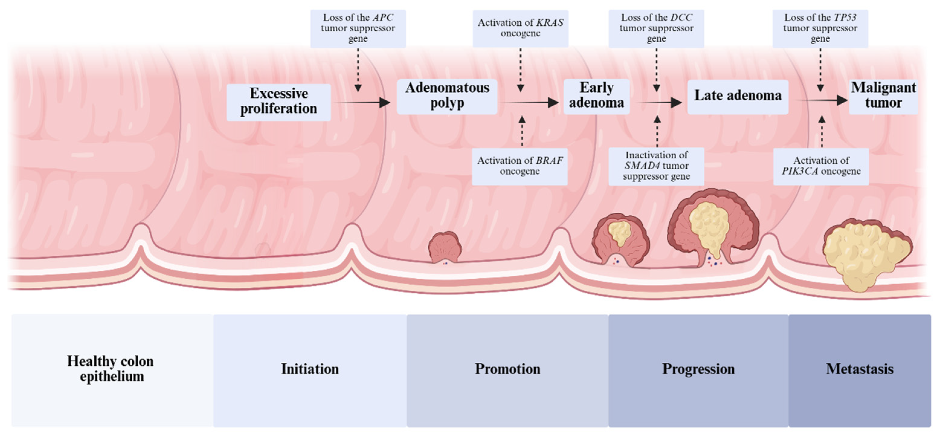
1.1. Colorectal Cancer
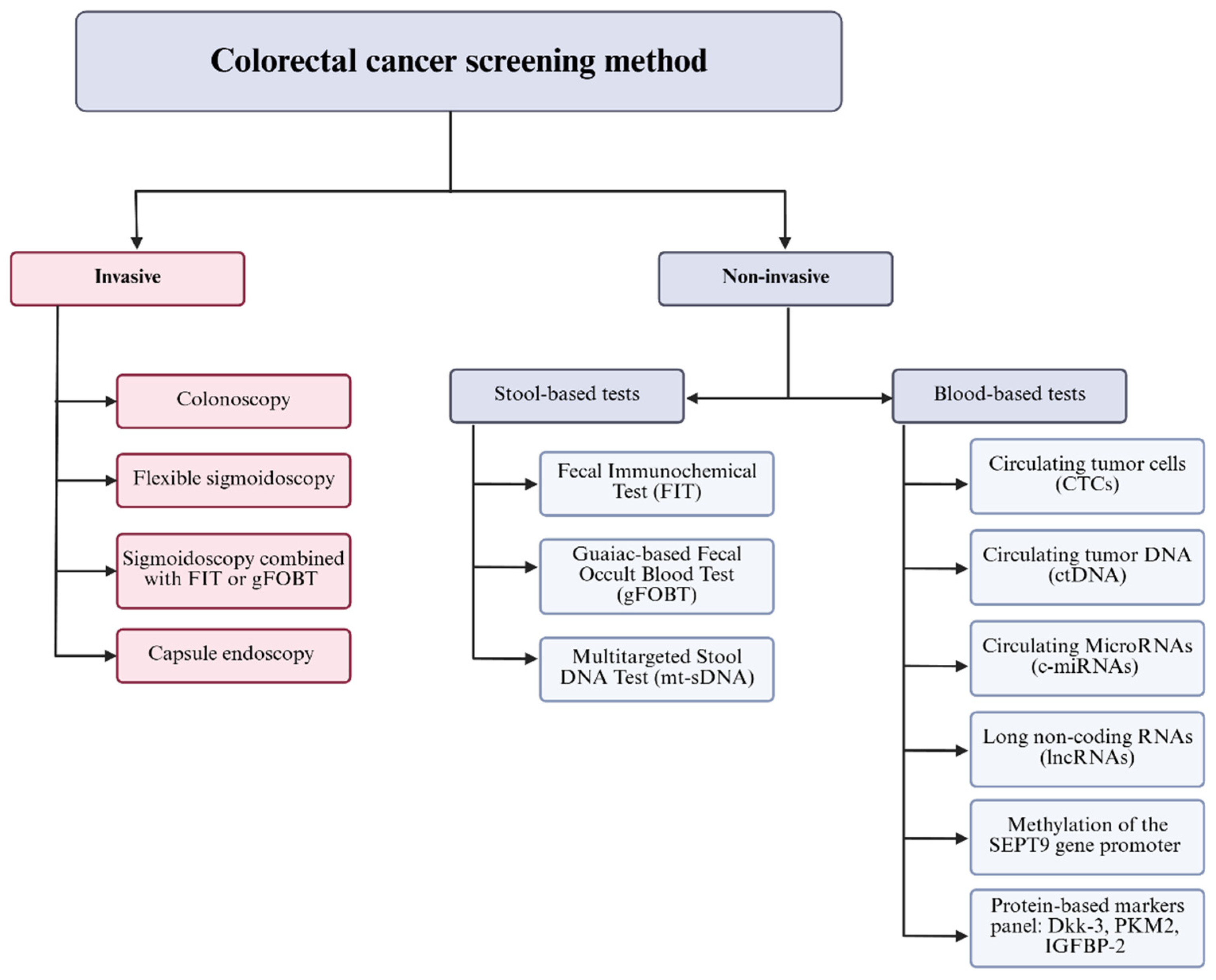
1.2. Screening Methods for CRC
1.2.1. Invasive Colorectal Cancer Screening
1.2.2. Non-Invasive Colorectal Cancer Screening
Stool-Based Tests
Blood and Liquid Biopsy-Based Tests
2. Exosomes
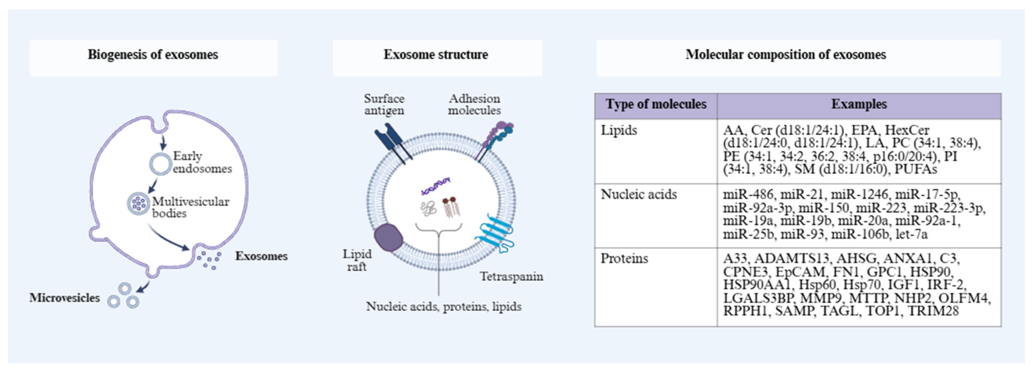
2.1. Structure and Characteristics of Exosomes
2.2. Exosome Isolation and Characterization
2.3. Exosomes as Potential Biomarkers in Oncological Diagnosis
2.4. Exosomal Biomarkers
2.4.1. Lipids
Exosomal Lipid Signatures in CRC
Stage-Dependent Variations in Exosomal Profiles
Exosome-Associated Inflammatory Biomarkers in CRC Development
2.4.2. miRNA
Exosomal miRNA Signatures in CRC
Stage-Dependent Variations in Exosomal Profiles
Exosome-Associated Inflammatory Biomarkers in CRC Development
2.4.3. Proteins
Exosomal Proteins Signatures in CRC
Stage-Dependent Variations in Exosomal Profiles
Exosome-Associated Inflammatory Biomarkers in CRC Development
3. Conclusions and Future Directions
Author Contributions
Funding
Institutional Review Board Statement
Informed Consent Statement
Data Availability Statement
Acknowledgments
Conflicts of Interest
Abbreviations
| A33 | transmembrane glycoprotein A33 |
| AA | arachidonic acid |
| ADAMTS13 | A disintegrin, and metalloproteinase with thrombospondin motifs 13 |
| AHSG | alpha 2-HS glycoprotein |
| AI | artificial intelligence |
| ALA | α-linolenic acid |
| ANXA1 | Annexin A1 |
| AP | adenomatous polyps |
| APC | adenomatous polyposis coli |
| ATX | autotaxin |
| AUC | area under the ROC curve |
| BRAF | B-Raf proto-oncogene, serine/threonine kinase |
| CA19-9 | carbohydrate antigen 19-9 |
| CCAT1 | colon cancer-associated transcript 1 |
| CEA | carcinoembryonic antigen |
| Cer | ceramides |
| CIMP | CpG island methylation phenotype |
| CIN | chromosomal instability |
| CNNs | convolutional neural networks |
| CPNE3 | copine protein III |
| CTCs | circulating tumor cells |
| cfDNA | cell-free DNA |
| CRC | colorectal cancer |
| ctDNA | circulating tumor DNA |
| C3 | complement component 3 |
| DCs | dendritic cells |
| DEPs | differentially expressed proteins |
| Dkk-3 | Dickkopf-3 |
| DLS | dynamic light scattering |
| EMT | endothelial–mesenchymal transition |
| EpCAM | epithelial cell adhesion molecule |
| EPA | eicosapentaenoic acid |
| ESCRT | endosomal sorting complex required for transport |
| EVs | extracellular vesicles |
| FAs | fatty acids |
| FDA | Food and Drug Administration |
| FIT | Fecal Immunochemical Test |
| FN1 | fibronectin 1 |
| FS | flexible sigmoidoscopy |
| gFOBT | guaiac-based Fecal Occult Blood Test |
| GLA | γ-linolenic acid |
| GOF | gain of oncogenic functions |
| GPC1 | glipican-1 |
| Her | hereditary non-polyposis colorectal cancer |
| HexCer | hexosylceramide |
| HOTAIR | HOX transcript antisense RNA |
| HP | hyperplastic polyps |
| HSP90AA1 | human stress-inducible 90 kDa heat shock protein alpha |
| Hsp60 | heat shock protein 60 |
| Hsp70 | heat shock protein 70 |
| IGF1 | insulin-like growth factor I |
| IGFBP-2 | insulin-like growth factor binding protein 2 |
| ILVs | intraluminal vesicles |
| IRF-2 | interferon regulatory factor-2 |
| KRAS | Kirsten rat sarcoma viral oncogene homolog |
| LA | linoleic acid |
| LGALS3BP | galectin-binding protein-3 |
| lncRNAs | long non-coding RNAs |
| LPA | lysophosphatidic acid |
| MDSCs | myeloid-derived suppressor cells |
| MMP9 | matrix metalloproteinase-9 |
| MSI | microsatellite instability |
| MTTP | microsomal triglyceride transfer protein |
| MUFA | monounsaturated fatty acid |
| MVBs | multivesicular bodies |
| mt-sDNA | multitargeted stool DNA test |
| NGS | next-generation sequencing |
| NK | natural killer |
| NHP2 | NHP2 ribonucleoprotein |
| NTA | nanoparticle tracking analysis |
| NSAIDs | non-steroidal anti-inflammatory drugs |
| OLFM4 | olfactomedin 4 |
| OXPHOS | mitochondrial oxidative phosphorylation |
| PA | phosphatidic acid |
| PC | phosphatidylcholine |
| PE | phosphatidylethanolamine |
| PGE2 | prostaglandin E2 |
| PI | phosphatidylinositol |
| PKM2 | pyruvate kinase M2 |
| PS | phosphatidylserine |
| PUFAs | polyunsaturated fatty acids |
| p53 | tumor protein P53 |
| RPPH1 | ribonuclease P RNA component H1 |
| SAMP | small archaeal modifier proteins |
| S1P | sphingosine-1-phosphate |
| SM | sphingomyelin |
| SMAD4 | SMAD family member 4 |
| SNAP23 | synaptosomal-associated protein 23 |
| SP | sphingolipid |
| SPE | exosomes purified from serum of patients |
| TAGL | transgelin |
| TCF | T cell factor |
| TEPs | tumor-educated platelets |
| TME | tumor microenvironment |
| TOP1 | topoisomerase 1 |
| TRIM28 | tripartite motif-containing 28 |
| Tregs | regulatory T cells |
| TP53 | tumor protein p53 |
| TUBB3 | tubulin β-III |
| WHO | World Health Organization |
References
- Morgan, E.; Arnold, M.; Gini, A.; Lorenzoni, V.; Cabasag, C.J.; Laversanne, M.; Vignat, J.; Ferlay, J.; Murphy, N.; Bray, F. Global Burden of Colorectal Cancer in 2020 and 2040: Incidence and Mortality Estimates from GLOBOCAN. Gut 2023, 72, 338–344. [Google Scholar] [CrossRef]
- Li, C.; Zheng, H.; Jia, H.; Huang, D.; Gu, W.; Cai, S.; Zhu, J. Prognosis of Three Histological Subtypes of Colorectal Adenocarcinoma: A Retrospective Analysis of 8005 Chinese Patients. Cancer Med. 2019, 8, 3411–3419. [Google Scholar] [CrossRef] [PubMed]
- Hultcrantz, R. Aspects of Colorectal Cancer Screening, Methods, Age and Gender. J. Intern. Med. 2021, 289, 493–507. [Google Scholar] [CrossRef] [PubMed]
- Stoffel, E.M.; Murphy, C.C. Epidemiology and Mechanisms of the Increasing Incidence of Colon and Rectal Cancers in Young Adults. Gastroenterology 2020, 158, 341–353. [Google Scholar] [CrossRef]
- Hossain, M.S.; Karuniawati, H.; Jairoun, A.A.; Urbi, Z.; Ooi, D.J.; John, A.; Lim, Y.C.; Kibria, K.M.K.; Mohiuddin, A.K.M.; Ming, L.C.; et al. Colorectal Cancer: A Review of Carcinogenesis, Global Epidemiology, Current Challenges, Risk Factors, Preventive and Treatment Strategies. Cancers 2022, 14, 1732. [Google Scholar] [CrossRef]
- Zhang, Y.; Wang, Y.; Zhang, B.; Li, P.; Zhao, Y. Methods and Biomarkers for Early Detection, Prediction, and Diagnosis of Colorectal Cancer. Biomed. Pharmacother. 2023, 163, 114786. [Google Scholar] [CrossRef]
- Balacescu, O.; Sur, D.; Cainap, C.; Visan, S.; Cruceriu, D.; Manzat-Saplacan, R.; Muresan, M.-S.; Balacescu, L.; Lisencu, C.; Irimie, A. The Impact of miRNA in Colorectal Cancer Progression and Its Liver Metastases. Int. J. Mol. Sci. 2018, 19, 3711. [Google Scholar] [CrossRef]
- Huang, D.; Sun, W.; Zhou, Y.; Li, P.; Chen, F.; Chen, H.; Xia, D.; Xu, E.; Lai, M.; Wu, Y.; et al. Mutations of Key Driver Genes in Colorectal Cancer Progression and Metastasis. Cancer Metastasis Rev. 2018, 37, 173–187. [Google Scholar] [CrossRef] [PubMed]
- Dow, L.E.; O’Rourke, K.P.; Simon, J.; Tschaharganeh, D.F.; van Es, J.H.; Clevers, H.; Lowe, S.W. Apc Restoration Promotes Cellular Differentiation and Reestablishes Crypt Homeostasis in Colorectal Cancer. Cell 2015, 161, 1539–1552. [Google Scholar] [CrossRef]
- Eklöf, V.; Wikberg, M.L.; Edin, S.; Dahlin, A.M.; Jonsson, B.-A.; Öberg, Å.; Rutegård, J.; Palmqvist, R. The Prognostic Role of KRAS, BRAF, PIK3CA and PTEN in Colorectal Cancer. Br. J. Cancer 2013, 108, 2153–2163. [Google Scholar] [CrossRef]
- Vogelstein, B.; Papadopoulos, N.; Velculescu, V.E.; Zhou, S.; Diaz, L.A.; Kinzler, K.W. Cancer Genome Landscapes. Science 2013, 339, 1546–1558. [Google Scholar] [CrossRef]
- Shibata, D.; Reale, M.A.; Lavin, P.; Silverman, M.; Fearon, E.R.; Steele, G.; Jessup, J.M.; Loda, M.; Summerhayes, I.C. The DCC Protein and Prognosis in Colorectal Cancer. N. Engl. J. Med. 1996, 335, 1727–1732. [Google Scholar] [CrossRef] [PubMed]
- Duman-Scheel, M. Deleted in Colorectal Cancer (DCC) Pathfinding: Axon Guidance Gene Finally Turned Tumor Suppressor. Curr. Drug Targets 2012, 13, 1445–1453. [Google Scholar] [CrossRef] [PubMed]
- Inokuchi, K.; Yamaguchi, H.; Hanawa, H.; Tanosaki, S.; Nakamura, K.; Tarusawa, M.; Miyake, K.; Shimada, T.; Dan, K. Loss of DCC Gene Expression Is of Prognostic Importance in Acute Myelogenous Leukemia. Clin. Cancer Res. 2002, 8, 1882–1888. [Google Scholar] [PubMed]
- Tamraz, M.; Al Ghossaini, N.; Temraz, S. Optimization of Colorectal Cancer Screening Strategies: New Insights. World J. Gastroenterol. 2024, 30, 3361–3366. [Google Scholar] [CrossRef]
- Lieberman, D.A.; Weiss, D.G.; Veterans Affairs Cooperative Study Group 380. One-Time Screening for Colorectal Cancer with Combined Fecal Occult-Blood Testing and Examination of the Distal Colon. N. Engl. J. Med. 2001, 345, 555–560. [Google Scholar] [CrossRef]
- Quintero, E.; Castells, A.; Bujanda, L.; Cubiella, J.; Salas, D.; Lanas, Á.; Andreu, M.; Carballo, F.; Morillas, J.D.; Hernández, C.; et al. Colonoscopy versus Fecal Immunochemical Testing in Colorectal-Cancer Screening. N. Engl. J. Med. 2012, 366, 697–706. [Google Scholar] [CrossRef]
- Brenner, H.; Stock, C.; Hoffmeister, M. Effect of Screening Sigmoidoscopy and Screening Colonoscopy on Colorectal Cancer Incidence and Mortality: Systematic Review and Meta-Analysis of Randomised Controlled Trials and Observational Studies. BMJ 2014, 348, g2467. [Google Scholar] [CrossRef]
- Cusumano, V.T.; May, F.P. Making FIT Count: Maximizing Appropriate Use of the Fecal Immunochemical Test for Colorectal Cancer Screening Programs. J. Gen. Intern. Med. 2020, 35, 1870–1874. [Google Scholar] [CrossRef]
- Rabeneck, L.; Rumble, R.B.; Thompson, F.; Mills, M.; Oleschuk, C.; Whibley, A.; Messersmith, H.; Lewis, N. Fecal Immunochemical Tests Compared with Guaiac Fecal Occult Blood Tests for Population-Based Colorectal Cancer Screening. Can. J. Gastroenterol. 2012, 26, 131–147. [Google Scholar] [CrossRef]
- Imperiale, T.F.; Ransohoff, D.F.; Itzkowitz, S.H.; Levin, T.R.; Lavin, P.; Lidgard, G.P.; Ahlquist, D.A.; Berger, B.M. Multitarget Stool DNA Testing for Colorectal-Cancer Screening. N. Engl. J. Med. 2014, 370, 1287–1297. [Google Scholar] [CrossRef]
- Marcuello, M.; Vymetalkova, V.; Neves, R.P.L.; Duran-Sanchon, S.; Vedeld, H.M.; Tham, E.; van Dalum, G.; Flügen, G.; Garcia-Barberan, V.; Fijneman, R.J.; et al. Circulating Biomarkers for Early Detection and Clinical Management of Colorectal Cancer. Mol. Asp. Med. 2019, 69, 107–122. [Google Scholar] [CrossRef]
- Xu, M.; Qi, P.; Du, X. Long Non-Coding RNAs in Colorectal Cancer: Implications for Pathogenesis and Clinical Application. Mod. Pathol. 2014, 27, 1310–1320. [Google Scholar] [CrossRef] [PubMed]
- Church, T.R.; Wandell, M.; Lofton-Day, C.; Mongin, S.J.; Burger, M.; Payne, S.R.; Castaños-Vélez, E.; Blumenstein, B.A.; Rösch, T.; Osborn, N.; et al. Prospective Evaluation of Methylated SEPT9 in Plasma for Detection of Asymptomatic Colorectal Cancer. Gut 2014, 63, 317–325. [Google Scholar] [CrossRef] [PubMed]
- Hung, C.-S.; Huang, C.-Y.; Lee, C.-H.; Chen, W.-Y.; Huang, M.-T.; Wei, P.-L.; Chang, Y.-J. IGFBP2 Plays an Important Role in Heat Shock Protein 27-Mediated Cancer Progression and Metastasis. Oncotarget 2017, 8, 54978–54992. [Google Scholar] [CrossRef]
- Peabody, J.; Rahim, A.; Wilcox, B.; McGehee, C.; Estigarribia, E.; Paculdo, D.; Arzadon, A.; Fugaro, S.; Tran, M.; Spitzer, G. Clinical Utility of a Blood-Based Protein Assay on Diagnostic Colonoscopy Referrals for Elevated-Risk Colorectal Cancer Patients in Primary Care. Am. J. Clin. Oncol. 2019, 42, 687. [Google Scholar] [CrossRef]
- Ullah, I.; Yang, L.; Yin, F.-T.; Sun, Y.; Li, X.-H.; Li, J.; Wang, X.-J. Multi-Omics Approaches in Colorectal Cancer Screening and Diagnosis, Recent Updates and Future Perspectives. Cancers 2022, 14, 5545. [Google Scholar] [CrossRef]
- Paraskar, G.; Bhattacharya, S.; Kuttiappan, A. The Role of Proteomics and Genomics in the Development of Colorectal Cancer Diagnostic Tools and Potential New Treatments. ACS Pharmacol. Transl. Sci. 2025, 8, 1227–1250. [Google Scholar] [CrossRef] [PubMed]
- Addissouky, T.A.; Sayed, I.E.T.E.; Ali, M.M.A.; Alubiady, M.H.S.; Wang, Y. Precision Medicine and Immunotherapy Advances Transforming Colorectal Cancer Treatment. J. Cancer Biol. 2024, 5, 38–43. [Google Scholar] [CrossRef]
- Goyal, H.; Mann, R.; Gandhi, Z.; Perisetti, A.; Ali, A.; Aman Ali, K.; Sharma, N.; Saligram, S.; Tharian, B.; Inamdar, S. Scope of Artificial Intelligence in Screening and Diagnosis of Colorectal Cancer. J. Clin. Med. 2020, 9, 3313. [Google Scholar] [CrossRef]
- Tokutake, K.; Morelos-Gomez, A.; Hoshi, K.; Katouda, M.; Tejima, S.; Endo, M. Artificial Intelligence for the Prevention and Prediction of Colorectal Neoplasms. J. Transl. Med. 2023, 21, 431. [Google Scholar] [CrossRef]
- Xu, H.; Tang, R.S.Y.; Lam, T.Y.T.; Zhao, G.; Lau, J.Y.W.; Liu, Y.; Wu, Q.; Rong, L.; Xu, W.; Li, X.; et al. Artificial Intelligence-Assisted Colonoscopy for Colorectal Cancer Screening: A Multicenter Randomized Controlled Trial. Clin. Gastroenterol. Hepatol. 2023, 21, 337–346.e3. [Google Scholar] [CrossRef]
- Baxter, N.N.; Goldwasser, M.A.; Paszat, L.F.; Saskin, R.; Urbach, D.R.; Rabeneck, L. Association of Colonoscopy and Death from Colorectal Cancer. Ann. Intern. Med. 2009, 150, 1–8. [Google Scholar] [CrossRef]
- Manser, C.N.; Bachmann, L.M.; Brunner, J.; Hunold, F.; Bauerfeind, P.; Marbet, U.A. Colonoscopy Screening Markedly Reduces the Occurrence of Colon Carcinomas and Carcinoma-Related Death: A Closed Cohort Study. Gastrointest. Endosc. 2012, 76, 110–117. [Google Scholar] [CrossRef]
- Kahi, C.J.; Imperiale, T.F.; Juliar, B.E.; Rex, D.K. Effect of Screening Colonoscopy on Colorectal Cancer Incidence and Mortality. Clin. Gastroenterol. Hepatol. 2009, 7, 770–775. [Google Scholar] [CrossRef] [PubMed]
- Nishihara, R.; Wu, K.; Lochhead, P.; Morikawa, T.; Liao, X.; Qian, Z.R.; Inamura, K.; Kim, S.A.; Kuchiba, A.; Yamauchi, M.; et al. Long-Term Colorectal-Cancer Incidence and Mortality after Lower Endoscopy. N. Engl. J. Med. 2013, 369, 1095–1105. [Google Scholar] [CrossRef]
- Ransohoff, D.F. How Much Does Colonoscopy Reduce Colon Cancer Mortality? Ann. Intern. Med. 2009, 150, 50–52. [Google Scholar] [CrossRef]
- Singh, H.; Nugent, Z.; Demers, A.A.; Kliewer, E.V.; Mahmud, S.M.; Bernstein, C.N. The Reduction in Colorectal Cancer Mortality After Colonoscopy Varies by Site of the Cancer. Gastroenterology 2010, 139, 1128–1137. [Google Scholar] [CrossRef] [PubMed]
- Sandler, R.S. Colonoscopy and Colorectal Cancer Mortality: Strong Beliefs or Strong Facts? Am. J. Gastroenterol. 2010, 105, 1633. [Google Scholar] [CrossRef] [PubMed]
- Neugut, A.I.; Lebwohl, B. Colonoscopy vs Sigmoidoscopy Screening: Getting It Right. JAMA 2010, 304, 461–462. [Google Scholar] [CrossRef]
- Jørgensen, O.D.; Kronborg, O.; Fenger, C.; Rasmussen, M. Influence of Long-Term Colonoscopic Surveillance on Incidence of Colorectal Cancer and Death from the Disease in Patients with Precursors (Adenomas). Acta Oncol. 2007, 46, 355–360. [Google Scholar] [CrossRef] [PubMed]
- Brenner, H.; Chang-Claude, J.; Seiler, C.M.; Rickert, A.; Hoffmeister, M. Protection From Colorectal Cancer After Colonoscopy. Ann. Intern. Med. 2011, 154, 22–30. [Google Scholar] [CrossRef]
- Jarbol, D.E.; Kragstrup, J.; Stovring, H.; Havelund, T.; de Muckadell, O.B.S. Proton Pump Inhibitor or Testing for Helicobacter Pylori as the First Step for Patients Presenting with Dyspepsia? A Cluster-Randomized Trial. Off. J. Am. Coll. Gastroenterol. 2006, 101, 1200. [Google Scholar] [CrossRef]
- Lieberman, D. A Call to Action--Measuring the Quality of Colonoscopy. N. Engl. J. Med. 2006, 355, 2588–2589. [Google Scholar] [CrossRef]
- Kaminski, M.F.; Regula, J.; Kraszewska, E.; Polkowski, M.; Wojciechowska, U.; Didkowska, J.; Zwierko, M.; Rupinski, M.; Nowacki, M.P.; Butruk, E. Quality Indicators for Colonoscopy and the Risk of Interval Cancer. N. Engl. J. Med. 2010, 362, 1795–1803. [Google Scholar] [CrossRef] [PubMed]
- Hoff, G.; Grotmol, T.; Skovlund, E.; Bretthauer, M.; Norwegian Colorectal Cancer Prevention Study Group. Risk of Colorectal Cancer Seven Years after Flexible Sigmoidoscopy Screening: Randomised Controlled Trial. BMJ 2009, 338, b1846. [Google Scholar] [CrossRef] [PubMed]
- Segnan, N.; Armaroli, P.; Bonelli, L.; Risio, M.; Sciallero, S.; Zappa, M.; Andreoni, B.; Arrigoni, A.; Bisanti, L.; Casella, C.; et al. Once-Only Sigmoidoscopy in Colorectal Cancer Screening: Follow-up Findings of the Italian Randomized Controlled Trial—SCORE. J. Natl. Cancer Inst. 2011, 103, 1310–1322. [Google Scholar] [CrossRef]
- Allison, J.E.; Tekawa, I.S.; Ransom, L.J.; Adrain, A.L. A Comparison of Fecal Occult-Blood Tests for Colorectal-Cancer Screening. N. Engl. J. Med. 1996, 334, 155–160. [Google Scholar] [CrossRef]
- Cooper, R.E.; Hutchinson, E.K.; Izzi, J.M. Evaluation of the Guaiac Fecal Occult Blood Test for Detection of Gastrointestinal Bleeding in the Rhesus Macaque (Macaca mulatta). J. Med. Primatol. 2020, 49, 16–25. [Google Scholar] [CrossRef]
- Diedrich, L.; Brinkmann, M.; Dreier, M.; Rossol, S.; Schramm, W.; Krauth, C. Is There a Place for Sigmoidoscopy in Colorectal Cancer Screening? A Systematic Review and Critical Appraisal of Cost-Effectiveness Models. PLoS ONE 2023, 18, e0290353. [Google Scholar] [CrossRef]
- Rabeneck, L.; Zwaal, C.; Goodman, J.H.; Mai, V.; Zamkanei, M. Cancer Care Ontario Guaiac Fecal Occult Blood Test (FOBT) Laboratory Standards: Evidentiary Base and Recommendations. Clin. Biochem. 2008, 41, 1289–1305. [Google Scholar] [CrossRef] [PubMed]
- Hewitson, P.; Glasziou, P.; Watson, E.; Towler, B.; Irwig, L. Cochrane Systematic Review of Colorectal Cancer Screening Using the Fecal Occult Blood Test (Hemoccult): An Update. Off. J. Am. Coll. Gastroenterol. 2008, 103, 1541. [Google Scholar] [CrossRef] [PubMed]
- Mandel, J.S.; Bond, J.H.; Church, T.R.; Snover, D.C.; Bradley, G.M.; Schuman, L.M.; Ederer, F. Reducing Mortality from Colorectal Cancer by Screening for Fecal Occult Blood. N. Engl. J. Med. 1993, 328, 1365–1371. [Google Scholar] [CrossRef]
- Fraser, C.G.; Matthew, C.M.; Mowat, N.A.G.; Wilson, J.A.; Carey, F.A.; Steele, R.J.C. Immunochemical Testing of Individuals Positive for Guaiac Faecal Occult Blood Test in a Screening Programme for Colorectal Cancer: An Observational Study. Lancet Oncol. 2006, 7, 127–131. [Google Scholar] [CrossRef]
- Huang, J.; Chen, M.; Chan, V.C.W.; Liu, X.; Zhong, C.; Lin, J.; Hang, J.; Zhong, C.C.; Yuan, J.; Wong, M.C.S. The Cost-Effectiveness of a Multi-Target Stool DNA-Based Screening (COLOTECT), FIT, Colonoscopy and No Screening for Colorectal Cancer. Cancer Rep. 2025, 8, e70176. [Google Scholar] [CrossRef]
- van Rossum, L.G.; van Rijn, A.F.; Laheij, R.J.; van Oijen, M.G.; Fockens, P.; van Krieken, H.H.; Verbeek, A.L.; Jansen, J.B.; Dekker, E. Random Comparison of Guaiac and Immunochemical Fecal Occult Blood Tests for Colorectal Cancer in a Screening Population. Gastroenterology 2008, 135, 82–90. [Google Scholar] [CrossRef] [PubMed]
- D’Souza, N.; Georgiou Delisle, T.; Chen, M.; Benton, S.; Abulafi, M. Faecal Immunochemical Test Is Superior to Symptoms in Predicting Pathology in Patients with Suspected Colorectal Cancer Symptoms Referred on a 2WW Pathway: A Diagnostic Accuracy Study. Gut 2021, 70, 1130–1138. [Google Scholar] [CrossRef]
- Berger, B.M.; Levin, B.; Hilsden, R.J. Multitarget Stool DNA for Colorectal Cancer Screening: A Review and Commentary on the United States Preventive Services Draft Guidelines. World J. Gastrointest. Oncol. 2016, 8, 450–458. [Google Scholar] [CrossRef]
- Lopes, S.R.; Martins, C.; Santos, I.C.; Teixeira, M.; Gamito, É.; Alves, A.L. Colorectal Cancer Screening: A Review of Current Knowledge and Progress in Research. World J. Gastrointest. Oncol. 2024, 16, 1119–1133. [Google Scholar] [CrossRef]
- Dolatkhah, R.; Dastgiri, S.; Jafarabadi, M.A.; Abdolahi, H.M.; Somi, M.H. Diagnostic accuracy of multitarget stool DNA testing for colorectal cancer screening: A systematic review and meta-analysis. Gastroenterol. Hepatol. 2022, 45, 753–766. [Google Scholar] [CrossRef]
- Ladabaum, U.; Mannalithara, A. Comparative Effectiveness and Cost Effectiveness of a Multitarget Stool DNA Test to Screen for Colorectal Neoplasia. Gastroenterology 2016, 151, 427–439.e6. [Google Scholar] [CrossRef]
- Eckmann, J.D.; Ebner, D.W.; Kisiel, J.B. Multi-Target Stool DNA Testing for Colorectal Cancer Screening: Emerging Learning on Real-World Performance. Curr. Treat. Options Gastroenterol. 2020, 18, 109–119. [Google Scholar] [CrossRef]
- Kisiel, J.B.; Limburg, P.J. Colorectal Cancer Screening With the Multitarget Stool DNA Test. Am. J. Gastroenterol. 2020, 115, 1737–1740. [Google Scholar] [CrossRef] [PubMed]
- Naber, S.K.; Knudsen, A.B.; Zauber, A.G.; Rutter, C.M.; Fischer, S.E.; Pabiniak, C.J.; Soto, B.; Kuntz, K.M.; Lansdorp-Vogelaar, I. Cost-Effectiveness of a Multitarget Stool DNA Test for Colorectal Cancer Screening of Medicare Beneficiaries. PLoS ONE 2019, 14, e0220234. [Google Scholar] [CrossRef]
- Coronado, G.D.; Jenkins, C.L.; Shuster, E.; Johnson, C.; Amy, D.; Cook, J.; Sahnow, S.; Zepp, J.M.; Mummadi, R. Blood-Based Colorectal Cancer Screening in an Integrated Health System: A Randomised Trial of Patient Adherence. Gut 2024, 73, 622–628. [Google Scholar] [CrossRef]
- Lofton-Day, C.; Model, F.; DeVos, T.; Tetzner, R.; Distler, J.; Schuster, M.; Song, X.; Lesche, R.; Liebenberg, V.; Ebert, M.; et al. DNA Methylation Biomarkers for Blood-Based Colorectal Cancer Screening. Clin. Chem. 2008, 54, 414–423. [Google Scholar] [CrossRef]
- Zapka, J.; Klabunde, C.N.; Taplin, S.; Yuan, G.; Ransohoff, D.; Kobrin, S. Screening Colonoscopy in the US: Attitudes and Practices of Primary Care Physicians. J. Gen. Intern. Med. 2012, 27, 1150–1158. [Google Scholar] [CrossRef] [PubMed]
- Juul, F.E.; Cross, A.J.; Schoen, R.E.; Senore, C.; Pinsky, P.F.; Miller, E.A.; Segnan, N.; Wooldrage, K.; Wieszczy-Szczepanik, P.; Armaroli, P.; et al. Effectiveness of Colonoscopy Screening vs Sigmoidoscopy Screening in Colorectal Cancer. JAMA Netw. Open 2024, 7, e240007. [Google Scholar] [CrossRef] [PubMed]
- Ahlquist, D.A. Stool-Based Tests Vs Screening Colonoscopy for the Detection of Colorectal Cancer. Gastroenterol. Hepatol. 2019, 15, 437–440. [Google Scholar]
- Niikura, R.; Nagata, N.; Yamada, A.; Honda, T.; Hasatani, K.; Ishii, N.; Shiratori, Y.; Doyama, H.; Nishida, T.; Sumiyoshi, T.; et al. Efficacy and Safety of Early vs Elective Colonoscopy for Acute Lower Gastrointestinal Bleeding. Gastroenterology 2020, 158, 168–175.e6. [Google Scholar] [CrossRef]
- Ibáñez, J.; Vanaclocha-Espí, M.; Pérez-Sanz, E.; Valverde, M.J.; Sáez-Lloret, I.; Molina-Barceló, A.; Salas, D. Severe Complications in Colorectal Cancer Screening Colonoscopies in the Valencian Community. Gastroenterol. Hepatol. 2018, 41, 553–561. [Google Scholar] [CrossRef]
- Fisher, D.A.; Maple, J.T.; Ben-Menachem, T.; Cash, B.D.; Decker, G.A.; Early, D.S.; Evans, J.A.; Fanelli, R.D.; Fukami, N.; Hwang, J.H.; et al. Complications of Colonoscopy. Gastrointest. Endosc. 2011, 74, 745–752. [Google Scholar] [CrossRef] [PubMed]
- Harlid, S.; Gunter, M.J.; Van Guelpen, B. Risk-Predictive and Diagnostic Biomarkers for Colorectal Cancer; a Systematic Review of Studies Using Pre-Diagnostic Blood Samples Collected in Prospective Cohorts and Screening Settings. Cancers 2021, 13, 4406. [Google Scholar] [CrossRef]
- Clark-Langone, K.M.; Wu, J.Y.; Sangli, C.; Chen, A.; Snable, J.L.; Nguyen, A.; Hackett, J.R.; Baker, J.; Yothers, G.; Kim, C.; et al. Biomarker Discovery for Colon Cancer Using a 761 Gene RT-PCR Assay. BMC Genom. 2007, 8, 279. [Google Scholar] [CrossRef] [PubMed]
- Hilal, G.; Reitzel, R.; Al Hamal, Z.; Chaftari, A.-M.; Al Wohoush, I.; Jiang, Y.; Hachem, R.; Raad, I.I. Novel Plasma Telomerase Detection Method to Improve Cancer Diagnostic Assessment. PLoS ONE 2017, 12, e0174266. [Google Scholar] [CrossRef]
- Danese, E.; Montagnana, M.; Minicozzi, A.M.; Matteis, G.D.; Scudo, G.; Salvagno, G.L.; Cordiano, C.; Lippi, G.; Guidi, G.C. Real-Time Polymerase Chain Reaction Quantification of Free DNA in Serum of Patients with Polyps and Colorectal Cancers. Clin. Chem. Lab. Med. 2010, 48, 1665–1668. [Google Scholar] [CrossRef]
- Ciombor, K.K.; Haraldsdottir, S.; Goldberg, R.M. How Can Next-Generation Sequencing (Genomics) Help Us in Treating Colorectal Cancer? Curr. Color. Cancer Rep. 2014, 10, 372–379. [Google Scholar] [CrossRef][Green Version]
- Kim, R.Y.; Xu, H.; Myllykangas, S.; Ji, H. Genetic-Based Biomarkers and next-Generation Sequencing: The Future of Personalized Care in Colorectal Cancer. Pers. Med. 2011, 8, 331–345. [Google Scholar] [CrossRef] [PubMed]
- Morasso, C.; Daveri, E.; Bonizzi, A.; Truffi, M.; Colombo, F.; Danelli, P.; Albasini, S.; Rivoltini, L.; Mazzucchelli, S.; Sorrentino, L.; et al. Raman Spectroscopy on Dried Blood Plasma Allows Diagnosis and Monitoring of Colorectal Cancer. MedComm 2024, 5, e774. [Google Scholar] [CrossRef]
- Bessa, X.; Vidal, J.; Balboa, J.C.; Márquez, C.; Duenwald, S.; He, Y.; Raymond, V.; Faull, I.; Burón, A.; Álvarez-Urturi, C.; et al. High Accuracy of a Blood ctDNA-Based Multimodal Test to Detect Colorectal Cancer. Ann. Oncol. 2023, 34, 1187–1193. [Google Scholar] [CrossRef]
- Ladabaum, U.; Mannalithara, A.; Weng, Y.; Schoen, R.E.; Dominitz, J.A.; Desai, M.; Lieberman, D. Comparative Effectiveness and Cost-Effectiveness of Colorectal Cancer Screening With Blood-Based Biomarkers (Liquid Biopsy) vs Fecal Tests or Colonoscopy. Gastroenterology 2024, 167, 378–391. [Google Scholar] [CrossRef]
- Rex, D.K.; Johnson, D.A.; Anderson, J.C.; Schoenfeld, P.S.; Burke, C.A.; Inadomi, J.M. American College of Gastroenterology Guidelines for Colorectal Cancer Screening 2008. Off. J. Am. Coll. Gastroenterol. 2009, 104, 739. [Google Scholar] [CrossRef]
- Mansour, H. Cell-Free Nucleic Acids as Noninvasive Biomarkers for Colorectal Cancer Detection. Front. Genet. 2014, 5, 182. [Google Scholar] [CrossRef]
- Danese, E.; Montagnana, M.; Lippi, G. Circulating Molecular Biomarkers for Screening or Early Diagnosis of Colorectal Cancer: Which Is Ready for Prime Time? Ann. Transl. Med. 2019, 7, 610. [Google Scholar] [CrossRef]
- Wills, B.; Gorse, E.; Lee, V. Role of Liquid Biopsies in Colorectal Cancer. Curr. Probl. Cancer 2018, 42, 593–600. [Google Scholar] [CrossRef]
- Normanno, N.; Cervantes, A.; Ciardiello, F.; Luca, A.D.; Pinto, C. The Liquid Biopsy in the Management of Colorectal Cancer Patients: Current Applications and Future Scenarios. Cancer Treat. Rev. 2018, 70, 1–8. [Google Scholar] [CrossRef] [PubMed]
- Vacante, M.; Ciuni, R.; Basile, F.; Biondi, A. The Liquid Biopsy in the Management of Colorectal Cancer: An Overview. Biomedicines 2020, 8, 308. [Google Scholar] [CrossRef] [PubMed]
- Alix-Panabières, C.; Pantel, K. Challenges in Circulating Tumour Cell Research. Nat. Rev. Cancer 2014, 14, 623–631. [Google Scholar] [CrossRef] [PubMed]
- Su, W.; Yu, H.; Jiang, L.; Chen, W.; Li, H.; Qin, J. Integrated Microfluidic Device for Enrichment and Identification of Circulating Tumor Cells from the Blood of Patients with Colorectal Cancer. Dis. Markers 2019, 2019, 8945974. [Google Scholar] [CrossRef]
- Wu, J.; Li, Z.; Zou, J.; Li, L.; Cui, N.; Hao, T.; Yi, K.; Yang, J.; Wu, Y. A Meta-Analysis of the Value of Circulating Tumor Cells in Monitoring Postoperative Recurrence and Metastasis of Colorectal Cancer. PLoS ONE 2022, 17, e0274282. [Google Scholar] [CrossRef]
- Thierry, A.R.; El Messaoudi, S.; Gahan, P.B.; Anker, P.; Stroun, M. Origins, Structures, and Functions of Circulating DNA in Oncology. Cancer Metastasis Rev. 2016, 35, 347–376. [Google Scholar] [CrossRef]
- Stroun, M.; Lyautey, J.; Lederrey, C.; Olson-Sand, A.; Anker, P. About the Possible Origin and Mechanism of Circulating DNA: Apoptosis and Active DNA Release. Clin. Chim. Acta 2001, 313, 139–142. [Google Scholar] [CrossRef]
- Osumi, H.; Shinozaki, E.; Yamaguchi, K.; Zembutsu, H. Clinical Utility of Circulating Tumor DNA for Colorectal Cancer. Cancer Sci. 2019, 110, 1148–1155. [Google Scholar] [CrossRef]
- Liu, H.-C.; Han, D.-S.; Hsu, C.-C.; Wang, J.-S. Circulating MicroRNA-486 and MicroRNA-146a Serve as Potential Biomarkers of Sarcopenia in the Older Adults. BMC Geriatr. 2021, 21, 86. [Google Scholar] [CrossRef]
- Schirripa, M.; Borelli, B.; D’Aurizio, R.; Lubrano, S.; Cremolini, C.; Zucchelli, G.; Antoniotti, C.; Marmorino, F.; Prete, A.A.; Murgioni, S.; et al. Early Modifications of Circulating microRNAs Levels in Metastatic Colorectal Cancer Patients Treated with Regorafenib. Pharmacogenomics J. 2019, 19, 455–464. [Google Scholar] [CrossRef]
- Van Roosbroeck, K.; Calin, G.A. Chapter Four—Cancer Hallmarks and MicroRNAs: The Therapeutic Connection. In Advances in Cancer Research; Croce, C.M., Fisher, P.B., Eds.; miRNA and Cancer; Academic Press: Cambridge, MA, USA, 2017; Volume 135, pp. 119–149. [Google Scholar]
- McDonald, J.S.; Milosevic, D.; Reddi, H.V.; Grebe, S.K.; Algeciras-Schimnich, A. Analysis of Circulating MicroRNA: Preanalytical and Analytical Challenges. Clin. Chem. 2011, 57, 833–840. [Google Scholar] [CrossRef]
- Coleman, D.; Kuwada, S. miRNA as a Biomarker for the Early Detection of Colorectal Cancer. Genes. 2024, 15, 338. [Google Scholar] [CrossRef]
- Wang, H.; Peng, R.; Wang, J.; Qin, Z.; Xue, L. Circulating microRNAs as Potential Cancer Biomarkers: The Advantage and Disadvantage. Clin. Epigenetics 2018, 10, 59. [Google Scholar] [CrossRef] [PubMed]
- Cheng, H.H.; Yi, H.S.; Kim, Y.; Kroh, E.M.; Chien, J.W.; Eaton, K.D.; Goodman, M.T.; Tait, J.F.; Tewari, M.; Pritchard, C.C. Plasma Processing Conditions Substantially Influence Circulating microRNA Biomarker Levels. PLoS ONE 2013, 8, e64795. [Google Scholar] [CrossRef] [PubMed]
- Healy, N.A.; Heneghan, H.M.; Miller, N.; Osborne, C.K.; Schiff, R.; Kerin, M.J. Systemic Mirnas as Potential Biomarkers for Malignancy. Int. J. Cancer 2012, 131, 2215–2222. [Google Scholar] [CrossRef] [PubMed]
- Høye, E.; Fromm, B.; Böttger, P.H.M.; Domanska, D.; Torgunrud, A.; Lund-Andersen, C.; Abrahamsen, T.W.; Fretland, Å.A.; Dagenborg, V.J.; Lorenz, S.; et al. A Comprehensive Framework for Analysis of microRNA Sequencing Data in Metastatic Colorectal Cancer. NAR Cancer 2022, 4, zcab051. [Google Scholar] [CrossRef] [PubMed]
- Zhao, W.; Song, M.; Zhang, J.; Kuerban, M.; Wang, H. Combined Identification of Long Non-Coding RNA CCAT1 and HOTAIR in Serum as an Effective Screening for Colorectal Carcinoma. Int. J. Clin. Exp. Pathol. 2015, 8, 14131–14140. [Google Scholar]
- Sole, C.; Arnaiz, E.; Manterola, L.; Otaegui, D.; Lawrie, C.H. The Circulating Transcriptome as a Source of Cancer Liquid Biopsy Biomarkers. Semin. Cancer Biol. 2019, 58, 100–108. [Google Scholar] [CrossRef]
- Shi, T.; Gao, G.; Cao, Y. Long Noncoding RNAs as Novel Biomarkers Have a Promising Future in Cancer Diagnostics. Dis. Markers 2016, 2016, 9085195. [Google Scholar] [CrossRef]
- Benes, V.; Castoldi, M. Expression Profiling of microRNA Using Real-Time Quantitative PCR, How to Use It and What Is Available. Methods 2010, 50, 244–249. [Google Scholar] [CrossRef]
- Yu, H.; Rohan, T. Role of the Insulin-Like Growth Factor Family in Cancer Development and Progression. J. Natl. Cancer Inst. 2000, 92, 1472–1489. [Google Scholar] [CrossRef]
- Renehan, A.G.; Jones, J.; Potten, C.S.; Shalet, S.M.; O’Dwyer, S.T. Elevated Serum Insulin-like Growth Factor (IGF)-II and IGF Binding Protein-2 in Patients with Colorectal Cancer. Br. J. Cancer 2000, 83, 1344–1350. [Google Scholar] [CrossRef]
- Hardt, P.D.; Ewald, N. Tumor M2 Pyruvate Kinase: A Tumor Marker and Its Clinical Application in Gastrointestinal Malignancy. Expert Rev. Mol. Diagn. 2008, 8, 579–585. [Google Scholar] [CrossRef]
- Li, R.; Liu, J.; Xue, H.; Huang, G. Diagnostic Value of Fecal Tumor M2-Pyruvate Kinase for CRC Screening: A Systematic Review and Meta-Analysis. Int. J. Cancer 2012, 131, 1837–1845. [Google Scholar] [CrossRef] [PubMed]
- Sithambaram, S.; Hilmi, I.; Goh, K.-L. The Diagnostic Accuracy of the M2 Pyruvate Kinase Quick Stool Test- A Rapid Office Based Assay Test for the Detection of Colorectal Cancer. PLoS ONE 2015, 10, e0131616. [Google Scholar] [CrossRef] [PubMed]
- Niehrs, C. Function and Biological Roles of the Dickkopf Family of Wnt Modulators. Oncogene 2006, 25, 7469–7481. [Google Scholar] [CrossRef]
- Krupnik, V.E.; Sharp, J.D.; Jiang, C.; Robison, K.; Chickering, T.W.; Amaravadi, L.; Brown, D.E.; Guyot, D.; Mays, G.; Leiby, K.; et al. Functional and Structural Diversity of the Human Dickkopf Gene Family. Gene 1999, 238, 301–313. [Google Scholar] [CrossRef]
- Sato, H.; Suzuki, H.; Toyota, M.; Nojima, M.; Maruyama, R.; Sasaki, S.; Takagi, H.; Sogabe, Y.; Sasaki, Y.; Idogawa, M.; et al. Frequent Epigenetic Inactivation of DICKKOPF Family Genes in Human Gastrointestinal Tumors. Carcinogenesis 2007, 28, 2459–2466. [Google Scholar] [CrossRef]
- Fung, K.Y.C.; Tabor, B.; Buckley, M.J.; Priebe, I.K.; Purins, L.; Pompeia, C.; Brierley, G.V.; Lockett, T.; Gibbs, P.; Tie, J.; et al. Blood-Based Protein Biomarker Panel for the Detection of Colorectal Cancer. PLoS ONE 2015, 10, e0120425. [Google Scholar] [CrossRef]
- Jeppesen, D.K.; Fenix, A.M.; Franklin, J.L.; Higginbotham, J.N.; Zhang, Q.; Zimmerman, L.J.; Liebler, D.C.; Ping, J.; Liu, Q.; Evans, R.; et al. Reassessment of Exosome Composition. Cell 2019, 177, 428–445.e18. [Google Scholar] [CrossRef] [PubMed]
- Kalluri, R. The Biology and Function of Exosomes in Cancer. J. Clin. Invest. 2016, 126, 1208–1215. [Google Scholar] [CrossRef]
- Rajagopal, C.; Harikumar, K.B. The Origin and Functions of Exosomes in Cancer. Front. Oncol. 2018, 8, 66. [Google Scholar] [CrossRef]
- Huda, M.N.; Nafiujjaman, M.; Deaguero, I.G.; Okonkwo, J.; Hill, M.L.; Kim, T.; Nurunnabi, M. Potential Use of Exosomes as Diagnostic Biomarkers and in Targeted Drug Delivery: Progress in Clinical and Preclinical Applications. ACS Biomater. Sci. Eng. 2021, 7, 2106–2149. [Google Scholar] [CrossRef]
- Rahmati, S.; Shojaei, F.; Shojaeian, A.; Rezakhani, L.; Dehkordi, M.B. An Overview of Current Knowledge in Biological Functions and Potential Theragnostic Applications of Exosomes. Chem. Phys. Lipids 2020, 226, 104836. [Google Scholar] [CrossRef] [PubMed]
- Jan, A.T.; Rahman, S.; Khan, S.; Tasduq, S.A.; Choi, I. Biology, Pathophysiological Role, and Clinical Implications of Exosomes: A Critical Appraisal. Cells 2019, 8, 99. [Google Scholar] [CrossRef] [PubMed]
- Sokolova, V.; Ludwig, A.-K.; Hornung, S.; Rotan, O.; Horn, P.A.; Epple, M.; Giebel, B. Characterisation of Exosomes Derived from Human Cells by Nanoparticle Tracking Analysis and Scanning Electron Microscopy. Colloids Surf. B Biointerfaces 2011, 87, 146–150. [Google Scholar] [CrossRef]
- Li, M.; Li, S.; Du, C.; Zhang, Y.; Li, Y.; Chu, L.; Han, X.; Galons, H.; Zhang, Y.; Sun, H.; et al. Exosomes from Different Cells: Characteristics, Modifications, and Therapeutic Applications. Eur. J. Med. Chem. 2020, 207, 112784. [Google Scholar] [CrossRef]
- Braicu, C.; Tomuleasa, C.; Monroig, P.; Cucuianu, A.; Berindan-Neagoe, I.; Calin, G.A. Exosomes as Divine Messengers: Are They the Hermes of Modern Molecular Oncology? Cell Death Differ. 2015, 22, 34–45. [Google Scholar] [CrossRef]
- Pocsfalvi, G.; Stanly, C.; Vilasi, A.; Fiume, I.; Capasso, G.; Turiák, L.; Buzas, E.I.; Vékey, K. Mass Spectrometry of Extracellular Vesicles. Mass. Spectrom. Rev. 2016, 35, 3–21. [Google Scholar] [CrossRef]
- Greening, D.W.; Xu, R.; Gopal, S.K.; Rai, A.; Simpson, R.J. Proteomic Insights into Extracellular Vesicle Biology—Defining Exosomes and Shed Microvesicles. Expert Rev. Proteom. 2017, 14, 69–95. [Google Scholar] [CrossRef]
- Han, Q.-F.; Li, W.-J.; Hu, K.-S.; Gao, J.; Zhai, W.-L.; Yang, J.-H.; Zhang, S.-J. Exosome Biogenesis: Machinery, Regulation, and Therapeutic Implications in Cancer. Mol. Cancer 2022, 21, 207. [Google Scholar] [CrossRef] [PubMed]
- Arya, S.B.; Collie, S.P.; Parent, C.A. The Ins-and-Outs of Exosome Biogenesis, Secretion, and Internalization. Trends Cell Biol. 2024, 34, 90–108. [Google Scholar] [CrossRef] [PubMed]
- Yan, W.; Jiang, S. Immune Cell-Derived Exosomes in the Cancer-Immunity Cycle. Trends Cancer 2020, 6, 506–517. [Google Scholar] [CrossRef] [PubMed]
- Mannavola, F.; Salerno, T.; Passarelli, A.; Tucci, M.; Internò, V.; Silvestris, F. Revisiting the Role of Exosomes in Colorectal Cancer: Where Are We Now? Front. Oncol. 2019, 9, 521. [Google Scholar] [CrossRef]
- Bhome, R.; Goh, R.W.; Bullock, M.D.; Pillar, N.; Thirdborough, S.M.; Mellone, M.; Mirnezami, R.; Galea, D.; Veselkov, K.; Gu, Q.; et al. Exosomal microRNAs Derived from Colorectal Cancer-Associated Fibroblasts: Role in Driving Cancer Progression. Aging 2017, 9, 2666–2694. [Google Scholar] [CrossRef]
- Wadhonkar, K.; Singh, N.; Heralde, F.M., III; Parihar, S.P.; Hirani, N.; Baig, M.S. Exosome-Derived miRNAs Regulate Macrophage-Colorectal Cancer Cell Cross-Talk during Aggressive Tumor Development. Color. Cancer 2023, 12, CRC40. [Google Scholar] [CrossRef]
- Zhang, Y.; Tang, S.; Gao, Y.; Lu, Z.; Yang, Y.; Chen, J.; Li, T. Application of Exosomal miRNA Mediated Macrophage Polarization in Colorectal Cancer: Current Progress and Challenges. Oncol. Res. 2024, 32, 61–71. [Google Scholar] [CrossRef]
- Di Bella, M.A. Overview and Update on Extracellular Vesicles: Considerations on Exosomes and Their Application in Modern Medicine. Biology 2022, 11, 804. [Google Scholar] [CrossRef] [PubMed]
- Kalluri, R.; LeBleu, V.S. The Biology, Function, and Biomedical Applications of Exosomes. Science 2020, 367, eaau6977. [Google Scholar] [CrossRef]
- Qiao, L.; Hu, S.; Huang, K.; Su, T.; Li, Z.; Vandergriff, A.; Cores, J.; Dinh, P.-U.; Allen, T.; Shen, D.; et al. Tumor Cell-Derived Exosomes Home to Their Cells of Origin and Can Be Used as Trojan Horses to Deliver Cancer Drugs. Theranostics 2020, 10, 3474–3487. [Google Scholar] [CrossRef]
- Wang, X.; Tian, L.; Lu, J.; Ng, I.O.-L. Exosomes and Cancer—Diagnostic and Prognostic Biomarkers and Therapeutic Vehicle. Oncogenesis 2022, 11, 54. [Google Scholar] [CrossRef]
- Cappello, F.; Fais, S. Extracellular Vesicles in Cancer Pros and Cons: The Importance of the Evidence-Based Medicine. Semin. Cancer Biol. 2022, 86, 4–12. [Google Scholar] [CrossRef] [PubMed]
- Jayaseelan, V.P. Emerging Role of Exosomes as Promising Diagnostic Tool for Cancer. Cancer Gene Ther. 2020, 27, 395–398. [Google Scholar] [CrossRef] [PubMed]
- Cheshomi, H.; Matin, M.M. Exosomes and Their Importance in Metastasis, Diagnosis, and Therapy of Colorectal Cancer. J. Cell. Biochem. 2019, 120, 2671–2686. [Google Scholar] [CrossRef]
- Huber, V.; Fais, S.; Iero, M.; Lugini, L.; Canese, P.; Squarcina, P.; Zaccheddu, A.; Colone, M.; Arancia, G.; Gentile, M.; et al. Human Colorectal Cancer Cells Induce T-Cell Death Through Release of Proapoptotic Microvesicles: Role in Immune Escape. Gastroenterology 2005, 128, 1796–1804. [Google Scholar] [CrossRef]
- Lou, J.; Huang, J.; Dai, X.; Xie, Y.; Dong, M.; Chen, B.; Zhao, J.; Zhou, H.; Zhou, B.; Yu, H. Knockdown of Tetraspanin 13 Inhibits Proliferation of Colorectal Cancer Cells. Int. J. Clin. Exp. Med. 2017, 10, 6387–6395. [Google Scholar]
- Ling, H.; Spizzo, R.; Atlasi, Y.; Nicoloso, M.; Shimizu, M.; Redis, R.S.; Nishida, N.; Gafà, R.; Song, J.; Guo, Z.; et al. CCAT2, a Novel Noncoding RNA Mapping to 8q24, Underlies Metastatic Progression and Chromosomal Instability in Colon Cancer. Genome Res. 2013, 23, 1446–1461. [Google Scholar] [CrossRef]
- Wang, X.; Ding, X.; Nan, L.; Wang, Y.; Wang, J.; Yan, Z.; Zhang, W.; Sun, J.; Zhu, W.; Ni, B.; et al. Investigation of the Roles of Exosomes in Colorectal Cancer Liver Metastasis. Oncol. Rep. 2015, 33, 2445–2453. [Google Scholar] [CrossRef]
- Lugini, L.; Valtieri, M.; Federici, C.; Cecchetti, S.; Meschini, S.; Condello, M.; Signore, M.; Fais, S. Exosomes from Human Colorectal Cancer Induce a Tumor-like Behavior in Colonic Mesenchymal Stromal Cells. Oncotarget 2016, 7, 50086–50098. [Google Scholar] [CrossRef]
- Ju, J. Implications of miRNAs in Colorectal Cancer Chemoresistance. Int. Drug Discov. 2011, 2011, 2063. [Google Scholar] [PubMed]
- Greening, D.W.; Gopal, S.K.; Mathias, R.A.; Liu, L.; Sheng, J.; Zhu, H.-J.; Simpson, R.J. Emerging Roles of Exosomes during Epithelial–Mesenchymal Transition and Cancer Progression. Semin. Cell Dev. Biol. 2015, 40, 60–71. [Google Scholar] [CrossRef] [PubMed]
- Mulvey, H.E.; Chang, A.; Adler, J.; Del Tatto, M.; Perez, K.; Quesenberry, P.J.; Chatterjee, D. Extracellular Vesicle-Mediated Phenotype Switching in Malignant and Non-Malignant Colon Cells. BMC Cancer 2015, 15, 571. [Google Scholar] [CrossRef]
- Rizk, N.I.; Abulsoud, A.I.; Kamal, M.M.; Kassem, D.H.; Hamdy, N.M. Exosomal-Long Non-Coding RNAs Journey in Colorectal Cancer: Evil and Goodness Faces of Key Players. Life Sci. 2022, 292, 120325. [Google Scholar] [CrossRef]
- Wang, Z.; Wang, Q.; Qin, F.; Chen, J. Exosomes: A Promising Avenue for Cancer Diagnosis beyond Treatment. Front. Cell Dev. Biol. 2024, 12, 1344705. [Google Scholar] [CrossRef]
- Andre, M.; Caobi, A.; Miles, J.S.; Vashist, A.; Ruiz, M.A.; Raymond, A.D. Diagnostic Potential of Exosomal Extracellular Vesicles in Oncology. BMC Cancer 2024, 24, 322. [Google Scholar] [CrossRef] [PubMed]
- Germain, N.; Dhayer, M.; Boileau, M.; Fovez, Q.; Kluza, J.; Marchetti, P. Lipid Metabolism and Resistance to Anticancer Treatment. Biology 2020, 9, 474. [Google Scholar] [CrossRef]
- Elsherbini, A.; Bieberich, E. Chapter Five—Ceramide and Exosomes: A Novel Target in Cancer Biology and Therapy. In Advances in Cancer Research; Chalfant, C.E., Fisher, P.B., Eds.; Sphingolipids in Cancer; Academic Press: Cambridge, MA, USA, 2018; Volume 140, pp. 121–154. [Google Scholar]
- Rice, G.E. Secretory Phospholipases and Membrane Polishing. Placenta 1998, 19, 13–20. [Google Scholar] [CrossRef]
- Skotland, T.; Hessvik, N.P.; Sandvig, K.; Llorente, A. Exosomal Lipid Composition and the Role of Ether Lipids and Phosphoinositides in Exosome Biology. J. Lipid Res. 2019, 60, 9–18. [Google Scholar] [CrossRef]
- Kopecka, J.; Trouillas, P.; Gašparović, A.Č.; Gazzano, E.; Assaraf, Y.G.; Riganti, C. Phospholipids and Cholesterol: Inducers of Cancer Multidrug Resistance and Therapeutic Targets. Drug Resist. Updates 2020, 49, 100670. [Google Scholar] [CrossRef] [PubMed]
- Taniguchi, M.; Okazaki, T. The Role of Sphingomyelin and Sphingomyelin Synthases in Cell Death, Proliferation and Migration—From Cell and Animal Models to Human Disorders. Biochim. Et Biophys. Acta Mol. Cell Biol. Lipids 2014, 1841, 692–703. [Google Scholar] [CrossRef] [PubMed]
- Beloribi-Djefaflia, S.; Siret, C.; Lombardo, D. Exosomal Lipids Induce Human Pancreatic Tumoral MiaPaCa-2 Cells Resistance through the CXCR4-SDF-1α Signaling Axis. Oncoscience 2014, 2, 15–30. [Google Scholar] [CrossRef] [PubMed]
- Shafi, O.; Rajpar, R.; Aakash; Waqas, M.; Haseeb, M.; Raveena; Kumari, M.; Kumar, A.; Kumari, M.; Yaqub, M.D. NF-κB Mediated Disruption of Pancreatic Signaling Pathways and Its Implications for the Risk of Pancreatic Adenocarcinoma: A Systematic Review. medRxiv 2024. [Google Scholar] [CrossRef]
- Zhang, H.; Xing, J.; Dai, Z.; Wang, D.; Tang, D. Exosomes: The Key of Sophisticated Cell-Cell Communication and Targeted Metastasis in Pancreatic Cancer. Cell Commun. Signal. 2022, 20, 9. [Google Scholar] [CrossRef]
- Khasabova, I.A.; Khasabov, S.G.; Johns, M.; Juliette, J.; Zheng, A.; Morgan, H.; Flippen, A.; Allen, K.; Golovko, M.Y.; Golovko, S.A.; et al. Exosome-Associated Lysophosphatidic Acid Signaling Contributes to Cancer Pain. Pain 2023, 164, 2684–2695. [Google Scholar] [CrossRef]
- Zhang, Q.; Deng, T.; Zhang, H.; Zuo, D.; Zhu, Q.; Bai, M.; Liu, R.; Ning, T.; Zhang, L.; Yu, Z.; et al. Adipocyte-Derived Exosomal MTTP Suppresses Ferroptosis and Promotes Chemoresistance in Colorectal Cancer. Adv. Sci. 2022, 9, 2203357. [Google Scholar] [CrossRef]
- Nishida-Aoki, N.; Izumi, Y.; Takeda, H.; Takahashi, M.; Ochiya, T.; Bamba, T. Lipidomic Analysis of Cells and Extracellular Vesicles from High- and Low-Metastatic Triple-Negative Breast Cancer. Metabolites 2020, 10, 67. [Google Scholar] [CrossRef]
- Prendeville, H.; Lynch, L. Diet, Lipids, and Antitumor Immunity. Cell. Mol. Immunol. 2022, 19, 432–444. [Google Scholar] [CrossRef]
- Gupta, P.; Kadamberi, I.P.; Mittal, S.; Tsaih, S.; George, J.; Kumar, S.; Vijayan, D.K.; Geethadevi, A.; Parashar, D.; Topchyan, P.; et al. Tumor Derived Extracellular Vesicles Drive T Cell Exhaustion in Tumor Microenvironment through Sphingosine Mediated Signaling and Impacting Immunotherapy Outcomes in Ovarian Cancer. Adv. Sci. 2022, 9, 2104452. [Google Scholar] [CrossRef]
- Bai, S.; Wang, Z.; Wang, M.; Li, J.; Wei, Y.; Xu, R.; Du, J. Tumor-Derived Exosomes Modulate Primary Site Tumor Metastasis. Front. Cell Dev. Biol. 2022, 10, 752818. [Google Scholar] [CrossRef]
- Olejarz, W.; Dominiak, A.; Żołnierzak, A.; Kubiak-Tomaszewska, G.; Lorenc, T. Tumor-Derived Exosomes in Immunosuppression and Immunotherapy. J. Immunol. Res. 2020, 2020, 6272498. [Google Scholar] [CrossRef] [PubMed]
- Brzozowski, J.S.; Jankowski, H.; Bond, D.R.; McCague, S.B.; Munro, B.R.; Predebon, M.J.; Scarlett, C.J.; Skelding, K.A.; Weidenhofer, J. Lipidomic Profiling of Extracellular Vesicles Derived from Prostate and Prostate Cancer Cell Lines. Lipids Health Dis. 2018, 17, 211. [Google Scholar] [CrossRef] [PubMed]
- Cheng, L.; Zhang, K.; Qing, Y.; Li, D.; Cui, M.; Jin, P.; Xu, T. Proteomic and Lipidomic Analysis of Exosomes Derived from Ovarian Cancer Cells and Ovarian Surface Epithelial Cells. J. Ovarian Res. 2020, 13, 9. [Google Scholar] [CrossRef]
- Bestard-Escalas, J.; Reigada, R.; Reyes, J.; de la Torre, P.; Liebisch, G.; Barceló-Coblijn, G. Fatty Acid Unsaturation Degree of Plasma Exosomes in Colorectal Cancer Patients: A Promising Biomarker. Int. J. Mol. Sci. 2021, 22, 5060. [Google Scholar] [CrossRef]
- Elmallah, M.I.Y.; Ortega-Deballon, P.; Hermite, L.; Pais-De-Barros, J.-P.; Gobbo, J.; Garrido, C. Lipidomic Profiling of Exosomes from Colorectal Cancer Cells and Patients Reveals Potential Biomarkers. Mol. Oncol. 2022, 16, 2710–2718. [Google Scholar] [CrossRef]
- Coviello, G.; Tutino, V.; Notarnicola, M.; Caruso, M.G. Erythrocyte Membrane Fatty Acids Profile in Colorectal Cancer Patients: A Preliminary Study. Anticancer Res. 2014, 34, 4775–4779. [Google Scholar] [PubMed]
- Huang, X.; Yuan, T.; Tschannen, M.; Sun, Z.; Jacob, H.; Du, M.; Liang, M.; Dittmar, R.L.; Liu, Y.; Liang, M.; et al. Characterization of Human Plasma-Derived Exosomal RNAs by Deep Sequencing. BMC Genom. 2013, 14, 319. [Google Scholar] [CrossRef] [PubMed]
- Yuan, T.; Huang, X.; Woodcock, M.; Du, M.; Dittmar, R.; Wang, Y.; Tsai, S.; Kohli, M.; Boardman, L.; Patel, T.; et al. Plasma Extracellular RNA Profiles in Healthy and Cancer Patients. Sci. Rep. 2016, 6, 19413. [Google Scholar] [CrossRef]
- Cheng, L.; Sharples, R.A.; Scicluna, B.J.; Hill, A.F. Exosomes Provide a Protective and Enriched Source of miRNA for Biomarker Profiling Compared to Intracellular and Cell-Free Blood. J. Extracell. Vesicles 2014, 3, 23743. [Google Scholar] [CrossRef]
- Xiao, Y.; Zhong, J.; Zhong, B.; Huang, J.; Jiang, L.; Jiang, Y.; Yuan, J.; Sun, J.; Dai, L.; Yang, C.; et al. Exosomes as Potential Sources of Biomarkers in Colorectal Cancer. Cancer Lett. 2020, 476, 13–22. [Google Scholar] [CrossRef]
- Liu, X.; Chen, X.; Zeng, K.; Xu, M.; He, B.; Pan, Y.; Sun, H.; Pan, B.; Xu, X.; Xu, T.; et al. DNA-Methylation-Mediated Silencing of miR-486-5p Promotes Colorectal Cancer Proliferation and Migration through Activation of PLAGL2/IGF2/β-Catenin Signal Pathways. Cell Death Dis. 2018, 9, 1037. [Google Scholar] [CrossRef]
- Ji, H.; Chen, M.; Greening, D.W.; He, W.; Rai, A.; Zhang, W.; Simpson, R.J. Deep Sequencing of RNA from Three Different Extracellular Vesicle (EV) Subtypes Released from the Human LIM1863 Colon Cancer Cell Line Uncovers Distinct Mirna-Enrichment Signatures. PLoS ONE 2014, 9, e110314. [Google Scholar] [CrossRef]
- Matsumura, T.; Sugimachi, K.; Iinuma, H.; Takahashi, Y.; Kurashige, J.; Sawada, G.; Ueda, M.; Uchi, R.; Ueo, H.; Takano, Y.; et al. Exosomal microRNA in Serum Is a Novel Biomarker of Recurrence in Human Colorectal Cancer. Br. J. Cancer 2015, 113, 275–281. [Google Scholar] [CrossRef]
- Ogata-Kawata, H.; Izumiya, M.; Kurioka, D.; Honma, Y.; Yamada, Y.; Furuta, K.; Gunji, T.; Ohta, H.; Okamoto, H.; Sonoda, H.; et al. Circulating Exosomal microRNAs as Biomarkers of Colon Cancer. PLoS ONE 2014, 9, e92921. [Google Scholar] [CrossRef]
- Liu, Y.D.; Zhuang, X.P.; Cai, D.L.; Cao, C.; Gu, Q.S.; Liu, X.N.; Zheng, B.B.; Guan, B.J.; Yu, L.; Li, J.K.; et al. Let-7a Regulates EV Secretion and Mitochondrial Oxidative Phosphorylation by Targeting SNAP23 in Colorectal Cancer. J. Exp. Clin. Cancer Res. 2021, 40, 31. [Google Scholar] [CrossRef] [PubMed]
- Qin, R.; Huang, Y.; Yao, Y.; Wang, L.; Zhang, Z.; Huang, W.; Su, Y.; Zhang, Y.; Guan, A.; Wang, H. The Role and Molecular Mechanism of Metabolic Reprogramming of Colorectal Cancer by UBR5 through PYK2 Regulation of OXPHOS Expression Study. J. Biochem. Mol. Toxicol. 2023, 37, e23376. [Google Scholar] [CrossRef] [PubMed]
- Ren, L.; Meng, L.; Gao, J.; Lu, M.; Guo, C.; Li, Y.; Rong, Z.; Ye, Y. PHB2 Promotes Colorectal Cancer Cell Proliferation and Tumorigenesis through NDUFS1-Mediated Oxidative Phosphorylation. Cell Death Dis. 2023, 14, 44. [Google Scholar] [CrossRef]
- Qiu, X.; Wang, A.; Wang, J.; Zhang, Z.; Tao, L. Mitochondrial Metabolic Reprogramming in Colorectal Cancer: Mechanisms of Resistance and Future Clinical Interventions. Cell Death Discov. 2025, 11, 375. [Google Scholar] [CrossRef] [PubMed]
- Pang, B.; Wu, H. Metabolic Reprogramming in Colorectal Cancer: A Review of Aerobic Glycolysis and Its Therapeutic Implications for Targeted Treatment Strategies. Cell Death Discov. 2025, 11, 321. [Google Scholar] [CrossRef]
- Qin, R.; Fan, X.; Huang, Y.; Chen, S.; Ding, R.; Yao, Y.; Wu, R.; Duan, Y.; Li, X.; Khan, H.U.; et al. Role of Glucose Metabolic Reprogramming in Colorectal Cancer Progression and Drug Resistance. Transl. Oncol. 2024, 50, 102156. [Google Scholar] [CrossRef]
- Fu, F.; Jiang, W.; Zhou, L.; Chen, Z. Circulating Exosomal miR-17-5p and miR-92a-3p Predict Pathologic Stage and Grade of Colorectal Cancer. Transl. Oncol. 2018, 11, 221–232. [Google Scholar] [CrossRef]
- Cooks, T.; Pateras, I.S.; Jenkins, L.M.; Patel, K.M.; Robles, A.I.; Morris, J.; Forshew, T.; Appella, E.; Gorgoulis, V.G.; Harris, C.C. Mutant P53 Cancers Reprogram Macrophages to Tumor Supporting Macrophages via Exosomal miR-1246. Nat. Commun. 2018, 9, 771. [Google Scholar] [CrossRef]
- Bao, H.; Peng, Z.; Cheng, X.; Jian, C.; Li, X.; Shi, Y.; Zhu, W.; Hu, Y.; Jiang, M.; Song, J.; et al. GABA Induced by Sleep Deprivation Promotes the Proliferation and Migration of Colon Tumors through miR-223-3p Endogenous Pathway and Exosome Pathway. J. Exp. Clin. Cancer Res. 2023, 42, 344. [Google Scholar] [CrossRef] [PubMed]
- Palmqvist, R.; Engarås, B.; Lindmark, G.; Hallmans, G.; Tavelin, B.; Nilsson, O.; Hammarström, S.; Hafström, L. Prediagnostic Levels of Carcinoembryonic Antigen and CA 242 in Colorectal Cancer: A Matched Case-Control Study. Dis. Colon Rectum 2003, 46, 1538. [Google Scholar] [CrossRef] [PubMed]
- Chen, Y.; Xie, Y.; Xu, L.; Zhan, S.; Xiao, Y.; Gao, Y.; Wu, B.; Ge, W. Protein Content and Functional Characteristics of Serum-Purified Exosomes from Patients with Colorectal Cancer Revealed by Quantitative Proteomics. Int. J. Cancer 2017, 140, 900–913. [Google Scholar] [CrossRef]
- Campanella, C.; Rappa, F.; Sciumè, C.; Marino Gammazza, A.; Barone, R.; Bucchieri, F.; David, S.; Curcurù, G.; Caruso Bavisotto, C.; Pitruzzella, A.; et al. Heat Shock Protein 60 Levels in Tissue and Circulating Exosomes in Human Large Bowel Cancer before and after Ablative Surgery. Cancer 2015, 121, 3230–3239. [Google Scholar] [CrossRef]
- Li, J.; Chen, Y.; Guo, X.; Zhou, L.; Jia, Z.; Peng, Z.; Tang, Y.; Liu, W.; Zhu, B.; Wang, L.; et al. GPC1 Exosome and Its Regulatory miRNAs Are Specific Markers for the Detection and Target Therapy of Colorectal Cancer. J. Cell. Mol. Med. 2017, 21, 838–847. [Google Scholar] [CrossRef]
- Liang, Z.; Liu, H.; Wang, F.; Xiong, L.; Zhou, C.; Hu, T.; He, X.; Wu, X.; Xie, D.; Wu, X.; et al. LncRNA RPPH1 Promotes Colorectal Cancer Metastasis by Interacting with TUBB3 and by Promoting Exosomes-Mediated Macrophage M2 Polarization. Cell Death Dis. 2019, 10, 829. [Google Scholar] [CrossRef]
- Sun, B.; Li, Y.; Zhou, Y.; Ng, T.K.; Zhao, C.; Gan, Q.; Gu, X.; Xiang, J. Circulating Exosomal CPNE3 as a Diagnostic and Prognostic Biomarker for Colorectal Cancer. J. Cell. Physiol. 2019, 234, 1416–1425. [Google Scholar] [CrossRef]
- Zhou, G.-Y.-J.; Zhao, D.-Y.; Yin, T.-F.; Wang, Q.-Q.; Zhou, Y.-C.; Yao, S.-K. Proteomics-Based Identification of Proteins in Tumor-Derived Exosomes as Candidate Biomarkers for Colorectal Cancer. World J. Gastrointest. Oncol. 2023, 15, 1227–1240. [Google Scholar] [CrossRef] [PubMed]
- Rai, A.; Greening, D.W.; Chen, M.; Xu, R.; Ji, H.; Simpson, R.J. Exosomes Derived from Human Primary and Metastatic Colorectal Cancer Cells Contribute to Functional Heterogeneity of Activated Fibroblasts by Reprogramming Their Proteome. Proteomics 2019, 19, 1800148. [Google Scholar] [CrossRef] [PubMed]
- Sun, B.; Zhou, Y.; Fang, Y.; Li, Z.; Gu, X.; Xiang, J. Colorectal Cancer Exosomes Induce Lymphatic Network Remodeling in Lymph Nodes. Int. J. Cancer 2019, 145, 1648–1659. [Google Scholar] [CrossRef] [PubMed]
- Liu, J.; Ren, L.; Li, S.; Li, W.; Zheng, X.; Yang, Y.; Fu, W.; Yi, J.; Wang, J.; Du, G. The Biology, Function, and Applications of Exosomes in Cancer. Acta Pharm. Sin. B 2021, 11, 2783–2797. [Google Scholar] [CrossRef]
- Tauro, B.J.; Greening, D.W.; Mathias, R.A.; Mathivanan, S.; Ji, H.; Simpson, R.J. Two Distinct Populations of Exosomes Are Released from LIM1863 Colon Carcinoma Cell-Derived Organoids. Mol. Cell. Proteom. 2013, 12, 587–598. [Google Scholar] [CrossRef]
- Liu, D.; Sun, J.; Zhu, J.; Zhou, H.; Zhang, X.; Zhang, Y. Expression and Clinical Significance of Colorectal Cancer Stem Cell Marker EpCAMhigh/CD44+ in Colorectal Cancer. Oncol. Lett. 2014, 7, 1544–1548. [Google Scholar] [CrossRef]
- Welt, S.; Ritter, G.; Williams, C., Jr.; Cohen, L.S.; John, M.; Jungbluth, A.; Richards, E.A.; Old, L.J.; Kemeny, N.E. Phase I Study of Anticolon Cancer Humanized Antibody A331. Clin. Cancer Res. 2003, 9, 1338–1346. [Google Scholar]
- Baptistella, A.R.; Salles Dias, M.V.; Aguiar, S.J.; Begnami, M.D.; Martins, V.R. Heterogeneous Expression of A33 in Colorectal Cancer: Possible Explanation for A33 Antibody Treatment Failure. Anti-Cancer Drugs 2016, 27, 734. [Google Scholar] [CrossRef]
- Hafez, F.; Shakweer, M.; Sabry, D.; Tawfik, A.; El-Beah, S.; Elsheshtawy, N.; Shash, L.; Salama, D.; Gaballah, A. TLR4, IgA and EpCAM Expression in Colorectal Cancer and Their Possible Association with Microbiota as a Pathogenic Factor; An Immunohistochemical and Genetic Study. Asian Pac. J. Cancer Prev. 2024, 25, 627–636. [Google Scholar] [CrossRef] [PubMed]
- Mathivanan, S.; Lim, J.W.E.; Tauro, B.J.; Ji, H.; Moritz, R.L.; Simpson, R.J. Proteomics Analysis of A33 Immunoaffinity-Purified Exosomes Released from the Human Colon Tumor Cell Line LIM1215 Reveals a Tissue-Specific Protein Signature. Mol. Cell. Proteom. 2010, 9, 197–208. [Google Scholar] [CrossRef]
- Gastpar, R.; Gehrmann, M.; Bausero, M.A.; Asea, A.; Gross, C.; Schroeder, J.A.; Multhoff, G. Heat Shock Protein 70 Surface-Positive Tumor Exosomes Stimulate Migratory and Cytolytic Activity of Natural Killer Cells. Cancer Res. 2005, 65, 5238–5247. [Google Scholar] [CrossRef] [PubMed]
- Asea, A.; Kraeft, S.K.; Kurt-Jones, E.A.; Stevenson, M.A.; Chen, L.B.; Finberg, R.W.; Koo, G.C.; Calderwood, S.K. HSP70 Stimulates Cytokine Production through a CD14-Dependant Pathway, Demonstrating Its Dual Role as a Chaperone and Cytokine. Nat. Med. 2000, 6, 435–442. [Google Scholar] [CrossRef]
- Maqsood, Q.; Sumrin, A.; Saleem, Y.; Wajid, A.; Mahnoor, M. Exosomes in Cancer: Diagnostic and Therapeutic Applications. Clin. Med. Insights Oncol. 2024, 18, 11795549231215966. [Google Scholar] [CrossRef]
- Huang, T.; Deng, C.-X. Current Progresses of Exosomes as Cancer Diagnostic and Prognostic Biomarkers. Int. J. Biol. Sci. 2019, 15, 1–11. [Google Scholar] [CrossRef]
- Ale Ebrahim, S.; Ashtari, A.; Zamani Pedram, M.; Ale Ebrahim, N.; Sanati-Nezhad, A. Publication Trends in Exosomes Nanoparticles for Cancer Detection. Int. J. Nanomed. 2020, 15, 4453–4470. [Google Scholar] [CrossRef] [PubMed]
- Shin, H.; Choi, B.H.; Shim, O.; Kim, J.; Park, Y.; Cho, S.K.; Kim, H.K.; Choi, Y. Single Test-Based Diagnosis of Multiple Cancer Types Using Exosome-SERS-AI for Early Stage Cancers. Nat. Commun. 2023, 14, 1644. [Google Scholar] [CrossRef]
- Yang, Z.; Tian, T.; Kong, J.; Chen, H. ChatExosome: An Artificial Intelligence (AI) Agent Based on Deep Learning of Exosomes Spectroscopy for Hepatocellular Carcinoma (HCC) Diagnosis. Anal. Chem. 2025, 97, 4643–4652. [Google Scholar] [CrossRef]
- Lu, D.; Shangguan, Z.; Su, Z.; Lin, C.; Huang, Z.; Xie, H. Artificial Intelligence-Based Plasma Exosome Label-Free SERS Profiling Strategy for Early Lung Cancer Detection. Anal. Bioanal. Chem. 2024, 416, 5089–5096. [Google Scholar] [CrossRef]
- Tkach, M.; Théry, C. Communication by Extracellular Vesicles: Where We Are and Where We Need to Go. Cell 2016, 164, 1226–1232. [Google Scholar] [CrossRef]
- Böing, A.N.; van der Pol, E.; Grootemaat, A.E.; Coumans, F.A.W.; Sturk, A.; Nieuwland, R. Single-Step Isolation of Extracellular Vesicles by Size-Exclusion Chromatography. J. Extracell. Vesicles 2014, 3, 23430. [Google Scholar] [CrossRef]
- Li, P.; Kaslan, M.; Lee, S.H.; Yao, J.; Gao, Z. Progress in Exosome Isolation Techniques. Theranostics 2017, 7, 789–804. [Google Scholar] [CrossRef]
- Lener, T.; Gimona, M.; Aigner, L.; Börger, V.; Buzas, E.; Camussi, G.; Chaput, N.; Chatterjee, D.; Court, F.A.; del Portillo, H.A.; et al. Applying Extracellular Vesicles Based Therapeutics in Clinical Trials—An ISEV Position Paper. J. Extracell. Vesicles 2015, 4, 30087. [Google Scholar] [CrossRef] [PubMed]
- Willms, E.; Johansson, H.J.; Mäger, I.; Lee, Y.; Blomberg, K.E.M.; Sadik, M.; Alaarg, A.; Smith, C.I.E.; Lehtiö, J.; El Andaloussi, S.; et al. Cells Release Subpopulations of Exosomes with Distinct Molecular and Biological Properties. Sci. Rep. 2016, 6, 22519. [Google Scholar] [CrossRef] [PubMed]
- He, M.; Crow, J.; Roth, M.; Zeng, Y.; Godwin, A.K. Integrated Immunoisolation and Protein Analysis of Circulating Exosomes Using Microfluidic Technology. Lab. Chip 2014, 14, 3773–3780. [Google Scholar] [CrossRef]
- Ullah, M.; Qian, N.P.M.; Yannarelli, G. Advances in Innovative Exosome-Technology for Real Time Monitoring of Viable Drugs in Clinical Translation, Prognosis and Treatment Response. Oncotarget 2021, 12, 1029–1031. [Google Scholar] [CrossRef] [PubMed]
- Mukerjee, N.; Bhattacharya, A.; Maitra, S.; Kaur, M.; Ganesan, S.; Mishra, S.; Ashraf, A.; Rizwan, M.; Kesari, K.K.; Tabish, T.A.; et al. Exosome Isolation and Characterization for Advanced Diagnostic and Therapeutic Applications. Mater. Today Bio 2025, 31, 101613. [Google Scholar] [CrossRef]
- Lee, K.W.A.; Chan, L.K.W.; Hung, L.C.; Phoebe, L.K.W.; Park, Y.; Yi, K.-H. Clinical Applications of Exosomes: A Critical Review. Int. J. Mol. Sci. 2024, 25, 7794. [Google Scholar] [CrossRef]
- Youssef, E.; Palmer, D.; Fletcher, B.; Vaughn, R. Exosomes in Precision Oncology and Beyond: From Bench to Bedside in Diagnostics and Therapeutics. Cancers 2025, 17, 940. [Google Scholar] [CrossRef] [PubMed]
- Welsh, J.A.; Goberdhan, D.C.I.; O’Driscoll, L.; Buzas, E.I.; Blenkiron, C.; Bussolati, B.; Cai, H.; Di Vizio, D.; Driedonks, T.A.P.; Erdbrügger, U.; et al. Minimal Information for Studies of Extracellular Vesicles (MISEV2023): From Basic to Advanced Approaches. J. Extracell. Vesicles 2024, 13, e12404. [Google Scholar] [CrossRef]
- Lipidomics Standards Initiative (LSI)—International Lipidomics Society. Available online: https://lipidomicssociety.org/interest_groups/lipidomics-standards-initiative-lsi/ (accessed on 15 December 2025).
- Singh, M.; Tiwari, P.K.; Kashyap, V.; Kumar, S. Proteomics of Extracellular Vesicles: Recent Updates, Challenges and Limitations. Proteomes 2025, 13, 12. [Google Scholar] [CrossRef]
- Ramirez, M.I.; Amorim, M.G.; Gadelha, C.; Milic, I.; Welsh, J.A.; Freitas, V.M.; Nawaz, M.; Akbar, N.; Couch, Y.; Makin, L.; et al. Technical Challenges of Working with Extracellular Vesicles. Nanoscale 2018, 10, 881–906. [Google Scholar] [CrossRef] [PubMed]
- Xu, C.; Mannucci, A.; Esposito, F.; Oliveres, H.; Alonso-Orduña, V.; Yubero, A.; Fernández-Martos, C.; Salud, A.; Gallego, J.; Martín-Richard, M.; et al. An Exosome-Based Liquid Biopsy Predicts Depth of Response and Survival Outcomes to Cetuximab and Panitumumab in Metastatic Colorectal Cancer: The EXONERATE Study. Clin. Cancer Res. 2025, 31, 1002–1015. [Google Scholar] [CrossRef] [PubMed]


Disclaimer/Publisher’s Note: The statements, opinions and data contained in all publications are solely those of the individual author(s) and contributor(s) and not of MDPI and/or the editor(s). MDPI and/or the editor(s) disclaim responsibility for any injury to people or property resulting from any ideas, methods, instructions or products referred to in the content. |
© 2026 by the authors. Licensee MDPI, Basel, Switzerland. This article is an open access article distributed under the terms and conditions of the Creative Commons Attribution (CC BY) license.
Share and Cite
Suska, K.; Piotrowski, M.; Jacenik, D.; Fichna, J. Diagnostic Potential of Exosomes in Colorectal Cancer: Current Advances and Future Perspectives. Molecules 2026, 31, 1339. https://doi.org/10.3390/molecules31081339
Suska K, Piotrowski M, Jacenik D, Fichna J. Diagnostic Potential of Exosomes in Colorectal Cancer: Current Advances and Future Perspectives. Molecules. 2026; 31(8):1339. https://doi.org/10.3390/molecules31081339
Chicago/Turabian StyleSuska, Kinga, Marcin Piotrowski, Damian Jacenik, and Jakub Fichna. 2026. "Diagnostic Potential of Exosomes in Colorectal Cancer: Current Advances and Future Perspectives" Molecules 31, no. 8: 1339. https://doi.org/10.3390/molecules31081339
APA StyleSuska, K., Piotrowski, M., Jacenik, D., & Fichna, J. (2026). Diagnostic Potential of Exosomes in Colorectal Cancer: Current Advances and Future Perspectives. Molecules, 31(8), 1339. https://doi.org/10.3390/molecules31081339