Article

Spontaneous Reduction of Cu(II) Complexes with Imidazole-Derived Ligands in Acetonitrile
by
Brenda Sánchez-Eguía
1,*,
Carolina Sánchez-López
2,
Marcos Flores-Álamo
1,
Nils Schuth
3,
Víctor M. Ugalde-Saldívar
1,
Virginia Gómez-Vidales
4,
Chiara E. Campi
5,
Juan Raúl Álvarez Idaboy
1,
Liliana Quintanar
2,3 and
Laura Gasque
1,*
1
Facultad de Química, Universidad Nacional Autónoma de México, Ciudad de Mexico 04510, Mexico
2
Center for Research in Aging, Ciudad de Mexico 14330, Mexico
3
Department of Chemistry, Center for Research and Advanced Studies (Cinvestav), Ciudad de Mexico 07350, Mexico
4
Instituto de Química, Universidad Nacional Autónoma de México, Circuito Exterior, Ciudad Universitaria, Ciudad de Mexico 04510, Mexico
5
Institute of Inorganic and Analytical Chemistry, Justus Liebig University Giessen, 35390 Giessen, Germany
*
Authors to whom correspondence should be addressed.
Molecules 2026, 31(8), 1245; https://doi.org/10.3390/molecules31081245
Submission received: 26 February 2026 / Revised: 30 March 2026 / Accepted: 2 April 2026 / Published: 9 April 2026

Abstract

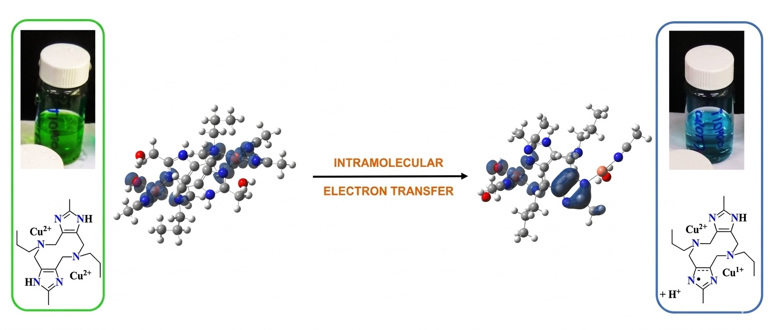
The spontaneous reduction of one Cu(II) center to Cu(I) in a series of three dinuclear copper complexes in acetonitrile is described. These complexes feature ligands that include nitrogen donors from a diazecine ring and imidazole, designated as promeim, thiopromeim, and thioenmeim; the latter two incorporate a thioether as a third donor component. The mechanism of metal reduction was elucidated through spectroscopic and spectrometric techniques (UV-vis, EPR, XANES, ESI-MS) and electrochemical tools, in combination with DFT electronic structure calculations. Based on these and on spectroelectrochemical results, a mechanism is proposed in which the one-electron reduction of one of the copper ions is achieved by a one-electron oxidation in the adjacent imidazole group, while the other copper ion remains as Cu(II). The persistent detection of superoxide and peroxide over long periods suggests a mechanism in which a catalytic cycle involving electron transfer occurs between copper, ligand, and dioxygen.

Graphical Abstract

1. Introduction

The main function of copper enzymes in biological systems is that of electron transfer, due to this metal ion’s ability to reversibly change oxidation states from II to I. It is noteworthy that in all of these enzymes, copper is always bound to at least one imidazole group from histidine residues [1,2,3,4].
Among the abundant copper complexes described in the literature over the years, there have been quite a few reports of a puzzling redox phenomenon that has been described as “autoreduction” or “spontaneous reduction” of Cu(II) to Cu(I) in a wide variety of chemical environments. Many of these involve bioinspired coordination complexes [5,6,7,8,9,10,11,12,13,14,15,16,17,18,19,20,21], and in some cases, the redox-active character of the ligands promotes metal reduction [22,23]. Even though in some of these articles the role of the solvent is mentioned and structural arguments are given for the stabilization of Cu(I) over Cu(II), most of these reports have not made clear the mechanism of electron transfer, nor do they identify the reducing species responsible for this transformation. In limited examples, electron transfer from the ligand to Cu(II) to create a free radical intermediate has been proposed [24,25,26]. The related phenomenon of coordination compound redox isomerism or valence tautomerism, which involves the interconversion of two electronic isomers or valence tautomers (i.e., species with identical chemical composition) with different charge distributions, has been amply described since the 1980s [27] and continues to be relevant [28,29]. Also recently, autoreduction of copper (II) has been reported on zeolite or molecular sieve-adsorbed copper salts, where coordinated -OH groups from the zeolite act as electron donors while being oxidized to molecular oxygen [30,31,32,33,34].
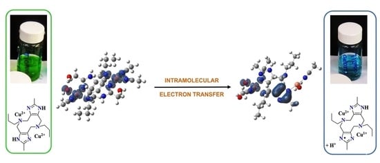
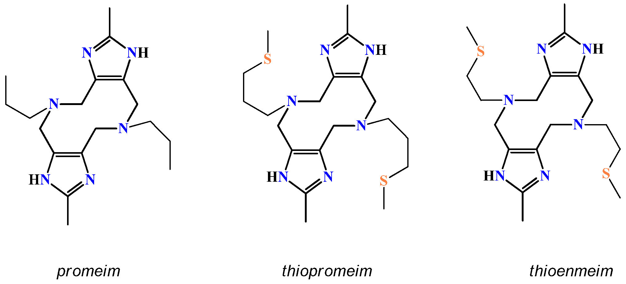
In this work, we describe the “autoreduction” reaction experienced by three dinuclear Cu(II) complexes with imidazole-containing ligands, promeim, thiopromeim, and thioenmeim (Scheme 1), when formed in acetonitrile using non-coordinating counteranions. Freshly prepared solutions exhibit a deep green color common for Cu(II) complexes and turn blue over time, following the kinetic pattern Cu2promeim >> Cu2thiopromeim >> Cu2thioenmeim. Attempts to isolate the product by slow evaporation of the solvent yielded a blue slurry and colorless crystals containing [Cu(MeCN)4]+ (Figure S1).
Analysis of this behavior was performed by spectroscopic, electrochemical, and computational means. These results, strongly supported by spectroelectrochemical experiments, led to a proposed mechanism in which the reduction of Cu(II) is accomplished by the loss of an electron from the ligand, possibly generating an intermediate species involving a radical cation in the imidazole moiety. In aerobic solutions, Cu(I) generated in this way reduces atmospheric O2, producing the superoxide ion. The latter can be detected by EPR spin trapping with DMPO at virtually any given moment, suggesting Cu(I) persists for long periods despite its reaction with dioxygen, and thus must be regenerated in a redox cycle involving Cu(II)/Cu(I), Imz/Imz· and O2/O2·−. This work addresses a transformation that takes place specifically in the presence of acetonitrile, favored by the well-known stabilizing effect this coordinated solvent has on Cu(I). These findings may be relevant to copper enzyme chemistry, since the imidazole group from histidine is a ubiquitous coordinating group in Cu redox enzymes. Even when acetonitrile is never present in these systems, there are steric and electronic factors that promote the necessary metal coordination geometry changes that enable reversible changes in oxidation state.

2. Results

2.1. Characterization of the Complexes in Their Initial State

2.1.1. Crystal Structures

Crystals of the thioether-containing ligand complexes [Cu2(thioenmeim)(H2O)2(NO3)2](NO3)2 and [Cu2(thiopromeim)(H2O)2(NO3)2](NO3)2 were easily obtained from methanol–water solutions. X-ray analysis revealed similar structures with the 10-membered central diazecine ring adopting a chair conformation, consistent with several complexes obtained by our group [35,36,37,38,39,40] (Figure 1A,B, CCDC: 2442956–2442957). Both complexes form centro-symmetric dinuclear molecules in which copper ions are five-coordinate in slightly distorted square-pyramidal geometry with τ parameter values [5] of 0.177 and 0.031, respectively. The Cu...Cu distances are 7.5129(6) Å for the former and 7.313(2) Å for the latter. In both complexes, the base of each pyramid is formed by two nitrogen atoms from the ligand, including a N(2) atom from the imidazole group and a N(1) atom from the tertiary amine, as well as an oxygen atom -O(1) from the nitrate group and a sulfur atom from the thiomethyl group. The apex of the pyramid is occupied by an oxygen atom from a coordinated water molecule, O(1W). Relevant bond distances and angles are listed in Table S2. Crystal packing and the description of network intermolecular interactions for these two complexes are presented in the Supplementary Materials (Figure S2A,B).
Crystals of the analogous copper complex with the promeim ligand proved elusive, probably due to this ligand’s lack of a third donor.

2.1.2. Electronic Spectra

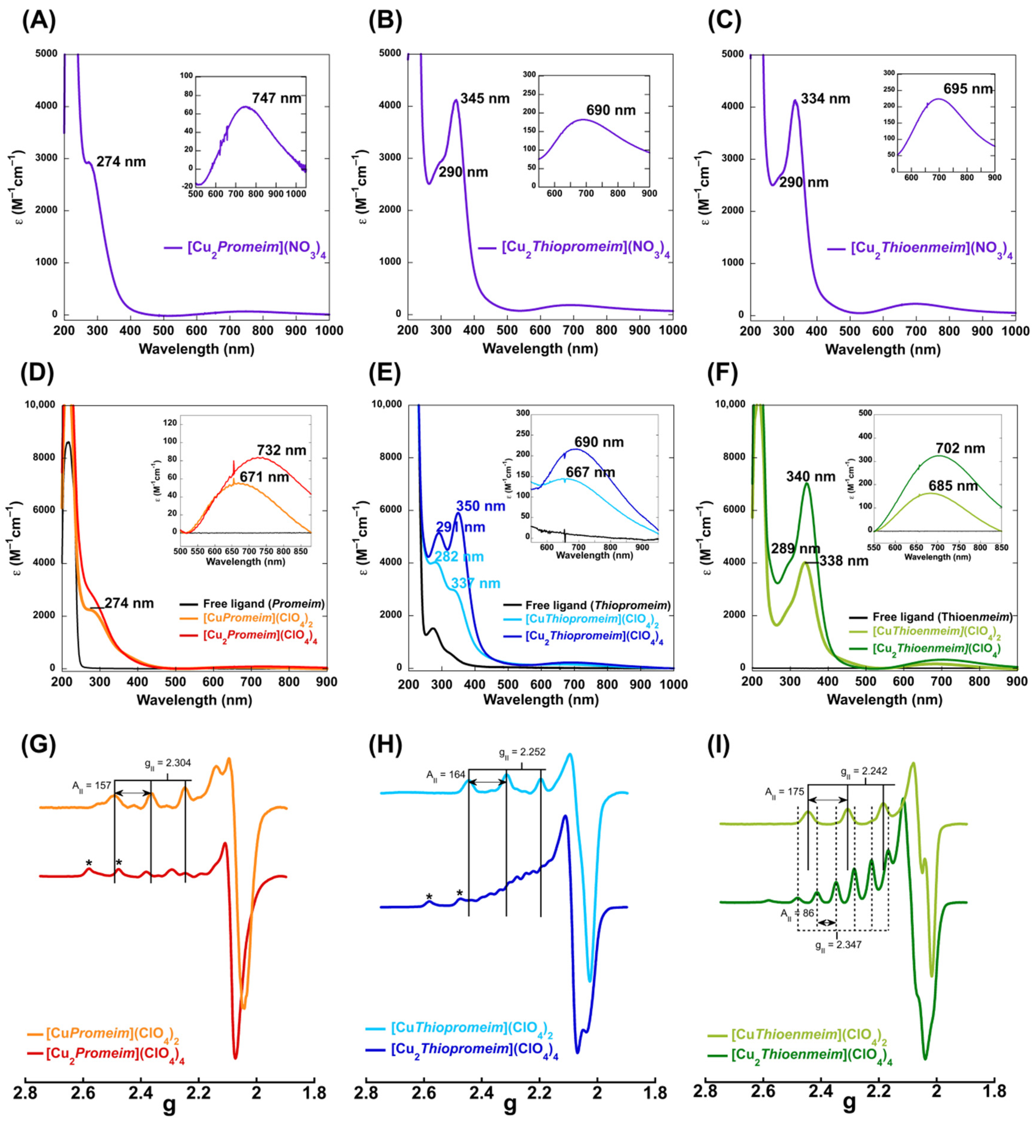
UV-vis electronic absorption spectra were initially obtained under the same conditions as those of crystallization, namely in methanol solution and with nitrates as counterions. The absorption spectrum of the [Cu2promeim](NO3)4 complex (Figure 2A) displays a ligand field absorption at 747 nm (ε = 67 M−1 cm−1) and a ligand-to-metal charge transfer (LMCT) band at 274 nm (ε = 2915 M−1 cm−1) assigned to an imidazole π to Cu(II) [41].
For the [Cu2thiopromeim](NO3)4 and [Cu2thioenmeim](NO3)4 complexes (Figure 2B,C), the ligand field transitions are shifted to 690 nm (ε = 180 M−1 cm−1) and to 695 nm (ε = 220 M−1 cm−1), respectively, while LMCT bands are observed at 345 nm (ε = 4118 M−1 cm−1) for [Cu2thiopromeim](NO3)4 and 334 nm (ε = 4132 M−1 cm−1) for [Cu2thioenmeim](NO3)4, corresponding to S → Cu(II) LMCT transitions [42]. Additionally, in both spectra, one shoulder is observed at 290 nm, corresponding to the imidazole π to Cu(II) LMCT transition.
A comparison of the electronic absorption spectra of solutions with 1:1 and 2:1 Cu:ligand ratios for the three ligands, as prepared in ethanol and using Cu(ClO4)2 as the copper source, is shown in Figure 2D–F. The promeim complexes at 1:1 and 2:1 Cu:ligand ratios exhibit very similar spectroscopic features, showing a slight increase in absorbance at 274 nm, assigned to an imidazole π-to-Cu(II) LMCT. Meanwhile, the ligand-field transitions shift from 671 to 732 nm. For the thiopromiem and thioenmeim complexes, larger differences are observed in the LMCT region. Specifically, the LMCT associated with the sulfur-to-Cu(II) transition increases in intensity and shifts slightly to a higher wavelength, from 337 to 350 nm for the thiopromeim complexes (Figure 2E) and from 338 to 340 nm for the thioenmeim complexes (Figure 2F). A similar trend (i.e., a shift to lower energy/higher wavelength is observed for the ligand field transitions when going from the 1:1 and 2:1 Cu:ligand ratios.

2.1.3. EPR Spectra

Solutions with 1:1 and 2:1 Cu:L ratios were also studied by electron paramagnetic resonance (EPR), using ethanol as a solvent and perchlorates as counterions. Under these conditions, their EPR spectra display signals with g|| > g > 2.0023 and a large parallel hyperfine splitting (A||), indicative of tetragonal Cu(II) centers with a dx2−y2 ground state [18]. The EPR spectrum of the Cu:promeim solution in a 1:1 ratio displays A|| = 157 × 10−4 cm−1 and g|| = 2.304 values, corresponding to a nitrogen-rich coordination mode. In contrast, the EPR spectrum of the Cu:thiopromeim 1:1 displays different A|| and g|| values of 164 × 10−4 cm−1 and 2.252, respectively, possibly reflecting an equatorial coordination sphere that includes nitrogen- and sulfur-based ligands. In addition, the other ligand that contains sulfur atoms in its structure, Cu:thioenmeim 1:1, displays signals with parameters very close to Cu:thiopromeim, with A|| = 175 × 10−4 cm−1 and a g|| = 2.242, supporting the participation of sulfur atoms in the coordination sphere to Cu(II) in a 1:1 ligand:metal ratio.
In contrast, the 2:1 Cu:ligand solutions show strikingly different EPR features, as compared to those of the 1:1 samples. In particular, for the thioenmeim ligand, a clear seven-line signal is observed in the parallel region of the spectra, indicative of electron spin coupling to two nuclear Cu(II) spins (I = 3/2). Several similar EPR spectra have been previously described by our group for dinuclear copper(II) complexes with analogous ligands [35,36,37,38,39,40] and by other research groups for other complexes with aliphatic spacers [43,44,45], in which separation distances range from 6.9 to 9.1 Å. The EPR spectrum of the 2:1 Cu:thioenmeim solution (green spectrum in Figure 2I) can be simulated by considering two Cu(II) centers, with their electron spins coupled to both metal nuclear spins, exhibiting a hyperfine splitting of A|| = 86 × 10−4 cm−1 and a g|| = 2.347 (Figure S3, Table S3). It should be noted that this hyperfine pattern is not evident in the spectra of the 2:1 Cu:promeim and Cu:thiopromeim solutions but could be observed only when acetates were used as counterions (Figure S4). In these two complexes, where there is no third donor atom (Cu2promeim) or where the third donor atom forms a larger, and thus less stable, chelate ring (Cu2thiopromeim), greater internal mobility might cause different conformations in which magnetic coupling cannot take place.
There may also have been a small amount of mononuclear species and a small amount of free copper in the solution (Figure 2G,H).

2.2. Transformations in MeCN

2.2.1. Electronic Spectra

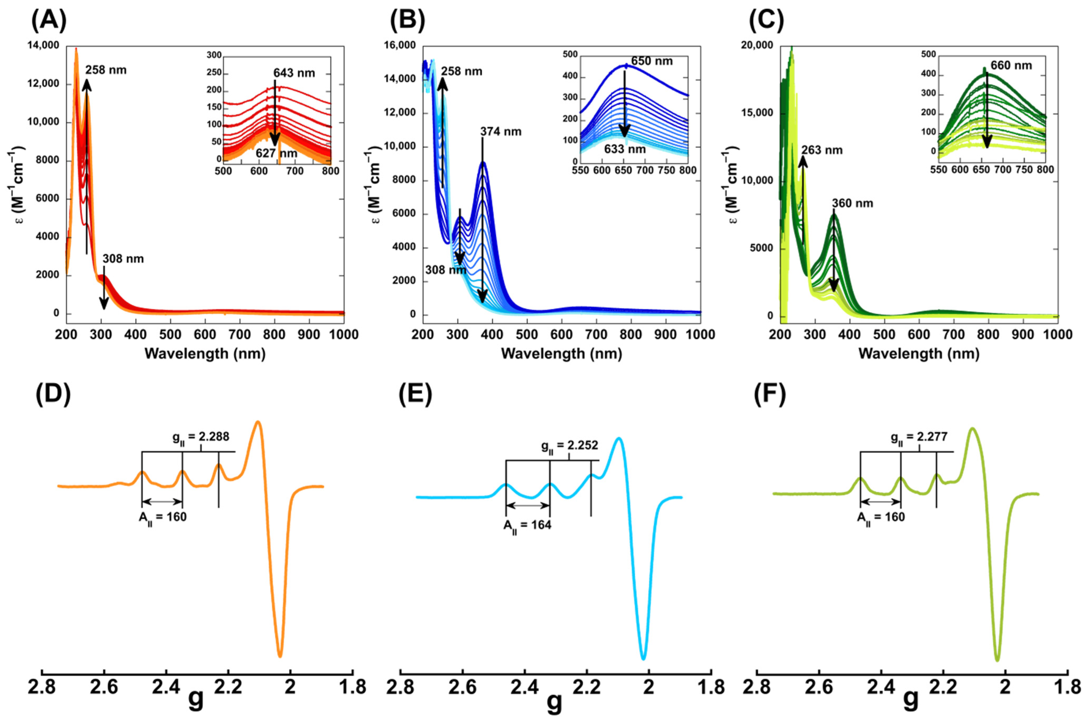
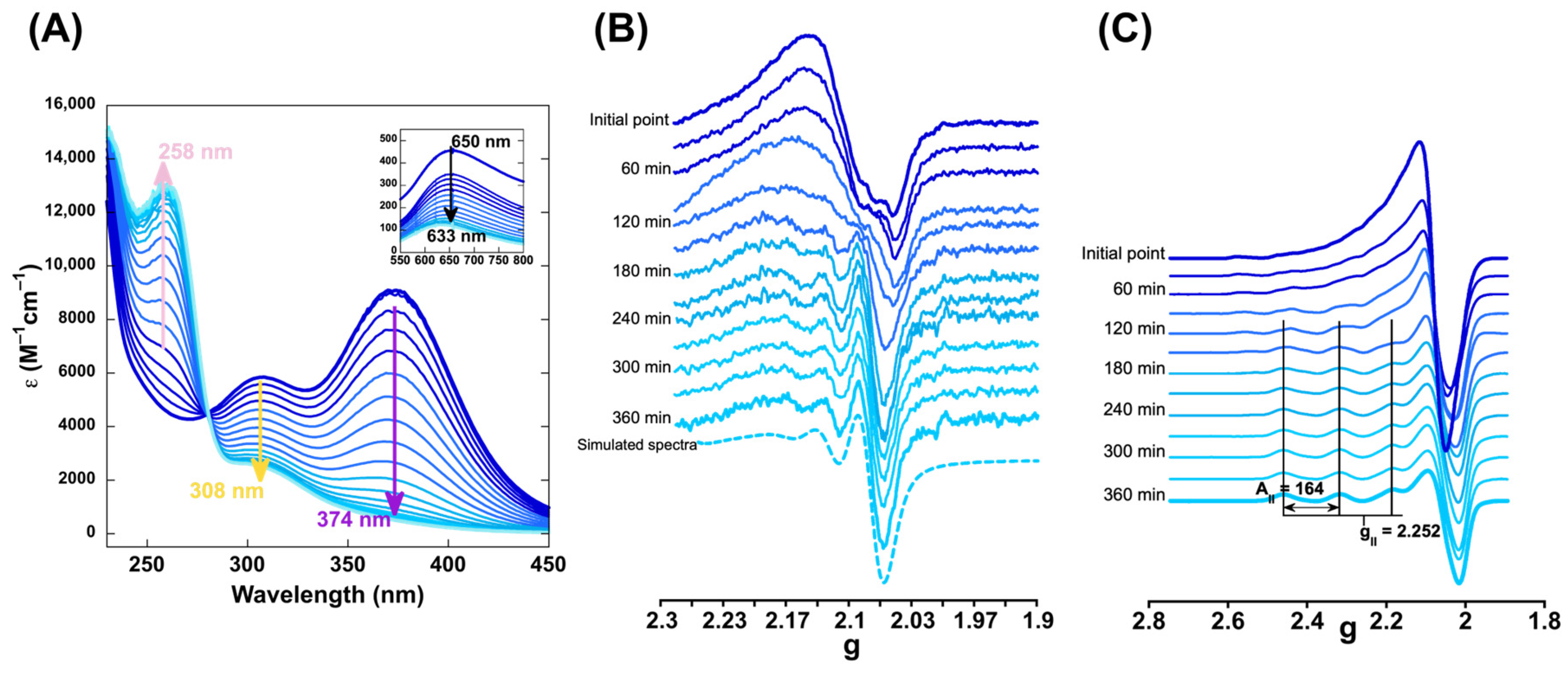
To gain insight into the reactivity of the dinuclear complexes under different conditions, their behavior in MeCN solvent was investigated. The dinuclear Cu complexes with promeim, thiopromeim, and thioenmeim, as described above, are stable in methanol or ethanol and readily obtained upon mixing the ligand with two equivalents of Cu(II) with non-coordinating anions (i.e., ClO4, BF4 or OTf). Caution! Perchlorates are explosive; handle with care. However, when prepared in acetonitrile, their solutions undergo marked color and spectroscopic changes, going from green to blue. This transformation was followed by UV-vis electronic absorption spectroscopy in the presence of 80% acetonitrile and 20% ethanol (since the ligand was insoluble in MeCN but soluble in EtOH), revealing the relative rate at which the three complexes undergo this change: Cu2promeim (reaction time ca. ½ h) << Cu2thiopromeim (ca. 3 h) << Cu2thioenmeim (>>24 h). Figure 3A–C show the changes in the UV-vis spectra of the MeCN/EtOH solutions of the three complexes. In the UV, the most noticeable changes for Cu2thiopromeim and Cu2thioenmeim are marked decreases in the 370 nm band associated with S → Cu LMCT, which is indicative of the loss of the thioether group from the coordination shell. For the three complexes, a decrease in 308 nm absorption, associated with imidazole → Cu LMCT, is observed, while there is a concomitant emergence of an intense band at ca. 258 nm.
In the visible region, the d-d band decreases in intensity, suggesting a partial reduction of Cu(II) to Cu(I), and its blue shift is indicative of a change in the coordination environment for the remaining Cu(II) species. The longer reaction rates observed for complexes with thioether-containing ligands may have been caused by a preliminary step where Cu-S bonds were substituted for Cu-NCMe bonds. This decoordination is expected to be slower for Cu2thioenmeim than for Cu2thiopromeim since, in the former, the sulfur atom forms part of a five-membered chelate ring with the metal ion, while in the latter, the chelate ring is six-membered. It is typical for complexes with analogous ligands that stability constants are larger for the complex with the five-membered ring-forming ligand. This trend has been observed in our lab in complexes with similar ligands [40]. After the loss of the thioether groups’ coordination, the three complexes can attain equivalent coordination environments.
It should be noted that at longer time points (ca. three weeks), when most of the solvent had slowly evaporated in open air, the acetonitrile solutions of these complexes yielded the colorless crystals of [Cu(MeCN)4]X (X = ClO4 or BF4) (Figure S1), further supporting that metal reduction occurs. From the remaining deep-blue semisolid, it was impossible to crystallize any species, so the new absorption band at 258 nm could potentially be associated with ligand oxidation (see below).
For the promeim complex, analysis by MS of this aged semisolid after extracting the copper ions showed that the ligand remained mostly intact, with less than 10% transformation (Figure S5).

2.2.2. EPR Spectra

The reactivity of the dinuclear Cu(II) complexes in MeCN/EtOH (80%/20%) solution was also studied by EPR spectroscopy. At the end of the transformation, the EPR spectra at 150 K were measured (Figure 3D–F), showing a single Cu(II) species with g|| = 2.288 and A|| = 160 × 10−4 cm−1 for the promeim complex, g|| = 2.252 and A|| = 164 × 10−4 cm−1 for the thiopromeim complex, and g|| = 2.277 and A|| = 160 × 10−4 cm−1 for the thioenmeim complex. These spectroscopic features are very similar to those observed for the Cu:ligand solutions in a 1:1 ratio (Figure 2G–I), suggesting the presence of a single Cu(II) ion in the complex at the end of the transformation. This scenario is consistent with one of the copper ions reducing to Cu(I).

2.2.3. X-Ray Absorption Spectroscopy (XAS)

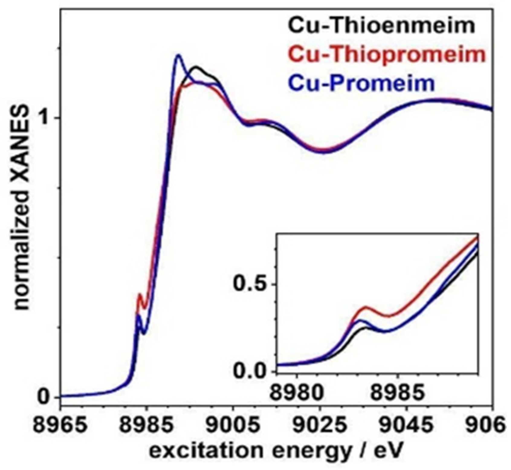
Copper K-edge XAS was also used to corroborate the formation of Cu(I) species upon the reaction of the dinuclear complexes in MeCN. The complexes were prepared using Cu(ClO4)2 dissolved in acetonitrile and mixed with the corresponding ligand in a 2:1 Cu:ligand stoichiometry to yield the dinuclear complexes at a high concentration (5 mM). Samples were frozen for XAS analysis after three days of transformation in MeCN. The presence of Cu(I) ions at the end point of the reaction can be confirmed from X-ray absorption near-edge structure (XANES) spectra (Figure 4).
For all three complexes, a rising edge feature at about 8982–8984 eV is observed, which can be assigned to the 1 s → 4 p electronic transition of Cu(I) ions present in the samples [46]. Previous studies by Solomon and coworkers showed that rising edge peak energies and intensities can be reasonably correlated with the coordination number [47]. Based on this, the edge features observed in Figure 4 (inset) can be ascribed to a tricoordinated Cu(I) species. In contrast, for Cu(II) species, it is important to mention that the pre-edge feature associated with the 1 s → 3 d transition (~8979 eV) of Cu(II) species is hardly observed due to its low intensity, since it is an electric dipole forbidden transition. Overall, these XAS data confirm that, upon reaction in MeCN, these dinuclear Cu complexes undergo reduction of one of the Cu(II) centers to Cu(I). Such metal reduction may occur at the expense of ligand oxidation, as will be proposed next.

2.2.4. Mass Spectra of Transformed Samples

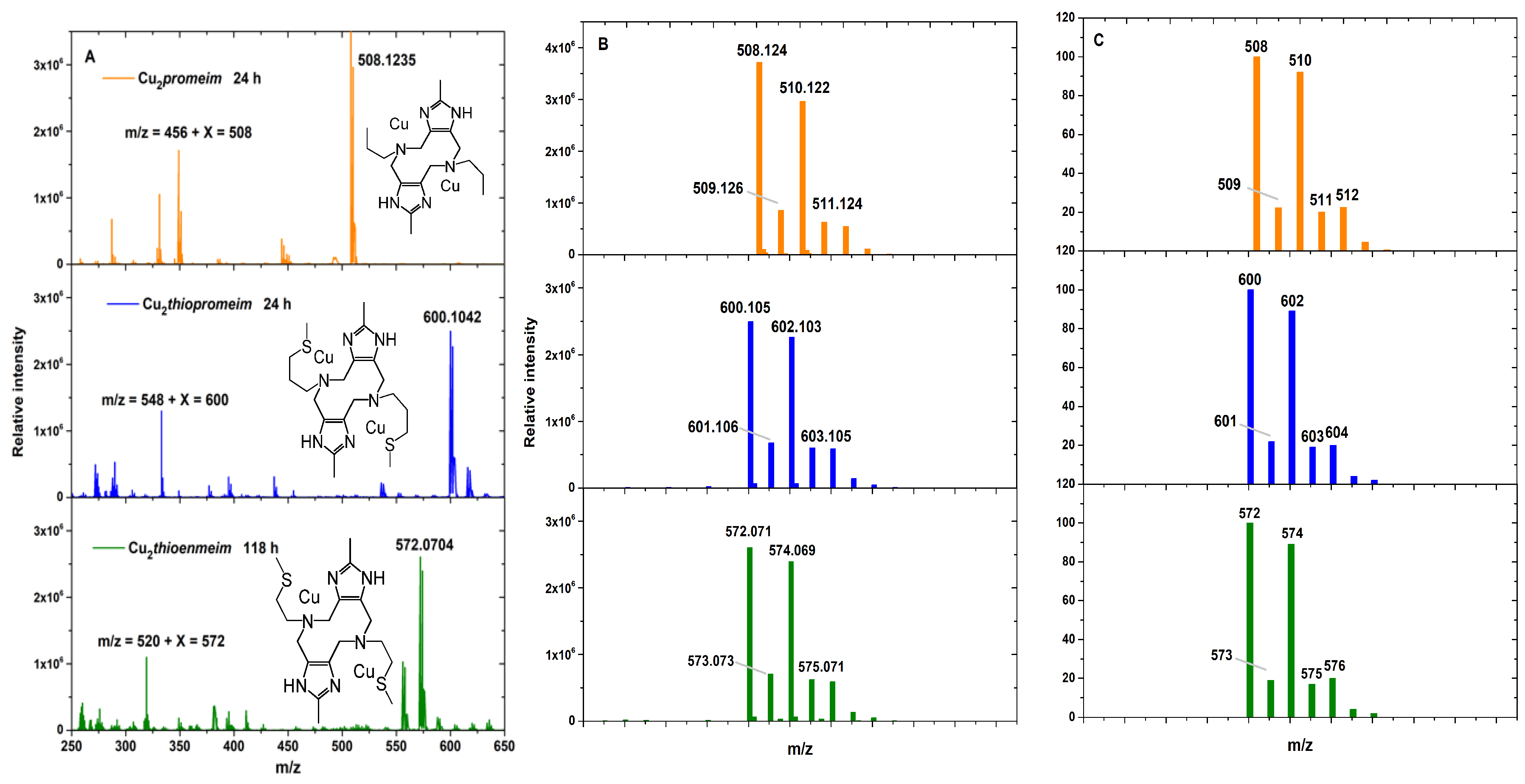
Transformation of the three complexes in acetonitrile solutions was also monitored at intervals by mass spectrometry until no further variations were observed: 24 h for Cu2promeim, 24 h for Cu2thiopromeim, and 118 h (ca. 5 days) for Cu2thioenmeim. The final main peaks for the three complexes were respectively 508.13, 600.1, and 572.07 m/z. All of them correspond to dinuclear species, as shown by the isotopic pattern in the simulated spectra (Figure 5). Remarkably, these masses correspond to an additional 52 u upon each of the formulae [Cu2promeim], [Cu2thiopromeim], and [Cu2thioenmeim], which have calculated masses of 456, 548, and 520 m/z, respectively. The 52 u could correspond to 2OH + H2O (originating from the hydrated Cu salts employed as the starting material). These findings suggest that the three complexes follow the same path, with an analogous final species, responsible for the blue color observed in all cases.

2.2.5. Kinetics of the Transformation of Cu2thiopromeim

Given its intermediate transformation time, Cu2thiopromeim was chosen to monitor the changes observed over time (in MeCN/EtOH (80%:20%)) in its electronic and EPR spectra (Figure 6).
The ligand field transition at 650 nm, the S → Cu LMCT at 370 nm and the imidazole → Cu LMCT at 308 nm decreased with time (Figure 6A). The intensity changes of these LMCT bands can be analyzed using a simple exponential fit, yielding a rate of decay that is comparable to that of the appearance of the new electronic transition at 258 nm.
The transformation was monitored simultaneously by EPR at room temperature (Figure 6B), showing evident changes in the spectrum over time, likely reflecting changes in Cu(II) coordination. In fact, the decay in EPR intensity at 3132 Gauss can be fit to a rate that is in the same order of magnitude as that of the decay of the LMCT bands observed by absorption (Figure S5), strongly suggesting that the loss of the dinuclear Cu(II) features correlates with the appearance of the 258 nm band and the changes in metal coordination detected by room temperature EPR. At the end of the transformation, the EPR spectrum at room temperature could be simulated (Figure 6B, dotted blue line) by considering only a mononuclear Cu(II) center with limited mobility and a correlation time (tR) of 0.18 ns with gx = gy = 2.077, gz = 2.254, Ax = Ay = 6.79 × 10⁻⁴ cm−1, and Az = 162 × 10−4 cm−1. In contrast, the time-resolved EPR data at 150 K reveal the loss of the signals associated with the dinuclear Cu2thiopromeim complex and the appearance of EPR signals (g|| = 2.252 and A|| = 164 × 10−4 cm−1) associated with the mononuclear Cu(II) complex (Figure 6). EPR intensity at 2902 Gauss, which had a major contribution from the dinuclear Cu species, also decayed at a rate in the same order of magnitude as the decay rates for the electronic absorption transitions associated with the Cu(II) dinuclear species (see Figure S6). Moreover, after 180 min of reaction, no further spectroscopic changes were observed; the EPR spectrum at 150 K was dominated by a mononuclear species, and spin quantitation confirmed a loss of about half of the EPR intensity (Figure S8). Overall, these data are consistent with the notion that one of the Cu(II) ions in the dinuclear complex undergoes reduction to Cu(I).

2.2.6. Electrochemical Measurements

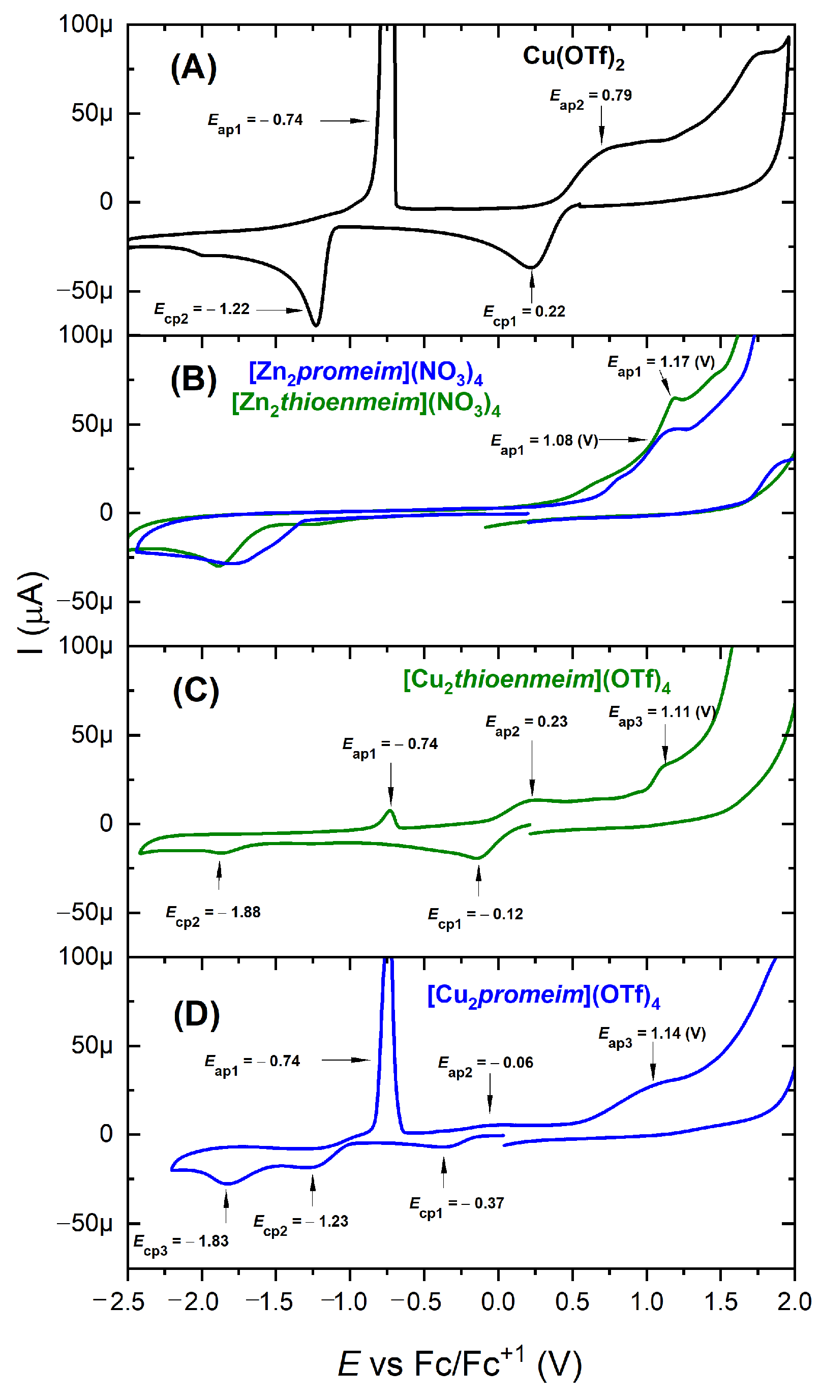
Electrochemical studies have provided relevant information in deciphering the electron transfer processes responsible for the spectroscopic changes described above. Given the different times at which each of the three systems undergoes the transformation, electrochemical measurements were used to confirm, firstly, the initial composition of the 2Cu:1 ligand systems, and secondly, their final composition. The electrochemical characterization of the initial state was achieved through CVs of the most inert system, (Cu2thioenmeim), while for the characterization of the final state, the most labile one, (Cu2promeim), was examined. All solutions were prepared in MeCN using Cu(OTf)2 and the corresponding ligands in a 2Cu:1ligand ratio. Cathodic scan CVs are shown in Figure 7. A MeCN solution of Cu(OTf)2 was used as a reference for uncomplexed Cu, while, given the insolubility of the ligands in MeCN, solutions of [Zn2promeim](NO3)4 and [Zn2thioenmeim](NO3)4 served as references for the ligands unbound to Cu.
In Figure 7A, the reduction wave at 0.22 V corresponds to Cu(II) → Cu(I) and the one at −1.22 V to Cu(I) → Cu°. The corresponding oxidation for Cu° → Cu(I) is observed at −0.74 V and for Cu(I) → Cu(II) at 0.79 V. In this case, the soluble copper species were the simple acetonitrile-solvated copper ions, namely [Cu(MeCN)4]+ and [Cu(MeCN)n]2+. In Figure 7B, the CVs of Zn2promeim and Zn2thioenmeim show signals ascribable to oxidation of the ligand, at 1.08 and 1.17 V.
The CV of a freshly prepared Cu2thioenmeim solution (Figure 7C) shows only two reduction waves: one ascribed to Cu(II) → Cu(I) at −0.12 V and that for Cu(I) → Cu° at −1.88 V. The corresponding oxidation waves are Cu° → Cu(I) at −0.74 V and Cu(I) → Cu(II) at −0.23 V. Finally, Figure 7D shows the CV for Cu2promeim measured approximately one hour after solution preparation, providing enough time for the transformation to reach its final state. In this case, three reduction waves can be observed: the first at −0.37 V, the second at −1.23 V, and the third at −1.83 V (Figure 7D).
Since the Cu(II)/Cu(I) pair is quasi-reversible in all cases, we were able to approximate the E1/2 values from the average (Eap2 + Ecp1)/2. These values are: 0.505 V for the solvated Cu ions, 0.055 V for Cu2thioenmeim, −0.192 for Cu2thiopromeim, and −0.215 V for the Cu2promeim copper complexes (Figure S10). The trend in these potentials agrees with expected values for Cu(II)/Cu(I) pairs in the various coordination environments. The largest value, found for the MeCN-solvated ions, is consistent with the great stability known for [Cu(MeCN)4]+. Concerning the other two copper solutions, it is well documented that copper complexes with sulfur-containing ligands display higher E1/2 values than those with exclusively nitrogen-donating ligands [48,49,50,51]. The comparative E1/2 values obtained for the studied complexes support the notion that copper ions in the presence of the thioenmeim ligand have a N2S + nMeCN coordination environment, while the coordination environment for copper ions in the presence of the promeim ligand is N2 + mMeCN. The signal observed at the lowest potential values for 2Cu:L systems (Ecp3 = −1.83 V for L = promeim and Ecp2 = −1.88 V for L = thioenmeim) corresponds to the Cu(I) → Cu° reductions. Most revealing is Ecp2, observed in the 2Cu:promeim system at −1.23 V, since it coincides with Ecp2 for the Cu(OTf)2 solution in MeCN, assigned above to [Cu(MeCN)4]+ → Cu°. This indicates the presence of [Cu(MeCN)4]+, which is consistent with the isolation of [Cu(MeCN)4]OTf crystals from aged similar solutions (Figure S1). It is also of interest to take note of the relative intensities of the first reduction peaks for Cu2promeim and Cu2thioenmeim, which were prepared with the same concentrations of ligand and Cu(OTf)2 and can be assumed to have similar diffusion coefficients. The marked decrease observed for this signal in Cu2promeim compared to Cu2thioenmeim agrees with a marked decrease in the concentration of Cu(II) in the former, which had already been reduced to a large extent at the time of measurement. Finally, in the CV for both Cu complexes, a third oxidation wave can be observed at ca. 1.1 V, attributed to the oxidation of the ligands, as supported by CV measurements performed on the analogous Zn2L complexes (Figure 7B).

2.2.7. UV-Vis Spectroelectrochemical Studies on [Zn2promeim]4+

The last-mentioned oxidation signal was decisive in proposing an electron donor for the reduction of Cu(II); the reasoning is outlined here:
-Concomitantly with the decrease in the LMCT band (Imz ⟶ Cu), we observed a surge in a very intense band at 258 nm. (Figure 3A–C).
-In all three copper complexes, cyclic voltammetry in acetonitrile showed an oxidation signal at ca. 1.2 V, ascribable to an electron loss in the ligand (Figure 7C,D).
-To prove this assignment, we performed cyclic voltammetry on the analogous Zn complex (since the ligand is insoluble in acetonitrile, and Zn(II) is not redox active beyond Zn(II)/Zn°). An oxidation signal was observed at practically the same potential value (Figure 7B).
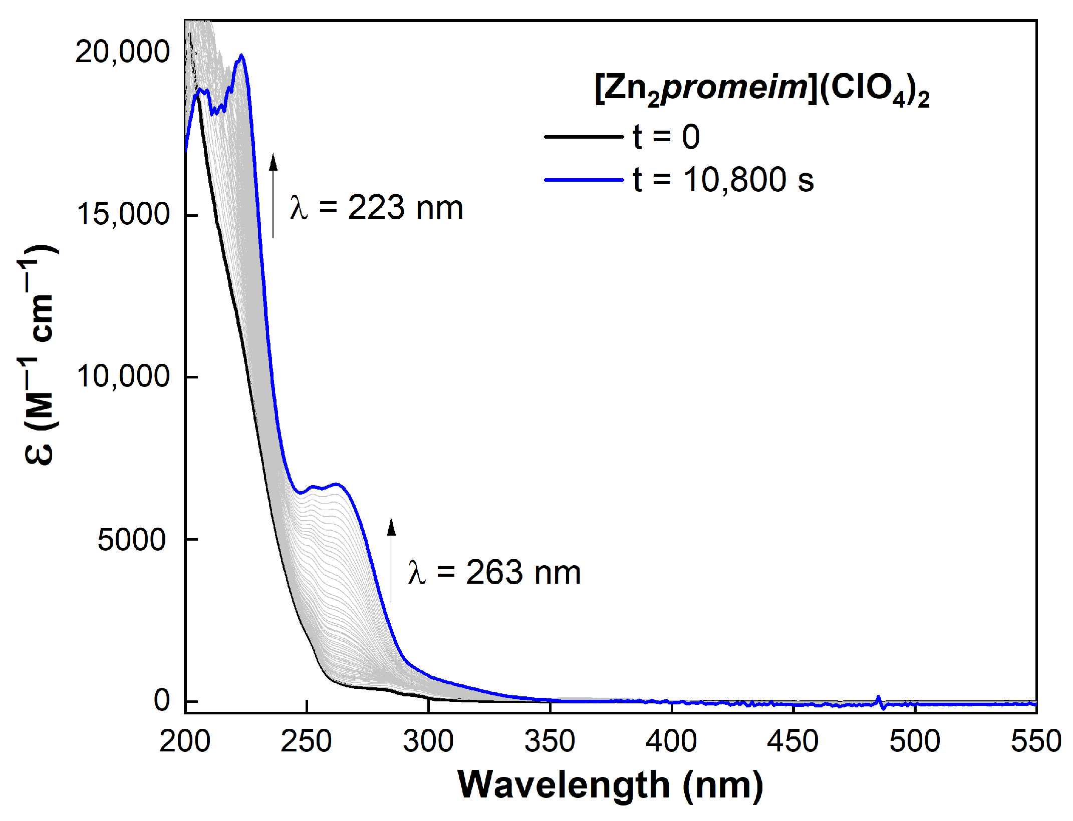
-We then performed electrochemical oxidation in a UV quartz cell to be able to apply the required potential and simultaneously observe the corresponding spectral changes.
-An absorption band at 263 nm surged with the application of the required potential (1.2–1.4 V). This wavelength value is very close to the band observed for the copper complex. (Figure 8).
-This strongly suggests that the oxidation of the ligand caused the new UV absorption.
-Given the chemical structure of the ligand, the HOMO (from which the electron is removed) is expected to have a significant contribution from the nitrogen atoms, particularly those from the heteroaromatic imidazole group. DFT modeling of the initial form of the complex confirms this assumption (vide infra).
This assumption is backed up by the previously determined redox potential for the histidine free radical generated by pulse radiolysis, which was found to be 1.17 V [52]. Also supporting this assignment is the corresponding absorption wavelength for the 2-methylimidazole radical, also generated by pulse radiolysis, which was reported at 285 nm [53].

2.3. Participation of Dioxygen

2.3.1. Identification of ROS in Cu2promeim Solutions

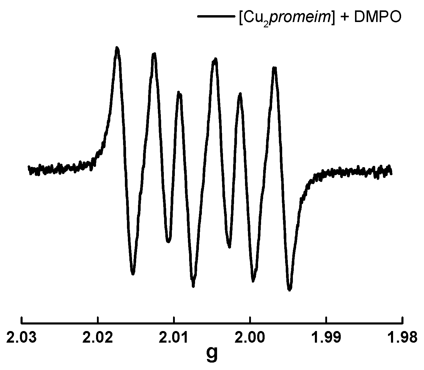
Attempting to identify a free radical generated during the reduction of the copper complexes, we recorded the EPR spectrum of Cu2promeim in MeCN/EtOH in the presence of the spin trap DMPO. The results, shown in Figure 9, agree with the presence of a superoxide radical O2 [54,55].
Strikingly similar results were obtained when this measurement was repeated on the same (aerated) solution after different times over several days (Figure S11). In contrast, measuring the spectra of a single sample in a sealed tube showed a marked decrease in the superoxide signal after several hours (Figure S12).
In parallel, the presence of H2O2 was detected in a MeCN/EtOH solution of Cu2promeim over a similar period of time, following the method described by Adak et al. [56] (Figure S13). This revealed that, given the availability of dioxygen, the formation of H2O2 continued for days without signs of coming to an end, providing further evidence of atmospheric oxygen reduction, since the superoxide ion, produced via a one-electron reaction with Cu(I), can subsequently undergo disproportionation to yield peroxide (H2O2) and molecular oxygen (O2), according to the reaction 2 O2· + 2 H+ ⟶ H2O2 + O2.

2.3.2. Cu2promeim Spontaneous Transformation in the Absence and Presence of Oxygen

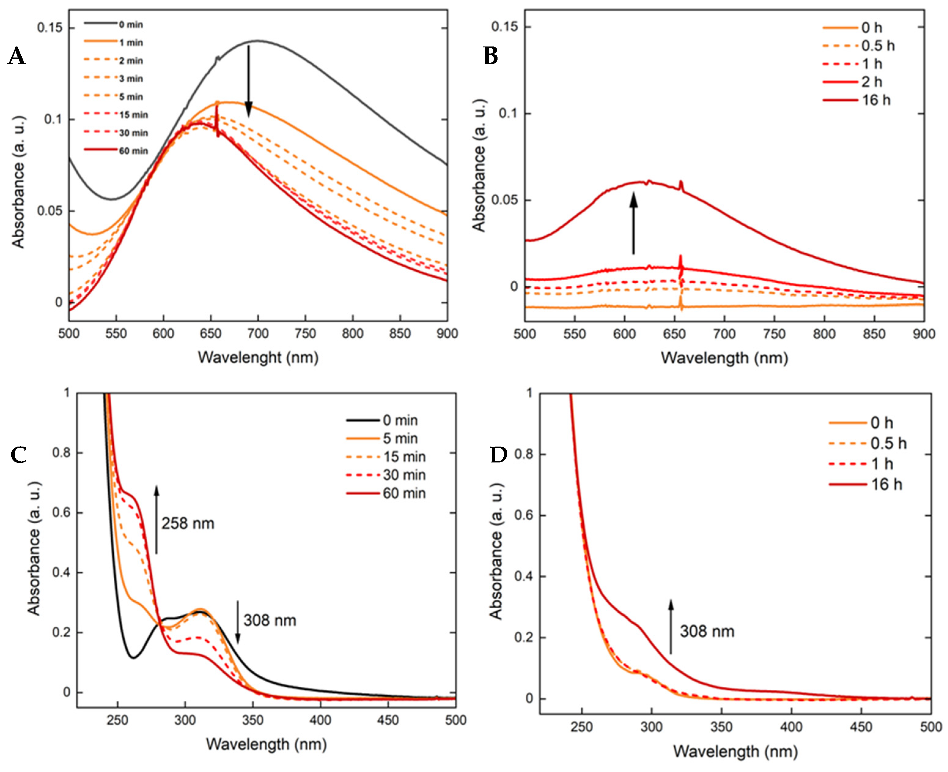
These results motivated us to compare the transformation of Cu2promeim in the absence and presence of dioxygen, followed by UV-vis spectroscopy. In Figure 10, we show the results of parallel experiments, in one case (A and C) starting from Cu(II), and in the other (B and D) starting from Cu(I).
When starting from CuII2promeim in the absence of O2, there was a slight blue shift and significant reduction in absorbance, which can be interpreted as a partial reduction of the Cu(II) ions, giving rise to the mixed-valence species CuICuIIpromeim. In contrast, when the measurement was performed starting from CuI2promeim, the absorbance of the surging d-d band after exposure to O2 hardly reached half of the value of the initial CuII2promeim, suggesting the permanence of the mixed-valence species CuICuIIpromeim, in an aerobic medium.

2.4. DFT Modeling of the Spontaneously Reduced Form of Cu2promeim

Density functional theory (DFT) calculations were carried out to model Cu2promeim, before and after intramolecular electron transfer. The optimized structure of the starting species comprises the dinuclear complex CuII2promeim in the usual chair conformation for these systems [35,36,37,38,39,40], in which each Cu(II) is coordinated to a heterocyclic nitrogen from an imidazole group and an aliphatic nitrogen from a tertiary amine. Coordination number five for both copper ions is completed with three acetonitrile molecules, two water molecules, and a hydroxo ion. In the optimized structure for the final state, one of the copper atoms maintained its original coordination environment, but many changes were found for the second site. The oxidation state was 1+ with a coordination number of 3, as determined by XAS. It was bonded to a water molecule, a MeCN molecule and a nitrogen atom from the imidazole ring, which had lost an electron to copper and became a radical, consistent with the spectroelectrochemical study. DFT modeling of this species with a radical on the imidazole ring only succeeded when this group was considered deprotonated.
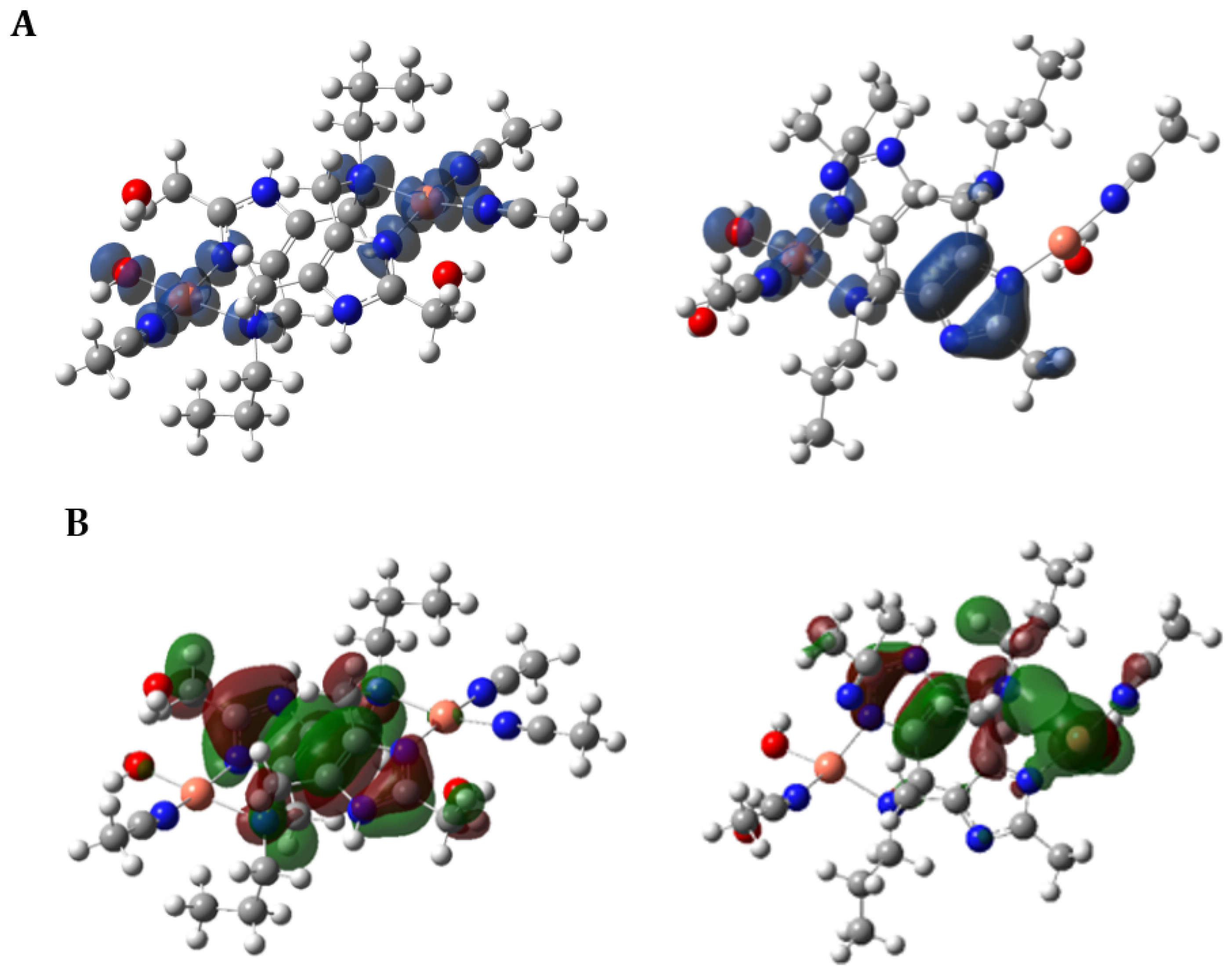
Taking all of this into account, the molecular mass for this species agrees with the main signal observed in the MS after the transformation (vide supra). Keeping in mind that MeCN molecules escape copper coordination during ESI, this mass corresponds to Cu2promeim + 52 mass units, which can be accomplished with Cu2promeim-H+ (from the imidazole deprotonation) + 2 H2O + OH. Figure 11A displays spin density plots for the optimized structures of the initial and final species, before and after electron transfer. In the former (left), the spin densities are symmetrically distributed among both Cu(II) atoms. In the latter (right), the second unpaired electron is clearly localized above and below the imidazole plane, indicating that an electron transfer occurred from the imidazole ring towards the Cu atom, which underwent reduction, leaving the unpaired electron in the π system of imidazole. The other Cu(II) atom remained unchanged. Figure 11B shows the HOMOs (the first doubly occupied molecular orbital) for the complex in its initial (left) and final (right) state. In the initial complex, the HOMO involved the π system of imidazole moieties. However, after electron transfer, it underwent a dramatic change, involving instead the σ bonding of the Cu(I) center with the imidazole.
During modeling of inner-sphere electron transfer, we consistently found that radical localization on the imidazole moiety was only feasible after deprotonation. Multiple attempts to optimize a structure containing a reduced Cu center and a neutral imidazole radical failed to converge to a stable stationary point; however, optimization became straightforward once deprotonation was introduced. This result indicates that deprotonation is a prerequisite for the subsequent electron transfer step.
These calculations represent the outcome of an extensive exploration, which included the evaluation of several DFT functionals, consideration of alternative ligand configurations, and a search for mechanistic pathways with a physically reasonable free-energy profile. For clarity and balance, we present here only the final results most relevant to the experimental observations since the primary aim of this work was experimental, with theoretical modeling included to support and rationalize the findings.

3. Discussion

So far, the spectroelectrochemical experiment backs up the proposal that the UV signal at ca. 260 nm is associated with an oxidation in the ligand. Given the structure of these ligands and considering the nature of the HOMO in Cu2promeim (Figure 11B), the leaving electron must come from the imidazole moiety, producing a free radical. In the copper complexes, the appearance of this UV signal is concomitant with the reduction of one Cu(II), while in the Zn analog, the signal surges when a potential of ca. 1.2 V is applied. DFT modeling supports an intermediate in which the loss of a proton accompanies the loss of the electron from the imidazole moiety, while the coordinated copper atom is reduced.
In contrast, a surprising observation was made when measuring EPR spectra in the presence of DMPO. Superoxide ion O2·− was detected at various times over several days.
Likewise, peroxide was detected chemically with KI during the same period, presumably from the disproportionation of superoxide. Figure S13 shows the formation of H2O2 (detected as I3) over three days in the same fully oxygenated solution. This indicates that, as long as dioxygen is available, H2O2 will continue to accumulate via partial disproportionation of the superoxide ion.
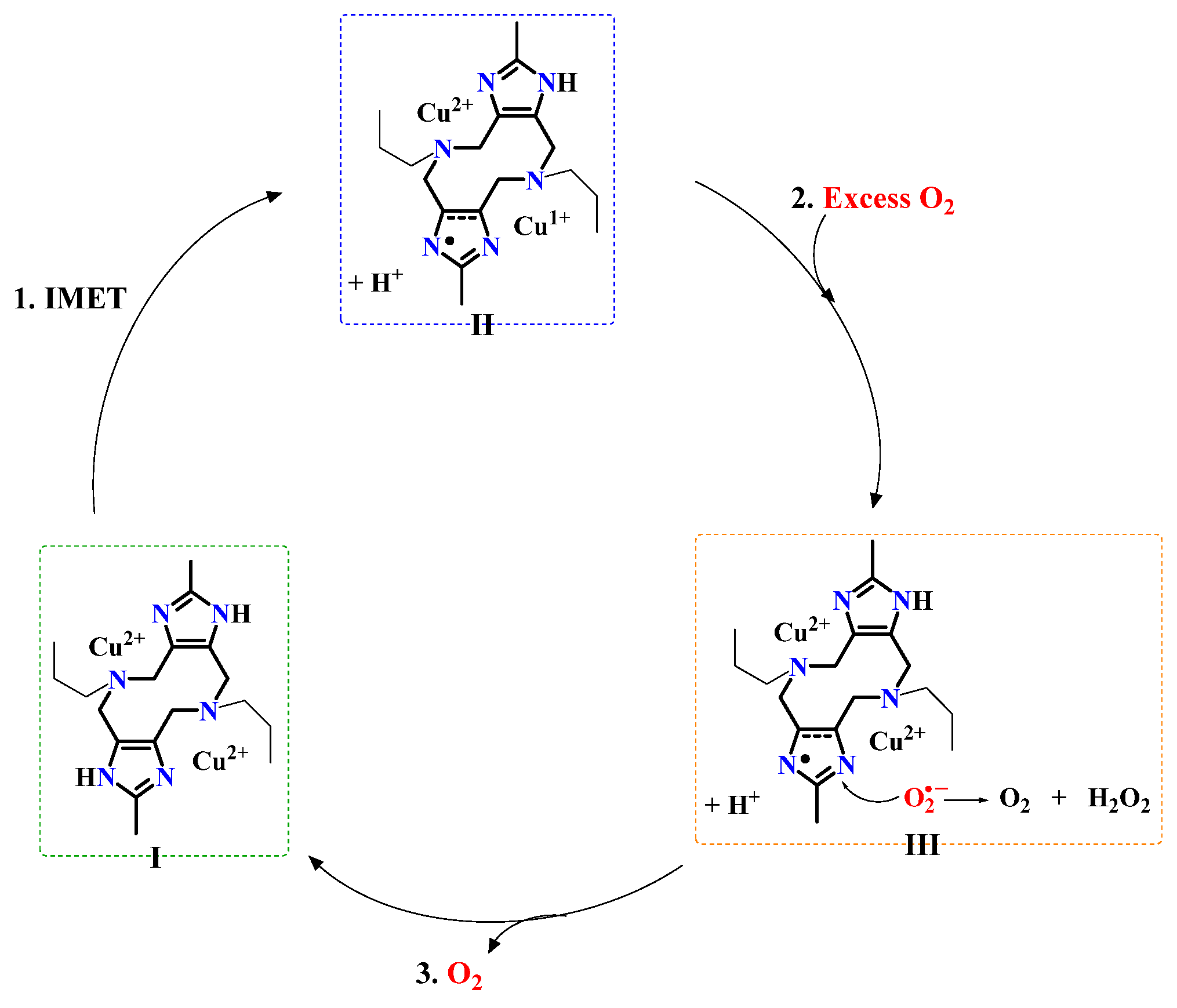
The crucial observation is that, given that O2·− is the product of the reaction of Cu(I) with fully available dioxygen, its presence at any given moment can only be explained by assuming that Cu(I) does not completely run out after long periods of time, even when the reaction is considered complete; i.e., it is being replenished. This would mean that as soon as Cu(I) is oxidized by dioxygen, the generated Cu(II) is re-reduced. The only available reducing agent for Cu(II) is the ligand, which was the initial electron donor; however, to perform this task, it would have to be regenerated from its oxidized form, putatively Imz·. The only available reducing agent for Imz· is the superoxide ion O2·− [57,58,59,60,61], which is generated at the Cu center. This proposal can be seen more clearly in Scheme 2.
Species I schematically represents the original complex, formed in situ upon mixing the copper salt and the ligand solutions. It rapidly converts to Species II, the nature of which is supported by the following:
-The persistence of the UV absorption at 258 nm, which spectroelectrochemistry assigns to an oxidation of the ligand.
-MS data confirming the dinuclear nature of the complex even after long reaction times.
-XAS measurements detecting the presence of Cu(I) (and Cu(II)) also after long reaction times.
-EPR spectra obtained after the transformation typical of a complex with a single Cu(II).
-Spin quantification after the transformation (Figure S8).
-The persistence of the d-d absorption at the end of the transformation, indicating the presence of Cu(II).
-The coincidence of the final d-d bands observed after CuII2promeim transforms under Ar and after CuI2promeim encounters O2.
-DFT modeling.
Step 1 in the proposed mechanism consists only of an intramolecular electron transfer, so it must be virtually instantaneous. Step 2, however, involves a bimolecular reaction (between the copper complex and O2), which presumably is not as fast as the first step, thus allowing the presence of Species II to be detected. Step 3 also consists of only an intramolecular electron transfer, with O2·− considered as part of the coordination center, so it is also expected to be faster than Step 2.

4. Materials and Methods

Reagents and solvents used were of reagent quality, purchased from MERCK Sigma Aldrich de México S de RL de CV, Toluca, México Caution! Perchlorates are explosive, handle with care.

4.1. Synthesis of the Ligands

Promeim [62] (2,8-Dimethyl-5,11-dipropyl-1,4,5,6,7,10,11,12 octahydroimidazo [4,5-h]-imidazo [4,5-c][1,6]diazeeine): First, 4.1 g (0.05 mol) of 2-methylimidazole and 2.95 g (0.05 mol) of n-propylamine were added to 20 mL of distilled water. To this mixture, 9.4 mL of a 35% formaldehyde solution (0.12 mol) was added slowly with stirring. The pH was adjusted to 11–12 with concentrated KOH. The mixture was refluxed for 24 h and then stirred for 3 days. The first microcrystalline white precipitate was filtered off and washed with cold water. The remaining solution was allowed to evaporate slowly. After two weeks, a second crop of a crystalline product was isolated, washed with cold water, and dried in air. Total yield: 5.8 g (70%). IR-ATR ν (cm−1): 3155, 2963, 2930, 2872, 2249, 1654, 1600, 1531, 1470, 1447, 1412, 1374, 1354, 1344, 1302, 1264, 1218, 1165, 1126, 1059, 1033, 1001, 962, 923, 837, 769, 746, 681, 645, 553, 505, 451, 426, 418. MS-DART (MeOH) m/z: 331 [C18H30N6] + H+. 1H-NMR (400 MHz, CD3OD): δ = 3.37 (s, 8H), 2.79 (t, J = 6.9 Hz, 4H), 2.35 (s, 6H), 1.68 (m, 4H), 1.02 (t, 6H). 13C NMR (400 MHz, CD3OD): δ 146.31 (NCN imidazole), 59.13 (NCH2 propyl), 46.51 (NCH2 diacezine), 22.53 (CH2 propyl), 13.57 (CH3 imidazole), 12.35 (CH3 propyl). Elemental Analysis for C18H30N6∙CH3OH∙2.5H2O Calculated: C, 55.99%; H, 9.65%; N, 20.62%. Found: C, 56.14%; H, 10.34%; N, 23.77. For this ligand, the crystal structure was previously determined.
Thiopromeim [63] (2,8-Dimethyl-5,11-bis(3-(methylthio) propyl)-1,4,5,6,7,10,11,12 octahydrodiimidazo [4,5-c:4′,5′-h][1,6]diazecine): In a round-bottom flask, 1.642 g (20 mmol) of 2-methylimidazole was dissolved in 70 mL of H2O with constant stirring. To this solution, 2.24 mL (20 mmol) of 3-(methylthio) propylamine was added, followed by the addition of 4.8 mL (60 mmol) of a 12.33 M solution of formaldehyde. To homogenize the reaction mixture, 30 mL of ethanol was added, and the mixture was brought to a basic pH with 2.78 mL (20 mmol) of triethylamine. The reaction mixture was kept under constant stirring at 75 °C in an oil bath for 72 h. After this time had passed, a creamy white precipitate was observed, which was filtered, washed with H2O, and left to dry in the oven for 24 h. Yield: 2.342 g (55.5%). IR-ATR ν (cm−1): 3030, 2986, 2915, 2817, 2653, 1668, 1596, 1539, 1447, 1409, 1332, 1042, 1005, 918, 773, 654. MS-DART (MeOH) m/z: 423 [C20H34N6S2] + H+. 1H-NMR (400 MHz, D2O): δ = 3.26 (s, 8H), 2.82 (t, J = 7.0 Hz, 4H), 2.56 (t, 4H), 2.24 (s, 6H), 2.02 (s, 6H), 1.83 (m, J = 7.2 Hz, 4H). Elemental Analysis for C20H34N6S2∙CH3OH Theoretical: C, 55.47%; H, 8.42%; N, 18.48%; S, 14.10%. Found: C, 55.08%; H, 8.04%; N, 19.77%; S, 13.31%. For this ligand, the crystal structure was previously determined.
Thioenmeim (2,8-Dimethyl-5,11-bis(2-(methylthio) ethyl)-1,4,5,6,7,10,11,12 octahydrodiimidazo [4,5-c:4′,5′-h][1,6]diazecine): In a round-bottom flask, 1.6422 g (20 mmol) of 2-methylimidazole was dissolved in 70 mL of H2O with constant stirring. To this solution, 1.86 mL (20 mmol) of 2-2-methylthio-ethylamine and 4.86 mL (60 mmol) of a 12.33 M solution of formaldehyde were added. To homogenize the reaction mixture, 10 mL of ethanol was incorporated into the reaction mixture. Finally, 2.8 mmol triethylamine were added to maintain a basic pH. The yellow solution was kept under constant stirring at 75 °C in an oil bath for 72 h. After this time, a white precipitate was observed, which was filtered, washed with H2O, and left to dry in the oven for 24 h. Yield: 1.41 g (35%). IR-ATR ν (cm−1): 3028, 2968, 2909, 2832, 2661, 1669, 1600, 1538, 1449, 1412, 1223, 1163, 1105, 1046, 965, 918, 849, 836, 787, 754, 688, 652, 630. MS-DART (MeOH) m/z: 395 [C18H30N6S2] + H+. 1H-NMR (400 MHz, D2O): δ = 3.29 (s, 8H), 2.96 (t, J = 6.9 Hz, 4H), 2.70 (t, J = 6.9 Hz, 4H), 2.25 (s, 6H), 2.07 (s, 6H). 13C NMR (400 MHz, CD3OD): δ 146.32 (NCN imidazole), 56.70 (NCH2 methylene), 46.58 (NCH2 diacezine), 33.79 (SCH2 methylene), 15.58 (SCH3 t), 13.59 (CH3 imidazole). Elemental Analysis for C18H30N6S2 Calculated: C, 54.79%; H, 7.66%; N, 21.30%; S, 16.25%. Found: C, 54.07%; H, 7.52%; N, 21.92%; S, 15.86%.

4.2. Copper Complexes Synthesis and Characterization

[Cu2promeim(H2O)6](NO3)4: In a round-bottom flask, 0.241 g of Cu(NO3)2·3H2O (1.0 mmol) was dissolved in 25 mL of methanol under constant stirring. To this solution, 0.192 g (0.5 mmol) of the promeim ligand was added stepwise. At the end, a blue-green solution was obtained, which was rotavaporated to remove the solvent. After complete dryness, the glassy solid was washed with diethyl ether. A blue-green glassy solid was isolated. Yield: 0.386 g (95%). IR-ATR ν (cm−1): 3541, 3298, 2979, 2881, 1755, 1714, 1657, 1580,1551, 1493, 1460, 1410, 1276, 1151, 1067, 1039, 1006, 933, 880, 848, 813, 771, 747, 710, 700, 674, 557, 509, 479, 452, 425. MS-ESI (MeOH) m/z: 454.8 [C18H30Cu2N6]-2H+. UV-vis MeOH solution 1 mM: λmax, ε (nm, M−1 cm−1): 274, 2900; 747, 70. Elemental Analysis for C18H42Cu2N10O18 Calculated: C, 26.57%; H, 5.20%; N, 17.21%. Found: C, 26.62%; H, 4.96%; N, 17.77.
[Cu2thiopromeim(H2O)2(NO3)2](NO3)2: The synthesis of the complex was performed by dissolving 0.241 g of Cu(NO3)2·3H2O (1.0 mmol) in 25 mL of methanol under constant stirring. First, 0.211 g (0.5 mmol) of thiopromeim was added stepwise to the copper salt-containing solution. The mixture turned cloudy, so approximately 5 mL of H2O was added to homogenize the solution. Finally, an emerald-green solution was obtained, which was left at room temperature to evaporate the solvent. After a few days, the formation of emerald green crystals was observed. Yield: 0.3875 g (94%). IR-ATR ν (cm−1): 3540, 3191, 3128, 3074, 3015, 2973, 2936, 2837, 2819, 2723, 1735, 1636, 1552, 1450, 1435, 1393, 1375, 1348, 1322, 1279, 1174, 1155, 1077, 1071, 1063, 1040, 1023, 1008, 978, 967, 921, 880, 861, 845, 822, 809, 789, 737, 720, 697, 684, 638, 597, 565, 534, 509, 484, 466, 444, 427. MS-ESI (MeOH) m/z: 546 [C20H34Cu2N6S2]-2H+. UV-vis MeOH solution 1 mM: λmax, ε (nm, M−1 cm−1): 290, 2900; 345, 4100; 693, 190. Elemental Analysis for C20H38Cu2N10O14S2. Calculated: C, 28.81%; H, 4.59%; N, 16.80%; S, 7.69%. Found: C, 29.04%; H, 4.47%; N, 16.76%; S, 7.50%.
[Cu2thioenmeim(H2O)2(NO3)2](NO3)2: The synthesis of the cupric complex was performed by dissolving 0.241 g of Cu(NO3)2·3H2O (1.0 mmol) in 25 mL of methanol under constant stirring. To this solution, 0.197 g (0.5 mmol) of the thioenmeim ligand was added stepwise. The mixture turned cloudy, so approximately 5 mL of H2O was added to homogenize the solution. Finally, an emerald green solution was obtained, which was left at room temperature to evaporate the solvent. After a few days, the formation of emerald green crystals was observed. Yield: 0.362 g (91%). IR-ATR ν (cm−1): 3393, 3196, 3120, 3073, 3047, 2973, 2948, 1750, 1638, 1551, 1473, 1445, 1391, 1370, 1350, 1298, 1284, 1256, 1201, 1148, 1087, 1072, 1065, 1036, 1018, 984, 946, 916, 879, 846, 829, 811, 787, 743, 721, 710, 682, 656, 638, 591, 505, 463, 426. MS-ESI (MeOH) m/z: 491 [C16H26Cu2N6S2]-2H+, the loss of the methyl group from the thioether; 518 [C18H30Cu2N6S2]-2H+. UV-vis MeOH solution 1 mM: λmax, ε (nm, M−1 cm−1): 290, 2750; 334, 4100; 695, 230. Elemental Analysis for C18H34Cu2N10O14S2 Calculated: C, 26.83%; H, 4.25%; N, 17.38%; S, 7.96%. Found: C, 27.08%; H, 4.33%; N, 17.09%; S, 7.83%.
Sample preparation for kinetic monitoring: Since the ligand was insoluble in acetonitrile, it was dissolved in ethanol, and the copper perchlorate was dissolved in acetonitrile. For the UV measurements, the concentrations in the cuvette were 0.4 mM in Cu and 0.2 mM in L, using either promeim, thiopromeim, or thioenmeim. For visible absorption, EPR and MS, the concentrations were 2.0 mM for Cu and 1.0 mM for the ligand. Details on the preparation of the samples are given in the Supplementary Materials.

5. Conclusions

The complete set of results obtained from the various techniques (in acetonitrile or in 4:1 acetonitrile/ethanol solutions) strongly suggests that in these complexes, the reduction of one Cu(II) to Cu(I) is indeed an intramolecular electron transfer reaction where the ligand acts as the electron donor, presumably from the imidazole moiety. A new intense band was observed in the UV spectra of the copper complexes undergoing spontaneous reduction, as well as in that of the analogous zinc complex when it was electrochemically oxidized.
While all three complexes undergo equivalent transformations, the different rates at which they reach the “final” state are associated with the initial loss of the thioether–copper bond in Cu2thiopromeim and Cu2thioenmein, which produces, at different times, a coordination environment equal to that of Cu2promeim. It is from this coordination state that the electron transfer proceeds, giving way to the “final” state. The various spectroscopic measurements performed on solutions of these compounds (UV-vis, EPR, MS, XANES), as well as the electrochemical determinations, are consistent with the DFT modeling of the Cu2promeim complex, which predicts a mixed-valence CuICuIIL· with a free radical on the imidazole moiety of the ligand.
Both the oxidation potentials obtained here for the ligands and the UV absorption wavelength of the transformed species coincide with previous reports for imidazole-derived radicals [52,53]. We propose that the apparent persistence of this mixed-valence complex with a free radical form of the ligand is maintained because it is not a static structure but part of a cyclic equilibrium of very fast reactions involving widely available dioxygen. Cu(I) is initially generated when Cu(II) accepts an electron from the ligand (which, according to DFT modeling, converts into radical Imz· with the loss of a proton) and is immediately reoxidized to Cu(II) by dioxygen, which then forms the superoxide ion. This superoxide ion can donate an electron to radical Imz·. With the reincorporation of a proton, the imidazole group is regenerated, and a new cycle can begin.
Most copper enzymes consistently feature coordinated imidazole residues and are usually involved in proton-coupled electron transfers with the generation of free radicals [64]. It is also known that Cu(I) interacts with molecular oxygen, generating different types of highly reactive copper–dioxygen adducts, including mononuclear end-on Cu(II)-O2·− like the one proposed here [65]. We hope that the findings presented here can be relevant in the understanding of the role of this substructure in biological systems.

Supplementary Materials

The following supporting information can be downloaded at: https://www.mdpi.com/article/10.3390/molecules31081245/s1. Ligand and compound characterization, X-ray diffraction refining data, and crystal packing, EPR simulation parameters, UV-vis titration of promeim and thioenmeim with Cu(II), spin quantification, anodic VC, superoxide EPR detection over time, H2O2 chemical detection, DFT cartesian coordinates and total energy of Cu2promeim before and after transformation [66,67,68,69,70,71,72,73,74,75,76].

Author Contributions

B.S.-E.: Investigation, laboratory work, DFT calculations and manuscript drafting. C.S.-L.: Investigation, UV-vis absorption and EPR experiments and simulations, manuscript writing. V.M.U.-S.: Electrochemical and spectro-electrochemical methodology and analysis, and overall analysis. J.R.Á.I.: DFT methodology and analysis. M.F.-Á.: Crystal structure determination. N.S.: XAS measurements. L.Q.: Experimental design and conceptualization of UV-vis absorption and EPR spectroscopic experiments and kinetic studies, data analysis and interpretation, and manuscript writing. C.E.C. UV-vis monitoring in the presence and absence of oxygen. V.G.-V.: EPR measurements of O2·−. L.G.: Conceptualization, supervision, overall analysis, and manuscript writing. All authors have read and agreed to the published version of the manuscript.

Funding

DGAPA-UNAM for postdoctoral funding. CONAHCYT—SECIHTI for grant PN2076. National Institutes of Health, National Institute of General Medical Sciences (Grant P30GM133894).

Institutional Review Board Statement

Not applicable.

Informed Consent Statement

Not applicable.

Data Availability Statement

The data presented in this study are available on request from the corresponding author.

Acknowledgments

BSE thanks: DGAPA-UNAM for postdoctoral funding, Jorge Peón and Ivan Castillo from IQ-UNAM for technical services, Everardo Tapia Mendoza and Emilio Iturbe Nava for kinetics ESI-MS studies at LANCIC-IQ UNAM. JRAI acknowledges the support of LANCAD UNAM-DGTIC-192 project at UNAM. NS was supported through grant PN2076 awarded to LQ from the National Council for Science and Technology of Mexico (CONAHCYT—SECIHTI). NS was a Deutsche Forschungsgemeinschaft International Fellow SCHU 33411/2-1. The use of the Stanford Synchrotron Radiation Lightsource, SLAC National Accelerator Laboratory, was supported by the US Department of Energy, Office of Science, Office of Basic Energy Sciences under Contract No. DE-AC02-76SF00515. The SSRL Structural Molecular Biology Program is supported by the DOE Office of Biological and Environmental Research and by the National Institutes of Health, National Institute of General Medical Sciences (Grant P30GM133894). LG deeply thanks Siegfried Schiendler for the measurement of the UV-vis spectra of Cu2promeim in the absence and presence of oxygen, and Gloria Acosta-Tejada for electrochemical measurements.

Conflicts of Interest

The authors declare no conflicts of interest.

Abbreviations

The following abbreviations are used in this manuscript:
MeCNAcetonitrile
cpCathodic peak
apAnodic peak

References

  1. Festa, R.A.; Thiele, D.J. Copper: An essential metal in biology. Curr. Biol. 2011, 21, R877–R883. [Google Scholar] [CrossRef]
  2. Kaim, W.; Rall, J. Copper—A “Modern” Bioelement. Angew. Chem. Int. Ed. Engl. 1996, 35, 43–60. [Google Scholar] [CrossRef]
  3. Karlin, K.D.; Yandell, J.K. Redox behavior of blue copper model complexes. Redox potentials and electron-transfer kinetics of some copper(II)-copper(I) complexes with nitrogen and thioether donors. Inorg. Chem. 1984, 23, 1184–1188. [Google Scholar] [CrossRef]
  4. Solomon, E.I.; Heppner, D.E.; Johnston, E.M.; Ginsbach, J.W.; Cirera, J.; Qayyum, M.; Kieber-Emmons, M.T.; Kjaergaard, C.H.; Hadt, R.G.; Tian, L. Copper Active Sites in Biology. Chem. Rev. 2014, 114, 3659–3853. [Google Scholar] [CrossRef]
  5. Addison, A.W.; Rao, T.N.; Reedijk, J.; van Rijn, J.; Verschoor, G.C. Synthesis, structure, and spectroscopic properties of copper(II) compounds containing nitrogen–sulphur donor ligands; the crystal and molecular structure of aqua [1,7-bis(N-methylbenzimidazol-2′-yl)-2,6-dithiaheptane]copper(II) perchlorate. J. Chem. Soc. Dalton Trans. 1984, 7, 1349–1356. [Google Scholar] [CrossRef]
  6. Beloglazkina, E.K.; Shimorsky, A.V.; Mazhuga, A.G.; Shilova, O.V.; Tafeenko, V.A.; Zyk, N.V. Formation of CuI(CH3CN)4ClO4 in the reactions of copper(II) perchlorate with acetonitrile in the presence of sulfur-containing organic compounds. Russ. J. Gen. Chem. 2009, 79, 1504–1508. [Google Scholar] [CrossRef]
  7. Beloglazkina, E.K.; Vorozhtsov, N.I.; Sviridova, L.A.; Grigorkevich, O.S.; Korablina, D.D.; Moiseeva, A.A.; Zyk, N.V.; Majouga, A.G. The first example of Cu(I) complex with 5-pyrazolyl-2-thioxotetrahydro-4H-imidazol-4-one: Synthesis and structural characterization. Inorg. Chem. Commun. 2016, 71, 86–89. [Google Scholar] [CrossRef]
  8. Doine, H.; Yano, Y.; Swaddle, T.W. Kinetics of the bis(2,9-dimethyl-1,10-phenanthroline)copper(I/II) self exchange reaction in solution. Inorg. Chem. 1989, 28, 2319–2322. [Google Scholar] [CrossRef]
  9. Fedorova, O.A.; Shepel, N.E.; Tokarev, S.D.; Lukovskaya, E.V.; Sotnikova, Y.A.; Moiseeva, A.A.; D’Aléo, A.; Fages, F.; Maurel, F.; Fedorov, Y.V. Intramolecular electron transfer in Cu(ii) complexes with aryl-imidazo-1,10-phenanthroline derivatives: Experimental and quantum chemical calculation studies. New J. Chem. 2019, 43, 2817–2827. [Google Scholar] [CrossRef]
  10. Itoh, S.; Kishikawa, N.; Suzuki, T.; Takagi, H.D. Syntheses, structural analyses and redox kinetics of four-coordinate [CuL2]2+ and five-coordinate [CuL2(solvent)]2+ complexes (L = 6,6′-dimethyl-2,2′-bipyridine or 2,9-dimethyl-1,10-phenanthroline): Completely gated reduction reaction of [Cu(dmp)2]2+ in nitromethane. Dalton Trans. 2005, 6, 1066–1078. [Google Scholar] [CrossRef]
  11. Kitagawa, S.; Munakata, M.; Higashie, A. Autoreduction of copper(II) complexes of 6,6′-diakyl-2,2′-bipyridine and characterization of their copper(I) complexes. Inorg. Chim. Acta 1984, 84, 79–84. [Google Scholar] [CrossRef]
  12. Konno, T.; Okamoto, K.; Hidaka, J. Synthesis and structure of unprecedented copper(I)-rhodium(III) CuI4RhIII4 and copper(I)-iridium(III) CuI4IrIII4 S-bridged polynuclear complexes containing .mu.2- and .mu.3-thiolato and coordinated disulfide: Spontaneous reduction of copper(II) to trigonal-planar copper(I). Inorg. Chem. 1992, 31, 3875–3876. [Google Scholar] [CrossRef]
  13. Kumari, S.; Muthuramalingam, S.; Dhara, A.K.; Singh, U.P.; Mayilmurugan, R.; Ghosh, K. Cu(i) complexes obtained via spontaneous reduction of Cu(ii) complexes supported by designed bidentate ligands: Bioinspired Cu(i) based catalysts for aromatic hydroxylation. Dalton Trans. 2020, 49, 13829–13839. [Google Scholar] [CrossRef]
  14. Levin, P.; Ruiz, M.C.; Romo, A.I.B.; Nascimento, O.R.; Di Virgilio, A.L.; Oliver, A.G.; Ayala, A.P.; Diógenes, I.C.N.; León, I.E.; Lemus, L. Water-mediated reduction of [Cu(dmp)2(CH3CN)]2+: Implications of the structure of a classical complex on its activity as an anticancer drug. Inorg. Chem. Front. 2021, 8, 3238–3252. [Google Scholar] [CrossRef]
  15. Maekawa, M.; Kitagawa, S.; Nakao, Y.; Sakamoto, S.; Yatani, A.; Mori, W.; Kashino, S.; Munakata, M. Syntheses, crystal structures and autoreduction behavior of antiferromagnetically coupled dicopper(II) oximato complexes. Inorg. Chim. Acta 1999, 293, 20–29. [Google Scholar] [CrossRef]
  16. Maiti, B.K.; Maia, L.B.; Moro, A.J.; Lima, J.C.; Cordas, C.M.; Moura, I.; Moura, J.J.G. Unusual Reduction Mechanism of Copper in Cysteine-Rich Environment. Inorg. Chem. 2018, 57, 8078–8088. [Google Scholar] [CrossRef] [PubMed]
  17. Malachowski, M.R.; Adams, M.; Elia, N.; Rheingold, A.L.; Kelly, R.S. Enforcing geometrical constraints on metal complexes using biphenyl-based ligands: Spontaneous reduction of copper(II) by sulfur-containing ligands. J. Chem. Soc. Dalton Trans. 1999, 13, 2177–2182. [Google Scholar] [CrossRef]
  18. Romo, A.I.B.; dos Reis, M.P.; Nascimento, O.R.; Bernhardt, P.V.; Rodríguez-López, J.; Diógenes, I.C.N. Interplay of electronic and geometric structure on Cu phenanthroline, bipyridine and derivative complexes, synthesis, characterization, and reactivity towards oxygen. Coord. Chem. Rev. 2023, 477, 214943. [Google Scholar] [CrossRef]
  19. Shearer, J.; Zhang, C.X.; Zakharov, L.N.; Rheingold, A.L.; Karlin, K.D. Substrate Oxidation by Copper−Dioxygen Adducts:  Mechanistic Considerations. J. Am. Chem. Soc. 2005, 127, 5469–5483. [Google Scholar] [CrossRef]
  20. Sutradhar, M.; Roy Barman, T.; Klanke, J.; Drew, M.G.B.; Rentschler, E. A novel Cu(II) dimer containing oxime-hydrazone Schiff base ligands with an unusual mode of coordination: Study of magnetic, autoreduction and solution properties. Polyhedron 2013, 53, 48–55. [Google Scholar] [CrossRef]
  21. Turnbull, M.M.; Pon, G.; Willett, R.D. Autoreduction of copper(II) by a non-chelating amine ligand: Synthesis and X-ray structure of tris (2,3-dimethylpyrazine) dicopper(I) perchlorate. Polyhedron 1991, 10, 1835–1838. [Google Scholar] [CrossRef]
  22. Werr, M.; Kaifer, E.; Enders, M.; Asyuda, A.; Zharnikov, M.; Himmel, H.-J. A Copper(I) Complex with Two Unpaired Electrons, Synthesised by Oxidation of a Copper(II) Complex with Two Redox-Active Ligands. Angew. Chem. Int. Ed. 2021, 60, 23451–23462. [Google Scholar] [CrossRef] [PubMed]
  23. Zhang, Y.-P.; He, Q.; Zhou, X.-H.; Liu, G.-H.; Yue, A.-Q.; Gao, C.-Y.; Zhao, J.-Z.; Du, W.-J.; Yan, S.-P. In situ reduction synthesis of quinoline-based copper(I) complexes: “Self-activating” chemical nuclease, antioxidation and anticancer activity. J. Mol. Struct. 2023, 1292, 136090. [Google Scholar] [CrossRef]
  24. Ihara, H.; Kakihana, Y.; Yamakage, A.; Kai, K.; Shibata, T.; Nishida, M.; Yamada, K.-i.; Uchida, K. 2-Oxo-histidine–containing dipeptides are functional oxidation products. J. Biol. Chem. 2019, 294, 1279–1289. [Google Scholar] [CrossRef]
  25. Schatz, M.; Becker, M.; Walter, O.; Liehr, G.; Schindler, S. Reactivity towards dioxygen of a copper(I) complex of tris(2-benzylaminoethyl)amine. Inorg. Chim. Acta 2001, 324, 173–179. [Google Scholar] [CrossRef]
  26. Sumalekshmy, S.; Gopidas, K.R. Reaction of aromatic amines with Cu(ClO4)2 in acetonitrile as a facile route to amine radical cation generation. Chem. Phys. Lett. 2005, 413, 294–299. [Google Scholar] [CrossRef]
  27. Abakumov, G.A.; Cherkasov, V.K.; Nevodchikov, V.I.; Kuropatov, V.A.; Yee, G.T.; Pierpont, C.G. Magnetic properties and redox isomerism for 4,4′-bis(semiquinone) complexes of copper. Inorg. Chem. 2001, 40, 2434–2436. [Google Scholar] [CrossRef] [PubMed]
  28. Summers, A.; Zahir, F.Z.M.; Turner, G.F.; Hay, M.A.; Riboldi-Tunnicliffe, A.; Williamson, R.; Bird, S.; Goerigk, L.; Boskovic, C.; Moggach, S.A. Putting the squeeze on valence tautomerism in cobalt-dioxolene complexes. Nat. Commun. 2024, 15, 8922. [Google Scholar] [CrossRef] [PubMed]
  29. Janetzki, J.T.; Brown, D.S.; Daumann, F.; Ismail, I.H.; Gable, R.W.; Hay, M.A.; Mulder, R.J.; Starikova, A.A.; Weber, B.; Giansiracusa, M.J.; et al. Thermal- and light-induced valence tautomerism with a concerted spin transition in an iron tris(diimine) complex. Chem. Sci. 2025, 16, 5857–5871. [Google Scholar] [CrossRef]
  30. Chen, D.; Yan, Y.; Guo, A.; Rizzotto, V.; Lei, H.; Qiao, Z.; Liang, H.; Jabłońska, M.; Jiang, X.; Jiang, J.; et al. Mechanistic insights into the promotion of low-temperature NH3-SCR catalysis by copper auto-reduction in Cu-zeolites. Appl. Catal. B Environ. 2023, 322, 122118. [Google Scholar] [CrossRef]
  31. Hutton, D.J.; Lopez, D.H.; Göltl, F. Thermodynamic driving forces for autoreduction of Cu sites in the zeolite SSZ-13. React. Chem. Eng. 2024, 9, 1685–1695. [Google Scholar] [CrossRef]
  32. Sushkevich, V.L.; Smirnov, A.V.; van Bokhoven, J.A. Autoreduction of Copper in Zeolites: Role of Topology, Si/Al Ratio, and Copper Loading. J. Phys. Chem. C 2019, 123, 9926–9934. [Google Scholar] [CrossRef]
  33. Sushkevich, V.L.; van Bokhoven, J.A. Revisiting copper reduction in zeolites: The impact of autoreduction and sample synthesis procedure. Chem. Commun. 2018, 54, 7447–7450. [Google Scholar] [CrossRef] [PubMed]
  34. Wilcox, L.N.; Rebolledo-Oyarce, J.; Mikes, A.D.; Wang, Y.; Schneider, W.F.; Gounder, R. Structure and Reactivity of Binuclear Cu Active Sites in Cu-CHA Zeolites for Stoichiometric Partial Methane Oxidation to Methanol. ACS Catal. 2024, 14, 3647–3663. [Google Scholar] [CrossRef]
  35. Costa, J.S.; Bandeira, N.A.G.; Le Guennic, B.; Robert, V.; Gamez, P.; Chastanet, G.; Ortiz-Frade, L.; Gasque, L. Coexistence of Intramolecular Ligand-Mediated and Through Hydrogen-Bond Magnetic Interactions in a Chain of Dicopper(II) Units. Inorg. Chem. 2011, 50, 5696–5705. [Google Scholar] [CrossRef]
  36. Gasque, L.; Ugalde-Saldívar, V.M.; Membrillo, I.; Olguín, J.; Mijangos, E.; Bernès, S.; González, I. A dicopper complex with distant metal centers. Structure, magnetic properties, electrochemistry and catecholase activity. J. Inorg. Biochem. 2008, 102, 1227–1235. [Google Scholar] [CrossRef]
  37. González-Sebastián, L.; Ugalde-Saldívar, V.M.; Mijangos, E.; Mendoza-Quijano, M.R.; Ortiz-Frade, L.; Gasque, L. Solvent and pH effects on the redox behavior and catecholase activity of a dicopper complex with distant metal centers. J. Inorg. Biochem. 2010, 104, 1112–1118. [Google Scholar] [CrossRef]
  38. Mendoza-Díaz, G.; Driessen, W.L.; Reedijk, J.; Gorter, S.; Gasque, L.; Thompson, K.R. Synthesis, characterization and X-ray structures of the new dinucleating ligand 2,8-dimethyl-1,4,5,6,7,10,11,12-octahydroimidazo[4,5-h] imidazo[4,5-c][1,6]diazecine-5,11-diethanoic acid and its Cu(II) complex; an alternating chain of Cu(II) ions, coupled both intramolecularly and intermolecularly. Inorg. Chim. Acta 2002, 339, 51–59. [Google Scholar] [CrossRef]
  39. Mendoza-Quijano, M.R.; Ferrer-Sueta, G.; Flores-Álamo, M.; Aliaga-Alcalde, N.; Gómez-Vidales, V.; Ugalde-Saldívar, V.M.; Gasque, L. Mechanistic insight on the catecholase activity of dinuclear copper complexes with distant metal centers. Dalton Trans. 2012, 41, 4985–4997. [Google Scholar] [CrossRef]
  40. Zerón, P.; Westphal, M.; Comba, P.; Flores-Álamo, M.; Stueckl, A.C.; Leal-Cervantes, C.; Ugalde-Saldívar, V.M.; Gasque, L. Dinuclear Copper(II) Complexes with Distant Metal Centers: Weaker Donor Groups Increase Catecholase Activity. Eur. J. Inorg. Chem. 2017, 2017, 56–62. [Google Scholar] [CrossRef]
  41. Bernarducci, E.; Schwindinger, W.F.; Hughey, J.L.; Krogh-Jespersen, K.; Schugar, H.J. Electronic spectra of copper(II)-imidazole and copper(II)-pyrazole chromophores. J. Am. Chem. Soc. 1981, 103, 1686–1691. [Google Scholar] [CrossRef]
  42. Lee, Y.; Lee, D.-H.; Narducci Sarjeant, A.A.; Zakharov, L.N.; Rheingold, A.L.; Karlin, K.D. Thioether Sulfur Oxygenation from O2 or H2O2 Reactivity of Copper Complexes with Tridentate N2Sthioether Ligands. Inorg. Chem. 2006, 45, 10098–10107. [Google Scholar] [CrossRef] [PubMed]
  43. Larin, G.M.; Minin, V.V.; Shul’gin, V.F. Exchange interactions in binuclear copper(II) complexes with hydrazones based on saturated dicarbohydrazides. Russ. Chem. Rev. 2008, 77, 451–465. [Google Scholar] [CrossRef]
  44. Larin, G.M.; Shul’gin, V.F. Weak spin-spin exchange coupling in copper(II) dimers with long copper-copper distances. Russ. J. Inorg. Chem. 2006, 51, S28–S48. [Google Scholar] [CrossRef]
  45. Tandon, S.S.; Thompson, L.K.; Bridson, J.N.; Dewan, J.C. Dinuclear copper(II) and cobalt(II) complexes of the tetradentate ligand 1,2,4,5-tetrakis(benzimidazol-2-yl)benzene (BTBI): Metallacyclic and nonmetallacyclic derivatives. X-ray crystal structures of [Cu2(BTBI)2Cl2][Cu2(BTBI)Cl2(DMF)4]Cl4.cntdot.12DMF and [Co2(BTBI)Br4].cntdot.4DMF. Inorg. Chem. 1994, 33, 54–61. [Google Scholar] [CrossRef]
  46. Brown, J.M.; Powers, L.; Kincaid, B.; Larrabee, J.A.; Spiro, T.G. Structural studies of the hemocyanin active site. 1. Extended x-ray absorption fine structure (EXAFS) analysis. J. Am. Chem. Soc. 1980, 102, 4210–4216. [Google Scholar] [CrossRef]
  47. Kau, L.S.; Spira-Solomon, D.J.; Penner-Hahn, J.E.; Hodgson, K.O.; Solomon, E.I. X-ray absorption edge determination of the oxidation state and coordination number of copper. Application to the type 3 site in Rhus vernicifera laccase and its reaction with oxygen. J. Am. Chem. Soc. 1987, 109, 6433–6442. [Google Scholar] [CrossRef]
  48. Ambundo, E.A.; Deydier, M.-V.; Grall, A.J.; Aguera-Vega, N.; Dressel, L.T.; Cooper, T.H.; Heeg, M.J.; Ochrymowycz, L.A.; Rorabacher, D.B. Influence of Coordination Geometry upon Copper(II/I) Redox Potentials. Physical Parameters for Twelve Copper Tripodal Ligand Complexes. Inorg. Chem. 1999, 38, 4233–4242. [Google Scholar] [CrossRef]
  49. Castillo, I.; Sánchez-Eguía, B.N.; Martínez-Alanis, P.R.; Ugalde-Saldívar, V.M.; Flores-Alamo, M. Synthesis, spectroscopic, and structural characterization of mixed thioether–benzimidazole copper complexes. Polyhedron 2015, 85, 824–829. [Google Scholar] [CrossRef]
  50. Kim, S.; Lee, J.Y.; Cowley, R.E.; Ginsbach, J.W.; Siegler, M.A.; Solomon, E.I.; Karlin, K.D. A N3S(thioether)-Ligated CuII-Superoxo with Enhanced Reactivity. J. Am. Chem. Soc. 2015, 137, 2796–2799. [Google Scholar] [CrossRef] [PubMed]
  51. Sánchez-Eguía, B.N.; Flores-Alamo, M.; Orio, M.; Castillo, I. Side-on cupric–superoxo triplet complexes as competent agents for H-abstraction relevant to the active site of PHM. Chem. Commun. 2015, 51, 11134–11137. [Google Scholar] [CrossRef]
  52. Navaratnam, S.; Parsons, B.J. Reduction potential of histidine free radicals: A pulse radiolysis study. J. Chem. Soc. Faraday Trans. 1998, 94, 2577–2581. [Google Scholar] [CrossRef]
  53. Bansal, K.M.; Sellers, R.M. Polarographic and optical pulse radiolysis study of the radicals formed by hydroxyl radical attack on imidazole and related compounds in aqueous solutions. J. Phys. Chem. 1975, 79, 1775–1780. [Google Scholar] [CrossRef]
  54. Clément, J.-L.; Ferré, N.; Siri, D.; Karoui, H.; Rockenbauer, A.; Tordo, P. Assignment of the EPR Spectrum of 5,5-Dimethyl-1-pyrroline N-Oxide (DMPO) Superoxide Spin Adduct. J. Org. Chem. 2005, 70, 1198–1203. [Google Scholar] [CrossRef] [PubMed]
  55. Gao, B.; Tian, C.; Guo, L.; Zhou, J.; Wang, Z.; Fu, C.; Ran, H.; Chen, W.; Huang, Q.; Wu, D.; et al. Copper Modulated Lead-Free Cs4MnSb2Cl12 Double Perovskite Microcrystals for Photocatalytic Reduction of CO2. Adv. Sci. 2024, 11, 2307543. [Google Scholar] [CrossRef]
  56. Adak, P.; Ghosh, B.; Bauzá, A.; Frontera, A.; Blake, A.J.; Corbella, M.; Das Mukhopadhyay, C.; Chattopadhyay, S.K. Catecholase activity, DNA binding and cytotoxicity studies of a Cu(ii) complex of a pyridoxal schiff base: Synthesis, X-ray crystal structure, spectroscopic, electrochemical and theoretical studies. RSC Adv. 2016, 6, 86851–86861. [Google Scholar] [CrossRef]
  57. Hayyan, M.; Hashim, M.A.; AlNashef, I.M. Superoxide Ion: Generation and Chemical Implications. Chem. Rev. 2016, 116, 3029–3085. [Google Scholar] [CrossRef]
  58. Mailer, K. Superoxide radical as electron donor for oxidative phosphorylation of ADP. Biochem. Biophys. Res. Commun. 1990, 170, 59–64. [Google Scholar] [CrossRef]
  59. Sawyer, D.T.; Valentine, J.S. How super is superoxide? Acc. Chem. Res. 1981, 14, 393–400. [Google Scholar] [CrossRef]
  60. Siraki, A.G.; Klotz, L.O.; Kehrer, J.P. 1.14—Free Radicals and Reactive Oxygen Species. In Comprehensive Toxicology, 3rd ed.; McQueen, C.A., Ed.; Elsevier: Amsterdam, The Netherlands, 2018; pp. 262–294. [Google Scholar]
  61. Galano, A.; Vargas, R.; Martínez, A. Carotenoids can act as antioxidants by oxidizing the superoxide radical anion. Phys. Chem. Chem. Phys. 2010, 12, 193–200. [Google Scholar] [CrossRef]
  62. Mendoza-Diaz, G.; Driessen, W.L.; Reedijk, J. 2,8-Dimethyl-5,11-dipropyl-1,4,5,6,7,10,11,12-octahydroimidazo[4,5-h]imidazo[4,5-c][1,6]diazecine Hexahydrate. Acta Crystallogr. Sect. C 1996, 52, 960–962. [Google Scholar] [CrossRef]
  63. Gasque, L.; Zermeño, E.; Bernés, S. 2,8-Dimethyl-5,11-bis[3-(methylsulfanyl)propyl]-1H,7H-diimidazo[c,h][1,6]diazecine. IUCrData 2019, 2019, x190441. [Google Scholar] [CrossRef]
  64. Frey, P.A.; Hegeman, A.D.; Reed, G.H. Free Radical Mechanisms in Enzymology. Chem. Rev. 2006, 106, 3302–3316. [Google Scholar] [CrossRef]
  65. Liu, J.J.; Diaz, D.E.; Quist, D.A.; Karlin, K.D. Copper(I)-Dioxygen Adducts and Copper Enzyme Mechanisms. Isr. J. Chem. 2016, 56, 738–755. [Google Scholar] [CrossRef]
  66. CrysAlisPro, version 1.171.36.32; Oxford Diffraction Ltd.: Abingdon, UK, 2013.
  67. Clark, R.C.; Reid, J.S. The analytical calculation of absorption in multifaceted crystals. Acta Cryst. 1995, A51, 887–897. [Google Scholar] [CrossRef]
  68. Sheldrick, G.M. SHELXT—Integrated space-group and crystal-structure determination. Acta Cryst. 2015, A71, 3–8. [Google Scholar] [CrossRef] [PubMed]
  69. Sheldrick, G.M. Crystal structure refinement with SHELXL. Acta Cryst. 2015, C71, 3–8. [Google Scholar] [CrossRef]
  70. Farrugia, L.J. WinGX and ORTEP for Windows: An update. J. Appl. Cryst. 2012, 45, 849–854. [Google Scholar] [CrossRef]
  71. Macrae, C.F.; Edgington, P.R.; McCabe, P.; Pidcock, E.; Shields, G.P.; Taylor, R.; Towler, M.; van de Streek, J. Mercury: Visualisation and analysis of crystal structures. J. Appl. Crystallogr. 2006, 39, 453–457. [Google Scholar] [CrossRef]
  72. Frisch, M.J.; Trucks, G.W.; Schlegel, H.B.; Scuseria, G.E.; Robb, M.A.; Cheeseman, J.R.; Scalmani, G.; Barone, V.; Mennucci, B.; Petersson, G.A.; et al. Gaussian 16; Gaussian, Inc.: Wallingford, CT, USA, 2009. [Google Scholar]
  73. Marenich, A.V.; Cramer, C.J.; Truhlar, D.G. Universal solvation model based on solute electron density and on a continuum model of the solvent defined by the bulk dielectric constant and atomic surface tensions. J. Phys. Chem. B 2009, 113, 6378–6396. [Google Scholar] [CrossRef]
  74. Zhao, Y.; Truhlar, D.G. The M06 suite of density functionals for main group thermochemistry, thermochemical kinetics, noncovalent interactions, excited states, and transition elements: Two new functionals and systematic testing of four M06-class functionals and 12 other functionals. Theor. Chem. Acc. 2008, 120, 215–241. [Google Scholar]
  75. Zhao, Y.; Schultz, N.E.; Truhlar, D.G. Design of density functionals by combining the method of constraint satisfaction with parametrization for thermochemistry, thermochemical kinetics, and noncovalent interactions. J. Chem. Theory Comput. 2006, 2, 364–382. [Google Scholar] [CrossRef] [PubMed]
  76. Lõkov, M.; Tshepelevitsh, S.; Heering, A.; Plieger, P.G.; Vianello, R.; Leito, I. On the basicity of conjugated nitrogen heterocycles in different media. Eur. J. Org. Chem. 2017, 2017, 4475–4489. [Google Scholar] [CrossRef]
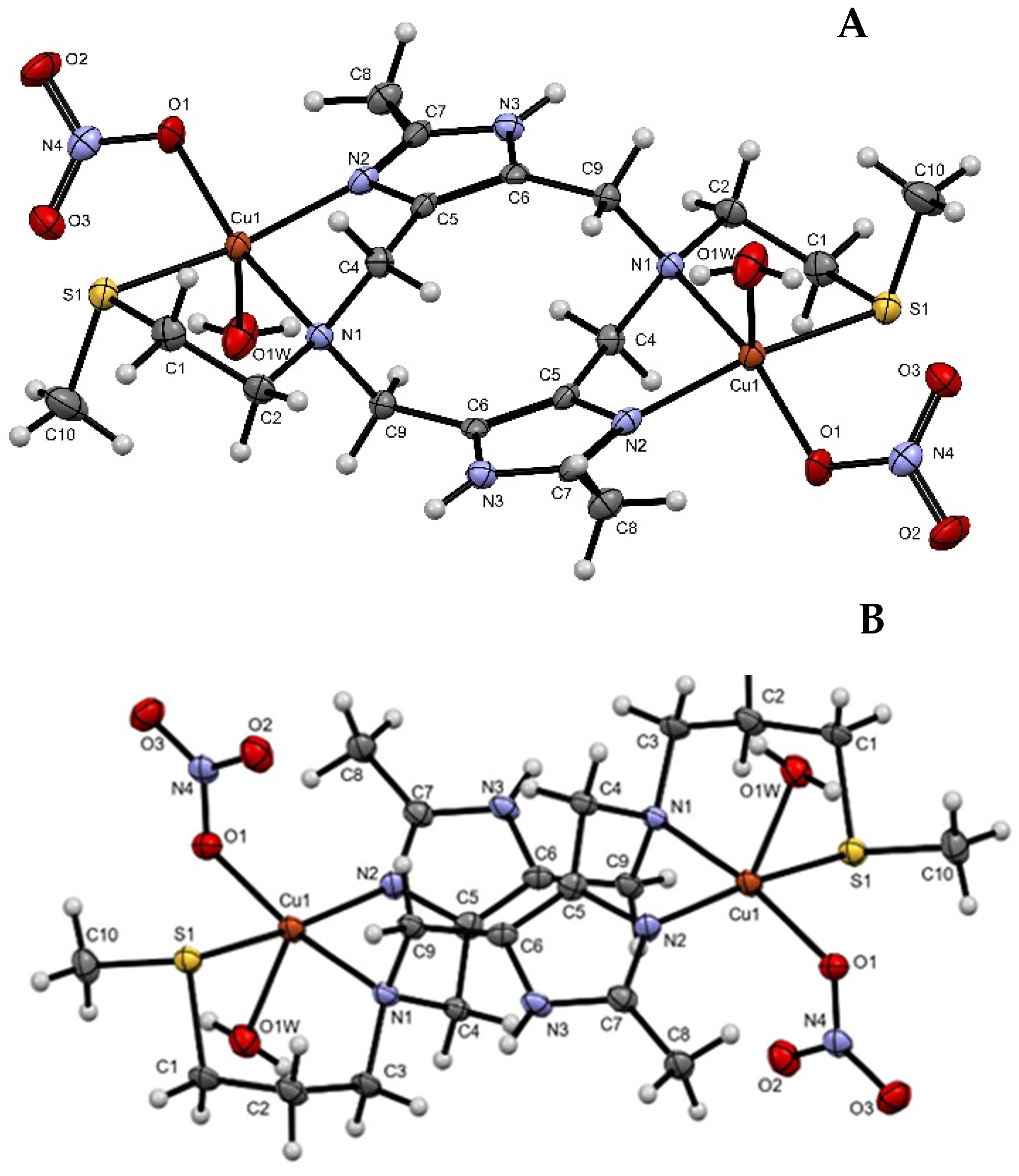
Figure 1. Perspective views for the structures of [Cu2thioenmeim(H2O)2(NO3)2](NO3)2 (A) and [Cu2thiopromeim(H2O)2(NO3)2](NO3)2 (B), with displacement ellipsoids at the 70% probability level for non-H atoms.
Molecules 31 01245 g001
Figure 2. Initial characterization of Cu2+-ligand complexes by UV-vis absorption and EPR spectroscopy. UV−Vis absorption spectra of methanolic solutions of Cu(II) complexes with promeim (A), thiopromeim (B), and thioenmeim (C), with 2.0 equiv. of Cu(NO3)2 salt. Corresponding UV-vis of solutions with 1.0 and 2.0 equivalents of Cu(ClO4)2 salt are shown in (DF). (GI) show the 150 K spectra of copper:ligand solutions in 1:1 and in 2:1 ratios for promeim, thiopromeim, and thioenmeim, respectively. Solutions in (DI) were prepared with Cu(ClO4)2. AII values in EPR spectra are given in 1 × 10⁻⁴ cm−1. Asterisks in (G,H) indicate signals associated with copper in solution.
Molecules 31 01245 g002
Figure 3. Transformation of the dinuclear complexes, monitored by UV-vis absorption and electron paramagnetic resonance (EPR). UV-vis absorption spectra showing the transformation of [Cu2promeim] (A), [Cu2thiopromeim] (B), and [Cu2thioenmeim] (C) complexes in MeCN/EtOH (80%/20%) over time. The EPR spectra at 150 K were recorded at the end of the reaction: (DF). AII values are given in 1 × 10⁻⁴ cm−1.
Molecules 31 01245 g003
Figure 4. Normalized copper K-edge XANES spectra of the copper complexes with promeim, thiopromeim, and thioenmeim in acetonitrile, at a 2:1 Cu:ligand stoichiometry and 5 mM concentration, using Cu(ClO4)2 as a metal source, frozen three days after preparation.
Molecules 31 01245 g004
Figure 5. ESI-MS spectra at the end of the transformation: (A) full spectra, (B) enlargement of the main peak, and (C) calculated isotopic distribution. Above, Cu2promeim; middle, Cu2thiopromeim and below, Cu2thioenmeim. Inset structures schematically describe the species with respective masses of 456, 548, and 520 mass units.
Molecules 31 01245 g005
Figure 6. Kinetics of the transformation of [Cu2thiopromeim](ClO4)4 in MeCN/EtOH as monitored by UV-vis absorption at room temperature and EPR at room temperature and 150 K. (A) Time-dependent UV-vis absorption spectra of the transformation of the [Cu2thiopromeim] complex in MeCN/EtOH; inset shows the decrease in the ligand field transition. Time-dependent EPR spectra of the transformation of the [Cu2thiopromeim] complex in MeCN/EtOH at room temperature (B) and 150 K (C).
Molecules 31 01245 g006
Figure 7. Comparison of cathodic CVs for (A) Cu(OTf)2; (B) [Zn2promeim](NO3)4 and [Zn2thioenmeim](NO3)4; (C) freshly prepared [Cu2thioenmeim](OTf)4 (prior to electron transfer); (D) aged [Cu2promeim](OTf)4 (after electron transfer). All measurements at 0.1 mM in acetonitrile.
Molecules 31 01245 g007
Figure 8. Spectro-electrochemistry of 0.1 mM in situ-generated [Zn2promeim](ClO4)4 with 0.1 M NBu4PF6 as the supporting electrolyte in acetonitrile. The working electrode was Pt mesh, the reference electrode was Ag/AgCl, and a Pt wire served as the counter electrode. Applied potential: 1.1–1.4 V.
Molecules 31 01245 g008
Figure 9. EPR spectrum of the transformation of [Cu2promeim] in the presence of excess DMPO, in MeCN:EtOH 4:1, 1 mM. The EPR spectrum shows a g = 2.0064 with AN = 1.31 mT, A = 0.830, A = was not defined.
Molecules 31 01245 g009
Figure 10. (A) Decrease in the d-d absorption for CuII2promeim. (B) Increase in the d-d absorption for CuI2promeim exposed to O2. (C) Decrease in the Imz ⟶ Cu LMCT band with simultaneous increase in the new absorption at 258 nm for CuII2promeim, in the absence of O2. (D) Increase in the Imz ⟶ Cu LMCT band when CuI2promeim is exposed to O2. Solutions in dry MeCN/EtOH (4:1) were handled under Ar; concentration of Cu2promeim was 0.2 mM and 1mM for UV and vis regions, respectively.
Molecules 31 01245 g010
Figure 11. (A): Spin density plots for the optimized structures of the initial (left) and final species (right). (B): HOMOs (first doubly occupied molecular orbital) for the complex in its initial (left) and final (right) state.
Molecules 31 01245 g011
Scheme 1. Dinucleating ligands used in this work.
Molecules 31 01245 sch001
Scheme 2. Proposed cycle involving electron transfer between copper, ligand, and dioxygen.
Molecules 31 01245 sch002
Disclaimer/Publisher’s Note: The statements, opinions and data contained in all publications are solely those of the individual author(s) and contributor(s) and not of MDPI and/or the editor(s). MDPI and/or the editor(s) disclaim responsibility for any injury to people or property resulting from any ideas, methods, instructions or products referred to in the content.