2.1. Polyphenolic Profile of Raspberry Leaves
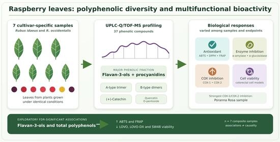
The detailed UPLC–PDA–Q/TOF-MS profile revealed pronounced qualitative and quantitative variation in the phenolic composition of the analyzed cultivar-specific composite raspberry leaf samples. Although all samples analyzed shared the same major classes of phenolic compounds, including phenolic acids, flavan-3-ols, flavonols, flavones, and ellagic acid derivatives, their relative abundances varied considerably among the samples, resulting in distinct phenolic profiles. In total, 37 phenolic compounds were identified or tentatively characterized (
Table 1), with flavan-3-ols, procyanidins, and flavonols representing the quantitatively predominant phenolic classes.
Total phenolic concentrations varied considerably among the analyzed cultivar-specific composite samples. The red-fruited raspberry cultivars exhibited the highest total phenolic concentrations (67.3–69.5 g/kg DW), followed by black raspberry cultivars (56.2–56.5 g/kg DW), while the yellow-fruited cultivars showed the lowest values (49.4–53.0 g/kg DW). These quantitative differences were accompanied by marked variation in the relative contribution of individual phenolic classes, indicating that the analyzed samples differed not only in total phenolic concentration, but also in the distribution of phenolic subclasses.
Although they shared the same main classes of phenolic metabolites, the analyzed cultivar-specific composite samples exhibited marked variation in the relative distribution of individual compounds. The higher total phenolic concentration observed in red fruit cultivars primarily reflected the high abundance of flavan-3-ols and procyanidins, which represented more than half of the quantified phenolic content. On the contrary, black raspberry leaves contained proportionally higher levels of flavonols together with selected derivatives of hydroxycinnamic acid, while yellow fruit cultivars displayed a more balanced phenolic profile, characterized by a lower proportion of flavan-3-ols and relatively greater contributions of low molecular weight phenolic acids and flavones. Therefore, the variation observed among the analyzed samples involved differences in the relative distribution of multiple phenolic subclasses rather than changes restricted to individual metabolites. Such compositional patterns may be related to differences in the regulation of phenylpropanoid metabolism; however, the present experimental design did not directly assess biosynthetic pathway activity or its genetic regulation. Further studies incorporating independent biological replication and targeted analysis of the regulation of the phenylpropanoid pathway would be required to determine the mechanisms underlying the observed differences in phenolic composition.
In addition to the variation in the abundance of the main phenolic classes, several metabolites showed distinct distribution patterns among the analyzed cultivar-specific composite samples. Within the present sample set, epicatechin was detected only in the three red-fruited cultivar samples, whereas quercetin O-rutinoside was detected only in the two black raspberry samples. Caffeic acid O-hexoside isomer 4 was particularly abundant in black raspberry samples, reaching concentrations markedly higher than those observed in the yellow-fruited samples, while it was not detected in the red-fruited samples. In contrast, quinic acid was detected in the yellow- and black-fruited samples but not in the red fruits, while apigenin O-pentoside occurred at comparatively high concentrations in the yellow-fruited samples. Collectively, these observations indicate that the composite samples differed not only in the abundance of the main phenolic classes but also in the distribution of individual metabolites.
Recent advances have demonstrated that proanthocyanidin accumulation is controlled by a complex regulatory network that involves the phenylpropanoid pathway, including chalcone synthase (CHS), dihydroflavonol reductase (DFR), anthocyanidin synthase (ANS), anthocyanidin reductase (ANR), and leucoanthocyanidin reductase (LAR). In addition to enzyme activities, metabolic channeling, intracellular transport, and transcriptional regulation contribute to genotype-dependent differences in flavan-3-ol accumulation [
18]. In raspberry leaves, Kobori et al. [
19] demonstrated that increased accumulation of catechin, epicatechin and procyanidins was accompanied by increased CHS expression, supporting the transcriptional regulation in flavan-3-ol accumulation. These findings provide a plausible biochemical context for the differences in the abundance of flavan-3-ol and procyanidin observed among the composite samples analyzed in the present study. However, because the expression or activity of enzymes involved in phenylpropanoid and flavonoid biosynthesis was not evaluated here, the metabolic mechanisms underlying these compositional differences cannot be established from the present data.
The phenolic composition observed in the present study is consistent with previous reports that identify raspberry leaves as rich sources of flavan-3-ols, flavonols, phenolic acids and ellagic acid derivatives [
20,
21,
22]. Similar LC–MS-based investigations have demonstrated that these compound classes constitute the dominant phenolics in raspberry leaves, although both the number of detected metabolites and their relative abundances vary considerably between studies. Such differences may result from multiple factors, including plant genotype, geographical origin, environmental conditions, development stage, and harvest time, as well as differences in extraction procedures and analytical platforms [
5,
12]. In particular, Pavlovi et al. [
23] reported substantial cultivar-dependent variation in the relative abundance of ellagitannins, flavonols and flavan-3-ols, emphasizing that genetic background is one of the principal determinants of the phenolic composition of raspberry leaves.
In the present study, the environmental and methodological sources of variation were reduced by cultivating all plants at the same experimental site under comparable agronomic conditions, collecting leaves at the same stage and on the same sampling date, and applying identical sample preparation and analytical procedures. This controlled sampling strategy strengthens the comparability of the phenolic profiles obtained for the analyzed cultivar-specific composite samples and reduces the variation attributable to differences in the cultivation site, harvest time, and analytical methodology. However, because only one pooled composite biological sample was analyzed per cultivar, the present study does not allow the estimation of biological variability to be estimated or the observed differences to be unequivocally attributed to cultivar or genotype.
An additional limitation arises from the taxonomic structure of the investigated material. The red- and yellow-fruited cultivars belong to Rubus idaeus L., while the two black raspberry cultivars belong to Rubus occidentalis L. Consequently, the identity of the species is confounded with the black-fruited group, and the effects of the species, cultivar/genotype, and fruit color group cannot be separated statistically within the present experimental design. Therefore, the observed phenolic patterns should be interpreted as compositional differences among the cultivar-specific composite samples analyzed rather than as independent effects of genotype or fruit color. Within R. idaeus, the red- and yellow-fruited samples also showed differences in both total phenolic concentration and the relative distribution of the main phenolic classes; however, the limited number of cultivars and the absence of independent biological replication preclude the generalization of these observations to fruit-color groups. More studies including a larger number of cultivars within each species and fruit color category, together with independent biological replication, will be required to unravel the relative contributions of genotype, species, and fruit color to the phenolic composition of the raspberry leaf.
The revised phytochemical profile also emphasizes the importance of considering identification confidence when interpreting the phenolic composition of raspberry leaves. Among the 37 compounds retained after re-evaluation, gallic acid, caffeic acid neochlorogenic acid, and (+)-catechin were confirmed by comparison with authentic standards (Level 1), while the remaining constituents were identified or tentatively characterized according to accurate mass, MS/MS fragmentation, and evidence from the literature (Levels 2–3). From a quality-control perspective, the consistent occurrence of selected constituents across all analyzed samples suggests their potential utility in a multimarker approach to standardizing raspberry leaf preparations. In particular, (+)-catechin, A-type procyanidin trimer, and quercetin O-pentoside isomer 2 may be considered candidate markers because they were detected in all seven samples and represent the main constituents of the quantitatively predominant flavan-3-ol/procyanidin and flavonol fractions. A multimarker strategy may provide a more representative assessment of phytochemical consistency than reliance on a single constituent, particularly given the differences in phenolic profiles observed among the raspberry types investigated. However, the present study was not designed to assess batch-to-batch reproducibility or the stability of the preparations during processing and storage. Therefore, the suitability of these compounds as quality control markers, including their concentration ranges and stability, should be validated using batches produced independently obtained from different harvests and subjected to defined processing and storage conditions before formal standardization criteria can be established.
2.2. Biological Activity
The antioxidant capacity of the raspberry leaf extracts varied among the cultivar-specific composite samples, as demonstrated by the ABTS, DPPH and FRAP assays (
Table 2).
Among the samples analyzed, the red-fruited cultivar samples generally showed higher ABTS, DPPH and FRAP values, whereas lower values were observed for the yellow-fruited samples and intermediate values for the black raspberry samples. This variation in antioxidant capacity was accompanied by differences in the phenolic composition between the samples analyzed, particularly in the abundance and relative distribution of the major phenolic classes. The potential relationships between phenolic composition and antioxidant capacity are further examined below using exploratory correlation analysis.
The high antioxidant potential of raspberry leaves is generally attributed to the presence of phenolic constituents, including hydrolyzable tannins, particularly ellagitannins, flavan-3-ols, procyanidins and flavonol glycosides, which are among the dominant classes of phenolic compounds in
Rubus leaves. Among the compounds identified in the present study, catechin derivatives, procyanidins, and quercetin glycosides are recognized as effective antioxidants due to their ability to donate hydrogen atoms or electrons, stabilize free radicals, and chelate transition metal ions. The relatively high abundance of these compounds in samples showing greater antioxidant capacity therefore provides a plausible biochemical basis for the observed pattern, although their individual contributions cannot be established from the present data. Previous studies have also reported associations between the phenolic composition of raspberry leaves and their antioxidant capacity. For example, Pavlović et al. [
23] reported variation in both phenolic profiles and antioxidant properties between leaf extracts from different raspberry and blackberries.
The relationship between phenolic composition and antioxidant capacity observed in the present study is consistent with previous studies on raspberry leaves. Ponder and Hallmann [
12] reported differences in both phenolic composition and antioxidant capacity between leaves of various cultivars of
R. idaeus and observed a significant relationship between total polyphenol content and in vitro antioxidant activity. Similarly, Varzaru et al. [
24] demonstrated a substantial antioxidant capacity of raspberry leaves using several complementary in vitro assays, including DPPH, ABTS, and hydroxyl radical scavenging assays, along with a high content of total polyphenols and flavonoids. Collectively, these studies support an association between the abundance of phenolic constituents and the antioxidant properties of raspberry leaves, while also indicating that the magnitude of antioxidant responses may vary depending on the phenolic composition and the assay employed.
Although all three antioxidant assays demonstrated substantial antioxidant capacity in raspberry leaf extracts, slight differences in cultivar classification were observed between ABTS, DPPH, and FRAP. Such variation is expected because these assays assess different aspects of antioxidant capacity and differ in their reaction mechanisms and sensitivity to individual antioxidant compounds. DPPH and ABTS assess primarily radical scavenging capacity through hydrogen atom or electron transfer reactions, while FRAP evaluates the ferric ion-reducing capacity of antioxidants. Therefore, antioxidant capacity depends not only on the total abundance of phenolic compounds but also on their molecular structure, redox properties, and relative proportions within the extract. Therefore, the structural diversity of the phenolic components detected in the raspberry leaf extracts, including flavan-3-ols, procyanidins, ellagic acid derivatives, and flavonol derivatives, may contribute to the differences in antioxidant responses observed between the analyzed samples and between the three assays.
Furthermore, the comprehensive characterization of the phenolic constituents presented by Alkhudaydi et al. [
22] confirmed that raspberry leaves are particularly rich in ellagitannins, flavan-3-ols, and flavonol derivatives, which collectively contribute to their high antioxidant potential and biological value.
The present results also support previous reports indicating that raspberry leaves are a rich source of antioxidant phytochemicals compared to several other aerial-plant materials. Zielonka-Brzezicka et al. [
25] demonstrated that raspberry leaf extracts exhibited consistently high antioxidant activity in the ABTS, DPPH and FRAP assays, confirming that leaves are a valuable source of antioxidant compounds within raspberry plants. Collectively, these findings reinforce the growing evidence that raspberry leaves, traditionally considered an agricultural by-product, constitute an attractive and sustainable source of natural antioxidants for the development of functional foods, nutraceuticals, and other value-added products.
Inflammation is a complex biological response that involves the activation of numerous signaling pathways and inflammatory mediators. Cyclooxygenases (COX-1 and COX-2) catalyze the conversion of arachidonic acid to prostaglandin precursors and represent important molecular targets in modulation of inflammatory processes. Therefore, inhibition of COX activity is commonly used as one of the in vitro indicators of the potential of bioactive compounds to modulate pathways associated with inflammation. In the present study, all raspberry leaf extracts inhibited both COX-1 and COX-2; however, considerable differences were observed among the cultivar-specific composite samples (
Table 2). The highest inhibitory activity against both cyclooxygenase isoforms was recorded for the yellow raspberry cultivar Poranna Rosa, while the lowest activity was observed for Glen Ample. These results demonstrate variation in COX-inhibitory activity among the analyzed raspberry leaf samples; however, this endpoint alone should not be interpreted as evidence of general anti-inflammatory activity.
Previous studies have identified raspberry leaves as a source of phenolic compounds with the potential to modulate inflammatory processes, including ellagitannins, flavan-3-ols, flavonols, and phenolic acids. These compounds have been reported to affect multiple inflammation-related targets and pathways, including cyclooxygenase and lipoxygenase activity, cellular antioxidant defense systems, and pro-inflammatory signaling pathways [
26]. Such effects are likely to reflect the combined contributions of multiple phenolic components rather than the activity of a single dominant compound, as also discussed in recent reviews of the biological properties of raspberry leaves and other plant materials. Interestingly, the pattern of COX inhibition did not parallel the total phenolic concentration or the abundance of the dominant flavan-3-ol and procyanidin fractions in the samples analyzed (
Table 1 and
Table 2). Although red-fruited samples generally contained higher concentrations of flavan-3-ols and procyanidins than yellow-fruited samples, the strongest inhibition of both COX-1 and COX-2 was observed for Poranna Rosa, which had a lower total phenolic concentration than the red-fruited samples. On the contrary, Glen Ample, despite its high total phenolic concentration and abundance of flavan-3-ols and procyanidins, showed the lowest inhibition of both cyclooxygenase isoforms. These observations indicate that the total phenolic concentration alone does not account for the pattern of COX inhibition observed in the analyzed raspberry leaf samples. Differences in the relative abundance of individual phenolic constituents, as well as possible additive or interactive effects within complex extracts, may contribute to the observed COX-inhibitory activity; however, the contributions of individual compounds and their interactions were not directly assessed in the present study.
Previous studies provide complementary evidence that raspberry leaf constituents can modulate inflammation-related processes. Maslov et al. [
27] demonstrated that raspberry leaf extracts significantly reduced carrageenan-induced inflammation in vivo and suggested that this activity was associated with the presence of catechins, ellagic acid derivatives, and other bioactive phenolics that act synergistically rather than independently. Luo et al. [
28] also reported variation in phenolic composition and inflammation-related activities in vitro among a diverse set of raspberry leaf samples, although their evaluation was based on hyaluronidase and inhibition of albumin denaturation rather than cyclooxygenase activity. Together, these studies indicate that the potential of raspberry leaf extracts to modulate inflammation-related processes may involve multiple phenolic constituents and may vary with the composition of the extract.
In the present study, the cultivation, sampling, and analytical conditions were standardized across the investigated plant material, thus improving the comparability of the analyzed samples. However, the observed differences in COX inhibition cannot be attributed specifically to genotype because the experimental design did not include independent biological replication at the cultivar level and the species identity was confounded with the black-fruited group. UPLC–PDA–Q/TOF-MS profiling nevertheless demonstrated marked variation in the relative abundance of phenolic acids, flavan-3-ols and procyanidins, ellagic acid derivatives, and flavonol glycosides among the analyzed composite samples. Together with the distinct pattern of COX inhibition, these compositional differences suggest that multiple constituents of extracts may contribute to the observed activity. However, the contribution of individual metabolites or interactions among them cannot be established from the present data.
Overall, the ability of all analyzed raspberry leaf extracts to inhibit both COX-1 and COX-2 provides evidence of their in vitro activity toward an inflammation-related molecular target. Together with their diverse phenolic composition, these findings support further investigation of raspberry leaves as a source of compounds capable of modulating inflammation-related pathways. Confirmation in complementary cellular and in vivo models, as well as identification of the constituents that contribute to COX inhibition, would be required before broader conclusions regarding anti-inflammatory efficacy or potential health applications can be drawn.
Natural polyphenols have attracted considerable attention as potential modulators of carbohydrate digestion due to their ability to inhibit digestive enzymes involved in the hydrolysis of dietary carbohydrates. Among these enzymes, α-amylase and α-glucosidase catalyze key steps in the breakdown of complex carbohydrates and oligosaccharides into absorbable monosaccharides. Their inhibition can delay carbohydrate hydrolysis and is therefore considered a relevant in vitro target in the search for food-derived compounds with the potential to modulate postprandial glucose availability.
The inhibitory activity against α-amylase and α-glucosidase varied markedly among the analyzed cultivar-specific composite samples (
Table 2). The Polesie sample showed the lowest IC
50 values for both α-amylase (0.11 mg/mL) and α-glucosidase (0.09 mg/mL), indicating the strongest inhibition of both enzymes among the analyzed samples. On the contrary, higher IC
50 values were generally observed for yellow-fruited samples. The highest overall values were recorded for Polana, with IC
50 values of 0.80 mg/mL for α-amylase and 0.76 mg/mL for α-glucosidase. Therefore, the samples analyzed showed substantial variation in their ability to inhibit carbohydrate-hydrolyzing enzymes. Raspberry leaves are rich in several phenolic classes with reported inhibitory effects on carbohydrate-hydrolyzing enzymes, including flavan-3-ols, procyanidins, ellagitannins, flavonols, and phenolic acids. Previous phytochemical studies have consistently identified these classes as important constituents of raspberry leaves [
23], providing a relevant compositional basis to investigate their potential contribution to α-amylase and α-glucosidase inhibition.
These results are consistent with those of Li et al. [
29], who demonstrated that raspberry leaf extracts possess strong inhibitory activity against both α-glucosidase and α-amylase and enhance glucose consumption in 3T3-L1 cells. Using affinity ultrafiltration combined with HPLC-MS/MS and molecular coupling, the authors identified epigallocatechin gallate, procyanidin C3, rutin, cyanidin-3-rutinoside, dihydromyricetin, and isovitexin as candidate contributors to enzyme-inhibiting activity of raspberry leaf extracts. Furthermore, these compounds interacted with key catalytic amino acid residues within the active sites of both enzymes, providing mechanistic evidence supporting their inhibitory potential.
The pattern of enzyme inhibition observed in the present study did not simply parallel the total phenolic concentration of the samples analyzed. Polesie, which showed the lowest IC50 values for both enzymes, was characterized by a high abundance of flavan-3-ols and procyanidins. Compounds belonging to these classes have previously been reported to inhibit α-glucosidase and α-amylase, and their inhibitory potency can be influenced by structural features such as the number and position of hydroxyl groups and the degree of polymerization. The relatively high abundance of flavan-3-ols and procyanidins in the Polesie sample therefore provides a plausible compositional context for its strong enzyme-inhibitory activity; however, the contribution of individual compounds cannot be established from the present data.
Wu et al. [
30] reported variation in α-glucosidase- and α-amylase-inhibitory activity among different raspberry tissues and related these differences to variation in phenolic composition, including procyanidins and ellagic acid derivatives. Together with the present results, these findings suggest that inhibition of carbohydrate-hydrolyzing enzymes by raspberry leaf extracts may be associated with the composition and relative abundance of multiple phenolic components rather than with total phenolic concentration alone.
For comparison, Bljajić et al. [
31] reported no detectable α-amylase inhibition by bilberry (
Vaccinium myrtillus L.) leaf extracts and α-glucosidase IC
50 values of 2.53 mg/mL and 0.29 mg/mL for aqueous and ethanolic extracts, respectively. Although direct quantitative comparisons between studies should be interpreted cautiously due to differences in extraction procedures and assay conditions, these data indicate that berry leaves can represent relevant sources of constituents capable of inhibiting carbohydrate-hydrolyzing enzymes.
Overall, the observed inhibition of α-amylase and α-glucosidase indicates that raspberry leaves contain constituents capable of interacting with carbohydrate-hydrolyzing enzymes. Together with their diverse phenolic composition, these findings support further investigation of raspberry leaves as a source of compounds potentially relevant to modulation of carbohydrate digestion. However, confirmation using complementary cellular, gastrointestinal, and in vivo models would be required before conclusions can be drawn regarding effects on postprandial glycemia or broader antidiabetic potential.
2.3. Effects of Raspberry Leaf Preparations on Colorectal Cancer Cell Viability
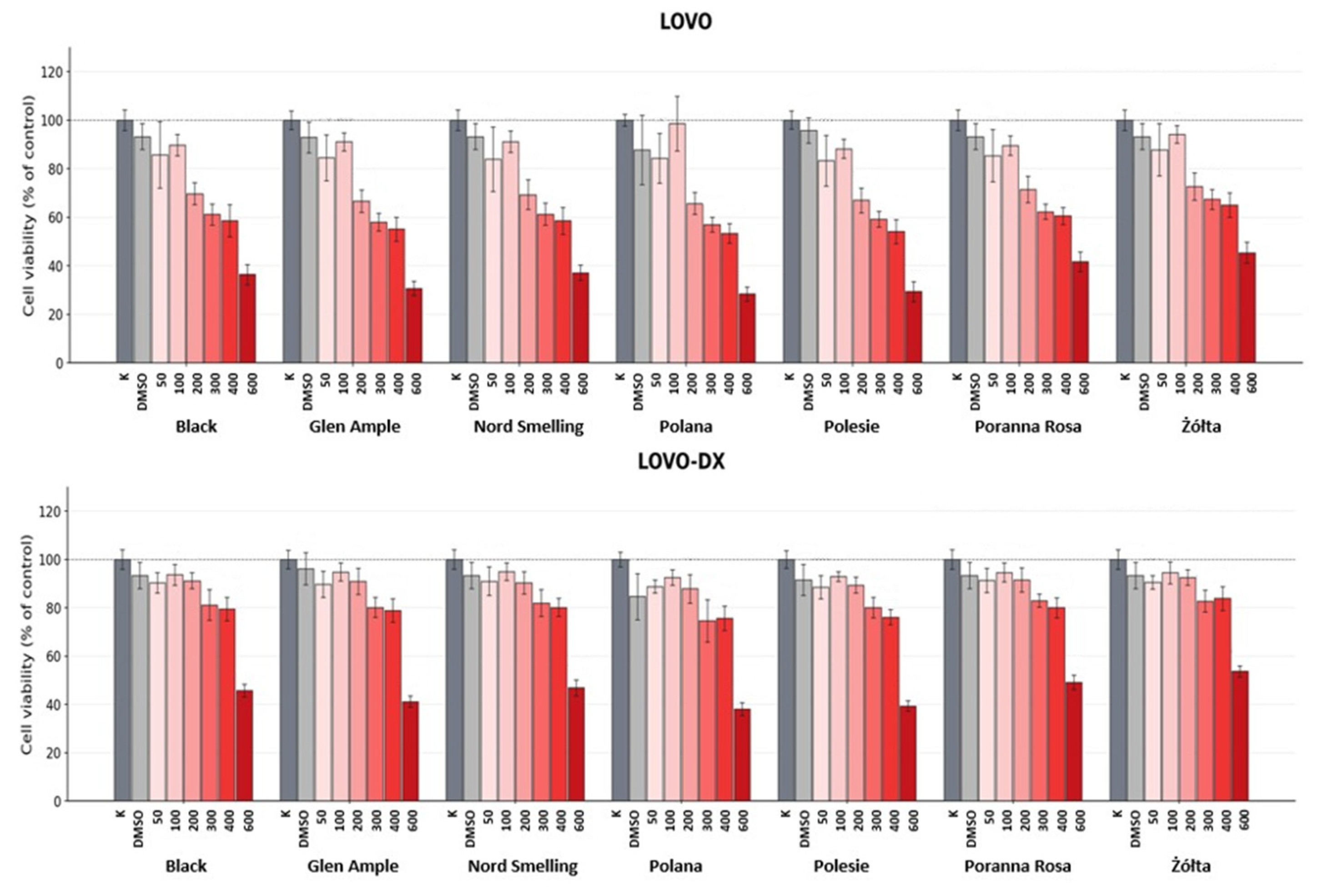
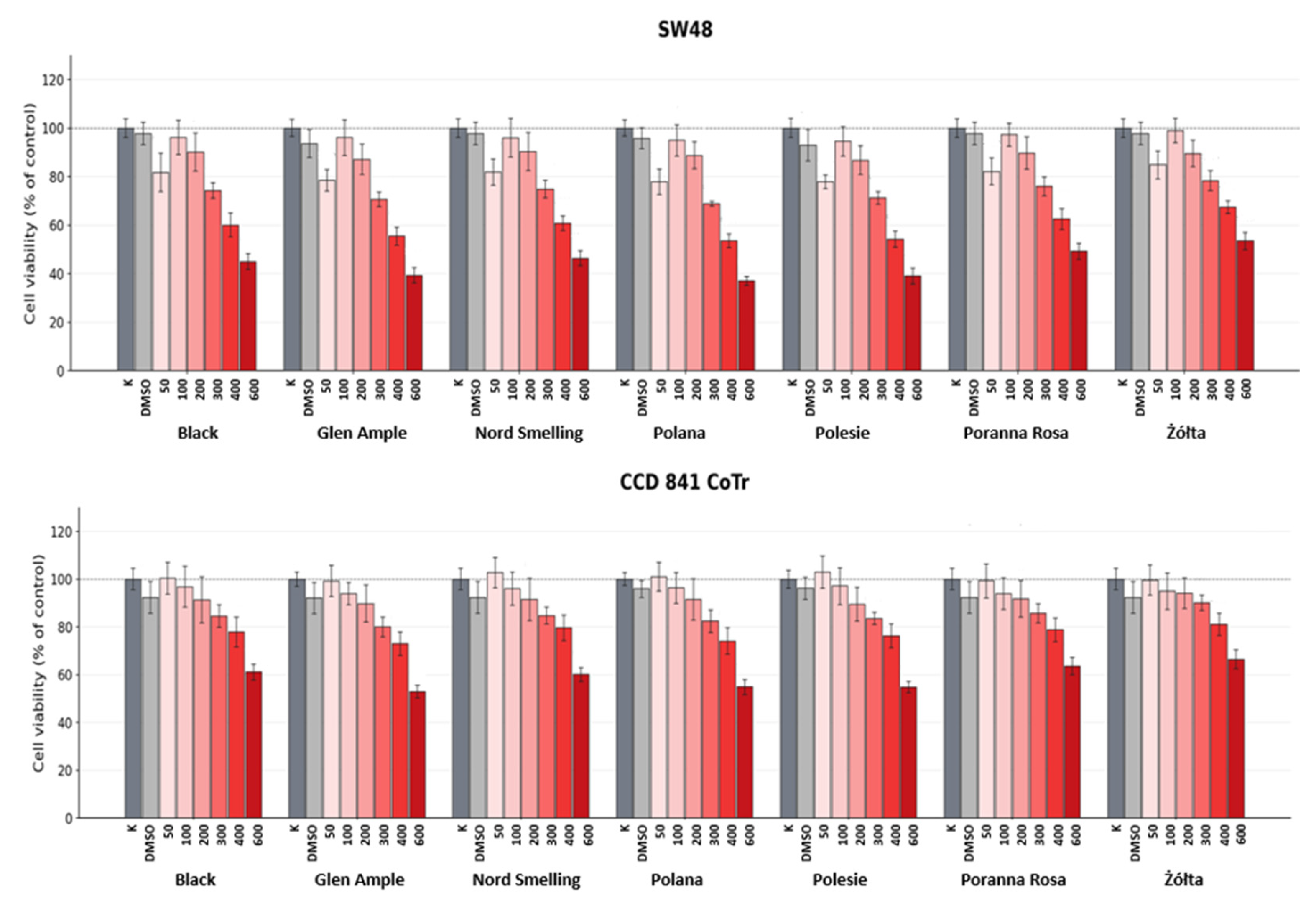
The effects of raspberry leaf preparations on cell viability were evaluated in four human colorectal cancer cell lines, including LOVO and its derivative LOVO-DX (although the resistant phenotype was not independently verified under the experimental conditions used in the present study) (
Figure 1), SW48 (
Figure 2), and HT29 (
Figure S1) and compared with their effects on the normal human colon epithelial cell line CCD 841 CoTr (
Figure 2).
Raspberry leaf preparations reduced the viability of LOVO, LOVO-DX and SW48 cells over the tested concentration range, although the magnitude of the response varied between the analyzed cultivar-specific composite samples. Overall, preparations from the red-fruited samples, particularly Polana, Polesie, and Glen Ample, tended to produce greater reductions in cancer cell viability, whereas the Żółta extract generally showed a weaker effect. In LOVO cells treated with 600 μg/mL, viability was reduced to 28.4%, 29.4%, and 31.7% following treatment with Polana, Polesie, and Glen Ample preparations, respectively, compared with 47.1% for the Żółta extract (
Figure 1).
The two black raspberry samples (R. occidentalis), Black and Nord Smelling, generally showed intermediate effects on cancer cell viability compared to the red- and yellow-fruited R. idaeus samples. Comparable patterns of viability reduction were observed in LOVO, LOVO-DX and SW48 cells, although the magnitude of the response varied between the analyzed samples and cell lines analyzed. LOVO-DX cells generally retained higher viability than parental LOVO cells following treatment with raspberry leaf preparations, suggesting lower sensitivity under the experimental conditions used. Nevertheless, all preparations reduced LOVO-DX cell viability over the tested concentration range. Compared to cancer cell lines, CCD 841 CoTr normal colon epithelial cells of CCD 841 CoTr generally retained higher viability, remaining above 50% throughout the tested concentration range. This pattern suggests a lower susceptibility of CCD 841 CoTr cells to the preparations under the conditions of the assay; quantitative evaluation of selectivity requires comparison of the corresponding IC50 values.
Unlike LOVO, LOVO-DX, and SW48 cells, HT29 cells showed a nonmonotonic and highly variable response to raspberry leaf preparations (
Figure S1), including an apparent increase in PrestoBlue signal at the highest concentration tested across all samples analyzed. Because the present data do not allow this response to be unequivocally distinguished from possible interference related to the assay, the HT29 results were not used for a comparative evaluation of the potency of the extract or IC
50 estimation and are presented separately in the
Supplementary Materials (Figure S1).
A methodological limitation of cell viability experiments is that concentration-matched cell-free controls containing raspberry leaf extract and PrestoBlue were not included. Consequently, direct chemical reduction of the resazurin reagent or interference with the fluorescence signal by extract constituents cannot be excluded. This limitation is particularly relevant to the nonmonotonic response but should also be considered when interpreting the results obtained for the other cell lines. Consequently, observed responses should be interpreted as changes in cell viability measured using the PrestoBlue assay, rather than as unequivocal evidence of cytotoxicity. Future studies should include extract-only background controls and confirm representative effects using an orthogonal viability or biomass-based assay.
The reduction in colorectal cancer cell viability observed in the present study is consistent with previous reports that describe growth-inhibiting effects of raspberry leaf extracts on colorectal cancer cells. Veljković et al. [
32] reported that methanolic extracts of wild
Rubus idaeus leaves inhibited the growth of HCT-116 colorectal cancer cells, with the most active population exhibiting IC
50 values of 162.38 μg/mL after 24 h and 95.69 μg/mL after 48 h, although considerable variability was observed among the analyzed populations. The authors also reported variations in phytochemical composition between the investigated raspberry populations, supporting a potential relationship between extract composition and effects on cancer cell growth. These findings are broadly consistent with the variation in cell viability responses observed among the raspberry leaf samples analyzed in the present study. However, a direct quantitative comparison of IC
50 values between studies should be interpreted with caution due to differences in plant material, extraction procedures, exposure time, viability assay (MTT vs. PrestoBlue
®), and colorectal cancer cell models.
The variation in cancer cell viability observed between the raspberry leaf samples was accompanied by differences in their phenolic composition. Samples that produced greater reductions in cell viability generally contained higher concentrations of total polyphenols and flavan-3-ols, while variation in several other phenolic groups, including phenolic acids and procyanidins, also broadly paralleled the observed differences in cancer cell viability. This pattern suggests a potential association between phenolic composition and the effects of raspberry leaf preparations on the viability of colorectal cancer cells, although the contributions of individual compounds or phenolic classes cannot be established from the present data. Raspberry leaves contain a structurally diverse mixture of catechins, procyanidins, derivatives of ellagic acids and flavonol glycosides, several of which have been associated with antiproliferative or cytotoxic effects in previous studies [
33]. Therefore, the observed responses may reflect the combined effects of multiple constituents of the extracts rather than the activity of a single dominant compound.
The reduction in cancer cell viability observed for polyphenol-rich plant preparations may involve several cellular mechanisms rather than a single pathway. Catechin and epicatechin have been reported to promote ROS-associated cell death signaling in colon cancer cells, and catechin has also been reported to induce G2/M cell-cycle arrest [
34]. Procyanidins, in turn, have been shown to inhibit proliferation and induce apoptosis associated with loss of mitochondrial membrane potential and caspase-3 activation in colorectal carcinoma cells, including HT-29 and LOVO cells [
35]. Ellagic acid has likewise been reported to increase ROS generation, induce G2/M cell-cycle arrest, and activate apoptosis-related pathways in colon adenocarcinoma cells [
36]. These findings provide a plausible mechanistic context for structurally related to those detected in raspberry leaves; however, they do not establish the mechanism responsible for the effects observed in the present study. Here, only cell viability was assessed, without measurements of intracellular ROS, mitochondrial function, apoptosis-related markers, or cell cycle distribution. Consequently, the observed reduction in colorectal cancer cell viability cannot be attributed to any specific molecular mechanism. More studies incorporating these endpoints are required to determine the pathways underlying responses to raspberry leaf preparations.
The raspberry leaf preparations also reduced the viability of LOVO-DX cells, although this cell line generally showed lower sensitivity to the preparations than the parental LOVO cells. Thus, the viability-reducing effects of the preparations were not restricted to the parental cell line. However, because resistance to doxorubicin was not experimentally verified under the conditions of the present study and the molecular mechanisms underlying the differential responses of LOVO and LOVO-DX cells were not investigated, these findings should not be interpreted as evidence that the components of raspberry leaf overcome multidrug resistance or chemoresistance. More studies incorporating appropriate chemotherapeutic controls and analysis of resistance-related mechanisms are required to address this question.
CCD 841 CoTr cells generally retained higher viability than colorectal cancer cell lines in the tested concentration range tested. The IC
50 for CCD 841 CoTr cells was not reached for any of the seven preparations and was therefore reported as >600 μg/mL (
Table S4). On the contrary, the IC
50 values for LOVO cells ranged from 426.7 to 552.9 μg/mL, while higher values were generally observed for LOVO-DX and SW48 cells. Consequently, the calculated selectivity indices could only be expressed as lower-bound estimates, ranging from >1.09 to >1.41 for LOVO, >1.01 to >1.12 for LOVO-DX, and >1.02 to >1.35 for SW48; for the Żółta preparation, SI could not be determined for LOVO-DX or SW48 because 50% viability was not reached in either the cancer or non-malignant cell line. These results indicate a lower apparent susceptibility of CCD 841 CoTr cells, particularly relative to LOVO cells, but do not support a strong claim of selective cytotoxicity [
33].
Finally, the present results further indicate that antioxidant capacity and effects on cancer cell viability should not be regarded as directly interchangeable biological properties. Although phenolic compounds can contribute to both endpoints, extracts exhibiting high antioxidant capacity do not necessarily produce the greatest reductions in cancer cell viability. Veljković et al. [
32] similarly reported no positive relationship between antioxidant capacity and anticancer activity among different wild raspberry populations, despite considerable variation in their phenolic composition. These observations suggest that the effects of raspberry leaf extracts on cancer cell viability may depend on the composition and relative abundance of individual phenolic components rather than on overall antioxidant capacity alone. In the present study, potential associations between specific phenolic classes and cell viability-related parameters were further examined using exploratory correlation analysis.
2.4. Integrated Correlation Analysis
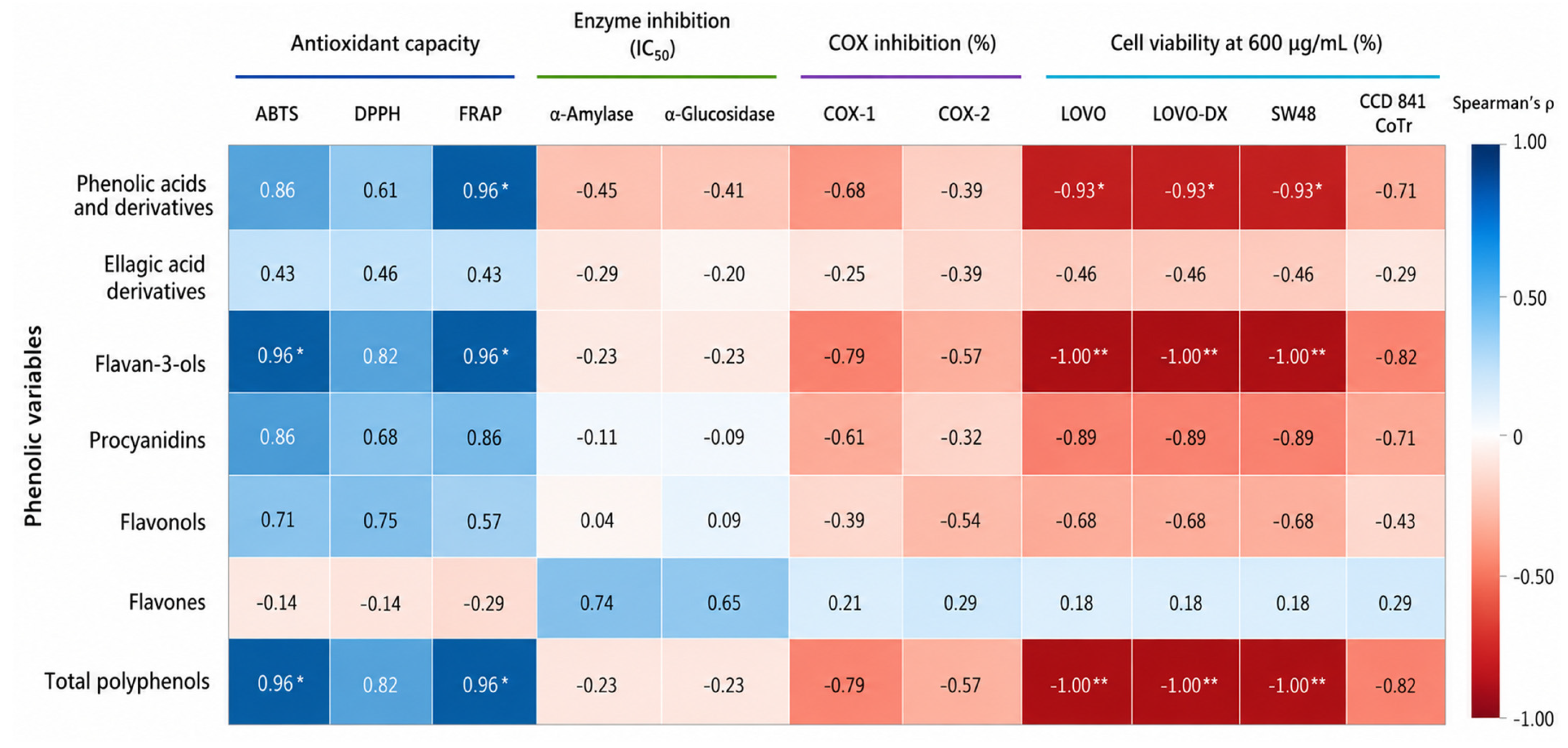
To explore the relationships between phenolic composition and biological responses of the analyzed raspberry leaf composite samples, Spearman’s rank correlation coefficients were calculated between the major phenolic groups and the corresponding biological activity endpoints (
Figure 3). Given the small number of analyzed samples (
n = 7), statistical significance was evaluated using exact two-sided permutation-based
p values, followed by Benjamini–Hochberg false discovery rate (FDR) correction in all tested correlations. The analysis was considered exploratory.
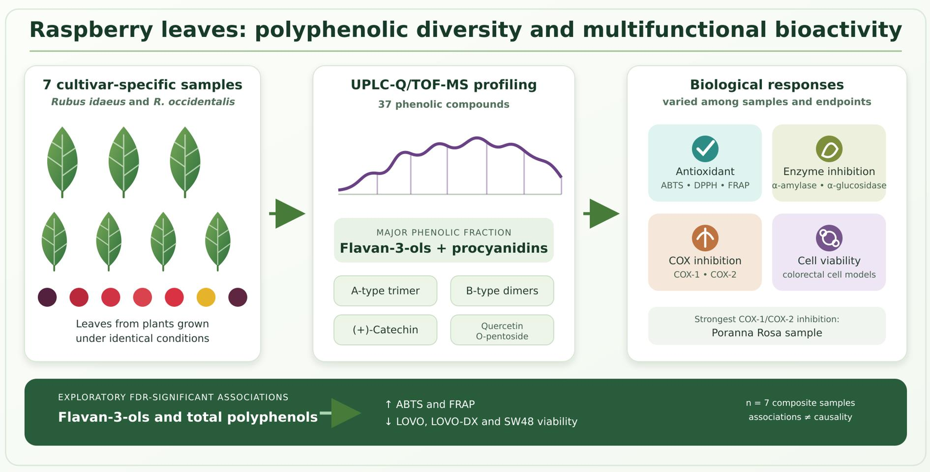
The strongest FDR-significant associations were observed for flavan-3-ols, total polyphenols, and phenolic acids and derivatives. Flavan-3-ol concentration was strongly positively correlated with the antioxidant capacity measured by ABTS (ρ = 0.96, exact p = 0.0028, q = 0.019) and FRAP (ρ = 0.96, exact p = 0.0028, q = 0.019). On the contrary, the concentration of flavan-3-ol showed a perfect inverse rank correlation with cell viability at 600 μg/mL in LOVO, LOVO-DX, and SW48 cells (ρ = 1.00, exact p = 0.00040, q = 0.0051 for each cell line).
The total polyphenol concentration showed the same correlation pattern, including positive associations with ABTS and FRAP (ρ = 0.96, q = 0.019 for both) and inverse associations with cell viability of LOVO, LOVO-DX, and SW48 cell viability (ρ = 1.00, q = 0.0051 for each). Because the total concentration of polyphenol incorporates the major phenolic subclasses and its ranking closely paralleled that of flavan-3-ols in the present dataset, these associations should not be interpreted as independent evidence supporting the contribution of flavan-3-ols.
Phenolic acids and their derivatives also showed significant FDR associations. Their concentration was positively correlated with FRAP antioxidant capacity (ρ = 0.96, exact p = 0.0028, q = 0.019) and inversely correlated with LOVO, LOVO-DX, and SW48 cell viability at 600 μg/mL (ρ = −0.93, exact p = 0.0067, q = 0.037 for each cell line). No FDR-significant associations were observed for α-amylase or α-glucosidase IC50, COX-1 or COX-2, or CCD 841 CoTr viability.
In general, the exploratory correlation analysis identified flavan-3-ols, total polyphenols, and phenolic acids and derivatives as the compositional variables most consistently associated with FRAP antioxidant capacity and colorectal cancer cell viability at 600 μg/mL in the present dataset, while significant associations with ABTS were observed for flavan-3-ols and total polyphenols. No phenolic group showed an FDR-significant association with DPPH, α-amylase or α-glucosidase IC50, COX-1 or COX-2 inhibition, or CCD 841 CoTr viability after exact permutation-based testing. These findings should be interpreted cautiously because the analysis was based on only seven cultivar-specific composite samples, several compositional variables were mathematically or biologically interrelated, and correlation does not establish causality. Therefore, the identified associations generate hypotheses for further investigation rather than demonstrating that a particular phenolic class determines the biological activity of raspberry leaves.