Article

Characterization of Forestiera tomentosa Fruit: Proximate Composition, Physicochemical Parameters, Phenolic Content, Antioxidant Capacity, and Toxicological Assessment


by
Salvador Hernández-Estrada
1,
Luis Antonio Ramirez-Contreras
1,
Luis Alfonso Hernández-Villaseñor
1,
Jorge Manuel Silva-Jara
2,
Efigenia Montalvo-González
3,
Zuamí Villagrán
1,
Noé Rodríguez-Barajas
1,
Jorge L. Mejía-Méndez
4,
Carlos Arnulfo Velázquez-Carriles
5,
Martin Zermeño-Ruiz
2 and
Luis Miguel Anaya-Esparza
6,*
1
Centro Universitario de Los Altos, Universidad de Guadalajara, Tepatitlan de Morelos 47620, Mexico
2
Centro Universitario de Ciencias Exactas e Ingenierias, Universidad de Guadalajara, Guadalajara 44430, Mexico
3
Laboratorio Integral de Investigación de Alimentos, Instituto Tecnológico de Tepic, Tecnológico Nacional de Mexico, Tepic 63175, Mexico
4
Escuela de Ingeniería y Ciencias, Tecnológico de Monterrey, Epigmenio González 500, San Pablo, Santiago de Querétaro 76130, Mexico
5
Centro Universitario de Tlajomulco, Universidad de Guadalajara, Tlajomulco de Zuñiga 45641, Mexico
6
Centro de Estudios para la Agricultura, la Alimentación y Crisis Climática, Centro Universitario de Los Altos, Universidad de Guadalajara, Tepatitlan de Morelos 47620, Mexico
*
Author to whom correspondence should be addressed.
Molecules 2026, 31(14), 2542; https://doi.org/10.3390/molecules31142542
Submission received: 17 June 2026 / Revised: 17 July 2026 / Accepted: 20 July 2026 / Published: 22 July 2026

Abstract

The demand for sustainable nutrients and bioactive compounds has increased interest in underutilized wild plants. Forestiera tomentosa, a Mexican drupe-bearing species, is largely unexplored. This study evaluated the proximate composition, physicochemical and functional properties, phenolic profile, antioxidant capacity, and toxicological safety of F. tomentosa fruit. The fruit showed high carbohydrate content [71.94% dry weight (DW)], with notable crude fiber (7.90% DW), protein (7.39% DW), and lipid (4.30% DW) contents. Analysis revealed a mildly acidic pH (5.66), titratable acidity of 0.32%, and total soluble solids of 2.33 °Brix. The fruit powder had a low water activity (0.42) and a favorable water solubility index (56.10%), oil absorption (4.54%), and foaming capacity (19.71%). The fruit contained high levels of soluble phenols (280.42 mg GAE/g DW), flavonoids (98.89 mg CE/g DW), anthocyanins (54.53 mg C3G/g DW), and condensed tannins (94.05 mg CE/g DW). High-performance liquid chromatography identified 20 phenolic compounds, with 3-(4-hydroxyphenyl) propionic acid, syringic acid, catechin, epicatechin, and gallocatechin being predominant. The fruit showed significant radical scavenging and reducing potential (DPPH, ABTS, and FRAP). Toxicological evaluation using the Artemia salina bioassay showed a 100% survival rate across all concentrations, indicating no acute toxicity. In silico ADMET predictions revealed favorable pharmacokinetic properties, including high intestinal absorption and compliance with Lipinski’s rule of five. These findings position F. tomentosa as a promising, non-toxic source of functional ingredients for the food, nutraceutical, and pharmaceutical industries, supporting biodiversity conservation and sustainable resource utilization. Further studies are needed to evaluate the potential health benefits of this fruit in vitro and in vivo.

1. Introduction

Native and edible fruits have attracted increasing scientific interest because they represent valuable sources of nutrients and bioactive phytochemicals with potential applications in the food, nutraceutical, cosmetic, and pharmaceutical industries. Despite this potential, many of these species remain undervalued due to limited scientific information on their chemical composition, phytochemical profiles, and biological properties, which restricts their integration into value chains and conservation strategies [1]. Therefore, the comprehensive characterization of underutilized wild fruits is essential to support their sustainable use, promote biodiversity conservation, and identify new plant resources with functional potential [2].
The family Olaceae comprises a moderately sized assemblage of woody plants, encompassing 28 genera, several of which are notably used to produce oils and fragrances. Nonetheless, this family also includes a lesser-known genus, Forestiera [3]. The genus Forestiera comprises small deciduous and semi-deciduous trees and shrubs distributed across the United States, Mexico, Central America, and South America, thriving in vegetation types characterized by distinct seasonal patterns. Furthermore, 14 species of this genus have been identified, with the majority (51%) located in Mexico [4,5,6]. The most frequently reported Forestiera species include F. neomexicana [7], F. isabelae [8], F. acuminata [9], F. veracruzana [6], F. angustifolia, F. pubescens [3], and F. tomentosa [10]. It is important to highlight that those studies on Forestiera species have focused on its morphology, palynology, and wood anatomy [5], whereas information regarding the chemical composition of its fruits remains scarce.
Forestiera tomentosa is a species native to and currently known only from Mexico, where it is distributed across several central and western states, including Aguascalientes, Colima, Guanajuato, Guerrero, Hidalgo, Michoacan, Oaxaca, Puebla, Queretaro, San Luis Potosi, Zacatecas, and Jalisco [11]. It is known as acebuche, arcibuche, aceitunilla, or granjeno and is a shrub or small tree (up to 6 m tall) with smooth, gray-to-blackish bark with leaves that are narrow to broadly elliptic, sometimes varying to oblong, lanceolate, or oblanceolate; moreover, it produces fruit of an ellipsoid shape of 7 to 11 mm long and 4 to 8 mm wide, and epicarp of a purple-blackish or deep-blue color when ripe, containing one seed of 4 mm, while its endocarp becomes rigid with maturation [12]. Additionally, the small drupes serve as a food source for local fauna, and these wild fruits are edible [13] and consumed by local people fresh and in salads, with an astringent flavor like that of red wine made from grapes (personal communication). To the best of our knowledge, no studies have comprehensively characterized the nutritional composition, physicochemical properties, phenolic profile, antioxidant capacity, or toxicological characteristics of F. tomentosa fruit. This knowledge gap highlights the need for future studies to develop sustainable processing strategies that maximize the utilization of this unexplored native fruit and support its potential applications in the food, nutraceutical, cosmetic, and pharmaceutical industries.
In this context, aligning with the shift from traditional medicine–food homology to a modern evidence-based paradigm [14], the aim of this study was to provide the first scientific characterization of F. tomentosa fruit by evaluating its proximate composition, physicochemical properties, phenolic profile, antioxidant capacity, and toxicological profile.

2. Results

Figure 1 illustrates the visual appearance of the Forestiera tomentosa tree, including bark, leaves, and fresh fruits (Figure 1a–d). Moreover, dried fruits (Figure 1e) and fruit aqueous extract (Figure 1f) are also shown.

2.1. Proximate Composition

The proximate composition of F. tomentosa fruit is shown in Table 1. The fresh fruit contains 67.85% moisture, whereas the dried fruit contains only 12.04%. The dried fruits are characterized by their concentration of available carbohydrates (71.94%, calculated by difference), including 21.40% reducing sugars and 7.90% crude fiber, followed by protein (7.39%), lipids (4.30%), and ash (4.33%).

2.2. Physicochemical Parameters and Functional Properties

Table 2 presents the physicochemical and functional properties of F. tomentosa fruits. The physicochemical parameters of the fresh fruits included a TSS content of 2.33 °Brix, a pH of 5.66, and a titratable acidity (TA) of 0.32%, yielding a TSS/TA ratio of 7.28. The color parameters of fresh fruits of F. tomentosa indicated a luminosity of 26.26, with positive a (1.01) and negative b (−15.30) coordinates. Additionally, the dried fruit powder exhibited a water activity of 0.40. The functional properties of the dried fruit included a water solubility index of 56%, a water absorption index of 1.80 g g−1, and a swelling power of 5.53%. Additionally, the fruit powder exhibited an oil absorption index of 2.48 g g−1, a foaming capacity of 19.71%, and an emulsifying capacity of 2.61%.

2.3. Phenolic Compounds and Antioxidant Capacity

The fruits of F. tomentosa contained 280 mg GAE/g of soluble phenols, 98.89 mg CE/g of flavonoids, 54.53 mg C3G/g of anthocyanins, and 94.95 mg CE/g of condensed tannins. Furthermore, F. tomentosa fruits exhibited antioxidant activity by DPPH (14.59 mmol TE/g), ABTS (12.04 mmol TE/g), and FRAP (1.92 mmol TE/g) assays (Table 3).
Additionally, twenty phenolic compounds were identified from F. tomentosa fruit by HPLC-DAD, including phenolic acids and flavonoids such as gallic, protocatechuic, neochlorogenic, 3,4-Dihydroxyphenylacetic, 4-Hydroxybenzoic, chlorogenic, 4-Hydroxyphenylacetic, vanillic, syringic, 3-(4-hydroxyphenyl) propionic, p-coumaric, trans-ferulic, ellagic, and salicylic acids, gallocatechin, catechin, epicatechin, rutin, and naringenin (Table 4). The most abundant compounds were 3-(4-hydroxyphenyl) propionic acid (172,711 µg/100 g), syringic acid (55,791 µg/100 g), catechin (29,630 µg/100 g), epicatechin (25,387 µg/100 g), gallocatechin (23,693 µg/100 g), and 4-Hydroxyphenylacetic acid (9229 µg/100 g).

2.4. Toxicity Bioassay in Artemia salina

Table 5 shows the results of the Artemia salina toxicity bioassay, in which the survival (%) of larvae was measured after 24 and 48 h of exposure to F. tomentosa extract at different concentrations. In all tested concentrations (1000–62.5 µg/mL), the survival rate of A. salina was 100%, like the synthetic seawater (negative control). On the other hand, the A. salina larvae exposed to sodium hypochlorite (positive control) died during the first 24 h of exposure.

2.5. In Silico Evaluation of Drug-likeness and Toxicity of Main Phenolic Compounds Identified from Forestiera tomentosa Fruit

Table 6 lists the physicochemical parameters (molecular weight, the number of hydrogen-bond acceptors and hydrogen-bond donors, lipophilicity, and the number of violations of Lipinski’s rule of five) of the main phenolics (syringic acid, catechin, epicatechin, gallocatechin, 3-(4-Hydroxyphenyl) propionic acid, and hydroxyphenylacetic acid) found in F. tomentosa fruit by HPLC-DAD.
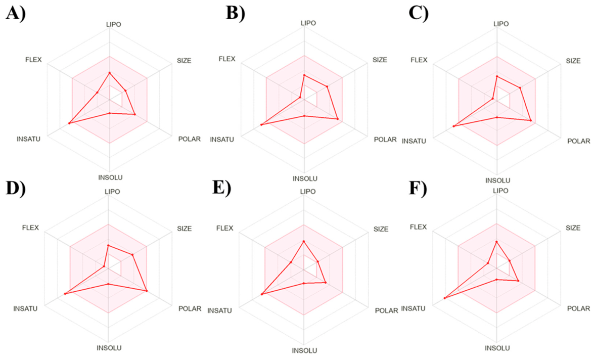
These compounds exhibited lipophilicity values ranging from 0.88 to 1.54 Log Po/w, and none violated any of Lipinski’s rules, except for gallocatechin, which violated one (NH or OH > 5). Furthermore, these compounds exhibited good physicochemical properties, including lipophilicity, size, polarity, insolubility, unsaturation, and flexibility (Figure 2). Lipinski’s rules are based on physicochemical parameters designed to assess chemical compounds and determine their pharmacokinetic properties. Smaller molecules, ideally with a molecular weight of 500 g/mol or less, and those with no more than 10 hydrogen-bond acceptors, up to 5 hydrogen-bond donors, and a lipophilicity of 5 or less generally cross cell membranes more efficiently. Consequently, they are considered superior drug candidates compared with larger molecules that exhibit excessive hydrogen bonding. In this evaluation, most of the compounds studied exhibited optimal physicochemical and pharmacokinetic properties, except for gallocatechin, which had more than five hydrogen-bond donors.
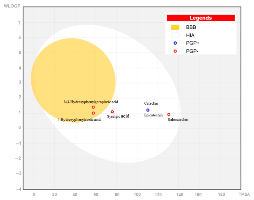
On the other hand, ADME characteristics are associated with the potential to achieve favorable oral bioavailability (OB), human intestinal absorption (HIA), and effective traversal of the blood–brain barrier (BBB) (Table 6). Notably, 3-(4-hydroxyphenyl)propionic acid (0.79), syringic acid (0.73), and 4-hydroxyphenylacetic acid (0.79) were distinguished by their OB values, whereas gallocatechin (0.35), epicatechin (0.39), and catechin (0.40) exhibited low OB values. For their part, syringic acid (0.99), catechin (0.99), and epicatechin (0.99) demonstrated the highest HIA values, compared with gallocatechin (0.98), 3-(4-hydroxyphenyl)propionic acid (0.98), and 4-hydroxyphenylacetic acid (0.95). Furthermore, 3-(4-hydroxyphenyl)propionic acid (0.45), 4-hydroxyphenylacetic acid (0.45), and syringic acid (0.43) are more likely to cross the BBB than gallocatechin (0.09), epicatechin (0.13), and catechin (0.14) (Figure 3).
Additionally, we assessed the toxicological properties of the six most prevalent compounds identified in F. tomentosa (Table 7). The clinical toxicity values ranged from 0.01 to 0.08, whereas the potential to inhibit the human ether-à-go-go-related gene (hERG) ranged from 3.71 × 10⁻³ to 0.30. Notably, syringic acid (0.69) exhibited a higher propensity to induce hepatotoxicity. All compounds showed a low probability of causing mutagenicity (0.05 to 0.34); however, catechin (0.34), epicatechin (0.33), and gallocatechin (0.33) exhibited the highest values. Similar trends were observed for carcinogenicity, with all compounds exhibiting a low probability of inducing carcinogenicity, ranging from 0.05 to 0.22; 4-hydroxy-phenylacetic acid (0.19) and 3-(4-hydroxy-phenyl) propionic acid (0.22) had higher values. Regarding the prediction of LD50 values (in humans), 4-hydroxyphenylacetic acid (1550 mg/kg), syringic acid (1700 mg/kg), and 3-(4-hydroxy-phenyl) propionic acid (2000 mg/kg) showed the lowest values, whereas catechin, epicatechin, and gallocatechin showed the highest values (10,000 mg/kg); moreover, these compounds showed a toxicity class ranging from four to six.
Finally, we performed molecular docking simulations of the six compounds with the three isoforms of the cytochrome P450 enzyme (CYP3A4, CYP2D6, and CYP2E1). Table 8 shows the binding scores (ΔGs) for the three target proteins. A ΔG (kcal/mol) < 0 suggests the possibility of spontaneous binding of the six compounds to the proteins. The results showed that the ΔG values for the three proteins were negative, indicating that all compounds spontaneously bound to the proteins.
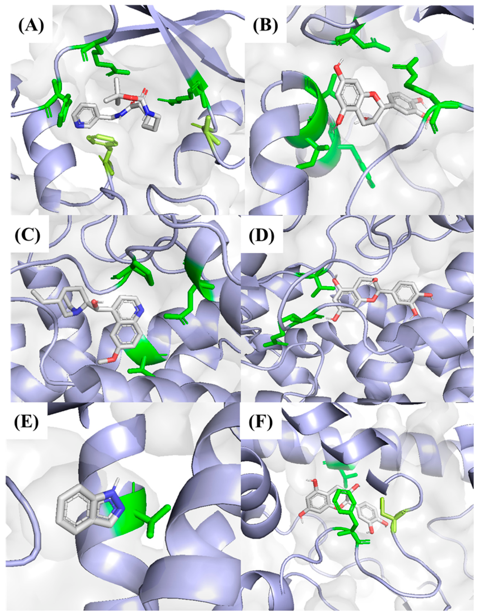
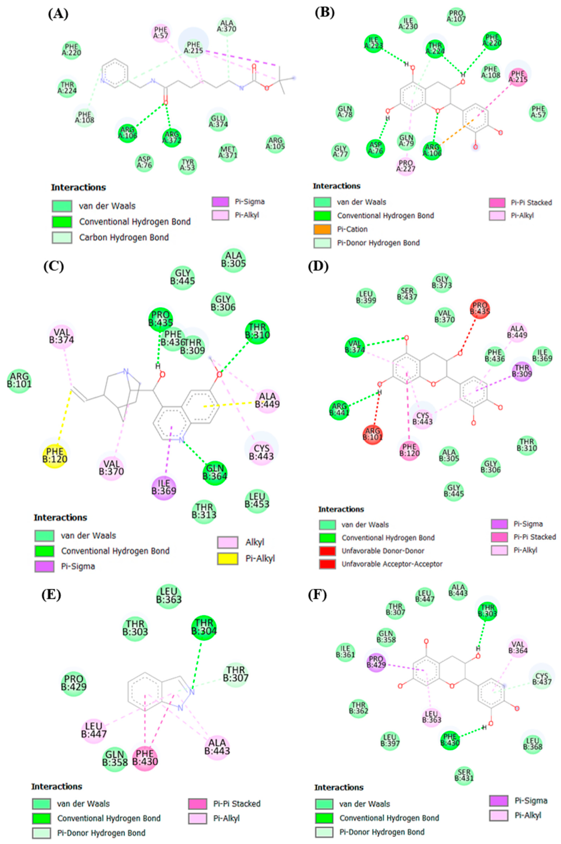
Figure 4 illustrates the 3D molecular docking between the target proteins and the compounds with the most favorable values, including controls for each enzyme. Furthermore, compared to the CYP3A4 inhibitor, tert-butyl {6-Oxo-6-[(Pyridin-3-ylmethyl)amino]hexyl}carbamate (−7.5 kcal/mol), epicatechin (−8.8 kcal/mol), gallocatechin (−8.4 kcal/mol), and catechin (−8.3 kcal/mol) showed better affinity. However, no compound showed greater affinity than the CYP2D6 control, quinine (−9.0 kcal/mol). All compounds showed better affinity than the CYP2E1 inhibitor indazole (−6.0 kcal/mol). In the CYP3A4–epicatechin complex, four hydrogen bonds (ASP76, ARG106, PHE220, ILE223, and THR224) were found, while in the control (CYP3A4-Tert-Butyl {6-Oxo-6-[(Pyridin-3-Ylmethyl) amino] hexyl} carbamate)) five hydrogen bonds were detected (ARG106, PHE108, PHE215, ALA370, ARG372). In the case of the CYP2D6–epicatechin complex, two hydrogen bonds were detected (VAL374 and ARG441), whereas in the control (CYP2D6–quinine), three hydrogen bonds were detected (THR310, GLN374, and PRO415). On the other hand, in the CYP2E1–catechin complex, there are two hydrogen bonds (THR303 and PHE430), while in its respective control (CYP2E1–indazole), only one hydrogen bond was detected (THR304) (Figure 5).

3. Discussion

The valorization of wild fruits has emerged as a critical strategy for identifying novel sources of nutrients and bioactive compounds while promoting the sustainable utilization of underexplored natural resources [2]. Forestiera tomentosa, an endemic Mexican species of the Oleaceae family, has remained scientifically overlooked, with its utilization traditionally limited to wood [5]. The fruits are small, fleshy drupes with an ellipsoid–oblong morphology, measuring approximately 6–10 mm in length and 3.5–5 mm in diameter. At physiological maturity, they exhibit a characteristic purple-to-deep-blue coloration, often covered by a pruinose layer that imparts a matte appearance [13]. According to local inhabitants, these fruits are edible and possess a distinctive astringent, wine-like flavor. However, no information about these fruits is available in the scientific literature.
Therefore, the present study provides novel insights into the proximal, physicochemical, phenolic, antioxidant, and toxicological profiles of F. tomentosa fruit, a genus that remains largely underexplored compared to other members of the Oleaceae family. In this context, comparable data for other Forestiera species are limited or nonexistent, further emphasizing the novelty and relevance of this study. In this context, in the absence of prior research on Forestiera tomentosa fruits or those of other Forestiera species, the findings were primarily compared with those reported for olive (Olea europaea) fruits, which are the most extensively studied both economically and scientifically within the Oleaceae family [16], to which F. tomentosa belongs [5]. Nonetheless, other wild edible drupes were selected for additional comparisons.
The proximate composition of F. tomentosa fruit suggested that this wild drupe may be a good source of carbohydrates, with appreciable amounts of crude fiber, protein, and fat. The composition of this fruit is comparable to that of other wild drupes, such as Prunus spinosa, which has been reported to contain high carbohydrate levels (88.51% DW) and moderate protein (6.46% DW) and fat (4.11% DW) contents [17,18]. Compared to olives, F. tomentosa showed comparable values for protein (1.45–5.99% DW) and crude fiber (1.29–3.91% DW) [19,20]; however, not for fat content, which typically ranges from 17 to 30% lipids depending on the cultivar [20]. Furthermore, the moisture content in fresh fruits of F. tomentosa is like that of olive fruits, with values ranging from 58 to 63% [18,20]. These results suggest that F. tomentosa fruit is a nutritive wild drupe with potential for both consumption and food production.
The physicochemical profile of F. tomentosa fruits, characterized by a mildly acidic pH, moderate titratable acidity, and low total soluble solids, results in a low TSS/TA ratio, a key factor in fruit flavor perception. Low TSS/TA ratios are consistently associated with a sour, non-sweet profile [21]. A comparable pH value (5.03–5.66) was reported in five Turkish olive fruit varieties [22]. Fresh olives are characterized by low sugar content and relatively high levels of organic acids and phenolic compounds, which contribute to their bitter and astringent taste rather than sweetness [23]. Conversely, Vaccinium corymbosum fruits typically exhibited a TSS/TA ratio higher than 10, indicating a sweeter flavor profile [24]. Furthermore, the purple-blackish color of F. tomentosa fruit is indicative of anthocyanin accumulation, like that of Vaccinum corymbosum fruits [25].
From a technological perspective, the dried fruit exhibited a low water activity, suggesting high microbiological stability and a reduced rate of degradative chemical reactions, thereby facilitating long-term storage without extensive use of preservatives [26]. For its part, the functional properties of F. tomentosa dried fruit, particularly its high water-solubility index, oil absorption, and foaming capacity, suggest its applicability as a functional ingredient in diverse food systems. High oil absorption capacity is commonly associated with enhanced flavor retention and mouthfeel in formulated foods, due to the binding of lipophilic compounds [27]. Likewise, solubility plays a critical role in the development of beverage formulations, where rapid dispersion and homogenization integration are essential for product stability and acceptance [28]. In addition, foaming capacity contributes to the formation and stabilization of air bubbles in bakery and aerated food systems, thereby directly influencing texture and structural properties [29]. The physicochemical and functional properties of fresh and dried F. tomentosa fruit can be beneficial for the development of foods as functional ingredients.
One of the most remarkable findings of this study is the high concentration of soluble phenols and flavonoids, including anthocyanins. These values are significantly higher than those reported for five green olive varieties (total phenolic content ranging from 8.74 to 18.8 mg GAE/g, total flavonoids values of 6.25 to 13.42 mg CE/g, and anthocyanins ranging from 0.001 to 0.541 mg C3G/g) [20] and other widely consumed superfruits such as Vaccinum corymbosum fruits with a polyphenol content of 5.9 mg GAE/100 g (DW) and anthocyanins with 1.7 mg GAE/100 g (DW) [30]. These elevated phenolic concentrations strongly correlate with the high antioxidant capacity observed across DPPH, ABTS, and FRAP [31]. These results suggested that F. tomentosa fruit may be an important source of antioxidant compounds.
The HPLC-DAD analysis revealed a diverse phenolic profile, including phenolic acids and flavonoids, with 3-(4-hydroxyphenyl) propionic acid and syringic acid as the predominant compounds. Additionally, the high levels of flavan-3-ols (catechin, epicatechin, and gallocatechin) could explain the astringent sensory perception of the fruit, as in other plant-based foods rich in catechin and tannins, including tea, cocoa, and wine [31,32]. These compounds have been associated with antioxidant, anti-inflammatory, and cardioprotective effects, playing a key role in modulating oxidative stress and supporting human health [33].
The absence of acute toxicity in the Artemia salina bioassay (at the evaluated concentrations) indicates low acute toxicity and provides strong preliminary evidence for the safety of F. tomentosa fruit [34]. This bioassay is widely recognized as a reliable screening tool for cytotoxicity and correlates well with more complex biological systems [35,36]. The application of computational biology and bioinformatics tools not only conserves time and resources but also accelerates and enhances the generation of results from in vitro and in vivo experiments, while facilitating future advancements [37]. The in silico analysis of phytochemical compounds through ADMET (absorption, distribution, metabolism, excretion, and toxicity) profiling has emerged as a valuable method for assessing drug-like characteristics and pharmacokinetic properties [38]. The in silico ADMET analysis further supports the safety and potential bioavailability of the identified major phenolic compounds. High predicted human intestinal absorption (HIA > 0.95) indicates good bioavailability, a critical factor for their biological efficacy. Moreover, compliance with Lipinski’s rule of five indicates favorable drug-likeness properties [39]. Although syringic acid exhibited a higher theoretical propensity for hepatotoxicity, the predicted LD50 values and toxicity classes indicate that all evaluated compounds are generally safe for consumption [40,41].
These results bridge the gap between traditional knowledge and scientific validation, positioning F. tomentosa as a promising, non-toxic source for the food and nutraceutical industries. Although F. tomentosa fruits are edible to local inhabitants, proper identification of the plant and moderation in consumption are required, especially if people are not accustomed to eating wild drupes.
Conversely, components in medicinal plant extracts can affect drug pharmacokinetics, potentially leading to toxicity or a lack of therapeutic effect, depending on whether the drug reaches excessively high or insufficient levels in tissues [42]. Molecular docking could help to explore the interactions between plant-derived chemical compounds and target molecules involved in critical metabolic processes, such as xenobiotic pathways [43]. Cytochrome P450 enzymes are part of a large family of proteins that are crucial for the metabolism of both foreign substances and naturally occurring compounds in the body [44]. In this study, molecular docking analysis was used to elucidate the interactions between compounds from F. tomentosa and cytochrome P450 isoforms. While all compound–protein interactions demonstrated low ∆G values, epicatechin and catechin were particularly notable for their binding affinities to the CYP3A4, CYP2D6, and CYP2EI isoforms. CYP3A4 is the most widely expressed CYP isoform and plays a pivotal role in pharmacology [45]. In an in vitro study, the amino acid THR224 has been identified as significant for substrate binding in the mechanism of action of CYP3A4 [46]. In this study, epicatechin interacts with THR224 through a hydrogen bond, which may contribute to its stability in binding to the enzyme. Although accounting for only 2–4% of total hepatic CYP content, CYP2D6 is essential for drug metabolism and processes approximately 20% of widely used medications [47]. In contrast to the results obtained in this study using in silico tools, in vitro research conducted by Satoh et al. [48] observed that catechins and epicatechins from green tea are weak inhibitors of CYP3A4 and CYP2D6. Meanwhile, gallate catechins were found to have a greater inhibitory effect. Additionally, site-directed mutagenesis assays have identified PHE120 as a fundamental amino acid in substrate orientation within the active site and therefore in the regiospecificity of CYP2D6 [49]. This study shows binding at this amino acid through π-alkyl and π-π stacked interactions by quinidine and epicatechin, respectively. CYP2EI is notable for its contribution to liver homeostasis and its ability to activate procarcinogens and convert certain drugs, including paracetamol and anesthetics [50]. The docking results revealed interactions between both imidazole (control) and catechin with THR303. The relevance of THR303 in the substrate recognition process, which is part of the active site of CYP2E1, has been demonstrated in both in silico and in vitro studies [51,52]. Although there is a lack of specific information on the interaction between catechin and CYP2E1, catechin concentrations in the range of 50–250 μM have been observed to exhibit both antioxidant and pro-oxidant effects in microsomal membranes expressing active CYP2E1 [53]. In contrast, epicatechin did not inhibit CYP2E1 and CYP3A4 in the membrane fraction of genetically modified Salmonella Typhimurium [54]. In addition, similar in silico approaches have been successfully employed to assess the potential of other fruits, such as goji berries, against aging-related diseases, highlighting the applicability of this method for prioritizing bioactive compounds in understudied species [55].
These findings open new directions for exploring the role of each compound present in F. tomentosa and the synergies among them, or with medications, with the aim of developing or using safe products from this plant material.

4. Future Prospects

Recent perspectives emphasize that the successful translation of underutilized food resources into value-added products requires an evidence-based approach integrating comprehensive chemical characterization, biological validation, safety assessment, sustainable processing, and value-chain development [56]. In this context, the findings of the present study provide an initial scientific foundation for exploring the future applications of F. tomentosa fruit, which may include the following strategic areas:
  • Food industry applications: The fruit can be used as a functional ingredient in developing food products, due to its bioactive compound content and antioxidant capacity. Aqueous fruit extracts can be used as a natural colorant in solid and liquid foods. Furthermore, this fruit can be explored to make fermented and non-fermented beverages.
  • Cosmetic and personal-care products: The high concentrations of flavonoids and tannins position F. tomentosa fruit as a candidate for developing cosmetic products, focusing on anti-aging and UV-protection products. Moreover, this fruit could be used as a sustainable alternative to synthetic dyes in makeup products.
  • Pharmaceutical and phytochemistry discovery: The identification of 20 phenolic compounds, particularly the abundance of 3-(4-Hydroxyphenyl) propionic acid and catechins, opens avenues for drug discovery. For example, plant-derived supplements or nutraceuticals with antidiabetic, anti-inflammatory, or antihypertensive properties, supported by further in silico, in vitro, in vivo, and clinical studies.
  • Sustainable pigment source: The deep blue-blackish color of the fruit and its anthocyanin content are clear indicators of its potential as a natural pigment source.
Future efforts should optimize anthocyanin extraction for use as a pH-sensitive colorant in biosensor development to detect microbial changes in food products.
5.
Biopesticides, biofertilizers, and plant growth promotion: Bioactive compounds from F. tomentosa fruit could serve as natural biopesticides and biofertilizers, supporting sustainable crop protection and soil health. Additionally, allelopathic effects may be harnessed for weed control and improved plant growth, encouraging agroecological practices and reducing reliance on synthetic chemicals.
6.
Green synthesis of nanoparticles: The reducing power and stabilizing capacity of the phenolic compounds found in F. tomentosa suggest its potential as a sustainable biorefinery for the green synthesis of metallic nanoparticles (i.e., Ag, Au, and Zn). These green-synthesized nanoparticles could exhibit antimicrobial and catalytic properties, offering an eco-friendly alternative to conventional chemical reduction methods.
7.
Comprehensive bioactive profiling: Using advanced omics technologies such as metabolomics, genomics, and transcriptomics allows for a more thorough understanding of the complete range of bioactive compounds in F. tomentosa fruit. These methods help identify new metabolites, clarify biosynthetic pathways, and aid in future breeding or biotechnological efforts to improve phytochemical yields.
8.
Preservation of traditional knowledge: Since there are no documented traditional uses of F. tomentosa, conducting ethnobotanical surveys is crucial. Recording any local or indigenous knowledge about the fruit will enhance scientific understanding, help preserve cultural heritage, and guide sustainable bioprospecting efforts.
9.
Sustainable value-chain development: The future commercialization of F. tomentosa fruit and its derivatives could support rural livelihoods, provided that sustainable harvesting protocols, cultivation systems, and conservation strategies are established to prevent overexploitation of natural populations. Such an approach would facilitate the development of resilient value chains while contributing to biodiversity conservation and the circular bioeconomy.
Although the findings of this study highlight the promising potential of F. tomentosa fruit for multiple industrial applications, the species is currently known only from wild populations in Mexico. Therefore, any future exploitation of this resource should be preceded by ecological assessments, population monitoring, and the development of sustainable harvesting protocols. Furthermore, studies on domestication, agronomic management, and cultivation practices are needed to ensure a reliable and sustainable supply of raw materials while preserving natural populations and maintaining biodiversity. Collectively, these actions will be essential for supporting the responsible valorization of F. tomentosa fruit, ensuring that its future commercial development contributes to biodiversity conservation, sustainable resource management, rural livelihoods, and the advancement of the circular bioeconomy.

5. Materials and Methods

5.1. Forestiera tomentosa Fruits

Forestiera tomentosa fruits (4.5 kg) were collected from wild trees located in Valle de Guadalupe, Jalisco, Mexico (coordinates: 102°36′45″ W, 20◦57′15″ N; altitude, 1973 m.a.s.l). The fruits were manually harvested in early June 2025 and transported from the University of Guadalajara to the Research Institute of Medical Sciences of the Center of Los Altos, on the same day of collection. Then, the fruits were manually cleaned of impurities, washed, and disinfected with a sodium hypochlorite solution at 200 ppm for 10 min. Fresh fruit samples were analyzed immediately after sanitization for all physicochemical determinations that require fresh fruit to minimize postharvest changes. The remaining fruits were dried in a forced-convection oven (LUZEREN®, DGH9070A, ISSE LABS S.A de C.V., Mexico City, Mexico) at 50 °C for 72 h. Afterward, fruits were ground using a Nutribullet® food processor (NB-101, Pacoima, CA, USA) and preserved in resealable metallic bags at ambient temperature until use. The plant material was identified by the biologist Juan Jose Montiel-Avila at Vivero Maculxóchitl (La Purificación, Texcoco, Estado de México, Mexico). The samples were deposited with the voucher number 1812LEZ.

5.2. Proximate, Physicochemical, and Functional Characterization

Proximate analysis was performed in accordance with AOAC guidelines [57]. Crude protein content was determined using the Kjeldahl method, employing a conversion factor of 6.25 (AOAC method 968.06). Lipid content was assessed via the Soxhlet extraction method (AOAC method 920.39), while crude fiber was quantified through an AOCS-approved procedure (Ba6a-05). Ash (AOAC method 942.05) and moisture (AOAC method 930.15) contents were measured by gravimetric methods, and available carbohydrates were calculated by difference. Reducing sugars were quantified using a colorimetric method with 3,5-dinitrosalicylic acid [58]. All measurements were conducted in triplicate, and the results are presented as percentages.
Regarding physicochemical parameters, total soluble solids (°Brix) were quantified using a portable refractometer (PAL87S, Atago, Co., Ltd., Tokyo, Japan) by placing a drop of fresh fruit juice directly onto the prism of the instrument. For pH determination, 20 g of fresh fruit was homogenized using a Nutribullet® blender (Nutribullet LLC, Los Angeles, CA, USA). The homogenate was filtered through cotton gauze, and the pH was measured by immersing the electrode of a previously calibrated pH meter (HI 207, HANNA, Bedford, UK) directly into the filtrate. Titratable acidity was determined by the pH-titration method (AOAC method 942.15) [57]. Briefly, 5 mL of the filtrate obtained for pH determination was mixed with 25 mL of distilled water and titrated with 0.1 M NaOH to a pH of 8.3 using a calibrated pH meter; the results were calculated as a percentage of citric acid. Titratable acidity was calculated using the following equation (Equation (1)).
T i t r a t a b l e   a c i d i t y %   c i t r i c   a c i d = V × N × m E q S a m p l e   v o l u m e × 100
where “V” is the volume of NaOH used (mL), “N” is the normality of NaOH, and “mEq” is the milliequivalent of citric acid (0.064 g/mEq).
Color was measured using a portable colorimeter (FRU, WR10QC, Shenzhen, China) on the CIELab scale. The sample was placed in a glass sample cup, and the colorimeter was positioned directly on the sample surface for measurement according to the manufacturer’s instructions. The activity water (aw) was measured using portable equipment (VTSYIQI, VTS160A, Hefei, China). The sample was placed in the manufacturer’s sample cup, and measurement was performed according to the manufacturer’s specifications. All measurements were conducted in triplicate.
For functional properties, the water solubility index (WSI, %) and water absorption index (WAI, g g−1) were measured employing the methodology described by Anderson et al. [59]. Briefly, the sample (2.5 g) was dispersed in distilled water (30 mL), vortex-mixed for 1 min, and incubated at 30 °C under continuous agitation (80 rpm) for 30 min. The suspension was subsequently centrifuged (Hermle Z32HK, Wehingen, Germany) at 6000 rpm for 10 min to separate the soluble and insoluble fractions, The supernatant was recovered and evaporated (100 °C for 24 h) to constant weight, and the recovered soluble solids were used to calculate WSI (Equation (2)), whereas WAI was calculated from the weight of the hydrated sediment after centrifugation (Equation (3)). All measurements were conducted in triplicate.
W a t e r   s o l u b i l i t y   i n d e x % = W e i g h t   o f   p e l l e t   a f t e r   c e n t r i f u g a t i o n W e i g h t   o f   s a m p l e × 100
W a t e r   a b s o r p t i o n   i n d e x g / g = W e i g h t   o f   t h e   p e l l e t   a f t e r   c e n t r i f u g a t i o n W e i g h t   o f   t h e   s a m p l e W e i g h t   o f   t h e   p e l l e t   a f t e r   c e n t r i f u g a t i o n × 100
The oil absorption capacity was determined as recommended by Nasrin et al. [60], using corn oil (g g−1). Briefly, 2.5 g of dry sample was mixed with 30 mL of corn oil, vortex-mixed for 1 min, and centrifuged at 10,000 rpm for 60 min. The supernatant was carefully decanted, and the weight of the resulting sediment was recorded. The results were calculated from the weight difference and expressed as grams of oil absorbed per gram of dry sample. The swelling power (%) was determined following the gravimetric method described by Uarrota et al. [61]. A dried fruit sample (100 mg) was mixed with 10 mL of distilled water and incubated in a water bath at 60 °C for 30 min. After cooling to room temperature, the suspension was centrifuged at 3000 rpm for 30 min. The weight of the hydrated sediment was used to calculate the swelling power after correcting for the sample’s initial moisture content, using the following Equation (4). All measurements were conducted in triplicate.
S w e l l i n g   p o w e r % = W e i g h t   o f   t h e   s e d i m e n t W e i g h t   o f   t h e   s a m p l e   w i t h   m o i s t u r e   c o r r e c t e d × 100
Emulsifying capacity (%) was determined using the method described by Yasumatsu et al. [62], with canola oil. Briefly, dried F. tomentosa fruit (2 g) were combined with distilled water (20 mL) and canola oil (20 mL), and the mixture was homogenized at 70 rpm for 20 min. After centrifugation at 10,000 rpm for 10 min, the height of the emulsion layer was measured using a digital vernier caliper (Steren HER-411, Shanghái, China), and the results were calculated using Equation (5). Foaming capacity (%) was determined following the methodology described by Piornos et al. [63]. A 1% (w/v) suspension of F. tomentosa dried fruit in distilled water was prepared and vortex-mixed for 1 min. The suspension was subjected to high-speed homogenization (LANKAI FSH-2A, Wuhan Baykul Electronic Commerce Co., Ltd., Wuhan, China) at 12,500 rpm for 60 s at room temperature. The height of the foam layer formed after homogenization was measured with a digital Vernier caliper, and the results were calculated using Equation (6). All measurements were conducted in triplicate.
E m u l s i f y i n g   c a p a c i t y % = H e i g h t   o f   e m u l s i f i e d   l a y e r H e i g h t   o f   w h o l e   l a y e r   i n   t h e   c o n i c a l   t u b e × 100
F o a m i n g   c a p a c i t y % = H e i g h t   o f   t h e   s u s p e n s i o n   a f t e r   h o m o g e n i z a t i o n m m H e i g h t   o f   t h e   s u s p e n s i o n   b e f o r e   h o m o g e n i z a t i o n ( m m ) H e i g h t   o f   t h e   s u s p e n t i o n   b e f o r e   h o m o g e n i z a t i o n ( m m ) × 100

5.3. Soluble Phenols, Flavonoids, Anthocyanins, Condensed Tannins, and Antioxidant Capacity

To quantify the soluble phenols, flavonoids, condensed tannins, and antioxidant capacity, an extraction was performed on fruit powder (0.5 g dried mass) mixed with a 80:20 methanol–acidified water (10 mL) solution (0.8% HCl, 72.8 g/L) [64,65]. The quantification of soluble phenols was based on the method described by Montreau [66], using the Folin–Ciocalteu colorimetric assay at 750 nm and a calibration curve of gallic acid (R2: 0.998), and expressed as milligrams of gallic acid equivalents per gram (mg GAE/g). For this, extract (12 µL) was mixed with 2N Folin–Ciocalteau solution (12 µL), 116 of 7.5% w/v Na2CO3, and distilled water (164 µL) in a 96-well microplate and incubated under agitation in an orbi-shaker (Benchmark, Orbi-Shaker 4-Plate BT1502, Sayreville, NJ, USA) at 200 rpm for 15 min in the dark.
The flavonoid content was determined by the aluminum chloride colorimetric assay at 510 nm, with catechin as the standard (R2: 0.999), and expressed as milligrams of catechin equivalents per gram (mg CE/g) [67]. Briefly, F. tomentosa fruit extract (100 µL) was combined with 5% (w/v) sodium nitrate solution (430 µL) and allowed to stand for 5 min. Subsequently, 10% (w/v) aluminum chloride solution (300 µL) was added, and the reaction mixture was maintained for an additional minute. Then, 1 M sodium hydroxide solution (440 µL) was incorporated, and the mixture was vortexed. An aliquot (200 µL) of the resulting reaction mixture was transferred to a 96-well microplate, and the absorbance was recorded.
The condensed tannins were determined using the vanillin–HCl method at 500 nm with a catechin calibration curve (R2: 0.998), and the results were expressed as milligrams catechin equivalents per gram (mg CE/g) [68]. In a test tube, F. tomentosa fruit extract (133 µL) was combined with 1 mL of 4% (w/v) vanillin in methanol and 500 µL of concentrated hydrochloric acid. After vortex mixing for 30 s, the reaction mixture was kept in the dark at 30 °C for 15 min. Subsequently, 200 µL of the resulting reaction mixture was transferred to a 96-well microplate, and the absorbance was recorded. On the other hand, to determine anthocyanin content, a new extraction was performed by mixing fruit powder (500 mg) with 10 mL of an acidified ethanol solution (85:15, v/v, ethanol and 1 N HCl, adjusted to pH 1). The mixture was magnetically stirred for 30 min and centrifuged at 3200 rpm. The resulting supernatant was transferred to a 10 mL volumetric flask and brought to volume with acidified ethanol. The absorbance was measured at 535 nm, and a calibration curve for cyanidin-3-O-glucoside was constructed (R2: 0.999), with results expressed as milligrams of cyanidin-3-O-glucoside equivalents per gram (mg C3OE/g) [69].
The antioxidant capacity was determined in the methanolic-water extracts. ABTS (2,2-azinobis-3-ethylbenzothiazoline-6-sulfonic acid) radical scavenging capacity was determined by combining F. tomentosa fruit extract (35 µL) with 7 mM ABTS working solution (265 µL) in a 96-well microplate. The reaction mixture was incubated in the dark with agitation at 200 rpm for 10 min, after which the absorbance was recorded at 734 nm [70]. DPPH (2,2-diphenyl-1-picrylhydrazyl) radical scavenging capacity was determined by mixing F. tomentosa fruit extract (40 µL) with 190 µM DPPH working solution (260 µL) in a 96-well microplate. The reaction mixture was incubated in the dark with agitation at 200 rpm for 30 min, after which the absorbance was recorded at 517 nm [71]. Ferric-reducing power (FRAP) was evaluated by combining F. tomentosa fruit extract (36 µL) with 264 µL FRAP working solution [0.3 M acetate buffer (pH 3.6), 20 mM FeCl3, and 10 mM TPTZ at a 10:1:1 (v/v/v) ratio], and distilled water (9 µL) in a 96-well microplate, and the reaction mixture was maintained in the dark with agitation at 200 rpm for 30 min, after which the absorbance was recorded at 595 nm [72]. In all antioxidant capacity assays, Trolox (6-hydroxy-2,5,7,8-tetramethylchroman-2-carboxylic acid) was used to construct standard calibration curves (R2: 0.999, 0.992, and 0.997, respectively), and results were expressed as Trolox-equivalent millimoles per gram (mmol TE/g).
All quantifications of this section were performed using a microplate reader (ACCURIS, SmartReader MR-9600, Accuris Instruments, Nanjing, China). All measurements were conducted in triplicate.

Phenolic Profile by HPLC-DAD

Phenolic compounds were identified and quantified by high-performance liquid chromatography using an Agilent 1260 Infinity HPLC system (Santa Clara, CA, USA) equipped with a diode-array detector (DAD) and a Poroshell 120 EC-C18 reversed-phase column (4.6 × 100 mm, 2.7 μm), following the methodology previously reported by [73] without modifications. The phenolic extract (Section 5.3) was concentrated under reduced pressure at 35 °C using a rotatory evaporator (Yamato RE300, Tokyo, Japan), reconstituted in 1 mL of methanol, and filtered through a 0.22 µm membrane before injection (10 μL). The mobile phase consisted of solvent A (0.1% trifluoroacetic acid in HPLC-grade water, v/v) and solvent B (acetonitrile), delivered at a flow rate of 0.5 mL/min using the following gradient: 0–10 min, 100% A; 10–15 min, 80% A and 20% B; 15–20 min, 75% A and 25% B; 20–35 min, 65% A and 35% B; 35–55 min, 25% A and 75% B; 55–57 min, 100% B; 57–62 min, 35% A and 65% B; 62–65 min, 65% A and 35% B, and 65–70 min, 100% A. The DAD monitored wavelengths between 270 and 320 nm, with phenolic acids detected at 270 nm, flavonoids between 270 and 280 nm, and hydroxybenzoic and hydroxycinnamic acids within the 280–320 nm range. Identification and quantification were performed using phenolic standards (Sigma-Aldrich, St. Louis, MO, USA), and the results were expressed as μg/100 g dried weight. All measurements were conducted in triplicate. Method validation data, including a representative HPLC-DAD chromatogram, calibration curve equations, coefficients of determination (R2), limits of detection (LOD), limits of quantification (LOQ), and information for each phenolic standard (CAS number and calibration range), are provided in the Supplementary Material (S2, Figure S1 and Table S1).

5.4. Toxicity Assay on Artemia salina

The Artemia salina assay was conducted in accordance with the methodology outlined by Castorena-Sánchez et al. [15]. A. salina cysts were incubated for 24 h at 28 °C in artificial seawater (30 g/L NaCl), under continuous illumination and aeration. Subsequently, ten A. salina nauplii (deposited in 5 mL of artificial seawater) were exposed to varying concentrations of F. tomentosa fruit extract (62.5, 125, 250, 500, 750, and 1000 µg/mL) in a six-well culture plate. The organisms were maintained in darkness, and after 24 h and 48 h exposure to F. tomentosa extract the survival rate was assessed using Equation (7). Sodium hypochlorite was employed as a positive control, while artificial seawater served as a negative control. To enhance visibility during counting, the larvae were observed under a light source to provide contrast. Results were obtained from three independent experiments and measured in triplicate.
S u r v i v a l % = 1 D e a d   n a u p l i   i n   t h e   b i o a s s a y T o t a l   n a u p l i   a t   t h e   b e g i n i n g × 100

5.5. Computational Prediction of Physicochemical, ADME, and Toxicological Properties

To the understand different characteristics of the six most relevant compounds identified in F. tomentosa by HPLC-DAD, the SMILES (Simplified Molecular Input Line Entry Specification) notation was retrieved from the PubChem database (https://pubchem.ncbi.nlm.nih.gov/compound/1548943; accessed on 5 March 2026) [74]. The physicochemical properties of the compounds were evaluated using SwissADME (https://www.swissadme.ch/; accessed on 3 March 2026) [39]. Furthermore, the ADME (absorption, distribution, metabolism, and excretion) and toxic traits of the compounds in the extract were examined using the ADMET-AI (https://admet.ai.greenstonebio.com/; accessed on 3 March 2026) [75]. Subsequently, to predict the LD50 and toxicity class of the compounds, the ProTox-3.0. (https://tox.charite.de/protox3/index.php?site=compound_input; accessed on 3 March 2026) server was employed [40].
Additionally, molecular docking was performed between the previously mentioned compounds and cytochrome P450 isoforms to explore the structural features of compounds present in F. tomentosa that might affect the activation of xenobiotic metabolic pathways. The chemical structures of each compound, including the controls, were retrieved in SDF format from the PubChem database (https://pubchem.ncbi.nlm.nih.gov/; accessed on 24 April 2026) [74]. The three-dimensional crystal structures of human CYP3A4 (PDB ID: 4D6Z), CYP2D6 (PDB ID: 4WNV), and CYP2E1 (PDB ID: 3E6I) were obtained from the Protein Data Bank (https://www.rcsb.org/; accessed on 24 April 2026). Prior to docking, the protein structure was prepared using PyMOL (v.3.1). Molecular docking simulations were carried out using CBDOCK2 (http://183.56.231.194:8001/cb-dock2/index.php; accessed on 24 April 2026) [76]. Before docking, the coordinates of the cavities were obtained using the “Search Cavities” function in the CBDOCK2 platform. The coordinates (x, y, and z) of the cavities used for CYP3A4, CYP2D6, and CYP2E1 were 14, 35, and −9; 5, −23, and 66; and 30, 36, and 16, respectively. The active site of the proteins explored was defined by the position of the co-crystallized ligand (control) in the PDB structure. Subsequently, the interactions between the ligands and proteins were visualized in BIOVIA Discovery Studio.

6. Conclusions

This study provides the first comprehensive characterization of Forestiera tomentosa fruit, demonstrating its potential as an underutilized native fruit with nutritional and functional value. Moreover, the results revealed that F. tomentosa fruit is a rich source of bioactive phenolic compounds, with 20 phenolic constituents identified that contribute to its antioxidant capacity. Additionally, the absence of acute toxicity in the Artemia salina assay, together with favorable in silico ADMET predictions, supports its potential safety for further investigations and applications in food-related products.
Future studies should focus on the complete nutritional characterization of this fruit, including its mineral and vitamin profiles and the assessment of antinutritional factors. Furthermore, the isolation and characterization of additional bioactive compounds, together with in vivo studies, are needed to validate their biological activities and to assess potential interactions between bioactive compounds from F. tomentosa and therapeutic agents.

Supplementary Materials

The following supporting information can be downloaded at https://www.mdpi.com/article/10.3390/molecules31142542/s1.

Author Contributions

Conceptualization, S.H.-E. and L.M.A.-E.; methodology, S.H.-E., L.A.H.-V., L.A.R.-C., E.M.-G., Z.V., N.R.-B., J.L.M.-M.; C.A.V.-C. and M.Z.-R.; software, L.A.R.-C., J.M.S.-J. and L.M.A.-E.; validation, J.M.S.-J., Z.V. and L.M.A.-E.; investigation, S.H.-E., L.A.H.-V., L.A.R.-C., E.M.-G., Z.V., N.R.-B., J.L.M.-M., C.A.V.-C. and M.Z.-R.; resources, J.M.S.-J., Z.V. and L.M.A.-E.; writing—original draft preparation, S.H.-E., L.A.H.-V., L.A.R.-C., J.M.S.-J., Z.V., N.R.-B., C.A.V.-C. and M.Z.-R., and L.M.A.-E.; writing—review and editing, J.M.S.-J., E.M.-G., Z.V., C.A.V.-C. and L.M.A.-E.; supervision, J.M.S.-J., E.M.-G., Z.V. and L.M.A.-E.; project administration, L.M.A.-E. All authors have read and agreed to the published version of the manuscript.

Funding

This research received no external funding.

Institutional Review Board Statement

Not applicable.

Informed Consent Statement

Not applicable.

Data Availability Statement

Data are available upon reasonable request from the corresponding author.

Acknowledgments

Salvador Hernández-Estrada (CVU: 1319414), Luis Alfonso Hernández-Villaseñor (CVU: 629990), and Luis Antonio Ramirez-Contreras (CVU: 1319432) gratefully acknowledge the financial support from the scholarship from SECIHTI-Mexico for Postgraduate studies in the Biosciences program from the Centro Universitario de Los Altos (CUALTOS) of the University of Guadalajara. Thanks to Jaime Venegas Urteaga and Yajaira Magaly Mercado Gómez for their technical support as part of their activities in the “Early incorporation into Research Program” at CUALTOS and to the Instituto de Investigación en Ciencias Médicas (IICM) at CUALTOS for infrastructure support.

Conflicts of Interest

The authors declare no conflicts of interest.

Abbreviations

ADMETAbsorption distribution, metabolism, excretion and toxicity
AOACAssociation of Official Analytical Communities
awActivity water
GAEGalic acid equivalents
CECatechin equivalents
C3OECyanidin-3-O-glucoside
ABTS2,2-azinobis-3-ethylbenzothiazoline-6-sulfonic acid
DPPH2,2-diphenyl-1-picrylhydrazyl
FRAPFerric-reducing power
DWDry weight
TROLOX6-hydroxy-2,5,7,8-tetramethylchroman-2-carboxylic acid
TETrolox equivalent
HPLC-DADHigh-performance liquid chromatography with diode array detection
SMILESSimplified molecular input line entry specification
LDLethal dose
TATitratable acidity
TSSTotal soluble solids
MWMolecular weight
nHBAsHydrogen-bond acceptors
nHBDsHydrogen-bond donors
LPLipophilicity
nLRThe number of violations of Lipinski’s rule of five
HIAHuman intestinal absorption
OBOral bioavailability
BBBBlood–brain barrier
WlogPWater partition coefficient
TPSATopological polar surface area
hERGHuman ether-à-go-go-related gene

References

  1. Kaur, K. Bioprospecting Wild Plants for Novel Antimicrobial Compounds with Pharmaceutical and Agricultural Value. Plant Sci. Rev. 2023, 4, 12–18. [Google Scholar] [CrossRef] [Scilit]
  2. Rout, P. Bioprospecting of Underutilized Mangrove Fruits Used by Coastal Communities in the Odisha Coast, India: A Review. Food Sci. Biotechnol. 2022, 31, 139–153. [Google Scholar] [PubMed]
  3. Dupin, J.; Raimondeau, P.; Hong-Wa, C.; Manzi, S.; Gaudeul, M.; Besnard, G. Resolving the Phylogeny of the Olive Family (Oleaceae): Confronting Information from Organellar and Nuclear Genomes. Genes 2020, 11, 1508. [Google Scholar] [CrossRef] [Scilit] [PubMed]
  4. Bassett, L.G.; Burkholder, J.F.; Forstner, M.R.J. Forestiera (Oleaceae) Is a Host Plant of Manduca Rustica (Sphin-gidae). J. Lepid. Soc. 2022, 76, 216–218. [Google Scholar] [CrossRef] [Scilit]
  5. Ruiz-Valencia, J.A.; Vázquez-Sánchez, M.; Burgos-Hernández, M.; Gutiérrez, J.; Terrazas, T. Wood Anatomy of Forestiera (Oleaceae) Species in Mexico. Acta Bot. Mex. 2021, 128, e1924. [Google Scholar]
  6. Palacios-Wassenaar, O.M.; Castillo-Campos, G. Forestiera veracruzana (Oleaceae), a New Species from the Ripa-rian Forests of Central Veracruz, Mexico. Phytotaxa 2021, 512, 190–196. [Google Scholar] [CrossRef] [Scilit]
  7. Nesom, G.L. Taxonomy of Forestiera pubescens and Forestiera neomexicana (Oleaceae). Lundellia 2009, 12, 8–14. [Google Scholar] [CrossRef] [Scilit]
  8. Hammel, B.E.; Cornejo, X. Forestiera isabelae (Oleaceae), Una Especie Nueva Para Costa Rica. Novon 2009, 19, 52–55. [Google Scholar] [CrossRef] [Scilit]
  9. Adams, S.B.; Hamel, P.B.; Connor, K.; Burke, B.; Gardiner, E.S.; Wise, D. Potential Roles of Fish, Birds, and Water in Swamp Privet (Forestiera acuminata) Seed Dispersal. Southeast. Nat. 2007, 64, 669–682. [Google Scholar] [CrossRef] [Scilit]
  10. De Ignacio, J.J. Notas Sobre La Taxonomía Actual Del Género Forestiera (Oleaceae); Las Especies Presentes En España. Bouteloua 2019, 28, 3–11. [Google Scholar]
  11. Villaseñor, J.L. Checklist of the Native Vascular Plants of México. Rev. Mex. Biodivers. 2016, 87, 559–902. [Google Scholar] [CrossRef] [Scilit]
  12. Rzedowski, J.; Calderón, G. Familia Oleaceae. In Flora del Bajío y de Regiones Adyacentes; Instituto de Ecología, A.C., Centro Regional del Bajío: Michoacán, México, 2004; Volume 124, pp. 1–12. [Google Scholar]
  13. Agencia de Bosques Urbanos, Aceitunilla/Granjeno 2024. Available online: https://bosquesamg.mx/arboretum/aceitunilla-granjeno/ (accessed on 27 April 2026).
  14. Sun-Waterhouse, D.-X.; Chen, X.-Y.; Liu, Z.-H.; Waterhouse, G.I.N.; Kang, W.-Y. Transformation from Traditional Medicine-Food Homology to Modern Food-Medicine Homology. Food Med. Homol. 2024, 1, 9420014. [Google Scholar] [CrossRef] [Scilit]
  15. Castorena-Sánchez, A.M.; Velázquez-Carriles, C.A.; López-Álvarez, M.A.; Serrano-Niño, J.C.; Cavazos-Garduño, A.; Garay-Martínez, L.E.; Silva-Jara, J.M. Magnesium Nanohydroxide (2D Brucite) as a Host Matrix for Thymol and Carvacrol: Synthesis, Characterization, and Inhibition of Foodborne Pathogens. Green Process. Synth. 2023, 12, 105–122. [Google Scholar] [CrossRef] [Scilit]
  16. Contreras-Angulo, L.A.; Laaroussi, H.; Ousaaid, D.; Bakour, M.; Lyoussi, B.; Ferreira-Santos, P. Sustainable Valorization of Olive Oil By-Products: Green Extraction of Phytochemicals, Encapsulation Strategies, and Food Applications. J. Food Sci. 2025, 90, e70412. [Google Scholar] [CrossRef] [Scilit] [PubMed]
  17. Vlaicu, P.A.; Varzaru, I.; Turcu, R.P.; Oancea, A.G.; Untea, A.E. Comparative Evaluation of Wild Plum (Prunus spinosa L.) Stone Fruits and Leaves: Insights into Nutritional Composition, Antioxidant Properties, and Polyphenol Profile. Foods 2026, 15, 142. [Google Scholar] [CrossRef] [Scilit] [PubMed]
  18. Barros, L.; Carvalho, A.M.; Morais, J.S.; Ferreira, I.C.F.R. Strawberry-Tree, Blackthorn and Rose Fruits: Detailed Characterisation in Nutrients and Phytochemicals with Antioxidant Properties. Food Chem. 2010, 120, 247–254. [Google Scholar] [CrossRef] [Scilit]
  19. Gagour, J.; El Ghailassi, K.; Ibourki, M.; Sakar, E.H.; Gharby, S. Physicochemical Traits of Olive Fruit and Oil from Eight Moroccan Wild Olive (Olea europaea L. Subsp. Oleaster) Populations. Biocatal. Agric. Biotechnol. 2024, 56, 103021. [Google Scholar] [CrossRef] [Scilit]
  20. Tariq, U.; Iahtisham-Ul-Haq; Butt, M.S.; Hameed, A.; Ali, A.; Abdi, G.; Aadil, R.M. Ripening-Induced Variations in the Physicochemical, Phytochemical, and Antioxidant Profile of Different Varieties of Olive (Olea europaea L.). Appl. Food Res. 2025, 5, 101343. [Google Scholar] [CrossRef] [Scilit]
  21. Zhou, J.; Yang, S.; Ma, Y.; Liu, Z.; Tu, H.; Wang, H.; Zhang, J.; Chen, Q.; He, W.; Li, M.; et al. Soluble Sugar and Organic Acid Composition and Flavor Evaluation of Chinese Cherry Fruits. Food Chem. X 2023, 20, 100953. [Google Scholar] [CrossRef] [Scilit] [PubMed]
  22. Tanilgan, K.; Özcanb, M.M.; Ünverb, A. Physical and Chemical Characteristics of Five Turkish Olive (Olea europea L.) Varieties and their Oils. Grasas y Aceites 2007, 58, 142–147. [Google Scholar] [CrossRef] [Scilit]
  23. Guo, Z.; Jia, X.; Zheng, Z.; Lu, X.; Zheng, Y.; Zheng, B.; Xiao, J. Chemical Composition and Nutritional Function of Olive (Olea europaea L.): A Review. Phytochem. Rev. 2018, 17, 1091–1110. [Google Scholar]
  24. Álvarez-Suárez, J.M.; Tejera, E.; González-Paramás, A.M.; Santos-Buelga, C. Red Berry Wines: Comprehensive Insights Into Composition, Technological Advances, Quality, And Safety Considerations. Compr. Rev. Food Sci. Food Saf. 2026, 25, e70386. [Google Scholar] [CrossRef] [Scilit] [PubMed]
  25. Xu, Y.; Wei, X.; Liu, Y.; Liu, C. Characterization of Postharvest Changes in Fruit Quality Traits of Highbush Blueberry (Vaccinium corymbosum L.) Cultivars. Horticulturae 2025, 11, 1250. [Google Scholar] [CrossRef] [Scilit]
  26. Peleg, M. Models of the Water Activity Effect on Microbial Growth Rate and Initiation. Appl. Microbiol. Biotechnol. 2022, 106, 1375–1382. [Google Scholar] [CrossRef] [Scilit] [PubMed]
  27. Shad, M.A.; Nawaz, H.; Noor, M.; Ahmad, H.B.; Hussain, M.; Choudhry, M.A. Functional Properties of Maize Flour and Its Blends with Wheat Flour: Optimization of Preparation Conditions by Response Surface Methodology. Pak. J. Bot. 2013, 45, 2027–2035. [Google Scholar]
  28. Mejía-Terán, A.; Blanco-Lizarazo, C.M.; Leiva Mateus, E.; Sotelo-Díaz, I. Techno-Functional and Physicochemical Properties of Corn Flours as Potential Food Ingredients. Appl. Food Res. 2024, 4, 100427. [Google Scholar] [CrossRef] [Scilit]
  29. Liang, G.; Chen, W.; Qie, X.; Zeng, M.; Qin, F.; He, Z.; Chen, J. Modification of Soy Protein Isolates Using Combined Pre-Heat Treatment and Controlled Enzymatic Hydrolysis for Improving Foaming Properties. Food Hydrocoll. 2020, 105, 105764. [Google Scholar] [CrossRef] [Scilit]
  30. Muñoz-Fariña, O.; López-Casanova, V.; García-Figueroa, O.; Roman-Benn, A.; Ah-Hen, K.; Bastias-Montes, J.M.; Quevedo-León, R.; Ravanal-Espinosa, M.C. Bioaccessibility of Phenolic Compounds in Fresh and Dehydrated Blueberries (Vaccinium corymbosum L.). Food Chem. Adv. 2023, 2, 100171. [Google Scholar] [CrossRef] [Scilit]
  31. Huang, R.; Xu, C. An Overview of the Perception and Mitigation of Astringency Associated with Phenolic Compounds. Compr. Rev. Food Sci. Food Saf. 2021, 20, 1036–1074. [Google Scholar] [PubMed]
  32. Kielhorn, S.; Thorngate, J.H., III. Oral sensations associated with the flavan-3-ols (+)-catechin and (−)-epicatechin. Food Qual. Prefer. 1999, 10, 109–116. [Google Scholar] [CrossRef] [Scilit]
  33. Shikha, D.; Singh, A.; Rangra, N.K.; Monga, V.; Bhatia, R. Insights to Therapeutic Potentials, Pharmaceutical Formulations, Chemistry and Analytical Methods of Catechin. Phytochem. Rev. 2024, 23, 1557–1598. [Google Scholar] [CrossRef] [Scilit]
  34. Romero-García, D.M.; Velázquez-Carriles, C.A.; Gomez, C.; Velázquez-Juárez, G.; Silva-Jara, J.M. Tannic Ac-id-Layered Hydroxide Salt Hybrid: Assessment of Antibiofilm Formation and Foodborne Pathogen Growth Inhibition. J. Food Sci. Technol. 2023, 60, 2659–2669. [Google Scholar] [CrossRef] [Scilit] [PubMed]
  35. Ntungwe, N.E.; Domínguez-Martín, E.M.; Roberto, A.; Tavares, J.; Isca, V.M.S.; Pereira, P.; Cebola, M.-J.; Rijo, P. Artemia Species: An Important Tool to Screen General Toxicity Samples. Curr. Pharm. Des. 2020, 26, 2892–2908. [Google Scholar] [CrossRef] [Scilit]
  36. Clarkson, C.; Maharaj, V.J.; Crouch, N.R.; Grace, O.M.; Pillay, P.; Matsabisa, M.G.; Bhagwandin, N.; Smith, P.J.; Folb, P.I. In Vitro Antiplasmodial Activity of Medicinal Plants Native to or Naturalised in South Africa. J. Eth-Nopharmacol. 2004, 92, 177–191. [Google Scholar] [CrossRef]
  37. Souza, H.C.A.; Souza, M.D.A.; Sousa, C.S.; Viana, E.K.A.; Alves, S.K.S.; Marques, A.O.; Ribeiro, A.S.N.; de Sousa do Vale, V.; Islam, M.T.; de Miranda, J.A.L.; et al. Molecular Docking and ADME-TOX Profiling of Moringa oleifera Constituents against SARS-CoV-2. Adv. Respir. Med. 2023, 91, 464–485. [Google Scholar] [CrossRef] [Scilit] [PubMed]
  38. Aljawobaei, W.; Ali, S.S.M.; Thippeswamy, N.B.; Achur, R. Molecular Docking and ADMET Analysis of Bioactive Compounds from Simarouba glauca Leaf Extracts for Anti-Inflammatory and Antioxidant Activities. Discov. Appl. Sci. 2025, 7, 1082. [Google Scholar] [CrossRef] [Scilit]
  39. Daina, A.; Michielin, O.; Zoete, V. SwissADME: A Free Web Tool to Evaluate Pharmacokinetics, Drug-Likeness and Medicinal Chemistry Friendliness of Small Molecules. Sci. Rep. 2017, 7, 42717. [Google Scholar] [CrossRef] [Scilit] [PubMed]
  40. Banerjee, P.; Kemmler, E.; Dunkel, M.; Preissner, R. ProTox 3.0: A Webserver for the Prediction of Toxicity of Chemicals. Nucleic Acids Res. 2024, 52, W513–W520. [Google Scholar] [CrossRef] [Scilit]
  41. Pires, D.E.V.; Blundell, T.L.; Ascher, D.B. PkCSM: Predicting Small-Molecule Pharmacokinetic and Toxicity Properties Using Graph-Based Signatures. J. Med. Chem. 2015, 58, 4066–4072. [Google Scholar] [CrossRef] [Scilit] [PubMed]
  42. Rombolà, L.; Scuteri, D.; Marilisa, S.; Watanabe, C.; Morrone, L.A.; Bagetta, G.; Corasaniti, M.T. Pharmacokinetic Interactions between Herbal Medicines and Drugs: Their Mechanisms and Clinical Relevance. Life 2020, 10, 106. [Google Scholar] [CrossRef] [Scilit] [PubMed]
  43. Ramirez-Contreras, L.A.; Anaya-Esparza, L.M.; Hernández-Estrada, S.; Hernández-Villaseñor, L.A.; Silva-Jara, J.M.; Hernández-Hernández, L.; Camargo-Hernández, G.; de Alba, A.F. Molecular Mechanism Analysis and Network Pharmacology of Capsaicin in Alzheimer’s and Parkinson’s Diseases: An in Silico Study. Explor. Neuroprot. Ther. 2025, 5, 1004132. [Google Scholar] [CrossRef] [Scilit]
  44. Hossam Abdelmonem, B.; Abdelaal, N.M.; Anwer, E.K.E.; Rashwan, A.A.; Hussein, M.A.; Ahmed, Y.F.; Khashana, R.; Hanna, M.M.; Abdelnaser, A. Decoding the Role of CYP450 Enzymes in Metabolism and Disease: A Comprehensive Review. Biomedicines 2024, 12, 1467. [Google Scholar] [CrossRef] [Scilit] [PubMed]
  45. Vander Schaaf, M.; Luth, K.; Townsend, D.M.; Chessman, K.H.; Mills, C.M.; Garner, S.S.; Peterson, Y.K. CYP3A4 Drug Metabolism Considerations in Pediatric Pharmacotherapy. Med. Chem. Res. 2024, 33, 2221–2235. [Google Scholar] [CrossRef] [Scilit]
  46. Sevrioukova, I.F.; Poulos, T.L. Structural and Mechanistic Insights into the Interaction of Cytochrome P4503A4 with Bromoergocryptine, a Type I Ligand. J. Biol. Chem. 2012, 287, 3510–3517. [Google Scholar] [CrossRef] [Scilit]
  47. Taylor, C.; Crosby, I.; Yip, V.; Maguire, P.; Pirmohamed, M.; Turner, R.M. A Review of the Important Role of CYP2D6 in Pharmacogenomics. Genes 2020, 11, 1295. [Google Scholar] [CrossRef] [Scilit] [PubMed]
  48. Satoh, T.; Fujisawa, H.; Nakamura, A.; Takahashi, N.; Watanabe, K. Inhibitory Effects of Eight Green Tea Catechins on Cytochrome P450 1A2, 2C9, 2D6, and 3A4 Activities. J. Pharm. Pharm. Sci. 2016, 19, 188–197. [Google Scholar] [CrossRef] [Scilit] [PubMed]
  49. Flanagan, J.U.; Maréchal, J.D.; Ward, R.; Kemp, C.A.; McLaughlin, L.A.; Sutcliffe, M.J.; Roberts, G.C.K.; Paine, M.J.I.; Wolf, C.R. Phe120 contributes to the regiospecificity of cytochrome P450 2D6: Mutation leads to the formation of a novel dextromethorphan metabolite. Biochem. J. 2004, 380, 353–360. [Google Scholar] [CrossRef] [Scilit] [PubMed]
  50. Harjumäki, R.; Pridgeon, C.S.; Ingelman-Sundberg, M. CYP2E1 in Alcoholic and Non-Alcoholic Liver Injury. Roles of Ros, Reactive Intermediates and Lipid Overload. Int. J. Mol. Sci. 2021, 22, 8221. [Google Scholar] [CrossRef] [Scilit] [PubMed]
  51. Liu, Y.; Liu, B.-Y.; Hao, P.; Li, X.; Li, Y.-X.; Wang, J.-F. π-π Stacking Mediated Drug-Drug Interactions in Human CYP2E1: Drug-Drug Interactions in Human CYP2E1. Proteins 2013, 81, 945–954. [Google Scholar] [CrossRef] [Scilit] [PubMed]
  52. Porubsky, P.R.; Meneely, K.M.; Scott, E.E. Structures of Human Cytochrome P-450 2E1. Insights into the Binding of Inhibitors and Both Small Molecular Weight and Fatty Acid Substrates: Insights into the Binding of Inhibitors and Both Small Molecular Weight and Fatty Acid Substrates. J. Biol. Chem. 2008, 283, 33698–33707. [Google Scholar] [CrossRef] [Scilit] [PubMed]
  53. Caro, A.A.; Davis, A.; Fobare, S.; Horan, N.; Ryan, C.; Schwab, C. Antioxidant and Pro-Oxidant Mechanisms of (+) Catechin in Microsomal CYP2E1-Dependent Oxidative Stress. Toxicol. Vitr. 2019, 54, 1–9. [Google Scholar] [CrossRef] [Scilit]
  54. Muto, S.; Fujita, K.-I.; Yamazaki, Y.; Kamataki, T. Inhibition by Green Tea Catechins of Metabolic Activation of Procarcinogens by Human Cytochrome P450. Mutat. Res. 2001, 479, 197–206. [Google Scholar] [CrossRef] [Scilit] [PubMed]
  55. Yao, R. An in-Silico Assessment Suggests the Potential Effects of Goji (Fruit of Lycium barbarum L.) against Aging-Related Diseases. Food Med. Homol. 2025, 2, 9420036. [Google Scholar] [CrossRef] [Scilit]
  56. Li, M.-Y.; Wu, G.-F.; Lu, F.; Geng, C.; Yu, S.; Gao, M.-L.; Gao, Y.; Zhang, X.-F.; Ling, J.-W.; Li, D.-L.; et al. Food and Medicine Homology Focus in 2026. Food Med. Homol. 2026, 3, 9420133. [Google Scholar] [CrossRef] [Scilit]
  57. AOAC. Official Methods of Analysis of the Association of Official Analytical Chemists, 15th ed.; AOAC: Washington, DC, USA, 2005. [Google Scholar]
  58. Lam, H.H.; Nguyen, T.M.T.; Do, T.A.S.; Dinh, T.H.; Dang-Bao, T. Quantification of Total Sugars and Reducing Sugars of Dragon Fruit-Derived Sugar-Samples by UV-Vis Spectrophotometric Method. IOP Conf. Ser. Earth Environ. Sci. 2021, 947, 012041. [Google Scholar] [CrossRef] [Scilit]
  59. Anderson, R.A.; Conway, H.F.; Peplinski, A. Gelatinization of Corn Grits by Roll Cooking, Extrusion Cooking and Steaming. Starke 1970, 22, 130–135. [Google Scholar] [CrossRef] [Scilit]
  60. Nasrin, T.A.A.; Noomhorm, A.; Anal, A.K. Physico-Chemical Characterization of Culled Plantain Pulp Starch, Peel Starch, and Flour. Int. J. Food Prop. 2015, 18, 165–177. [Google Scholar]
  61. Uarrota, V.G.; Amante, E.R.; Demiate, I.M.; Vieira, F.; Delgadillo, I.; Maraschin, M. Physicochemical, Thermal, and Pasting Properties of Flours and Starches of Eight Brazilian Maize Landraces (Zea mays L.). Food Hydrocoll. 2013, 30, 614–624. [Google Scholar] [CrossRef] [Scilit]
  62. Yasumatsu, K.; Sawada, K.; Moritaka, S.; Misaki, M.; Toda, J.; Wada, T.; Ishii, K. Whipping and Emulsifying Properties of Soybean Products. Agric. Biol. Chem. 1972, 36, 719–727. [Google Scholar] [CrossRef]
  63. Piornos, J.A.; Burgos-Díaz, C.; Morales, E.; Rubilar, M.; Acevedo, F. Highly Efficient Encapsulation of Linseed Oil into Alginate/Lupin Protein Beads: Optimization of the Emulsion Formulation. Food Hydrocoll. 2017, 63, 139–148. [Google Scholar] [CrossRef] [Scilit]
  64. Namvar, K.; Mohammadi, A.; Ataei Salehi, E.; Feyzi, P. Evaluation of Solvent Effect (Methanol: Water Mixture) on the Phenolic Content and Antioxidant Activities of Stachys turcomanica Trautv. Pharm. Sci. 2017, 23, 244–248. [Google Scholar] [CrossRef] [Scilit]
  65. Pérez-Jiménez, J.; Arranz, S.; Tabernero, M.; Díaz- Rubio, M.E.; Serrano, J.; Goñi, I.; Saura-Calixto, F. Updated Methodology to Determine Antioxidant Capacity in Plant Foods, Oils and Beverages: Extraction, Measurement and Expression of Results. Food Res. Int. 2008, 41, 274–285. [Google Scholar] [CrossRef] [Scilit]
  66. Montreau, F.R. Sur Le Dosage Des Composes Phenoliques Totaux Dans Les Vins Par La Methode Folin-Ciocalteu. OENO One 1972, 6, 397–404. [Google Scholar] [CrossRef] [Scilit]
  67. Esmaeili, A.K.; Taha, R.M.; Mohajer, S.; Banisalam, B. Antioxidant Activity and Total Phenolic and Flavonoid Content of Various Solvent Extracts from in Vivo and in Vitro Grown Trifolium pratense L. (Red Clover). BioMed Res. Int. 2015, 2015, 643285. [Google Scholar] [CrossRef] [Scilit]
  68. Broadhurst, R.B.; Jones, W.T. Analysis of Condensed Tannins Using Acidified Vanillin. J. Sci. Food Agric. 1978, 29, 788–794. [Google Scholar] [CrossRef] [Scilit]
  69. Amador-Rodríguez, K.Y.; Martínez-Bustos, F.; Pérez-Cabrera, L.E.; Posadas-Del-Río, F.A.; Chávez-Vela, N.A.; Sandoval-Cardoso, M.L.; Guevara-Lara, F. Effect of Huitlacoche (Ustilago maydis DC Corda) Paste Addition on Functional, Chemical and Textural Properties of Tortilla Chips. Food Sci. Technol. 2015, 35, 452–459. [Google Scholar] [CrossRef] [Scilit]
  70. Stämpfli, R.; Brühwiler, P.; Mourad, S.; Verdejo, R.; Shaffer, M. Antioxidant Activity Applying an Improved ABTS Radical Cation Decolorization Assay. Free Radic. Biol. Med. 1999, 26, 1231–1237. [Google Scholar] [CrossRef] [Scilit]
  71. Prior, R.L.; Wu, X.; Schaich, K. Standardized Methods for the Determination of Antioxidant Capacity and Phenolics in Foods and Dietary Supplements. J. Agric. Food Chem. 2005, 53, 4290–4302. [Google Scholar] [CrossRef] [Scilit] [PubMed]
  72. Benzie, I.F.F.; Strain, J.J. The Ferric Reducing Ability of Plasma (FRAP) as a Measure of “Antioxidant Power”: The FRAP Assay. Anal. Biochem. 1996, 239, 70–76. [Google Scholar] [CrossRef] [Scilit] [PubMed]
  73. Nolasco-González, Y.; Chacón-López, M.A.; Ortiz-Basurto, R.I.; Aguilera-Aguirre, S.; González-Aguilar, G.A.; Rodríguez-Aguayo, C.; Navarro-Cortez, M.C.; García-Galindo, H.S.; García-Magaña, M.d.L.; Meza-Espinoza, L.; et al. Annona Muricata Leaves as a Source of Bioactive Compounds: Extraction and Quantification Using Ultrasound. Horticulturae 2022, 8, 560. [Google Scholar] [CrossRef] [Scilit]
  74. Kim, S.; Chen, J.; Cheng, T.; Gindulyte, A.; He, J.; He, S.; Li, Q.; Shoemaker, B.A.; Thiessen, P.A.; Yu, B.; et al. PubChem 2023 Update. Nucleic Acids Res. 2023, 51, D1373–D1380. [Google Scholar] [PubMed]
  75. Swanson, K.; Walther, P.; Leitz, J.; Mukherjee, S.; Wu, J.C.; Shivnaraine, R.V.; Zou, J.; Valencia, A. ADMET-AI: A Machine Learning ADMET Platform for Evaluation of Large-Scale Chemical Libraries. Bioinformatics 2024, 40, btae416. [Google Scholar] [CrossRef] [Scilit] [PubMed]
  76. Liu, Y.; Yang, X.; Gan, J.; Chen, S.; Xiao, Z.X.; Cao, Y. CB-Dock2: Improved Protein-Ligand Blind Docking by Integrating Cavity Detection, Docking and Homologous Template Fitting. Nucleic Acids Res. 2022, 50, W159–W164. [Google Scholar] [CrossRef] [Scilit] [PubMed]
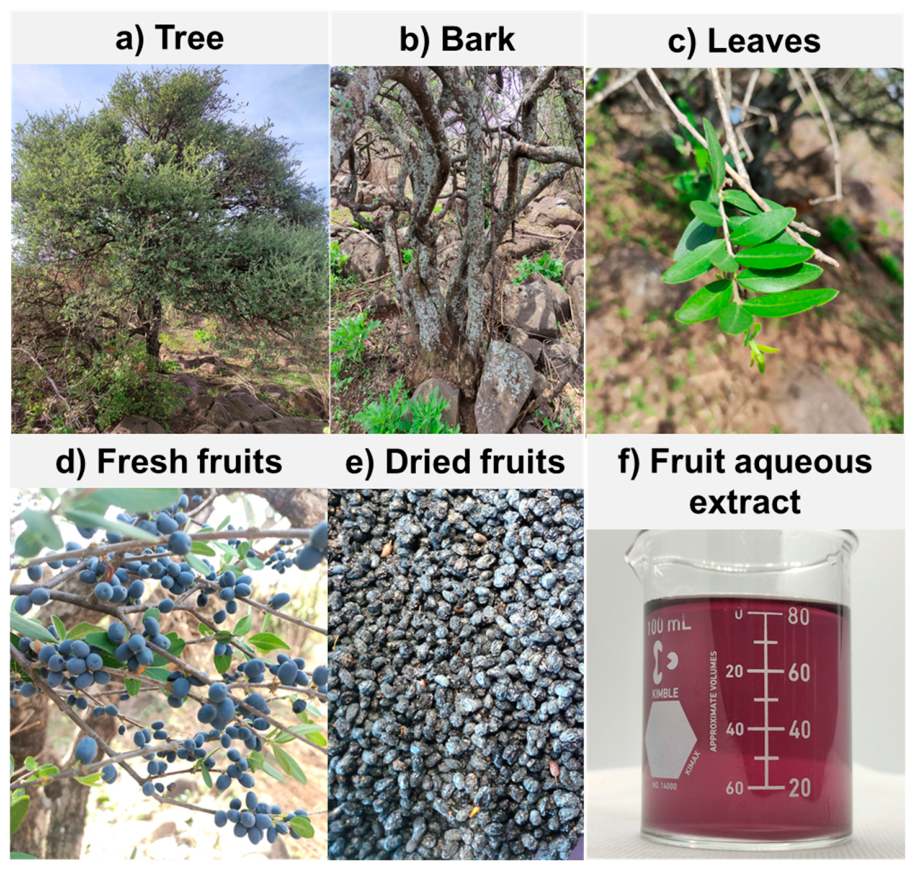
Figure 1. Visual appearance of Forestiera tomentosa tree (a), bark (b), leaves (c), fresh fruits (d), dried fruits (e), and fruit aqueous extract (f).
Molecules 31 02542 g001
Figure 2. The radar image obtained from SwissADME for the top 6 compounds present in the extract: (A) syringic acid, (B) catechin, (C) epicatechin, (D) gallocatechin, (E) 3-(4-Hydroxyphenyl) propionic acid, and (F) 4-hydroxyphenylacetic acid.
Molecules 31 02542 g002
Figure 3. Egg plot from SwissADME showed the water partition coefficient (WlogP) versus the topological polar surface area (TPSA) for the six compounds with the highest presence in F. tomentosa.
Molecules 31 02542 g003
Figure 4. 3D representation of the complexes: (A) CYP3A4-Tert-Butyl {6-Oxo-6-[(Pyridin-3-Ylmethyl)amino]hexyl}carbamate; (B) CYP3A4–epicatechin; (C) CYP2D6–quinine; (D) CYP2D6-epicatechin; (E) CYP2E1–indazole; (F) CYP2E1–catechin. The prominent green residues represent conventional hydrogen bonds, while the lime-green residues represent a carbon–hydrogen bond in (A) and a pi-donor hydrogen bond in (F).
Molecules 31 02542 g004
Figure 5. Favorable interactions of complexes: (A) CYP3A4-Tert-Butyl {6-Oxo-6-[(Pyridin-3-Ylmethyl)amino]hexyl}carbamate; (B) CYP3A4–epicatechin; (C) CYP2D6–quinine; (D) CYP2D6–epicatechin; (E) CYP2E1–indazole; (F) CYP2E1–catechin.
Molecules 31 02542 g005
Table 1. Proximal composition of Forestiera tomentosa fruit.
ParameterContent (%)
* Proteins7.39
* Lipids4.30
Available carbohydrates71.94
* Crude fiber7.90
** Reducing sugars21.40 ± 0.05
* Ash4.33
* Moisture in dried fruits12.04
Moisture in fresh fruits67.85 ± 0.12
* Values correspond to the mean values reported by an accredited external analytical laboratory and presented on a dry-matter basis (Supplementary Material, S1). ** Results are mean ± standard deviation of triplicate determinations. Results were calculated using average dry-matter-basis data; ▲ Results are presented on a wet-matter basis.
Table 2. Physicochemical parameters and functional properties of Forestiera tomentosa fruit.
ParameterContent
Physicochemical parameters
* pH5.66 ± 0.02
* Titratable acidity (% citric acid)0.32 ± 0.07
* Total soluble solids (TSS, °Brix)2.33 ± 0.01
TSS/TA ratio7.28
* Luminosity26.26 ± 1.61
* a (coordinate)1.01 ± 0.35
* b (coordinate)−15.30 ± 0.53
Water activity0.42 ± 0.01
Functional properties
Water solubility index (%) 56.10 ± 0.19
Water absorption index (g g−1)1.80 ± 0.24
Swelling power (%)2.48 ± 0.11
Oil absorption index (g g−1)4.54 ± 0.07
Emulsifying capacity (%)2.61 ± 0.19
Foaming capacity (%)19.71 ± 0.89
Results are the mean ± standard deviation of triplicate determinations. Results are presented on a dry-matter basis. * Results are presented on a fresh basis.
Table 3. Phenolic compounds content and antioxidant capacity of Forestiera tomentosa fruit.
ParameterContent
Phenolic compounds
Total soluble phenols (mg GAE/g)280.42 ± 7.84
Total flavonoids (mg CE/g)98.89 ± 9.39
Total anthocyanins (mg C3G/g)54.53 ± 3.07
Condensed tannins (mg CE/g)94.95 ± 2.05
Antioxidant capacity
DPPH (mmol TE/g)14.59 ± 0.26
ABTS (mmol TE/g)12.04 ± 0.13
FRAP (mmol TE/g)1.92 ± 0.29
Results are mean ± standard deviation of triplicate determinations. GAE: Gallic acid equivalent; CE: catechin equivalent; C3G: Cyanidin-3-glucoside. All results are presented on a dry-matter basis.
Table 4. HPLC-DAD phenolic profile of Forestiera tomentosa fruit.
CompoundContent (µg/100 g DW)
Gallic acid2411.98 ± 340.76
Protocatechuic acid730.55 ± 6.70
Gallocatechin23,693.03 ± 196.43
Neochlorogenic acid3496.48 ± 3.14
3,4-Dihydroxyphenylacetic acid2696.84 ± 10.92
4-Hydroxybenzoic acid1876.46 ± 22.27
Chlorogenic acid1330.67 ± 4.03
4-Hydroxyphenylacetic acid9229.56 ± 70.98
Vanillic acid3691.48 ± 40.14
Syringic acid55,791.95 ± 32.82
Caffeic acid882.44 ± 3.63
Catechin29,630.66 ± 122.63
Epicatechin25,387.03 ± 69.41
3-(4-hydroxyphenyl) propionic acid172,711.40 ± 63.68
Rutin2621.29 ± 55.37
p-Coumaric acid3670.09 ± 13.89
Trans-ferulic acid824.35 ± 5.27
Ellagic acid2507.57 ± 1.86
Salicylic acid1158.56 ± 22.31
Naringenin1730.70 ± 6.52
Results are mean ± standard deviation of triplicate determinations. DW: Dry weight.
Table 5. Bioassay in Artemia salina larvae after 24 and 48 h of exposure to Forestiera tomentosa aqueous extract.
Concentration (µg/mL)Survival Rate (%)
24 h48 h
1000100100
500100100
250100100
125100100
62.5100100
1000100100
Synthetic seawater (Negative control)100100
Sodium hypochlorite solution at 10% (positive control)00
Results are the mean of three independent experiments (n = 3). Toxicity was evaluated using a viability scale: 100–91% non-toxic, 90–51% slightly toxic, 50–11% toxic, and 10–0% highly toxic [15].
Table 6. In silico analysis of physicochemical and ADME properties of six major phytochemicals identified in Forestiera tomentosa fruit.
CompoundMWnHBAnHBDLPnLROB *HIA **BBB ***
Syringic acid198.17521.5400.730.990.43
Catechin290.27651.3300.400.990.14
Epicatechin290.27651.4700.390.990.13
Gallocatechin306.27761.4710.350.980.09
3-(4-Hydroxyphenyl) propionic acid166.17321.1900.790.980.45
4-Hydroxyphenylacetic acid152.15320.8800.790.950.45
MW: Molecular weight (g/mol); nHBA: hydrogen-bond acceptors; nHBD: hydrogen-bond donors; LP: lipophilicity; nLR: number of violations of Lipinski’s rule of five; HIA: human intestinal absorption; OB: oral bioavailability; BBB: blood–brain barrier. * A value closer to 1 indicates the capacity of the compound to be absorbed from the human gastrointestinal system into the bloodstream of the human body. ** A value closer to 1 indicates better compound performance in terms of absorption and availability at the site of action. *** A value closer to 1 indicates a higher probability that the compound can cross the BBB.
Table 7. In silico analysis of toxicological properties of F. tomentosa fruit compounds.
CompoundClinical ToxicityhERG BlockingDrug-Induced Liver InjuryMutagenicity *Carcinogenicity **Predicted LD50 (mg/kg)Toxicity Class
Syringic acid0.010.010.690.050.0517004
Catechin0.060.250.260.340.0510,0006
Epicatechin0.060.230.250.330.0510,0006
Gallocatechin0.080.300.340.330.0410,0006
3-(4-Hydroxyphenyl) propionic acid0.050.010.320.050.2220004
4-Hydroxyphenylacetic acid0.063.71 × 10−30.300.050.1915504
* Value closer to 1 indicates a higher probability that the compound produces the effect. ** Toxicity is classified as 1–6; class 1 is the most toxic and lethal, while class 6 is non-toxic.
Table 8. Detailed molecular docking information for Forestiera tomentosa compounds with cytochrome P450 isoforms.
CompoundPubChem CIDCYP3A4
(kcal/mol)
CYP2D6
(kcal/mol)
CYP2E1
(kcal/mol)
Syringic acid10742−6.3−6.0−6.4
Catechin9064−8.3−7.7−9.2
Epicatechin72276−8.8−7.8−7.9
Gallocatechin65084−8.4−7.5−9.0
3-(4-Hydroxyphenyl) propionic acid10394−6.4−6.8−6.9
4-Hydroxyphenylacetic acid127−6.6−6.3−6.7
Control for CYP3A4 (Tert-Butyl {6-Oxo-6-[(Pyridin-3-Ylmethyl)amino]hexyl}carbamate)91885508−7.5------
Control for CYP2D6 (quinine)3034034---−9.0---
Control for CYP2E1 (Indazole)9221------−6.0
Disclaimer/Publisher’s Note: The statements, opinions and data contained in all publications are solely those of the individual author(s) and contributor(s) and not of MDPI and/or the editor(s). MDPI and/or the editor(s) disclaim responsibility for any injury to people or property resulting from any ideas, methods, instructions or products referred to in the content.