Article

Identification of Pyrrolo [2,3-b] Pyridine Derivatives as Novel JAK1 Inhibitors for the Treatment of Inflammatory Bowel Disease

1
College of Pharmacy, Taizhou University, Taizhou 225300, China
2
Jiangsu Key Laboratory of Drug Design and Optimization, China Pharmaceutical University, Nanjing 210009, China
3
Nanjing Linkinovo Biological Technology Research Institute, Nanjing 211100, China
*
Authors to whom correspondence should be addressed.
Molecules 2026, 31(13), 2236; https://doi.org/10.3390/molecules31132236
Submission received: 28 May 2026 / Revised: 18 June 2026 / Accepted: 20 June 2026 / Published: 25 June 2026
(This article belongs to the Special Issue Small-Molecule Drug Design and Discovery)

Abstract

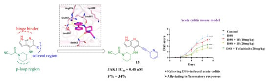
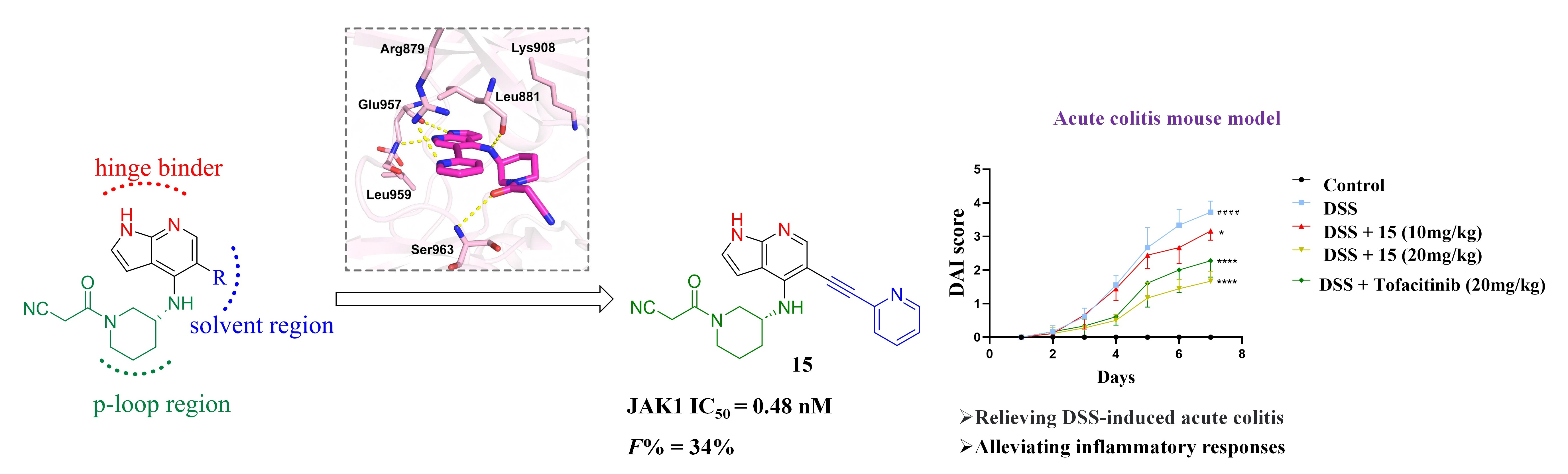
Treatment of inflammatory bowel disease (IBD) remains a major medical challenge due to the lack of safe and effective therapeutic agents. JAK1 has been validated as a key therapeutic target that modulates the pathological progression of IBD. In this study, using tofacitinib as the lead compound, we adopted a scaffold growth strategy to design and synthesize a series of pyrrolo [2,3-b] pyridine derivatives as novel JAK1 inhibitors for the treatment of IBD. Among them, compound 15 exerted potent inhibitory activity against JAK1 with an IC50 value of 0.48 nM. Western blot results showed that compound 15 significantly inhibited LPS-induced STAT1/3 phosphorylation in RAW264.7 cells. In addition, 15 exhibited satisfactory metabolic stability and oral bioavailability. In the DSS-induced colitis model, 15 remarkably ameliorated inflammatory symptoms, promoted epithelial repair, and inhibited the production of pro-inflammatory cytokines such as TNF-α and IL-6. Therefore, compound 15 is regarded as a promising candidate for the treatment of IBD.

Graphical Abstract

1. Introduction

Inflammatory bowel disease (IBD) is a chronic, recurrent inflammatory disorder of the gastrointestinal tract, mainly including Crohn’s disease (CD) and ulcerative colitis (UC) [1,2,3,4,5,6]. It is primarily characterized by recurrent intestinal inflammation, impaired intestinal epithelial barrier function and intestinal microecological dysbiosis [7,8,9,10]. Studies have demonstrated that inhibiting multiple cytokine-driven inflammatory pathways is effective for the treatment of IBD [11,12,13,14,15]. Janus kinases (JAKs) interact with signal transducers and activators of transcription (STAT) proteins and play a pivotal role in cytokine receptor signaling cascades [16,17,18]. Therefore, JAKs represent promising therapeutic targets for IBD [19,20,21,22].
JAKs constitute a small family of receptor-associated tyrosine kinases and play a crucial role in cellular responses to cytokines, consisting mainly of four members: JAK1, JAK2, JAK3 and TYK2 [9,23]. Pro-inflammatory cytokines bind to cell membrane receptors and activate JAKs, which further activate STATs via phosphorylation. Subsequently, activated STATs translocate into the nucleus to regulate the transcription of inflammation-related genes, thereby mediating and amplifying intestinal inflammatory responses [24,25,26]. In the JAK-STAT signaling pathway, JAK1 acts as a core shared molecule for multiple signaling cascades, and it is the only subtype capable of forming heterodimers with the other three JAK members and phosphorylating all STAT proteins [27,28,29].
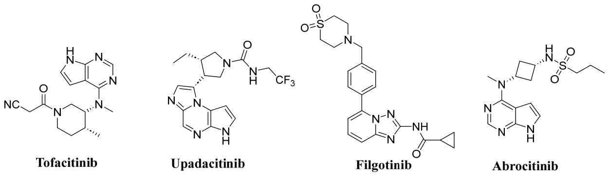
Multiple JAK1 inhibitors are currently commercially available, including tofacitinib, upadacitinib, abrocitinib and filgotinib, etc. [30,31,32,33]. (Figure 1). Notably, tofacitinib exerts a prominent therapeutic effect on the remission of UC, and is particularly applicable to patients with inadequate response to conventional therapy, making it a first-line agent for UC management in clinical practice [19,34,35]. Nevertheless, the clinical use of tofacitinib is associated with a variety of adverse reactions, such as decreased hemoglobin, reduced absolute neutrophil count, elevated total cholesterol, and increased susceptibility to various infections [36,37,38]. More importantly, the FDA has recently issued a boxed warning: tofacitinib administered at a dose of 10 mg twice daily may increase the risks of thrombosis and mortality [39,40]. Accordingly, there is an urgent need to develop novel low-toxicity tofacitinib derivatives for the treatment of IBD.
Accordingly, using tofacitinib as the lead compound, we designed and synthesized a series of pyrrolo [2,3-b] pyridine derivatives via the scaffold growth strategy as novel JAK1 inhibitors for the treatment of IBD. Systematic structure–activity relationship (SAR) studies revealed that compound 15 exhibited potent JAK1 inhibitory activity. Notably, in the DSS-induced acute colitis model, compound 15 markedly alleviated inflammatory symptoms, promoted epithelial repair, and suppressed the production of pro-inflammatory cytokines.

2. Results and Discussion

2.1. Molecular Design

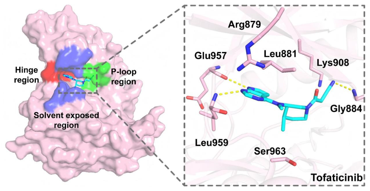
We first analyzed the binding mode of tofacitinib to JAK1 using the co-crystal structure (PDB: 3EYG) (Figure 2). The pyrrolopyrimidine moiety in the tofacitinib molecule mimics the purine moiety of ATP/ADP and stably occupies the hinge region of the ATP-binding pocket of JAK1 protein. Specifically, the pyrrolopyrimidine scaffold forms hydrogen bonds with the Leu959 and Glu957 residues in the hinge region. Meanwhile, the cyanoamide-substituted piperidine moiety of tofacitinib mimics the cyclopentane and phosphate moieties of ATP/ADP. The cyanoamide fragment extends into the P-loop region, where the cyano group forms an additional hydrogen bond with the Gly884 residue. This chemical architecture and preferred conformation confer high binding affinity of tofacitinib to JAK1 protein. Furthermore, we found that the solvent-exposed region of tofacitinib is not fully occupied, leaving considerable chemical modification space that can be exploited to further improve the physicochemical properties and other relevant characteristics of the compound.
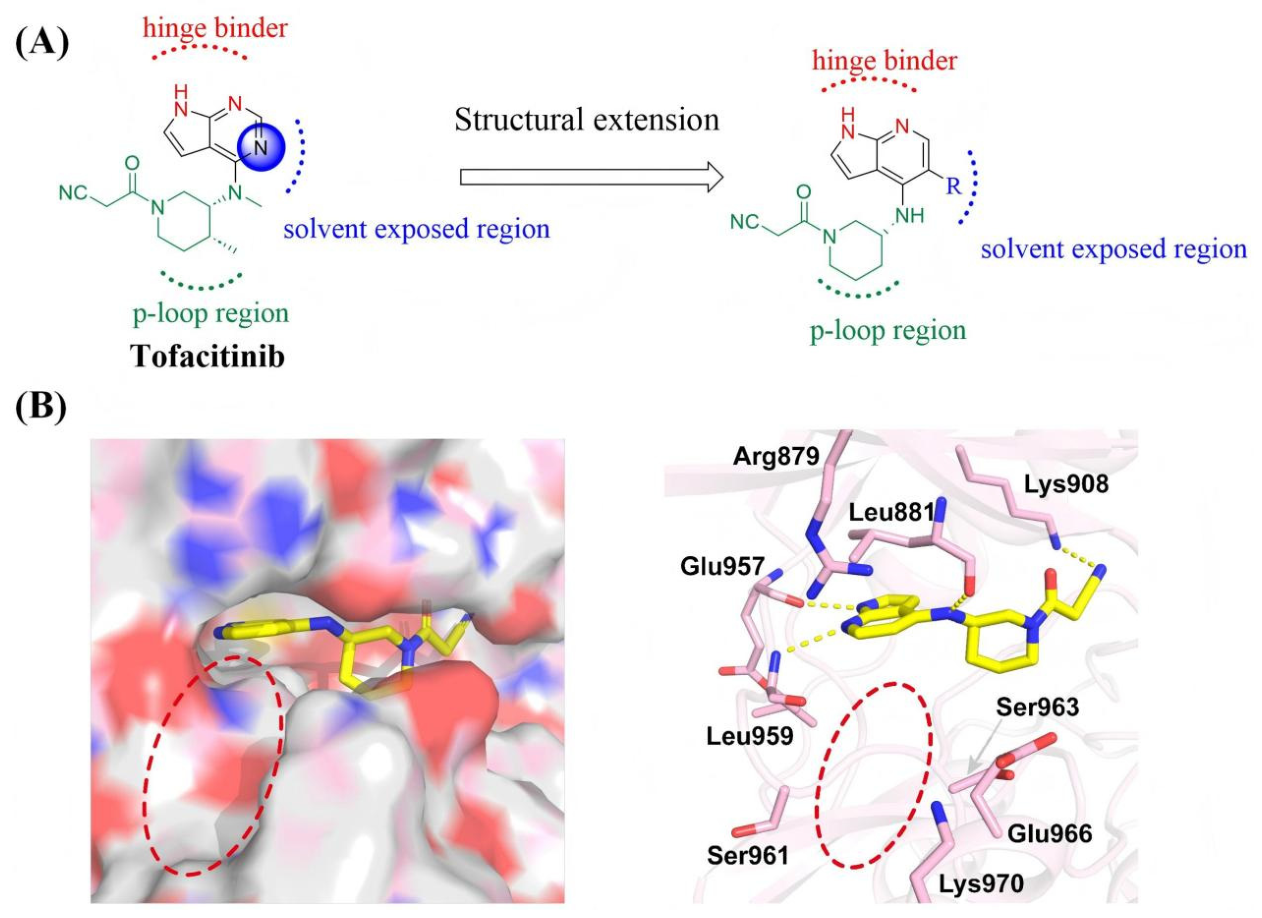
Building on these findings, we conducted rational structural design using tofacitinib as the lead compound. To achieve scaffold extension, we performed a C-N atom replacement in the core backbone, and designed a novel class of JAK1 inhibitors bearing a pyrrolopyridine scaffold (Figure 3A). To elucidate the interactions between this scaffold and JAK1 protein, we performed molecular docking studies, and the results are shown in Figure 3B. Docking results revealed that the pyrrolopyridine scaffold forms a stable hydrogen bond network with residues Leu959 and Glu957 in the hinge region of JAK1, and its cyanoamide-substituted piperidine moiety can well occupy the hydrophobic pocket of the protein. Furthermore, we identified a set of key amino acid residues, including Arg879, Ser961, Ser963, Glu966 and Lys970, distributed in the solvent-exposed region of JAK1. Given that introducing appropriate substituents into the solvent-exposed region could potentially establish additional interactions with these residues, we adopted the scaffold growth strategy by introducing linear moieties or bulky aromatic groups into this region to fully occupy the pocket. Aiming to form additional hydrophobic interactions and hydrogen bonds with the surrounding amino acid residues in the solvent-exposed region, we finally designed and synthesized a series of novel small-molecule JAK1 inhibitors.

2.2. Structure−Activity Relationship (SAR) Discussion

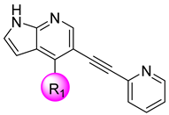
Next, we conducted a systematic SAR study on the R substituents of the pyridine ring, with detailed results presented in Table 1. Based on the distance characteristics between the amino acid residues surrounding the solvent-exposed region of the JAK1 protein and the molecular scaffold, we first introduced 5,6-fused heteroaromatic substituents to evaluate their effects on JAK1 inhibitory activity. Among them, compound 9 bearing a pyrazolopyridine group (IC50 = 3.532 ± 0.190 nM), compound 10 containing a pyrrolopyridine moiety (IC50 = 36.5 ± 3.694 nM), and compound 11 (IC50 = 90.07 ± 5.940 nM) all exhibited inferior inhibitory potency to tofacitinib. We then further investigated the impact of introducing 6,6-fused heteroaromatic substituents on biological activity. The results showed that compounds incorporating quinoline (12), isoquinoline (13), and quinazoline (14) moieties had similar degrees of loss in inhibitory efficacy, with IC50 values all falling within the range of 35–50 nM. Notably, when the solvent-exposed region was occupied by a bicyclic aromatic system, all tested compounds showed varying degrees of decreased JAK1 inhibitory activity. A plausible explanation is that a certain torsional angle formed between the pyrrolopyridine scaffold and the bicyclic structural system may increase the steric hindrance, which in turn restricts the formation of the preferred molecular conformation and weakens the complementarity and binding interaction with the ATP-binding pocket of JAK1 protein. Building on these findings, we further introduced a linear alkynylpyridine moiety at this position to obtain compound 15. This compound displayed potent JAK1 inhibitory activity (IC50 = 0.48 ± 0.427 nM), with significantly improved JAK1 inhibitory potency compared to tofacitinib. This finding indicates that the linear alkynylpyridine structure can effectively adapt to the spatial constraints of the solvent-exposed region and enhance the binding interaction between the molecule and the protein. In summary, compound 15 exhibited the optimal JAK1 inhibitory activity, which defined the direction for further structural modifications in subsequent studies.
Then, we investigated the effects of different types of aminopiperidine scaffolds on JAK1 inhibitory potency. The results presented in Table 2 demonstrate that both compound 16 (IC50 = 10.54 ± 1.548 nM) and compound 18 (IC50 = 23.27 ± 1.209 nM) resulted in reduced JAK1 inhibitory activity. Furthermore, compound 17 exhibited a markedly decreased JAK1 inhibitory activity with an IC50 value of 61.74 ± 10.720 nM. It is hypothesized that the S-configured R1 group orients toward the hydrophilic side of the binding pocket and forms unfavorable interactions with the polar residues within the pocket, thereby attenuating the binding interaction between the small molecule and JAK1 protein.

2.3. Molecular Docking and MD Study of Compound 9 and 15

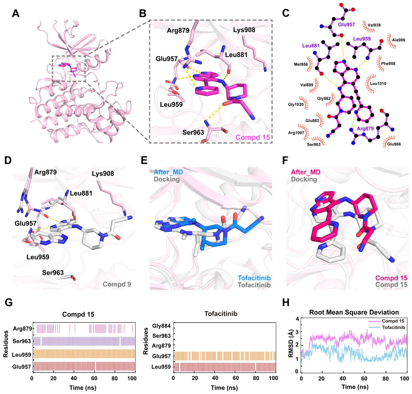
To elucidate the binding mode of compound 15, we performed molecular docking simulations using the JAK1 co-crystal structure (PDB: 3EYG) as the template (Figure 4A–C). The docking results revealed that compound 15 adopted a binding mode similar to that of tofacitinib. Its pyrrolopyridine scaffold formed a stable hydrogen bond network with the hinge region residues Glu957 and Leu959. Meanwhile, the aminopiperidine moiety occupied the polar pocket and established hydrogen bond interactions with residues Leu881 and Ser963. Furthermore, the lipophilic alkynylpyridine group introduced into the solvent-exposed region formed a hydrogen bond between its pyridine nitrogen and residue Arg879. This interaction simultaneously induced a conformational rearrangement of the compound, rendering its molecular structure better accommodated to the ATP-binding cavity of JAK1 protein. To further elucidate the reasons for the low potency of bicyclic aromatic substituted compounds, we performed molecular docking analysis on the representative compound 9 and compared its binding pose with that of compound 15 (Figure 4D). The docking results reveal that the bicyclic heteroaromatic substituent of compound 9 has a large volume and rigid conformation. It occupies a broader spatial range within the solvent-exposed region of JAK1. This region features a narrow and elongated topology due to steric constraints imposed by surrounding amino acid residues. Consequently, the bulky bicyclic moiety reduces the spatial complementarity between the ligand and the binding pocket. Further analysis indicates that compound 9 adopts an altered binding orientation relative to compound 15. This shift prevents its cyanoamide-substituted piperidine fragment from maintaining polar interactions with Ser963. Collectively, although compound 9 can still fit into the narrow subpocket of the solvent-exposed region of JAK1, its large bicyclic substituent impedes the formation of the optimal binding conformation and disrupts the interaction network with peripheral amino acid residues. This is likely the primary reason why compounds 914 exhibit weaker inhibitory activity than compound 15.
To further evaluate the binding stability of compound 15, we performed 100 ns molecular dynamics (MD) simulations on compound 15 and tofacitinib. Root-mean-square deviation (RMSD) analysis of the ligands indicated that both compounds reached a relatively stable binding state throughout the simulation (Figure 4H). A comparison between the initial MD conformation and representative conformations after simulation revealed that both ligands remained within the ATP-binding pocket of JAK1 after the 100 ns simulation. No obvious dissociation or large-scale displacement was observed, indicating that both compounds could maintain basically stable binding modes (Figure 4E,F). Further interaction analysis revealed distinct differences in dynamic binding behaviors between compound 15 and tofacitinib. During molecular dynamics (MD) simulations, compound 15 stably sustained polar interactions with the main chains of Glu957 and Leu959 in the hinge region, and also formed persistent polar contacts with the hydroxyl group of Ser963. Moreover, the alkynylpyridine moiety of compound 15 extended toward the solvent-exposed side of the ATP-binding pocket, enabling the formation of relatively stable π–cation interactions with Arg879. In contrast, tofacitinib retained the canonical hinge region interactions with the main chains of Glu957 and Leu959. However, it lacks the linear extended alkynylpyridine fragment found in compound 15, and thus failed to establish continuous interactions with Ser963 or Arg879 throughout the MD simulation. Additionally, the interaction with Gly884 observed in the initial binding pose of tofacitinib was not sustained during the dynamic simulation (Figure 4G). These results demonstrate that compound 15 not only retains the canonical binding mode with the hinge region, but also further strengthens the intermolecular interaction network on the solvent-exposed side of the ATP-binding pocket via its alkynylpyridine fragment. This additional network of interactions, particularly the persistent contacts with Ser963 and Arg879, may help improve the binding stability and potential pocket residence capacity of compound 15 within the JAK1 ATP-binding pocket. Collectively, these observations provide further structural and dynamic evidence explaining why compound 15 exhibits superior inhibitory activity relative to tofacitinib.

2.4. Immunoblotting Confirmed the Inhibition of 15 in the JAK/STAT Signaling Pathway

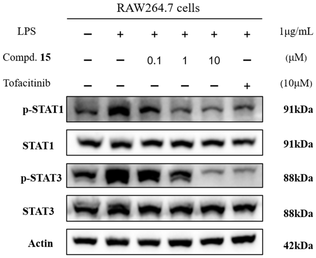
JAK kinase activation is initiated by distinct cytokines, which induces JAK dimerization and mediates STAT protein phosphorylation. To verify the efficacy of compound 15, we performed Western blot analysis in RAW264.7 cells using tofacitinib as the positive control. Upon LPS stimulation, cells were treated with different concentrations of compound 15, and the inhibitory effects on STAT phosphorylation were examined, as shown in Figure 5. Following stimulation with 1 μg/mL LPS, compound 15 inhibited the phosphorylation levels of STAT1/3 in a dose-dependent manner. Notably, at a concentration of 10 μM, compound 15 markedly inhibited LPS-induced phosphorylation, and at this concentration, it exhibited a phosphorylation inhibitory effect comparable to that of tofacitinib. Collectively, compound 15 demonstrated favorable inhibitory activity against the JAK/STAT signaling pathway at the cellular level.

2.5. Drug-like Properties Evaluation of 15

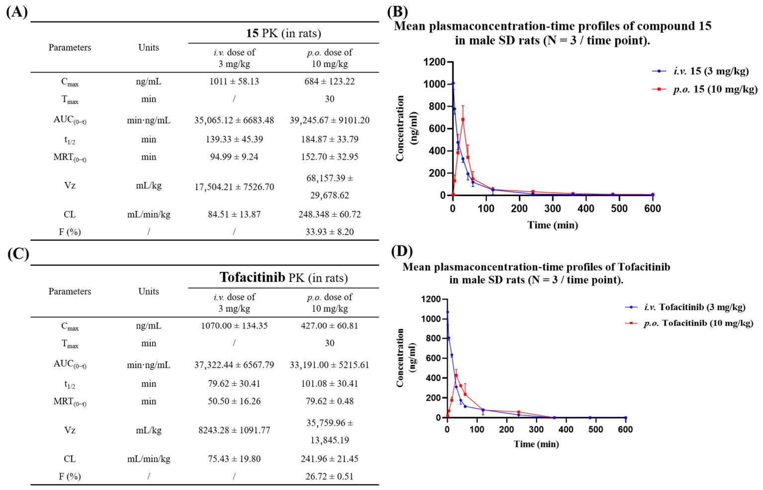
Given its favorable JAK1 potency, we evaluated the drug-like properties of compound 15. As shown in Table 3, compound 15 had an aqueous solubility of 462.34 μg/mL and exhibited excellent stability in simulated gastric fluid (SGF), simulated intestinal fluid (SIF), rat plasma, and rat liver microsomes (RLMs). We further assessed the pharmacokinetic (PK) profiles of compound 15 and tofacitinib. Two compounds were respectively dissolved in a vehicle consisting of 10% DMSO, 40% PEG 400, and 50% water, and administered to SD rats via intravenous (IV: 3 mg/kg) and oral (PO: 10 mg/kg) routes. The corresponding pharmacokinetic parameters are presented in Figure 6. A comparative analysis of key pharmacokinetic (PK) parameters revealed that compound 15 exhibited higher oral bioavailability than tofacitinib under identical experimental conditions. This indicates that compound 15 has favorable oral absorption and undergoes less hepatic first-pass metabolism, so ideal plasma drug concentrations can be achieved at low oral doses. At the oral dose of 10 mg/kg, the maximum plasma concentration (Cmax) of compound 15 was 684 ng/mL, with the area under the concentration–time curve (AUC) of 39,245.67 min·ng/mL. In comparison, tofacitinib showed a Cmax of 427 ng/mL and an AUC of 33,191.00 min·ng/mL. The higher systemic exposure of compound 15 at the same dose facilitates sustained pharmacological effects. The oral elimination half-life (T1/2) of compound 15 was 184.87 min, which was markedly longer than that of tofacitinib (101.08 min). A prolonged half-life corresponds to slower in vivo elimination, which is expected to reduce dosing frequency and improve patient compliance. Additionally, compound 15 displayed a moderate clearance rate and a larger apparent volume of distribution, suggesting a better potential for tissue distribution in vivo. Collectively, these findings support further in vivo evaluation of compound 15.

2.6. Evaluation of the Therapeutic Effect of 15 in an Acute Mice Colitis Model

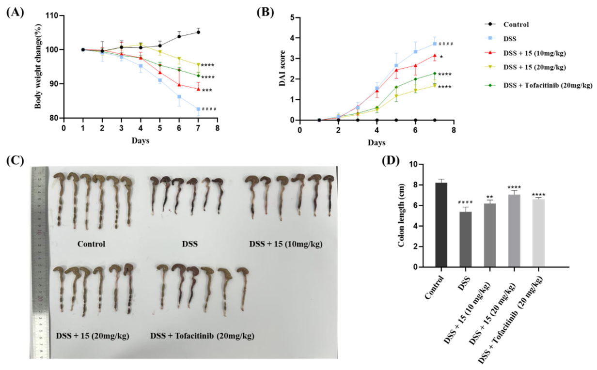
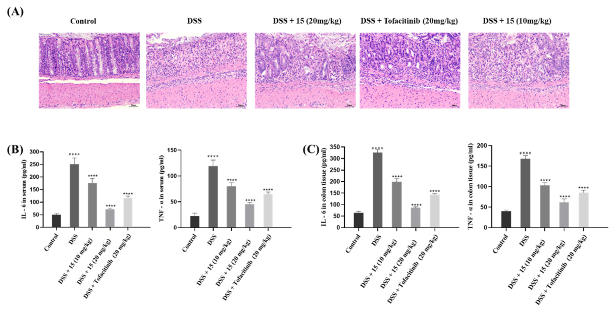
We established dextran sulfate sodium (DSS)-induced acute ulcerative colitis mouse models to further evaluate the in vivo therapeutic efficacy of compound 15. Acute murine colitis was induced using 3% (w/v) DSS, and tofacitinib was selected as the positive control. Tofacitinib (20 mg/kg) and compound 15 (10 mg/kg or 20 mg/kg) were administered orally once daily (qd) for 7 consecutive days. The severity of colitis was assessed by measuring body weight, disease activity index (DAI) score, and colon length. Compared with the normal control group, mice in the DSS model group developed severe colitis symptoms, manifested as significant body weight loss, elevated DAI scores, and shortened colon length (Figure 7A–D). Treatment with compound 15 significantly ameliorated these symptoms, as evidenced by attenuated body weight loss, reduced DAI scores, and preserved colon length. Its therapeutic efficacy was comparable to that of tofacitinib at the same dose (20 mg/kg). Notably, a significant dose–response relationship was observed, with the higher dose of compound 15 (20 mg/kg) exhibiting more pronounced therapeutic effects. Histopathological examination of hematoxylin and eosin (H&E)-stained colon tissue sections further revealed that compound 15 alleviated DSS-induced tissue damage and inflammatory cell infiltration (Figure 8A). Subsequently, the levels of pro-inflammatory cytokines (IL-6 and TNF-α) in colon tissues and serum were further evaluated by enzyme-linked immunosorbent assay (ELISA) (Figure 8B,C). The results showed that the levels of pro-inflammatory cytokines in colon tissues and serum were significantly elevated in the DSS model group, whereas treatment with compound 15 markedly downregulated these inflammatory markers, demonstrating potent anti-inflammatory activity. Collectively, these experimental results demonstrated that compound 15 significantly attenuates DSS-induced acute colitis symptoms in mice, indicating its promising in vivo therapeutic potential.

2.7. Synthesis of the Target Compounds

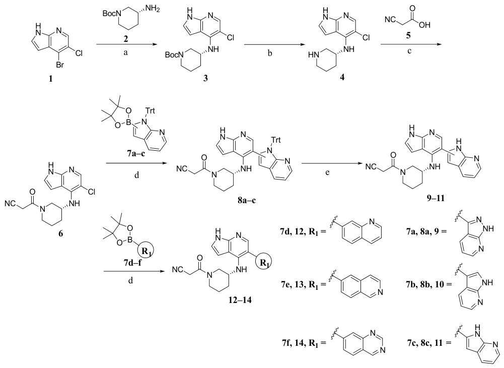
The structures and synthetic routes of the target compounds are detailed in Scheme 1 and Scheme 2. The target compounds 914 were synthesized following the synthetic procedure outlined in Scheme 1. Starting from compound 1, intermediate 3 was obtained via Buchwald–Hartwig cross-coupling reaction, followed by Boc deprotection with an appropriate amount of trifluoroacetic acid to afford intermediate 4. Condensation of intermediate 4 with cyanoacetic acid in anhydrous dichloromethane as the solvent yielded the key intermediate 6. Intermediate 6 was converted to intermediates 8a–c and target compounds 1214 via Suzuki cross-coupling reaction, and final deprotection with an appropriate amount of trifluoroacetic acid furnished the target compounds 911.
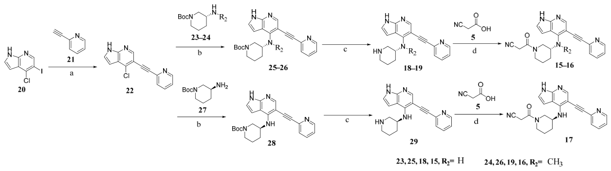
Target compounds 1518 were synthesized following the procedure outlined in Scheme 2. Starting from compound 20, intermediate 22 was obtained via Sonogashira cross-coupling reaction. Compounds 25, 26 and 28 were prepared via Buchwald–Hartwig cross-coupling reaction, followed by Boc deprotection with an appropriate amount of trifluoroacetic acid to afford intermediates 18, 19 and 29. Subsequent condensation with cyanoacetic acid in anhydrous dichloromethane as the solvent yielded target compounds 15, 16 and 17.

3. Experimental Section

3.1. Chemistry

All chemical solvents and reagents were purchased from commercial sources unless otherwise stated. Reactions were monitored by thin-layer chromatography (TLC) on 0.25 mm silica gel plates (GF-254) and visualized under UV light at wavelengths of 254 and 365 nm. Column chromatography was performed on silica gel (200–300 mesh) for the separation and purification of target compounds. Chemical structures were identified by 1H NMR spectroscopy, 13C NMR spectroscopy, and high-resolution mass spectrometry (HRMS). 1H NMR and 13C NMR spectra were recorded on a Bruker AVANCE 300 spectrometer (Bruker, Leipzig, Germany) using deuterated solvents with tetramethylsilane (TMS) as the internal standard. HRMS spectra were recorded on a Waters Q-Tof micro mass spectrometer. Compound purities were determined by high-performance liquid chromatography (HPLC) on a Shimadzu instrument (Shimadzu, Kyoto, Japan) equipped with an Inertsil C18 column (4.6 mm × 250 mm, 5 μm) using a methanol/water mobile phase at a flow rate of 1 mL/min, with detection at 254 nm. All final compounds were confirmed to have purities >95% by HPLC analysis.

3.1.1. Procedure for Preparation of 22

4-Chloro-5-(pyridin-2-ylethynyl)-1H-pyrrolo [2,3-b]pyridine (22)
To 25 mL of acetonitrile, compounds 20 (1.00 g, 3.60 mmol, 1.00 equiv) and 21 (555.12 mg, 5.93 mmol, 1.50 equiv), Pd(PPh3)2Cl2 (126.36 mg, 0.18 mmol, 0.05 equiv), CuI (68.40 mg, 0.36 mmol, 0.10 equiv), and triethylamine (1.18 g, 10.80 mmol, 3.00 equiv) were added. The reaction mixture was degassed and placed under an argon atmosphere, then heated to reflux at 90 °C. Upon completion of the reaction, the solvent was evaporated under reduced pressure. The residue was extracted with ethyl acetate, and the organic layer was washed sequentially with water (2 × 100 mL) and brine (1 × 100 mL), dried over anhydrous sodium sulfate, and concentrated under reduced pressure to afford the crude product. Purification by column chromatography (DCM:MeOH = 250:1, v/v) afforded 728 mg of compound 22 as a yellow solid in 80% yield. 1H NMR (300 MHz, DMSO-d6) δ 12.39 (s, 1H), 8.65 (d, J = 4.8 Hz, 1H), 8.52 (s, 1H), 7.91–7.86 (m, 1H), 7.76–7.67 (m, 2H), 7.48–7.43 (m, 1H), 6.63–6.59 (m, 1H). MS (ESI) m/z: 254.1 [M + H]+.

3.1.2. Procedure for General Preparation of 3 and 25, 26, 28

tert-butyl (R)-3-((5-(pyridin-2-ylethynyl)-1H-pyrrolo [2,3-b]pyridin-4-yl)amino)piperidine-1-carboxylate (25)
To 5 mL of dioxane, compound 22 (250.00 mg, 0.99 mmol, 1.00 equiv), 4 g (296.80 mg, 1.48 mmol, 1.50 equiv), t-BuONa (596.44 mg, 5.93 mmol, 6.00 equiv), 1,3-bis(2,6-diisopropylphenyl)imidazolium chloride (63.65 mg, 0.15 mmol, 0.15 equiv) and Pd2(dba)3 (63.78 mg, 0.07 mmol, 0.07 equiv) were added. The reaction mixture was degassed and purged with argon, then heated to reflux at 110 °C. Upon completion of the reaction, the solvent was evaporated under reduced pressure. The residue was extracted with ethyl acetate, and the organic layer was washed sequentially with water (2 × 100 mL) and brine (1 × 100 mL), dried over anhydrous sodium sulfate, and concentrated under reduced pressure. Purification by column chromatography (DCM:MeOH = 200:1, v/v) afforded 268 mg of compound 25 as a yellow solid in 65% yield. 1H NMR (300 MHz, DMSO-d6) δ 11.64 (s, 1H), 8.59 (dd, J = 5.0, 1.7 Hz, 1H), 8.08 (s, 1H), 7.90–7.70 (m, 1H), 7.60 (d, J = 7.8 Hz, 1H), 7.40–7.32 (m, 1H), 7.26 (t, J = 2.9 Hz, 1H), 6.66 (s, 1H), 5.83 (d, J = 8.5 Hz, 1H), 4.20–4.28 (m, 1H), 3.95–3.89 (m, 1H), 3.57 (d, J = 12.8 Hz, 1H), 3.26–3.07 (m, 2H), 2.15–1.97 (m, 1H), 1.78–1.65 (m, 2H), 1.60–1.52 (m, 1H), 1.24 (s, 9H). MS (ESI) m/z: 418.2 [M + H]+.
tert-butyl (R)-3-(methyl(5-(pyridin-2-ylethynyl)-1H-pyrrolo [2,3-b]pyridin-4-yl)amino)piperidine-1-carboxylate (26)
Compound 26 was synthesized according to the same procedure as 25 using 22 and 24. The crude product is used directly in the next step without purification.
(S)-3-((5-(pyridin-2-ylethynyl)-1H-pyrrolo [2,3-b]pyridin-4-yl)amino)piperidine-1-carboxylate (28)
Compound 28 was synthesized according to the same procedure as 25 using 22 and 27. The crude product is used directly in the next step without purification.
tert-butyl (R)-3-((5-chloro-1H-pyrrolo [2,3-b]pyridin-4-yl)amino)piperidine-1-carboxylate (3)
Compound 3 was synthesized according to the same procedure as 25 using 1 and 2. yield: 66%. 1H NMR (300 MHz, DMSO-d6) δ 11.51 (s, 1H), 7.89 (s, 1H), 7.26 (t, J = 3.0 Hz, 1H), 6.56 (d, J = 3.5 Hz, 1H), 5.46 (d, J = 8.3 Hz, 1H), 4.08–4.03 (m, 1H), 3.87 (d, J = 13.0 Hz, 1H), 3.57 (dd, J = 12.5, 5.7 Hz, 1H), 3.22 (s, 1H), 3.13–3.07 (m, 1H), 2.02–1.59 (m, 4H), 1.30 (s, 9H). MS (ESI) m/z: 351.1 [M + H]+.

3.1.3. Procedure for General Preparation of 4, 18, 19 and 29

(R)-N-(piperidin-3-yl)-5-(pyridin-2-ylethynyl)-1H-pyrrolo [2,3-b]pyridin-4-amine (18)
To a mixture of compound 25 (200.00 mg, 0.63 mmol, 1.00 equiv) and dichloromethane (10 mL), 5 mL of trifluoroacetic acid (TFA) at room temperature was added. The reaction mixture was stirred at room temperature for 2 h. Upon completion of the reaction, the solvent was evaporated under reduced pressure. The residue was extracted with ethyl acetate, and the organic layer was washed sequentially with saturated sodium bicarbonate solution (2 × 100 mL) and brine (1 × 100 mL), dried over anhydrous sodium sulfate, and concentrated under reduced pressure to afford the crude product. Purification by column chromatography (DCM:MeOH = 25:1, v/v) afforded 170 mg of compound 18 as a white solid in 85% yield. 1H NMR (300 MHz, DMSO-d6) δ 11.61 (s, 1H), 8.59 (s, 1H), 8.05 (s, 1H), 7.83 (t, J = 7.7 Hz, 1H), 7.69 (d, J = 7.9 Hz, 1H), 7.36 (s, 1H), 7.23 (s, 1H), 6.65 (s, 1H), 6.22 (d, J = 8.7 Hz, 1H), 4.29 (s, 1H), 3.14 (d, J = 11.5 Hz, 1H), 2.82 (s, 2H), 2.72 (s, 1H), 1.92 (s, 1H), 1.70–1.54 (m, 4H); HRMS (ESI): calcd for C19H19N5, [M + H]+ 318.1713, found 318.1719; HPLC (MeOH:H2O with 0.1% NH3 = 80:20) tR = 5.07 min, 98.01%.
(R)-N-methyl-N-(piperidin-3-yl)-5-(pyridin-2-ylethynyl)-1H-pyrrolo [2,3-b]pyridin-4-amine (19)
Compound 19 was synthesized according to the same procedure as 18 using 26. The crude product is used directly in the next step without purification.
(S)-N-(piperidin-3-yl)-5-(pyridin-2-ylethynyl)-1H-pyrrolo [2,3-b]pyridin-4-amine (29)
29 was synthesized according to the same procedure as 18 using 28. The crude product is used directly in the next step without purification.
(R)-5-chloro-N-(piperidin-3-yl)-1H-pyrrolo [2,3-b]pyridin-4-amine (4)
Compound 4 was synthesized according to the same procedure as 18 using 3. The crude product is used directly in the next step without purification.

3.1.4. Procedure for General Preparation of 6, 15, 16 and 17

(R)-3-oxo-3-(3-((5-(pyridin-2-ylethynyl)-1H-pyrrolo [2,3-b] pyridin-4-yl) amino) piperidin-1-yl) propanenitrile (15)
To 10 mL of dichloromethane compound 18 (150.00 mg, 0.47 mmol, 1.00 equiv), compound 5 (60.56 mg, 0.71 mmol, 1.50 equiv), and DCC (205.66 mg, 0.94 mmol, 2.00 equiv) were added. The reaction mixture was degassed and purged with nitrogen, then stirred at 40 °C for 2 h. Upon completion of the reaction, the solvent was evaporated under reduced pressure. The residue was extracted with ethyl acetate, and the organic layer was washed sequentially with saturated sodium bicarbonate solution (2 × 100 mL) and brine (1 × 100 mL), dried over anhydrous sodium sulfate, and concentrated under reduced pressure to afford the crude product. Purification by column chromatography (DCM:MeOH = 75:1, v/v) afforded 108 mg of compound 15 as a yellow solid in 60% yield. 1H NMR (300 MHz, DMSO-d6) δ 11.64 (s, 1H), 8.63–8.55 (m, 1H), 8.07 (s, 1H), 7.84 (t, J = 7.6 Hz, 1H), 7.69–7.56 (m, 1H), 7.42–7.31 (m, 1H), 7.26 (s, 1H), 6.70 (d, J = 11.3 Hz, 1H), 5.92–5.86 (m, 1H), 4.45–4.32 (m, 1H), 4.24 (s, 1H), 4.12 (s, 1H), 4.03–3.76 (m, 1H), 3.52 (d, J = 11.6 Hz, 1H), 3.20 (d, J = 11.5 Hz, 1H), 3.14–2.89 (m, 1H), 2.10 (s, 1H), 1.74 (s, 3H); 13C NMR (75 MHz, DMSO-d6) δ 161.66, 149.79, 149.02, 147.47, 147.09, 142.79, 136.34, 126.27, 122.54, 115.95, 104.44, 99.85, 94.64, 92.93, 85.60, 50.17, 48.75, 46.80, 45.38, 30.29, 24.75, 22.97; HRMS (ESI): calcd for C22H20N6O, [M + H]+ 385.1771, found 385.1782; HPLC (MeOH:H2O with 0.1% NH3 = 80:20) tR = 5.32 min, 98.86%.
(R)-3-(3-(methyl(5-(pyridin-2-ylethynyl)-1H-pyrrolo [2,3-b] pyridin-4-yl) amino) piperidin-1-yl)-3-oxopropanenitrile (16)
Compound 16 was synthesized according to the same procedure as 15 using 19 and 5. Yellow solid (yield: 56%). 1H NMR (300 MHz, DMSO-d6) δ 11.77 (s, 1H), 8.58 (d, J = 4.8 Hz, 1H), 8.18 (d, J = 6.0 Hz, 1H), 7.82 (t, J = 7.5 Hz, 1H), 7.57 (t, J = 6.0 Hz, 1H), 7.38–7.32 (m, 2H), 6.63 (s, 1H), 4.33–4.28 (m, 1H), 4.07–4.01 (m, 2H), 3.99–3.89 (m, 1H), 3.69–3.51 (m, 1H), 3.24 (s, 3H), 3.09–2.89 (m, 2H), 2.15 (s, 1H), 2.01 (d, J = 13.1 Hz, 1H), 1.75 (d, J = 12.7 Hz, 1H), 1.53 (d, J = 13.0 Hz, 1H); HRMS (ESI): calcd for C23H22N6O, [M + H]+ 399.1928, found 399.1935; HPLC (MeOH:H2O with 0.1% NH3 = 80:20) tR = 5.40 min, 96.55%.
(S)-3-oxo-3-(3-((5-(pyridin-2-ylethynyl)-1H-pyrrolo [2,3-b] pyridin-4-yl) amino) piperidin-1-yl) propanenitrile (17)
Compound 17 was synthesized according to the same procedure as 15 using 29 and 5. Yellow solid (yield: 50%). 1H NMR (300 MHz, DMSO-d6) δ 11.65 (s, 1H), 8.59 (d, J = 4.7 Hz, 1H), 8.08 (d, J = 3.5 Hz, 1H), 7.84 (t, J = 7.9 Hz, 1H), 7.70–7.55 (m, 1H), 7.37 (t, J = 6.2 Hz, 1H), 7.26 (s, 1H), 6.70 (d, J = 10.9 Hz, 1H), 5.95–5.89 (m, 1H), 4.50–4.24 (m, 1H), 4.15–3.98 (m, 2H), 3.85–3.45 (m, 2H), 3.25–2.85 (m, 2H), 2.10 (s, 1H), 1.63 (s, 3H); HRMS (ESI): calcd for C22H20N6O, [M + H]+ 385.1771, found 385.1769; HPLC (MeOH:H2O with 0.1% NH3 = 80:20) tR = 6.67 min, 99.15%.
(R)-3-(3-((5-chloro-1H-pyrrolo [2,3-b] pyridin-4-yl) amino) piperidin-1-yl)-3-oxopropanenitrile (6)
Compound 6 was synthesized according to the same procedure as 15 using 4 and 5. yield: 60%. 1H NMR (300 MHz, DMSO-d6) δ 11.51 (s, 1H), 7.89 (d, J = 2.6 Hz, 1H), 7.26 (t, J = 2.9 Hz, 1H), 6.66–6.56 (m, 1H), 5.59–5.51 (m, 1H), 4.39 (dd, J = 12.1, 3.2 Hz, 1H), 4.19–3.98 (m, 2H), 3.99–3.95 (m, 1H), 3.79–3.51 (m, 1H), 3.24–3.01 (m, 1H), 3.00–2.67 (m, 1H), 2.04 (d, J = 12.7 Hz, 1H), 1.76–1.66 (m, 3H). MS (ESI) m/z: 318.1 [M + H]+.

3.1.5. Procedure for General Preparation of 911

(R)-3-(3-((5-(1H-pyrazolo [3,4-b] pyridin-3-yl)-1H-pyrrolo [2,3-b] pyridin-4-yl) amino) piperidin-1-yl)-3-oxopropanenitrile (9)
To 5 mL of a mixed solvent of 1,2-dimethoxyethane and water, compound 6 (100.00 mg, 0.32 mmol, 1.00 equiv), compound 7a (77.12 mg, 0.32 mmol, 1.00 equiv), Cs2CO3 (208.33 mg, 0.64 mmol, 2.00 equiv), and XphosPdG2 (24.35 mg, 0.03 mmol, 0.10 equiv) were added. The reaction mixture was degassed and purged with argon, then heated to reflux at 100 °C. Upon completion of the reaction, the solvent was evaporated under reduced pressure. The residue was extracted with ethyl acetate, and the organic layer was washed sequentially with water (2 × 100 mL) and brine (1 × 100 mL), dried over anhydrous sodium sulfate, and concentrated under reduced pressure. Subsequently, 5 mL of dichloromethane and 0.5 mL of trifluoroacetic acid were added to the residue, and the mixture was stirred at room temperature for 2 h. Upon completion of the reaction, the solvent was evaporated under reduced pressure. The residue was extracted with ethyl acetate, and the organic layer was washed sequentially with saturated aqueous sodium bicarbonate solution (2 × 100 mL) and brine (1 × 100 mL), dried over anhydrous sodium sulfate, and concentrated under reduced pressure to afford the crude product. Purification by column chromatography (DCM:MeOH = 70:1, v/v) afforded 76 mg of compound 9 as a yellow solid in 60% yield. 1H NMR (300 MHz, DMSO-d6) δ 13.79 (d, J = 9.4 Hz, 1H), 11.52 (s, 1H), 8.60 (d, J = 4.4 Hz, 1H), 8.50 (d, J = 7.6 Hz, 2H), 8.21 (d, J = 8.3 Hz, 1H), 7.27 (s, 2H), 6.73 (s, 1H), 4.31 (d, J = 12.4 Hz, 1H), 4.10 (t, J = 14.8 Hz, 2H), 3.92–3.70 (m, 1H), 3.69–3.48 (m, 1H), 3.25–3.02 (m, 2H), 2.12 (s, 1H), 1.69 (d, J = 35.6 Hz, 3H); 13C NMR (75 MHz, DMSO-d6) δ 162.11, 152.54, 150.47, 144.37, 143.64, 136.39, 130.13, 128.57, 127.62, 120.90, 116.69, 114.17, 106.72, 100.48, 50.83, 50.20, 47.67, 46.10, 30.99, 25.43, 24.00; HRMS (ESI): calcd for C21H20N8O, [M + H]+ 401.1833, found 401.1835; HPLC (MeOH:H2O with 0.1% NH3 = 80:20) tR = 4.74 min, 99.86%.
(R)-3-(3-(1H,1′H-[3,5′-bipyrrolo [2,3-b] pyridine]-4′-ylamino) piperidin-1-yl)-3-oxopropanenitrile (10)
Compound 10 was synthesized according to the same procedure as 9 using 6 and 7b. Yellow solid (yield: 55%). 1H NMR (300 MHz, DMSO-d6) δ 11.89 (t, J = 4.9 Hz, 1H), 11.38 (s, 1H), 8.28 (d, J = 4.6 Hz, 1H), 7.81 (d, J = 9.3 Hz, 1H), 7.74 (d, J = 8.3 Hz, 1H), 7.55 (d, J = 2.5 Hz, 1H), 7.24 (d, J = 4.1 Hz, 1H), 7.15–7.05 (m, 1H), 6.65 (dd, J = 13.4, 13.6 Hz, 1H), 5.05–4.89 (m, 1H), 4.41–4.06 (m, 1H), 4.03–3.89 (m, 2H), 3.87–3.71 (m, 1H), 3.66–3.42 (m, 1H), 2.94 (s, 1H), 2.72 (t, J = 11.2 Hz, 1H), 2.01–1.92 (m, 1H), 1.54 (s, 2H), 1.37 (s, 1H); 13C NMR (75 MHz, DMSO-d6) δ 162.04, 149.28, 145.33, 143.42, 127.82, 125.63, 122.94, 119.40, 116.71, 115.98, 109.63, 107.10, 106.73, 106.12, 100.28, 51.00, 49.88, 47.90, 46.00, 31.46, 25.43, 24.18; HRMS (ESI): calcd for C22H21N7O, [M + H]+ 400.1880, found 400.1881; HPLC (MeOH:H2O with 0.1% NH3 = 80:20) tR = 4.54 min, 99.76%.
(R)-3-(3-(1H,1′H-[2,5′-bipyrrolo [2,3-b] pyridine]-4′-ylamino) piperidin-1-yl)-3-oxopropanenitrile (11)
Compound 11 was synthesized according to the same procedure as 9 using 6 and 7c. Black solid (yield: 40%). 1H NMR (300 MHz, DMSO-d6) δ 11.90 (s, 1H), 11.48 (s, 1H), 8.19 (d, J = 4.6 Hz, 1H), 8.01 (s, 1H), 7.94 (d, J = 7.5 Hz, 1H), 7.26 (s, 1H), 7.13–7.02 (m, 1H), 6.68 (d, J = 13.3 Hz, 1H), 6.58 (s, 1H), 5.67 (t, J = 10.0 Hz, 1H), 4.27 (d, J = 12.7 Hz, 1H), 4.06 (s, 2H), 3.92 (s, 1H), 3.59 (dd, J = 13.5, 13.6 Hz, 1H), 3.14–2.84 (m, 2H), 2.08–1.96 (m, 1H), 1.61 (s, 3H); 13C NMR (75 MHz, DMSO-d6) δ 162.22, 150.25, 149.57, 144.98, 142.58, 136.05, 127.76, 123.22, 121.43, 116.72, 116.25, 105.81, 105.53, 100.77, 99.07, 50.94, 49.74, 47.74, 46.12, 31.10, 25.47, 23.85; HRMS (ESI): calcd for C22H21N7O, [M + H]+ 400.1914, found 400.1901; HPLC (MeOH:H2O with 0.1% NH3 = 80:20) tR = 4.74 min, 99.07%.

3.1.6. Procedure for General Preparation of 1214

(R)-3-oxo-3-(3-((5-(quinolin-7-yl)-1H-pyrrolo [2,3-b] pyridin-4-yl) amino) piperidin-1-yl) propanenitrile (12)
To 5 mL of a mixed solvent of 1,2-dimethoxyethane and water, compound 6 (100.00 mg, 0.32 mmol, 1.00 equiv), compound 7d (80.08 mg, 0.32 mmol, 1.00 equiv), Cs2CO3 (208.32 mg, 0.64 mmol, 2.00 equiv), and XphosPdG2 (24.12 mg, 0.03 mmol, 0.10 equiv) were added. The reaction mixture was degassed and purged with argon, then heated to reflux at 100 °C. Upon completion of the reaction, the solvent was evaporated under reduced pressure. The residue was extracted with ethyl acetate, and the organic layer was washed sequentially with water (2 × 100 mL) and brine (1 × 100 mL), dried over anhydrous sodium sulfate, and concentrated under reduced pressure. Purification by column chromatography (DCM:MeOH = 90:1, v/v) afforded 104 mg of compound 12 as a yellow solid in 80% yield. 1H NMR (300 MHz, DMSO-d6) δ 11.45 (s, 1H), 8.94 (s, 1H), 8.41 (d, J = 8.2 Hz, 1H), 8.06–8.02 (m, 2H), 7.83 (s, 1H), 7.70–7.64 (m, 1H), 7.55 (d, J = 8.4 Hz, 1H), 7.28 (s, 1H), 6.67 (d, J = 8.4 Hz, 1H), 5.18–5.10 (m, 1H), 4.25–4.08 (m, 1H), 4.03 (d, J = 5.6 Hz, 1H), 3.93 (d, J = 8.3 Hz, 1H), 3.83 (s, 1H), 3.66–3.42 (m, 1H), 3.10–2.74 (m, 2H), 1.98 (s, 1H), 1.54 (s, 3H); 13C NMR (75 MHz, DMSO-d6) δ 162.16, 151.33, 150.40, 148.68, 145.09, 144.32, 139.69, 136.34, 129.54, 129.25, 127.31, 123.31, 121.89, 116.67, 114.23, 106.69, 100.47, 50.86, 50.06, 47.63, 46.09, 30.93, 25.49, 23.88; HRMS (ESI): calcd for C24H22N6O, [M + H]+ 411.1928, found 411.1926; HPLC (MeOH:H2O with 0.1% NH3 = 80:20) tR = 4.70 min, 99.55%.
(R)-3-(3-((5-(isoquinolin-6-yl)-1H-pyrrolo [2,3-b] pyridin-4-yl) amino) piperidin-1-yl)-3-oxopropanenitrile (13)
Compound 13 was synthesized according to the same procedure as 12 using 6 and 7e. Yellow solid (yield: 75%). 1H NMR (300 MHz, DMSO-d6) δ 11.45 (s, 1H), 9.34 (d, J = 2.9 Hz, 1H), 8.52 (d, J = 5.7 Hz, 1H), 8.19 (d, J = 8.5 Hz, 1H), 7.97 (d, J = 5.7 Hz, 1H), 7.87 (d, J = 5.8 Hz, 1H), 7.83 (s, 1H), 7.75 (t, J = 8.3 Hz, 1H), 7.27 (s, 1H), 6.72–6.60 (m, 1H), 5.18–5.13 (m, 1H), 4.20 (d, J = 12.9 Hz, 1H), 4.01 (d, J = 5.3 Hz, 1H), 3.94–3.80 (m, 2H), 3.55–3.50 (m, 1H), 3.04–2.72 (m, 2H), 1.97 (d, J = 11.3 Hz, 1H), 1.55–1.45 (m, 3H); 13C NMR (75 MHz, DMSO-d6) δ 162.11, 152.54, 150.47, 145.21, 144.37, 143.64, 140.82, 136.39, 130.13, 128.57, 127.18, 123.31, 120.90, 116.69, 114.17, 106.72, 100.48, 50.83, 50.20, 47.67, 46.10, 30.99, 25.43, 24.00; HRMS (ESI): calcd for C24H22N6O, [M + H]+ 411.1928, found 411.1931; HPLC (MeOH:H2O with 0.1% NH3 = 80:20) tR = 4.53 min, 99.78%.
(R)-3-(3-((5-(isoquinolin-6-yl)-1H-pyrrolo [2,3-b] pyridin-4-yl) amino) piperidin-1-yl)-3-oxopropanenitrile (14)
Compound 14 was synthesized according to the same procedure as 12 using 6 and 7f. Yellow solid (yield: 78%). 1H NMR (300 MHz, DMSO-d6) δ 11.50 (s, 1H), 9.62 (d, J = 2.7 Hz, 1H), 9.31 (s, 1H), 8.22 (d, J = 8.4 Hz, 1H), 8.00 (d, J = 9.9 Hz, 1H), 7.88–7.81 (m, 2H), 7.28 (s, 1H), 6.68 (d, J = 14.6 Hz, 1H), 5.36–5.30 (m, 1H), 4.25–4.08 (m, 1H), 4.07–3.94 (m, 2H), 3.87 (s, 1H), 3.56–3.52 (m, 1H), 3.07–2.72 (m, 2H), 1.99 (s, 1H), 1.65–1.45 (m, 3H); 13C NMR (75 MHz, DMSO-d6) δ 162.16, 160.66, 155.84, 150.43, 145.43, 145.27, 144.45, 130.87, 128.71, 127.96, 124.06, 123.38, 116.66, 113.71, 106.73, 100.65, 50.82, 50.25, 47.62, 46.15, 30.92, 25.47, 23.96; HRMS (ESI): calcd for C23H21N7O, [M + H]+ 412.1880, found 412.1882; HPLC (MeOH:H2O with 0.1% NH3 = 80:20) tR = 7.28 min, 93.18%.

3.2. JAK1 Inhibition Assay

The inhibitory activity of compounds against JAK1 kinase was performed by Beijing Aisiyipu Bio-technology Co. Ltd. (Beijing, China). The inhibitory activity assay of JAK1 was conducted using the ADP-Glo™ Kinase Assay. Prepare 2× ATP/Substrate solution (2 μM concentration) and 2× kinase solution (25 nM concentration) with kinase reaction buffer. A total of 100 nL of 15 dilutions was transferred to a 384-well assay plate using an Echo 655 liquid handler (Beckman Coulter Life Sciences, Indianapolis, IN, USA). After centrifugation, 4 μL of 2× kinase solution was added, and the plate was centrifuged at 1000 rpm for 1 min and incubated at 25 °C for 10 min. The reaction was initiated by adding 4 μL of 2× ATP/substrate solution, followed by centrifugation at 1000 rpm for 1 min and incubation at 25 °C for 60 min. A total of 4 μL of ADP-Glo reagent was then added to terminate the reaction. After centrifugation at 1000 rpm for 1 min, the plate was incubated at 25 °C for 40 min. Subsequently, 8 μL of kinase detection reagent was added, and the plate was centrifuged at 1000 rpm for 1 min and incubated at 25 °C for a further 40 min. Luminescence signals were recorded using a multimode microplate reader (Tecan, Männedorf, Switzerland).

3.3. Molecular Docking and MD

The X-ray crystal structure of JAK1 (3EYG) was downloaded from the Protein Data Bank (http://www.rcsb.org). Molecular docking was carried out using the extra precision (XP) mode of Glide in the Schrödinger Suite. Protein preparation was performed with the Protein Preparation Wizard (default settings), involving hydrogen addition, water removal, generation of het states via Epik, and subsequent structure optimization and minimization. The binding pocket was defined using the lattice generation tool with default parameters. Ligand structures were prepared using LigPrep v3.6 within Maestro 12.1. Docking between JAK1 and the prepared ligands was performed with the Glide docking wizard. Binding modes were analyzed using PyMOL.
Molecular dynamics simulations were performed to evaluate the dynamic binding stability of compound 15 and tofacitinib in complex with JAK1. The initial structure of the JAK1–tofacitinib complex was obtained from the co-crystal structure of JAK1 with tofacitinib (PDB ID: 3EYG), whereas the initial structure of the JAK1–compound 15 complex was derived from the molecular docking model. The protein was treated with the CHARMM36 force field, and ligand parameters were generated using CGenFF v3.0.1. All complexes were placed in a 10 × 10 × 10 nm3 cubic periodic water box, solvated with the TIP3P water model, and neutralized by adding Na+ or Cl ions. After energy minimization, the systems were equilibrated sequentially under NVT and NPT ensembles. Subsequently, each system was subjected to a 100 ns production MD simulation. Long-range electrostatic interactions were treated using the PME method, and bonds involving hydrogen atoms were constrained using the LINCS algorithm with a 2 fs time step. After the simulations, ligand RMSD values were calculated, and the frequencies of key protein–ligand interactions were analyzed. For the interaction timeline analysis, one frame was extracted every 1 ns from each trajectory.

3.4. Cell Culture and Reagents

Lipopolysaccharide (LPS) was purchased from Biyuntian Biotechnology (Shanghai, China). RAW 264.7 cells were cultured in high-glucose DMEM containing double antibiotics, with 10% fetal bovine serum (FBS) added to the medium. The culture conditions were 37°C in a humidified incubator with 5% CO2.

3.5. Western Blotting

Proteins were separated by 12% sodium dodecyl sulfate–polyacrylamide gel electrophoresis (SDS-PAGE) and then transferred onto polyvinylidene fluoride (PVDF) membranes. After blocking with 5% non-fat skim milk at room temperature for 45 min, the membranes were washed three times with PBST, followed by incubation with primary antibodies overnight at 4 °C. The membranes were then incubated with horseradish peroxidase (HRP)-conjugated secondary antibodies at room temperature for 45 min, and washed three times with PBST again. Finally, protein bands were visualized using enhanced chemiluminescence (ECL) substrate and captured with a chemiluminescence imaging system.

3.6. Aqueous Solubility Study

Solubility was determined by high-performance liquid chromatography (HPLC) according to a previously reported method [41,42]. A stock solution of 15 was prepared and diluted to concentrations ranging from 0.23 to 500 μg/mL to establish a standard curve of peak area versus concentration. An excess amount of 15 was added to 1 mL of purified water to form a saturated solution at 37°C. Following centrifugation at 3000 rpm, the supernatant was analyzed on an HPLC system (Shimadzu LC-20AT, Nakagyo-ku, Japan). The saturated solubility was then calculated using the standard curve equation.

3.7. Simulated Gastric and Intestinal Fluid Stability

The SGF and SIF stabilities were measured according to the previously reported method [43]. A solution of 15 (10 μL, 10 mM in DMSO) was added to 990 μL of SGF or SIF. The mixture was incubated at 37 °C. Aliquots (50 μL) were collected at 0, 0.25, 0.5, 1, 2, 4, 8, 10, and 12 h and analyzed by HPLC (Shimadzu LC-20AT). All experiments were performed in triplicate.

3.8. Rat Plasma Stability

The stability of 15 in rat plasma was evaluated using a previously reported method [44,45,46]. Specifically, 15 was dissolved in DMSO to prepare a 10 mM stock solution. Then, 4 μL of this compound solution was added to 996 μL of prewarmed rat plasma (maintained at 37 °C) for incubation. At different time points (0, 0.25, 0.5, 1, 2, 4, 8, 10, and 12 h), an equal volume of cold acetonitrile was added to terminate the reaction. After centrifugation, the supernatant was transferred to a new 96-well plate and mixed with purified water (v:v = 1:2). The concentration of 15 was quantified by liquid chromatography–tandem mass spectrometry (LC-MS/MS).

3.9. RLM Stability

The stability of 15 in rat liver microsomes was evaluated using a previously reported method [47]. Specifically, 15 was preincubated with rat liver microsomes (RLMs, 0.5 mg/mL) in 100 mM phosphate buffer (pH 7.4) at 37 °C for 5 min. The metabolic reaction was initiated by the addition of 1 mM NADPH. Following incubation at 37 °C for designated time intervals (0, 0.16, 0.25, 0.33, 0.5, 0.75, 1, 1.5, and 2 h), cold acetonitrile was introduced to terminate the reaction and precipitate proteins. After centrifugation, the resulting supernatants were collected and subjected to quantitative analysis via LC-MS/MS.

3.10. In Vivo Pharmacokinetics Study

Six healthy male Sprague–Dawley rats (6–8 weeks old, 180 ± 30 g) obtained from GemPharmatech were randomly divided into two groups (n = 3 per group). Compound 15 was administered intravenously (3 mg/kg) and orally (10 mg/kg), respectively. Blood samples were collected at designated time points (0.016, 0.083, 0.25, 0.5, 0.75, 1, 2, 4, 6, 8, and 10 h) into heparinized Eppendorf tubes, immediately centrifuged at 12,000 rpm for 5 min at 4 °C, and analyzed by LC-MS/MS (Shimadzu LCMS-8050).

3.11. Animal

Animal studies were conducted according to protocols approved by the Institutional Animal Care and Use Committee of China Pharmaceutical University, and the protocol code is YSL-202601049. All animals were used appropriately in a scientifically valid and ethical manner. Male C57BL/6 mice (6–8 weeks old, 18–20 g) (Gempharmatech, Nanjing, China) were acclimatized 3 days prior to experimentation.

3.12. DSS-Induced Colitis Model

DSS was dissolved in drinking water to a concentration of 3% (w/v). C57BL/6 mice were separated into five groups (n = 6 per group): (1) control group, (2) DSS model group, (3) DSS + 15 (10 mg/kg) group, (4) DSS + 15 (20 mg/kg) group, (5) DSS + tofacitinib (20 mg/kg) group. Mice were given 3% (w/v) DSS in drinking water for 7 days to construct the acute colitis model. Compound 15 (10 and 20 mg/kg) and tofacitinib (20 mg/kg) were ground and suspended in DMSO/PEG400/H2O (v:v:v = 1:4:5) and administered by gavage daily. Mice were monitored daily for behavioral changes, body weight, and disease activity index (DAI). The detailed criteria for DAI scoring are shown in Table 4. At the end of the experiment, the mice were executed and the colons were collected, measured for their length, and further analyzed.
DAI was calculated using the following formula: DAI score = (weight loss score + fecal trait score + blood in stool score)/3.

3.13. IL-6 and TNF-α Production

Levels of IL-6 (EK0410/EK0411, Boster, Wuhan, China) and TNF-α (EK0525/EK0527, Boster, China) were detected using commercially available kits according to the manufacturer’s instructions.

3.14. Histopathological Assessment

The resected colonic tissues were fixed in 10% neutral buffered formalin for 24 h. The specimens were further dehydrated and embedded in paraffin. To carry out a histological examination, 4 µm sections of fixed embedded colon tissues were cut and stained with H&E. Pathologists who were unaware of the treatment performed histologic assessments based on a scoring system.

3.15. Statistical Analysis

Data are expressed as the mean ± SD. Statistical comparisons were performed using one-way ANOVA with Tukey’s multiple comparisons test in GraphPad Prism 8.0. Statistical significance was considered at p < 0.05.

4. Conclusions

In summary, a series of novel pyrrolo [2,3-b] pyridine derivatives were developed in this study via a structure-based drug design (SBDD) strategy using tofacitinib as the lead compound. Among them, compound 15 is a potent JAK1 inhibitor with an IC50 value of 0.48 nM. Molecular docking results demonstrated that the alkynylpyridine moiety in compound 15 forms a stable hydrogen bond with the Arg879 residue, and its molecular structure exhibits better complementarity to the ATP-binding pocket of JAK1 protein. Consequently, compound 15 shows significantly enhanced JAK1 inhibitory activity compared to tofacitinib. At the cellular level, compound 15 exhibited favorable inhibitory activity against the JAK/STAT signaling pathway. Furthermore, compound 15 displayed significantly improved pharmacokinetic properties, including excellent in vitro stability and an oral bioavailability of 34% in rats. In the DSS-induced acute colitis model, compound 15 significantly reduced the DAI score, inhibited the production of pro-inflammatory cytokines (TNF-α and IL-6), and ameliorated histopathological features. Although short-term in vivo administration observations reveal good tolerability of this compound at therapeutic doses, comprehensive safety evaluations including acute and chronic toxicity tests, cardiovascular safety assays, and dedicated investigations into class-wide risks such as immunosuppression and increased infection susceptibility of JAK inhibitors have not been performed in the present study. In subsequent research, we will conduct a full panel of preclinical safety assessments in a phased manner to fully characterize the safety profile of compound 15. Overall, compound 15, as a novel and potent JAK1 inhibitor, possesses promising potential for the treatment of inflammatory bowel disease.

Supplementary Materials

The following supporting information can be downloaded at https://www.mdpi.com/article/10.3390/molecules31132236/s1. Supplementary information contains the detection of raw data of Western blot, and structure characterization data of all target compounds.

Author Contributions

S.W.: Writing—original draft, visualization, validation, methodology, investigation, formal analysis, data curation. C.X.: Validation, investigation, formal analysis, data curation. L.-M.D.: Visualization, investigation, formal analysis, data curation. J.J.: Formal analysis, data curation. H.-K.S.: Formal analysis, data curation. S.-X.-W.L.: Data curation. Y.M.: Writing—review and editing, supervision, funding acquisition, conceptualization. H.S.: Writing—review and editing, project administration, conceptualization. Z.-Y.J.: Writing—review and editing, supervision, project administration, funding acquisition, conceptualization. All authors have read and agreed to the published version of the manuscript.

Funding

This research was funded by the Nanjing Science and Technology Plan Project (202304009), the Natural Science Foundation of the Jiangsu Higher Education Institutions of China (24KJA350004 and 23KJB360016) and Taizhou Natural Science Foundation (TZ202511). The APC was funded by Taizhou Natural Science Foundation (TZ202511).

Institutional Review Board Statement

The animal study protocol was approved by the Ethics Committee of China Pharmaceutical University (protocol code YSL-202601049, approval date: 21 January 2026).

Informed Consent Statement

Not applicable.

Data Availability Statement

The original contributions presented in this study are included in the article/Supplementary Material. Further inquiries can be directed to the corresponding authors.

Acknowledgments

We would like to express our gratitude to Can Xiao and Li-Min Du for their support of this study.

Conflicts of Interest

Authors Shun-Xin Wang Lin and Hao Sun were employed by the company Nanjing Linkinovo Biological Technology Research Institute. The remaining authors declare that the research was conducted in the absence of any commercial or financial relationships that could be construed as a potential conflict of interest.

Abbreviations

IBD, inflammatory bowel disease; CD, Crohn’s disease; UC, ulcerative colitis; DAI, disease activity index; DSS, dextran sulfate sodium; ELISA, enzyme-linked immunosorbent assay; H&E, hematoxylin and eosin; SAR, structure−activity relationship; WB, Western blot; SGF, simulated gastric fluid; SIF, simulated intestinal fluid; RLM, rat liver microsomes.

References

  1. Bai, R.; Jie, X.; Yao, C.; Xie, Y. Discovery of small-molecule candidates against inflammatory bowel disease. Eur. J. Med. Chem. 2020, 185, 111805. [Google Scholar] [CrossRef] [PubMed]
  2. Shuai, K.; Liu, B. Regulation of JAK–STAT signalling in the immune system. Nat. Rev. Immunol. 2003, 3, 900–911. [Google Scholar] [CrossRef] [PubMed]
  3. Baumgart, D.C.; Carding, S.R. Inflammatory bowel disease: Cause and immunobiology. Lancet 2007, 369, 1627–1640. [Google Scholar] [CrossRef] [PubMed]
  4. Zhao, Z.; Lu, H.; Wang, J.; Wu, T.; Xu, S.; Ge, Y.; You, Q.; Jiang, Z.; Lu, M. Discovery of β-amino acid substituted naphthalene sulfonamide derivatives as potent Kelch-like ECH-associated protein 1−nuclear factor erythroid 2-related factor 2 (Keap1−Nrf2) protein−protein interaction inhibitors for ulcerative colitis management. Eur. J. Med. Chem. 2025, 288, 117384. [Google Scholar] [CrossRef] [PubMed]
  5. Liang, X.; Xie, Y.; Liu, X.; Xu, H.; Ren, H.; Tang, S.; Liu, Q.; Huang, M.; Shao, X.; Li, C.; et al. Discovery of Novel Imidazo [4,5-c]quinoline Derivatives to Treat Inflammatory Bowel Disease (IBD) by Inhibiting Multiple Proinflammatory Signaling Pathways and Restoring Intestinal Homeostasis. J. Med. Chem. 2022, 65, 11949–11969. [Google Scholar] [CrossRef] [PubMed]
  6. Li, G.; Ou, Y.; Liu, Y.; Wang, J.; He, Z.; Deng, X.; Pan, W.; Peng, M.; Zhou, C.; Li, J.; et al. Development of potent JAK1/2 PROTAC degraders for the treatment of inflammatory bowel diseases. Eur. J. Med. Chem. 2026, 302, 118374. [Google Scholar] [CrossRef] [PubMed]
  7. Sobczak, M.; Fabisiak, A.; Murawska, N.; Wesołowska, E.; Wierzbicka, P.; Wlazłowski, M.; Wójcikowska, M.; Zatorski, H.; Zwolińska, M.; Fichna, J. Current overview of extrinsic and intrinsic factors in etiology and progression of inflammatory bowel diseases. Pharmacol. Rep. 2014, 66, 766–775. [Google Scholar] [CrossRef] [PubMed]
  8. Park, J.H.; Peyrin-Biroulet, L.; Eisenhut, M.; Shin, J.I. IBD immunopathogenesis: A comprehensive review of inflammatory molecules. Autoimmun. Rev. 2017, 16, 416–426. [Google Scholar] [CrossRef] [PubMed]
  9. Zhang, C.; Qi, W.; Li, Y.; Tang, M.; Yang, T.; Liu, K.; Chen, Y.; Deng, D.; Xiang, M.; Chen, L. Discovery of 3-(4-(2-((1H-Indol-5-yl)amino)-5-fluoropyrimidin-4-yl)-1H-pyrazol-1-yl)propanenitrile Derivatives as Selective TYK2 Inhibitors for the Treatment of Inflammatory Bowel Disease. J. Med. Chem. 2021, 64, 1966–1988. [Google Scholar] [CrossRef] [PubMed]
  10. Yang, T.; Cui, X.; Tang, M.; Qi, W.; Zhu, Z.; Shi, M.; Yang, L.; Pei, H.; Zhang, W.; Xie, L.; et al. Identification of a Novel 2,8-Diazaspiro [4.5]decan-1-one Derivative as a Potent and Selective Dual TYK2/JAK1 Inhibitor for the Treatment of Inflammatory Bowel Disease. J. Med. Chem. 2022, 65, 3151–3172. [Google Scholar] [CrossRef] [PubMed]
  11. Neurath, M.F. Cytokines in inflammatory bowel disease. Nat. Rev. Immunol. 2014, 14, 329–342. [Google Scholar] [CrossRef] [PubMed]
  12. Kołodziejska-Sawerska, A.; Rychlik, A.; Depta, A.; Wdowiak, M.; Nowicki, M.; Kander, M. Cytokines in canine inflammatory bowel disease. Pol. J. Vet. Sci. 2013, 16, 165–171. [Google Scholar] [CrossRef] [PubMed]
  13. Wong, S.C.; MacRae, V.E.; McGrogan, P.; Ahmed, S.F. The Role of Pro-Inflammatory Cytokines in Inflammatory Bowel Disease Growth Retardation. J. Pediatr. Gastroenterol. Nutr. 2006, 43, 144–155. [Google Scholar] [CrossRef] [PubMed]
  14. Spinelli, F.R.; Colbert, R.A.; Gadina, M. JAK1: Number one in the family; number one in inflammation? Rheumatology 2021, 60, ii3–ii10. [Google Scholar] [CrossRef] [PubMed]
  15. De Vries, L.C.S.; Duarte, J.M.; De Krijger, M.; Welting, O.; Van Hamersveld, P.H.P.; Van Leeuwen-Hilbers, F.W.M.; Moerland, P.D.; Jongejan, A.; D’Haens, G.R.; De Jonge, W.J.; et al. A JAK1 Selective Kinase Inhibitor and Tofacitinib Affect Macrophage Activation and Function. Inflamm. Bowel Dis. 2019, 25, 647–660. [Google Scholar] [CrossRef] [PubMed]
  16. Hu, X.; Li, J.; Fu, M.; Zhao, X.; Wang, W. The JAK/STAT signaling pathway: From bench to clinic. Signal Transduct. Target. Ther. 2021, 6, 402. [Google Scholar] [CrossRef] [PubMed]
  17. Xin, P.; Xu, X.; Deng, C.; Liu, S.; Wang, Y.; Zhou, X.; Ma, H.; Wei, D.; Sun, S. The role of JAK/STAT signaling pathway and its inhibitors in diseases. Int. Immunopharmacol. 2020, 80, 106210. [Google Scholar] [CrossRef] [PubMed]
  18. Samra, S.; Bergerson, J.R.E.; Freeman, A.F.; Turvey, S.E. JAK-STAT signaling pathway, immunodeficiency, inflammation, immune dysregulation, and inborn errors of immunity. J. Allergy Clin. Immunol. 2025, 155, 357–367. [Google Scholar] [CrossRef] [PubMed]
  19. Leonard, K.A.; Madge, L.A.; Krawczuk, P.J.; Wang, A.; Kreutter, K.D.; Bacani, G.M.; Chai, W.; Smith, R.C.; Tichenor, M.S.; Harris, M.C.; et al. Discovery of a Gut-Restricted JAK Inhibitor for the Treatment of Inflammatory Bowel Disease. J. Med. Chem. 2020, 63, 2915–2929. [Google Scholar] [CrossRef] [PubMed]
  20. Soendergaard, C.; Bergenheim, F.H.; Bjerrum, J.T.; Nielsen, O.H. Targeting JAK-STAT signal transduction in IBD. Pharmacol. Ther. 2018, 192, 100–111. [Google Scholar] [CrossRef] [PubMed]
  21. Mou, Y.; Wen, S.; Wang, Y.; Zhao, Y.; Li, Y.-P.; Sha, H.-K.; Gui, L.-J.; Jiang, Z.-Y.; Xu, X.-M. Discovery of Novel Tofacitinib–ADTOH Molecular Hybridization Derivatives for the Treatment of Ulcerative Colitis. Antioxidants 2025, 14, 325. [Google Scholar] [CrossRef] [PubMed]
  22. Xu, L.; Xu, F.; Yao, Q.; Wong, V.K.W. Ruxolitinib alleviates DSS-induced acute ulcerative colitis by inhibiting STAT1 phosphorylation and reducing MDSC infiltration. Front. Pharmacol. 2025, 16, 1572534. [Google Scholar] [CrossRef] [PubMed]
  23. Yue, H.; Bao, Y.; Xing, J.; Wang, Y.; Zhang, N.; Wang, T.; Jia, D.; Li, C.; Han, L.; Wang, Z.; et al. Identification of 2,4-Diaminopyrimidine Derivatives as Novel Gut-Restricted Selective JAK1 Inhibitors for the Treatment of Inflammatory Bowel Disease. J. Med. Chem. 2026, 69, 6084–6110. [Google Scholar] [CrossRef] [PubMed]
  24. Salas, A.; Hernandez-Rocha, C.; Duijvestein, M.; Faubion, W.; McGovern, D.; Vermeire, S.; Vetrano, S.; Vande Casteele, N. JAK–STAT pathway targeting for the treatment of inflammatory bowel disease. Nat. Rev. Gastroenterol. Hepatol. 2020, 17, 323–337. [Google Scholar] [CrossRef] [PubMed]
  25. Schwartz, D.M.; Kanno, Y.; Villarino, A.; Ward, M.; Gadina, M.; O’Shea, J.J. JAK inhibition as a therapeutic strategy for immune and inflammatory diseases. Nat. Rev. Drug Discov. 2017, 16, 843–862. [Google Scholar] [CrossRef] [PubMed]
  26. Schwartz, D.M.; Bonelli, M.; Gadina, M.; O’Shea, J.J. Type I/II cytokines, JAKs, and new strategies for treating autoimmune diseases. Nat. Rev. Rheumatol. 2015, 12, 25–36. [Google Scholar] [CrossRef] [PubMed]
  27. O’Shea, J.J.; Schwartz, D.M.; Villarino, A.V.; Gadina, M.; McInnes, I.B.; Laurence, A. The JAK-STAT Pathway: Impact on Human Disease and Therapeutic Intervention. Annu. Rev. Med. 2015, 66, 311–328. [Google Scholar] [CrossRef] [PubMed]
  28. Levy, D.E.; Darnell, J.E. STATs: Transcriptional control and biological impact. Nat. Rev. Mol. Cell Biol. 2002, 3, 651–662. [Google Scholar] [CrossRef] [PubMed]
  29. McBride, K.M. Regulated nuclear import of the STAT1 transcription factor by direct binding of importin-alpha. EMBO J. 2002, 21, 1754–1763. [Google Scholar] [CrossRef] [PubMed]
  30. Sandborn, W.J.; Su, C.; Sands, B.E.; D’Haens, G.R.; Vermeire, S.; Schreiber, S.; Danese, S.; Feagan, B.G.; Reinisch, W.; Niezychowski, W.; et al. Tofacitinib as Induction and Maintenance Therapy for Ulcerative Colitis. N. Engl. J. Med. 2017, 376, 1723–1736. [Google Scholar] [CrossRef] [PubMed]
  31. Duggan, S.; Keam, S.J. Upadacitinib: First Approval. Drugs 2019, 79, 1819–1828. [Google Scholar] [CrossRef] [PubMed]
  32. Van Rompaey, L.; Galien, R.; van der Aar, E.M.; Clement-Lacroix, P.; Nelles, L.; Smets, B.; Lepescheux, L.; Christophe, T.; Conrath, K.; Vandeghinste, N.; et al. Preclinical Characterization of GLPG0634, a Selective Inhibitor of JAK1, for the Treatment of Inflammatory Diseases. J. Immunol. 2013, 191, 3568–3577. [Google Scholar] [CrossRef] [PubMed]
  33. Vazquez, M.L.; Kaila, N.; Strohbach, J.W.; Trzupek, J.D.; Brown, M.F.; Flanagan, M.E.; Mitton-Fry, M.J.; Johnson, T.A.; TenBrink, R.E.; Arnold, E.P.; et al. Identification of N-{cis-3-[Methyl(7H-pyrrolo [2,3-d]pyrimidin-4-yl)amino]cyclobutyl}propane-1-sulfonamide (PF-04965842): A Selective JAK1 Clinical Candidate for the Treatment of Autoimmune Diseases. J. Med. Chem. 2018, 61, 1130–1152. [Google Scholar] [CrossRef] [PubMed]
  34. Fernández-Clotet, A.; Castro-Poceiro, J.; Panés, J. Tofacitinib for the treatment of ulcerative colitis. Expert Rev. Clin. Immunol. 2018, 14, 881–892. [Google Scholar] [CrossRef] [PubMed]
  35. Izzo, R.; Bevivino, G.; Monteleone, G. Tofacitinib for the treatment of ulcerative colitis. Expert Opin. Investig. Drugs 2016, 25, 991–997. [Google Scholar] [CrossRef] [PubMed]
  36. Sandborn, W.J.; Ghosh, S.; Panes, J.; Vranic, I.; Su, C.; Rousell, S.; Niezychowski, W. Tofacitinib, an Oral Janus Kinase Inhibitor, in Active Ulcerative Colitis. N. Engl. J. Med. 2012, 367, 616–624. [Google Scholar] [CrossRef] [PubMed]
  37. Sandborn, W.J.; Ghosh, S.; Panes, J.; Vranic, I.; Wang, W.; Niezychowski, W.; Vermeire, S.A.R.A.; Dewit, O.; Peeters, H.; Stehlik, J.; et al. A Phase 2 Study of Tofacitinib, an Oral Janus Kinase Inhibitor, in Patients With Crohn’s Disease. Clin. Gastroenterol. Hepatol. 2014, 12, 1485–1493.e1482. [Google Scholar] [CrossRef] [PubMed]
  38. Zhao, J.; Zhang, B.; Mao, Q.; Ping, K.; Zhang, P.; Lin, F.; Liu, D.; Feng, Y.; Sun, M.; Zhang, Y.; et al. Discovery of a Colon-Targeted Azo Prodrug of Tofacitinib through the Establishment of Colon-Specific Delivery Systems Constructed by 5-ASA–PABA–MAC and 5-ASA–PABA–Diamine for the Treatment of Ulcerative Colitis. J. Med. Chem. 2022, 65, 4926–4948. [Google Scholar] [CrossRef] [PubMed]
  39. Deepak, P.; Alayo, Q.A.; Khatiwada, A.; Lin, B.; Fenster, M.; Dimopoulos, C.; Bader, G.; Weisshof, R.; Jacobs, M.; Gutierrez, A.; et al. Safety of Tofacitinib in a Real-World Cohort of Patients With Ulcerative Colitis. Clin. Gastroenterol. Hepatol. 2021, 19, 1592–1601.e1593. [Google Scholar] [CrossRef] [PubMed]
  40. Lightner, A.L.; Vaidya, P.; Holubar, S.; Warusavitarne, J.; Sahnan, K.; Carrano, F.M.; Spinelli, A.; Zaghiyan, K.; Fleshner, P.R. Perioperative safety of tofacitinib in surgical ulcerative colitis patients. Color. Dis. 2021, 23, 2085–2090. [Google Scholar] [CrossRef] [PubMed]
  41. Chen, X.; Cao, D.; Liu, C.; Meng, F.; Zhang, Z.; Xu, R.; Tong, Y.; Xin, Y.; Zhang, W.; Kang, W.; et al. Discovery of 1H-Imidazo [4,5-b]pyridine Derivatives as Potent and Selective BET Inhibitors for the Management of Neuropathic Pain. J. Med. Chem. 2023, 66, 8725–8744. [Google Scholar] [CrossRef] [PubMed]
  42. Chen, X.; Wu, T.; Du, Z.; Kang, W.; Xu, R.; Meng, F.; Liu, C.; Chen, Y.; Bao, Q.; Shen, J.; et al. Discovery of a brain-permeable bromodomain and extra terminal domain (BET) inhibitor with selectivity for BD1 for the treatment of multiple sclerosis. Eur. J. Med. Chem. 2024, 265, 116080. [Google Scholar] [CrossRef] [PubMed]
  43. Xin, Y.; Xiao, C.; Wang, N.; Wu, H.; Kang, W.; Chen, X.; Liu, C.; You, Q.; Jiang, Z.; Guo, X. Rational Design of Novel Quinazolinone–Pyrrolodihydropyrrolone Analogs as PIM/HDAC Dual-Target Inhibitors for the Treatment of Acute Myelocytic Leukemia. J. Med. Chem. 2025, 68, 11062–11080. [Google Scholar] [CrossRef] [PubMed]
  44. Zhu, P.-J.; Yu, Z.-Z.; Lv, Y.-F.; Zhao, J.-L.; Tong, Y.-Y.; You, Q.-D.; Jiang, Z.-Y. Discovery of 3,5-Dimethyl-4-Sulfonyl-1H-Pyrrole-Based Myeloid Cell Leukemia 1 Inhibitors with High Affinity, Selectivity, and Oral Bioavailability. J. Med. Chem. 2021, 64, 11330–11353. [Google Scholar] [CrossRef] [PubMed]
  45. Wen, S.; Li, Y.; Li, X.; Huo, F.; Chen, Q.; Sha, H.; Wang, Y.; Zhao, Y.; Jiang, Z.; Mou, Y. Discovery of a Prodrug of Bardoxolone Methyl for the Treatment of Acute Lung Injury. ChemMedChem 2026, 21, e202501062. [Google Scholar] [CrossRef] [PubMed]
  46. Zhu, T.; Tu, J.; Sun, S.; Zhao, Z.; Lu, H.; Wen, S.; Xu, S.; Ge, Y.; You, Q.; Sheng, C. A bioorthogonal antifungal system driven by an all-in-one tetrazine reagent for precise imaging and therapy. Acta Pharm. Sin. B, 2026; in press. [CrossRef]
  47. Zhou, H.-S.; Hu, L.-B.; Zhang, H.; Shan, W.-X.; Wang, Y.; Li, X.; Liu, T.; Zhao, J.; You, Q.-D.; Jiang, Z.-Y. Design, Synthesis, and Structure–Activity Relationships of Indoline-Based Kelch-like ECH-Associated Protein 1-Nuclear Factor (Erythroid-Derived 2)-Like 2 (Keap1-Nrf2) Protein–Protein Interaction Inhibitors. J. Med. Chem. 2020, 63, 11149–11168. [Google Scholar] [CrossRef] [PubMed]
Figure 1. Structures of representative JAK1 inhibitors tofacitinib, upadacitinib, filgotinib and abrocitinib.
Molecules 31 02236 g001
Figure 2. Co-crystal structure analysis of tofacitinib (cyan) and JAK1 (pink) complex (PDB: 3EYG). Yellow dashed lines represent hydrogen bonds.
Molecules 31 02236 g002
Figure 3. Development strategy of JAK1 inhibitors. (A) Rational design of target compounds and hit optimization regions. (B) 3D binding poses of pyrrolo [2,3-b] pyridine scaffold (yellow) in ATP-binding pocket of JAK1 protein (pink) (PDB: 3EYG). The red dashed line indicates the solvent-exposed region. Yellow dashed lines represent hydrogen bonds.
Molecules 31 02236 g003
Figure 4. Molecular docking and molecular dynamics simulation analyses of JAK1 inhibitors. (A) Overall docking pose of compound 15 in the ATP-binding pocket of JAK1. (B) Detailed interactions between compound 15 and key residues of JAK1. (C) Two-dimensional interaction diagram of compound 15 in the JAK1 binding pocket. (D) Docking pose of compound 9 in the JAK1 ATP-binding pocket. The compound 15-JAK1 binding model was used as a structural reference, with compound 15 omitted for clarity. (E) Superposition of the initial structures and representative conformations after 100 ns MD simulations for tofacitinib. (F) Superposition of the initial structures and representative conformations after 100 ns MD simulations for compound 15. (G) Time evolution of key JAK1–ligand interactions during the 100 ns MD simulations. The trajectories were analyzed every 1 ns, and each vertical line represents one detected interaction. Arg879 indicates a π-cation interaction; the other residues indicate hydrogen bonds. (H) Ligand RMSD profiles of compound 15 and tofacitinib during the 100 ns MD simulations. Pink and blue indicate compound 15 and tofacitinib, respectively. Yellow dashed lines indicate hydrogen bonds or polar interactions. The figures were prepared using PyMOL 2.6.2.
Molecules 31 02236 g004
Figure 5. Compound 15 inhibited LPS-induced phosphorylation of STATs in vitro. Cells were stimulated with 1 μg/mL LPS, immediately followed by the addition of different concentrations of the test compounds, and co-incubated for 24 h.
Molecules 31 02236 g005
Figure 6. Pharmacokinetic properties of compound 15 and Tofacitinib. (A) In vivo PK parameters of 15 in male rats. (B) Concentration–time curves of 15 after intravenous injection (3 mg/kg) or oral administration (10 mg/kg) in SD rats. (C) In vivo PK parameters of Tofacitinib in male rats. (D) Concentration–time curves of Tofacitinib after intravenous injection (3 mg/kg) or oral administration (10 mg/kg) in SD rats. The values shown are the means ± SD (n = 3).
Molecules 31 02236 g006
Figure 7. Therapeutic efficacy of compound 15 in the acute mice colitis model (n = 6 per group). (A) Changes in body weight across different treatment groups. (B) DAI scores in different groups. (C) Gross views of the colons in the different experimental groups at the end of the administration. (D) Colon length measurements for mice in different groups. Values in (A,B,D) are presented as means ± SD (n = 6). Statistical significance was determined by one-way ANOVA with Tukey’s multiple comparisons test: #### p < 0.0001, vs. control group; **** p < 0.0001, *** p < 0.001, ** p < 0.01, * p < 0.05, vs. DSS group.
Molecules 31 02236 g007
Figure 8. (A) Representative H&E staining images of colon sections (Scale bar = 50 μm). (B) Levels of IL-6 and TNF-α in serum. (C) Levels of IL-6 and TNF-α in the colon tissue. Values in (B,C) are presented as means ± SD (n = 6). Statistical significance was determined by one-way ANOVA with Tukey’s multiple comparisons test: #### p < 0.0001, vs. control group; **** p < 0.0001, vs. DSS group.
Molecules 31 02236 g008
Scheme 1. Synthetic route of compounds 914. Reagents and conditions: (a) Pd2(dba)3, 1,3-Bis (2,6-diisopropylphenyl) imidazolium chloride, Sodium tert-butoxide, 1,4-dioxane, 100 °C, 2 h, 66%; (b) CF3COOH, CH2Cl2, rt, 1 h; (c) 2-cyanoacetic acid, DCC, N,N-Diisopropylethylamine, CH2Cl2, rt, 6 h, 60%; (d) XPhos Pd G2, Cs2CO3, 1,2-dimethoxyethane and water, 100 °C, 2 h, 75–80%; (e) CF3COOH, CH2Cl2, rt, 1 h, 40–60%.
Molecules 31 02236 sch001
Scheme 2. Synthetic route of compounds 1518. Reagents and conditions: (a) ethynylpyridine, (PPh3)2PdCl2, CuI, triethylamine, CH3CN, 90 °C, 4 h, 80%; (b) Pd2(dba)3, 1,3-Bis (2,6-diisopropylphenyl) imidazolium chloride, Sodium tert-butoxide, 1,4-dioxane, 100 °C, 2 h, 65%; (c) CF3COOH, CH2Cl2, rt, 1 h, 85%; (d) 2-cyanoacetic acid, DCC, N,N-Diisopropylethylamine, CH2Cl2, rt, 6 h, 50–60%.
Molecules 31 02236 sch002
Table 1. Structures and enzymatic potencies for compounds 915.
Molecules 31 02236 i001
Compd.RJAK1 IC50 (nM) a
9Molecules 31 02236 i0023.532 ± 0.190
10Molecules 31 02236 i00336.5 ± 3.694
11Molecules 31 02236 i00490.07 ± 5.940
12Molecules 31 02236 i00548.78 ± 0.799
13Molecules 31 02236 i00647.72 ± 6.074
14Molecules 31 02236 i00739.64 ± 2.687
15Molecules 31 02236 i0080.48 ± 0.427
Tofaticinib/2.66 ± 0.359
a Measured at 2 μM ATP concentration. Compounds were assayed at least twice and the IC50 was reported as the geometric mean.
Table 2. Structures and enzymatic potencies for compounds 1618.
Molecules 31 02236 i009
Compd.R1JAK1 IC50 (nM) a
16Molecules 31 02236 i01010.54 ± 1.548
17Molecules 31 02236 i01161.74 ± 10.720
18Molecules 31 02236 i01223.27 ± 1.209
Tofaticinib/2.66 ± 0.359
a Measured at 2 μM ATP concentration. Compounds were assayed at least twice and the IC50 was reported as the geometric mean.
Table 3. Solubility a of 15 in water, stability in SIF b, SGF b, rat plasma b and metabolic stability in RLM in vitro.
Parameters15Parameters15
Solubility (μg/mL)462.34RLM T1/2 (min)131.20 ± 1.90
SIF94.67RLM Clint (μL/min/mg)5.28 ± 0.08
SGF96.24RLM 30 min remaining (%)81.28 ± 2.99
Rat plasma85.91--
a Solubility testing by HPLC method. b Remaining (%) after 12 h of incubation.
Table 4. Scoring criteria for DAI.
ScoresWeight Loss (%)Fecal TraitBlood in Stool
00normalnormal
1<5soft stooloccult blood positive
25–10pasty stoolslight visible blood
310–15watery stoolobvious hematochezia
4>15liquid stoolgross bloody stool
Disclaimer/Publisher’s Note: The statements, opinions and data contained in all publications are solely those of the individual author(s) and contributor(s) and not of MDPI and/or the editor(s). MDPI and/or the editor(s) disclaim responsibility for any injury to people or property resulting from any ideas, methods, instructions or products referred to in the content.