1. Introduction
T-cell acute lymphoblastic leukemia (T-ALL) is an aggressive hematologic malignancy, accounting for approximately 15% of pediatric and 25% of adult acute lymphoblastic leukemia cases [
1]. Despite advances in combination chemotherapy, the long-term survival rate for patients with relapsed or refractory T-ALL remains below 10% [
2], underscoring the urgent need for novel therapeutic strategies that can overcome drug resistance while minimizing systemic toxicity.
Proteasome inhibitors such as bortezomib (BTZ) have demonstrated efficacy in T-ALL by promoting the accumulation of misfolded proteins, thereby inducing endoplasmic reticulum (ER) stress and triggering apoptosis [
3]. Preclinical data indicate that BTZ exhibits only modest single-agent cytotoxicity ex vivo in T-ALL models but shows enhanced efficacy when combined with agents such as dexamethasone or histone deacetylase (HDAC) inhibitors, suggesting that BTZ monotherapy is insufficient [
4].
ER stress activates the unfolded protein response (UPR), a conserved adaptive signaling network mediated by three principal sensors: protein kinase RNA-like ER kinase (PERK), inositol-requiring enzyme 1 (IRE1), and activating transcription factor 6 (ATF6). Upon ER stress, GRP78 dissociates from these sensors, resulting in the activation of downstream pathways, including PERK-mediated phosphorylation of eIF2α, IRE1-dependent activation of stress kinases such as JNK, ATF6 cleavage and transcriptional regulation of ER chaperones. These pathways collectively determine cell fate by balancing adaptive responses and apoptotic signaling.
In multiple myeloma, BTZ-induced ER stress resulting from misfolded protein accumulation activates a GRP78 (78 kDa glucose-regulated protein)-mediated unfolded protein response (UPR) that promotes cell survival. However, targeting GRP78 disrupts this adaptive mechanism and enhances the cytotoxicity of proteasome inhibitors—a vulnerability that has been exploited in combination therapy strategies. A 2023 study demonstrated that combining the GRP78 inhibitor HA15 with BTZ significantly increased ER stress markers (GRP78, ATF4, CHOP, XBP1), inhibited colony formation, and enhanced apoptosis compared to BTZ alone. Importantly, this combination more effectively suppressed tumor growth in vivo, confirming that HA15 sensitizes cells to proteasome inhibition [
5]. However, the therapeutic potential of modulating GRP78 in T-ALL remains largely unexplored.
Natural products have historically served as a prolific source of anticancer agents [
6]. Among these, phenanthrene-type alkaloids and related compounds derived from medicinal orchids exhibit diverse bioactivities, including cytotoxicity against solid tumors [
7]. Phenanthrenoid alkaloids, characterized by their rigid tricyclic architectures analogous to anthracycline antibiotics [
8], are emerging as modulators of endoplasmic reticulum (ER) stress in preclinical cancer models [
9,
10,
11]. However, their potential in hematologic malignancies and the underlying mechanisms of action remain underexplored. In particular, the ability of such compounds to disrupt ER homeostasis and modulate chaperone function has not been systematically investigated in T-ALL models.
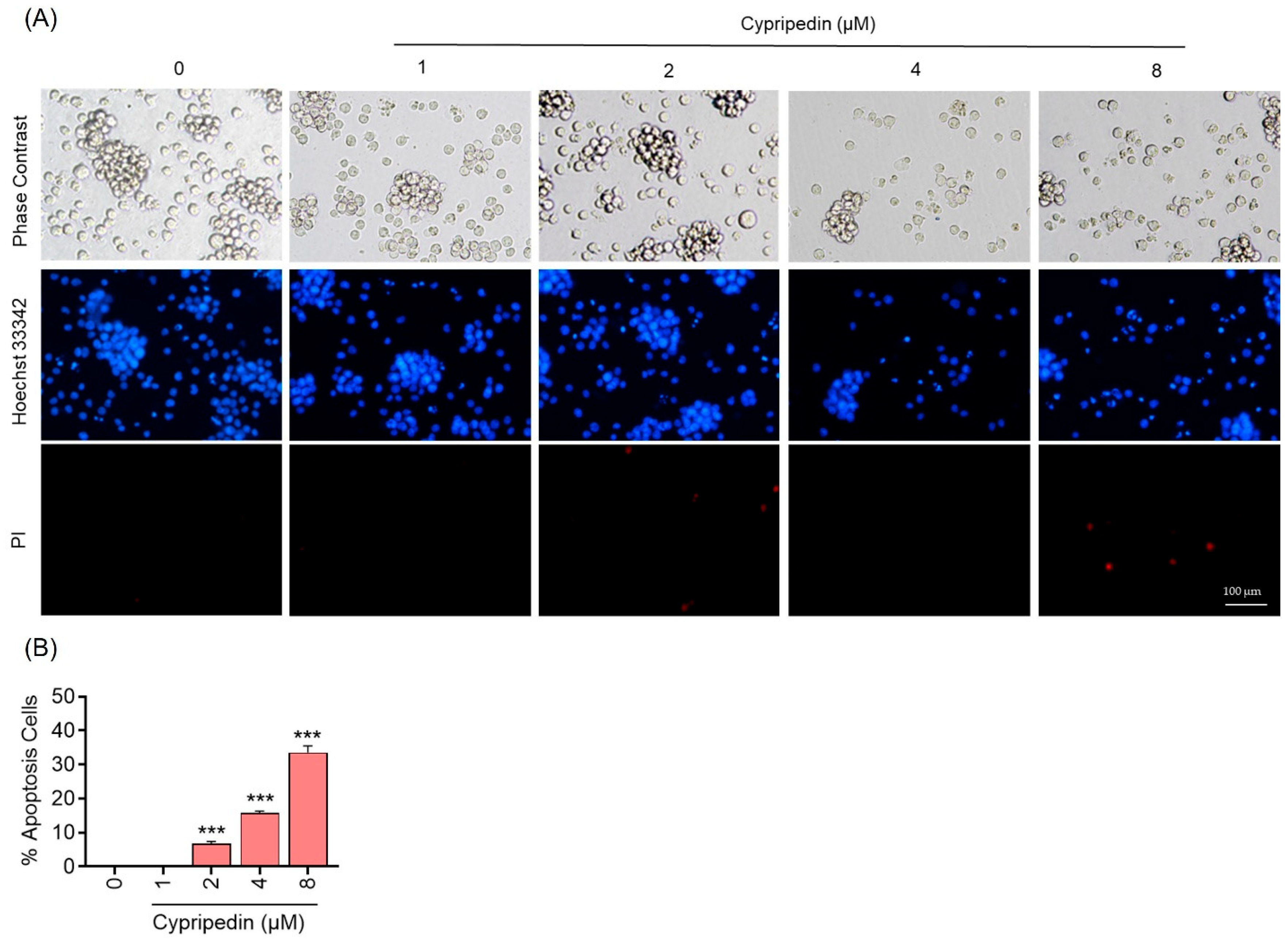
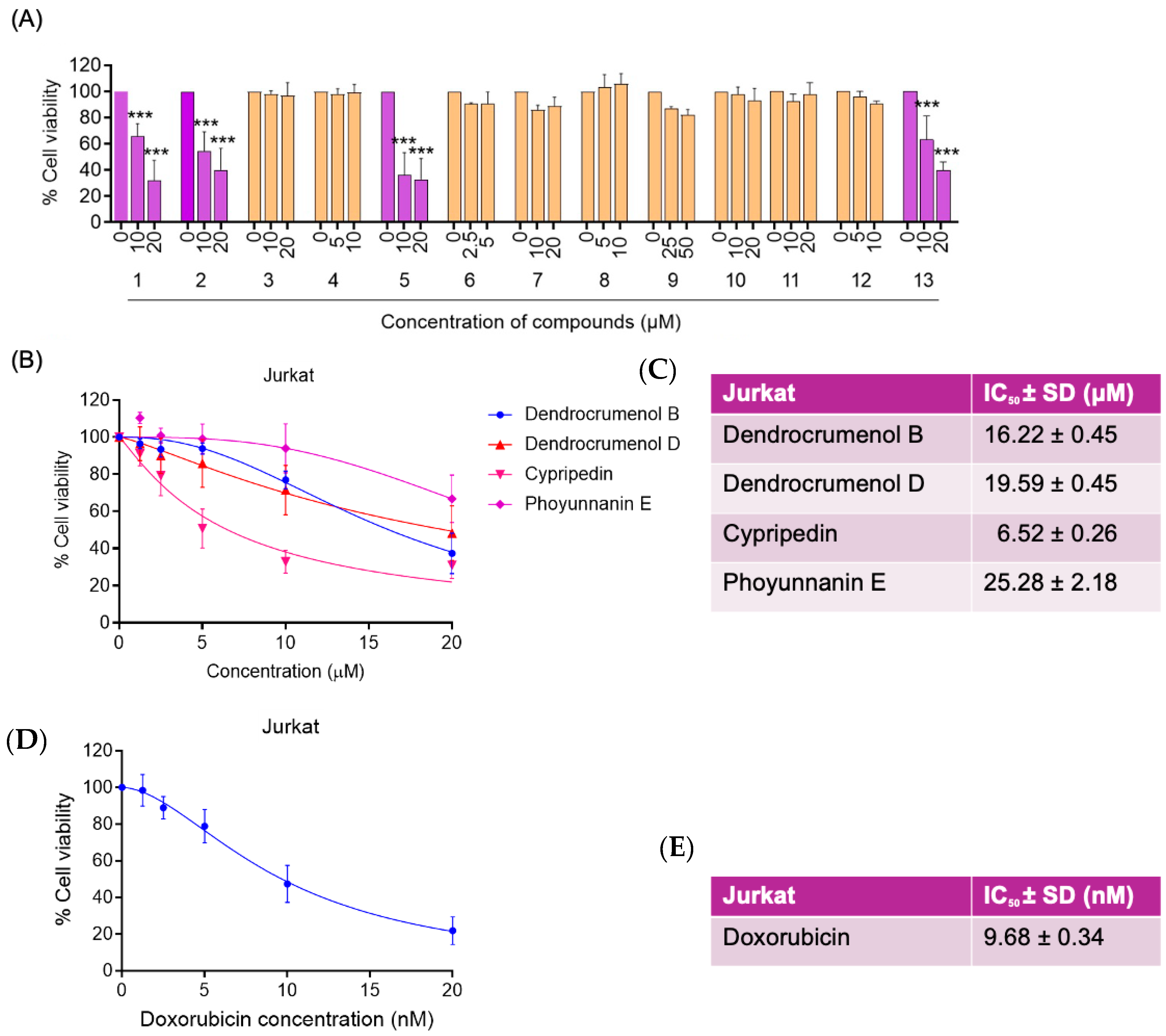
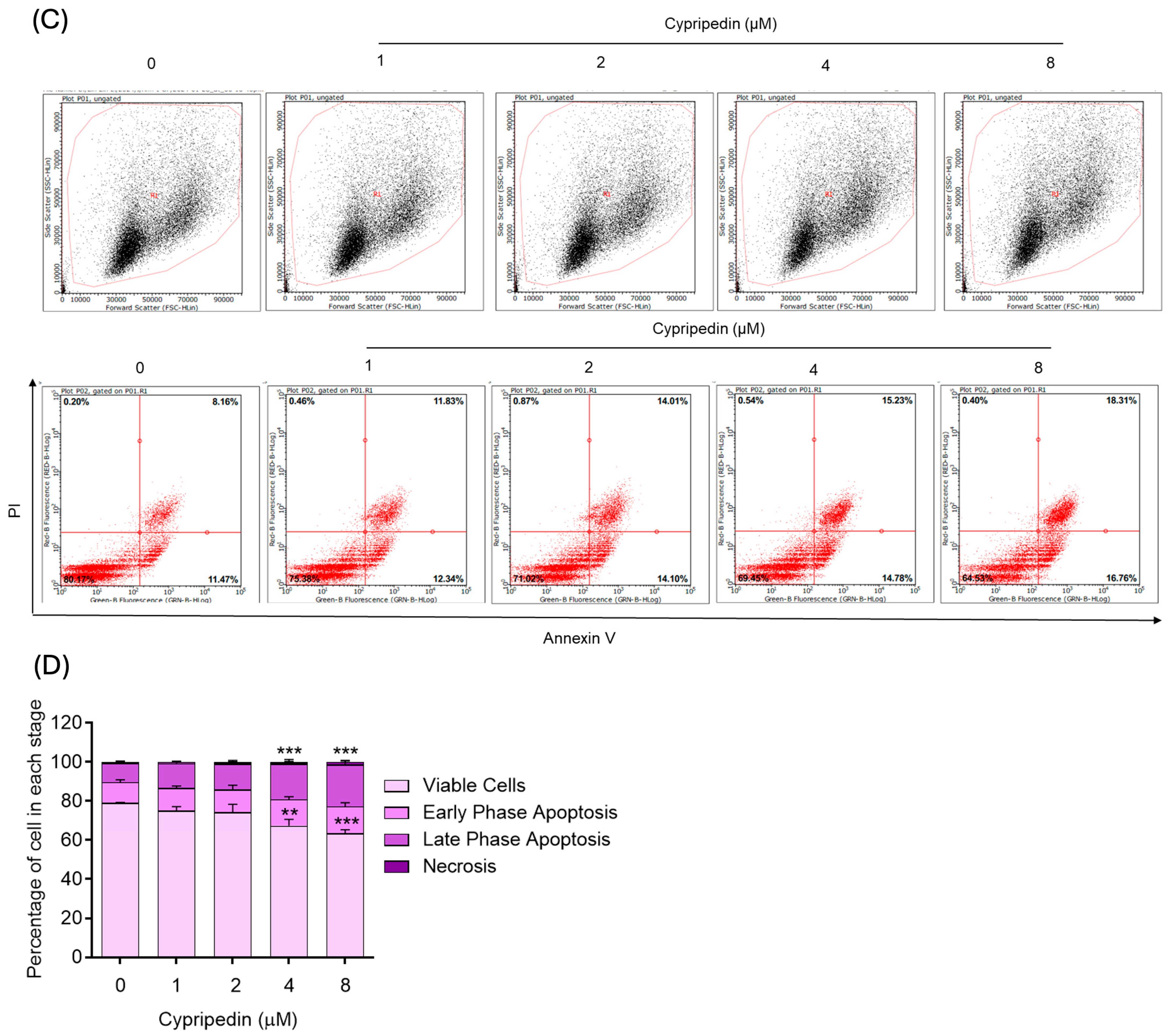
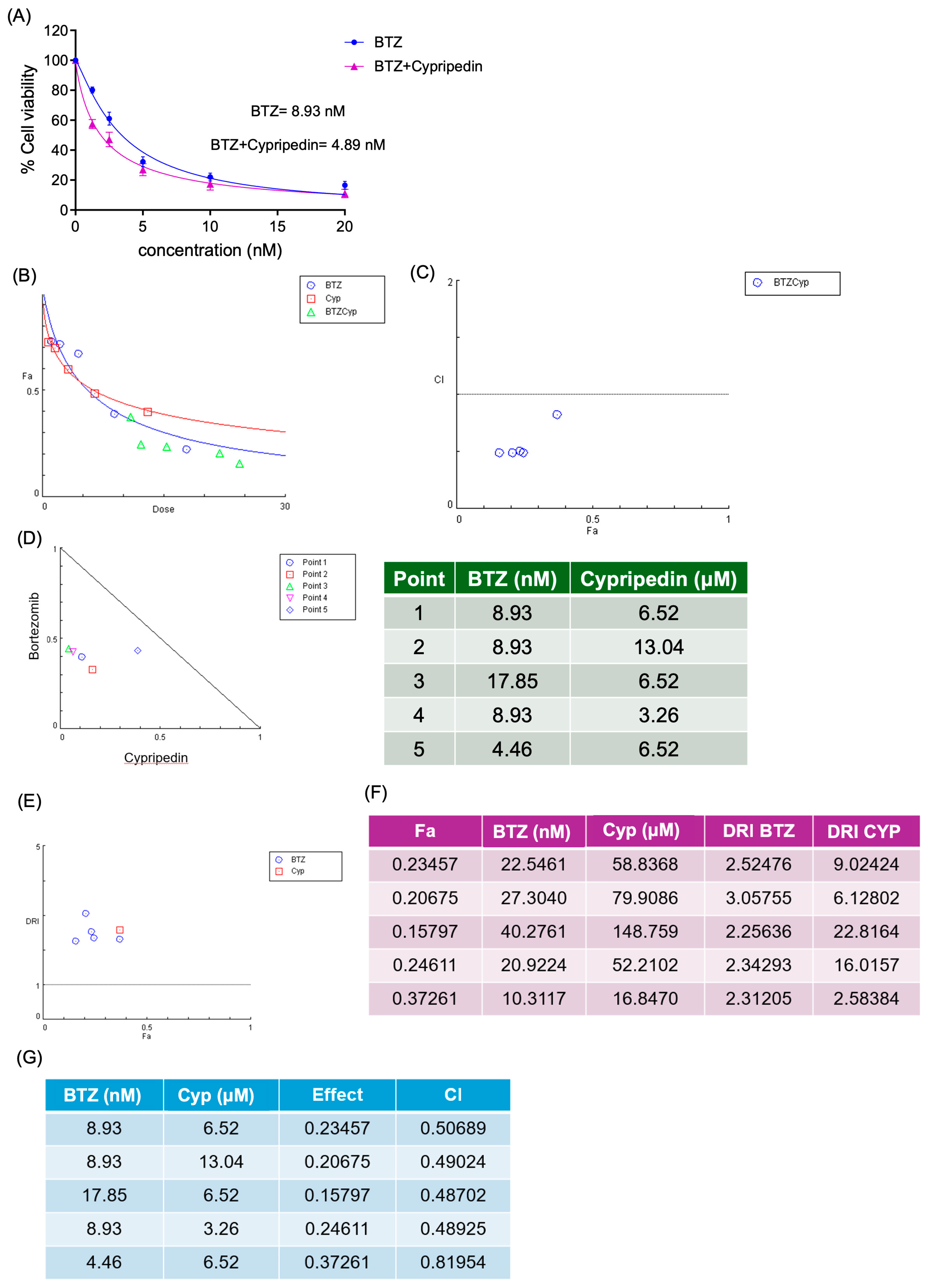
Here, we performed a medium-throughput screen of thirteen naturally derived phenanthrene-type compounds to evaluate their cytotoxic effects in the Jurkat T-ALL cell line. Cypripedin was identified as the most potent compound, exhibiting low-micromolar IC50 values. Its pro-apoptotic activity was subsequently characterized using complementary assays. We further evaluated the potential synergy between Cypripedin and the proteasome inhibitor bortezomib, which revealed strong synergism by Chou–Talalay analysis and a twofold reduction in the IC50 of bortezomib.
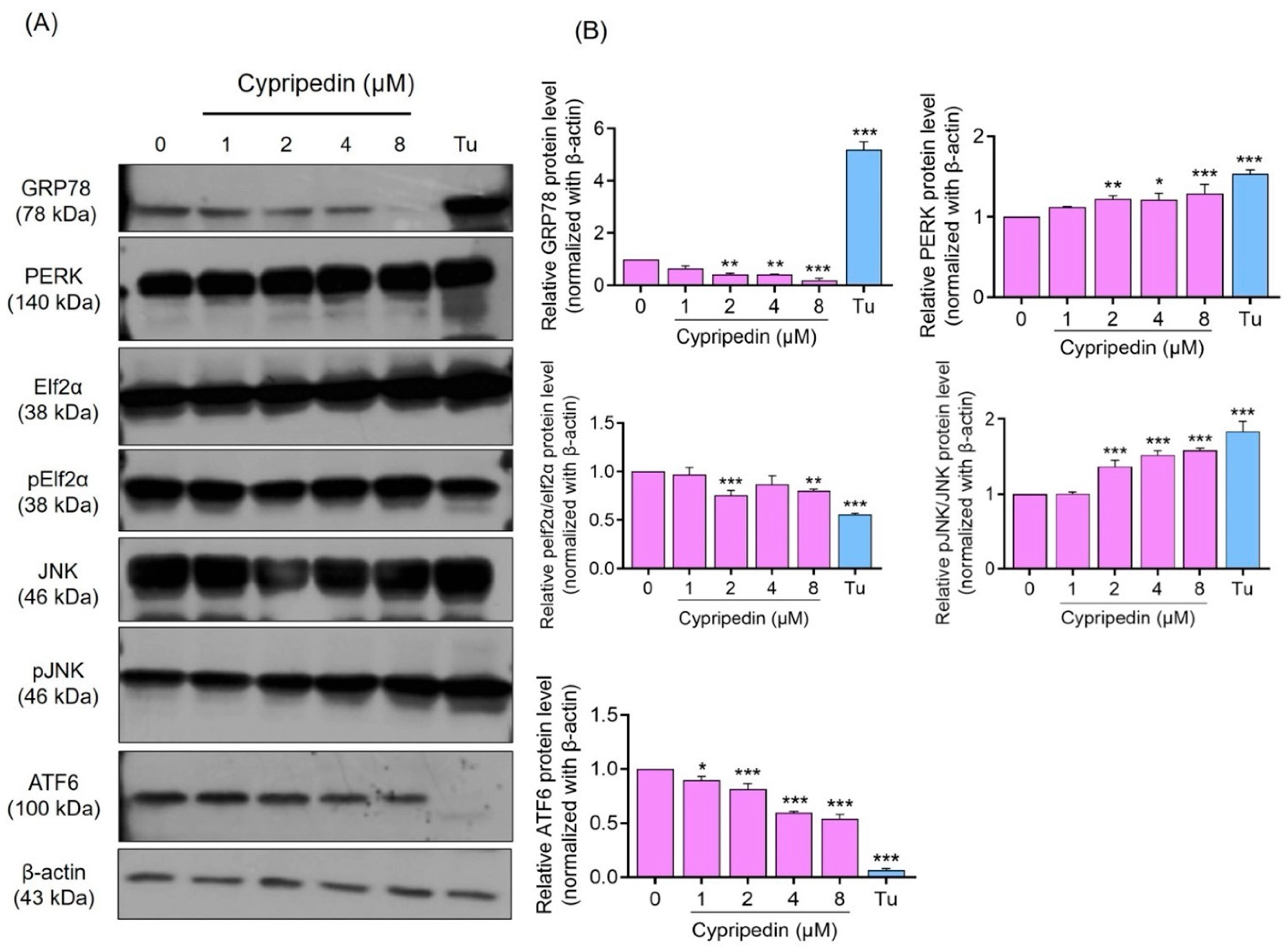
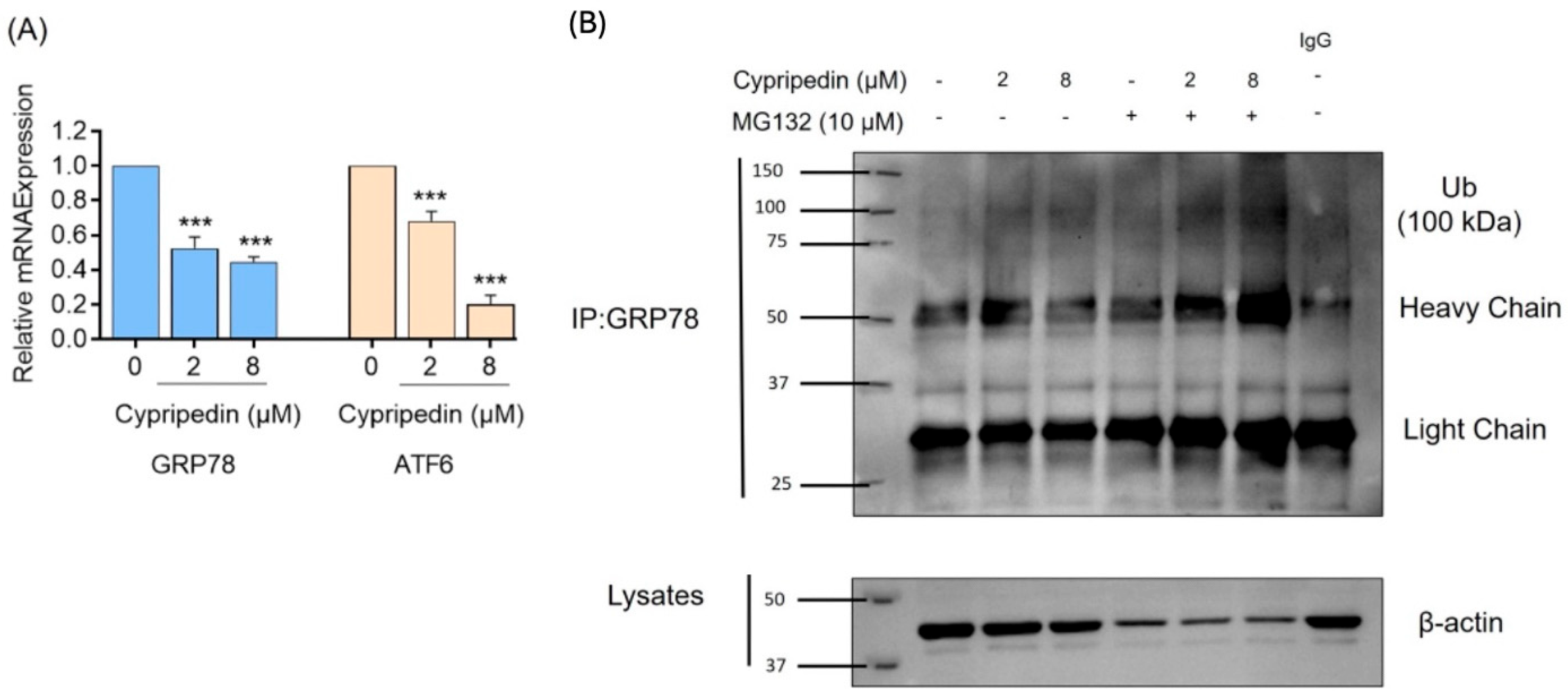
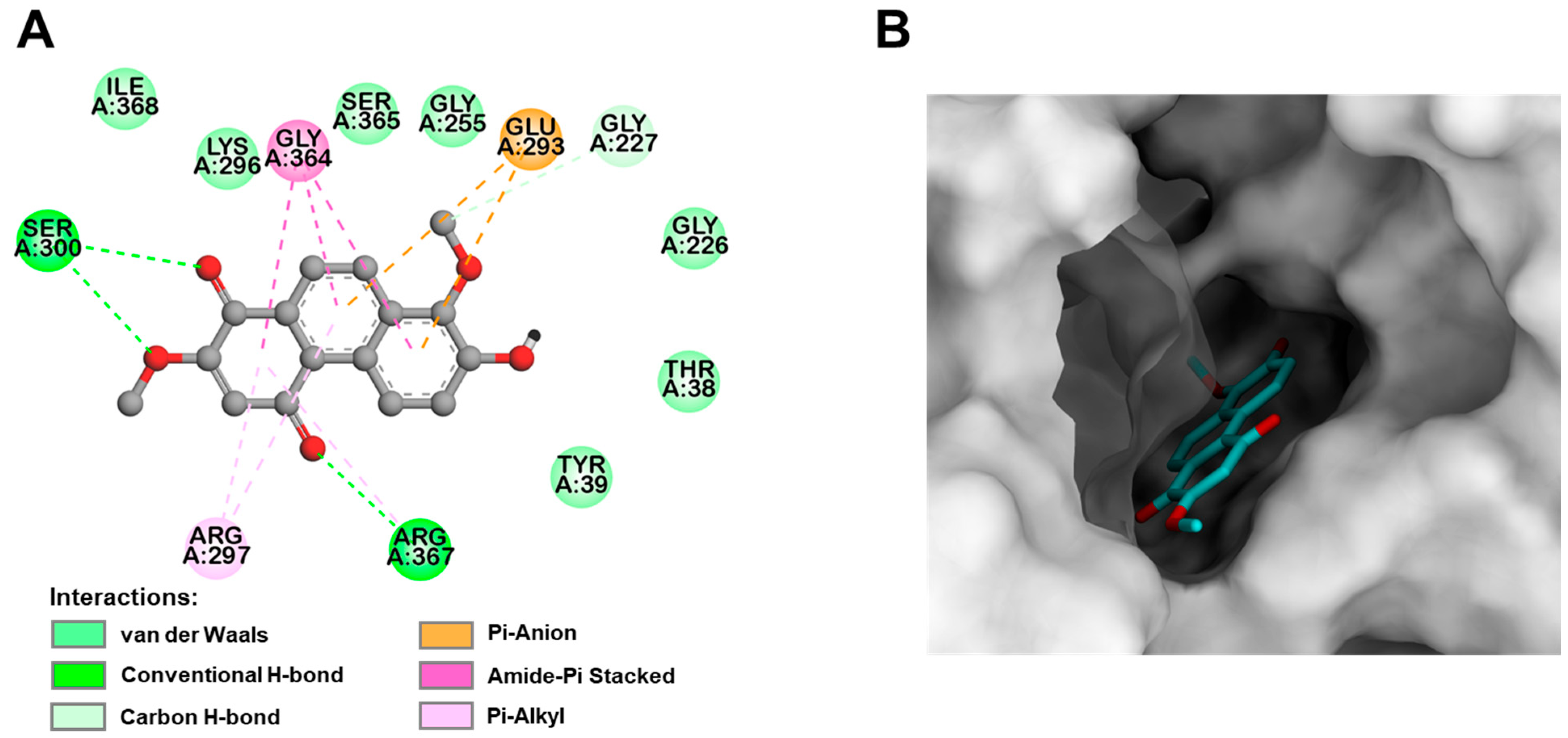
To elucidate the mechanistic basis of these effects, we examined ER stress activation through key unfolded protein response (UPR) markers, including GRP78, PERK/eIF2α, JNK, and ATF6. We also assessed the transcriptional regulation of UPR-related genes and demonstrated that Cypripedin is associated with increased ubiquitination of GRP78. Finally, in silico molecular docking supported these findings by predicting direct binding of Cypripedin to the ATPase cleft of GRP78. This integrated approach identifies Cypripedin as a novel natural compound that engages ER stress signaling to induce apoptosis and enhance the cytotoxic effects of proteasome inhibition in T-ALL cells.
2. Materials and Methods
2.1. Cell Culture and Reagents
Jurkat T-ALL cells (accession number CVCL_0367, Homo sapiens) were acquired from the American Type Culture Collection (ATCC, Manassas, VA, USA). These cells were cultured in RPMI 1640 medium (Cat no. 11875-093, Gibco, Grand Island, NY, USA), supplemented with 10% fetal bovine serum (FBS; Cat no. SV30160.03, HyClone™, Cytiva, Global Life Sciences, Vienna, Austria), 2 mM L-glutamine (Ref no. 35050-061, Gibco, Gaithersburg, MA, USA), and 100 U/mL of antibiotic–antimycotic (Ref no. 15240-062, Gibco, Grand Island, NY, USA). Cells were incubated at 37 °C in a humidified atmosphere containing 5% CO2.
All cell culture reagents, including RPMI 1640 medium, L-glutamine, FBS, antibiotic–antimycotic, and phosphate-buffered saline (PBS), were purchased from Gibco (Grand Island, NY, USA). Additional reagents used in the study included Cell Proliferation Reagent WST-1 (Cat no. 05 015 944 001), bovine serum albumin (BSA; Cat no. 9048-46-8), Tunicamycin (product no: 654380), Hoechst 33342 (Cat no. B2261), and propidium iodide (PI; Cat no. P4170), all obtained from Sigma-Aldrich (St. Louis, MO, USA). MG132 (cat no: 2194S) was purchased from cell signaling. Bortezomib (Cat no: sc-217785 was obtained from Santa Cruz Biotechnology, Inc., Dallas, TX, USA.)
Primary antibodies for ER stress-related proteins were purchased from Cell Signaling Technology (Beverly, MA, USA), including rabbit monoclonal antibodies against PERK (140 kDa, Cat no: 5683), Elf2α (38 kDa, Cat no: 9722S), phospho-Elf2α (38 kDa, Cat no: 3597S), JNK (46, 54 kDa, Cat no: 9252S), phospho-JNK (46, 54 kDa, Cat no: 9255S), and ATF-6 (D4Z8V) (100 kDa, Cat no: 65880). Additional antibodies, including GRP78 (78 kDa, Cat no: ab21685) and ubiquitin (Cat no: ab134953), were obtained from Abcam (Waltham, MA, USA).
2.2. Extraction and Structure Elucidation of Cypripedin
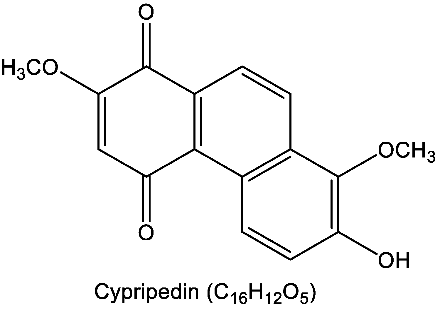
Cypripedin was extracted from Dendrobium densiflorum using methanol and subsequently purified via C-18 column chromatography with an H
2O-MeOH gradient (as described by Treesuwan S. et al. [
12]. Briefly, its structure was confirmed by NMR analysis (
Scheme 1), and its purity verified by both HPLC and NMR exceeded 95%, the material used in this study. Spectral data of cypripedin: The structure of cypripedin was elucidated using NMR analysis performed on a Bruker Avance DPX-300 FT-NMR (Billerica, MA, USA) spectrometer, and its mass spectrum was obtained with a Bruker microTOF mass spectrometer (ESI-MS, Billerica, MA, USA); C
16H
12O
5; HR-ESI-MS [M+Na]
+ at
m/z 307.0582 (calcd. for 307.0582, C
16H
12O
5Na);
1H NMR (300 MHz, acetone-
d6) δ: 9.27 (1H, d,
J = 9.6 Hz, H-5), 8.37 (1H, d,
J = 9.0 Hz, H-9), 8.09 (1H, d,
J = 9.0 Hz, H-10), 7.43 (1H, d,
J = 9.6 Hz, H-6), 6.20 (1H, s, H-3), 3.94 (3H, s, MeO-8), 3.93 (3H, s, MeO-2);
13C NMR (75 MHz, acetone-
d6) δ: 189.1 (C-4), 181.2 (C-1), 159.6 (C-2), 149.5 (C-7), 141.1 (C-8), 134.0 (C-8a), 130.0 (C-10a), 128.3 (C-4a), 127.2 (C-9), 126.2 (C-5), 125.7 (C-4b), 123.4 (C-6), 122.7 (C-10), 111.9 (C-3), 61.5 (MeO-8), 56.7 (MeO-2).
2.3. Preparation of Stock Solutions of Natural Products
Thirteen bioactive compounds were obtained from Assoc. Prof. Boonchoo Sritularak, which have been previously identified. The natural products were dissolved in dimethyl sulfoxide (DMSO) to prepare 50 mM stock solutions and subsequently stored at −20 °C. The DMSO concentration used in all experiments was kept below 0.1%, a level that exhibited no cytotoxic effects.
2.4. WST1 Cell Proliferation Assay
A cell viability assay was conducted to assess the cytotoxic effects of the natural products on Jurkat T-ALL cells. Jurkat cells were seeded at a density of 1 × 10⁵ cells per well (100 µL) in 96-well plates and incubated under 5% CO2 conditions. Cells were then treated with various concentrations of natural products for 24 h. Following treatment, 10 µL of WST-1 cell proliferation reagent was added to each well containing 100 µL of cell suspension. After a 2.5 h incubation with WST-1, absorbance was measured using a microplate reader. The absorbance of the formazan product was recorded within the peak absorption of 450 nm, and a reference wavelength above 600 nm was used. Relative cell viability for each treatment group was determined by comparing it to the untreated control group.
2.5. Nuclear Staining Assay
Apoptotic and necrotic cell death were assessed through dual staining with Hoechst 33,342 and propidium iodide (PI). Jurkat T-ALL cells were plated in 96-well plates at a density of 1 × 10⁵ cells per well (100 µL) and incubated at 37 °C. The cells were then exposed to different concentrations of Cypripedin (0–8 µM) for 24 h. Following treatment, the cells were co-stained with Hoechst 33342 (10 µg/mL) and PI (0.02 µg/mL) for 30 min at 37 °C. Apoptotic cells showing nuclear condensation and fragmentation were detected with Hoechst 33342, while necrotic cells were identified by PI-positive staining. Stained cells were observed and imaged using a fluorescence microscope (Olympus IX51 equipped with a DP70 digital camera, Olympus, Tokyo, Japan).
2.6. Apoptosis Assay by Annexin V Staining
Apoptosis at early and late stages, along with necrotic cell death, was evaluated using a fluorescein isothiocyanate (FITC)-Annexin V/propidium iodide (PI) apoptosis detection kit, following the manufacturer’s protocol (ImmunoTools, catalog no. 3149001, Friesoythe, Germany). Cells were incubated with Annexin V-FITC and PI in binding buffer for 15 min at RT. The stained cells were then analyzed by flow cytometry using the Benchtop Guava® easyCyte HT system operated with guavaSoft™ software (version 3.3, EMD Millipore, Billerica, MA, USA).
2.7. Drug Combination Analysis
Initially, the IC50 values of Cypripedin and Bortezomib were determined individually using the WST-1 cell proliferation assay. Following this, a combination treatment analysis was conducted to assess potential synergistic effects. Cypripedin and Bortezomib were administered both individually and in combination at a fixed ratio derived from their respective dose–response curves. The combination index (CI) was calculated using CompuSyn software (version 1.0; T.C. Chou and N. Martin, Memorial Sloan-Kettering Cancer Center, New York, NY, USA), and an isobologram was generated to quantitatively evaluate the nature of the drug interaction.
2.8. Western Blot Analysis
Jurkat T-ALL cells were seeded in 6-well plates at a density of 1 × 10⁶ cells per well (1 mL) and treated with Cypripedin at concentrations ranging from 0 to 8 µM for 24 h at 37 °C. After treatment, cells were collected and washed with ice-cold PBS. Lysis was performed using a buffer containing 50 mM HEPES (pH 7.5), 150 mM NaCl, 5 mM EDTA, 1% Triton X-100, 1 mM PMSF, and 2 µg/mL pepstatin A, supplemented with a complete protease inhibitor cocktail (EASYpack, Roche, cat no: 04693116001, Mannheim, Germany). The cells were lysed on ice for 40 min. Lysates were then centrifuged at 12,000× g for 15 min at 4 °C, and protein concentrations were measured using the BCA protein assay kit (Thermo-Fisher Scientific, Rockford, IL, USA).
Equal amounts of protein were mixed with loading buffer and denatured by heating at 95 °C for 5 min. Proteins were separated by SDS-PAGE and transferred onto 0.45 µm nitrocellulose membranes. Membranes were blocked with 5% non-fat milk in TBST (25 mM Tris-HCl, pH 7.5, 125 mM NaCl, 0.05% Tween-20) for 1 h at room temperature, then incubated overnight at 4 °C with primary antibodies diluted 1:1000 in 5% BSA in TBST. β-Actin was used as a loading control.
The following day, membranes were washed three times with TBST (5 min each) and then incubated with HRP-conjugated secondary antibodies (anti-rabbit or anti-mouse IgG, diluted 1:2000 in 5% skim milk in TBST) for 1 h at room temperature. After further washing with TBST, protein bands were visualized using a chemiluminescent substrate (SuperSignal West Pico, Pierce, Rockford, IL, USA) then subjected to the X-ray film exposure. The molecular weight were compared against the marker (Biotinylated Protein Ladder Detection PackTM, Cell Signaling Technology, cat no. 7727, Danvers, MA, USA). Signal intensity was measured by densitometry using ImageJ software (version 1.52a, National Institutes of Health, Bethesda, MD, USA).
2.9. Quantitative Analysis for Real-Time PCR Analysis
Total RNA was isolated from Jurkat T-ALL cells treated with Cypripedin (1 × 10⁶ cells per well in 6-well plates) using GENEzol reagent. The extracted RNA was then used for complementary DNA (cDNA) synthesis with SuperScript III reverse transcriptase (Invitrogen, Carlsbad, CA, USA). Reverse transcription quantitative PCR (RT-qPCR) was conducted using 100 ng of cDNA in a 20 µL reaction volume with Luna® Universal qPCR Master Mix (New England Biolabs, Ipswich, MA, USA). Amplification was performed on a CFX96 Real-Time PCR system (Bio-Rad, Hercules, CA, USA). The thermal cycling conditions included an initial denaturation at 95 °C for 1 min, followed by 45 cycles of denaturation at 95 °C for 15 s and annealing at 60 °C for 30 s. A melting curve analysis was performed at the end of the run to confirm primer specificity. The targeted gene of primers are:
GRP78 (Fwd) GTTCTTCAATGGCAAGGAACCATC Tm = 63.5 °C
GRP78 (Rev) CCATCCTTTCGATTTCTTCAGGTG Tm = 63.5 °C
ATF 6 (Fwd) GCCTTTATTGCTTCCAGCAG Tm = 54.5 °C
ATF 6 (Rev) TGAGACAGCAAAACCGTCTG Tm = 55.5 °C
GAPDH (Fwd) GCTCAGAACACCTATGGGGAA Tm = 59.8 °C
GAPDH (Rev) CATCGCCCCACTTGATTTGG Tm = 59.8 °C
The PCR products were normalized against GAPDH, which served as internal control. Relative mRNA expression levels of the target genes were calculated using the comparative Cq (ΔΔCq) method.
2.10. Immunoprecipitation
Jurkat T-ALL cells treated with Cypripedin were harvested using cold PBS and lysed on ice with a buffer containing 50 mM HEPES (pH 7.5), 150 mM NaCl, 5 mM EDTA, 1% Triton X-100, 1 mM PMSF, and 2 μg/mL pepstatin A (catalog no. 9803, Cell Signaling, Danvers, MA, USA), supplemented with a complete protease inhibitor cocktail (EASYpack, Roche, catalog no. 04693116001, Mannheim, Germany). The lysates were incubated on ice for 40 min and then centrifuged at 12,000× g for 15 min at 4 °C. Protein concentrations were determined using the BCA protein assay kit (Thermo Fisher Scientific, Rockford, IL, USA).
Immunoprecipitation was performed using the Dynabeads™ Protein G Immunoprecipitation Kit (Thermo Fisher Scientific, Waltham, MA, USA). Magnetic beads were incubated with 1.5 μL of GRP78 antibody in 100 μL of antibody binding and washing buffer for 15 min at room temperature with rotation. The bead–antibody complexes were resuspended in lysis buffer and incubated overnight at 4 °C to allow binding of the GRP78 antigen. The bead–antibody–antigen complexes were washed three times with 200 μL washing buffer, then resuspended in 30 μL lysis buffer mixed with 5 μL of 6× sample buffer and denatured at 95 °C for 5 min. The samples were loaded onto a 10% SDS-PAGE gel for protein separation.
Following electrophoresis, proteins were transferred to a 0.45 μm nitrocellulose membrane. The membrane was blocked in 5% nonfat dry milk prepared in TBST (25 mM Tris-HCl, pH 7.4; 125 mM NaCl; 0.05% Tween 20) for 1 h and 30 min. It was then incubated overnight at 4 °C with the primary antibody against ubiquitin. The next day, the membrane was incubated for 1 h at room temperature with HRP-conjugated secondary anti-mouse IgG antibody diluted 1:2000 in 5% skim milk in TBST. Protein bands were visualized using enhanced chemiluminescence and quantified by densitometry with the iBright™ CL 1500 Imaging System (Invitrogen™, cat no. A44240, Carlsbad, CA, USA) and the molecular weight were compared against the marker (Precision Plus Protein Dual Color StandardsTM, BioRad, cat no.1610374, Hercules, CA, USA). Band intensities were analyzed using ImageJ software (version 1.52a, National Institutes of Health, Bathesda, MD, USA).
2.11. Statistical Analysis
Results are presented as the mean ± standard deviation (SD) based on three or more independent biological replicates. Statistical analysis for multiple group comparisons was conducted using one-way ANOVA followed by a post hoc test with GraphPad Prism version 9.0 (GraphPad Software, La Jolla, CA, USA). Differences were considered statistically significant at p-values below 0.05.
Statistical Analysis—DepMap
Statistical comparisons between groups were performed using a two-sided Mann–Whitney U test implemented in the SciPy library. This non-parametric test was selected due to the non-normal and highly skewed distribution of dependency scores, which are bounded between 0 and 1 and exhibit a pronounced ceiling effect. A p-value < 0.05 was considered statistically significant.
2.12. Molecular Docking
The X-ray crystal structure of human GRP78 ATPase domain (PDB ID: 3LDP [
13]) was downloaded from the RCSB Protein Data Bank (
http://www.rcsb.org/). Prior to docking simulation, all solvent molecules and the co-crystallized ligand were removed to prepare the protein structure. The SMILES representations of the ligands were obtained from the PubChem database (
http://pubchem.ncbi.nlm.nih.gov/) and converted into 3D PDB format using the Online SMILES Translator and Structure File Generator (
http://cactus.nci.nih.gov/translate/, accessed on 15 March 2025). PDBQT file formats for GRP78 protein and the ligands were generated using AutoDockFR 1.0 [
14]. Molecular docking was carried out using AutoDock Vina version 1.2.7 [
15]. The docking grid was centered on the coordinates of the original co-crystallized ligand within the active site, using a grid box size of 20 × 20 × 20 Å and center coordinates x = 17.52, y = −9.15, and z = 5.35. The best docking pose with the lowest AutoDock Vina score of the GRP78–Cypripedin complex was selected for further analysis. 3D visualization was performed using ChimeraX 1.10 [
16], and 2D interaction diagram was analyzed using Discovery Studio Visualizer 2024 (BIOVIA, San Diego, CA, USA).
2.13. DepMap
2.13.1. DepMap Data Acquisition and Preprocessing
Genome-wide CRISPR gene dependency data were obtained from the Cancer Dependency Map (DepMap Public release 25Q3). Gene-level dependency probabilities were downloaded as the CRISPRGeneDependency.csv dataset. Cell line annotations, including lineage classification and standardized cell line names, were retrieved from the corresponding Model.csv file.
The dependency score for HSPA5 (GRP78) was extracted using the annotated gene identifier (“HSPA5 (3309)”). Cell line identifiers in the dependency matrix (ModelID) were matched to annotation data via an inner merge to obtain lineage information and standardized cell line names. Data preprocessing and integration were performed using Python (version 3.11.3; Clang 14.0.6) with the pandas library.
2.13.2. Lineage Grouping and Data Stratification
Cell lines were categorized based on the “OncotreeLineage” annotation. For comparative analysis, cell lines were grouped into lymphoid and non-lymphoid (“Other”) categories. The lymphoid group included hematopoietic malignancies of lymphoid origin, while all remaining lineages were classified as “Other”.
2.13.3. Data Visualization
Dependency distributions were visualized using boxplots with overlaid individual data points (strip plots) generated using the seaborn and matplotlib libraries. Boxplots represent the interquartile range (IQR), with median values indicated by central lines and whiskers extending to 1.5 × IQR. Individual cell lines are displayed as semi-transparent points to illustrate data distribution and outliers.
4. Discussion
Our study identifies Cypripedin as a potent inducer of apoptosis in Jurkat T-ALL cells, demonstrating an IC
50 of 6.52 µM and robust activation of intrinsic apoptotic pathways. The compound’s ability to synergize with bortezomib—evidenced by combination index values below 0.5 and 1.8-fold reduction in the proteasome inhibitor IC
50—suggests a promising strategy to enhance therapeutic efficacy while potentially lowering the required dose and mitigating systemic toxicity. Apoptosis induction by Cypripedin was confirmed through complementary assays. Hoechst 33342/PI staining provided morphological evidence of chromatin condensation, while Annexin V/PI flow cytometry quantified both early and late apoptotic populations. These multimodal data validate Cypripedin’s pro-apoptotic activity and are consistent with prior reports of phenanthrene derivatives inducing cytotoxicity via mitochondrial pathways in solid tumor models [
17].
Mechanistically, Western blot analysis revealed activation of multiple branches of the unfolded protein response (UPR), including the alteration of GRP78, phosphorylated PERK and eIF2α, ATF6 and upregulation of p-JNK. This broad engagement of ER stress signaling distinguishes Cypripedin from many natural products, which typically target individual UPR nodes. Notably, at higher concentrations, Cypripedin selectively downregulated GRP78 and ATF6 transcripts, suggesting a shift from adaptive to terminal UPR—a transition aligned with ER stress-mediated apoptosis mechanisms described by Hetz [
18].
A key novel finding of this study is the promotion of GRP78 ubiquitination by Cypripedin, demonstrated by immunoprecipitation assays and enhanced by proteasome blockade with MG132. GRP78, a central chaperone that maintains ER protein-folding homeostasis, is frequently upregulated in hematologic malignancies and has been implicated in chemoresistance [
19]. By promoting GRP78 turnover, Cypripedin compromises the cell’s ability to manage ER stress, thereby sensitizing cells to proteasome inhibition. Molecular docking analysis supports this proposed mechanism, predicting that Cypripedin binds within the ATPase cleft of GRP78, forming hydrogen bonds with Ser300 and Arg367, and engaging surrounding hydrophobic residues. This interaction is likely to destabilize the chaperone’s conformation, potentially exposing lysine residues for recognition by E3 ubiquitin ligases and facilitating ubiquitination. Future studies should include site-directed mutagenesis of these contact residues, along with biophysical assays such as thermal shift and surface plasmon resonance, to validate direct binding.
Comparatively, other group have reported on natural products that modulate ER stress—for example, 7,8-dihydroxyflavone suppressing GRP78 in metabolic disease models and icariin inhibiting the IRE1α–XBP1 pathway in neurons [
20,
21]. However, few studies have demonstrated both induction of ER stress and targeted degradation of a key UPR chaperone. Cypripedin’s dual functionality—activating stress responses while promoting chaperone depletion—introduces a novel paradigm in natural product pharmacology. Therapeutically, the observed synergy with bortezomib highlights the potential for combination regimens in T-ALL and possibly other malignancies that rely on proteostasis. The ability to lower effective doses of proteasome inhibitors may reduce associated toxicities, such as peripheral neuropathy and myelosuppression, which currently limit clinical use. Furthermore, targeting GRP78 may help overcome resistance mechanisms not addressed by existing therapies. While our in vitro data are comprehensive, in vivo validation is critical. Studies on pharmacokinetics, bioavailability, and toxicity are required to assess translational potential. Additionally, identifying the specific E3 ligases responsible for GRP78 ubiquitination could provide mechanistic insight and identify new therapeutic targets. Structure–activity relationship (SAR) studies of Cypripedin and its analogs may further enhance its binding affinity and selectivity.
In conclusion, Cypripedin is a multifaceted natural compound that induces ER stress-mediated apoptosis and potentiates the cytotoxic effects of proteasome inhibition, with associated GRP78 ubiquitination. This integrated mechanism of action represents a novel therapeutic avenue for T-ALL and other proteostasis-dependent cancers, meriting further preclinical investigation, particularly in animal models.




 ,
,