Review

PKM2-Mediated Glycolytic Reprogramming in Thyroid Cancer: Mechanistic Insights and Therapeutic Potential


1
Department of Surgery (Thyroid & Breast), Zhongshan Hospital, Fudan University, Shanghai 200030, China
2
School of Basic Medical Sciences, Fudan University, Shanghai 200433, China
*
Authors to whom correspondence should be addressed.
Molecules 2026, 31(11), 1811; https://doi.org/10.3390/molecules31111811
Submission received: 6 May 2026 / Revised: 18 May 2026 / Accepted: 19 May 2026 / Published: 25 May 2026
(This article belongs to the Section Medicinal Chemistry)

Abstract

Thyroid cancer (TC) is an endocrine malignant tumor with the fastest-growing incidence worldwide. It has complex pathological types and significant heterogeneity, with great differences in clinical prognosis among different subtypes. Among them, aggressive subtypes, such as radioiodine-refractory (RAI-R) TC and anaplastic thyroid cancer (ATC), have become a major challenge in current clinical diagnosis and treatment, due to limited treatment options and high risks of recurrence and metastasis. Tumor metabolic reprogramming is one of the characteristics of cancer, among which the Warburg effect plays a driving role. As a rate-limiting enzyme in the glycolytic pathway, pyruvate kinase M2 (PKM2), with its unique functional plasticity, has become a linchpin of glycolytic metabolism and malignant phenotypes of tumor cells. This article will systematically review the functional regulatory mechanisms of PKM2, its specific role in TC, and explore the targeted therapeutic strategies and research prospects of TC with PKM2, providing a new theoretical basis and potential plans for the clinical diagnosis and treatment.

1. Introduction

Thyroid cancer (TC), the most common endocrine malignancy, continues to exhibit a rising global incidence, posing a significant public health challenge [1,2]. Globally, the incidence of TC is showing a continuous upward trend. According to the statistics from the International Agency for Research on Cancer (IARC) of the World Health Organization, there were 586,000 new cases of TC and about 44,000 deaths worldwide in 2020, and the incidence rate in women was significantly higher than that in men, with a male-to-female incidence ratio of about 1:3, and the age of onset was largely in young and middle-aged groups [3,4,5]. In China, the incidence of TC is also rising rapidly. Data released by the National Cancer Center of China in 2022 showed that TC has ranked among the top three malignant tumors in Chinese women, and the incidence is showing a clear younger trend, posing a severe challenge to public health [6].
According to histological phenotypes and differentiation degree, thyroid cancer is mainly divided into four categories: differentiated thyroid cancer (DTC), anaplastic thyroid cancer (ATC), medullary thyroid cancer (MTC), and other rare subtypes [7]. Among them, DTC accounts for the highest proportion, approximately 90% of all TC cases, and is further divided into papillary thyroid cancer (PTC) and follicular thyroid cancer (FTC). Such tumors have low malignant degree and good prognosis after early surgery combined with radioactive iodine (RAI) therapy, but some patients still experience recurrence, metastasis, or radioiodine resistance [8]. Although ATC accounts for less than 5% of cases, it has an extremely high malignant degree, strong invasiveness, poor sensitivity to radiotherapy and chemotherapy, and the median survival time is usually less than 1 year [9]. MTC originates from parafollicular C cells of the thyroid gland, and its occurrence is closely related to RET proto-oncogene mutations, with both hereditary and sporadic hallmarks, and its prognosis is between DTC and ATC [6,10].
Standard treatment approaches for DTC include surgery, thyroid hormone suppression therapy, and RAI therapy [11,12]. However, approximately 5% to 15% of DTC patients develop RAI-refractory (RAI-R) disease, wherein tumor cells lose the capacity to uptake iodine, rendering RAI therapy ineffective [13]. One study reported that up to 60% of patients with aggressive metastatic DTC develop resistance to RAI treatment [14]. The emergence of RAI resistance significantly worsens patient prognosis and elevates the risk of distant metastasis [15]. For RAI-R DTC patients, treatment options become markedly of disease recurrence and limited. Multi-kinase inhibitors (MKIs), such as sorafenib and lenvatinib, have demonstrably improved progression-free survival in these patients, yet their objective response rates (ORR) typically range from 12% to 27%, often accompanied by significant toxicities that impair quality of life [16,17].
The occurrence and development of TC is a complex process involving multiple gene mutations and aberrant activation of signaling pathways. The identification of driving genes provides an important basis for understanding their pathogenesis and precise treatment. The BRAF V600E mutation is the most common gene mutation in TC, with an incidence rate of 40–66% in PTC and about 24% in ATC [18,19]. This mutation can make the BRAF protein continuously activated, independent of upstream RAS signals, break the balance of cell growth by phosphorylating the downstream MEK-ERK pathway, and promote metabolic reprogramming, which is closely related to tumor TNM stage, lymph node metastasis, and poor prognosis [20,21]. RAS gene mutations (including NRAS, HRAS, and KRAS) are more common in FTC, MTC, and some PTC, with an incidence rate of about 10–20%, regulating tumor cell growth and survival by activating PI3K-AKT and MAPK pathways [22]. Additionally, RET/PTC rearrangement, TERT promoter mutation, P53 mutation, etc., are also cornerstones of different subtypes of TC. Among them, the TERT promoter mutation is more common in advanced DTC and ATC, indicating increased tumor invasiveness and poor prognosis [23].

2. Metabolic Reprogramming, Warburg Effect and PKM2

Tumor metabolic reprogramming is widely recognized as one of the central characteristics of malignant tumors, among which the Warburg effect (aerobic glycolysis) is the most typical manifestation [24]. This phenomenon describes how tumor cells preferentially choose the glycolytic pathway to convert glucose into lactic acid even under aerobic conditions, rather than performing efficient energy metabolism through oxidative phosphorylation (OXPHOS). This phenomenon is significantly different from the metabolic mode of normal cells [25]. The Warburg effect is of great significance for the malignant proliferation and microenvironmental adaptation of tumor cells. Its value lies not only in rapidly providing energy for tumor cells but also, more importantly, in shunting glucose metabolic intermediates (such as triose phosphate, pyruvate, ribose phosphate, etc.) to biosynthetic pathways such as nucleotides, amino acids, and lipids through regulating glycolytic flux, providing sufficient material basis for the rapid proliferation of tumor cells [24,26,27]. At the same time, the Warburg effect helps tumor cells adapt to harsh tumor microenvironments (TME) such as hypoxia and nutrient deficiency by maintaining intracellular reactive oxygen species (ROS) homeostasis and regulating fundamental signaling pathways [28].
Pyruvate kinase (PK) is a principal terminal enzyme in the glycolytic pathway, which can catalyze the conversion of phosphoenolpyruvate (PEP) to pyruvate and generate ATP. Its activity directly regulates the rate and direction of glycolytic flux [29]. According to the specificity of gene coding and tissue distribution, the pyruvate kinase family is mainly divided into four subtypes: liver-type (PKL), red blood cell-type (PKR), muscle-type 1 (PKM1), and muscle-type 2 (PKM2) [30]. Among them, PKL is chiefly expressed in tissues such as the liver, kidneys, and small intestine, participating in the regulation of glucose metabolism homeostasis; PKR is specifically expressed in red blood cells and renal medulla, ensuring the energy supply of red blood cells; both PKM1 and PKM2 are encoded by the PKM gene and produced through alternative splicing, and their tissue distribution and functional characteristics have significant differences [31].
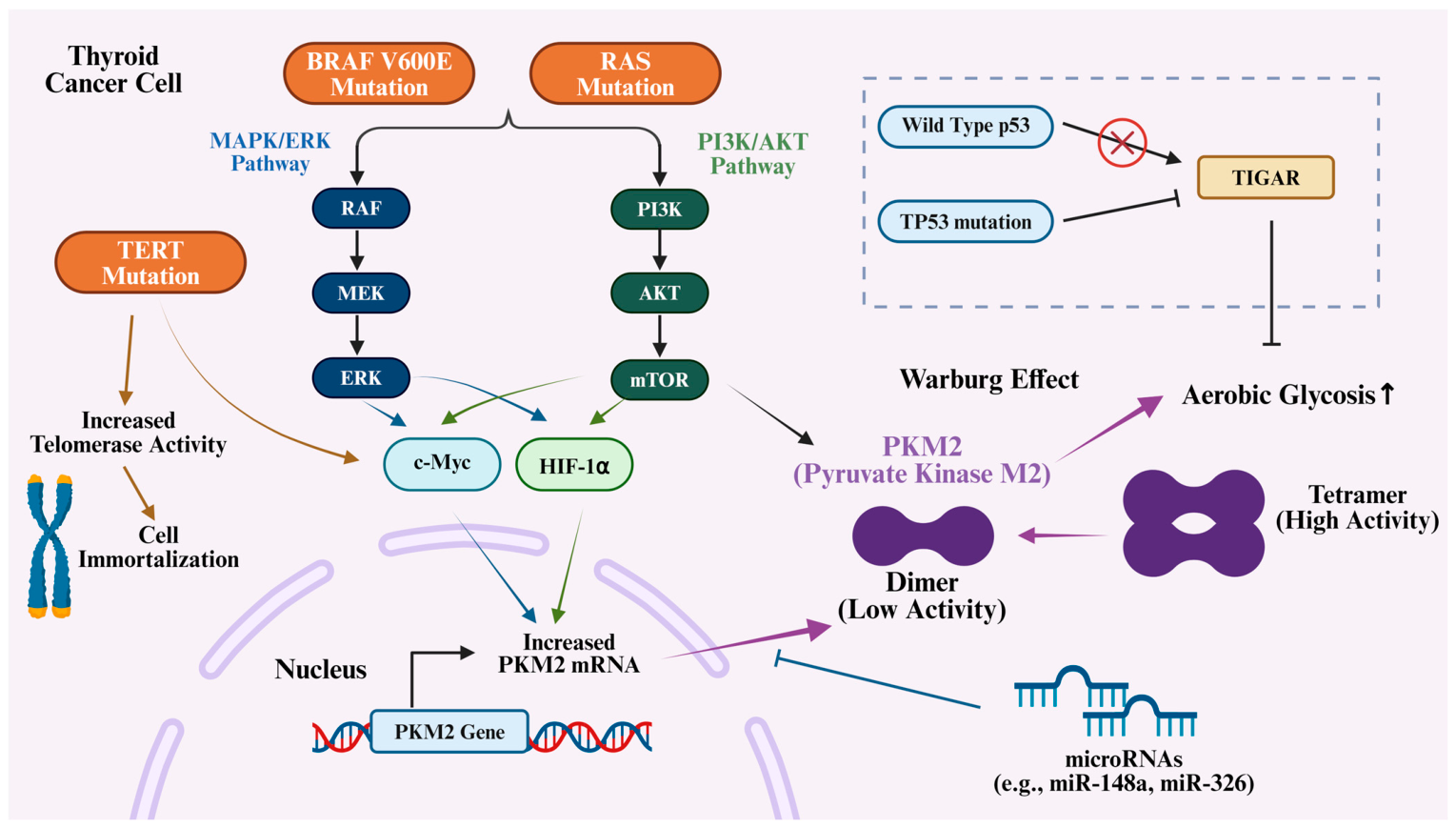
PKM1 is mostly found in metabolically active normal tissues such as myocardium, skeletal muscle, and brain tissue. Because it contains exon 9 of the PKM gene, it can stably exist in the form of a high-activity tetramer, with strong enzyme activity and is not easily regulated by external signals, which can efficiently catalyze glycolytic reactions and ensure cell energy supply. In contrast, PKM2 contains exon 10 of the PKM gene, which is mostly enriched in embryonic tissues, stem cells, and malignant tumor cells, with high structural and functional plasticity, and can dynamically switch between tetramer and dimer forms: the tetramer form of PKM2 has high enzyme activity, promoting the complete progress of glycolysis; the dimer form has significantly reduced enzyme activity, which can lead to the accumulation of glycolytic intermediates and provide raw materials for biosynthetic pathways [32,33,34]. As shown in Figure 1, cancer cells preferentially undergo aerobic glycolysis through metabolic reprogramming, particularly the Warburg effect, to support their rapid proliferation. This process involves the dimer form of PKM2, which features low enzyme activity compared with the tetramer form of PKM1. Therefore, PKM2 plays a universal and critical role in cancer metabolic reprogramming. As a rate-limiting enzyme in the glycolytic pathway, PKM2 promotes aerobic glycolysis and biosynthetic processes by modulating metabolic flux in tumor cells to meet the demands of rapid proliferation [35,36,37].

3. Regulation and Modification of PKM2

In TC cells, BRAF V600E mutation can induce PKM2 expression by activating the downstream MAPK signaling pathway [38,39]. Even under normoxic conditions, BRAF V600E-mediated MAPK overactivation can upregulate PKM2 expression through the stabilization of c-Myc and HIF-1α [40,41]. This upregulation promotes PKM2 to form its low-activity dimeric form, thereby enhancing aerobic glycolysis, which provides biosynthetic precursors for rapid tumor growth [33]. Besides the MAPK/ERK signaling pathway, the BRAF V600E mutation targets the PI3K pathway as well [42]. This is also a possible axis that connects BRAF V600E mutation and PKM2, because a previous study shows AKT kinase, as a key component of the PI3K/Akt signaling pathway, can phosphorylate the Ser37 site of PKM2, thereby affecting the cellular localization and function of PKM2 [43].
RAS mutations are a common driving factor for thyroid follicular carcinoma [44]. Its carcinogenic core lies in the continuous activation of downstream MAPK and PI3K-AKT signaling pathways; therefore, it has similarities with BRAF V600E in regulating PKM2 [45]. The incidence of TERT promoter mutations is increased in poorly differentiated cancer, undifferentiated cancer, and invasive papillary carcinoma, and is closely related to poor prognosis [46]. Its direct function is to enhance telomerase activity, maintain telomere length, and promote cell immortality [47]. Although not directly encoding signal proteins, TERT mutations often coexist with BRAF or RAS mutations, and this synergistic effect greatly enhances the invasiveness of tumors [48]. From a metabolic perspective, upregulation of telomerase activity is associated with cellular metabolic reprogramming. There are studies suggesting that TERT may affect the activity of transcription factors, such as c-Myc in a non-telomerase-dependent manner, and c-Myc is a key transcriptional regulator of PKM2 expression [49,50].
P53 mutations are extremely common in progressive TC, especially ATC [51]. The wild-type p53 protein is an important negative regulator of cellular metabolism, which can inhibit glycolysis and promote OXPHOS [52]. p53 can inhibit glycolysis flux by inducing the expression of TIGAR (p53-induced glycolysis and apoptosis regulatory factor). When p53 mutates and becomes inactive, this braking effect on glycolysis is released, which may lead to a general upregulation of glycolytic enzymes, including PKM2, accelerating the Warburg effect [53].
Beyond genetic mutations, epigenetic regulation via miRNAs adds another layer of control over PKM2 expression. Those pathways uniquely modify PKM2 in TC are summarized in Figure 2. Other PKM2 regulation mechanisms are listed in Table 1, and these provide potential research directions for future experimental validation.
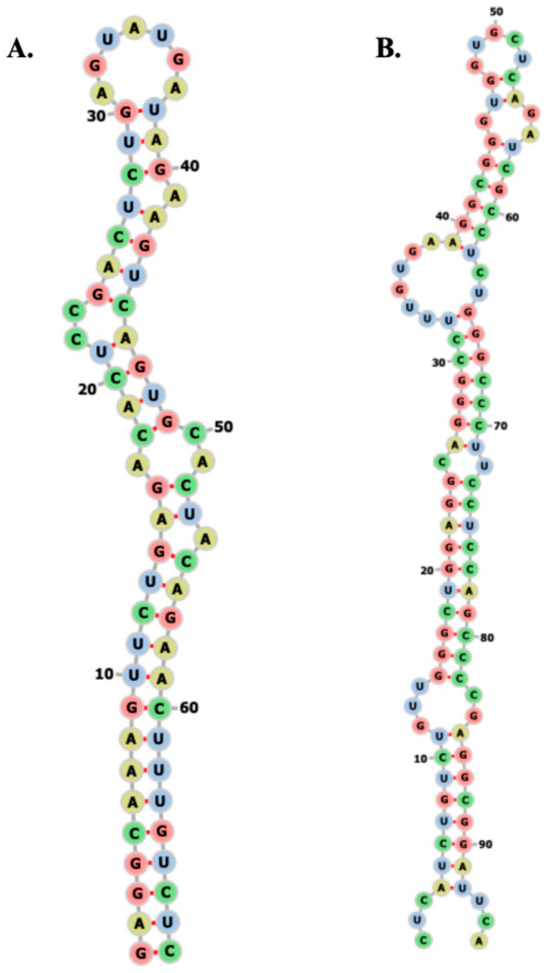
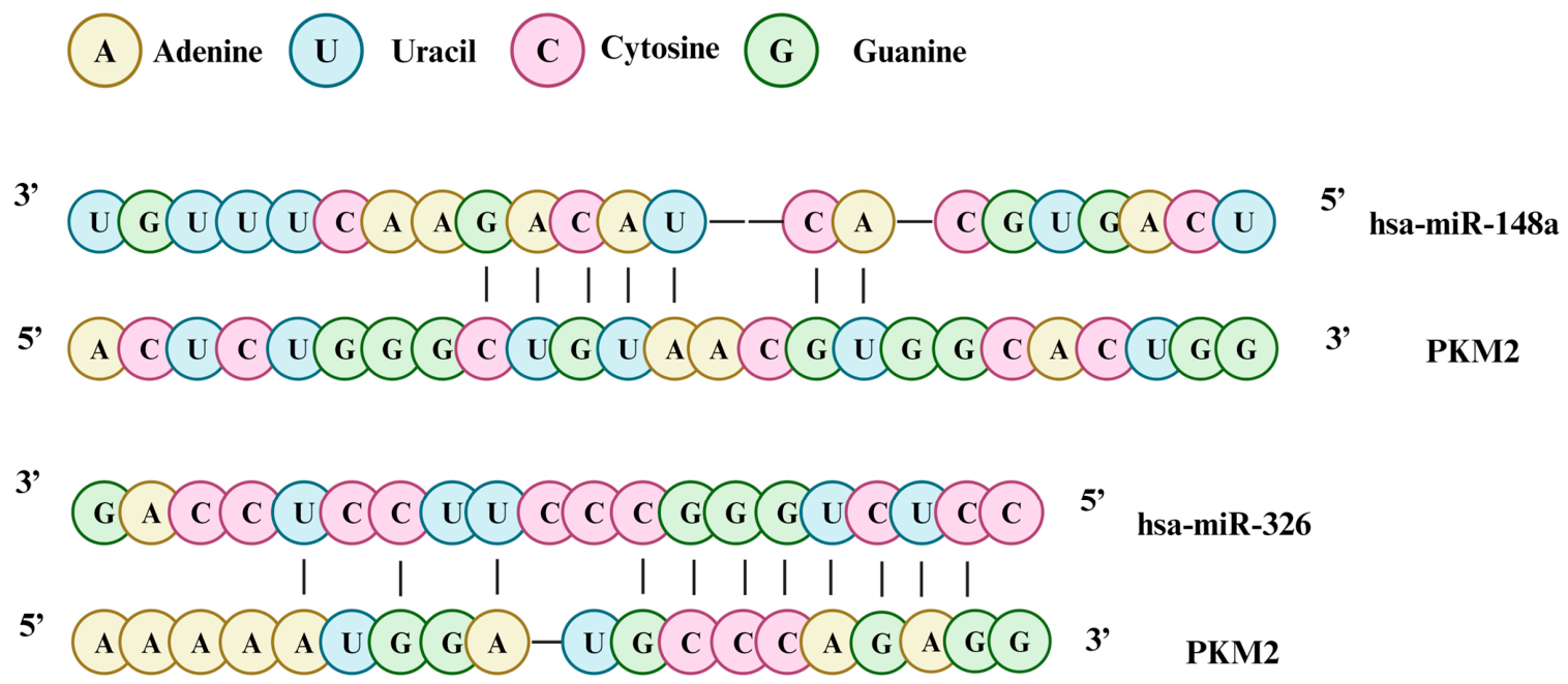
As a class of non-coding small RNAs, miRNAs can induce transcriptional or translational inhibition by binding to the 3′ untranslated region (3′UTR) of target gene mRNAs, participating in the post-transcriptional regulation of PKM2 and enriching the regulatory dimensions of PKM2 [80,81]. As a tumor-suppressive miRNA, miR-122 is downregulated in TC. It can directly target the 3′UTR of PKM2 to inhibit PKM2 expression, thereby reversing the Warburg effect and inhibiting tumor cell proliferation [55]; while miR-326 synergistically inhibits the invasion and metastasis of thyroid cancer cells by targeting PKM2 and downstream signaling pathway molecules. In addition, miR-148a, miR-326, etc., have also been confirmed to regulate PKM2 expression, and anomalous expression of these miRNAs in TC further disrupts the expression balance of PKM2 and promotes TC progression [54]. Figure 3 demonstrates the secondary structures of pre-miR-148a and pre-miR-326. They are obtained from miRBase (http://www.mirbase.org) (accessed on 8th April, 2026) and visualized using RNAfold. Figure 4 shows predicted putative miR-148a and miR-326-binding sites in the 3′UTR of PKM2.
Post-translational modification (PTM) is the central way to regulate PKM2 enzyme activity, subcellular localization, and protein–protein interactions, mainly including phosphorylation, acetylation, oxidation, and other modification types. Different modification methods can dynamically regulate its function by changing the spatial conformation of PKM2. These modifications are abnormally activated in TC, further enhancing the pro-cancer function of PKM2 [82,83].
Phosphorylation modification is the most common PTM method of PKM2. A variety of kinases can regulate its enzyme activity, subcellular localization, and non-enzymatic functions by phosphorylating different amino acid sites of PKM2, among which FGFR1, AKT, and ERK are focal regulatory kinases [58]. FGFR1 can specifically phosphorylate the Tyr105 site of PKM2, inducing PKM2 to dissociate from the high-activity tetramer into the low-activity dimer, inhibiting its enzyme activity, and promoting the nuclear translocation of the dimer form of PKM2 to exert the function of a transcriptional coactivator. The activation of the FGFR1-PKM2 signaling axis in TC can synergize with the BRAF V600E mutation to promote tumor metabolic reprogramming and invasion and metastasis [59].
AKT can enhance the interaction between PKM2 and HIF-1α by phosphorylating the Ser37 site of PKM2, promote PKM2 nuclear translocation, and inhibit its enzyme activity, enhancing the Warburg effect [60], while ERK, as a pivotal downstream kinase of the MAPK pathway, can phosphorylate the Thr365 site of PKM2, regulate its binding ability to β-catenin, and promote the expression of proliferation-related genes [58]. In TC, BRAF V600E and RAS mutations can activate AKT and ERK kinases, leading to dysregulated phosphorylation modification of PKM2, forming a “driver gene-kinase-PKM2” regulatory pathway, which synergistically promotes tumor progression [57]. Phosphorylation modifications at different sites can precisely regulate the functional state of PKM2 through synergistic or antagonistic effects, providing multi-dimensional targets for the targeted therapy of TC.
Tripartite motif-containing protein 35 (TRIM35) is a member of the RBCC family, which has a highly conserved sequence: a RING domain followed by one or two B-Box domains, and then a coiled-coil domain [84]. In hepatocellular carcinoma, the coiled-coil domain mediates the reduction in the Warburg effect and cancer cell proliferation. In addition, TRIM35 also inhibits the tumorigenicity of HCC cells by blocking PKM2 Tyr105 phosphorylation [56,85].
Crotonylation of PKM2 can upregulate and promote its nuclear translocation, subsequently impacting metabolic reprogramming and fostering phenotypic switching in vascular smooth muscle cells [61]. Similar crotonylation in tumors may also drive tumor progression by affecting PKM2’s subcellular localization and function [86].
Succinylation modification maintains the low-activity state by changing the conformation of PKM2 and inhibiting its tetramer formation [62]. Studies have shown that SIRT5 (a desuccinylase) removes succinylation modifications on specific lysine residues of PKM2, thereby promoting tetramer formation. Conversely, accumulation of succinylation disrupts the tetramer interface, maintaining PKM2 in a low-activity dimeric/monomeric state [63].
PKM2 is also modified by O-linked β-N-acetylglucosamine (O-GlcNAc). The enzyme OGT (O-GlcNAc transferase) attaches an O-GlcNAc group to PKM2. Through mass spectrometry, the researchers identified Thr405 as the specific site of this modification. Thr405 is located at the critical interface where PKM2 monomers bind to form a tetramer. The addition of the O-GlcNAc group creates steric hindrance, destabilizing the highly active tetramer and shifting PKM2 into the low-activity dimeric or monomeric state [64].
Acetylation modification predominantly regulates the balance between dimer and tetramer of PKM2 and its enzyme activity by changing the spatial conformation of PKM2, among which p300 is the core acetyltransferase regulating PKM2 acetylation [69,74]. p300 can specifically acetylate sites such as Lys305 and Lys433 of PKM2, inducing PKM2 to dissociate from the high-activity tetramer into the low-activity dimer, inhibiting its glycolytic enzyme activity, and promoting the nuclear translocation of the dimer to enhance its interaction with transcription factors and exert the function of a transcriptional coactivator [68]. However, the deacetylase SIRT1 can reverse the acetylation at Lys305 site, forming a dynamic balance of acetylation modification. This balance is disrupted in non-small cell lung cancer (NSCLC), becoming a crucial regulatory mechanism of metabolic reprogramming [65].
Under the oxidative stress microenvironment, PKM2 can undergo oxidative modification, among which Cys358 is a cardinal oxidative site [87]. Oxidative modification can directly regulate the enzyme activity and function of PKM2 [70]. Due to the active metabolism and hypoxic microenvironment of cancer cells, the intracellular level of ROS is significantly increased, which can induce oxidative modification of the Cys358 site of PKM2, forming disulfide bonds, resulting in complete loss of PKM2 enzyme activity, and promoting its nuclear translocation to interact with transcription factors such as HIF-1α and p53 to regulate downstream gene expression [88]. Oxidatively modified PKM2 can help TC cells resist ROS-induced apoptosis by activating antioxidant stress-related genes, and enhance the Warburg effect to adapt to the oxidative stress microenvironment [71].
Fructose-1,6-bisphosphate (FBP) is a salient allosteric activator of PKM2, which can specifically bind to the allosteric site of PKM2, inducing its transformation from the low-activity dimer to the high-activity tetramer, significantly enhancing its glycolytic enzyme activity, promoting the complete progress of glycolysis, and reducing the accumulation of intermediates [75,77]. In the early stage of TC, the glucose concentration in the TME is relatively sufficient, and the FBP level is high, which can activate the enzyme activity of PKM2 to provide energy for tumor cells; while in the advanced stage of tumor, local hypoxia and nutrient deficiency lead to a decrease in FBP concentration, and PKM2 returns to the low-activity dimer form, initiating metabolic reprogramming to provide raw materials for biosynthesis [74]. In addition, the allosteric activation of PKM2 by FBP can also inhibit its nuclear translocation function, reducing the pro-cancer effect mediated by non-enzymatic activity [72].
Serine can bind to the active center of PKM2, competitively inhibit its enzyme activity, and promote its nuclear translocation to exert the function of a transcriptional coactivator [78]. Serine regulates PKM2 enzymatic activity by binding to its amino acid binding pocket. Chaneton et al. resolved the three-dimensional structure of the PKM2–L-serine complex at a resolution of 2.30 Å using X-ray crystallography, providing direct evidence for the molecular details of the serine–PKM2 interaction. Structural analysis revealed that serine binding to PKM2 primarily relies on a key residue in the AA binding pocket—His464 is essential for serine binding and activation of PKM2; the H464A mutant completely loses both serine binding ability and the corresponding activation effect [79].

4. Function of PKM2 in Cancer

PKM2 promotes aerobic glycolysis, diverting metabolic intermediates into biosynthetic pathways to meet the macromolecular demands for nucleotides, amino acids, and lipids required for rapid tumor cell proliferation. TC cells exhibit metabolic reprogramming characteristics such as mitochondrial dysfunction, activated glycolysis, lipid metabolism imbalance, and glutamine dependence [89,90]. For example, recent research indicates that transketolase (TKT) synergizes with PKM2 to promote renal cell carcinoma progression by enhancing glycolysis [91].
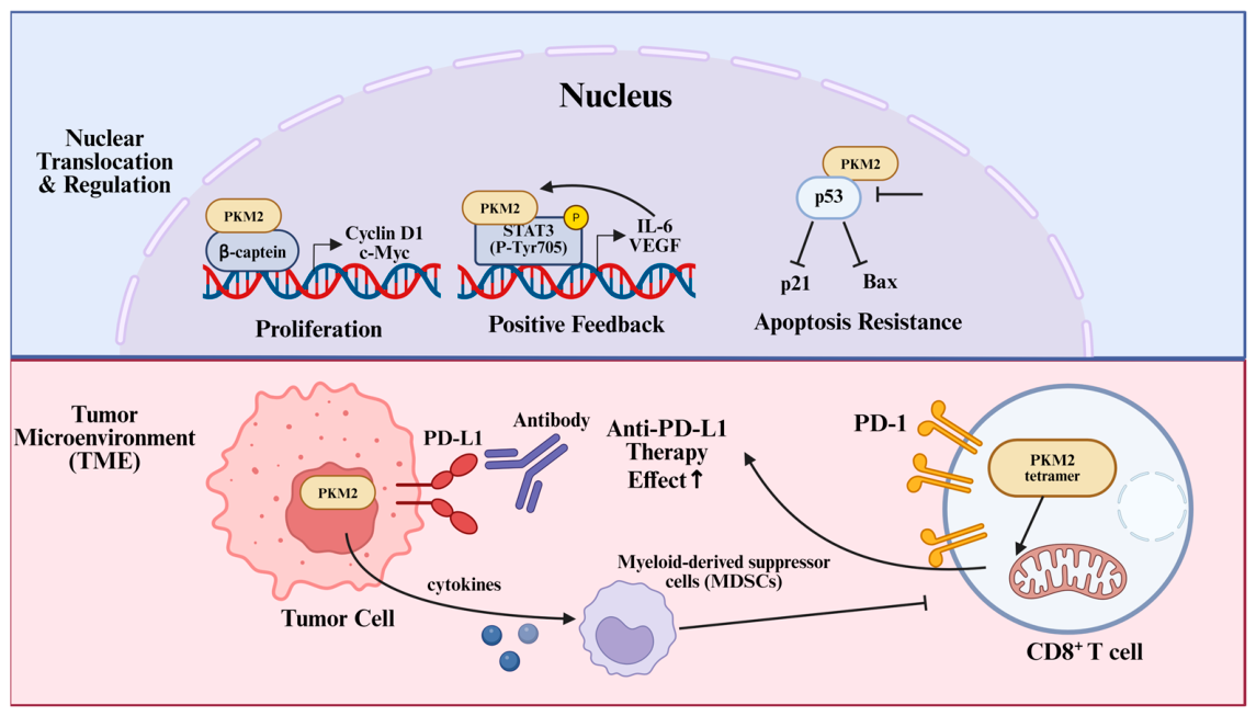
The non-metabolic functions of PKM2 also play a critical role in TC progression [92,93]. As shown in Figure 5, PKM2 can translocate into the cell nucleus, acting as a transcriptional coactivator involved in regulating gene expression, thereby influencing tumor cell proliferation, invasion, and metastasis. The interaction with β-catenin is one of the dominant mechanisms by which PKM2 regulates tumor proliferation. Nuclear PKM2 can bind to β-catenin, promote its translocation into the nucleus, and enhance the binding ability of β-catenin to T-cell factor/lymphoid enhancer factor (TCF/LEF) family transcription factors, upregulating the expression of proliferation-related genes, such as Cyclin D1 and c-Myc, and promoting the cell cycle progression of TC cells [94,95]. In addition, PKM2 can enhance the stability of β-catenin by phosphorylating it, inhibit its ubiquitin-dependent degradation, and further strengthen the pro-cancer signal mediated by β-catenin [90,96].
As a primary regulatory factor of inflammation and tumor progression, STAT3 is disorderedly activated in PTC and can interact with nuclear PKM2 to form a positive feedback regulatory loop [97,98]. PKM2 can activate the transcriptional function of STAT3 by phosphorylating the Tyr705 site of STAT3, upregulating the expression of genes such as IL-6 and VEGF; while activated STAT3 can in turn promote PKM gene transcription, enhance PKM2 expression, and synergistically promote the proliferation, invasion, and immune suppression of TC cells [99,100].
Existing clinical studies and basic experiments have confirmed that PKM2 shows abnormally high expression in TC tissues, and its expression level is significantly correlated with tumor invasiveness and clinical prognosis, having potential value as a prognostic biomarker for TC [101]. In clinical sample studies, detection by immunohistochemistry, real-time fluorescence quantitative PCR, Western blotting, and other technologies found that the positive expression rate and expression level of PKM2 in TC tissues were significantly higher than those in adjacent normal thyroid tissues, and there were obvious subtype-specific differences—in ATC, highly invasive PTC (such as diffuse sclerosing type, tall cell type), and MTC, the expression level of PKM2 was significantly increased, while in low-malignancy classic PTC and FTC, its expression level was relatively low, suggesting that PKM2 expression is positively correlated with the malignant degree of TC [102].
Analysis of clinicopathological correlation showed that high PKM2 expression was closely related to adverse clinical characteristics of TC patients. Specifically, the expression level of PKM2 was positively correlated with tumor size. In TC tissues with a maximum diameter >2cm, the positive expression rate of PKM2 was significantly higher than that in small tumor tissues; Meanwhile, patients with high PKM2 expression were more likely to have cervical lymph node metastasis, distant metastasis (such as lung metastasis, bone metastasis), and a later TNM stage (higher proportion of stage III–IV) [101]. Survival analysis results further confirmed that the disease-free survival (DFS) and overall survival (OS) of patients in the high PKM2 expression group were significantly shorter than those in the low expression group. Multivariate COX regression analysis showed that high PKM2 expression was an independent risk factor for poor prognosis of TC patients [103].
Evidence from cell and animal experiments further verified the pro-cancer effect of PKM2 in TC. In vitro experiments, high expression of PKM2 was detected in TC cell lines (papillary cancer TPC-1, B-CPAP cells, anaplastic cancer 8505C, SW1736 cells, medullary cancer TT, MZ-CRC-1 cells), and after knocking down PKM2 expression, the proliferation activity and colony formation ability of cancer cells were significantly decreased, while overexpression of PKM2 could enhance the malignant phenotype of cancer cells [99,102]. In vivo xenograft model studies showed that when PKM2-silenced TPC-1 cells were subcutaneously injected into nude mice, the growth rate of xenografts was significantly slowed down, and the tumor volume and weight were significantly smaller than those in the control group [104]; In the meantime, in the lung metastasis model, PKM2 knockdown could significantly reduce the number of lung metastases in nude mice, confirming that PKM2 plays an irreplaceable role in the in vivo growth and metastasis of TC [105].
Besides TC, PKM2 is widely investigated in other cancers. The interaction between PKM2 and p53 has a bidirectional regulatory characteristic, participating in the precise regulation of the cell cycle and apoptosis [106]. Under stress conditions, such as oxidative stress and DNA damage, nuclear PKM2 can bind to p53, inhibit the transcriptional activity of p53, downregulate the expression of apoptosis-related genes, such as p21 and Bax, and help cancer cells resist stress-induced apoptosis [107], while in low-malignancy TC cells, p53 can in turn inhibit the nuclear translocation function of PKM2, maintain its cytoplasmic enzyme activity, and avoid metabolic reprogramming. Meanwhile, p53 mutation leads to loss of function, breaking the balance of interaction with PKM2 via the mTOR signaling pathway, and further enhancing the pro-cancer function of PKM2 [108].
As shown in Figure 5, PKM2’s role in the TME also includes the regulation of immune cells, particularly macrophage polarization [109]. Macrophages can polarize into two functionally distinct phenotypes based on microenvironmental signals: M1, which exhibits pro-inflammatory and anti-tumor effects, and M2, which promotes tissue repair, angiogenesis, and tumor progression [110,111]. PKM2 can regulate M1/M2 polarization of macrophages and cytokine secretion, thus affecting inflammation and tumor progression [112]. While lactic acid, which increases during the Warburg effect, has the same effect on TAMs [113]. It can also accelerate the formation of cancer-associated fibroblasts (CAFs) in pancreatic cancer [114]. PKM2 dimer promotes the release of cytokines by tumor cells, thereby recruiting myeloid-derived suppressor cells (MDSCs). These cytokines bind to the surface receptors of MDSCs and activate related signaling pathways. This process subsequently inhibits the activity of T cells, thereby affecting tumor development [115,116,117]. On the opposite, PKM2 tetramer can enhance mitochondrial biogenesis and effector function of CD8+ T cells. This phenomenon can improve the efficacy of PD-L1 therapy [118].

5. PKM2 in Different Subtypes of TC

DTC is primarily divided into PTC and FTC, which exhibit significant differences in molecular characteristics and clinical behavior [39,119].
PTC is the most common subtype of TC, generally with a good prognosis but a nearly 40% of central lymph node metastasis [120]. One study found that the expression of glycolytic enzymes, including PKM2, is linked to aggressive features of PTC, and this association differs in patients with or without chronic lymphocytic thyroiditis (CLT) [121]. Specifically, studies have found that PKM2 mRNA expression may serve as a biomarker for lymph node metastasis in PTC [122]. Single-cell transcriptomic analysis has revealed heterogeneity in tumor and immune cells during PTC lymph node metastasis, as well as ITGA2-mediated metabolic reprogramming and immune crosstalk [123,124,125].
FTC is the second most common type of TC, characterized by lower aggressiveness but sometimes developing distant metastases. Unlike PTC, FTC typically metastasizes hematogenously rather than via lymph nodes [126]. While PKM2 also promotes proliferation and metastasis in other tumors, direct research on the specific role of PKM2 in FTC is limited.
While research on the role of PKM2 in ATC remains limited, evidence from other malignancies has established a significant correlation between nuclear PKM2 accumulation and tumor proliferation [127]. Therefore, future studies should explore whether nuclear PKM2 levels vary across different thyroid cancer subtypes and further investigate their potential association with cell proliferation, migration, and metastatic capacity.
In RAI-R TC, except that PKM2 dimer has a negative effect on SLC5A5 and thyroglobulin through histone H3 acetylation, PKM2 promotes radioiodine resistance in other ways. The therapeutic mechanism of radioactive 131I involves inducing DNA double-strand breaks via its emitted beta particles to kill cancer cells. However, cancer cells often repair these damages by activating DNA damage response pathways, leading to treatment resistance [128].
Meanwhile, RAI treatment causes cellular damage through the production of free radicals and ROS. To counteract this oxidative stress, cancer cells typically enhance their antioxidant defense systems. PKM2 can activate glucose-6-phosphate dehydrogenase (G6PD), a key enzyme in the pentose phosphate pathway (PPP), thereby increasing levels of NADPH and glutathione (GSH) [129]. NADPH and GSH are important reducing agents that can scavenge ROS generated by 131I therapy, protecting cancer cells from oxidative stress-induced death. This enhanced antioxidant capacity further promotes TC cells’ resistance to RAI.
Last but not least, PKM2 can form an axis with factors like HIF-1α, inducing the expression of EMT (epithelial–mesenchymal transition)-related genes, such as ZEB1, and stemness-related genes, such as OCT4 and NANOG, thereby promoting tumor cell dedifferentiation, migration, invasion, and cancer stem cell-like properties [130,131]. These characteristics are very likely to contribute to the development of radioiodine resistance. Nuclear PKM2, acting as an RNA-binding protein, specifically binds to G-quadruplex structures in pre-mRNA and prevents the binding of inhibitory RNA-binding proteins, thereby promoting the expression of rG4me (pre-mRNAs containing G-quadruplexes), a process also associated with EMT and tumor metastasis [105].

6. Therapeutic Strategies Targeting PKM2

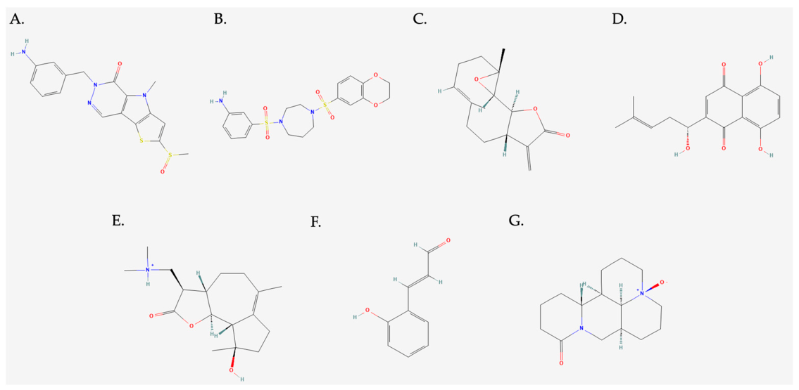
Based on the irreplaceable role of PKM2 in TC metabolic reprogramming, malignant progression, and radioiodine resistance, targeting PKM2 has become a potential direction for the precise treatment. According to the functional characteristics of PKM2, single-drug therapeutic strategies such as activating PKM2, inhibiting PKM2, and targeting upstream regulatory factors have been developed. Concurrently, based on its synergistic relationship with driver genes and the immune microenvironment, combined therapeutic strategies have shown better application prospects. The goal of the PKM2 activation strategy is to induce PKM2 to transform from the low-activity dimer to the high-activity tetramer through small-molecule compounds and stabilize its enzyme activity [32]. To systematically review the current progress in PKM2-targeted drug development and its status in thyroid cancer research, Table 2 categorizes and summarizes reported PKM2-targeted drugs, particularly highlighting those that have not yet been definitively validated in thyroid cancer. The chemical structures of TEPP-46, DASA-58, parthenolide, shikonin, DMAMCL, 2-hydroxycinnamaldehyde, and oxymatrine are shown in Figure 6. At present, representative small-molecule activators such as TEPP-46 and DASA-58 have been found, and their research in other cancer models provides experimental support for this strategy.
Research on TEPP-46 in kidney disease has made great progress. After TEPP-46 activates PKM2, it can inhibit the accumulation of HIF-1α, which has been proven to induce abnormal glycolysis and EMT in kidney cells. Inhibition of the EMT process can reduce the generation of fibroblasts and the deposition of extracellular matrix in the renal interstitium, thereby blocking the progression of renal fibrosis [132]. TEPP-46-mediated PKM2 activation can repair mitochondrial function, balance the metabolic imbalance between glycolysis and mitochondrial OXPHOS, reduce kidney cell damage, and delay the progression of diabetic kidney disease [133].
DASA-58 binds to the allosteric site of PKM2, prompting it to form a stable tetramer structure. This conformation is resistant to the inhibitory effect of tyrosine phosphorylated proteins, thereby continuously activating the pyruvate kinase activity of PKM2, reducing its nuclear translocation, and further regulating downstream metabolic and signaling pathways. DASA-58 can block LPS-induced HIF-1α expression in bone marrow-derived macrophages, downregulate HIF-1α-dependent target genes such as IL-1β, and inhibit inflammation-related metabolic reprogramming; enhance the metabolic switch from glycolysis to OXPHOS, reduce the aerobic glycolysis level of tumor cells, decrease lactate production and ATP supply, and inhibit tumor proliferation and metastasis; reduce PKM2 nuclear translocation, inhibit its binding to transcription factors (such as HIF-1α, β-catenin), and downregulate the expression of proliferation and invasion-related genes (such as GLUT1, HK2, VEGF) [134,135,136].
As a naturally derived PKM2 inhibitor, Shikonin can specifically bind to the active center of PKM2, inhibit its catalytic activity, and prevent the interaction between PKM2 and transcription factors, dual-blocking its enzymatic and non-enzymatic activities [137]. In vitro experiments have confirmed that Shikonin can significantly inhibit the proliferation of various subtypes of TC cells (papillary cancer, anaplastic cancer, medullary cancer), induce cell cycle arrest in the G2/M phase, activate the caspase-dependent apoptotic pathway, and has a more significant inhibitory effect on cancer cells with high PKM2 expression [138]. In BRAF V600E-positive TC cells, Shikonin can effectively block the BRAF-PKM2 crosstalk loop, downregulate the expression of pro-cancer genes such as GLUT1 and Cyclin D1, thus inhibiting cell invasion and metastasis [139].
Table 2. Current PKM2-targeting therapies.
CategoryAgent(s)MechanismEvidence in
Thyroid Cancer?
References
ActivatorsTEPP-46 (PubChem CID: 44246499),
DASA-58 (PubChem CID: 44543605)
Stabilizes PKM2 tetramer, enhances enzymatic activity, promotes pyruvate production, inhibits LPS-induced HIF-1α and IL-1β, attenuates proinflammatory M1 macrophages and promotes anti-inflammatory M2 macrophages.No[132]
PA-12Stimulates PKM2 pyruvate kinase activity (in vitro AC50 4.92 μM), inhibits lung cancer cell viability under hypoxia.No[140]
Parthenolide
dimers (PubChem CID: 6473881)
Activates PKM2 (AC50 15 μM), promotes tetramer formation, reduces nuclear translocation, inhibits proliferation and metastasis, induces apoptosis in glioblastoma cells.No[141]
InhibitorsShikonin (PubChem CID: 479503)Direct PKM2 enzymatic inhibition, suppresses aerobic glycolysis, induces apoptosis, inhibits tumor growth.No[142]
Naphthoquinone
derivatives
Potent PKM2 inhibitors, induce apoptosis and autophagy through Akt/mTOR suppression.No[143]
DMAMCL (PubChem CID: 133081974)Targets PKM2, rewires aerobic glycolysis, suppresses glioblastoma cell proliferation and tumor growth.No[144]
FV-429 (PubChem SID: 433986466)Regulates nuclear translocation of PKM2, induces mitochondrial apoptosis, inhibits glycolysis in pancreatic cancer cells.No[145]
2-Hydroxycinnamaldehyde (PubChem CID: 5318169)Directly targets and inhibits PKM2, inhibits cancer cell proliferation and tumor growth.No[146]
Oxymatrine (PubChem CID: 24864132)Attenuates PKM2-mediated aerobic glycolysis, inhibits colorectal cancer metastasis.No[147]

7. Discussion and Future Directions

This review systematically summarizes the central role, regulatory network, specific functions, and targeted therapeutic strategies of PKM2 in TC metabolic reprogramming and occurrence and development. Combined with evidence from clinical samples, cell, and animal experiments, we establish PKM2’s intrinsic status as an indispensable pro-cancer molecule in TC.
Through its dual functions of enzymatic and non-enzymatic activities, PKM2 constructs a complex regulatory network in TC cells. On the one hand, in its low-activity dimeric form, it triggers the Warburg effect, shunting glycolytic intermediates to support tumor biosynthesis, thereby meeting the demands of rapid tumor cell proliferation for macromolecules, such as nucleotides, amino acids, and lipids. This metabolic reprogramming not only provides the necessary building blocks for tumor growth but also helps tumor cells adapt to various stresses within the TME, such as nutrient deprivation, hypoxia, and oxidative stress. On the other hand, PKM2 can translocate into the cell nucleus, acting as a transcriptional coactivator that synergizes with factors such as HIF-1α, β-catenin, and STAT3 to regulate the expression of genes related to cell proliferation, invasion, metastasis, and apoptosis resistance.
Concurrently, PKM2 forms specific regulatory axes with driver genes, like BRAF V600E, RAS, and c-Myc, which not only promote TC subtype progression and enhance malignant phenotypes, but also facilitate radioiodine resistance through the “BRAF-PKM2-NIS” axis, becoming a pivotal obstacle in the clinical diagnosis and treatment of TC. Furthermore, PTMs of PKM2 critically influence its conformation, stability, subcellular localization, and function, thereby finely tuning its enzymatic and non-metabolic roles. Hypoxia within the TME and M2 polarization of tumor-associated macrophages (TAMs) also regulate PKM2 expression and activity through mechanisms such as the HIF-1α pathway and cytokine secretion, forming a vicious cycle that drives tumor progression.
Based on PKM2’s fundamental role in TC, strategies targeting PKM2, including PKM2 activators, inhibitors, and indirect modulators, have shown potential application value. These strategies aim to inhibit tumor growth by stabilizing the high-activity tetrameric form of PKM2 or by blocking its enzymatic activity and non-metabolic functions. These studies provide novel insights and targets for the precise treatment of TC.
Despite significant progress in understanding PKM2’s role in TC, several research gaps remain, necessitating further exploration. Future research should focus on the following five dimensions:
i.
Subtype-specific mechanism research
Significant differences exist in the biological characteristics, driver gene profiles, and metabolic patterns among various TC subtypes (differentiated, anaplastic, and medullary cancer). However, the subtype-specificity of the underlying PKM2 regulatory network remains unclear. Existing studies predominantly focus on PTC, with insufficient exploration of PKM2’s regulatory mechanisms in FTC, ATC and MTC. Moving forward, targeted research should be conducted for different subtypes to clarify variations in PKM2 expression levels, regulatory factors, and functions among various subtypes. For instance, elucidating the possible regulatory mode of the RET-RAS-PKM2 axis in MTC and the dominant role of PKM2 non-enzymatic activity in ATC will provide a theoretical basis for formulating subtype-specific targeted therapeutic strategies, avoiding “one-size-fits-all” treatment plans, and improving treatment accuracy [10,39].
ii.
Exploration of TME interaction mechanism
TME is an integral factor affecting TC progression and treatment sensitivity [148]. At present, research on the role of PKM2 in the interaction between TC cells and microenvironment components is still scarce. Future work needs to focus on exploring the interaction mechanism between PKM2-mediated metabolic reprogramming and CAFs, immune cells. On one hand, it is crucial to clarify how metabolic products such as lactate and ROS, regulated by PKM2, regulate CAF activation and immune cell infiltration and function [109,149]. On the other hand, analyzing the reverse regulatory effect of cytokines secreted by microenvironmental components on PKM2 expression and activity will elucidate the vicious cycle mechanism of metabolic reprogramming and immune microenvironment remodeling, and provide more robust theoretical support for the combined application of PKM2-targeted drugs and immunotherapy [112,150].
iii.
Optimization and innovation of targeted drugs
Existing PKM2-targeted drugs have problems such as poor tissue specificity, high off-target toxicity risk, and insufficient precision of function regulation, which restrict their clinical translation [151]. Future drug optimization should focus on two directions: First, developing thyroid tissue-specific delivery systems, such as nanocarriers coupled with thyroid-stimulating hormone receptors and NIS, to achieve precise delivery of PKM2 targeted drugs to TC cells, improve drug concentration in tumor tissues, and reduce damage to normal metabolically active tissues [104]; second, develop highly specific PKM2 allosteric modulators that break the “all-or-nothing” regulatory mode of existing drugs, and precisely regulate the enzymatic/non-enzymatic activities of PKM2 [151]. For example, for patients with TC radioiodine resistance, developing modulators that can selectively activate PKM2 enzymatic activity and restore NIS function without affecting its normal physiological functions would improve treatment safety and effectiveness [152].
iv.
Clinical transformation verification of biomarkers
Existing evidence suggests that PKM2 has potential as a biomarker for TC, but it lacks verification in large-sample clinical cohort studies, and its clinical application value is not yet clear [121,153]. In further investigations, multi-center, large-sample clinical studies should be carried out to verify the biomarker function of PKM2 from three dimensions: First, its early diagnostic value, by detecting PKM2 expression level in serum and fine-needle aspiration samples, and clarifying its diagnostic efficacy for distinguishing benign and malignant thyroid lesions; Second, its prognostic evaluation value, by verifying the correlation between PKM2 expression level and patient DFS and OS through long-term follow-up, establishing its status as an independent risk factor for poor prognosis; Third, its efficacy prediction value, by focusing on analyzing the correlation between PKM2 expression and the efficacy of RAI therapy, BRAF inhibitor therapy, and PKM2 targeted therapy, constructing an efficacy prediction model based on PKM2, and realizing precise stratification and individualized treatment of patients [121,152].
v.
Multi-omics integration to analyze regulatory networks
The role of PKM2 in TC is not regulated by a single pathway but involves a complex network of multiple levels, including metabolism, signaling pathways, and gene transcription [38,154]. In subsequent studies, it is necessary to carry out integrated analysis with the help of multi-omics technologies, such as transcriptomics, proteomics, metabolomics, and phosphoproteomics [119,124]. This approach will allow researchers to screen targeting genes and proteins regulated by PKM2 through transcriptomics and proteomics, clarify PKM2-mediated glycolytic flux changes and key metabolites through metabolomics, and construct a molecular network map of PKM2 regulation in TC through combined modeling of multi-omics data. This will help explore key targets that synergize with PKM2, provide new ideas for developing multi-target combined therapeutic strategies, and further improve the overall efficacy of TC treatment [155].

8. Conclusions

Despite significant progress in understanding PKM2’s role in thyroid cancer, numerous challenges and unresolved questions remain. Future research should focus on elucidating the precise regulatory mechanisms of PKM2 across different thyroid cancer subtypes, thoroughly exploring the complex interactions between PKM2 and the TME, and optimizing the specificity and efficacy of targeted drugs. Concurrently, large-scale clinical validation of PKM2 as a biomarker, coupled with multi-omics integration to analyze its regulatory network, will facilitate the translation of PKM2 from basic research to clinical practice. Ultimately, this will provide more precise and effective diagnostic and therapeutic strategies for thyroid cancer patients, significantly improving their prognosis and quality of life.

Author Contributions

Conceptualization, Q.W. and Z.A.; methodology, Q.W.; software, S.L., W.L. and J.Z.; validation, S.L. and W.L.; formal analysis, Q.W.; investigation, S.L. and C.Z.; resources, Z.A.; data curation, Q.W.; writing—original draft preparation, S.L. and L.R.; writing—review and editing, Q.W. and Z.A.; visualization, S.L. and W.L.; supervision, Z.A.; project administration, Z.A.; funding acquisition, Z.A. All authors have read and agreed to the published version of the manuscript.

Funding

This research was funded by Shanghai Innovation Medical Device Application Demonstration Project 2023, grant number 23SHS05200, and Scientific Research Development Fund of Zhongshan Hospital, Fudan University, grant numbers 2025XKPT58 and 2025XKPT58-1.

Institutional Review Board Statement

Not applicable.

Informed Consent Statement

Not applicable.

Data Availability Statement

No new data were created or analyzed in this study.

Conflicts of Interest

The authors declare no conflicts of interest.

Abbreviations

The following abbreviations are used in this manuscript:
PKM2Pyruvate kinase muscle type 2
IARCInternational Agency for Research on Cancer
DTCDifferentiated thyroid cancer
ATCAnaplastic thyroid cancer
MTCMedullary thyroid cancer
PTCPapillary thyroid cancer
FTCFollicular thyroid cancer
RAIRadioactive iodine
RAI-RRAI-refractory
MKIsMulti-kinase inhibitors
ORRObjective response rate
TMETumor microenvironment
ROSReactive oxygen species
PKPyruvate Kinase
PEPPhosphoenolpyruvate
PKLPyruvate kinase liver type
PKRPyruvate kinase red blood cell type
PKM1Pyruvate kinase muscle type 1
3′UTR3′-untranslated region
PTMPost-translational modification
O-GlcNAcO-linked β-N-acetylglucosamine
TRIM35Tripartite motif-containing protein 35
NSCLCNon-small cell lung cancer
FBPFructose-1,6-bisphosphate
TKTTransketolase
TCFT-cell factor
LEFLymphoid enhancer factor
EMTEpithelial–mesenchymal transition
OXPHOSOxidative phosphorylation
NISNa+-iodine symporter
TAMsTumor-associated macrophages
CAFsCancer associated fibroblast
DFSDisease-free survival
OSOverall survival

References

  1. Boucai, L.; Zafereo, M.; Cabanillas, M.E. Thyroid Cancer: A Review. JAMA 2024, 331, 425–435. [Google Scholar] [CrossRef]
  2. He, T.; Li, M.; Gao, Z.-L.; Li, X.-Y.; Zhong, H.-R.; Ding, C.-S.; Cai, H.-W. Analysis of Delayed Initial Radioactive Iodine Therapy and Clinical Outcomes in Papillary Thyroid Cancer: A Two-Center Retrospective Study. Nucl. Med. Commun. 2024, 45, 779–787. [Google Scholar] [CrossRef]
  3. Zhao, Z.; Fan, Y.; Sun, P.; Zhang, S.; Xu, M.; Li, J.; Xu, P. Temporal Trends and Geographic Disparities in Thyroid Cancer Burden: A Global Analysis from 1990 to 2021. Front. Nutr. 2025, 12, 1613737. [Google Scholar] [CrossRef]
  4. Sung, H.; Ferlay, J.; Siegel, R.L.; Laversanne, M.; Soerjomataram, I.; Jemal, A.; Bray, F. Global Cancer Statistics 2020: GLOBOCAN Estimates of Incidence and Mortality Worldwide for 36 Cancers in 185 Countries. CA Cancer J. Clin. 2021, 71, 209–249. [Google Scholar] [CrossRef]
  5. Li, M.; Dal Maso, L.; Pizzato, M.; Vaccarella, S. Evolving Epidemiological Patterns of Thyroid Cancer and Estimates of Overdiagnosis in 2013–2017 in 63 Countries Worldwide: A Population-Based Study. Lancet Diabetes Endocrinol. 2024, 12, 824–836. [Google Scholar] [CrossRef] [PubMed]
  6. Han, B.; Zheng, R.; Zeng, H.; Wang, S.; Sun, K.; Chen, R.; Li, L.; Wei, W.; He, J. Cancer Incidence and Mortality in China, 2022. J. Natl. Cancer Cent. 2024, 4, 47–53. [Google Scholar] [CrossRef]
  7. Grani, G.; Sponziello, M.; Filetti, S.; Durante, C. Thyroid Nodules: Diagnosis and Management. Nat. Rev. Endocrinol. 2024, 20, 715–728. [Google Scholar] [CrossRef] [PubMed]
  8. Jeong, S.I.; Kim, W.; Yu, H.W.; Choi, J.Y.; Ahn, C.H.; Moon, J.H.; Choi, S.I.; Cha, W.; Jeong, W.-J.; Park, S.Y.; et al. Incidence and Clinicopathological Features of Differentiated High-Grade Thyroid Carcinomas: An Institutional Experience. Endocr. Pathol. 2023, 34, 287–297. [Google Scholar] [CrossRef]
  9. Pavlidis, E.T.; Galanis, I.N.; E Pavlidis, T. Update on Current Diagnosis and Management of Anaplastic Thyroid Carcinoma. World J. Clin. Oncol. 2023, 14, 570–583. [Google Scholar] [CrossRef]
  10. Oczko-Wojciechowska, M.; Czarniecka, A.; Gawlik, T.; Jarzab, B.; Krajewska, J. Current Status of the Prognostic Molecular Markers in Medullary Thyroid Carcinoma. Endocr. Connect. 2020, 9, R251–R263. [Google Scholar] [CrossRef]
  11. Zhang, L.; Feng, Q.; Yu, H.W.; Choi, J.Y.; Ahn, C.H.; Moon, J.H.; Choi, S.I.; Cha, W.; Jeong, W.-J.; Park, S.Y.; et al. Molecular Basis and Targeted Therapy in Thyroid Cancer: Progress and Opportunities. Biochim. Biophys. Acta (BBA)—Rev. Cancer 2023, 1878, 188928. [Google Scholar] [CrossRef]
  12. Lee, E.K. Systemic Therapy for Differentiated Thyroid Cancer with Distant Metastasis. J. Korean Med. Assoc. 2024, 67, 484–491. [Google Scholar] [CrossRef]
  13. Díaz Vico, T.; Martínez-Amores Martínez, B.; Góngora, L.M.; Jiménez-Fonseca, P.; Martín, P.P.; Torrente, I.G.; Muñoz-Nájar, A.G.; Durán-Poveda, M. Systemic Therapeutic Options in Radioiodine-Refractory Differentiated Thyroid Cancer: Current Indications and Optimal Timing. Cancers 2025, 17, 1800. [Google Scholar] [CrossRef]
  14. Liu, Y.; Wang, J.; Hu, X.; Pan, Z.; Xu, T.; Xu, J.; Jiang, L.; Huang, P.; Zhang, Y.; Ge, M. Radioiodine Therapy in Advanced Differentiated Thyroid Cancer: Resistance and Overcoming Strategy. Drug Resist. Updates 2023, 68, 100939. [Google Scholar] [CrossRef] [PubMed]
  15. Pappa, T.; Wirth, L. An Update on Redifferentiation Strategies for Radioactive Iodine-Refractory Differentiated Thyroid Carcinoma. Endocrine 2024, 87, 1–10. [Google Scholar] [CrossRef]
  16. Cheng, L.; Newbold, K. Systemic Therapy for Advanced Thyroid Cancer—New Personalized Options. Drugs 2025, 85, 1381–1390. [Google Scholar] [CrossRef] [PubMed]
  17. Hamidi, S.; Hofmann, M.-C.; Iyer, P.C.; Cabanillas, M.E.; Hu, M.I.; Busaidy, N.L.; Dadu, R. Review Article: New Treatments for Advanced Differentiated Thyroid Cancers and Potential Mechanisms of Drug Resistance. Front. Endocrinol. 2023, 14, 1176731. [Google Scholar] [CrossRef] [PubMed]
  18. Landa, I.; Ibrahimpasic, T.; Boucai, L.; Sinha, R.; Knauf, J.A.; Shah, R.H.; Dogan, S.; Ricarte-Filho, J.C.; Krishnamoorthy, G.P.; Xu, B.; et al. Genomic and Transcriptomic Hallmarks of Poorly Differentiated and Anaplastic Thyroid Cancers. J. Clin. Investig. 2016, 126, 1052–1066. [Google Scholar] [CrossRef]
  19. Xing, M.; Alzahrani, A.S.; Carson, K.A.; Viola, D.; Elisei, R.; Bendlova, B.; Yip, L.; Mian, C.; Vianello, F.; Tuttle, R.M.; et al. Association Between BRAF V600E Mutation and Mortality in Patients With Papillary Thyroid Cancer. JAMA 2013, 309, 1493. [Google Scholar] [CrossRef]
  20. Xing, M. BRAF Mutation in Thyroid Cancer. Endocr. Relat. Cancer 2005, 12, 245–262. [Google Scholar] [CrossRef]
  21. Behnagh, A.K.; Eghbali, M.; Abdolmaleki, F.; Ghadikolaei, O.A.; Asl, P.R.; Afsharpad, M.; Cheraghi, S.; Honardoost, M. An Overview on Prevalence and Detection Approaches of BRAF V600E Mutation in Anaplastic Thyroid Carcinoma: A Systematic Review and Meta-Analysis. Iran. J. Public Health 2024, 53, 1496–1507. [Google Scholar] [CrossRef]
  22. Xing, M. Molecular Pathogenesis and Mechanisms of Thyroid Cancer. Nat. Rev. Cancer 2013, 13, 184–199. [Google Scholar] [CrossRef]
  23. Liu, X.; Bishop, J.; Shan, Y.; Pai, S.; Liu, D.; Murugan, A.K.; Sun, H.; El-Naggar, A.K.; Xing, M. Highly Prevalent TERT Promoter Mutations in Aggressive Thyroid Cancers. Endocr.-Relat. Cancer 2013, 20, 603–610. [Google Scholar] [CrossRef]
  24. Hanahan, D.; Weinberg, R.A. Hallmarks of Cancer: The Next Generation. Cell 2011, 144, 646–674. [Google Scholar] [CrossRef]
  25. Vander Heiden, M.G.; Cantley, L.C.; Thompson, C.B. Understanding the Warburg Effect: The Metabolic Requirements of Cell Proliferation. Science 2009, 324, 1029–1033. [Google Scholar] [CrossRef]
  26. Liberti, M.V.; Locasale, J.W. The Warburg Effect: How Does It Benefit Cancer Cells? Trends Biochem. Sci. 2016, 41, 211–218. [Google Scholar] [CrossRef]
  27. DeBerardinis, R.J.; Lum, J.J.; Hatzivassiliou, G.; Thompson, C.B. The Biology of Cancer: Metabolic Reprogramming Fuels Cell Growth and Proliferation. Cell Metab. 2008, 7, 11–20. [Google Scholar] [CrossRef]
  28. Wu, C.-A.; Chao, Y.; Shiah, S.-G.; Lin, W.-W. Nutrient Deprivation Induces the Warburg Effect through ROS/AMPK-Dependent Activation of Pyruvate Dehydrogenase Kinase. Biochim. Biophys. Acta (BBA)—Mol. Cell Res. 2013, 1833, 1147–1156. [Google Scholar] [CrossRef]
  29. Schormann, N.; Hayden, K.L.; Lee, P.; Banerjee, S.; Chattopadhyay, D. An Overview of Structure, Function, and Regulation of Pyruvate Kinases. Protein Sci. 2019, 28, 1771–1784. [Google Scholar] [CrossRef]
  30. Israelsen, W.J.; Vander Heiden, M.G. Pyruvate Kinase: Function, Regulation and Role in Cancer. Semin. Cell Dev. Biol. 2015, 43, 43–51. [Google Scholar] [CrossRef]
  31. Noguchi, T. Regulation of pyruvate kinase gene expression and its clinical application. Rinsho Byori 1990, 38, 868–875. [Google Scholar]
  32. Anastasiou, D.; Yu, Y.; Israelsen, W.J.; Jiang, J.-K.; Boxer, M.B.; Hong, B.S.; Tempel, W.; Dimov, S.; Shen, M.; Jha, A.; et al. Pyruvate Kinase M2 Activators Promote Tetramer Formation and Suppress Tumorigenesis. Nat. Chem. Biol. 2012, 8, 839–847. [Google Scholar] [CrossRef]
  33. Christofk, H.R.; Vander Heiden, M.G.; Harris, M.H.; Ramanathan, A.; Gerszten, R.E.; Wei, R.; Fleming, M.D.; Schreiber, S.L.; Cantley, L.C. The M2 Splice Isoform of Pyruvate Kinase Is Important for Cancer Metabolism and Tumour Growth. Nature 2008, 452, 230–233. [Google Scholar] [CrossRef]
  34. Mazurek, S. Pyruvate Kinase Type M2: A Key Regulator of the Metabolic Budget System in Tumor Cells. Int. J. Biochem. Cell Biol. 2011, 43, 969–980. [Google Scholar] [CrossRef]
  35. Monteiro, F.; Shetty, S.S. Natural Antioxidants as Inhibitors of Pyruvate Kinase M2 in Warburg Phenotypes. J. Herb. Med. 2023, 42, 100750. [Google Scholar] [CrossRef]
  36. Chiu, C.-F.; Guerrero, J.; Regalado, R.; Zhou, J.; Notarte, K.; Lu, Y.-W.; Encarnacion, P.; Carles, C.; Octavo, E.; Limbaroc, D.; et al. Insights into Metabolic Reprogramming in Tumor Evolution and Therapy. Cancers 2024, 16, 3513. [Google Scholar] [CrossRef]
  37. Shuvalov, O.; Daks, A.; Fedorova, O.; Petukhov, A.; Barlev, N. Linking Metabolic Reprogramming, Plasticity and Tumor Progression. Cancers 2021, 13, 762. [Google Scholar] [CrossRef]
  38. Zhang, T.; Han, H.; Zhang, T.; Zhang, Y.; Ma, L.; Yang, Z.; Zhao, Y.X. Oncogenic Mutation-Driven Metabolism-Immunity Regulatory Axis: Potential Prospects for Thyroid Cancer Precision Therapy. Biochim. Biophys. Acta (BBA)—Rev. Cancer 2025, 1880, 189459. [Google Scholar] [CrossRef]
  39. Li, Z.; Wang, N.; Li, X.; Xie, Y.; Dou, Z.; Xin, H.; Lin, Y.; Si, Y.; Feng, T.; Wang, G. Thyroid Cancer: From Molecular Insights to Therapy (Review). Oncol. Lett. 2025, 30, 520. [Google Scholar] [CrossRef]
  40. Masoud, G.N.; Li, W. HIF-1α Pathway: Role, Regulation and Intervention for Cancer Therapy. Acta Pharm. Sin. B 2015, 5, 378–389. [Google Scholar] [CrossRef]
  41. Bharti, J.; Gogu, P.; Pandey, S.K.; Verma, A.; Yadav, J.P.; Singh, A.K.; Kumar, P.; Dwivedi, A.R.; Pathak, P. BRAF V600E in Cancer: Exploring Structural Complexities, Mutation Profiles, and Pathway Dysregulation. Exp. Cell Res. 2025, 446, 114440. [Google Scholar] [CrossRef]
  42. Lasolle, H.; Schiavo, A.; Tourneur, A.; Gillotay, P.; Fonseca, B.d.F.d.; Ceolin, L.; Monestier, O.; Aganahi, B.; Chomette, L.; Kizys, M.M.L.; et al. Dual Targeting of MAPK and PI3K Pathways Unlocks Redifferentiation of Braf-Mutated Thyroid Cancer Organoids. Oncogene 2024, 43, 155–170. [Google Scholar] [CrossRef]
  43. Liu, R.; Liu, D.; Xing, M. The Akt Inhibitor MK2206 Synergizes, but Perifosine Antagonizes, the BRAFV600E Inhibitor PLX4032 and the MEK1/2 Inhibitor AZD6244 in the Inhibition of Thyroid Cancer Cells. J. Clin. Endocrinol. Metab. 2012, 97, E173–E182. [Google Scholar] [CrossRef]
  44. Nikiforova, M.N.; Lynch, R.A.; Biddinger, P.W.; Alexander, E.K.; Dorn, G.W.; Tallini, G.; Kroll, T.G.; Nikiforov, Y.E. RAS Point Mutations and PAX8-PPARγ Rearrangement in Thyroid Tumors: Evidence for Distinct Molecular Pathways in Thyroid Follicular Carcinoma. J. Clin. Endocrinol. Metab. 2003, 88, 2318–2326. [Google Scholar] [CrossRef]
  45. Bahar, M.E.; Kim, H.J.; Kim, D.R. Targeting the RAS/RAF/MAPK Pathway for Cancer Therapy: From Mechanism to Clinical Studies. Signal Transduct. Target. Ther. 2023, 8, 455. [Google Scholar] [CrossRef]
  46. Matsuse, M.; Mitsutake, N. TERT Promoter Mutations in Thyroid Cancer. Endocr. J. 2023, 70, 1035–1049. [Google Scholar] [CrossRef]
  47. Landa, I. InTERTwined: How TERT Promoter Mutations Impact BRAFV600E-Driven Thyroid Cancers. Curr. Opin. Endocr. Metab. Res. 2023, 30, 100460. [Google Scholar] [CrossRef]
  48. Sang, Y.; Hu, G.; Xue, J.; Chen, M.; Hong, S.; Liu, R. Risk Stratification by Combining Common Genetic Mutations and TERT Promoter Methylation in Papillary Thyroid Cancer. Endocrine 2024, 85, 304–312. [Google Scholar] [CrossRef]
  49. Shim, H.; Dolde, C.; Lewis, B.C.; Wu, C.S.; Dang, G.; Jungmann, R.A.; Dalla-Favera, R.; Dang, C.V. C-Myc Transactivation of LDH-A: Implications for Tumor Metabolism and Growth. Proc. Natl. Acad. Sci. USA 1997, 94, 6658–6663. [Google Scholar] [CrossRef]
  50. Maida, Y.; Yasukawa, M.; Furuuchi, M.; Lassmann, T.; Possemato, R.; Okamoto, N.; Kasim, V.; Hayashizaki, Y.; Hahn, W.C.; Masutomi, K. An RNA-Dependent RNA Polymerase Formed by TERT and the RMRP RNA. Nature 2009, 461, 230–235. [Google Scholar] [CrossRef]
  51. Lacka, K.; Maciejewski, A.; Tyburski, P.; Manuszewska-Jopek, E.; Majewski, P.; Więckowska, B. Rationale for Testing TP53 Mutations in Thyroid Cancer—Original Data and Meta-Analysis. Int. J. Mol. Sci. 2025, 26, 1035. [Google Scholar] [CrossRef] [PubMed]
  52. Liu, Y.; Gu, W. The Complexity of P53-Mediated Metabolic Regulation in Tumor Suppression. Semin. Cancer Biol. 2022, 85, 4–32. [Google Scholar] [CrossRef] [PubMed]
  53. Koo, K.Y.; Moon, K.; Song, H.S.; Lee, M.-S. Metabolic Regulation by P53: Implications for Cancer Therapy. Mol. Cells 2025, 48, 100198. [Google Scholar] [CrossRef]
  54. Yu, G.; Sun, W.; Shen, Y.; Hu, Y.; Liu, H.; Li, W.; Wang, Y. PKM2 Functions as a Potential Oncogene and Is a Crucial Target of miR-148a and miR-326 in Thyroid Tumorigenesis. Am. J. Transl. Res. 2018, 10, 1793–1801. [Google Scholar]
  55. Liu, A.M.; Xu, Z.; Shek, F.H.; Wong, K.-F.; Lee, N.P.; Poon, R.T.; Chen, J.; Luk, J.M. miR-122 Targets Pyruvate Kinase M2 and Affects Metabolism of Hepatocellular Carcinoma. PLoS ONE 2014, 9, e86872. [Google Scholar] [CrossRef]
  56. Rahman, M.A.; Salajegheh, A.; Smith, R.A.; Lam, A.K. Multiple Proliferation-Survival Signalling Pathways Are Simultaneously Active in BRAF V600E Mutated Thyroid Carcinomas. Exp. Mol. Pathol. 2015, 99, 492–497. [Google Scholar] [CrossRef]
  57. Yang, W.; Zheng, Y.; Xia, Y.; Ji, H.; Chen, X.; Guo, F.; Lyssiotis, C.A.; Aldape, K.; Cantley, L.C.; Lu, Z. ERK1/2-Dependent Phosphorylation and Nuclear Translocation of PKM2 Promotes the Warburg Effect. Nat. Cell Biol. 2012, 14, 1295–1304. [Google Scholar] [CrossRef]
  58. Kachel, P.; Trojanowicz, B.; Sekulla, C.; Prenzel, H.; Dralle, H.; Hoang-Vu, C. Phosphorylation of Pyruvate Kinase M2 and Lactate Dehydrogenase A by Fibroblast Growth Factor Receptor 1 in Benign and Malignant Thyroid Tissue. BMC Cancer 2015, 15, 140. [Google Scholar] [CrossRef][Green Version]
  59. Salani, B.; Ravera, S.; Amaro, A.; Salis, A.; Passalacqua, M.; Millo, E.; Damonte, G.; Marini, C.; Pfeffer, U.; Sambuceti, G.; et al. IGF1 Regulates PKM2 Function through Akt Phosphorylation. Cell Cycle 2015, 14, 1559–1567. [Google Scholar] [CrossRef] [PubMed]
  60. Chen, Z.; Wang, Z.; Guo, W.; Zhang, Z.; Zhao, F.; Zhao, Y.; Jia, D.; Ding, J.; Wang, H.; Yao, M.; et al. TRIM35 Interacts with Pyruvate Kinase Isoform M2 to Suppress the Warburg Effect and Tumorigenicity in Hepatocellular Carcinoma. Oncogene 2015, 34, 3946–3956. [Google Scholar] [CrossRef] [PubMed]
  61. Cao, S.-H.; Ma, R.-Y.; Cao, T.; Hu, T.; Yang, S.; Ren, Z.-Y.; Niu, J.-L.; Zheng, M.-Q.; Han, M.; Dong, L.-H. PKM2 Crotonylation Reprograms Glycolysis in VSMCs, Contributing to Phenotypic Switching. Oncogene 2025, 44, 1990–2003. [Google Scholar] [CrossRef]
  62. Ye, X.; Niu, X.; Xu, Y.; Li, Z.; Yu, Y.; Chen, Z.; Lu, S. Desuccinylation of Pyruvate Kinase M2 by SIRT5 Contributes to Antioxidant Response and Tumor Growth. Oncotarget 2017, 8, 6984–6993. [Google Scholar]
  63. Wang, F.; Wang, K.; Xu, W.; Zhao, S.; Ye, D.; Wang, Y.; Xu, Y.; Zhou, L.; Chu, Y.; Zhang, C.; et al. SIRT5 Desuccinylates and Activates Pyruvate Kinase M2 to Block Macrophage IL-1β Production and to Prevent DSS-Induced Colitis in Mice. Cell Rep. 2017, 19, 2331–2344. [Google Scholar] [CrossRef] [PubMed]
  64. Wang, Y.; Liu, J.; Jin, X.; Zhang, D.; Li, D.; Hao, F.; Feng, Y.; Gu, S.; Meng, F.; Tian, M.; et al. O-GlcNAcylation Destabilizes the Active Tetrameric PKM2 to Promote the Warburg Effect. Proc. Natl. Acad. Sci. USA 2017, 114, 13732–13737. [Google Scholar] [CrossRef]
  65. Park, S.-H.; Ozden, O.; Liu, G.; Song, H.Y.; Zhu, Y.; Yan, Y.; Zou, X.; Kang, H.-J.; Jiang, H.; Principe, D.R.; et al. SIRT2-Mediated Deacetylation and Tetramerization of Pyruvate Kinase Directs Glycolysis and Tumor Growth. Cancer Res. 2016, 76, 3802–3812. [Google Scholar] [CrossRef]
  66. Nandi, S.; Dey, M. Biochemical and Structural Insights into How Amino Acids Regulate Pyruvate Kinase Muscle Isoform 2. J. Biol. Chem. 2020, 295, 5390–5403. [Google Scholar] [CrossRef] [PubMed]
  67. K433 Acetylation Switches PKM2 to a Nuclear Kinase. Cancer Discov. 2013, 3, 1325. [CrossRef][Green Version]
  68. Lv, L.; Xu, Y.-P.; Zhao, D.; Li, F.-L.; Wang, W.; Sasaki, N.; Jiang, Y.; Zhou, X.; Li, T.-T.; Guan, K.-L.; et al. Mitogenic and Oncogenic Stimulation of K433 Acetylation Promotes PKM2 Protein Kinase Activity and Nuclear Localization. Mol. Cell 2013, 52, 340–352. [Google Scholar] [CrossRef]
  69. Lv, L.; Li, D.; Zhao, D.; Lin, R.; Chu, Y.; Zhang, H.; Zha, Z.; Liu, Y.; Li, Z.; Xu, Y.; et al. Acetylation Targets the M2 Isoform of Pyruvate Kinase for Degradation through Chaperone-Mediated Autophagy and Promotes Tumor Growth. Mol. Cell 2011, 42, 719–730. [Google Scholar] [CrossRef] [PubMed]
  70. Irokawa, H.; Numasaki, S.; Kato, S.; Iwai, K.; Inose-Maruyama, A.; Ohdate, T.; Hwang, G.-W.; Toyama, T.; Watanabe, T.; Kuge, S. Comprehensive Analyses of the Cysteine Thiol Oxidation of PKM2 Reveal the Effects of Multiple Oxidation on Cellular Oxidative Stress Response. Biochem. J. 2021, 478, 1453–1470. [Google Scholar] [CrossRef]
  71. Xing, M. Oxidative Stress: A New Risk Factor for Thyroid Cancer. Endocr.-Relat. Cancer 2012, 19, C7–C11. [Google Scholar] [CrossRef] [PubMed]
  72. Macpherson, J.A.; Theisen, A.; Masino, L.; Fets, L.; Driscoll, P.C.; Encheva, V.; Snijders, A.P.; Martin, S.R.; Kleinjung, J.; E Barran, P.; et al. Functional Cross-Talk between Allosteric Effects of Activating and Inhibiting Ligands Underlies PKM2 Regulation. eLife 2019, 8, e45068. [Google Scholar] [CrossRef]
  73. Cui, Y.; Sun, Y.; Li, D.; Zhang, Y.; Zhang, Y.; Cao, D.; Cao, X. The Crosstalk among the Physical Tumor Microenvironment and the Effects of Glucose Deprivation on Tumors in the Past Decade. Front. Cell Dev. Biol. 2023, 11, 1275543. [Google Scholar] [CrossRef]
  74. Nandi, S.; Razzaghi, M.; Srivastava, D.; Dey, M. Structural Basis for Allosteric Regulation of Pyruvate Kinase M2 by Phosphorylation and Acetylation. J. Biol. Chem. 2020, 295, 17425–17440. [Google Scholar] [CrossRef]
  75. Gavriilidou, A.F.M.; Holding, F.P.; Mayer, D.; Coyle, J.E.; Veprintsev, D.B.; Zenobi, R. Native Mass Spectrometry Gives Insight into the Allosteric Binding Mechanism of M2 Pyruvate Kinase to Fructose-1,6-Bisphosphate. Biochemistry 2018, 57, 1685–1689. [Google Scholar] [CrossRef] [PubMed]
  76. Duan, S.; Wu, M.; Zhang, Z.J.; Chang, S. The Potential Role of Reprogrammed Glucose Metabolism: An Emerging Actionable Codependent Target in Thyroid Cancer. J. Transl. Med. 2023, 21, 735. [Google Scholar] [CrossRef]
  77. Upadhyay, S.; Bhardwaj, M. Impact of Cancer-Associated PKM2 Mutations on Enzyme Activity and Allosteric Regulation: Structural and Functional Insights into Metabolic Reprogramming. Biochemistry 2025, 64, 1463–1475. [Google Scholar] [CrossRef]
  78. Ye, J.; Mancuso, A.; Tong, X.; Ward, P.S.; Fan, J.; Rabinowitz, J.D.; Thompson, C.B. Pyruvate Kinase M2 Promotes de Novo Serine Synthesis to Sustain mTORC1 Activity and Cell Proliferation. Proc. Natl. Acad. Sci. USA 2012, 109, 6904–6909. [Google Scholar] [CrossRef] [PubMed]
  79. Chaneton, B.; Hillmann, P.; Zheng, L.; Martin, A.C.L.; Maddocks, O.D.K.; Chokkathukalam, A.; Coyle, J.E.; Jankevics, A.; Holding, F.P.; Vousden, K.H.; et al. Serine Is a Natural Ligand and Allosteric Activator of Pyruvate Kinase M2. Nature 2012, 491, 458–462. [Google Scholar] [CrossRef]
  80. Gebert, L.F.R.; MacRae, I.J. Regulation of microRNA Function in Animals. Nat. Rev. Mol. Cell Biol. 2019, 20, 21–37. [Google Scholar] [CrossRef]
  81. Bartel, D.P. MicroRNAs. Cell 2004, 116, 281–297. [Google Scholar] [CrossRef]
  82. Wang, P.; Sun, C.; Zhu, T.; Xu, Y. Structural Insight into Mechanisms for Dynamic Regulation of PKM2. Protein Cell 2015, 6, 275–287. [Google Scholar] [CrossRef]
  83. Prakasam, G.; Iqbal, M.A.; Bamezai, R.N.K.; Mazurek, S. Posttranslational Modifications of Pyruvate Kinase M2: Tweaks That Benefit Cancer. Front. Oncol. 2018, 8, 22. [Google Scholar] [CrossRef]
  84. Hatakeyama, S. TRIM Proteins and Cancer. Nat. Rev. Cancer 2011, 11, 792–804. [Google Scholar] [CrossRef] [PubMed]
  85. Hitosugi, T.; Kang, S.; Heiden, M.G.V.; Chung, T.-W.; Elf, S.; Lythgoe, K.; Dong, S.; Lonial, S.; Wang, X.; Chen, G.Z.; et al. Tyrosine Phosphorylation Inhibits PKM2 to Promote the Warburg Effect and Tumor Growth. Sci. Signal. 2009, 2, ra73. [Google Scholar] [CrossRef] [PubMed]
  86. Sabari, B.R.; Tang, Z.; Huang, H.; Yong-Gonzalez, V.; Molina, H.; Kong, H.E.; Dai, L.; Shimada, M.; Cross, J.R.; Zhao, Y.; et al. Intracellular Crotonyl-CoA Stimulates Transcription through P300-Catalyzed Histone Crotonylation. Mol. Cell 2015, 58, 203–215. [Google Scholar] [CrossRef]
  87. Anastasiou, D.; Poulogiannis, G.; Asara, J.M.; Boxer, M.B.; Jiang, J.-K.; Shen, M.; Bellinger, G.; Sasaki, A.T.; Locasale, J.W.; Auld, D.S.; et al. Inhibition of Pyruvate Kinase M2 by Reactive Oxygen Species Contributes to Cellular Antioxidant Responses. Science 2011, 334, 1278–1283. [Google Scholar] [CrossRef]
  88. Luo, W.; Hu, H.; Chang, R.; Zhong, J.; Knabel, M.; O’Meally, R.; Cole, R.N.; Pandey, A.; Semenza, G.L. Pyruvate Kinase M2 Is a PHD3-Stimulated Coactivator for Hypoxia-Inducible Factor 1. Cell 2011, 145, 732–744. [Google Scholar] [CrossRef]
  89. Ping, P.; Ma, Y.; Xu, X.; Li, J. Reprogramming of Fatty Acid Metabolism in Thyroid Cancer: Potential Targets and Mechanisms. Chin. J. Cancer Res. 2025, 37, 227–249. [Google Scholar] [CrossRef]
  90. Li, S.; Han, H.; Yang, K.; Li, X.; Ma, L.; Yang, Z.; Zhao, Y.-X. Exosome-Mediated Metabolic Reprogramming: Effects on Thyroid Cancer Progression and Tumor Microenvironment Remodeling. Mol. Cancer 2025, 24, 247. [Google Scholar] [CrossRef] [PubMed]
  91. Wang, Q.; Tang, A.; Zhuang, Q.; Xu, H.; Xu, S.; Zhang, J.; Wang, Y.; Li, L.; Chu, S.; Wang, Y.; et al. TKT Drives Renal Cell Carcinoma Progression through Metabolic Reprogramming and Synergistic Interaction with PKM2. Cell Death Discov. 2025, 11, 537. [Google Scholar] [CrossRef]
  92. Chen, J.; Zhou, Q.; Feng, J.; Zheng, W.; Du, J.; Meng, X.; Wang, Y.; Wang, J. Activation of AMPK Promotes Thyroid Cancer Cell Migration through Its Interaction with PKM2 and β-Catenin. Life Sci. 2019, 239, 116877. [Google Scholar] [CrossRef] [PubMed]
  93. Yang, W.; Xia, Y.; Hawke, D.; Li, X.; Liang, J.; Xing, D.; Aldape, K.; Hunter, T.; Yung, W.K.A.; Lu, Z. PKM2 Phosphorylates Histone H3 and Promotes Gene Transcription and Tumorigenesis. Cell 2012, 150, 685–696. [Google Scholar] [CrossRef] [PubMed]
  94. Yang, W.; Xia, Y.; Ji, H.; Zheng, Y.; Liang, J.; Huang, W.; Gao, X.; Aldape, K.; Lu, Z. Nuclear PKM2 Regulates β-Catenin Transactivation upon EGFR Activation. Nature 2011, 480, 118–122. [Google Scholar] [CrossRef] [PubMed]
  95. He, Y.; Chen, W.; Yu, L.; Liu, Y.; Cheng, Y.; Li, X.; Yu, G. FOXA2 Suppresses PKM2 Transcription and Affects the Wnt/Β-catenin Activity to Block Aerobic Glycolysis in Thyroid Carcinoma. Clin. Exp. Pharmacol. Physiol. 2023, 50, 561–572. [Google Scholar] [CrossRef]
  96. Canal, F.; Perret, C. PKM2: A New Player in the β-Catenin Game. Future Oncol. 2012, 8, 395–398. [Google Scholar] [CrossRef]
  97. Li, Y.-J.; Zhang, C.; Martincuks, A.; Herrmann, A.; Yu, H. STAT Proteins in Cancer: Orchestration of Metabolism. Nat. Rev. Cancer 2023, 23, 115–134. [Google Scholar] [CrossRef]
  98. Silva, J.; Soares, P.; Trovisco, V.; Knauf, J.; Fagin, J.; Bromberg, J. Abstract 3138: Stat3 Signaling in Thyroid Cancer. Cancer Res. 2010, 70, 3138. [Google Scholar] [CrossRef]
  99. Liu, B.; Song, M.; Qin, H.; Zhang, B.; Liu, Y.; Sun, Y.; Ma, Y.; Shi, T. Phosphoribosyl Pyrophosphate Amidotransferase Promotes the Progression of Thyroid Cancer via Regulating Pyruvate Kinase M2. OncoTargets Ther. 2020, 13, 7629–7639. [Google Scholar] [CrossRef]
  100. Demaria, M.; Poli, V. PKM2, STAT3 and HIF-1α: The Warburg’s Vicious Circle. JAK-STAT 2012, 1, 194–196. [Google Scholar] [CrossRef]
  101. Hao, Z.; Wang, Y.; Li, J.; Liu, W.; Zhao, W.; Wang, J. Expression of HIF-1α/PKM2 Axis Correlates to Biological and Clinical Significance in Papillary Thyroid Carcinoma. Medicine 2023, 102, e33232. [Google Scholar] [CrossRef]
  102. Feng, C.; Gao, Y.; Wang, C.; Yu, X.; Zhang, W.; Guan, H.; Shan, Z.; Teng, W. Aberrant Overexpression of Pyruvate Kinase M2 Is Associated With Aggressive Tumor Features and the BRAF Mutation in Papillary Thyroid Cancer. J. Clin. Endocrinol. Metab. 2013, 98, E1524–E1533. [Google Scholar] [CrossRef]
  103. Xintaropoulou, C.; Ward, C.; Wise, A.; Queckborner, S.; Turnbull, A.; Michie, C.O.; Williams, A.R.W.; Rye, T.; Gourley, C.; Langdon, S.P. Expression of Glycolytic Enzymes in Ovarian Cancers and Evaluation of the Glycolytic Pathway as a Strategy for Ovarian Cancer Treatment. BMC Cancer 2018, 18, 636. [Google Scholar] [CrossRef]
  104. Yang, X.; Li, W.; Han, X.; Wang, J.; Dai, J.; Ye, X.; Meng, M. Apatinib Weakens Proliferation, Migration, Invasion, and Angiogenesis of Thyroid Cancer Cells through Downregulating Pyruvate Kinase M2. Sci. Rep. 2024, 14, 879. [Google Scholar] [CrossRef]
  105. Anastasakis, D.G.; Apostolidi, M.; Garman, K.A.; Polash, A.H.; Umar, M.I.; Meng, Q.; Scutenaire, J.; Jarvis, J.E.; Wang, X.; Haase, A.D.; et al. Nuclear PKM2 Binds Pre-mRNA at Folded G-Quadruplexes and Reveals Their Gene Regulatory Role. Mol. Cell 2024, 84, 3775–3789.e6. [Google Scholar] [CrossRef]
  106. Wu, H.; Yang, P.; Hu, W.; Wang, Y.; Lu, Y.; Zhang, L.; Fan, Y.; Xiao, H.; Li, Z. Overexpression of PKM2 Promotes Mitochondrial Fusion through Attenuated P53 Stability. Oncotarget 2016, 7, 78069–78082. [Google Scholar] [CrossRef]
  107. Xia, L.; Wang, X.-R.; Wang, X.L.; Liu, S.H.; Ding, X.W.; Chen, G.Q.; Lu, Y. A Novel Role for Pyruvate Kinase M2 as a Corepressor for P53 during the DNA Damage Response in Human Tumor Cells. J. Biol. Chem. 2016, 291, 26138–26150. [Google Scholar] [CrossRef]
  108. Dando, I.; Cordani, M.; Donadelli, M. Mutant P53 and mTOR/PKM2 Regulation in Cancer Cells. IUBMB Life 2016, 68, 722–726. [Google Scholar]
  109. Li, K.; Wu, M.; Zhang, Q.; Wu, J.; Ding, X.; Xiao, W. PKM2: A Gatekeeper in Macrophage Metabolic Reprogramming. J. Pharm. Anal. 2026, 2026, 101564. [Google Scholar] [CrossRef]
  110. Zhang, Q.; Liu, L.; Gong, C.-Y.; Shi, H.-S.; Zeng, Y.-H.; Wang, X.-Z.; Zhao, Y.-W.; Wei, Y.-Q. Prognostic Significance of Tumor-Associated Macrophages in Solid Tumor: A Meta-Analysis of the Literature. PLoS ONE 2012, 7, e50946. [Google Scholar] [CrossRef]
  111. Bingle, L.; Brown, N.J.; Lewis, C.E. The Role of Tumour-associated Macrophages in Tumour Progression: Implications for New Anticancer Therapies. J. Pathol. 2002, 196, 254–265. [Google Scholar] [CrossRef]
  112. Li, Y.; Zhu, L.; Liu, L.; Li, B. Role of Macrophage PKM2 in Inflammation and Tumor Progression and Its Targeted Therapy. Biochim. Biophys. Acta (BBA)—Rev. Cancer 2025, 1880, 189478. [Google Scholar] [CrossRef]
  113. Colegio, O.R.; Chu, N.-Q.; Szabo, A.L.; Chu, T.; Rhebergen, A.M.; Jairam, V.; Cyrus, N.; Brokowski, C.E.; Eisenbarth, S.C.; Phillips, G.M.; et al. Functional Polarization of Tumour-Associated Macrophages by Tumour-Derived Lactic Acid. Nature 2014, 513, 559–563. [Google Scholar] [CrossRef]
  114. Bhagat, T.D.; Von Ahrens, D.; Dawlaty, M.; Zou, Y.; Baddour, J.; Achreja, A.; Zhao, H.; Yang, L.; Patel, B.; Kwak, C.; et al. Lactate-Mediated Epigenetic Reprogramming Regulates Formation of Human Pancreatic Cancer-Associated Fibroblasts. eLife 2019, 8, e50663. [Google Scholar] [CrossRef]
  115. Yang, J.; Ren, B.; Yang, G.; Wang, H.; Chen, G.; You, L.; Zhang, T.; Zhao, Y. The Enhancement of Glycolysis Regulates Pancreatic Cancer Metastasis. Cell. Mol. Life Sci. 2020, 77, 305–321. [Google Scholar] [CrossRef]
  116. Hou, P.; Luo, L.; Chen, H.-Z.; Chen, Q.-T.; Bian, X.-L.; Wu, S.-F.; Zhou, J.-X.; Zhao, W.-X.; Liu, J.-M.; Wang, X.-M.; et al. Ectosomal PKM2 Promotes HCC by Inducing Macrophage Differentiation and Remodeling the Tumor Microenvironment. Mol. Cell 2020, 78, 1192–1206.e10. [Google Scholar] [CrossRef]
  117. Shi, H.; Han, X.; Sun, Y.; Shang, C.; Wei, M.; Ba, X.; Zeng, X. Chemokine (C-X-C Motif) Ligand 1 and CXCL 2 Produced by Tumor Promote the Generation of Monocytic Myeloid-derived Suppressor Cells. Cancer Sci. 2018, 109, 3826–3839. [Google Scholar] [CrossRef]
  118. Mortazavi Farsani, S.S.; Soni, J.; Jin, L.; Yadav, A.K.; Bansal, S.; Mi, T.; Hilakivi-Clarke, L.; Clarke, R.; Youngblood, B.; Cheema, A.; et al. Pyruvate Kinase M2 Activation Reprograms Mitochondria in CD8 T Cells, Enhancing Effector Functions and Efficacy of Anti-PD1 Therapy. Cell Metab. 2025, 37, 1294–1310.e7. [Google Scholar] [CrossRef]
  119. Abooshahab, R.; Zarkesh, M.; Hedayati, M. Metabolomics Fingerprinting of Thyroid Malignancies: A GC/MS-Based Approach for Subtype Classification and Biomarker Discovery. BMC Cancer 2025, 25, 1586. [Google Scholar] [CrossRef]
  120. Zhang, T.-T.; Qi, X.-Z.; Chen, J.-P.; Shi, R.-L.; Wen, S.-S.; Wang, Y.-L.; Ji, Q.-H.; Shen, Q.; Zhu, Y.-X.; Qu, N. The Association between Tumor’s Location and Cervical Lymph Nodes Metastasis in Papillary Thyroid Cancer. Gland Surg. 2019, 8, 557–568. [Google Scholar] [CrossRef]
  121. Kim, W.W.; Kim, D.; Kim, J.K.; Kang, S.-W.; Lee, J.; Jeong, J.J.; Nam, K.-H.; Chung, W.Y. The Effect of Glycolytic Enzyme Expression and Thyroiditis on the Aggressiveness of Papillary Thyroid Carcinoma: A Retrospective Cohort Study. Ann. Surg. Treat. Res. 2025, 109, 169. [Google Scholar] [CrossRef]
  122. Kim, W.W.; Kim, D.; Kim, J.K.; Kang, S.-W.; Lee, J.; Jeong, J.J.; Nam, K.-H.; Chung, W.Y. The Influence of Glycolytic Enzyme Expression on the Aggressiveness of Papillary Thyroid Carcinoma Using TCGA Data. J. Endocr. Surg. 2025, 25, 13. [Google Scholar] [CrossRef]
  123. Ning, Z.; Yi, H.; Yang, T.; Liu, J.; Su, S.; He, T.; Huang, H.; Xie, M.; Li, H.; Tang, Y.; et al. Single-Cell Perspective on the Monocyte-to-HDL Cholesterol Ratio as a Metastasis Biomarker in Papillary Thyroid Cancer. BMC Cancer 2025, 25, 1203. [Google Scholar]
  124. Zhan, Z.; Li, N.; Sun, Y.; Chen, L.; Yin, J.; Shan, Y.; Zeng, J.; Li, Z.; Tan, H.; Tang, N.; et al. Single-Cell Transcriptomics Reveals ITGA2-Mediated Metabolic Reprogramming and Immune Crosstalk in Pediatric Thyroid Carcinogenesis. Adv. Sci. 2025, 12, e04088. [Google Scholar]
  125. Chen, Y.; Zhao, J.; Sun, Y.; Yang, Z.; Yang, C.; Zhu, D. Single-Cell RNA Sequencing Reveals Tumor Cell and Immune Cell Variations Associated with Lymphatic Metastasis in Papillary Thyroid Cancer. Endocr. Connect. 2025, 14, e250514. [Google Scholar] [CrossRef]
  126. Zhang, T.; He, L.; Wang, Z.; Dong, W.; Sun, W.; Zhang, P.; Zhang, H. Risk Factors for Death of Follicular Thyroid Carcinoma: A Systematic Review and Meta-Analysis. Endocrine 2023, 82, 457–466. [Google Scholar] [CrossRef]
  127. Gao, X.; Wang, H.; Yang, J.J.; Liu, X.; Liu, Z.-R. Pyruvate Kinase M2 Regulates Gene Transcription by Acting as a Protein Kinase. Mol. Cell 2012, 45, 598–609. [Google Scholar] [CrossRef]
  128. Sizemore, S.T.; Zhang, M.; Cho, J.H.; Sizemore, G.M.; Hurwitz, B.; Kaur, B.; Lehman, N.L.; Ostrowski, M.C.; Robe, P.A.; Miao, W.; et al. Pyruvate Kinase M2 Regulates Homologous Recombination-Mediated DNA Double-Strand Break Repair. Cell Res. 2018, 28, 1090–1102. [Google Scholar] [CrossRef] [PubMed]
  129. Bailleul, J.; Ruan, Y.; Abdulrahman, L.; Scott, A.J.; Yazal, T.; Sung, D.; Park, K.; Hoang, H.; Nathaniel, J.; Chu, F.-I.; et al. M2 Isoform of Pyruvate Kinase Rewires Glucose Metabolism during Radiation Therapy to Promote an Antioxidant Response and Glioblastoma Radioresistance. Neuro-Oncology 2023, 25, 1989–2000. [Google Scholar] [CrossRef]
  130. Zhou, X.; Li, Y.; Pan, M.; Lu, T.; Liu, C.; Wang, Z.; Tang, F.; Hu, G. PKM2 Promotes Lymphatic Metastasis of Hypopharyngeal Carcinoma via Regulating Epithelial-Mesenchymal Transition: An Experimental Research. Diagn. Pathol. 2024, 19, 48. [Google Scholar] [CrossRef]
  131. Shi, Y.; Liu, N.; Lai, W.; Yan, B.; Chen, L.; Liu, S.; Liu, S.; Wang, X.; Xiao, D.; Liu, X.; et al. Nuclear EGFR-PKM2 Axis Induces Cancer Stem Cell-like Characteristics in Irradiation-Resistant Cells. Cancer Lett. 2018, 422, 81–93. [Google Scholar] [CrossRef]
  132. Liu, H.; Takagaki, Y.; Kumagai, A.; Kanasaki, K.; Koya, D. The PKM2 Activator TEPP-46 Suppresses Kidney Fibrosis via Inhibition of the EMT Program and Aberrant Glycolysis Associated with Suppression of HIF-1α Accumulation. J. Diabetes Investig. 2021, 12, 697–709. [Google Scholar] [CrossRef]
  133. Park, J.; Joo, Y.S.; Ryu, J.; Nam, B.; Park, J.T.; Yoo, T.-H.; Kang, S.-W.; Han, S.H. Pyruvate Kinase M2 Activation Maintains Mitochondrial Metabolism by Regulating the Interaction between HIF-1α and PGC-1α in Diabetic Kidney Disease. Mol. Med. 2025, 31, 266. [Google Scholar] [CrossRef]
  134. Boschert, V.; Teusch, J.; Müller-Richter, U.D.A.; Brands, R.C.; Hartmann, S. PKM2 Modulation in Head and Neck Squamous Cell Carcinoma. Int. J. Mol. Sci. 2022, 23, 775. [Google Scholar] [CrossRef]
  135. Apostolidi, M.; Vathiotis, I.A.; Muthusamy, V.; Gaule, P.; Gassaway, B.M.; Rimm, D.L.; Rinehart, J. Targeting Pyruvate Kinase M2 Phosphorylation Reverses Aggressive Cancer Phenotypes. Cancer Res. 2021, 81, 4346–4359. [Google Scholar] [CrossRef]
  136. Chen, D.-Q.; Han, J.; Liu, H.; Feng, K.; Li, P. Targeting Pyruvate Kinase M2 for the Treatment of Kidney Disease. Front. Pharmacol. 2024, 15, 1376252. [Google Scholar] [CrossRef] [PubMed]
  137. Sun, Q.; Gong, T.; Liu, M.; Ren, S.; Yang, H.; Zeng, S.; Zhao, H.; Chen, L.; Ming, T.; Meng, X.; et al. Shikonin, a Naphthalene Ingredient: Therapeutic Actions, Pharmacokinetics, Toxicology, Clinical Trials and Pharmaceutical Researches. Phytomedicine 2022, 94, 153805. [Google Scholar] [CrossRef]
  138. Duan, D.; Zhang, B.; Yao, J.; Liu, Y.; Fang, J. Shikonin Targets Cytosolic Thioredoxin Reductase to Induce ROS-Mediated Apoptosis in Human Promyelocytic Leukemia HL-60 Cells. Free. Radic. Biol. Med. 2014, 70, 182–193. [Google Scholar] [CrossRef]
  139. Yang, C.; Yang, L.; Li, D.; Tan, J.; Jia, Q.; Sun, H.; Meng, Z.; Wang, Y. Shikonin Inhibits the Growth of Anaplastic Thyroid Carcinoma Cells by Promoting Ferroptosis and Inhibiting Glycolysis. Heliyon 2024, 10, e34291. [Google Scholar] [CrossRef] [PubMed]
  140. Kim, D.J.; Park, Y.S.; Kim, N.D.; Min, S.H.; You, Y.-M.; Jung, Y.; Koo, H.; Noh, H.; Kim, J.-A.; Park, K.C.; et al. A Novel Pyruvate Kinase M2 Activator Compound That Suppresses Lung Cancer Cell Viability under Hypoxia. Mol. Cells 2015, 38, 373–379. [Google Scholar] [CrossRef] [PubMed]
  141. Ding, Y.; Xue, Q.; Liu, S.; Hu, K.; Wang, D.; Wang, T.; Li, Y.; Guo, H.; Hao, X.; Ge, W.; et al. Identification of Parthenolide Dimers as Activators of Pyruvate Kinase M2 in Xenografts of Glioblastoma Multiforme in Vivo. J. Med. Chem. 2020, 63, 1597–1611. [Google Scholar] [CrossRef]
  142. Zhao, X.; Zhu, Y.; Hu, J.; Jiang, L.; Li, L.; Jia, S.; Zen, K. Shikonin Inhibits Tumor Growth in Mice by Suppressing Pyruvate Kinase M2-Mediated Aerobic Glycolysis. Sci. Rep. 2018, 8, 14517. [Google Scholar] [CrossRef]
  143. Ning, X.; Qi, H.; Li, R.; Jin, Y.; McNutt, M.A.; Yin, Y. Synthesis and Antitumor Activity of Novel 2, 3-Didithiocarbamate Substituted Naphthoquinones as Inhibitors of Pyruvate Kinase M2 Isoform. J. Enzym. Inhib. Med. Chem. 2018, 33, 126–129. [Google Scholar] [CrossRef]
  144. Guo, J.; Xue, Q.; Liu, K.; Ge, W.; Liu, W.; Wang, J.; Zhang, M.; Li, Q.-Y.; Cai, D.; Shan, C.; et al. Dimethylaminomicheliolide (DMAMCL) Suppresses the Proliferation of Glioblastoma Cells via Targeting Pyruvate Kinase 2 (PKM2) and Rewiring Aerobic Glycolysis. Front. Oncol. 2019, 9, 993. [Google Scholar] [CrossRef]
  145. Jin, X.; Min, Q.; Wang, D.; Wang, Y.; Li, G.; Wang, Z.; Guo, Y.; Zhou, Y. FV-429 Induces Apoptosis by Regulating Nuclear Translocation of PKM2 in Pancreatic Cancer Cells. Heliyon 2024, 10, e29515. [Google Scholar] [CrossRef]
  146. Yoon, Y.J.; Kim, Y.-H.; Jin, Y.; Chi, S.-W.; Moon, J.H.; Han, D.C.; Kwon, B.-M. 2′-Hydroxycinnamaldehyde Inhibits Cancer Cell Proliferation and Tumor Growth by Targeting the Pyruvate Kinase M2. Cancer Lett. 2018, 434, 42–55. [Google Scholar] [CrossRef]
  147. Li, X.; Sun, J.; Xu, Q.; Duan, W.; Yang, L.; Wu, X.; Lu, G.; Zhang, L.; Zheng, Y. Oxymatrine Inhibits Colorectal Cancer Metastasis via Attenuating PKM2-Mediated Aerobic Glycolysis. Cancer Manag. Res. 2020, 12, 9503–9513. [Google Scholar] [CrossRef]
  148. Zhu, L.; Jing, X.; Ahn, B.-C. Turning Tumor Microenvironmental Foes to Friends: A New Opportunity for Thyroid Cancer Therapy and Redifferentiation. Oral Oncol. 2025, 168, 107513. [Google Scholar] [CrossRef]
  149. Zhang, Y.; Wang, Y.; Zhao, G.; Orsulic, S.; Matei, D. Metabolic Dependencies and Targets in Ovarian Cancer. Pharmacol. Ther. 2023, 245, 108413. [Google Scholar] [CrossRef] [PubMed]
  150. Liu, W.; Wu, J.; Zhang, X.; Zhang, Y.; Zeng, X.; Peng, X. PKM2 Orchestrates Tumor Progression via Metabolic Reprogramming and MDSCs-Mediated Immune Suppression in the Tumor Microenvironment. Front. Immunol. 2025, 16, 1588019. [Google Scholar] [CrossRef] [PubMed]
  151. Kremer, D.M.; Lyssiotis, C.A. Targeting Allosteric Regulation of Cancer Metabolism. Nat. Chem. Biol. 2022, 18, 441–450. [Google Scholar] [CrossRef] [PubMed]
  152. Wang, D.; Liu, X.; Li, M.; Ning, J. HIF-1α Regulates the Cell Viability in Radioiodine-resistant Papillary Thyroid Carcinoma Cells Induced by Hypoxia through PKM2/NF-κB Signaling Pathway. Mol. Carcinog. 2024, 63, 238–252. [Google Scholar] [CrossRef]
  153. Li, H.; Sun, D.; Jin, K.; Wang, X. Identification of Novel Gene Signature Predicting Lymph Node Metastasis in Papillary Thyroid Cancer via Bioinformatics Analysis and in Vitro Validation. Int. J. Gen. Med. 2025, 18, 1463–1479. [Google Scholar] [CrossRef]
  154. Zulfareen, L.; Taiyab, A.; Hasan, G.M.; Hassan, I. A Review on the Role of Pyruvate Kinase M2 in Cancer: From Metabolic Switch to Transcriptional Regulation. Int. J. Biol. Macromol. 2025, 330, 148067. [Google Scholar] [CrossRef] [PubMed]
  155. Jiang, H.; Hu, B.; Xu, X.; He, F.; Zhou, Y.; Xie, X.; Zhang, C. Cellular and Molecular Determinants of Lymph Node Metastasis in Papillary Thyroid Carcinoma: Integrated Multi-Omics Profiling and Machine Learning Models. Comput. Biol. Chem. 2026, 121, 108857. [Google Scholar] [CrossRef] [PubMed]
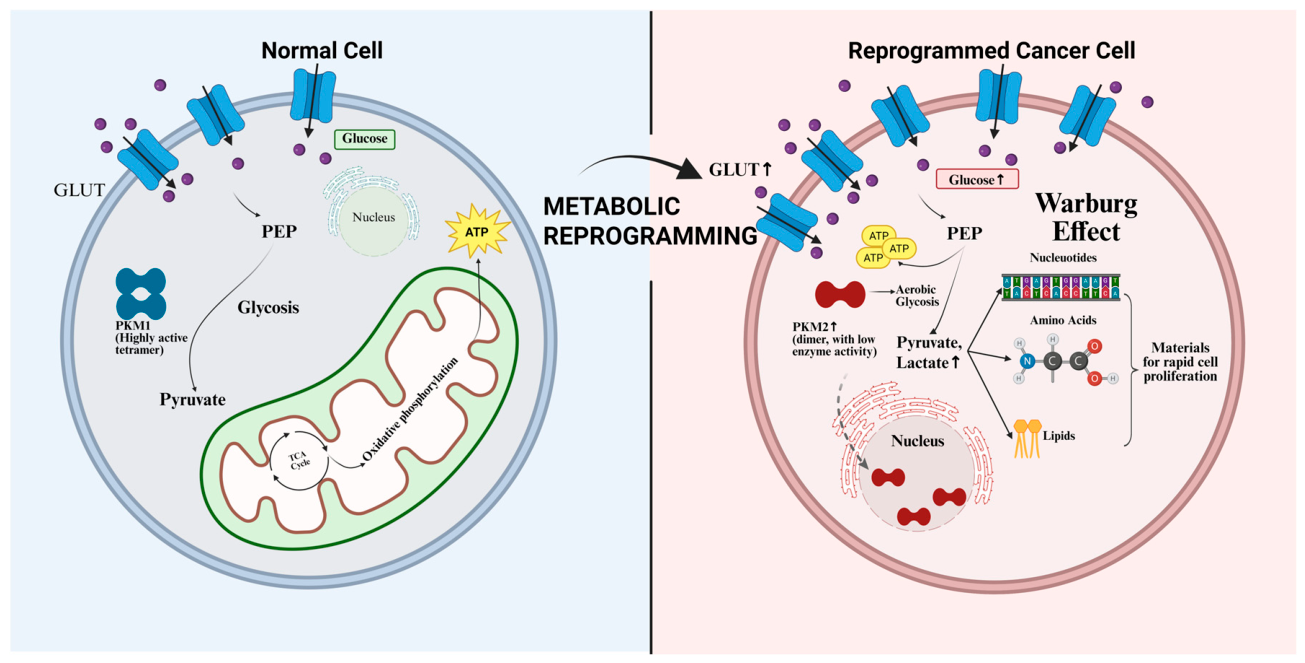
Figure 1. Metabolic reprogramming, Warburg effect, and the role of pyruvate kinase M2 (PKM2) in thyroid cancer. (Left) Normal cellular metabolism: Under normal physiological conditions, glucose is converted into pyruvate through glycolysis by the highly active tetramer PKM1, which then enters mitochondria for the tricarboxylic acid cycle (TCA cycle) and oxidative phosphorylation, producing a large amount of ATP. (Right) Metabolic reprogramming and Warburg effect in thyroid cancer cells: Cancer cells exhibit metabolic reprogramming, prioritizing glycolysis even under aerobic conditions, converting most glucose to lactate and excreting it from the cell, a phenomenon known as the “Warburg effect”. In thyroid cancer cells, abundant pyruvate kinase M2 (PKM2) typically exists in a low activity dimer form, leading to the accumulation of glycolytic intermediates in the cytoplasm. These accumulated intermediate products are diverted to the biosynthetic pathway, providing large molecular precursors, such as nucleotides, amino acids, and lipids, for the rapid proliferation of cancer cells. PKM2 can simultaneously relocate to the cell nucleus, which will be discussed later. Created in Biorender. Shenshen Li. (2026) http://BioRender.com/.
Molecules 31 01811 g001
Figure 2. BRAF V600E and RAS mutations activate the MAPK/ERK (RAF→MEK→ERK) and PI3K/AKT (PI3K→AKT→mTOR) signaling cascades, respectively. Both pathways converge on the transcription factors c-Myc and HIF-1α, which translocate into the nucleus and promote the transcriptional upregulation of the PKM2 gene, leading to increased PKM2 mRNA levels. Additionally, the BRAF V600E mutation enhances telomerase activity, contributing to cell immortalization. The interconversion between the tetramer and dimer forms is a key regulatory switch in tumor metabolic reprogramming. Post-transcriptional regulation by microRNAs (e.g., miR-148a, miR-326) modulates PKM2 expression by targeting PKM2 mRNA. In the context of tumor suppressor loss, wild-type p53 normally activates TIGAR (TP53-induced glycolysis and apoptosis regulator), which suppresses aerobic glycolysis; however, p53 mutations abrogate this regulatory axis, resulting in the loss of TIGAR-mediated glycolytic inhibition and further enhancement of the Warburg effect. Created in Biorender. Shenshen Li. (2026) http://BioRender.com/.
Molecules 31 01811 g002
Figure 3. (A) pre-miR-148a; (B) pre-miR-326. They are obtained from miRBase (http://www.mirbase.org) (accessed on 8 April 2026) and visualized using RNAfold.
Molecules 31 01811 g003
Figure 4. Predicted putative miR-148a and miR326-binding sites in the 3′-untranslated region (UTR) of PKM2. Created in Biorender. Shenshen Li. (2026) http://BioRender.com/.
Molecules 31 01811 g004
Figure 5. Nuclear PKM2 drives tumor proliferation and apoptosis resistance via β-captein, STAT3, and downregulate p53 transcription. In the TME, PKM2 promotes cytokine expression and MDSCs accumulation, suppressing T cells. PKM2 tetramer in CD8+ T cells can enhance the effect of anti-PD-L1 therapy. Created in Biorender. Shenshen Li. (2026) http://BioRender.com/.
Molecules 31 01811 g005
Figure 6. Chemical structures of the seven compounds evaluated in this study. All chemical structures were retrieved from the PubChem database (https://pubchem.ncbi.nlm.nih.gov) (accessed on 17 May 2026). (A) TEPP-46, (B) DASA-58, (C) Parthenolide, (D) Shikonin, (E) DMAMCL, (F) 2-Hydroxycinnamaldehyde, (G) Oxymatrine.
Molecules 31 01811 g006
Table 1. PKM2 regulation mechanisms in other tumors.
Regulatory MechanismMechanism of ActionEffect on PKM2TissueReferences
miR-122, miR-148a, miR-326Binds to the 3′UTR of PKM2 mRNAInhibits PKM2 transcriptionHepatocellular carcinoma (HCC) and thyroid Cancer [54,55]
PhosphorylationFGFR1, AKT, ERK can phosphorylate at the Tyr105, Ser37, Thr365 sites, respectively. TRIM35 can inhibit phosphorylation at Tyr105.Destabilize the active tetrameric PKM2 and lead to nuclear translocation of PKM2FGFR1: Thyroid cancer
AKT and ERK: No specific tumor
TRIM35: HCC
[56,57,58,59,60]
CrotonylationCrotonylation site at Lys305 Vascular smooth muscle cell[61]
SuccinylationSuccinylation at Lys498. The process can be reversed by SIRT5.No specific tumor[62,63]
O-GlcNAcylationTargets Thr405 and Ser406, residues of the region. No specific tumor[64]
Acetylationp300 can acetylate at Lys305 and Lys433 site of PKM2, while SIRT1 can reverse the acetylation at Lys305. Breast cancer and non-small cell lung cancer (NSCLC)[65,66,67,68,69]
Oxidative ModificationOxidation at Cys358No specific tumor[70,71]
Metabolite Regulation
Fructose-1,6-bisphosphate (FBP)Binds to the allosteric site of PKM2Induces its transformation from low-activity dimer to high-activity tetramer, significantly enhancing its glycolytic enzyme activity, inhibits nuclear translocationNo specific tumor[72,73,74,75,76,77]
SerineCompetitively binds to the active center of PKM2Competitively inhibits its enzyme activity, promotes nuclear translocationNo specific tumor[78,79]
Disclaimer/Publisher’s Note: The statements, opinions and data contained in all publications are solely those of the individual author(s) and contributor(s) and not of MDPI and/or the editor(s). MDPI and/or the editor(s) disclaim responsibility for any injury to people or property resulting from any ideas, methods, instructions or products referred to in the content.