Article

Molecular and Immobilized Tripodal Phosphine Ligands and Their Trinuclear Palladium Complexes

Department of Chemistry, Texas A&M University, College Station, TX 77842-3012, USA
*
Author to whom correspondence should be addressed.
Molecules 2025, 30(7), 1616; https://doi.org/10.3390/molecules30071616
Submission received: 27 February 2025 / Revised: 29 March 2025 / Accepted: 30 March 2025 / Published: 4 April 2025
(This article belongs to the Special Issue Fundamental Aspects of Chemical Bonding—2nd Edition)

Abstract

:
The synthesis and characterization of the tripodal phosphines RSi(CH2CH2PPh2)3 (R = Me, OMe, OEt) (13) is described. The 1H NMR spectra of all phosphines display virtual coupling patterns. The ligands form the corresponding trinuclear Pd complexes [RSi(CH2CH2PPh2)3]2(PdCl2)3 (46) with three PdCl2 moieties sandwiched between two tripodal ligands. The complexes 4, 5, and 7 (R = OH) have been analyzed by single crystal X-ray diffraction. The coordination at the Pd center is square planar with the phosphine groups occupying trans positions. The 31P{1H} MAS NMR spectra of polycrystalline 1 are in accordance with the packing motif of the molecules in the unit cell. The tripodal ligand 3 has successfully been immobilized on silica as 3i. It coordinates PdCl2 on the surface, as demonstrated by 31P{1H} MAS NMR. Hereby, the cis coordination is prevalent when 3i has maximal surface coverage. At low surface coverage, one tripodal linker can accommodate trans coordination at the metal center. A surface-bound trinuclear Pd complex has been generated, as well as a heterobimetallic Pd/Cu complex. All surface species have been characterized by 31P{1H} MAS NMR.

1. Introduction

General Introduction

One of our main research interests is immobilizing molecular catalysts on solid supports to allow for easy separation from the reaction mixtures and recycling [1]. Silica has been mostly chosen as the support material because it is inexpensive and available in a variety of pore and particle sizes. It is temperature stable and mechanically robust and can be separated by settling. It retains the immobilized catalyst even in cases when metal nanoparticles form [2,3,4]. Bifunctional phosphines have proven to be favorable linkers because they can coordinate metal complexes and irreversibly bind to silica surfaces. The latter can be accomplished by creating phosphonium salts on the surface [2,4,5], however, the more straightforward way of immobilization is via the reaction of alkoxysilyl groups with surface silanol and siloxane groups [6]. Catalysts immobilized with mono-, di-, and triphosphine linkers [1,3] incorporating ethoxysilyl groups could be recycled many times in a batchwise manner [3]. In general, chelating phosphine ligands increase the lifetimes of the immobilized catalysts because leaching is diminished, as demonstrated by HRMAS NMR spectroscopy earlier [7,8,9]. Tripodal phosphine linkers, in particular, offer the potential to coordinate two different metals with one linker [10], as desirable, for example, for the Sonogashira reaction [9]. Tripodal phosphine linkers have successfully been applied for immobilizing Wilkinson-type rhodium hydrogenation catalysts [2,3]. However, one last challenge remains. After several catalytic runs, most often metal nanoparticles form because of the mobility of the phosphine linkers in the presence of a solvent that allows for contact of the metal centers [2,3]. This process occurs especially when linkers with long, flexible methylene chains of up to 11 CH2 segments are used [2,3]. In order to suppress the nanoparticle formation, while retaining the favorable features of the chelate phosphines, we opted for tripodal phosphine linkers with shorter methylene chains incorporating alkoxy groups for immobilization on silica (Figure 1). Unfortunately, the tripodal phosphine congeners with only one methylene group tend to decompose when immobilized on silica [11,12].
However, the tripodal ligand 3 with ethylene groups separating the ethoxysilyl and phosphine groups, can be bound to silica as 3i without decomposition (Figure 2). The tripod structure remains intact, as demonstrated by solid-state NMR spectroscopy.
In this contribution, we describe the synthesis and characterization of the previously unused tripodal phosphine linkers of the type RSi(CH2CH2PPh2)3 (R = Me, OMe, OEt) (13) and their trinuclear Pd complexes [RSi(CH2CH2PPh2)3]2(PdCl2)3 (46) (Figure 1). The complexes 4, 5, and 7 (R = OH) have been analyzed by single crystal X-ray diffraction. It has been demonstrated by 31P{1H} MAS NMR that the tripodal ligand 3 can successfully be immobilized on silica as 3i and that PdCl2 and CuCl can be bound to it. Diverse mono- and heterodinuclear surface-bound complexes could be identified (Figure 2).

2. Results and Discussion

2.1. Tripodal Phosphine Ligands

The tripodal phosphine ligands 13 (Figure 1) have been synthesized by radical-initiated hydrophosphination of the corresponding trivinylsilanes with HPPh2. The reactions could be initiated by UV irradiation, as described previously for 1 [13], and the tripodal phosphine EtOSi(CH2PPh2)3 [11,12]. Alternatively, AIBN (azobisisobutyronitrile) can function as a radical initiator, as described for the synthesis of tripodal phosphine ligands with long methylene chains incorporating ethoxysilyl groups [2,3]. The tripodal phosphines 13 were obtained as colorless solids in yields higher than 84%.
The 31P{1H} NMR signals of the tripodal ligands 13 are found within the narrow chemical shift range from −9.4 to −9.9 ppm (Figure S1). There is only one clean singlet for each tripodal ligand, indicating that all PPh2 groups are chemically equivalent.
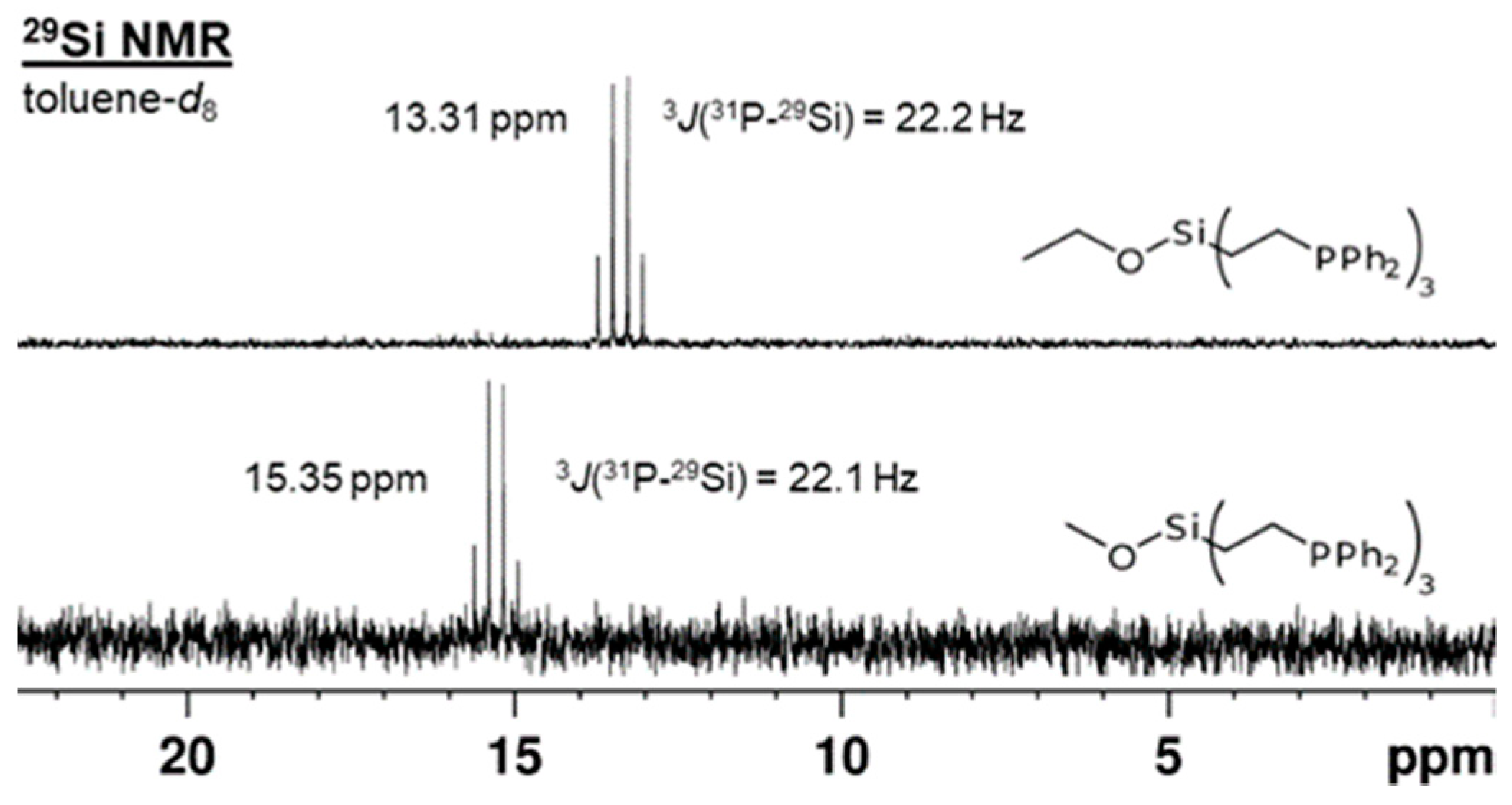
Direct proof for the presence of three equivalent phosphorus atoms in the tripodal ligands 13 can be derived from the 29Si{1H} NMR spectra (Figure 3). The 29Si{1H} signals are 1:3:3:1 quartets with 3J(31P-29Si) coupling constants of 21.2 Hz (1), 22.1 Hz (2), and 22.2 Hz (3), in accordance with values for similar systems that range from 12.1–27.8 Hz [14,15,16]. The δ(29Si) of 13 lie within the expected chemical shift range for tetraalkyl- and alkoxytrialkylsilanes from 0 to 20 ppm [11,12].
The 1H NMR spectra of all tripodal ligands show virtual couplings for the signals of the diastereotopic ethylene protons (Figures S2–S5). The signal shapes are textbook examples of virtual couplings [1,11,17,18]. The shapes and the spans of the signals, defined as the Hz distances between the outer lines, do not change when the strength of the external magnetic field is increased from 400 to 500 MHz. For example, the methylene signals of 3 at 1.87 ppm and 0.67 ppm retain the distances of 17.7 Hz and 27.0 Hz between their outer lines (Figure S5). When the solvent is changed from CDCl3 to C6D6, a slight change of the distances to 17.3 Hz and 27.2 Hz occurs.
In addition to the 1H NMR spectra, 13C{1H} NMR (Figures S6 and S7) confirms the presence of the methyl, methoxy, and ethoxy groups bound to Si in 13. The phenyl carbon signals have the expected chemical shifts and display the characteristic 1–3J(31P-13C) couplings of the ipso, ortho, and meta carbon nuclei with 31P that have been observed for tripodal diphenylalkylphosphines previously [2,3,11].

2.2. Trinuclear Palladium Complexes

When the tripodal phosphines 13 are reacted with (PhCN)2PdCl2 the complexes 46 result as polycrystalline solids (Figure 1). The yields are respectable but not quantitative because of the intrinsic potential of tripodal ligands to form coordination polymers in solution. Consequently, the 31P{1H} NMR signals undergo a characteristic downfield shift that indicates the coordination of the phosphines to the metal centers (Figure S1 and Figure 4). All phosphorus nuclei are chemically equivalent due to the symmetry of the complexes and therefore, their signals show no couplings.
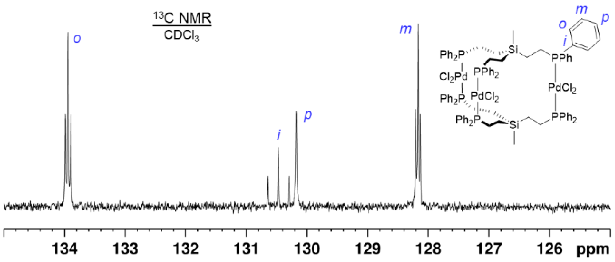
However, the 13C{1H} NMR spectrum of 4, for example, shows virtual couplings for the ipso, ortho, and meta carbons (Figure 5 and Figure S8) [1,11,17,18]. This means that different 31P nuclei communicate with each other and the 13C{1H} NMR signals are split into triplets by seemingly identical phosphorus coupling partners [17,18]. In principle, the J(31P-13C) couplings could be propagated along the pathway over the Si atom. However, in this case, more complex coupling patterns would be expected and the 13C{1H} NMR signal of the carbon bound to Si should also be split into a multiplet. This is not the case (Figure S8) but the alkyl carbon bound to 31P features a virtual triplet. Therefore, it can be concluded that this triplet and the virtual couplings to the aryl carbon nuclei are due to the coupling pathway over the Pd center.

2.3. X-Ray Crystallography

The trinuclear Pd complexes 4 and 5 could be crystallized from a solvent mixture of DCM with ether and single crystal X-ray diffraction confirmed the structures depicted in Figure 1. Unfortunately, high-quality crystals of 6 could not be obtained because the ethoxysilyl group was prone to alkoxy exchange and hydrolysis. Attempts to crystallize 6 in the presence of MeOH led to alkoxy exchange and crystals of 5. Single crystals of 5 obtained from MeOH (5a) show significant structural differences compared to the crystals of 5 obtained from DCM/ether mixtures (5b). Traces of water in the solvent used for crystallizing 6 led to the X-ray structure of the corresponding silanol 7 (R = OH). All data and measurement details for X-ray diffraction are reported in the Supplementary Materials and in ref. [19]. The single crystal structures and unit cells of 4, 5, and 7, as well as solid-state NMR spectra of polycrystalline 4 are displayed in Figure 6, Figure 7, Figure 8, Figure 9, Figure 10 and Figure 11, Figures S9 and S10.
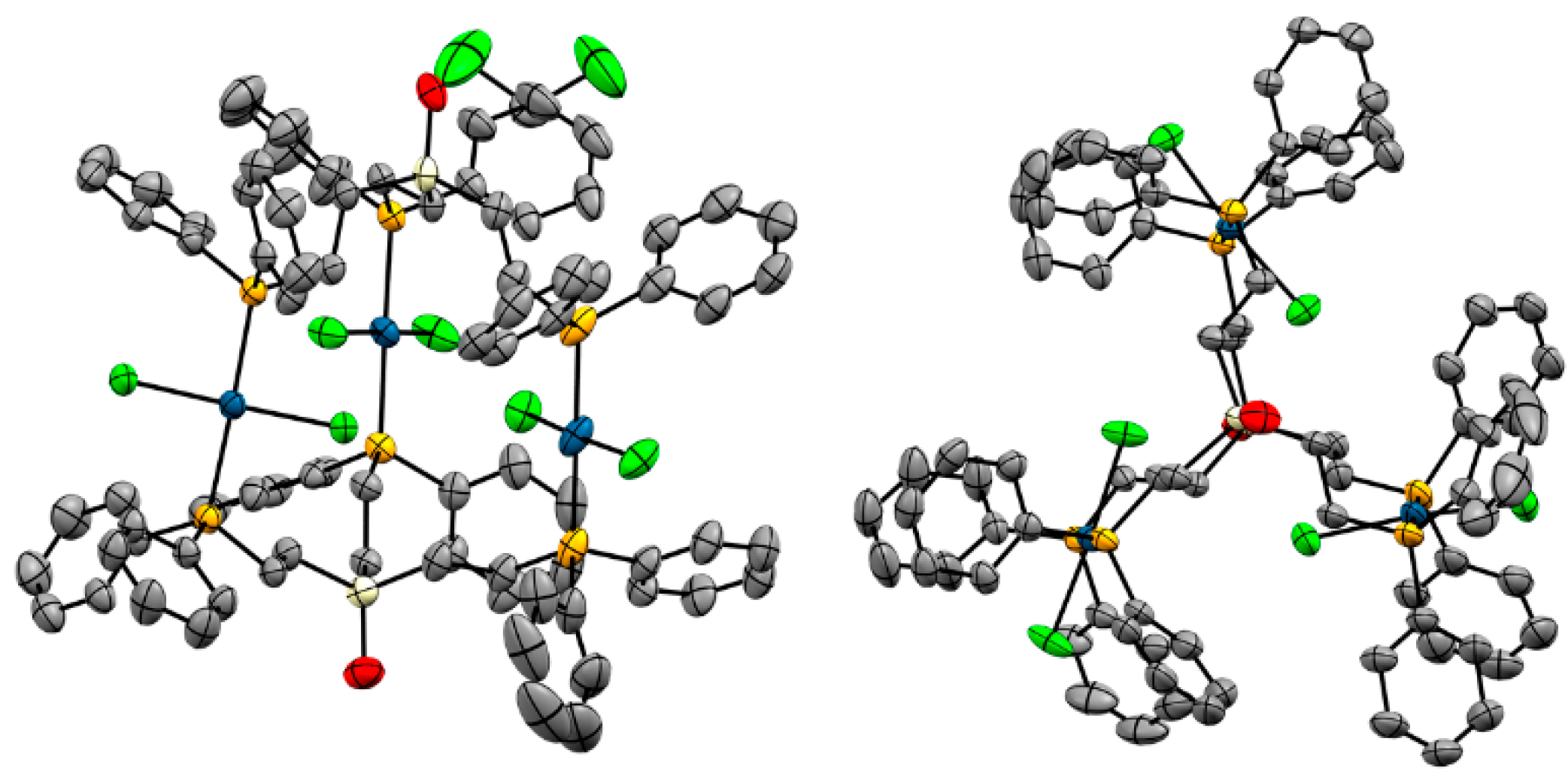
The structure of 4 and its unit cell are displayed in Figure 6 and Figure 7. Three PdCl2 units are sandwiched between two tripodal phosphine ligands 1. The phosphine groups occupy the trans positions and all P–Pd–P axes are nearly parallel.
Figure 6. Single crystal X-ray structure of one molecule of 4. View from the side (left) and along the Si···Si axis (right). Hydrogen atoms, solvent molecules, and disordered phenyl rings are omitted for clarity.
Molecules 30 01616 g006
Interestingly, the view from the top of the complex along the Si···Si axis reveals that the ethylene groups of the tripodal ligands are nearly eclipsed with very small dihedral angles (Figure 6). Even the phenyl groups are parallel. The orientation of the Cl atoms, however, appears to be random and dominated by the packing motif. Under the conditions of the X-ray diffraction measurement the Cl–Pd–Cl moieties of 4 do not rotate although the available space would allow for this [20].
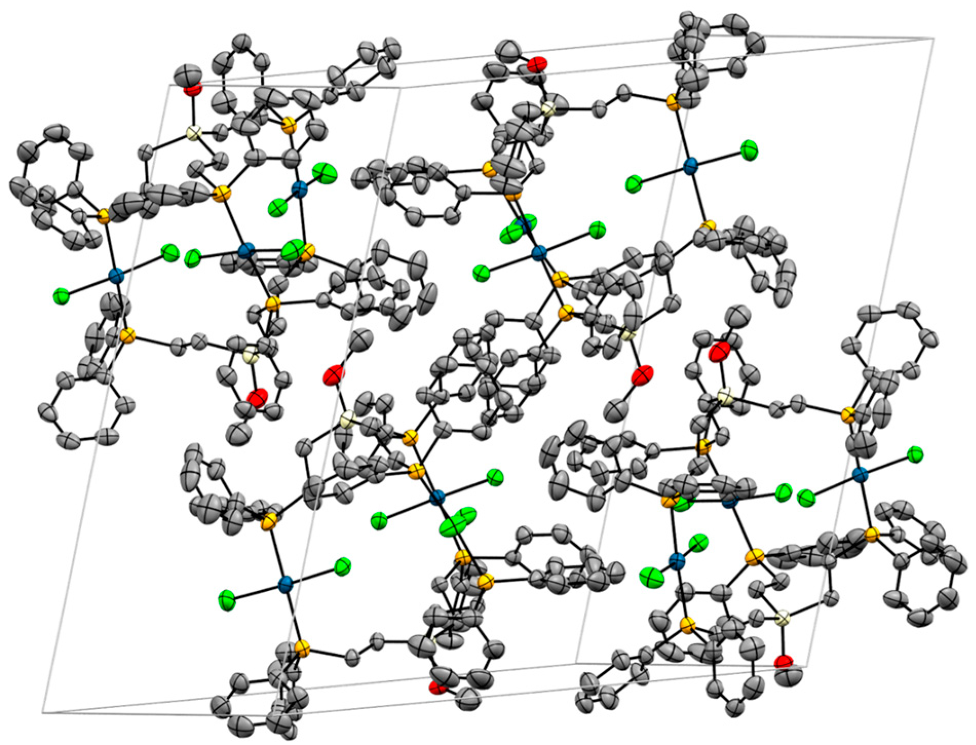
Figure 7. Unit cell of the single crystal X-ray structure of 4, incorporating six solvent molecules (DCM). Hydrogen atoms and disordered phenyl rings are omitted for clarity.
Molecules 30 01616 g007
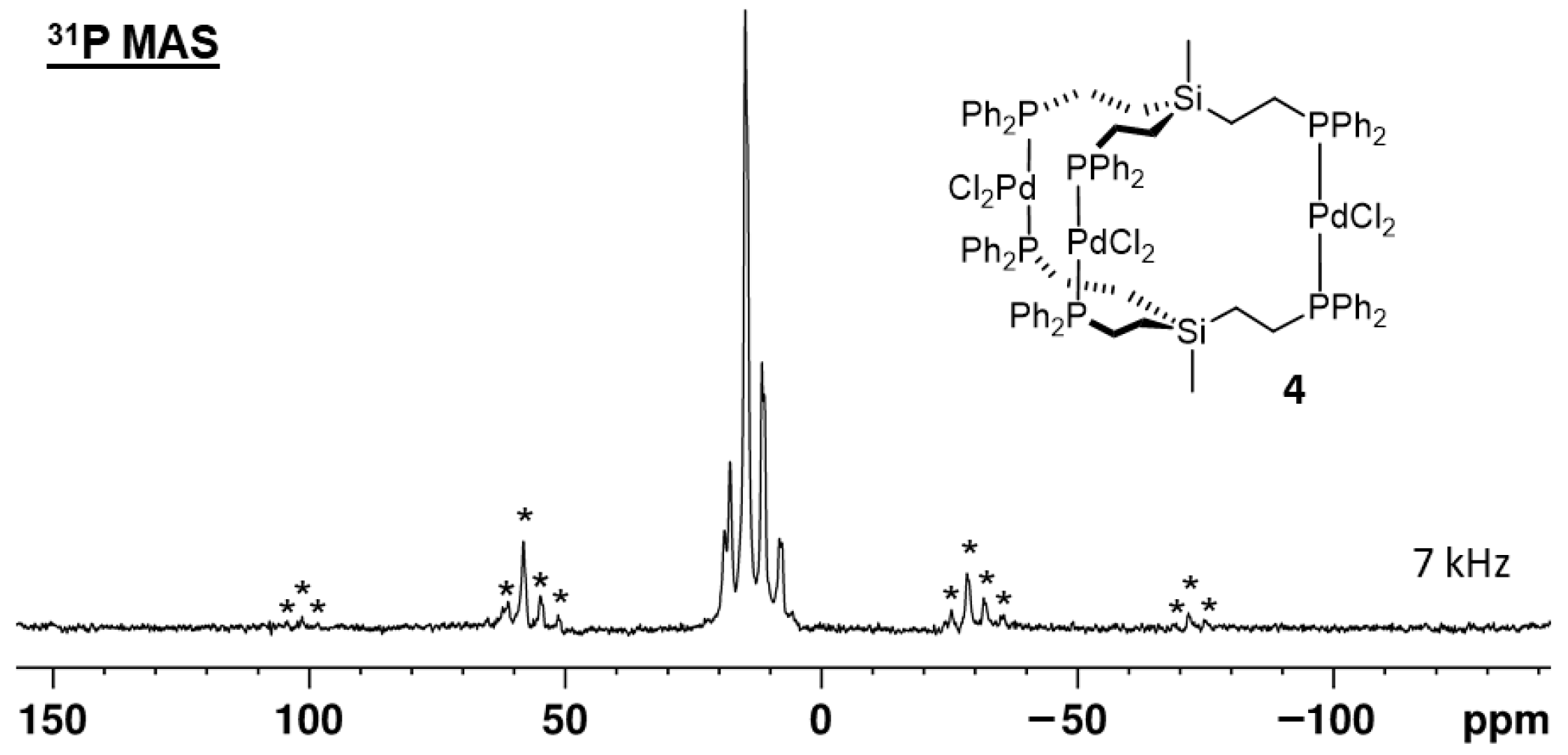
The unit cell of 4 is composed of two molecules. The remaining voids are filled with six DCM molecules. Regarding the arrangement of the complexes within the unit cell, there are 12 magnetically inequivalent phosphorus nuclei. However, the two complex molecules have a similar orientation in space, and, therefore, pairs of two phosphorus nuclei in analogous positions are expected to yield similar chemical shifts in the 31P{1H} MAS NMR spectrum of solid 4. To test this hypothesis, we recorded the 31P{1H} MAS spectra of polycrystalline 4 at different rotational speeds (Figure 8 and Figure S11). As expected, the spectra show multiple, partly overlapping, signals with at least six isotropic lines. The chemical shift range of the isotropic lines corresponds to the δ(31P) of alkyldiphenylphosphines trans-coordinated to PdCl2 [7,8,9].
Figure 8. 31P{1H} MAS NMR spectrum of polycrystalline 4, rotated with a spinning frequency of 7 kHz. Asterisks denote rotational sidebands.
Molecules 30 01616 g008
Figure 9 displays the structure of 5, viewed from the side and along the Si···Si axis. The structural features are very similar to those discussed above for 4, although the alignment of the ligand ethylene groups is not quite as precisely eclipsed as in 4. The biggest difference is visible in the unit cell of 5 (Figure 10). Four trinuclear complexes are densely packed in the unit cell, with disordered solvent molecules filling the voids.
Figure 9. Single crystal X-ray structure of one molecule of 5a. View from the side (left) and along the Si···Si axis (right). Hydrogen atoms and disordered solvent molecules are omitted for clarity.
Molecules 30 01616 g009
Figure 10. Unit cell of 5a. Hydrogen atoms and disordered solvent molecules are omitted for clarity.
Molecules 30 01616 g010
The structure of one molecule of 7 resembles more closely the one of 4 (Figure 11). However, the unit cell is different and shows a very complex packing motif (Figure S12).
Figure 11. Single crystal X-ray structure of one molecule of 7 with one included dichloromethane molecule. View from the side (left) and along the Si···Si axis (right). Hydrogen atoms are omitted for clarity.
Molecules 30 01616 g011
Interestingly, the trinuclear sandwich motif we found for 47 has only been described previously for two Pd complexes [21] and one Ni complex [22]. In the case of PdI2 and PdCl2, the tripodal ligands were O=P(CH2CH2PPh2)3 and O=P(CH2CH2P(S)Ph2)3, respectively. The ligand in the trinuclear NiCl2 complex was CH3C(CH2CH2PPh2)3. In the following, we include these complexes in our discussion of the X-ray data.
The unit cell and molecular structure of 4 and the complex Pd3I6(pOp3)2 [21] are nearly identical, with close to perfect eclipse of the methylene and phenyl groups as observed in Figure 6.
In the square planar complexes with trans coordination of the ligands L, ideally the angles L–M–L should be 180°. However, the values provided in Table 1 show that there is significant scatter, sometimes within the same complex. For example, the structure of 5, derived from Et2O/DCM crystallization (5b), shows one significantly smaller L–M–L value of 165.45 (3)° as compared to 176.33 (3)° and 176.45 (3)°. In other words, this complex has an indentation on one side. The most symmetric shape is reported for the complex with an additional S atom that extends the tripodal ligand in Pd3Cl6(pOp3S3)2 [21].
The X–M–X angles of the PdCl2 complexes are closer to the ideal 180° than the PdI2 or NiCl2 complexes (Table 2). These differences are probably due to the smaller atomic radius of Ni(II) and the larger radius of iodide in the complex Pd3I6(pOp3)2, leading to interactions between the halide and phenyl and methylene groups (Table 2). These interactions are minimized by the additional sulfur atom of the complex Pd3Cl3(pOp3S3)2, which possesses X–M–X angles of 179.5 (1)°, which are closest to the ideal 180° [21].
With the exception of the complex Pd3Cl3(pOp3S3)2, the ethylene groups in all tripodal ligands in the sandwich complexes in Table 3 are eclipsed. The L–Center–Center–L dihedral angles amount to less than 12.7° for all complexes, except for Pd3Cl3(pOp3S3)2 (50.68°). The eclipsed ethylene groups in the complex 4, 5a, and 7 are visible, for example, in the views along the Si···Si axes (Figure 6, Figure 9, and Figure 11).
The distances between the central atoms of the ligands vary within a large range from 4.85 (9) to 8.530 (2) Å (Table 4). These distances correlate inversely with the distances between the metal centers. For example, complex 7 has the largest distance between the centers, corresponding to relatively small M-M distances. This is understandable, as elongating the “balloons” at the top and bottom will result in more svelt molecules with reduced circumference in the middle due to the flexible nature of the tripodal ligands. Overall, Pd3Cl3(pOp3S3)2 has the longest metal-metal distances, however, the extra sulfur atom between the metal and center has to be acknowledged. Interestingly, for the center-center and metal-metal distances, the nature of the metal atom, whether it is Pd or Ni, does not seem to play a crucial role (Table 4).

2.4. Immobilization on Silica

One of the beneficial features of immobilized tripodal ligands is that coordination polymers can no longer occur. With low surface coverages, coordination of the metal by a second ligand is no longer possible because of the large, fixed distance between the ligands. In contrast to adsorbed species, covalently silica-bound ligands are not mobile on the surface. Furthermore, once the linker containing the alkoxysilyl group is bound to silica, it is no longer water-sensitive. In contrast to 6, it incorporates a Si-O-C linkage that is easily hydrolyzed to a silanol group to form 7, once a siloxane (Si-O-Si) group is formed by covalent binding to the silica surface, the linker is no longer prone to hydrolysis. In order to remove a phosphine linker from the silica surface, harsh conditions are needed, as described earlier [23,24].
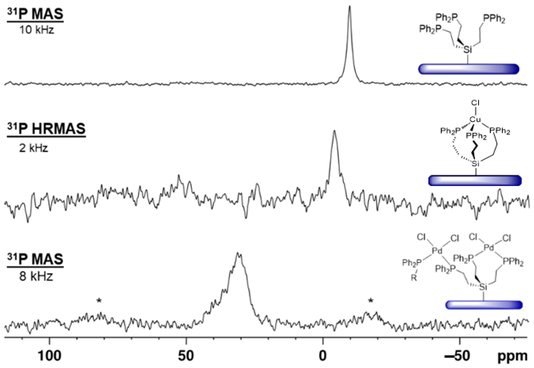
The tripodal phosphine ligand 3 has successfully been immobilized on silica as 3i according to optimized procedures [1,6,23,24]. The 29Si CP/MAS (cross-polarization in combination with magic angle spinning) shows the corresponding signal at 10 ppm, in accordance with the chemical shift of 29Si{1H} NMR signals of silica-tethered trialkylethoxysilanes [3]. In the 31P{1H} MAS spectrum a signal at –10 ppm is found (Figure 12, top), in agreement with the δ(31P) value of the molecular ligand 3 in solution (−9.39 ppm).
When the ligand 3i, immobilized on silica with maximal surface coverage [23,24], is treated with (MeCN)2PdCl2 all phosphine groups are coordinated to the metal center. The signal is broad with a characteristic CSA (chemical shift anisotropy) that manifests in large first-order rotational sidebands even at 8 kHz rotational frequency (Figure 12, bottom) [9]. The chemical shift of about 30 ppm indicates that the phosphines are mostly cis-coordinated to the metal center [7,8,9]. Since the surface coverage is dense, the Pd complex can span different neighboring tripodal ligands as depicted in Figure 12.
The immobilized ligand 3i is not limited to coordinating Pd. When 1.5 equivalents of CuCl are added, the surface-bound triphosphine copper chloride complex depicted in Figure 12 (middle) is obtained. The chemical shift of about −5 ppm for the 31P{1H} HRMAS (high-resolution magic angle spinning) [7,8,9] NMR signal corresponds well to the value reported earlier [9,25].
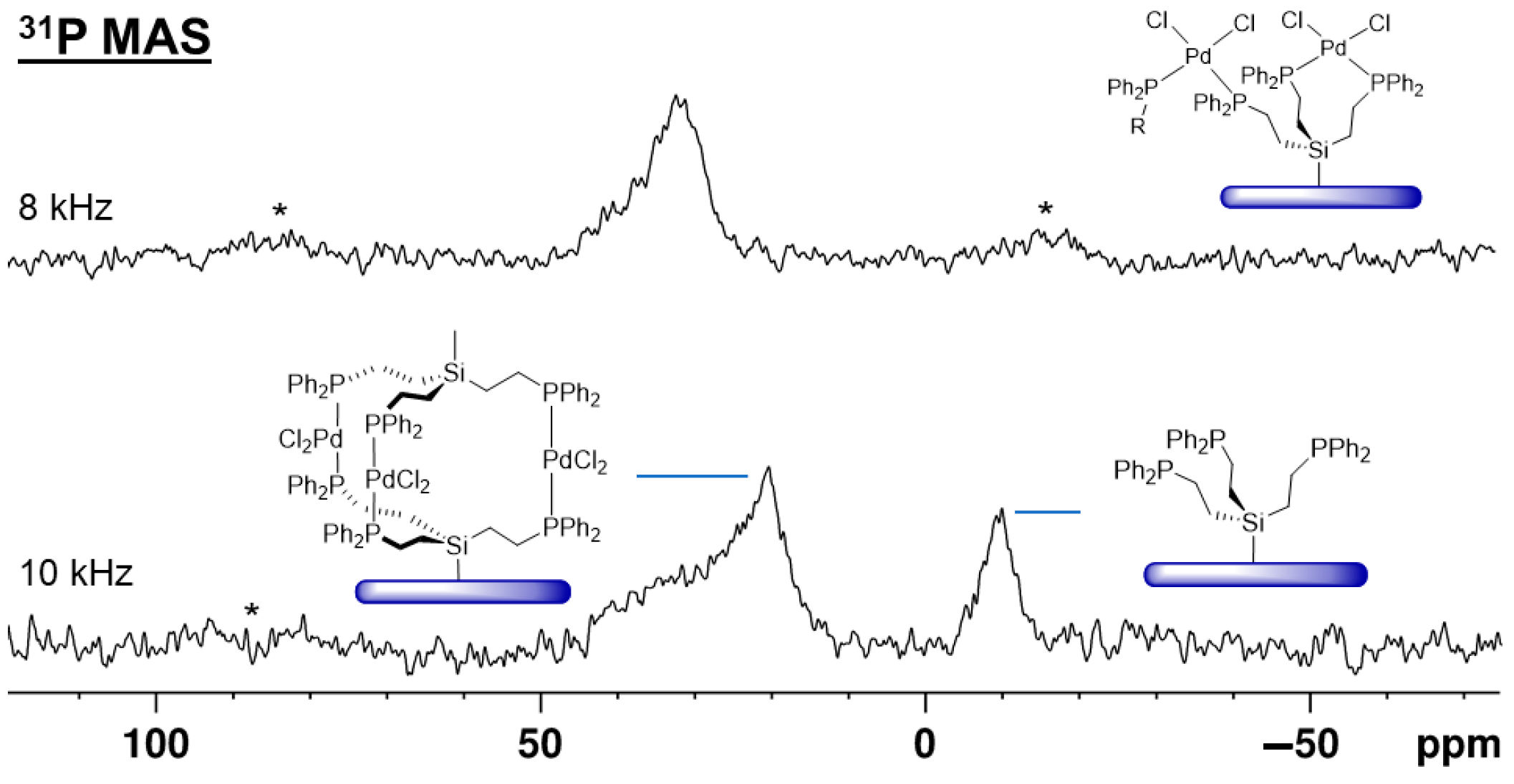
Interestingly, treating the Pd complex, cis-coordinated by 3i (Figure 13, top), with the tripodal ligand 1, the signal for the uncoordinated surface-bound phosphine 3i reappears (Figure 13, bottom). This means that ligand 1 replaces the phosphine groups of 3i at the Pd center. Furthermore, the cis-coordinated Pd complex mostly turns into the surface-bound sandwich complex shown in Figure 13. This immobilized complex corresponds to the molecular complex 4, with trans coordination of the phosphine ligands and a 31P chemical shift of about 22 ppm. These results are in accordance with earlier studies that suggested that the PdCl2 moiety is able to move from one surface-bound phosphine ligand to a more favorable one in the presence of a solvent [7,8,9]. In the presented scenario, trans coordination at the Pd center is clearly preferred and is the driving force for the formation of the immobilized trinuclear Pd complex.
When the ligand 3i is immobilized with only 24% of the maximal surface coverage and treated with one equivalent of (MeCN)2PdCl2, the 31P{1H} MAS signal of the uncoordinated ligand is still visible at about –10 ppm (Figure 14, top). This means that in the solid state, there is no intra- or intermolecular rapid ligand exchange, as in the presence of a solvent [7,8,9]. Furthermore, the signal of cis-coordinated phosphines appears at about 30 ppm, and in addition, a resonance for trans-coordinated phosphine groups (Figure 14, top) at ca. 22 ppm. Since ligand 3i is dilute on the surface in this case, the tripodal ligand obviously has a large enough span to accommodate the intramolecular trans coordination of Pd.
One of our key interests is creating an efficient immobilized Sonogashira Pd/Cu catalyst system that has been described for coupling iodobenzene and phenylacetylene using mono- and bidentate phosphine linkers previously [7,8,9]. The tripodal ligand 3i allows for both metals to be bound with one linker molecule. This is favorable because the Pd component does not have to migrate to the Cu complex prior to the onset of catalysis [9]. Starting with the ligand 3i that has been tethered to silica with a low surface coverage of 24% and then treated with PdCl2 (Figure 14, top), the Cu complex can be bound by the remaining uncoordinated phosphine group. Accordingly, after treatment with (MeCN)3CuCl, the signal for uncoordinated phosphine groups has disappeared, while the resonance for Cu-bound phosphines emerges at about –3.5 ppm (Figure 14, bottom). There is an obvious preference for the trans coordination at Pd to persist, while the signal for the cis-coordinated phosphines is diminished in intensity. Preliminary experiments show that the combined immobilization of the Pd and Cu components leads to an active Sonogashira catalyst and detailed catalysis studies using this system will be undertaken to compare it with previously reported catalysis results [8,9].
Finally, it should be pointed out that the 31P{1H} MAS signals obtained for the phosphine groups coordinated to the Pd center are not identical to the phosphine oxide signal. When the immobilized ligand 3i is reacted with aqueous H2O2/acetone, all phosphines are turned into phosphine oxides [26]. The signal appears at 38 ppm, downfield-shifted enough to distinguish it from the resonances of Pd-coordinated phosphines (Figure S13).

3. Conclusions

Three tripodal phosphine ligands incorporating ethylene spacers and silyl groups have been synthesized and characterized by 1H, 13C{1H}, 29Si{1H}, and 31P{1H} NMR spectroscopy (13). All phosphines displayed virtual couplings for the ethylene proton signals and the 29Si resonances were split into quartets due to the couplings to three 31P nuclei. The trinuclear Pd complexes 46 have been synthesized with tripodal ligands and characterized by single-crystal X-ray diffraction and multinuclear NMR spectroscopy. The structures display trans coordination for all three Pd centers that are sandwiched between two tripodal ligands. The ethylene groups of the tripodal ligands are eclipsed.
In summary, the described NMR spectroscopic and X-ray diffraction results expand the general understanding of molecular and surface-bound metal complexes with tripodal phosphine ligands. Pd- and Cu-containing complexes could be generated on a silica surface and the foundation for the next generation of immobilized Sonogashira catalyst systems with two different metals bound to one tripodal ligand has been established [9,10]. Preliminary results indicate that the presented immobilized Pd/Cu system, in combination with phenylacetylene and iodobenzene, is an active Sonogashira catalyst under the conditions described previously [7,8,9]. In future projects, the catalytic activities of these systems will be explored in detail, optimized, and compared to those of Sonogashira catalysts with separate Pd and Cu components on a silica surface [7,8,9].

4. Experimental Section

4.1. General Information

All reactions were performed under an inert atmosphere unless specified otherwise. The solvents were dried in a conventional solvent purification apparatus and subsequently kept under purified nitrogen, with the exception of methanol. Methanol was degassed by 3 freeze-pump-thaw cycles and kept over 4 Å molecular sieves for 3 days before use. The melting points and ranges were obtained with an Optimelt melting point apparatus using sealed capillaries. The compounds started melting at the given lower values and reached the clear points at the high values. The silica (average pore diameter 40 Å, particle size 0.06−0.2 mm, specific surface area 750 m2/g) was acquired from SiliCycle and dried at 300 °C and 0.1 Torr for 3 days before use in order to allow for the ethoxysilane to bind to the surface [27]. The maximal surface coverage (100%) was determined by offering a weighed excess of 3 in the immobilization step. The original amount of 3 minus the residual 3 found in the supernatant and washings after the immobilization corresponds to the surface-bound 3i with maximal surface coverage.

4.2. NMR Spectroscopy

The 1H, 13C{1H}, 29Si{1H}, and 31P{1H} solution NMR spectra were recorded on a Varian VnmrS 500, Varian Inova 500, or Bruker Avance Neo 400 NMR spectrometer. 1H and 13C{1H} NMR spectra were internally referenced to solvent signals. The 29Si{1H} NMR spectra were externally referenced to hexamethyldisiloxane (δ = 6.53 ppm). 31P{1H} NMR spectra were externally referenced to chlorodiphenylphosphine (δ = +81.92 ppm). The solid-state NMR spectra were measured with a Bruker Avance 400 MHz NMR spectrometer. All solid-state NMR measurements were recorded with the Bruker HPDEC standard pulse program with high-power 1H decoupling (31P) and cross-polarization (29Si). Typically, for 31P{1H} MAS measurements, 1024 scans with a pulse delay of 7 s were recorded to result in spectra with decent S/N ratios within 2 h. For 29Si{1H} NMR measurements 7000 scans with a pulse delay of 6 s were recorded. 31P{1H} MAS spectra were externally referenced to ammoniumdihydrogen phosphate (δ = 0.81 ppm). 29Si CP/MAS spectra were externally referenced to tetrakis(trimethylsilyl)silane (δ = 8.62 ppm). The spectra were processed using exponential multiplication with a linebroadening factor of 80 Hz prior to FT. The 31P{1H} MAS halfwidth of 3i was determined with no linebroadening. The halfwidth of 8i 31P{1H} MAS was obtained with the minimal effective linebroadening of 30 Hz, and likewise 3i 29Si CP/MAS with a linebroadening factor of 80 Hz.

4.3. X-Ray Diffraction

All details are provided in reference [19] and the Supplementary Materials.

4.4. Syntheses

MeSi(CH2CH2PPh2)3 (1). A flask was charged with trivinylmethylsilane (0.1692 g, 1.362 mmol), diphenylphosphine (2.945 g, 15.82 mmol), and AIBN (0.0552 g, 0.336 mmol, 1.92 mol%) and the reaction mixture was stirred for 72 h at 70 °C. Excess diphenylphosphine was vacuum-distilled from the product at 230 °C, yielding a viscous yellow oil. High purity was obtained by recrystallizing 1 from a 28 wt% solution in a 5:2 mixture of toluene and methanol by slow cooling. A colorless solid was obtained (0.7814 g, 1.144 mmol, 83.99% yield) with a mp of 98–101 °C, in accordance with the literature [13].
1H NMR (CDCl3, 500.13 MHz): δ (ppm) = 7.31–7.36 (m, 12H, Haryl), 7.28–7.31 (m, 18H, Haryl), 1.80–1.85 (m, 6H, PCH2), 0.55–0.61 (m, 6H, SiCH2), −0.02 (s, 3H, CH3); 13C{1H} NMR (CDCl3, 125.77 MHz): δ (ppm) = 136.65 (d, 1J(31P-13C) = 14.2 Hz, Ci), 132.69 (d, 2J(31P-13C) = 17.9 Hz, Co), 128.36 (s, Cp), 128.36 (d, 3J(31P-13C) = 6.4 Hz, Cm), 21.45 (d, 1J(31P-13C) = 14.2 Hz, PCH2), 10.85 (d, 2J(31P-13C) = 10.8 Hz, SiCH2), −5.69 (s, SiCH3); 31P{1H} NMR (CDCl3, 202.28 MHz): δ (ppm) = −9.88 (s); 29Si{1H} NMR (CDCl3, 79.37 MHz): δ (ppm) = 6.27 (q, 3J(31P-29Si) = 21.2 Hz) [28].
MeOSi(CH2CH2PPh2)3 (2). Trivinylmethoxysilane (0.1589 g, 1.133 mmol), diphenylphosphine (3.947 g, 21.20 mmol), AIBN (0.0801 g, 0.488 mmol, 2.14 mol%), and toluene (1.9 mL) were combined in a flask and stirred for 48 h at 80 °C. The excess of diphenylphosphine was vacuum-distilled from the reaction mixture at 230 °C and a viscous yellow oil was obtained. Three drops of diethyl ether were added and the mixture was cooled to 5 °C. After 18 h a colorless solid formed (0.6678 g, 0.9556 mmol, 84.34%). mp 75–77 °C.
1H NMR (CDCl3, 499.69 MHz): δ (ppm) = 7.33–7.38 (m, 12H, Ho), 7.28–7.31 (m, 18H, Haryl), 3.31 (s, 3H, CH3), 1.85–1.90 (m, 6H, PCH2), 1.64–1.71 (m, 6H, SiCH2); 13C{1H} NMR (CDCl3, 125.65 MHz): δ (ppm) = 138.69 (d, 1J(31P-13C) = 14.0 Hz, Ci), 132.88 (d, 2J(31P-13C) = 18.0 Hz, Co), 128.73 (s, Cp), 128.54 (d, 3J(31P-13C) = 6.6 Hz, Cm), 50.94 (s, CH3), 20.75 (d, 1J(31P-13C) = 14.6 Hz, PCH2), 8.11 (d, 2J(31P-13C) = 11.3 Hz SiCH2); 31P{1H} NMR (CDCl3, 202.28 MHz): δ (ppm) = −9.83 (s). 29Si{1H} NMR (toluene-d8, 99.30 MHz): δ (ppm) = 15.35 (q, 3J(31P-29Si) = 22.1 Hz).
EtOSi(CH2CH2PPh2)3 (3). Trivinylethoxysilane (0.2774 g, 1.798 mmol), diphenylphosphine (3.966 g, 21.30 mmol), AIBN (0.0672 g, 0.409 mmol, 1.75 mol%), and toluene (1.3 mL) were placed in a flask and stirred together for 24 h at 70 °C. The mixture was cooled to RT and additional AIBN (0.547 g, 0.333 mmol, 1.40 mol%) was added. The reaction mixture was stirred for 48 h at 70 °C. The excess of diphenylphosphine was vacuum-distilled from the reaction mixture at 230 °C and a viscous yellow oil was obtained. After cooling to RT a colorless solid formed in quantitative yield (1.282 g, 1.798 mmol). mp 89–95 °C.
1H NMR (CDCl3, 400.09 MHz): δ (ppm) = 7.32–7.37 (m, 12H, Ho), 7.27–7.31 (m, 18H, Haryl), 3.52 (q, 3J(1H-1H) = 7.0 Hz, 2H, OCH2), 1.84–1.89 (m, 6H, PCH2), 1.09 (t, 3J(1H-1H) = 7.0 Hz, 3H, CH3) 0.62–0.71 (m, 6H, SiCH2); 13C{1H} NMR (CDCl3, 100.63 MHz): δ (ppm) = 138.76 (d, 1J(31P-13C) = 14.2 Hz, Ci), 132.89 (d, 2J(31P-13C) = 18.0 Hz, Co), 128.71 (s, Cp), 128.53 (d, 3J(31P-13C) = 6.5 Hz, Cm), 58.82 (s, OCH2), 20.83 (d, 1J(31P-13C) = 14.6 Hz, PCH2), 18.69 (s, CH3), 8.50 (d, 2J(31P-13C) = 11.1 Hz, SiCH2); 31P{1H} NMR (CDCl3, 161.96 MHz): δ (ppm) = −9.39 (s); 29Si{1H} NMR (toluene-d8, 99.30 MHz): δ (ppm) = 13.31 (q, 3J(31P-29Si) = 22.2 Hz).
[MeSi(CH2CH2PPh2)3]2(PdCl2)3 (4). A solution of (PhCN)2PdCl2 (0.2771 g, 0.7224 mmol) in 60 mL of toluene was added dropwise to a toluene solution of 1 (0.5150 g, 0.7224 mmol). The mixture was stirred overnight at RT and yellow precipitate of 4 formed. The toluene was decanted, and the complex dissolved 230 mL of hot benzene and filtered while hot. The solvent was removed in vacuo and a yellow solid was obtained (0.6060 g, 0.3095 mmol, 64.27%). For growing crystals of sufficient quality for single crystal X-ray diffraction diethyl ether was layered over a DCM solution of 4.
1H NMR (CDCl3, 499.69 MHz): δ (ppm) = 7.71–7.77 (m, 24H, Ho), 7.42 (t, 3J(1H-1H) = 7.3 Hz, 12H, Hp), 7.35 (t, 3J(1H-1H) = 7.3 Hz, 24H, Hm), 2.37–2.44 (m, 12H, PCH2), 1.01–1.08 (m, 12H, SiCH2), −0.28 (s, 6H, CH3); 13C{1H} NMR (CDCl3, 125.65 MHz): δ (ppm) = 133.95 (virtual t, 1J(31P-13C) = 5.9 Hz, Ci), 130.47 (virtual t, 2J(31P-13C) = 22.1 Hz, Co), 130.18 (s, Cp), 128.17 (virtual t, 3J(31P-13C) = 4.8 Hz, Cm), 19.82 (virtual t, 1J(31P-13C) = 12.3 Hz, PCH2), 7.29 (s, SiCH2), −6.32 (s, SiCH3); 31P{1H} NMR (CDCl3, 202.34 MHz): δ (ppm) = 22.34 (s). mp 220 °C.
[MeOSi(CH2CH2PPh2)3]2(PdCl2)3 (5). A solution of 2 (82.1 mg, 0.117 mmol) in 25 mL of a 5:1 mixture of DCM with MeCN was combined with (PhCN)2PdCl2 (61.0 mg, 0.159 mmol) and stirred for 2 h at RT. The solution turned yellow. It was filtered and the solvent was removed in vacuo. The residue was washed with 10 mL toluene, yielding 5 as a yellow solid (172.5 mg, 0.08939 mmol, 76.4%). For growing crystals of sufficient quality for single-crystal X-ray diffraction diethyl ether was layered over a DCM solution of 5.
1H NMR (CDCl3, 499.69 MHz): δ (ppm) = 7.70–7.76 (m, 24H, Ho), 7.42 (t, 3J(1H-1H) = 7.2 Hz, 12H, Hp), 7.36 (t, 3J(1H-1H) = 7.3 Hz, 24H, Hm), 3.05 (s, 6H, OCH3) 2.37–2.47 (m, 12H, PCH2), 1.08–1.16 (m, 12H, SiCH2); 31P{1H} NMR (CDCl3, 162.00 MHz): δ (ppm) = 21.71 (s).
[EtOSi(CH2CH2PPh2)3]2(PdCl2)3 (6). (MeCN)2PdCl2 (22.9 mg, 0.0883 mmol) was added to a solution of 3 (42.0 mg, 0.0589 mmol) in 10 mL DCM and the reaction mixture was stirred overnight at RT. The yellow solution was filtered and the solvent was removed in vacuo. The solid yellow residue was washed with 15 mL diethyl ether and pure 6 was obtained (83.1 mg, 0.0425 mmol, 72.1%).
1H NMR (CDCl3, 499.69 MHz): δ (ppm) = 7.72–7.77 (m, 24H, Ho), 7.42 (t, 3J(1H-1H) = 7.3 Hz, 12H, Hp), 7.36 (t, 3J(1H-1H) = 7.4 Hz, 24H, Hm), 3.24 (q, 3J(1H-1H) = 7.0 Hz, 4H, OCH2) 2.44–2.49 (m, 12H, PCH2), 1.04–1.11 (m, 12H, SiCH2), 0.83 (t, 3J(1H-1H) = 7.0 Hz. 6H, CH3); 31P{1H} NMR (CDCl3, 202.34 MHz): δ (ppm) = 21.68 (s).
Representative procedure for the immobilization of 3 (3i). A flask was charged with 3 (0.2979 g, 0.4179 mmol), toluene (30 mL) and dried silica (3.1137 g) and the mixture was stirred for 58 h at 70 °C. The supernatant was decanted and the silica was washed three times with 20 mL aliquots of toluene, then three times with 20 mL of acetone. The supernatant and washings were stripped of the solvent in vacuo, leaving 0.1367 g of residue (51.7 mg, 0.0725 mmol 3 per g silica), corresponding to about 5.8 molecules per 100 nm2 of surface area).
31P{1H} MAS NMR (162.00 MHz, 10 kHz rotational frequency): δ (ppm) = −9.74 (∆ν1/2 = 210 Hz). 29Si CP/MAS NMR (79.51 MHz, 4 kHz rotational frequency): δ (ppm) = 9.45 (∆ν1/2 = 330 Hz).
Representative procedure for generating the immobilized Pd complex using 3i with maximal surface coverage. A flask was charged with 3i (1.1306 g, 51.7 mg 3 per g silica, 0.0820 mmol 3i), (MeCN)2PdCl2 (32.8 mg, 0.126 mmol, 1.54 eq), and acetonitrile (30 mL) and was stirred for 18 h at RT. The supernatant was decanted and the silica was washed three times with 20 mL aliquots of acetonitrile.
31P{1H} MAS NMR (162.00 MHz, 10 kHz rotational frequency): δ (ppm) = 31.3 (∆ν1/2 = 1.10 kHz).
Representative procedure for generating the immobilized Cu complex using 3i. A flask was charged with 3i with 23% surface coverage (0.1105 g, 11.8 mg 3 per g silica, 0.00183 mmol 3i), CuCl (2.5 mg, 0.025 mmol, 14 eq), and acetonitrile (5 mL) and the reaction mixture was stirred for 18 h at RT. The supernatant was decanted and the silica was washed three times with 5 mL aliquots of acetonitrile.
31P{1H} HRMAS NMR (acetonitrile, 162.00 MHz, 2 kHz rotational frequency): δ (ppm) = −3.5 (∆ν1/2 = 510 Hz).

Supplementary Materials

The following supporting information can be downloaded at: https://www.mdpi.com/article/10.3390/molecules30071616/s1, Figure S1: 31P{1H} NMR spectra of 1-6 in the indicated solvents; Figure S2: 1H NMR spectrum of 1 in C6D6. The asterisks denote traces of HPPh2 and s stems from a solvent impurity; Figure S3: 1H NMR spectrum of 2 in C6D6; Figure S4: 1H NMR spectrum of 2 in CDCl3; Figure S5: 1H NMR spectrum of 3 in CDCl3; Figure S6: 13C{1H} NMR spectrum of 2 in CDCl3; Figure S7: 13C{1H} NMR spectrum of 3 in CDCl3; Figure S8: 13C{1H} NMR spectrum of 4 in CDCl3; Figure S9: Single crystal X-ray structure of one molecule of 5b. View from the side (left) and along the Si···Si axis (right). Hydrogen atoms and disordered solvent molecules are omitted for clarity; Figure S10: Unit cell of 5b. Hydrogen atoms and disordered solvent molecules are omitted for clarity; Figure S11: 31P{1H} MAS NMR spectrum of polycrystalline 4 recorded at 4 kHz spinning frequency. Rotational sidebands are indicated with asterisks; Figure S12: Unit cell of 7. Hydrogen atoms and disordered solvent molecules are omitted for clarity; Table S1: Crystallographic data for 4, 5a, 5b, and 7; Figure S13: 31P{1H} MAS NMR spectra of 3i (top) and of 3i after oxidation with hydrogen peroxide (bottom) at the indicated rotational speeds. References [29,30,31,32,33,34,35,36] are cited in the supplementary materials.

Author Contributions

Conceptualization, J.B.; methodology, J.B., M.K, K.J.C. and N.B.; software, M.R.K., K.J.C. and N.B.; validation, M.R.K., K.J.C. and N.B.; formal analysis, J.B., M.R.K., K.J.C. and N.B.; investigation, M.R.K., K.J.C. and N.B.; resources, J.B.; data curation, J.B., M.R.K., K.J.C. and N.B.; writing-original draft preparation, J.B.; writing-review and editing, J.B. and M.R.K.; visualization, M.R.K. and K.J.C.; supervision, J.B.; project administration, J.B.; funding acquisition, J.B. All authors have read and agreed to the published version of the manuscript.

Funding

This work was supported by the National Science Foundation (CHE-1900100). We thank Miranda Lobermeier and Michael O’Brien for helping with some of the experiments.

Institutional Review Board Statement

Not applicable.

Informed Consent Statement

Not applicable.

Data Availability Statement

CCDC 2201608 (4), 2201218 (5a), 2422836 (5b), and 2201217 (7) contain the crystallographic data for this paper. These data can be obtained free of charge via www.ccdc.cam.ac.uk/data_request/cif, or by emailing data_request@ccdc.cam.ac.uk, or by contacting The Cambridge Crystallographic Data Centre, 12 Union Road, Cambridge CB2 1EZ, UK. Available online: www.ccdc.cam.ac.uk/data_request/cif (accessed on 1 January 2025).

Conflicts of Interest

There are no conflicts of interest to declare.

References

  1. Blümel, J. Linkers and Catalysts Immobilized on Oxide Supports: New Insights by Solid-State NMR Spectroscopy. Coord. Chem. Rev. 2008, 252, 2410–2423. [Google Scholar]
  2. Guenther, J.; Reibenspies, J.; Blümel, J. Synthesis, Immobilization, CP- and HR-MAS NMR of a New Chelate Phosphine Linker System, and Catalysis by Rhodium Adducts thereof. Adv. Synth. Catal. 2011, 353, 443–460. [Google Scholar] [CrossRef]
  3. Guenther, J.; Reibenspies, J.; Blümel, J. Synthesis and Characterization of Tridentate Phosphine Ligands Incorporating Long Methylene Chains and Ethoxysilane Groups for Immobilizing Molecular Rhodium Catalysts. Mol. Catal. 2019, 479, 110629. [Google Scholar]
  4. Baker, J.H.; Bhuvanesh, N.; Blümel, J. Tetraphosphines with Tetra(biphenyl)silane and -Stannane Cores as Rigid Scaffold Linkers for Immobilized Catalysts. J. Organomet. Chem. 2017, 847, 193–203. [Google Scholar]
  5. Yang, Y.; Beele, B.; Blümel, J. Easily Immobilized Di- and Tetraphosphine Linkers: Rigid Scaffolds that Prevent Interactions of Metal Complexes with Oxide Supports. J. Am. Chem. Soc. 2008, 30, 3771–3773. [Google Scholar] [CrossRef]
  6. Blümel, J. Reactions of Ethoxysilanes with Silica: A Solid-State NMR Study. J. Am. Chem. Soc. 1995, 117, 2112–2113. [Google Scholar]
  7. Pope, J.C.; Posset, T.; Bhuvanesh, N.; Blümel, J. The Palladium Component of an Immobilized Sonogashira Catalyst System: New Insights by Multinuclear HRMAS NMR Spectroscopy. Organometallics 2014, 33, 6750–6753. [Google Scholar]
  8. Posset, T.; Guenther, J.; Pope, J.C.; Oeser, T.; Blümel, J. Immobilized Sonogashira Catalyst Systems: New Insights by Multinuclear HRMAS NMR Studies. Chem. Commun. 2011, 47, 2059–2061. [Google Scholar]
  9. Posset, T.; Blümel, J. New Mechanistic Insights Regarding Pd/Cu Catalysts for the Sonogashira Reaction: HRMAS NMR Studies of Silica-Immobilized Systems. J. Am. Chem. Soc. 2006, 128, 8394–8395. [Google Scholar] [CrossRef]
  10. Cluff, K.J.; Bhuvanesh, N.; Blümel, J. Monometallic Ni(0) and Heterobimetallic Ni(0)/Au(I) Complexes of Tripodal Phosphine Ligands: Characterization in Solution and in the Solid State and Catalysis. Chem. Eur. J. 2015, 21, 10138–10148. [Google Scholar]
  11. Fetouaki, R.; Seifert, A.; Bogza, M.; Oeser, T.; Blümel, J. Synthesis, immobilization, and solid-state NMR of new phosphine linkers with long alkyl chains. Inorg. Chim. Acta 2006, 359, 4865–4873. [Google Scholar] [CrossRef]
  12. Bogza, M. New Dppm-, Dppp-, and Tripod-Type Linkers Containing Ethoxysilane Groups for Immobilization, and their Ni, Pd, W, and Rh Complexes. Ph.D. Thesis, University of Heidelberg, Heidelberg, Germany, 2007. [Google Scholar]
  13. Hendriksen, D.E.; Oswald, A.A.; Ansell, G.B.; Leta, S.; Kastrup, R.V. Selective rhodium-catalyzed hydroformylation with the tri- and tetraphosphine ligands (CH3)1,0Si(CH2CH2PPh2)3,4. Formation of Rh[Si(CH2CH2PPh2)3](CO) via CH3-Si bond cleavage and structure of this Rh(I)-Si bonded complex. Organometallics 1989, 8, 1153–1157. [Google Scholar] [CrossRef]
  14. Grobe, J.; Wehmshulte, R. Alternativ-Liganden. XXX [1] Neue Tripod-Liganden XM’ (OCH2PMe2)n(CH2CH2PMe2)3-n (M’ = Si; Ge; n = 0–3) für Käfigstrukturen. Z. Anorg. Allg. Chem. 1993, 619, 563–575. [Google Scholar] [CrossRef]
  15. Grobe, J.; Hildebrandt, W.; Martin, R.; Walter, A. Alternativ-Liganden. XXI. Neue Donor/Akzeptor-Liganden Me2PCH2CH2SiFnMe3-n, Me2PCH2CH2SiR(C6H4F)2 und (2-Me2PC6H4)SiXMe2. Z. Anorg. Allg. Chem. 1991, 592, 121–140. [Google Scholar] [CrossRef]
  16. Joslin, F.L.; Stobart, S.R. (Phosphinoalkyl)silanes. 2. Synthesis and spectroscopic properties of the poly(phosphinoalkyl)silanes SiHR[(CH2)nPR’2]2 (R = Me or Ph; n = 2 or 3; R’ = Ph or cyclohexyl) and SiH[(CH2)nPR’2]3 (R = Me or Ph; n = 2, R’ = Ph or cyclohexyl; n = 3, R’ = Ph). Inorg. Chem. 1993, 32, 2221–2223. [Google Scholar] [CrossRef]
  17. Hersh, W.H. False AA’X Spin-Spin Coupling System in 13C NMR: Examples Involving Phosphorus and a 20-Year-Old Mystery in Off-Resonance Decoupling. J. Chem. Educ. 1997, 74, 1485–1488. [Google Scholar] [CrossRef]
  18. Hersh, W.H.; Xu, P.; Wang, B.; Yom, J.W.; Simpson, C.K. Synthesis of Tungsten Carbonyl and Nitrosyl Complexes of Monodentate and Chelating Aryl-N-sulfonylphosphoramides, the First Members of a New Class of Electron-Withdrawing Phosphine Ligands. Comparative IR and 13C and 31P NMR Study of Related Phosphorus Complexes. Inorg. Chem. 1996, 35, 5453–5459. [Google Scholar]
  19. Available online: www.ccdc.cam.ac.uk/data_request/cif (accessed on 1 January 2025).
  20. Zarcone, S.R.; Chu, G.M.; Ehnbom, A.; Cardenal, A.; Fiedler, T.; Bhuvanesh, N.; Hall, M.B.; Gladysz, J.A. Toward Frameworks with Multiple Aligned and Interactive Fe(CO)3 Rotators: Syntheses and Structures of Diiron Complexes Linked by Two trans-Diaxial α,ω-Diphosphine Ligands Ar2P(CH2)nPAr2. Inorg. Chem. 2021, 60, 3314–3330. [Google Scholar] [CrossRef]
  21. Aizawa, S.; Kawamoto, T.; Nishigaki, S.; Sasaki, A. Preparation and properties of sandwiched trinuclear palladium(II) complexes with tridentate phosphine and phosphine sulfide ligands. J. Organomet. Chem. 2011, 696, 2471–2476. [Google Scholar] [CrossRef]
  22. Cecconi, F.; Midollini, S.; Orlandini, A.; Sacconi, L. Trimeric nickel(II) complexes with bridging triphosphine ligand 1,1,1-tris(diphenylphosphinoethyl)ethane (etp3). X-ray structure of the complex [Ni3Cl6(etp3)2]·0.7CHCl3. Inorg. Chim. Acta. 1980, 42, 59–63. [Google Scholar] [CrossRef]
  23. Shakeri, E.; Blümel, J. Creating Well-Defined Monolayers of Phosphine Linkers Incorporating Ethoxysilyl Groups on Silica Surfaces for Superior Immobilized Catalysts. Appl. Surf. Sci. 2023, 615, 156380–156387. [Google Scholar] [CrossRef]
  24. Merckle, C.; Blümel, J. Bifunctional Phosphines Immobilized on Inorganic Oxides. Chem. Mater. 2001, 13, 3617–3623. [Google Scholar] [CrossRef]
  25. Lamahewage, S.N.S.; Dorn, R.W.; Atterberry, B.A.; Gi, E.; Kimball, M.R.; Blümel, J.; Vela, J.; Rossini, A.J. Accelerated acquisition of wideline solid-state NMR spectra of spin 3/2 nuclei by frequency-stepped indirect detection experiments. Phys. Chem. Chem. Phys. 2024, 26, 5081. [Google Scholar] [CrossRef]
  26. Hoefler, J.C.; Vu, A.; Perez, A.J.; Blümel, J. Immobilized Di(hydroperoxy)propane Adducts of Phosphine Oxides as Traceless and Recyclable Oxidizing Agents. Appl. Surf. Sci. 2023, 629, 157333. [Google Scholar] [CrossRef]
  27. Behringer, K.D.; Blümel, J. Reactions of Ethoxysilanes with Silica: A Solid-State NMR Study. J. Liq. Chromatogr. Rel. Technol. 1996, 19, 2753–2765. [Google Scholar] [CrossRef]
  28. Guenther, J. Tridentate Phosphine Linkers for Immobilized Catalysts: Development and Characterization of Immobilized Rhodium Complexes and Solid-State NMR Studies of Polymers. Ph.D. Thesis, Texas A&M University, College Station, TX, USA, 2012. [Google Scholar]
  29. Bruker. APEX3; Bruker AXS Inc.: Madison, WI, USA, 2015. [Google Scholar]
  30. Bruker. SADABS; Bruker AXS Inc.: Madison, WI, USA, 2001. [Google Scholar]
  31. Sheldrick, G.M. A short history of SHELX. Acta Crystallogr. Sect. A 2008, A64, 112–122. [Google Scholar] [CrossRef]
  32. Sheldrick, G.M. SHELXT—Integrated space-group and crystal-structure determination. Acta Crystallogr. Sect. A Found. Adv. 2015, 71, 3–8. [Google Scholar] [CrossRef]
  33. Sheldrick, G.M. Crystal structure refinement with SHELXL. Acta Crystallogr. Sect. C Struct. Chem. 2015, 71, 3–8. [Google Scholar] [CrossRef]
  34. Spek, A.L. Structure validation in chemical crystallography. Acta Crystallogr. Sect. D Struct. Biol. 2009, 65, 148–155. [Google Scholar] [CrossRef]
  35. Dolomanov, O.V.; Bourhis, L.J.; Gildea, R.J.; Howard, J.A.K.; Puschmann, H. OLEX2: A complete structure solution, refinement and analysis program. J. Appl. Cryst. 2009, 42, 339–341. [Google Scholar] [CrossRef]
  36. Taylor, R.; Macrae, C.F. Rules governing the crystal packing of mono- and dialcohols. Acta Cryst. 2001, B57, 815–827. [Google Scholar] [CrossRef] [PubMed]
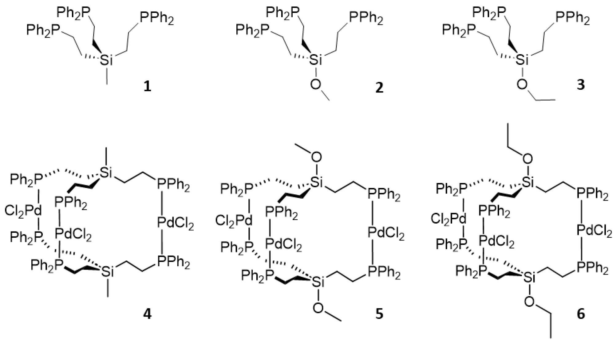
Figure 1. Tripodal phosphine ligands 13 and the corresponding trinuclear Pd complexes 46.
Molecules 30 01616 g001
Figure 2. Immobilized tripodal phosphine ligand 3i and the corresponding trinuclear Pd, Cu, and Pd/Cu complexes.
Molecules 30 01616 g002
Figure 3. 29Si{1H} NMR spectra of 2 (bottom) and 3 (top) in toluene-d8.
Molecules 30 01616 g003
Figure 4. 31P{1H} NMR spectrum of 1 and corresponding PdCl2 complex 4 in C6D6.
Molecules 30 01616 g004
Figure 5. 13C{1H} NMR spectrum of 4 in CDCl3. The ipso, ortho, and meta carbon signals show virtual couplings.
Molecules 30 01616 g005
Figure 12. 31P{1H} MAS NMR spectrum of the immobilized tripodal linker 3i (top), 31P HRMAS of the immobilized CuCl complex (middle), recorded in the presence of acetonitrile, and 31P{1H} MAS spectrum of the cis-coordinated PdCl2 complex (bottom) at the indicated rotational speeds. Asterisks denote rotational sidebands.
Molecules 30 01616 g012
Figure 13. 31P{1H} MAS spectrum of the Pd complex immobilized with maximal surface coverage of 3i (top), and the spectrum obtained after treatment with 1 (bottom) at the indicated rotational speeds. The asterisks denote rotational sidebands.
Molecules 30 01616 g013
Figure 14. 31P{1H} MAS NMR spectra (10 kHz) of immobilized tripodal linker 3i (24% surface coverage) cis- and trans-coordinated to PdCl2 (top), and the same material after addition of the Cu component (bottom). P stands for uncoordinated Ph2P groups, and Cu-P indicates the signal of phosphine groups coordinated to Cu.
Molecules 30 01616 g014
Table 1. Selected bond angles in trinuclear metal complexes (°). pOp3 stands for the ligand O=P(CH2CH2PPh2)3, pOp3S3 for O=P(CH2CH2P(S)Ph2)3 and etp for the ligand CH3C(CH2CH2PPh2)3 [21,22]. L = P, except for pOp3S3 where L = S.
ComplexL–M–L (°)
4 165.68 (3) 175.79 (3)176.09 (3)
5a174.22 (3)174.71 (3)178.24 (3)
5b165.45 (3)176.33 (3)176.45 (3)
7177.22 (4)179.02 (4)179.31 (5)
Pd3I6(pOp3)2 [21]175.8 (2)176.4 (2)178.2 (2)
Pd3Cl6(pOp3S3)2 [21]168.1 (1)168.1 (1)168.1 (1)
Ni3Cl6(etp)2 [22]167.62171.80176.54
Table 2. Selected bond angles in trinuclear metal complexes (°). pOp3 stands for the ligand O=P(CH2CH2PPh2)3, pOp3S3 for O=P(CH2CH2P(S)Ph2)3 and etp for CH3C(CH2CH2PPh2)3. X = Cl, I [21,22].
ComplexX–M–X
4178.76 (3)173.04 (4)171.76 (4)
5a168.83 (5)176.18 (4)176.89 (4)
5b179.23 (3)174.14 (3)172.24 (3)
7174.09 (5)176.63 (5)174.20 (5)
Pd3I6(pOp3)2 [21]160.26 (7)172.6 (2)/158.5 (2)169.38 (7)
Pd3Cl6(pOp3S3)2 [21]179.5 (1)179.5 (1)179.5 (1)
Ni3Cl6(etp)2 [22]161.97166.85166.31
Table 3. Selected dihedral angles in trinuclear metal complexes (°). pOp3 stands for the ligand O=P(CH2CH2PPh2)3, pOp3S3 for O=P(CH2CH2P(S)Ph2)3 and etp for CH3C(CH2CH2PPh2)3 [21,22]. L = P, except for pOp3S3 where L = S. Center = Si (4, 5a, 5b, 7), P for the other Pd complexes, and C for the Ni complex.
ComplexDihedral Angles
L–Center–Center–L (°)
42.11 (2)2.93 (3)8.14 (3)
5a4.41 (3)8.95 (3)12.68 (3)
5b2.91 (3)1.90 (2)7.40 (3)
75.43 (3)6.27 (3)11.72 (3)
Pd3I6(pOp3)2 [21]2.0 (1)0.6 (1)0.8 (1)
Pd3Cl6(pOp3S3)2 [21]50.6850.6850.68
Ni3Cl6(etp)2 [22]0.370.102.35
Table 4. Selected center-center and metal-metal distances in the listed trinuclear complexes (Å). pOp3 stands for the ligand O=P(CH2CH2PPh2)3, pOp3S3 for O=P(CH2CH2P(S)Ph2)3 and etp for CH3C(CH2CH2PPh2)3 [21,22]. M = Pd, Ni. Center = Si (4, 5a, 5b, 7), P for the other Pd complexes, and C for the Ni complex.
ComplexCenter–CenterM–M
47.364 (1)7.4963 (8)6.8894 (8)7.5769 (7)
5a8.363 (1)7.0903 (6)7.2621 (7)7.1109 (6)
5b7.453 (1)7.4188 (5)7.8960 (6)7.4932 (5)
78.530 (2)7.1131 (8)6.8095 (7)7.1727 (7)
Pd3I6(pOp3)2 [21]6.882 (9)7.73 (1)7.35 (1)7.325 (8)
Pd3Cl6(pOp3S3)2 [21]4.85 (9)7.616 (8)7.62 (1)7.616 (7)
Ni3Cl6(etp)2 [22]6.0517.7836.8937.27
Disclaimer/Publisher’s Note: The statements, opinions and data contained in all publications are solely those of the individual author(s) and contributor(s) and not of MDPI and/or the editor(s). MDPI and/or the editor(s) disclaim responsibility for any injury to people or property resulting from any ideas, methods, instructions or products referred to in the content.