Article

Preclinical Theranostic Profiling of [64Cu]Cu-Acetate in Prostate Cancer

1
Department of Pharmacology and Physiology, Faculty of Medicine and Health Sciences, Université de Sherbrooke, Sherbrooke, QC J1H 5N4, Canada
2
Institute of Pharmacology, Faculty of Medicine and Health Sciences, Université de Sherbrooke, Sherbrooke, QC J1H 5N4, Canada
3
Department of Chemistry, Faculty of Sciences, Université de Sherbrooke, Sherbrooke, QC J1H 5N4, Canada
4
Department of Medical Imaging and Radiation Sciences, Faculty of Medicine and Health Sciences, Université de Sherbrooke, Sherbrooke, QC J1H 5N4, Canada
5
Institut de Recherche sur le Cancer de l’Université de Sherbrooke (IRCUS), Université de Sherbrooke, Sherbrooke, QC J1H 5N4, Canada
*
Authors to whom correspondence should be addressed.
Molecules 2025, 30(19), 3957; https://doi.org/10.3390/molecules30193957
Submission received: 28 August 2025 / Revised: 26 September 2025 / Accepted: 26 September 2025 / Published: 2 October 2025
(This article belongs to the Special Issue Applications of Radiochemistry in Healthcare)

Abstract

Copper plays a critical role in cancer biology, with tumor cells exhibiting abnormal copper metabolism that drives proliferation and tumor growth. A limited number of preclinical and clinical studies have reported promising theranostic potential of copper-based radionuclides, such as 64Cu, for both diagnostic imaging and targeted radiotherapy in diverse cancers, including prostate cancer (PCa). In this work, we evaluated the cellular uptake and antitumor efficacy of [64Cu]Cu-acetate using both cellular and animal models of PCa. Uptake assays revealed that ~70% of the administered dose (10 kBq) was internalized by PC-3 cells within 24 h, predominantly localizing to the cytoplasm, with around 9% detected in the nucleus. These results were corroborated by comparable natural Cu-acetate uptake levels (at equimolar dose) in PC-3 cells, as quantified by ICP-MS. Clonogenic assays revealed a dose-dependent reduction in survival following treatment with [64Cu]Cu-acetate (3 and 6 MBq), whereas its non-radioactive counterpart [NatCu]Cu-acetate, even at excess concentrations (10 µM), had no significant effect. Ex vivo biodistribution studies showed selective tumor accumulation/retention alongside expected hepatic uptake. Clear tumor visualization was achieved using μPET imaging with [64Cu]Cu-acetate (10 MBq iv). A single higher dose (65 MBq iv) effectively reduced tumor growth in a subcutaneous PC-3 xenograft mouse model, without systemic toxicity, as evidenced by stable body weight. Together, these results further support the theranostic potential of [64Cu]Cu in PCa.

1. Introduction

Copper (Cu) plays a vital role in numerous cellular processes, including the regulation of growth and proliferation [1,2]. Notably, cancer cells often exhibit altered Cu metabolism compared to normal cells, partly due to dysregulated expression of the high-affinity Cu transporter 1 (CTR1, also known as SLC31A1). CTR1 expression has been reported to vary across different malignancies, including prostate cancer (PCa), glioblastoma, melanoma, lung, breast, and liver cancers [3,4,5,6]. While some studies suggest that CTR1 may contribute to elevated intracellular Cu in tumors [7,8,9,10], its specific role in PCa remains unclear [6,11]. Nevertheless, these observations indicate that Cu accumulation in tumors likely involves multiple mechanisms, providing a rational for metabolic imaging using positron emission tomography (PET).
Indeed, Cu-based radionuclides have demonstrated promising theranostic potential for both diagnostic imaging and targeted radiotherapy in cancers, including PCa [1,12,13,14,15,16,17]. Among the various Cu radioisotopes available for cancer imaging and therapy [18], the cyclotron-produced Cu-64 ([64Cu]Cu) stands out as one of the most versatile [13,14,19]. This interest is largely driven by its favorable decay properties. Its β+ emission (17.8%) enables high-resolution PET imaging for tumor visualization [20,21], while its β decay (38.4%) and electron capture (EC, 43.5%) support targeted radionuclide therapy or endoradiotherapy [18].
The therapeutic efficacy of [64Cu]Cu is closely linked to its nuclear localization and the emission of short-range, high linear energy transfer Auger electrons during EC decay, which can directly damage DNA [22,23]. In cancer cells, nuclear and perinuclear accumulation may be facilitated by endogenous Cu-binding proteins such as Atox1, a key player in intracellular Cu trafficking [24,25]. This targeting mechanism could enhance the radiobiological impact of [64Cu]Cu2+, particularly in tumors with elevated levels of Atox1 or related Cu-transport pathways.
Additionally, its relatively long half-life of 12.7 h, although shorter than that of commonly used therapeutic radionuclides such as 177Lu, 90Y, 131I or 161Tb, provides a suitable time window for both imaging and therapeutic evaluation in preclinical and early translational settings [20]. Its hepatobiliary excretion also helps reduce background signal in the pelvic region, resulting in improved imaging contrast [13,26]. While high activity levels required for radiotherapy may pose a risk of hepatic radiotoxicity [13], efficient clearance and the liver’s regenerative capacity are expected to mitigate this concern [27].
Together, these features contribute to a high tumor-to-background ratio, positioning [64Cu]Cu as a promising agent for both detection and treatment of PCa [28].
However, experimental evidence supporting the theranostic potential of [64Cu]Cu ions, and particularly their therapeutic efficacy in PCa, remains limited. This study aims to build on prior observations by providing new data on the uptake, biodistribution, and notably, the therapeutic impact of [64Cu]Cu2+ in PCa models.

2. Results

2.1. Cellular Accumulation and Subcellular Localization of [64Cu]Cu-Acetate in PC-3 Cells

To evaluate the kinetics of [64Cu]Cu-acetate ([64Cu]Cu(OAc)2) uptake in PC-3 cells, total cell-associated radioactivity was measured at multiple time points and expressed as the percentage of the injected dose per 106 cells (%ID/106 cells) (Figure 1A). A significant time-dependent increase in cellular accumulation was observed. Uptake was minimal at 15 min (4.8% ID/106 cells) but increased significantly at 4 h (56.6% ID/106 cells) and further at 24 h (66.8% ID/106 cells) (**** p < 0.0001). These findings indicate a progressive and efficient uptake of [64Cu]Cu-acetate in PCa cells over time. Uptake of nonradioactive natural Cu-acetate (NatCu-acetate; [63/65Cu]Cu-acetate) was also evaluated using inductively coupled plasma–mass spectrometry (ICP-MS) following treatment of PC-3 cells with 0.5 µM for 24 h, to validate the previous findings (Figure 1B). Treated cells showed a significant increase in Cu content, up to ~45 ppb per 106 cells compared to untreated controls, which showed baseline Cu levels of ~20 ppb per 106 cells, most likely due to prior exposure to Cu present in the serum-containing culture medium (RPMI with 10% FBS: ~200 nM Cu; present study). This high intracellular level of Cu indicates the ability of PC-3 cells to maintain exogenous Cu. The consistency between γ-counter and ICP-MS results underscores the time-dependent uptake of [64Cu]Cu-acetate in PC-3 PCa cells. To further evaluate intracellular distribution, subcellular fractionation was performed at the 24 h time point. Trypan blue staining confirmed the purity of the nuclear fraction. Quantitative analysis revealed that most of the radioactivity was retained in the cytoplasmic compartment (~67% of total cell-associated 64Cu-acetate), with only a small fraction localized to the nucleus (~9%) (Figure 1C).

2.2. In Vitro Radiocytotoxicity of [64Cu]Cu-Acetate in PC-3 Cells

Clonogenic assays were conducted to assess the cytotoxic effects of [64Cu]Cu-acetate on PC-3 PCa cells (Figure 1D). Clonal efficiency was assessed following treatment with increasing doses of 64Cu-acetate (1.2, 3, and 6 MBq) and compared to untreated controls. A clear dose-dependent decline in clonal survival was observed. At 3 MBq, a significant decrease in colony formation was detected (p < 0.05), which was further pronounced at 6 MBq (p < 0.01). Notably, there was no detectable effect on clonogenic efficiency when PC-3 cells were treated with the stable, non-radioactive counterpart, NatCu-acetate even at a higher concentration (10 µM).

2.3. Ex Vivo Biodistribution of [64Cu]Cu-Acetate and [64Cu]CuCl2 in PC-3 Tumor Xenograft-Bearing Nude Mice

A comparative biodistribution study was performed to evaluate the tissue distribution of 64Cu from its acetate and chloride salt forms in male nude mice bearing subcutaneous PC-3 tumors. One-hour post-injection (0.19–0.37 MBq; 5–10 µCi), the liver and kidneys exhibited the highest uptake among all organs, as shown in Table 1. Liver accumulation was highest for both 64Cu-acetate (31.56 ± 11.33% ID/g) and 64Cu-chloride (29.98 ± 4.69% ID/g), indicating hepatic clearance as the primary excretion route. Both radiotracers also showed notable kidney uptake (9.26 ± 2.52 and 9.66 ± 1.65% ID/g, respectively), suggesting a secondary but limited role for renal elimination. Although CTR1, the major Cu transporter, is expressed in the brain [29], brain uptake was the lowest among all organs tested, in line with previous findings [5,30]. This minimal accumulation is likely due to the inability of 64Cu to cross the intact blood–brain barrier. A trend toward higher tumor uptake was observed for 64Cu-chloride compared to 64Cu-acetate (5.02 ± 5.18 vs 1.96 ± 0.21% ID/g), but the difference was not statistically significant (p > 0.05).

2.4. In Vivo PET/CT Imaging of [64Cu]Cu-Acetate in PC-3 Xenograft-Bearing Nude Mice

PET/CT images of [64Cu]Cu-acetate showed clear radiotracer accumulation in both the left and right PC-3 tumors (Figure 2A). Coronal and sagittal scans demonstrated symmetric uptake in subcutaneous flank tumors, with low background signal in surrounding tissues. Static PET scans were used to evaluate the in vivo distribution of 64Cu-acetate in PC-3 tumor-bearing nude mice. To further assess tissue distribution, ex vivo gamma counting was performed on the same animals following PET imaging. Organs were collected and analyzed for radioactivity content, enabling direct comparison between imaging-derived uptake and actual tissue radioactivity. As summarized in Figure 2, biodistribution analysis showed tracer accumulation in the liver and kidneys, consistent with the hepatic and renal clearance reported for [64Cu]CuCl2 [9]. The quantitative uptake values obtained ex vivo (% ID/g) corroborated the in vivo PET measurements, particularly in muscle and kidney, while slight variations were observed in tumor and liver values (Figure 2B). Minor differences in tumor and liver uptake likely reflect regional heterogeneity or differences in partial volume of interest. Interestingly, tumor uptake of [64Cu]Cu-acetate increased over time (1 h vs. 24 h), concomitant with a reduction in peripheral organ uptakes, suggesting enhanced tumor selectivity and prolonged retention in tumor tissue relative to non-target tissues (see Table 1 and Figure 2B).

2.5. Therapeutic Effect of [64Cu]Cu-Acetate on Tumor Growth in PC-3 Tumor-Bearing Mice

The therapeutic effect of [64Cu]Cu-acetate was assessed in male nude mice bearing subcutaneous PC-3 xenografts following a single intravenous dose (65 MBq; 1.5–1.75 mCi) (Figure 3). Tumor growth was monitored and compared to that in a saline-treated control group. While tumors continued to grow in both groups, mice treated with 64Cu-acetate showed a significantly slower rate of tumor progression (Figure 3A). By day 22, tumor volume in the control group had increased nearly tenfold, whereas growth in the [64Cu]Cu-acetate treated group remained substantially limited, supporting a therapeutic effect. To evaluate potential systemic toxicity, body weight was measured throughout the study (Figure 3B). No significant weight loss was observed in the [64Cu]Cu-acetate group compared to controls, suggesting that the treatment was well tolerated.

3. Discussion

To date, few studies have explored the dual use of unchelated 64Cu as both a PET imaging agent and a therapeutic radionuclide, with most existing data based on 64Cu-chloride. Our study reinforces and broadens this body of evidence by showing that 64Cu-acetate can likewise fulfill both functions in PCa.
At the cellular level, [64Cu]Cu-acetate exhibited rapid and substantial internalization of ~67% ID/106 cells at 24 h, with predominant cytoplasmic distribution. Cellular uptake at 24 h was comparable to levels observed at 4 h, indicating a prolonged intracellular retention time. Notable nuclear localization was observed at 24 h post-injection, with ~9% of the activity detected in the nuclear fraction. Similar findings were reported in PC-3 cells exposed to [64Cu]CuCl2, where cytoplasmic predominance and limited nuclear uptake were observed after 3 h of exposure [11]. This could reflect preferential trafficking of 64Cu toward vesicular and trans-Golgi pathways mediated by ATP7A and ATP7B [32]. Such compartmentalization may explain the cytoplasmic and nuclear uptake values of 64Cu observed in our study.
Functionally, [64Cu]Cu-acetate significantly reduced clonogenic survival in the androgen-independent, castration-resistant PC-3 cells in a dose-dependent manner. These findings are in agreement with previous work by Guerreiro et al., who reported that [64Cu]CuCl2 (2.8 MBq) inhibited clonogenic survival in PC-3 cells while sparing non-tumoral prostate cells (RWPE-1) [11].
Ex vivo organ biodistribution revealed similar tumor uptake and clearance profiles for [64Cu]CuCl2 and [64Cu]Cu-acetate, with both tracers showing dual hepatobiliary and renal elimination, and predominant liver clearance. The comparable tumor uptake observed with these two kit-ready Cu formulations likely reflects rapid in vivo binding of free Cu2+ to plasma proteins, such as albumin, ceruloplasmin, and transcuprein, resulting in similar biodistribution and dosimetry between the two formulations [33,34]. Thus, the primary advantage of using the acetate form would be pharmaceutical rather than dosimetric: acetate formulations are neutral and isotonic, whereas CuCl2 is typically obtained in 0.1–1 M HCl and requires buffering before administration (IAEA guidelines [35]). Accordingly, the acetate form offers formulation benefits without altering the radiation burden. Moreover, in accordance with the ex vivo findings, [64Cu]Cu-acetate enabled clear visualization of subcutaneous PCa xenografts using µPET imaging, with % ID/g values (2–5% ID/g at 24 h) comparable to those reported in previous studies (~4–6% ID/g at 24 h) [9,10].
In the context of endoradiotherapy, earlier preclinical studies using [64Cu]CuCl2 in glioblastoma (human U87-MG) and melanoma (mouse B16F10 or human A375M) xenografts models demonstrated marked tumor growth delay, survival benefit, and selective tumor uptake of [64Cu]Cu [4,5]. Our data confirms that the therapeutic potential of [64Cu]Cu extends beyond the tumor types and is not dependent on the specific formulation used. Notably, even a single administered dose of [64Cu]Cu-acetate effectively reduced PCa growth without inducing weight loss or acute toxicity. These results support the feasibility of Cu-based radiotherapy in PCa and open the door for multi-dose treatment protocols, which based on prior success in glioblastoma models [5], may further improve therapeutic outcomes. As noted in the Introduction, although concerns have been raised regarding radiation exposure to non-target organs—particularly the liver [9,10], a major site of Cu accumulation, clinical data to date suggest a favorable safety profile of [64Cu]Cu-based agents [13,14,30]. Furthermore, the efficient clearance of [64Cu]Cu, combined with the liver’s natural regenerative capacity, is anticipated to minimize the risk of hepatotoxicity.
A theranostic approach using [64Cu]Cu may be particularly valuable in PCa cases that lack established molecular targets such as PSMA or GRPR, as well as in those involving emerging receptor biomarkers currently under investigation (e.g., NTR1, NPR-A, B1R) [31,36,37]. As noted by Cantiello et al. [38], up to 10% of PCa cases, including aggressive subtypes like neuroendocrine PCa, do not express PSMA or standard epithelial markers, leading to false-negative results on PSMA-targeted PET imaging and complicating diagnosis and staging. A recent triple-tracer PET imaging study in patients with metastatic PCa revealed that ~53% would have qualified for PSMA radioligand therapy, while none were eligible for DOTATATE-based radiotherapy [39]. Similarly, GRPR-targeted agents such as NeoBOMB1 face limitations due to heterogeneous GRPR expression in PCa. In this context, [64Cu]Cu PET/CT offers a promising alternative as a complementary imaging and therapeutic tool in cases refractory to standard molecular imaging approaches. Its tumor uptake is driven by Cu metabolism and transporter expression, which may remain active even in PSMA- or GRPR-negative lesions. While intratumoral heterogeneity also affects Cu-based tracers, preclinical studies have shown that 64Cu uptake can vary across and within tumor models, highlighting the need for individualized assessment [40,41].
In conclusion, pharmaceutical grade [64Cu]Cu exhibits potential as a bona fide theranostic agent, with encouraging results for both PET imaging and targeted radiotherapy of PCa. Nonetheless, further studies are required to validate its clinical utility and to optimize its application in therapeutic settings.

4. Materials and Methods

4.1. Production of [64Cu]Cu

[64Cu]Cu was produced via the 64Ni(p,n)64Cu nuclear reaction using a TR-19 or TR-24 cyclotron (ACSI, Advanced Cyclotron Systems, Inc., Richmond, BC, Canada) and an electroplated 64Ni-enriched target on a rhodium disc. Following the previously described procedure [42], the resulting [64Cu]CuCl2 was recovered in 0.1 M HCl. [64Cu]Cu-acetate was then prepared by dissolving [64Cu]CuCl2 in 0.1 M ammonium acetate buffer (pH 5.5). The apparent molar activity (AMA) of the resulting [64Cu]Cu-acetate, averaging 350 ± 40 MBq/nmol, was determined by ligand titration following the method of McCarthy et al. [43], using NOTA (1,4,7-triazacyclononane-N,N′,N″-triacetic acid) as the chelator in place of TETA (1,4,8,11-tetraazacyclotetradecane-N,N′,N″,N‴-tetraacetic acid). Briefly, serial dilution titrations were performed by adding increasing amounts of NOTA to aliquots containing a fixed amount of [64Cu]Cu under standardized radiolabeling conditions (pH ~5.5, room temperature, 15 min). Complexation efficiency was assessed by radio-TLC, and the lowest concentration of NOTA yielding ≥95–97% complexation was used to calculate the AMA. This calculation assumes a 1:1 chelation stoichiometry and provides an estimate of the amount of non-radioactive (carrier) copper present in the sample. The resulting [64Cu]Cu was reconstituted in phosphate-buffered saline (PBS, pH 7.4) for in vitro studies or in isotonic saline for in vivo studies.

4.2. Cell Cultures

The human prostate adenocarcinoma cell line PC-3 was obtained from the American Type Culture Collection (ATCC, Manassas, VA, USA). The cells were cultured in RPMI-1640 medium (Wisent Inc., Saint-Jean-Baptiste, QC, Canada) supplemented with 10% heat-inactivated fetal bovine serum (FBS, Wisent Inc.) and 1% penicillin/streptomycin (Pen/Strep). Cultures were maintained at 37 °C in a humidified 5% CO2 incubator. For passaging, adherent cells were detached using 0.05% trypsin/EDTA (Wisent Inc.) or StemPros Accutase® (Life Technologies, Carlsbad, CA, USA), with selection based on experimental requirements.

4.3. In Vitro Cellular Uptake and Nuclear Localization of [64Cu]Cu-Acetate

Cellular uptake and nuclear localization studies were performed by incubating PC-3 cells with [64Cu]Cu-acetate (10 kBq) for 0.25 h, 4 h, or 24 h at 37 °C. At each time point, the culture medium was collected, and the cells were washed with Hank’s balanced salt solution (HBSS, Wisent Inc). Whole cells were collected using Accutase, and nuclei were isolated according to the previously described methods [37,44]. Radioactivity in the medium, whole cells, and nuclei was quantified using a gamma counter (Cobra II auto-gamma counter, Packard, Minneapolis, MN, USA). Nuclear uptake was calculated as the percentage of radioactivity in purified nuclei relative to the total cellular radioactivity.

4.4. Cell Uptake Analysis of NatCu-Acetate Measured by ICP-MS

PC-3 cells were incubated for 24 h at 37 °C with 500 nM NatCu-acetate (Sigma-Aldrich; 229601, Burlington, MA, USA) or left untreated as a control in RPMI-1640 medium with 1% FBS. Following washing and cell harvesting, pellets were digested in a HNO3/H2O2 50:50 mixture and analyzed for Cu content using inductively coupled plasma-mass spectrometry (ICP-MS, PerkinElmer Elan DRC II, Woodbridge, ON, Canada). Samples and calibration standards were diluted with 2% HNO3/0.4% H2O2 prior to analysis.

4.5. Clonogenic Assay

PC-3 cells were seeded in a 6-well plate and treated 48 h later with different doses (1.2, 3, and 6 MBq; corresponding to about 2.5, 5 and 10 nM, respectively) of [64Cu]Cu-acetate or with 5 µM of NatCu-acetate. The cells were then incubated at 37 °C for 10–14 days to allow colony formation. After incubation, colonies were stained and counted [45].

4.6. Subcutaneous Tumor Xenograft Model

PC-3 xenograft tumors were established in male Nu/Nu nude mice (Crl:NU-Foxn1nu: 4–6 weeks old; Charles River Laboratories, Inc., Wilmington, MA, USA) by subcutaneous injection of 2.5 × 10⁶ cells suspended in 50 μL of a 1:1 mixture of serum-free medium and Matrigel (#356234, Sigma-Aldrich, Burlington, MA, USA) into both flanks. Within 10–12 days post-inoculation, the tumors reached the appropriate size, after which the mice were used for ex vivo and in vivo studies.

4.7. Biodistribution and PET Imaging in PC-3 Xenograft Mice

Biodistribution of [64Cu]Cu-acetate and [64Cu]Cu-chloride was performed in PC-3 tumor-bearing nude mice through intravenous injection of 5–10 µCi in the caudal vein under isoflurane anesthesia. At 1 h post-injection, isoflurane-anesthetized mice were euthanized by 5% CO2 inhalation. Organs were collected, washed in 0.9% saline, and radioactivity was measured using a Hidex automated gamma counter (Cobra II auto-gamma counter, Packard, Minneapolis, MN USA). Tissue radioactivity was quantified and expressed as percentage injected dose per gram (% ID/g), using external 64Cu standards to account for physical decay. In vivo distribution of [64Cu]Cu-acetate was further evaluated in PC-3 xenograft-bearing nude mice by small-animal PET imaging using a LabPET8 scanner (Gamma Medica–IDEAS Inc.; 7.5-cm axial and 10-cm transaxial field of view, Northridge, CA, USA), in conjunction with X-ray CT acquired on a MILabs U-CT system (Utrecht, The Netherlands) for anatomical correlation [46]. For PET imaging, 6–9 MBq in 200 μL of [64Cu]Cu-acetate was administered via the tail vein of the animal, and a 30-min static acquisition was performed 24 h post-injection, followed by a CT scan. The percentage of injected dose per gram (% ID/g) of tissues was calculated using a calibration phantom loaded with a known activity concentration [47]. Tumor uptake was quantified by placing a volume of interest (VOI) over the central region of the tumor, and the mean activity was calculated from voxels within the upper quartile of signal intensity. To validate PET findings, the same animals were euthanized following imaging, and ex vivo biodistribution was performed as described.

4.8. Radiotherapy with [64Cu]Cu-Acetate in PC-3 Xenograft Mice

The therapeutic efficacy of [64Cu]Cu-acetate was investigated in PC-3 xenograft-bearing mice. When the tumor reached a diameter of 50–200 mm3, the mice were randomly divided into two groups (6–7 animals per group). One group received 65 MBq (1.5–1.75 mCi) of [64Cu]Cu-acetate, while the other group was considered the control and received an equivalent volume of isotonic saline (100 µL). Although no dosimetry was performed, the radiation dose used was based on previous studies and falls within the typical range for 64Cu-based internal radiotherapy in mice [4,48,49]. Tumor volume was measured with a digital vernier caliper and calculated using the formula: length × width2. Normalized tumor growth was calculated by dividing tumor volumes at each time point by the initial volume measured on the day of treatment. The body weight of the mice was measured every 2 days to assess any potential toxicity. A tumor diameter of 1000 mm3, the development of an ulcerated necrotic area within the tumor, and any movement restrictions or weight loss exceeding 20% were considered endpoints for study termination.

4.9. Statistical Analyses

Statistical analyses were performed using GraphPad Prism software (version 10.2; GraphPad Software, San Diego, CA, USA). Data are reported as mean ± SEM or mean ± SD, as specified. Group comparisons were performed using one-way ANOVA followed by Dunnett’s test, or an unpaired Student’s t test, as appropriate. A p value < 0.05 was considered statistically significant.

Author Contributions

Conceptualization, B.G. and F.G.; methodology, B.G. and F.G.; investigation, S.G.K., M.S. and C.D.; data analysis, S.G.K., M.S. and C.D.; resources, Y.D., B.G. and F.G.; data curation, S.G.K., M.S. and C.D.; writing—original draft preparation, S.G.K., M.S., B.G. and F.G.; writing—review and editing, Y.D.; supervision, Y.D., B.G. and F.G.; project administration, F.G.; funding acquisition, Y.D., B.G. and F.G. All authors have read and agreed to the published version of the manuscript.

Funding

This project was funded by Movember Discovery Grant from Prostate Cancer Canada (Grant number D2014-15) and the Canadian Institutes of Health Research Grant (MOP-166037). Y.D., B.G. and F.G. are members of the Fonds de Recherche du Québec Santé (FRQS)-funded Centre de Recherche du Centre Hospitalier Universitaire de Sherbrooke (CR-CHUS).

Institutional Review Board Statement

The animal study protocol was approved by the Animal Care Committee (CPA) of the Faculty of Medicine and Health Sciences (FMSS) at the Université de Sherbrooke (protocols #392-15BR (14 July 2015) and #2019-2371 (5 March 2020)) and followed guidelines of Canadian Council on Animal Care.

Informed Consent Statement

Not applicable.

Data Availability Statement

All data generated or analysed during this study are available from the corresponding authors upon reasonable request.

Acknowledgments

The production of [64Cu], PET imaging and biodistribution studies were conducted at the Sherbrooke Molecular Imaging Centre (CIMS). We thank S. Ait-Mohand for producing the [64Cu] salts. We also thank O. Sarrhini from the CIMS/CR-CHUS Image Visualization and Analysis Platform (PAVI) for his technical assistance with the analysis of PET data. The authors used ChatGPT (GPT-4 Omni, OpenAI) to assist with grammar and language editing during manuscript preparation. All AI-assisted content was reviewed and verified by the authors, who take full responsibility for the integrity and accuracy of the manuscript.

Conflicts of Interest

The authors declare no conflicts of interest.

Abbreviations

CTR1Copper transporter 1
FBSFetal bovine serum
PBSPhosphate-buffered saline
HBSSHank’s balanced salt solution
ICP-MSInductively coupled plasma–mass spectrometry
PCaProstate cancer
Pen/StrepPenicillin/Streptomycin
PETPositron emission tomography

References

  1. Capriotti, G.; Piccardo, A.; Giovannelli, E.; Signore, A. Targeting Copper in Cancer Imaging and Therapy: A New Theragnostic Agent. J. Clin. Med. 2022, 12, 223. [Google Scholar] [CrossRef] [PubMed]
  2. Brady, D.C.; Crowe, M.S.; Turski, M.L.; Hobbs, G.A.; Yao, X.; Chaikuad, A.; Knapp, S.; Xiao, K.; Campbell, S.L.; Thiele, D.J.; et al. Copper Is Required for Oncogenic BRAF Signalling and Tumorigenesis. Nature 2014, 509, 492–496. [Google Scholar] [CrossRef] [PubMed]
  3. Kim, K.I.; Jang, S.J.; Park, J.H.; Lee, Y.J.; Lee, T.S.; Woo, K.S.; Park, H.; Choe, J.G.; An, G.I.; Kang, J.H. Detection of Increased 64Cu Uptake by Human Copper Transporter 1 Gene Overexpression Using PET with 64CuCl2 in Human Breast Cancer Xenograft Model. J. Nucl. Med. 2014, 55, 1692–1698. [Google Scholar] [CrossRef] [PubMed]
  4. Qin, C.; Liu, H.; Chen, K.; Hu, X.; Ma, X.; Lan, X.; Zhang, Y.; Cheng, Z. Theranostics of Malignant Melanoma with 64CuCl2. J. Nucl. Med. 2014, 55, 812–817. [Google Scholar] [CrossRef]
  5. Ferrari, C.; Asabella, A.N.; Villano, C.; Giacobbi, B.; Coccetti, D.; Panichelli, P.; Rubini, G. Copper-64 Dichloride as Theranostic Agent for Glioblastoma Multiforme: A Preclinical Study. Biomed. Res. Int. 2015, 2015, 129764. [Google Scholar] [CrossRef]
  6. Kong, F.-S.; Ren, C.-Y.; Jia, R.; Zhou, Y.; Chen, J.-H.; Ma, Y. Systematic Pan-Cancer Analysis Identifies SLC31A1 as a Biomarker in Multiple Tumor Types. BMC Med. Genomics 2023, 16, 61. [Google Scholar] [CrossRef]
  7. Chakravarty, R.; Chakraborty, S.; Dash, A. 64Cu2+ Ions as PET Probe: An Emerging Paradigm in Molecular Imaging of Cancer. Mol. Pharm. 2016, 13, 3601–3612. [Google Scholar] [CrossRef]
  8. Speltri, G.; Porto, F.; Boschi, A.; Uccelli, L.; Martini, P. Recent Advances in Preclinical Studies of the Theranostic Agent [64Cu]CuCl2. Molecules 2024, 29, 4085. [Google Scholar] [CrossRef]
  9. Cai, H.; Wu, J.; Muzik, O.; Hsieh, J.-T.; Lee, R.J.; Peng, F. Reduced 64Cu Uptake and Tumor Growth Inhibition by Knockdown of Human Copper Transporter 1 in Xenograft Mouse Model of Prostate Cancer. J. Nucl. Med. 2014, 55, 622–628. [Google Scholar] [CrossRef]
  10. Peng, F.; Lu, X.; Janisse, J.; Muzik, O.; Shields, A.F. PET of Human Prostate Cancer Xenografts in Mice with Increased Uptake of 64CuCl2. J. Nucl. Med. 2006, 47, 1649–1652. [Google Scholar]
  11. Guerreiro, J.F.; Alves, V.; Abrunhosa, A.J.; Paulo, A.; Gil, O.M.; Mendes, F. Radiobiological Characterization of 64CuCl2 as a Simple Tool for Prostate Cancer Theranostics. Molecules 2018, 23, 2944. [Google Scholar] [CrossRef]
  12. Paparo, F.; Peirano, A.; Matos, J.; Bacigalupo, L.; Rossi, U.; Mussetto, I.; Bottoni, G.; Ugolini, M.; Introini, C.; Grillo Ruggieri, F.; et al. Diagnostic Value of Retrospectively Fused 64CuCl2 PET/MRI in Biochemical Relapse of Prostate Cancer: Comparison with Fused 18F-Choline PET/MRI, 64CuCl2 PET/CT, 18F-Choline PET/CT, and mpMRI. Abdom. Radiol. 2020, 45, 3896–3906. [Google Scholar] [CrossRef]
  13. Piccardo, A.; Paparo, F.; Puntoni, M.; Righi, S.; Bottoni, G.; Bacigalupo, L.; Zanardi, S.; DeCensi, A.; Ferrarazzo, G.; Gambaro, M.; et al. 64CuCl2 PET/CT in Prostate Cancer Relapse. J. Nucl. Med. 2018, 59, 444–451. [Google Scholar] [CrossRef]
  14. Capasso, E.; Durzu, S.; Piras, S.; Zandieh, S.; Knoll, P.; Haug, A.; Hacker, M.; Meleddu, C.; Mirzaei, S. Role of 64CuCl2 PET/CT in Staging of Prostate Cancer. Ann. Nucl. Med. 2015, 29, 482–488. [Google Scholar] [CrossRef]
  15. Wang, Q.; Song, D.; Ma, X.; Wu, X.; Jiang, L. Preclinical PET Imaging Study of Lung Cancer with 64CuCl2. Ann. Nucl. Med. 2020, 34, 653–662. [Google Scholar] [CrossRef] [PubMed]
  16. Wachsmann, J.; Peng, F. Molecular Imaging and Therapy Targeting Copper Metabolism in Hepatocellular Carcinoma. World J. Gastroenterol. 2016, 22, 221–231. [Google Scholar] [CrossRef] [PubMed]
  17. Fiz, F.; Bottoni, G.; Ugolini, M.; Righi, S.; Cirone, A.; Garganese, M.C.; Verrico, A.; Rossi, A.; Milanaccio, C.; Ramaglia, A.; et al. Diagnostic and Dosimetry Features of [64Cu]CuCl2 in High-Grade Paediatric Infiltrative Gliomas. Mol. Imaging Biol. 2023, 25, 391–400. [Google Scholar] [CrossRef]
  18. Ahmedova, A.; Todorov, B.; Burdzhiev, N.; Goze, C. Copper Radiopharmaceuticals for Theranostic Applications. Eur. J. Med. Chem. 2018, 157, 1406–1425. [Google Scholar] [CrossRef]
  19. Peng, F. Recent Advances in Cancer Imaging with 64CuCl2 PET/CT. Nucl. Med. Mol. Imaging 2022, 56, 80–85. [Google Scholar] [CrossRef]
  20. Gutfilen, B.; Souza, S.A.; Valentini, G. Copper-64: A Real Theranostic Agent. Drug Des. Devel. Ther. 2018, 12, 3235–3245. [Google Scholar] [CrossRef]
  21. Wang, H.; Chen, X. Visualization of Copper Metabolism by 64CuCl2-PET. Mol. Imaging Biol. 2012, 14, 14–16. [Google Scholar] [CrossRef]
  22. Gutfilen, B.; Duatti, A. Editorial: In-Vivo Targeting of Nuclear DNA with Radioactive Copper-64 Ions. Front. Med. 2023, 10, 1334294. [Google Scholar] [CrossRef]
  23. Buchegger, F.; Perillo-Adamer, F.; Dupertuis, Y.M.; Delaloye, A.B. Auger radiation targeted into DNA: A therapy perspective. Eur. J. Nucl. Med. Mol. Imaging 2023, 33, 1352–1363. [Google Scholar] [CrossRef] [PubMed]
  24. Hatori, Y.; Lutsenko, S. The Role of Copper Chaperone Atox1 in Coupling Redox Homeostasis to Intracellular Copper Distribution. Antioxidants 2016, 5, 25. [Google Scholar] [CrossRef] [PubMed]
  25. Beaino, W.; Guo, Y.; Chang, A.J.; Anderson, C.J. Roles of Atox1 and P53 in the Trafficking of Copper-64 to Tumor Cell Nuclei: Implications for Cancer Therapy. J. Biol. Inorg. Chem. 2014, 19, 427–438. [Google Scholar] [CrossRef] [PubMed]
  26. Righi, S.; Ugolini, M.; Bottoni, G.; Puntoni, M.; Iacozzi, M.; Paparo, F.; Cabria, M.; Ceriani, L.; Gambaro, M.; Giovanella, L.; et al. Biokinetic and Dosimetric Aspects of 64CuCl2 in Human Prostate Cancer: Possible Theranostic Implications. EJNMMI Res. 2018, 8, 18. [Google Scholar] [CrossRef]
  27. Wadas, T.J.; Wong, E.H.; Weisman, G.R.; Anderson, C.J. Copper Chelation Chemistry and Its Role in Copper Radiopharmaceuticals. Curr. Pharm. Des. 2007, 13, 3–16. [Google Scholar] [CrossRef]
  28. Yang, N.; Guo, X.-Y.; Ding, J.; Wang, F.; Liu, T.-L.; Zhu, H.; Yang, Z. Copper-64 Based PET-Radiopharmaceuticals: Ways to Clinical Translational. Semin. Nucl. Med. 2024, 54, 792–800. [Google Scholar] [CrossRef]
  29. Fu, X.; Zhang, Y.; Jiang, W.; Monnot, A.D.; Bates, C.A.; Zheng, W. Regulation of Copper Transport Crossing Brain Barrier Systems by Cu-ATPases: Effect of Manganese Exposure. Toxicol. Sci. 2014, 139, 432–451. [Google Scholar] [CrossRef]
  30. Avila-Rodriguez, M.A.; Rios, C.; Carrasco-Hernandez, J.; Manrique-Arias, J.C.; Martinez-Hernandez, R.; García-Pérez, F.O.; Jalilian, A.R.; Martinez-Rodriguez, E.; Romero-Piña, M.E.; Diaz-Ruiz, A. Biodistribution and Radiation Dosimetry of [64Cu]Copper Dichloride: First-in-Human Study in Healthy Volunteers. EJNMMI Res. 2017, 7, 98. [Google Scholar] [CrossRef]
  31. Ghanaatgar Kasbi, S.; Savard, M.; Couture, F.; Dubuc, C.; Dumulon-Perreault, V.; Nepveu-Traversy, M.-E.; Ait-Mohand, S.; Sabbagh, R.; Geha, S.; Guérin, B.; et al. Theranostic Potential of a New 64Cu-Labeled NOTA-R954 Peptide Conjugate for Kinin B1R Expressing Prostate Cancer. Pharmaceutics 2025, 17, 1215. [Google Scholar] [CrossRef]
  32. Lutsenko, S. Dynamic and Cell-Specific Transport Networks for Intracellular Copper Ions. J. Cell Sci. 2021, 134, jcs240523. [Google Scholar] [CrossRef]
  33. Kirsipuu, T.; Zadorožnaja, A.; Smirnova, J.; Friedemann, M.; Plitz, T.; Tõugu, V.; Palumaa, P. Copper(II)-Binding Equilibria in Human Blood. Sci. Rep. 2020, 10, 5686. [Google Scholar] [CrossRef]
  34. Linder, M.C. Ceruloplasmin and Other Copper Binding Components of Blood Plasma and Their Functions: An Update. Metallomics 2016, 8, 887–905. [Google Scholar] [CrossRef]
  35. International Atomic Energy Agency (IAEA). Copper-64 Radiopharmaceuticals: Production, Quality Control and Clinical Applications; IAEA Radioisotopes and Radiopharmaceuticals Series No. 7; IAEA: Vienna, Austria, 2022. [Google Scholar]
  36. Chaudhary, P.K.; Kim, S. An Insight into GPCR and G-Proteins as Cancer Drivers. Cells 2021, 10, 3288. [Google Scholar] [CrossRef] [PubMed]
  37. Li, X.; Cai, H.; Wu, X.; Li, L.; Wu, H.; Tian, R. New Frontiers in Molecular Imaging Using Peptide-Based Radiopharmaceuticals for Prostate Cancer. Front. Chem. 2020, 8, 583309. [Google Scholar] [CrossRef] [PubMed]
  38. Cantiello, F.; Crocerossa, F.; Cascini, G.L.; Russo, G.I.; Ferro, M.; Cimino, S.; Lucarelli, G.; Damiano, R. 64CuCl2 PET/CT as a Potential New Imaging Method in Prostate Cancer: Illusion or Reality? Minerva Urol. Nephrol. 2021, 73, 668–671. [Google Scholar] [CrossRef]
  39. Pouliot, F.; Saad, F.; Rousseau, E.; Richard, P.O.; Zamanian, A.; Probst, S.; Lévesque, É.; Castonguay, V.; Marcoux, N.; Lodde, M.; et al. Intrapatient Intermetastatic Heterogeneity Determined by Triple-Tracer PET Imaging in mCRPC Patients and Correlation to Survival: The 3TMPO Cohort Study. J. Nucl. Med. 2024, 65, 1710–1717. [Google Scholar] [CrossRef]
  40. Zhang, W.; Wang, T.; Wang, Y.; Zhu, F.; Shi, H.; Zhang, J.; Wang, Z.; Qu, M.; Zhang, H.; Wang, T.; et al. Intratumor Heterogeneity and Clonal Evolution Revealed in Castration-Resistant Prostate Cancer by Longitudinal Genomic Analysis. Transl. Oncol. 2022, 16, 101311. [Google Scholar] [CrossRef]
  41. Haffner, M.C.; Zwart, W.; Roudier, M.P.; True, L.D.; Nelson, W.G.; Epstein, J.I.; De Marzo, A.M.; Nelson, P.S.; Yegnasubramanian, S. Genomic and Phenotypic Heterogeneity in Prostate Cancer. Nat. Rev. Urol. 2021, 18, 79–92. [Google Scholar] [CrossRef]
  42. Zeisler, S.K.; Pavan, R.A.; Orzechowski, J.; Langlois, R.; Rodrigue, S.; van Lier, J.E. Production of 64Cu on the Sherbrooke TR-PET Cyclotron. J. Radioanal. Nucl. Chem. 2003, 257, 175–177. [Google Scholar] [CrossRef]
  43. McCarthy, D.W.; Shefer, R.E.; Klinkowstein, R.E.; Bass, L.A.; Margeneau, W.H.; Cutler, C.S.; Anderson, C.J.; Welch, M.J. Efficient production of high specific activity 64Cu using a biomedical cyclotron. Nucl. Med. Biol. 1997, 24, 35–43. [Google Scholar] [CrossRef] [PubMed]
  44. Wang, M.; Caruano, A.L.; Lewis, M.R.; Meyer, L.A.; VanderWaal, R.P.; Anderson, C.J. Subcellular Localization of Radio-labeled Somatostatin Analogues: Implications for Targeted Radiotherapy of Cancer. Cancer Res. 2003, 63, 6864–6869. [Google Scholar] [PubMed]
  45. Dubuc, C.; Savard, M.; Bovenzi, V.; Lessard, A.; Côté, J.; Neugebauer, W.; Geha, S.; Chemtob, S.; Gobeil, F. Antitumor Activity of Cell-Penetrant Kinin B1 Receptor Antagonists in Human Triple-Negative Breast Cancer Cells. J. Cell. Physiol. 2019, 234, 2851–2865. [Google Scholar] [CrossRef]
  46. Bergeron, M.; Cadorette, J.; Tetrault, M.-A.; Beaudoin, J.-F.; Leroux, J.-D.; Fontaine, R.; Lecomte, R. Imaging Performance of LabPET APD-Based Digital PET Scanners for Pre-Clinical Research. Phys. Med. Biol. 2014, 59, 661–678. [Google Scholar] [CrossRef]
  47. Khosravifarsani, M.; Ait-Mohand, S.; Paquette, B.; Sanche, L.; Guérin, B. In Vivo Behavior of [64Cu]NOTA-Terpyridine Platinum, a Novel Chemo-Radio-Theranostic Agent for Imaging and Therapy of Colorectal Cancer. Front. Med. 2022, 9, 975213. [Google Scholar] [CrossRef]
  48. Yoshii, Y.; Furukawa, T.; Kiyono, Y.; Watanabe, R.; Mori, T.; Yoshii, H.; Asai, T.; Okazawa, H.; Welch, M.J.; Fujibayashi, Y. Internal Radiotherapy with Copper-64-Diacetyl-Bis(N4-Methylthiosemicarbazone) Reduces CD133+ Highly Tumorigenic Cells and Metastatic Ability of Mouse Colon Carcinoma. Nucl. Med. Biol. 2011, 38, 151–157. [Google Scholar] [CrossRef]
  49. Métivier, C.; Le Saëc, P.; Gaschet, J.; Chauvet, C.; Marionneau-Lambot, S.; Hofgaard, P.O.; Bogen, B.; Pineau, J.; Le Bris, N.; Tripier, R.; et al. Preclinical Evaluation of a 64Cu-Based Theranostic Approach in a Murine Model of Multiple Myeloma. Pharmaceutics 2023, 15, 1817. [Google Scholar] [CrossRef]
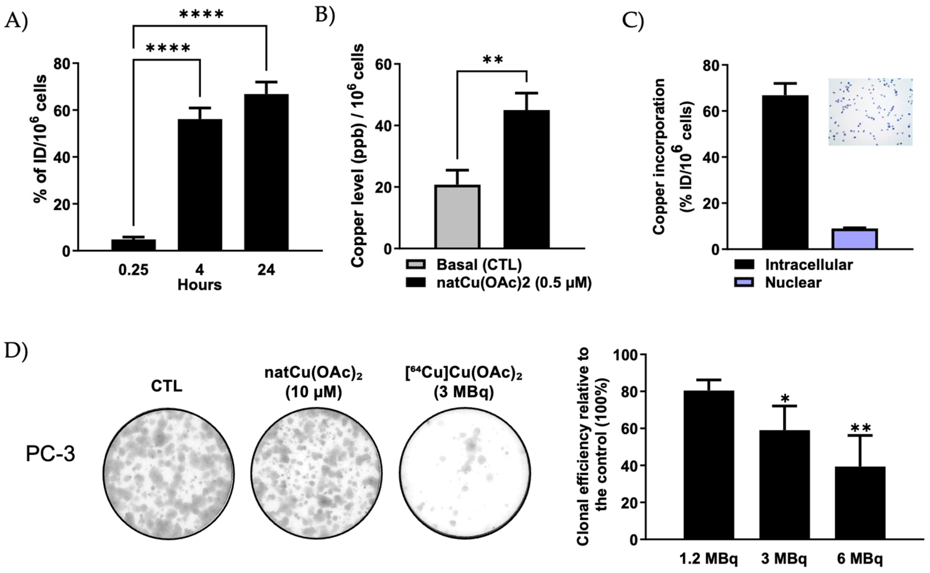
Figure 1. Cellular uptake, subcellular distribution, and radiotoxic effects of [64Cu]Cu-acetate, in PCa cells. (A) Time-dependent uptake of [64Cu]Cu-acetate (10 kBq) in PC-3 cells at 0.25 h, 4 h, and 24 h post-incubation. Data are expressed as a percentage of the injected dose per 106 cells (%ID/106 cells). A significant increase in uptake was observed at 4 h and 24 h compared to 0.25 h (**** p < 0.0001). n = 4. Statistical analysis was performed using ordinary one-way ANOVA with Dunnett’s multiple comparisons test. (B) Quantification of Cu accumulation in PC-3 cells following 24 h treatment with NatCu-acetate (0.5 µM), measured by ICP-MS. A significant increase in intracellular Cu was observed compared to basal levels (** p < 0.01, unpaired t-test). n = 7–8. (C) Subcellular distribution of [64Cu]Cu-acetate in PC-3 cells after 24 h. Cu incorporation was predominantly cytoplasmic, with ~9% nuclear accumulation. Nuclear fraction purity was validated by optical microscopy following trypan blue staining (inset). n = 4. (D) Clonogenic survival of PC-3 cells following exposure to increasing activities of [64Cu]Cu-acetate (1.2, 3, and 6 MBq). Representative colony formation images (left) and quantification of clonal efficiency relative to untreated control (CTL; 100%) are shown (right). Data are presented as mean ± SEM from four independent experiments. Statistical analysis was performed using one-way ANOVA followed by Dunnett’s test. * p < 0.05, ** p < 0.01 vs. control.
Molecules 30 03957 g001
Figure 2. PET imaging and biodistribution of [64Cu]Cu-acetate in PC-3 tumor-bearing mice. (A) Representative coronal and sagittal PET/CT images acquired 24 h post-injection of [64Cu]Cu-acetate (7 MBq) in mice bearing subcutaneous PC-3 tumors. Yellow arrows indicate the locations of left and right tumors. (B) Quantitative uptake values (% ID/g) of [64Cu]Cu-acetate in selected tissues of PC-3 tumor–bearing mice, measured 24 h post-injection by in vivo PET imaging and ex vivo gamma counting performed on the same animals. Data are presented as mean ± SD (n = 3).
Molecules 30 03957 g002
Figure 3. Therapeutic efficacy and tolerability of [64Cu]Cu-acetate in PC-3 tumor-bearing mice. (A) Tumor growth curves of mice treated with [64Cu]Cu-acetate or saline vehicle control (CTL). Treatment significantly delayed tumor progression compared with control. Data represent the mean ± SEM from 11–13 tumors per group (n = 6–7 mice/group); * p < 0.05, ** p < 0.01, unpaired Student’s t test with Welch’s correction. (B) Body weight monitoring over the study period showed no significant weight loss in either group (n = 6–7 mice/group). Data are presented as mean ± SEM. Control group data previously reported in [31], under identical conditions (n = 16 vs. n = 13).
Molecules 30 03957 g003
Table 1. Ex vivo biodistribution of [64Cu]Cu(OAc)2 and [64Cu]CuCl2 in PC-3 xenograft mice at 1 h post-injection (p.i.).
OrganBloodPlasmaKidneyPancreasLiverHeartLungTumorMuscleBoneBrain
[64Cu]Cu(OAc)21.95
± 0.65
2.96
± 0.86
9.26
± 2.52
3.15
± 1.18
31.56
± 11.33
2.84
± 0.43
4.19
± 1.49
1.96
± 0.21
0.61
± 0.13
0.81
± 0.08
0.36
± 0.10
[64Cu]CuCl23.23
± 1.97
2.28
± 0.44
9.66
± 1.65
2.35
± 0.41
29.98
± 4.69
3.52
± 1.35
5.64
± 3.78
5.02
± 5.18
0.64
± 0.16
1.11
± 0.59
0.34
± 0.08
Values are expressed as mean ± SD (n = 3).
Disclaimer/Publisher’s Note: The statements, opinions and data contained in all publications are solely those of the individual author(s) and contributor(s) and not of MDPI and/or the editor(s). MDPI and/or the editor(s) disclaim responsibility for any injury to people or property resulting from any ideas, methods, instructions or products referred to in the content.