Article

Cancer Chemopreventive Potential of Claoxylon longifolium Grown in Southern Thailand: A Bioassay-Guided Isolation of Vicenin 1 as the Active Compound and In Silico Studies on Related C-Glycosyl Flavones

by
Chuanchom Khuniad
1,2,*,
Lutfun Nahar
3,*,
Anupam D. Talukdar
4,
Rajat Nath
5,
Kenneth J. Ritchie
1 and
Satyajit D. Sarker
1,*
1
Centre for Natural Products Discovery (CNPD), School of Pharmacy and Biomolecular Sciences, Liverpool John Moores University, Liverpool L3 3AF, UK
2
Department of Thai Traditional Medicine, Faculty of Health and Sports Science, Thaksin University, Phathalung 93210, Thailand
3
Laboratory of Growth Regulators, Palacký University and Institute of Experimental Botany, The Czech Academy of Sciences, Šlechtitelů 27, 78371 Olomouc, Czech Republic
4
Department of Life Science and Bioinformatics, Assam University, Silchar 788011, India
5
Department of Biotechnology and Microbiology, School of Natural Sciences, Techno India University, Agartala 799004, India
*
Authors to whom correspondence should be addressed.
Molecules 2025, 30(15), 3173; https://doi.org/10.3390/molecules30153173
Submission received: 1 July 2025 / Revised: 22 July 2025 / Accepted: 23 July 2025 / Published: 29 July 2025

Abstract

Claoxylon longifolium (Euphorbiaceae) is an indigenous vegetable that has been used in southern Thai traditional medicine and cuisine. A bioassay-guided approach was adopted to investigate the phytochemicals and chemopreventive potential of C. longifolium leaves and stems. Phytochemical investigation of the active MeOH fractions afforded six known compounds, including caffeic acid (1), isovitexin (2), and vicenins 1–3 (3–5) from leaves and hexadecanoic acid methyl ester (6) from stems. Their structures were determined by spectroscopic means. Ten constituents were tentatively identified from the oily fractions of stems by GC-MS. Non-cytotoxic concentrations of compounds 16 were identified using the MTT cell viability assay. The ability of compounds 16 at non-cytotoxic concentrations to induce Nrf2 activation, correlating to their potential chemopreventive properties, was determined using a luciferase reporter assay in the AREc32 cell line. Only vicenin 1 (3) was considered to be a potent chemopreventive compound, as it increased luciferase activity by 2.3-fold. In silico studies on compounds 25 and vitexin (16) revealed the potential of these compounds as cancer chemopreventive and chemotherapeutic agents. This study provides the first report on the chemopreventive properties of C. longifolium. All identified and isolated compounds are reported here for the first time from this species.

Graphical Abstract

1. Introduction

In consideration of global cancer incidence and mortality rates reported by the International Agency for Research on Cancer (IARC), cancer is a major threat to human health worldwide [1]. As of 2022, there have been almost 20 million new cancer cases. Furthermore, 16.8% of all deaths (9.7 million) were cancer deaths, which makes it the second leading cause of death worldwide behind cardiovascular diseases. Almost one-half of all new cases (49.2%) and the majority (56.1%) of cancer deaths globally were estimated to occur in Asia in 2022. Hence, Asia has the highest burden of cancer compared to the other regions in the world. According to the GLOBOCAN 2022 database, created by the IARC, the number of new cancer cases is projected to reach 35 million by 2050, representing a 77% increase from 2022 [1]. Therefore, the search for new alternative cancer prevention and treatment options is still necessary.
Cancer chemoprevention is defined as the use of “nontoxic” natural or synthetic chemical agents to inhibit, slow down, or reverse the process of carcinogenesis by blocking or suppressing specific molecular events and signalling pathways associated with cancer development [2]. The goal of cancer chemoprevention is to decrease cancer incidence and concurrently reduce treatment-related adverse effects and mortality rates, making it an effective strategy for cancer control and management [3,4].
Nuclear factor erythroid 2-related factor 2 (Nrf2) is a cytoprotective transcription factor that regulates the expression of many cellular antioxidants and detoxification enzymes [5]. Nrf2 binds to the antioxidant response element (ARE) in the promoter regions of many cytoprotective genes, thereby promoting their expression. Thus, the Nrf2-ARE signalling pathway is recognized as a potential molecular target for cancer chemoprevention because the activation of this pathway induces the production of a battery of cytoprotective and detoxifying genes that may be a promising approach to prevent or suppress carcinogenic progression [6,7]. To date, numerous natural products that exert chemopreventive properties have been discovered as Nrf2 activators such as curcumin, diallyl sulfide, resveratrol, sulforaphane and tert-butylhydroquinone (tBHQ) [8,9,10].
Thai traditional medicine (TTM) uses many natural products to treat diseases. Even though modern medicine has made noteworthy progress, many people who live in rural communities of southern Thailand still rely on traditional healers and medicinal plants for their primary healthcare needs [11,12]. Importantly, Thailand is a biodiversity-rich country with more than 11,000 different plant species documented in the Flora of Thailand [13], many of which have medicinal properties and could be a potential source for cancer chemopreventive agents. Claoxylon longifolium (Blume) Endl. ex Hassk. (Euphorbiaceae), locally known as “Tamad” or “Phak wan chang” (Figure 1) is native to Northeast India, from Southeast Asia to Yunnan of southwestern China, throughout West Malaysia up to Borneo, Java and New Guinea [14,15]. The distribution of this species in Thailand is in the southern peninsula, except Kanchanaburi province (Figure 2) [15,16].
In Thailand, Malaysia, and the Philippines, the young shoots and leaves of this plant are used as vegetables and food ingredients [14,17,18]. Folk healers in southern Thailand use the whole plant to treat menstrual fever and leaves to treat blurred vision [19]. Moreover, decoction of leaves/roots can be drunk as tonic and restorative remedies [20,21]. Interestingly, this species is used by traditional Malay midwives as an ingredient in an herbal bath for postpartum recovery and rejuvenation therapy [22]. Preliminary phytochemical screening of C. longifolium leaves revealed the presence of alkaloids, flavonoids and terpenes [23,24]. Hu et al. [25] isolated nine phenolic compounds, including cleomiscosin A, cleomiscosin C, ficusal, scopoletin, ferulaldehyde, isovanillic acid, protocatechuic acid, vanillin and p-hydroxybenzaldehyde from the stems of C. longifolium. To the best of our knowledge, there is no published work on pharmacological studies available in any well-known scientific database. Therefore, based on the traditional uses and previously reported phytochemical studies of C. longifolium, it was hypothesised that this plant could have chemopreventive activity, and accordingly, this study aimed to investigate the phytochemicals and chemopreventive effect of C. longifolium leaves and stems growing in southern Thailand. This paper reports, for the first time, the bioassay-guided isolation of vicenin 1 (3) as a potential chemopreventive agent from the methanolic extract of the leaves of C. longifolium, isolation and identification of five other known compounds from the leaves and stems, GC-MS analysis of the stem extract, and in silico studies with vicenins 1–3 (3–5) and related compounds for cancer chemopreventive and cancer chemotherapeutic potential.

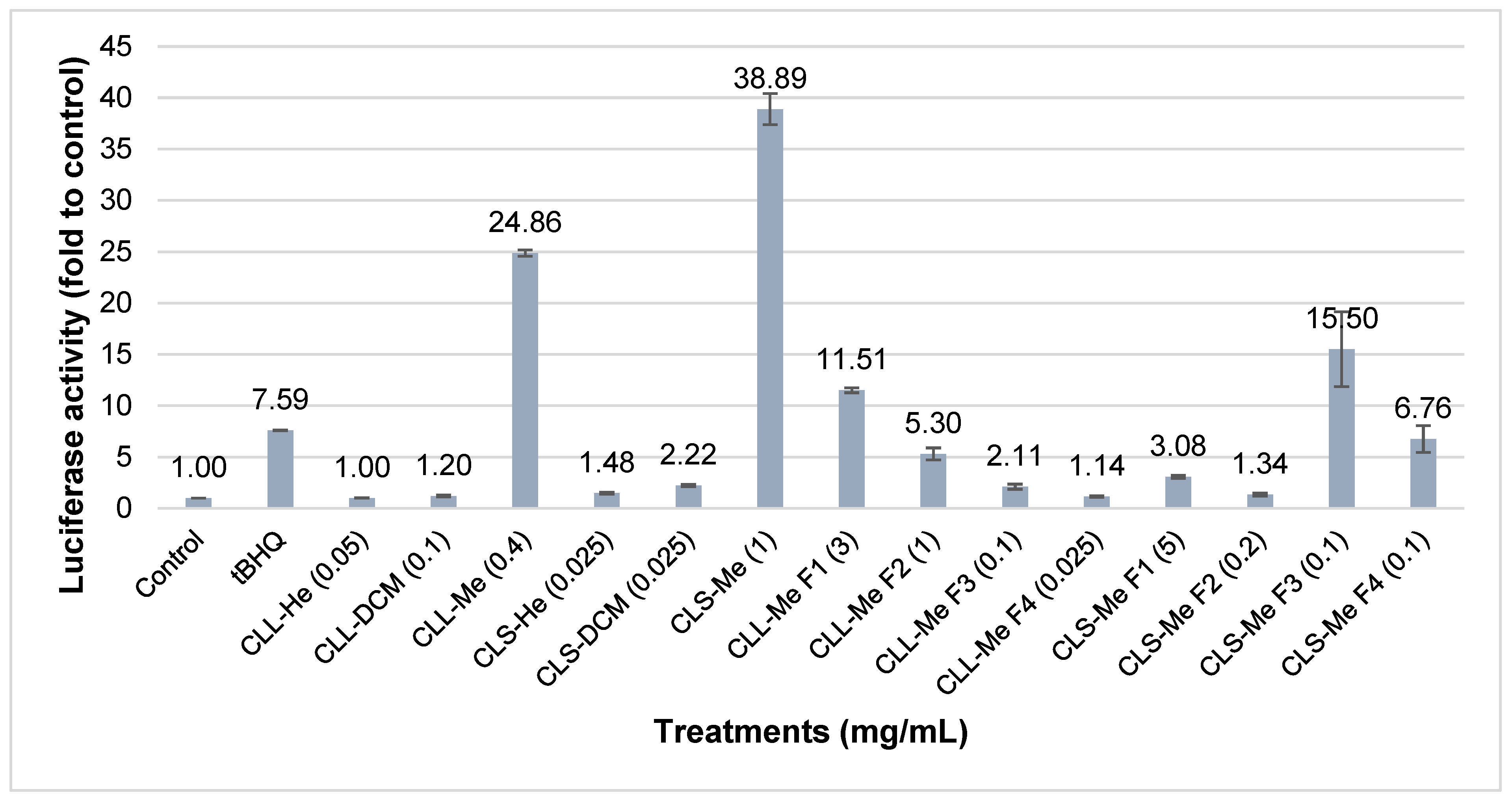
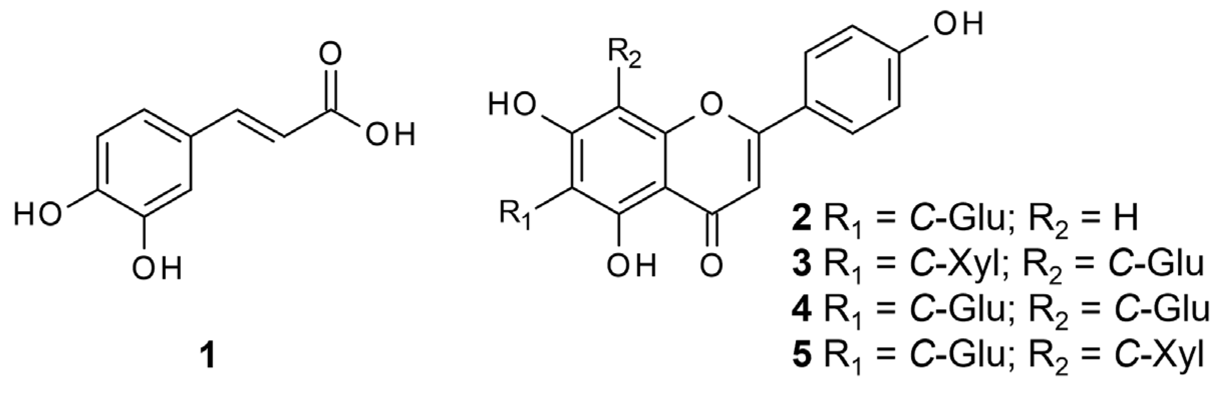
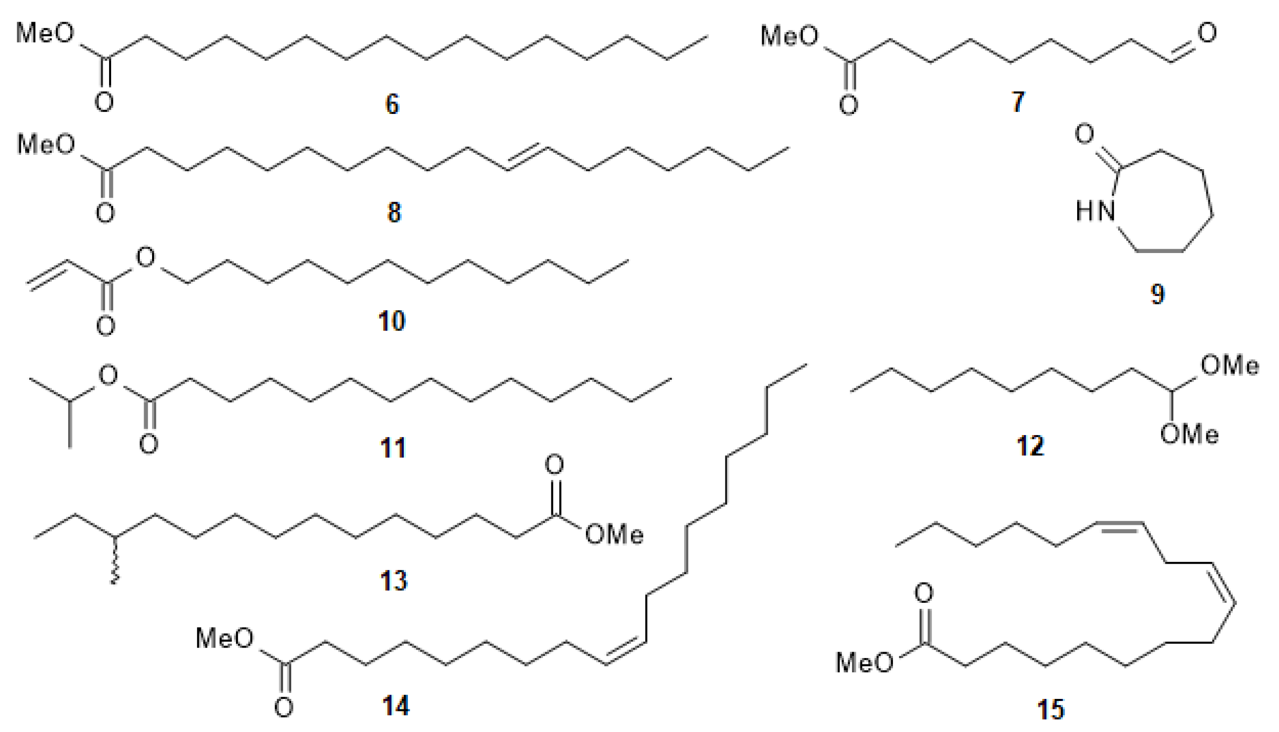
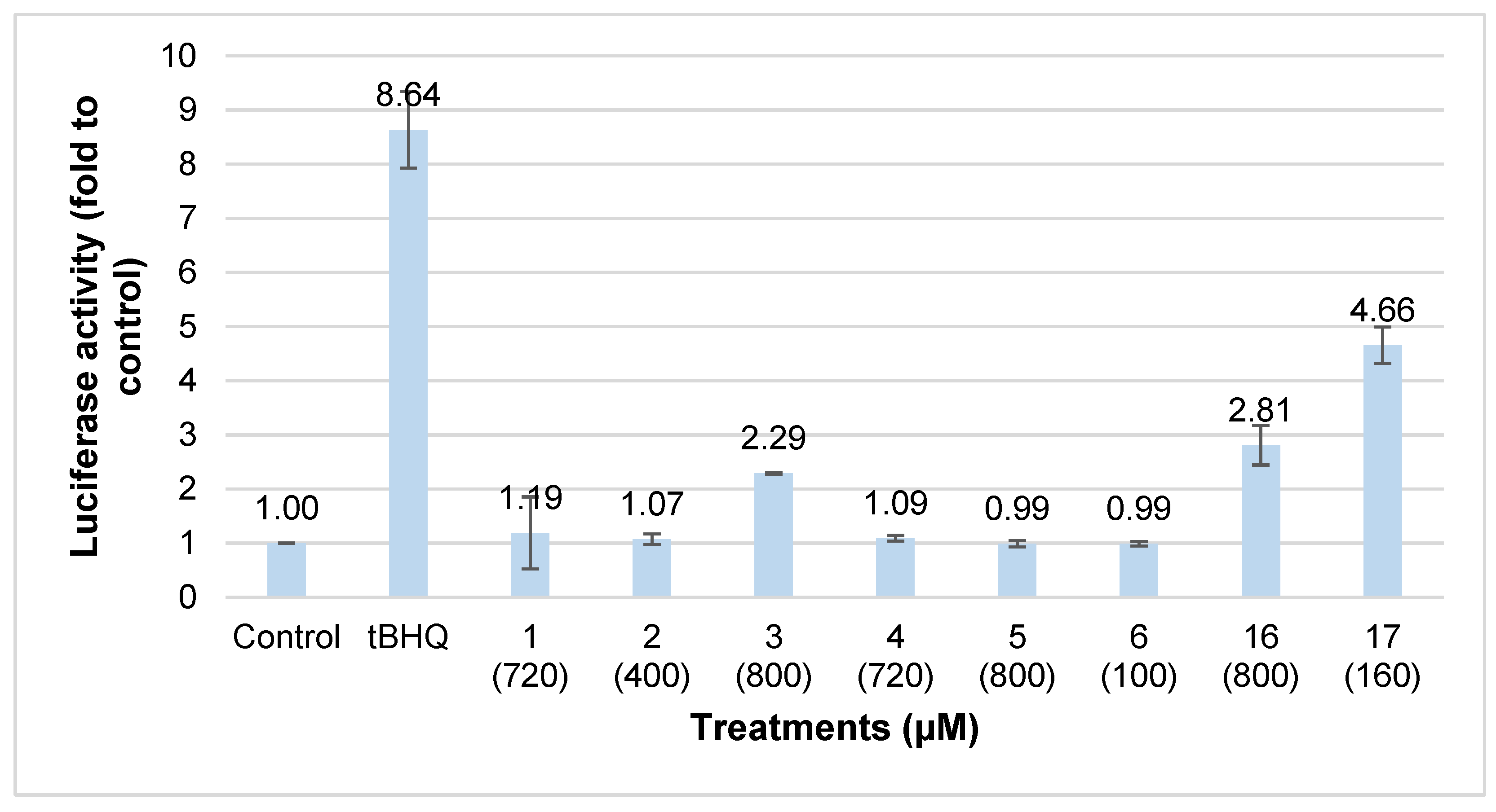
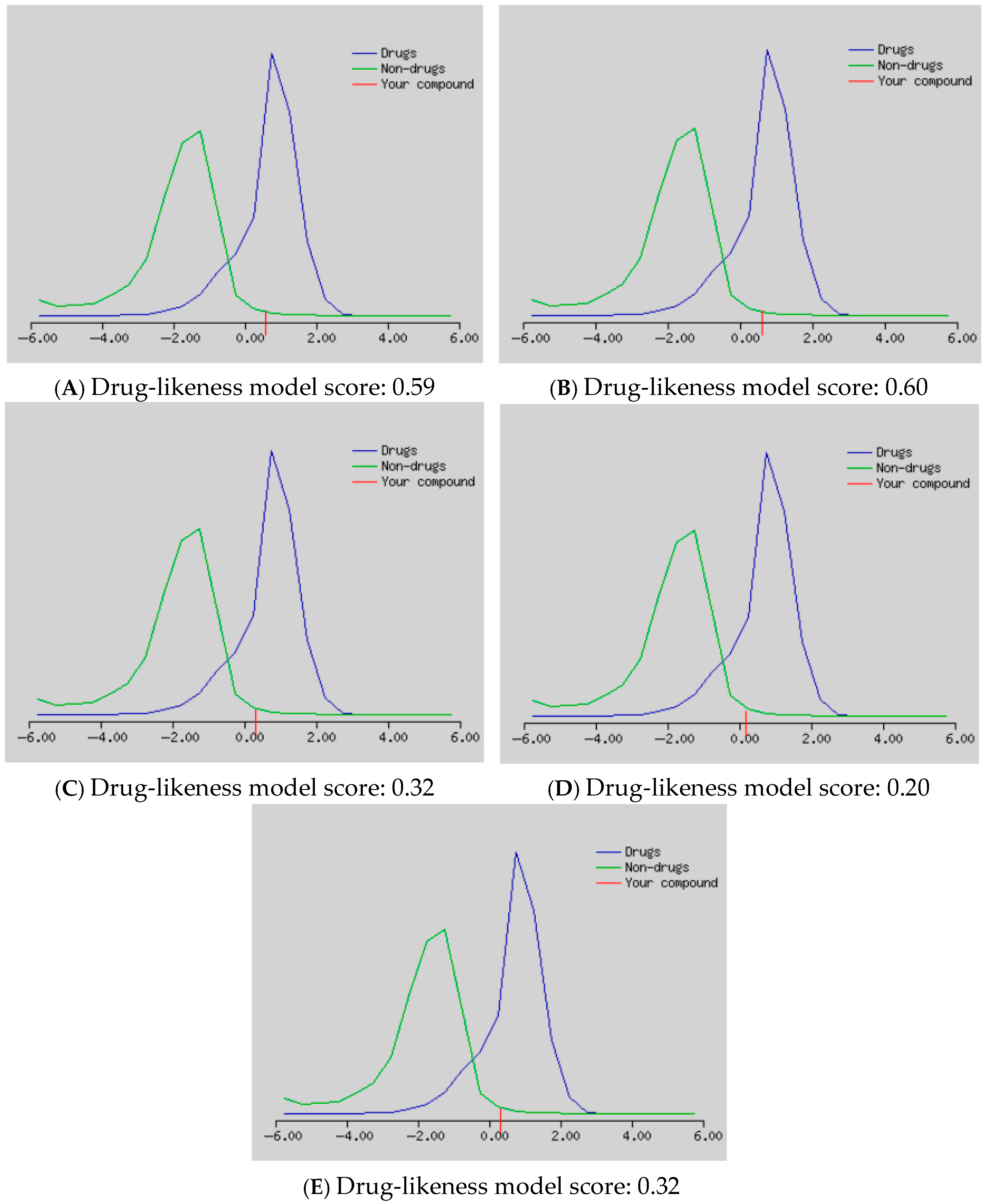
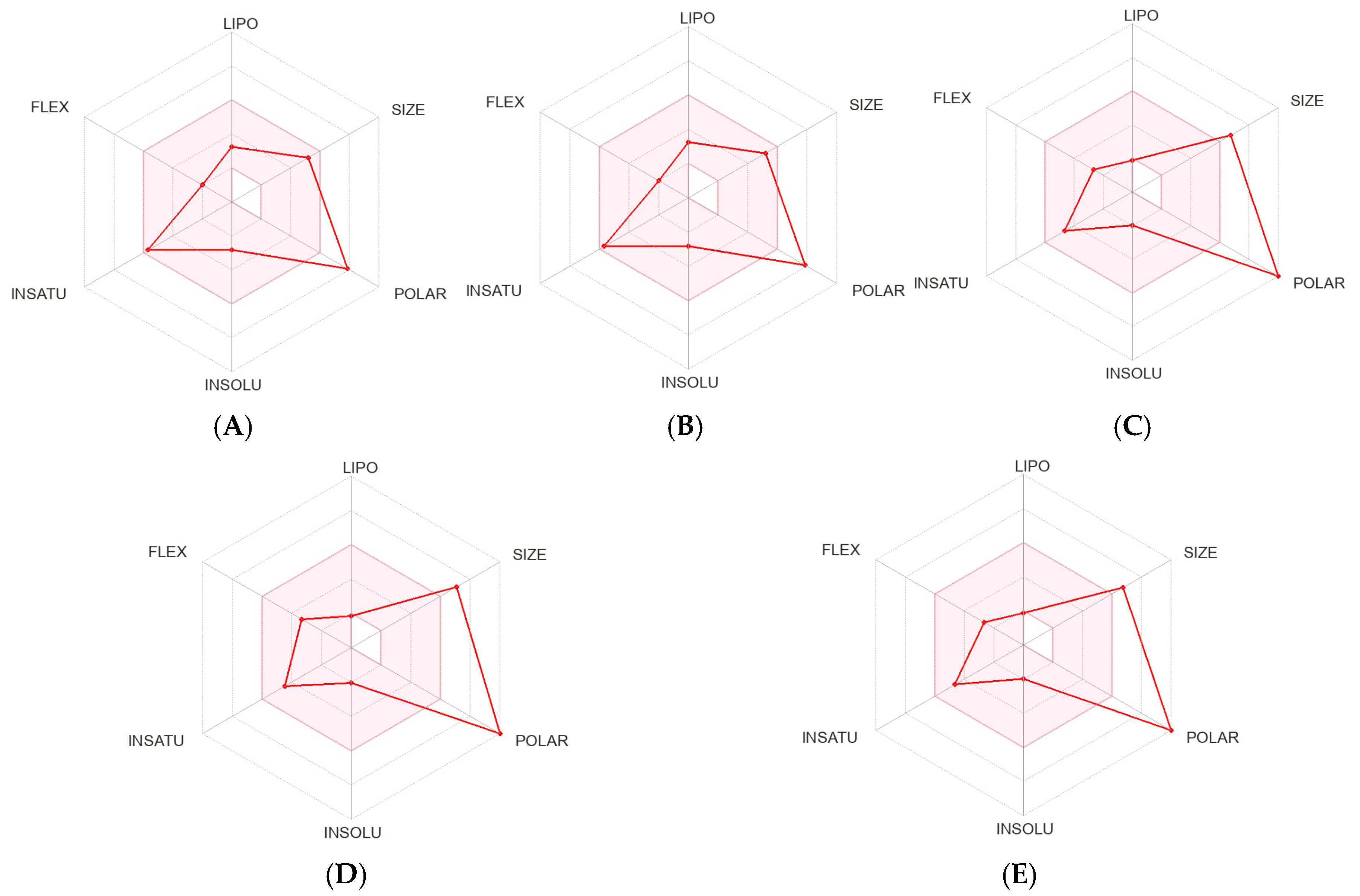
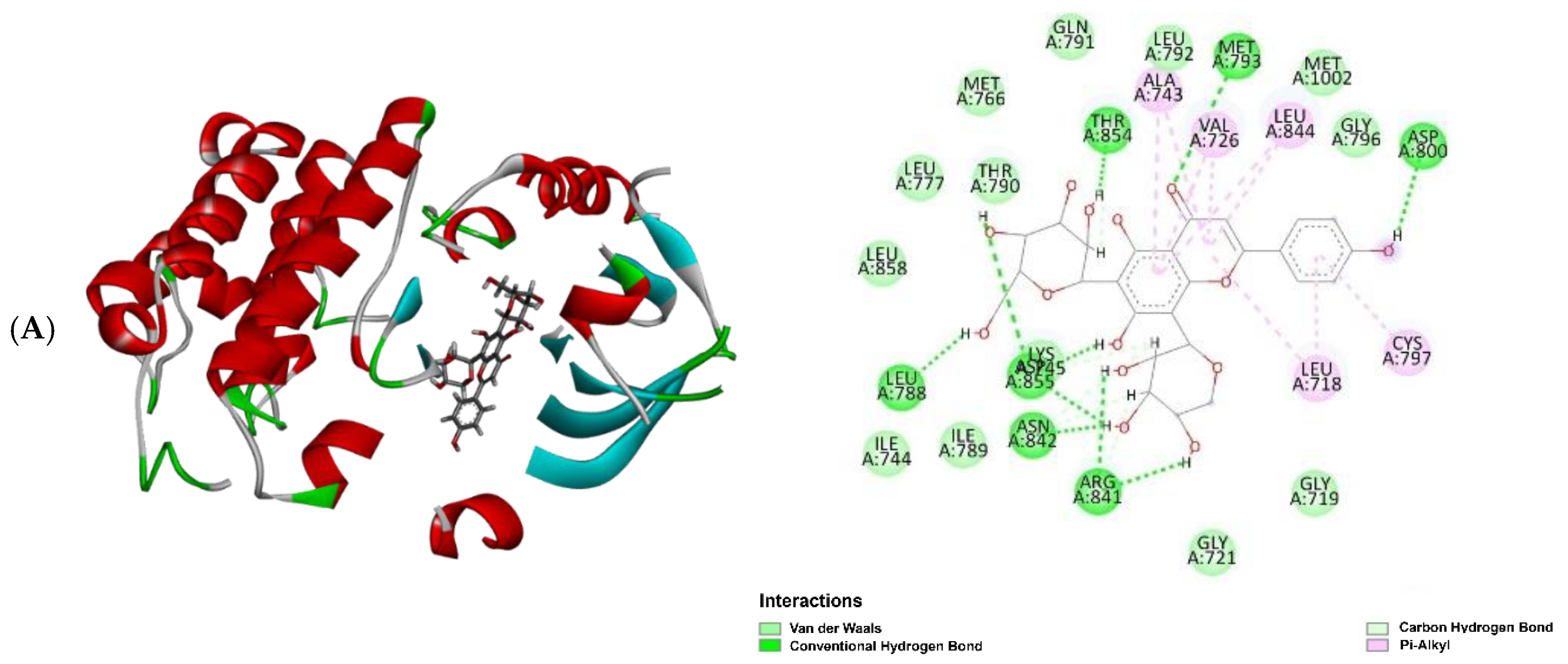
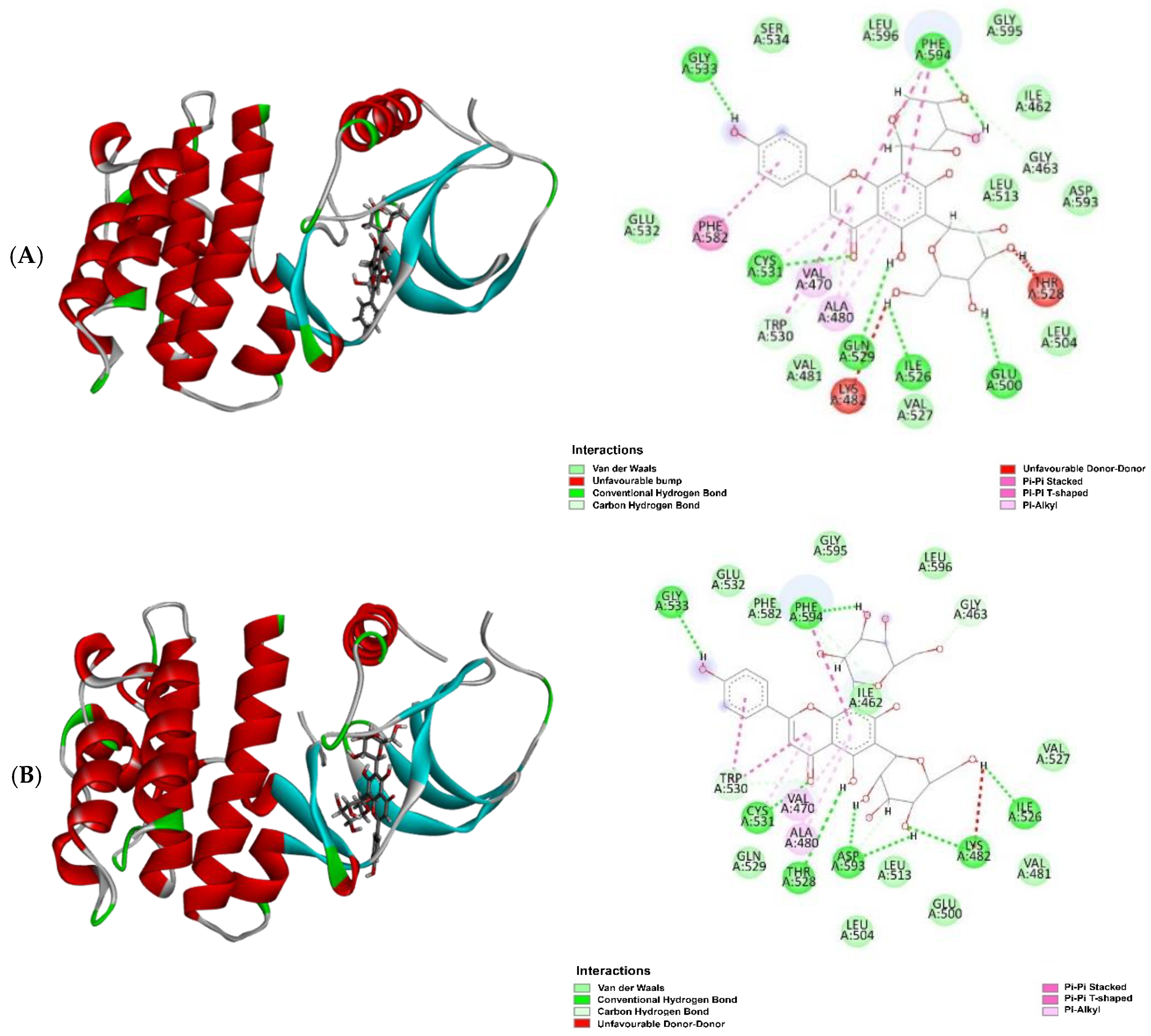
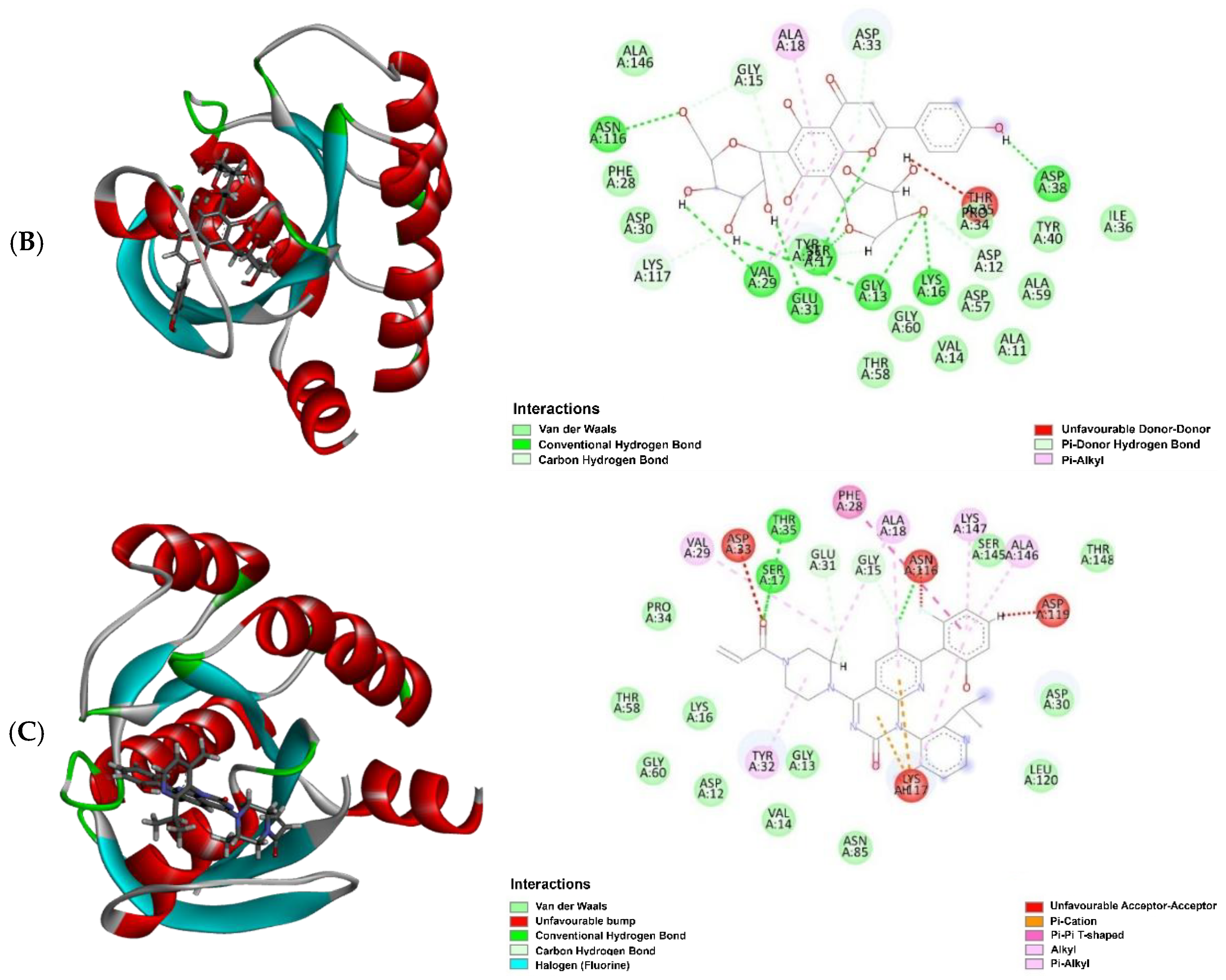
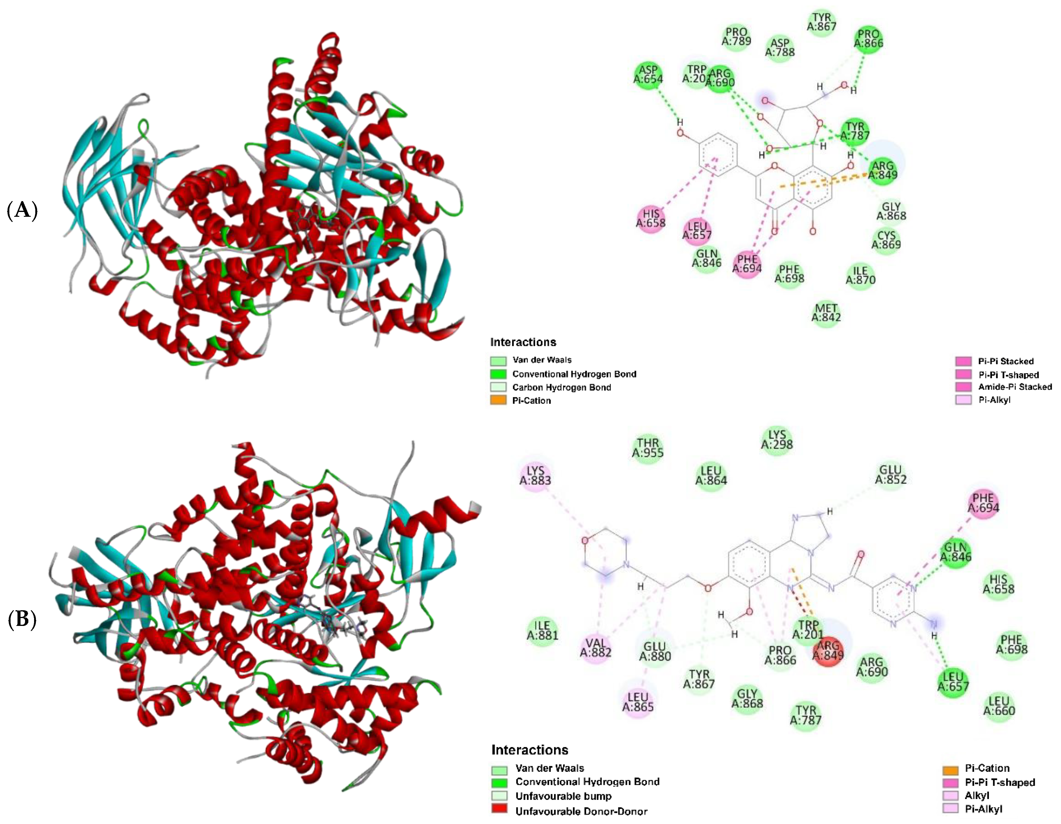
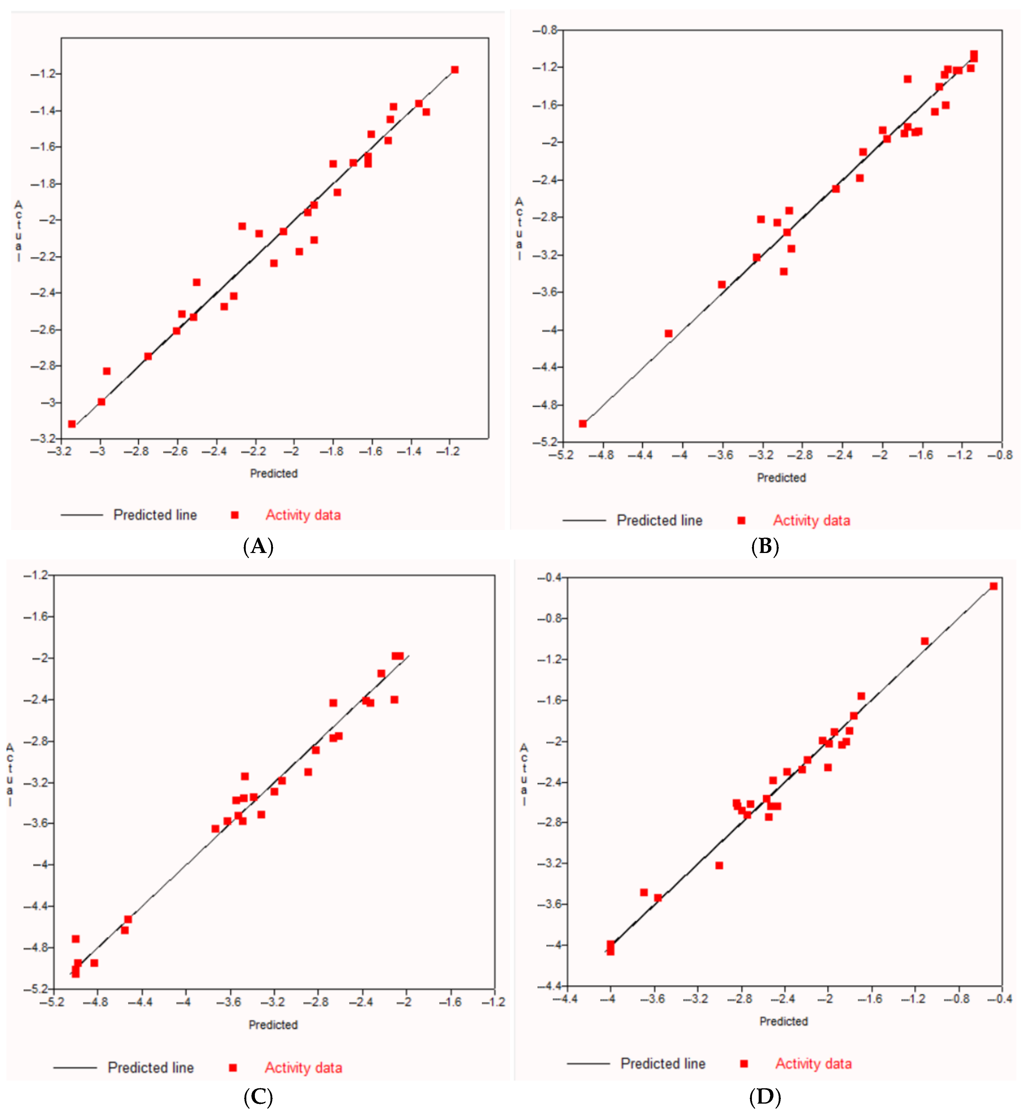
2. Results and Discussion

The n-hexane, dichloromethane (DCM), and methanol (MeOH) extracts obtained from leaves and stems of C. longifolium were evaluated for the ability to activate the Nrf2-ARE pathway using the luciferase reporter assay. Before conducting the luciferase reporter assay, the extracts with various concentrations (0.0125 to 1 mg/mL) were screened for their cytotoxicity in AREc32 cells by the MTT assay to determine a non-cytotoxic concentration of each extract (concentrations causing no more than 10% cell death). tBHQ at 10 μM, used as a positive control, caused a 6-fold activation of Nrf2 activity compared to the control. According to McMahon et al. [26], chemicals causing a >2-fold increase in the luciferase activity were defined as active. The MeOH extract of stems (1 mg/mL) was the most active with a 38.9-fold induction of luciferase activity (relative to control) followed by the MeOH extract of leaves (0.4 mg/mL) with a 24.9-fold induction and the DCM extract of stems (0.025 mg/mL) with a 2.2-fold induction (Figure 3). Fractionation of the active MeOH extracts followed by evaluation of their chemopreventive potential revealed MeOH fractions F1, F2 and F3 of the leaves and MeOH fractions F1, F3 and F4 of stems as active fractions, having a luciferase induction of more than 2-fold (Figure 3). Overall, upon fractionation by solid-phase extraction (SPE), the chemopreventive activity of MeOH fractions of leaves and stems was lower than respective crude extracts. In this case, the plausible explanation could be that the activities of the crude extracts resulted from the combined action of various compounds with synergistic or additive effects [27,28]. Moreover, in some cases, multiple constituents are required to observe the biological effect or get a better effect [29].
As the MeOH SPE fractions F1 and F2 of leaves and F3 and F4 of stems were overall the most active fractions, the phytochemical investigation was conducted on those active MeOH fractions of C. longifolium using a combination of reversed-phase solid-phase extraction (SPE) and preparative/semi-preparative/analytical HPLC techniques. The chromatographic separation of the active fractions F1 and F2 obtained from leaves led to the isolation and purification of five known compounds: a phenolic acid, caffeic acid (1) [30] and four C-glycosyl flavones, isovitexin (2) [31,32] and vicenins 1–3 (3–5) [33,34,35,36] (Figure 4), while the active fraction F4 of stems afforded hexadecanoic acid methyl ester (6). The structures of all isolated compounds were elucidated by extensive 1D and 2D NMR spectroscopic (1H, DEPTQ, COSY, NOESY, HSQC and HMBC) and mass spectrometric analyses, as well as by comparison with respective published spectral data in the literature.
The chromatographic separation of the active MeOH fraction 4 of C. longifolium stems afforded a single compound, which was characterised as hexadecanoic acid methyl ester (6) by spectroscopic means (NMR and LC-MS) and by comparison of its spectral data with the published data [37]. GC-MS analysis and comparison with the National Institute of Standards and Technology (NIST) mass spectral library led to the tentative identification of ten phytochemicals in the active MeOH fractions F3 and F4 of C. longifolium stems including 9-oxononanoic acid methyl ester (7), 11-octadecenoic acid methyl ester (8), caprolactam (9), dodecyl acrylate (10), hexadecanoic acid methyl ester (6), isopropyl myristate (11), nonanal dimethyl acetal (12), 12-methyltetradecanoic acid methyl ester (13), (Z)-9-octadecenoic acid methyl ester (14) and (Z,Z)-9,12-octadecadienoic acid methyl ester (15) (Figure 5). Although caprolactam (9) was identified in this fraction, it is not a naturally occurring compound and is most likely a contaminant from leaching from plastic bags used for storing plant materials.
The tentatively identified compounds (by GC-MS) with their retention times, molecular formulas, molecular weights, and peak areas (%) are listed in Table 1. Thus, active MeOH fractions F3 and F4 of C. longifolium stems contained fatty acid methyl esters. Some of those compounds identified from the stems of C. longifolium have been reported to possess various biological activities. For instance, 11-octadecenoic acid methyl ester (8) and (Z)-9-octadecenoic acid methyl ester (14) were fatty acid methyl esters and have been revealed to exhibit cancer preventive activity [38,39]. 9-Oxononanoic acid methyl ester (7), (Z,Z)-9,12-octadecadienoic acid methyl ester (15), dodecyl acrylate (10) and hexadecanoic acid methyl ester (6) were previously acknowledged for anticancer [39,40,41,42,43,44] and antioxidant activities [44,45,46,47]. These compounds might contribute cancer chemopreventive effect of active MeOH fractions F3 and F4 of C. longifolium stems. Although fatty acid methyl esters occur in plants, the possibility of them being artefacts, especially when MeOH is used for extraction, cannot be ruled out. The identified compounds and biological activities related to medicinal uses are given in Table 2.
To the best of our knowledge, isolated compounds 1–6 and the GC-MS-assisted identified compounds 7–15 are reported here for the first time from C. longifolium. However, the nine phenolic compounds previously isolated from C. longifolium stems [26] were not found in the present study. It could be because the plant material was collected from different environmental conditions, affecting the profile of secondary metabolites [58]. Moreover, compound isolation was conducted from the most active fractions, not all fractions. This species belongs to the Euphorbiaceae family, which produces a wide range of secondary metabolites such as alkaloids, fatty acids, phenolic compounds, steroids and terpenoids [59,60,61].
The ability of compounds 1–6 to induce Nrf2 activation, correlating to their potential chemopreventive property, was determined by the luciferase reporter assay. AREc32 cells were incubated with each isolated compound at non-cytotoxic concentrations for 24 h. The luciferase activity of the isolated compounds is presented in Figure 6.
Among the tested compounds, only vicenin 1 (3) at a non-cytotoxic concentration was considered a chemopreventive compound as it increased luciferase activity by 2.3-fold. To compare structure-function relationships, vitexin (16) and rosmarinic acid (17) were evaluated for their chemopreventive activities against AREc32 cells using the luciferase assay. The luciferase activity produced by the commercially obtained compounds 16 at 800 µM and 17 at 160 µM was 2.8 and 4.7-fold relative to the control, respectively (Figure 7). The similar features between compounds 16 and 3, active flavone C-glycosides were the glucose unit attached to C-8 on the aglycone. This structural feature might be responsible for the activity. The rest of the isolated phenolic compounds showed no chemopreventive activity, which could be due to the glucose attachment to C-6 for compounds 2 and 5 and an additional glucose moiety at C-6 of compound 4. Hence, the sugar position and the number of sugar moieties might affect the activity. Vicenin 1 (3), vitexin (16) and rosmarinic acid (17) have been proven to possess anti-inflammatory and antioxidant effects [62,63,64,65], which are intricately linked with cancer chemoprevention and chemotherapy [66].
In silico studies for bioactivity of five flavone C-glycosides, including isovitexin (2), vitexin (16), and vicenins 1–3 (3–5), were conducted, aiming to establish their drug-likeness properties and to predict the possible mechanism of action behind the cancer chemopreventive and chemotherapeutic effects. The PASS (prediction of activity spectra of substances) prediction analysis revealed the cancer chemopreventive and chemotherapeutic potentials of the selected C-glycosyl flavones (Table 3) as their Pa values exceeded 0.7. It was found that all the tested compounds showed high potential antineoplastic activity. Moreover, all the tested compounds except compound 16 were predicted to have enormous potential as anticarcinogenic. However, only compounds 24 and 16 had the potential as antimutagenic and apoptosis agonists. Additionally, only compounds 4 and 5 showed potential chemopreventive activity.
Drug-likeness analysis results of the tested compounds predicted by MolSoft are illustrated in Figure 7. Regarding drug-likeness scores, the compounds with positive values (nearer to 1) represent drug-like molecules [67]. Thus, all compounds showed good drug-likeness scores between 0.20 and 0.60. Vitexin (16) and isovitexin (2) had the best drug-likeness scores of 0.60 and 0.59, respectively.
The results of ADMET prediction for the tested compounds are presented in Table 4. Their bioavailability radar included six important physicochemical properties for oral bioavailability (lipophilicity, size, polarity, solubility, flexibility and saturation). In terms of size, vicenins 1–3 (3–5) exceeded the optimal size of the molecule (150–500 g/mol). All tested compounds exhibited a high degree of saturation, having fraction Csp3 values ranging from 0.29 to 0.44, which passed the filter of fraction Csp3 > 0.25. Furthermore, all tested compounds showed high polarity values, displaying topological polar surface area (TPSA) more than 130 Å2. Hence, these compounds were too polar and consequently predicted not to be orally bioavailable. Drugs with higher TPSA values are less lipid soluble [68]. For lipophilicity, the logP values of all compounds using a logP of a reference compound (XLOGP3) were in the optimal range (−0.7 to +5.0). The water solubility parameter is important for absorption. When three different topological models (ESOL, Ali and Silicos-IT) were applied, all compounds were predicted to be soluble to very soluble in water, meaning that they can be excreted in the kidneys and urine.
In terms of pharmacokinetics, all compounds showed low gastrointestinal (GI) absorption. As a result, bioavailability is predicted to be poor. Notably, low oral bioavailability due to limited absorption is one of the issues found in flavonoids [69]. Alternatively, other administration routes, such as inhalation, injection, or the preparation of special dosage forms, such as nanoformulation, may require enhancing the bioavailability. Moreover, all compounds demonstrated no predictable blood-brain barrier (BBB) permeability, which means they were nontoxic or had a low toxicity to the brain. Additionally, none of the compounds were predicted to show any inhibition of CYP450 subtypes. This indicated that these compounds were not causing drug-drug interactions. Furthermore, negative log Kp values of the tested compounds revealed that all compounds exhibited low skin permeability. The medical chemistry parameters revealed that the tested compounds exhibited no structural alerts against PAINS (Pan-assay interference compounds) and Brenk’s filters. The summary of the prediction of oral bioavailability is plotted as the bioavailability radar with six parameters (Figure 8). Ideally, drug-likeness by this model requires the red lines assumed by the tested molecules to be entirely located in the pink zone of the plot [70]. Therefore, isovitexin (2), vitexin (16) and vicenins 1–3 (3–5) were predicted not to be orally bioavailable, because they were too polar and compounds 3–5 had high molecular weights.
Molecular docking results revealed that the experimental compounds displayed significant contacts with the critical residues, demonstrating the ability of these proteins to alter their activity. Vicenin 3 (5) had the highest binding efficiency against three target proteins, except for PI3K, among all other compounds, including the positive controls (marketed drug). Docking analysis suggested the experimental compounds had cancer chemopreventive and chemotherapeutic potentials, whether it is involved with Nrf2 activation or other molecular mechanisms. If the top one compound is considered against each protein, compound 5 showed the best inhibition against EGFR and BRAF targets with a MolDock score of −142.57 and −162.63, respectively, whereas the positive control (marketed drug) gefitinib displayed a MolDock score of −141.80 and dabrafenib showed −137.99. Similarly, against KRAS and PI3K, the best inhibition was shown by vicenin 2 (4) and vitexin (16) with a MolDock score of −154.39 and −133.27, respectively, while the positive control sotorasib exhibited −150.41 and copanlisib showed −126.30. The entire MolDock scores of the experimental compounds against four cancer-related target proteins are displayed in Table 5. Docking pose and 2D interactions of best ligands and positive controls with target receptors are shown in Figure 9, Figure 10, Figure 11 and Figure 12.
QSAR models were used to acquire a better understanding of the structure–activity relationships. The prediction of biological activities was achieved quantitatively as a 50% inhibitory concentration (IC50) of a tested compound required to express a biological response. From the QSAR equation, the IC50 value of the experimental compounds of each target was calculated (Table 6). The multiple regression plots (linear) for the cancer-related target proteins are shown in Figure 13. From QSAR studies, it was found that the lowest IC50 value was shown by vicenin 2 (4) for the target EGFR as 2.45 nM. For the target BRAF, the lowest IC50 value was predicted to be 11.75 nM from vicenins 1 (3) and 3 (5), while the IC50 value of compound 4 was 58.88 nM. This means changing the type of sugar moieties of glycosylated compounds can make a difference in their activities [71,72]. The predicted IC50 value of vitexin (16) was 165.96 µM, considering the lowest value for the target KRAS. Compounds 3 and 5 also showed the lowest IC50 value (64.57 nM) for the target PI3K. These results were a supporting indicator if we consider vicenins 1 (3) and 3 (5) as potential contributors to anticancer activity.
In silico studies and literature reviews suggested that these flavone C-glycosides have potential as therapeutically acceptable cancer chemotherapeutic agents. Moreover, these compounds also have cancer chemopreventive prospects. In silico study results supported the reported potential anticancer properties of these compounds. However, extensive in vitro or in vivo preclinical studies are required, as well as clinical trials. Although, the in silico studies showed the cancer chemopreventive activity profiles of isovitexin (2), vitexin (16) and vicenins 1–3 (3–5), when in vitro chemopreventive activity using the luciferase assay was performed, compounds 2, 4 and 5 were not active against AREc 32 cells in terms of activation of Nrf2 activity. The in vitro luciferase assay results of compounds 3 and 16 found that these compounds induced Nrf2 activation in AREc32 cells. Therefore, the results of the in vitro studies matched the findings from the in silico work. It demonstrates the need for conducting in vitro and in vivo studies together with in silico studies. In silico is a prediction based on numerous factors such as molecular structure, physicochemical, and pharmacokinetic properties, but the reality may be different from in silico prediction. Thus, in silico prediction is not always 100% accurate. Even if the results from the in vitro cancer chemopreventive effect indicated that compounds 4 and 5 were not Nrf2 activators, it does not mean that compounds 4 and 5 do not exert a chemopreventive effect. These two compounds may be contributing to cancer chemoprevention through other signalling pathways, not necessarily through Nrf2 activation.
Apart from the Nrf2/ARE signalling pathway, there are many other cellular signalling pathways that play important roles in cancer chemoprevention [2]. In recent years, growing evidence has revealed the significance of natural products in affecting the numerous essential cellular signalling pathways associated with cancer such as activator protein-1 (AP-1), Janus kinase/signal transducers and activators of transcription (JAK/STAT), mitogen-activated protein kinase (MAPK), nuclear factor-Kappa B (NF-κB), phosphatidylinositol 3-kinase/protein kinase B/mammalian target of rapamycin (PI3K-AKT-mTOR) pathways [73]. Notably, these natural products prevent cancer cell proliferation, development, and metastasis by inducing cell cycle arrest, promoting apoptosis, generating reactive oxygen species (ROS), inducing/inhibiting several signalling pathways, inhibiting angiogenesis and regulating transcription factors [74].

3. Materials and Methods

3.1. Chemicals, Reagents, and Materials

All chemicals were purchased from Sigma-Aldrich (Dorset, UK). Solvents were purchased from Fisher Scientific (Loughborough, UK) and used without further purification. Deuterated NMR solvents, including methanol (CD3OD), pyridine (Pyr-d5) and DMSO (DMSO-d6), were purchased from Cambridge Isotope Laboratories (Tewksbury, MA, USA). Cell culture reagents (0.25% trypsin-EDTA, penicillin-streptomycin and phosphate-buffered saline (PBS)) were purchased from Thermo Fisher Scientific (Paisley, UK), while basal medium (DMEM), fetal bovine serum (FBS) and geneticin G418 were purchased from Biosera (Neuville, France). Luciferase assay kit reagent was purchased from Promega (Madison, WI, USA). Specialized white 96-well flat-bottom plates were from Greiner Bio-One (Frickenhausen, Germany), while clear 96-well flat-bottom plates were from Falcon (Falcon, NC, USA).

3.2. General Experimental Procedures

Analytical HPLC was performed on a Dionex Ultimate 3000 UHPLC (Thermo Fisher Scientific, Waltham, MA, USA) coupled with a diode array detector (DAD). MeOH crude extracts and MeOH fractions were analyzed on a reversed-phase Phenomenex column (Luna 5 μm C18(2) 100 Å, 150 mm × 4.6 mm, serial no. H21-192754, Phenomenex, Torrance, CA, USA). Preparative HPLC system of a Dionex Ultimate 3000 UHPLC (Thermo Fisher Scientific, MA, USA) or a Jasco LC-4000 Series (Jasco, Tokyo, Japan) and a reverse phase Phenomenex column (Luna 10 μm C18(2) 100 Å, 150 mm × 21.2 mm, serial no. H18-196260, Phenomenex, CA, USA) were used to isolate compounds from active fractions. The purification of impure compounds was accomplished on an Agilent 1260 Infinity series semi-preparative HPLC system coupled with a DAD detector using an ACE 10 C18-HL column (150 mm × 10 mm, Hichrom Ltd., Lutterworth, UK). A flow rate of 1 mL/min, 2 mL/min and 10 mL/min was used for analytical, semi-preparative and preparative HPLC, respectively. The column temperature was maintained at 25 °C. The chromatogram was monitored at variable UV-vis wavelengths (220, 254, 280 and 320 nm). The NMR experiments were performed in CD3OD, Pyr-d5 or DMSO-d6 solution on a Bruker AMX-600 NMR spectrometer (600 MHz for 1H, and 150 MHz for 13C) (Bruker, Billerica, MA, USA). ESI-MS data were recorded through a Micromass LCT time-of-flight (TOF) mass spectrometer (Waters, Manchester, UK) equipped with an electrospray ionisation (ESI) source. The GC-MS analysis of oily fractions of C. longifolium stems was performed using an Agilent 6890N network GC system equipped with an Agilent 5790 mass selective detector (Agilent, Santa Clara, CA, USA) and a capillary column HP-5MS (30 m × 0.25 mm i.d., 0.25 μm film thickness). The oven temperature was programmed as follows: the beginning of 150 °C kept for 3 min, rising by 20 °C/min to 280 °C and maintaining for 4 min. The run time was 14 min. The mass-spectrometric analysis was conducted in the range of 41–500 m/z using electronic ionisation energy, the EI mode at 70 eV. The MS source temperature was 230 °C. Identification of the detected components was performed by comparing their mass spectra with the reference spectra in NIST MS Search software version 3.0.

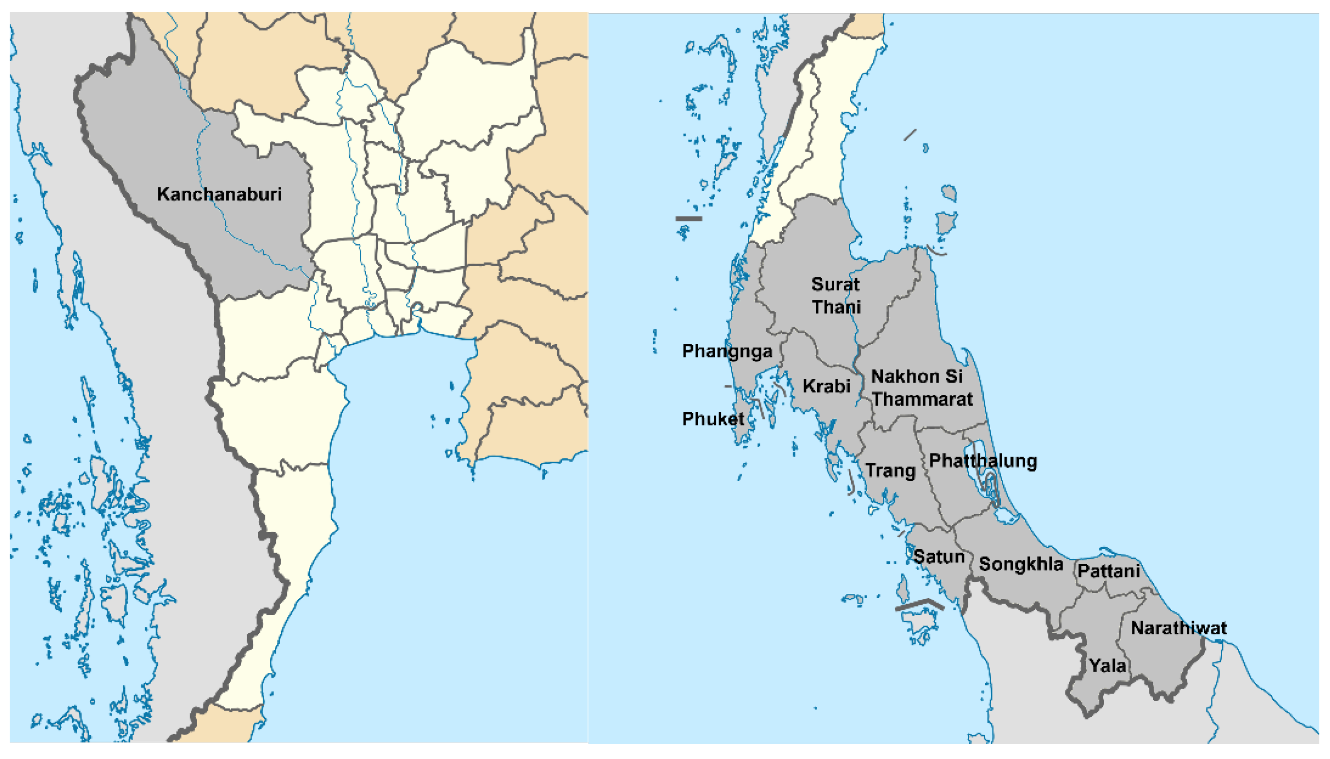
3.3. Plant Materials

Leaves (CLL) and stems (CLS) of Claoxylon longifolium (Blume) Endl. ex Hassk. were collected by Dr Chuanchom Khuniad from Phatthalung Province, southern Thailand (Latitude: 7°37′4.30″ N; Longitude: 100°04′40.51″ E), during April and May 2021. Its voucher specimen (BKF NO.228283) was prepared and deposited in the Forest Herbarium (BKF), Department of National Park, Wildlife and Plant Conservation, Ministry of Natural Resources, Bangkok, Thailand.

3.4. Extraction and Isolation of Compounds

Leaves (278.0 g) and stems (473.0 g) of C. longifolium were washed, sliced into small pieces, and dried in a solar greenhouse dryer at a temperature not exceeding 55 °C. Dried leaves and stems were ground into powder with a spice grinder. Powdered leaves and stems were Soxhlet-extracted separately and consecutively with n-hexane, dichloromethane (DCM) and methanol (MeOH) (900 mL, ten cycles each) [75]. The resulting extracts were concentrated separately to dryness under reduced pressure using a rotary evaporator at a maximum temperature of 45 °C (BUCHI, Flawil, Switzerland). The crude extracts at a non-cytotoxic concentration were screened for their chemopreventive potential using a luciferase reporter assay in AREc32 cells. MeOH extracts of leaves and stems were found to be the most active extracts. A portion of each active MeOH extract (2 g) was subjected to solid-phase extraction (SPE) on a Strata C18-E reversed-phase cartridge (20 g) (Phenomenex, CA, USA), previously washed with MeOH (50 mL), followed by equilibration with water (100 mL) [76]. The SPE cartridge was eluted with a step gradient consisting of 20, 50, 80% MeOH in water and 100% MeOH (200 mL each) to obtain four MeOH fractions (F1–F4), respectively. All fractions were concentrated to dryness by a rotary evaporator combined with a nitrogen blowdown concentrator and kept at 4 °C for further analyses. MeOH fractions F1 and F2 of leaves (CLL-Me F1 and F2) were analyzed by preparative HPLC (Jasco) with a mobile phase gradient of water (A) and methanol (B), both containing 0.1% TFA: 30–50% A in B for 40 min, monitored at wavelength 220 nm. Preparative HPLC analysis of CLL-Me F1 using the above method yielded caffeic acid (1, 5.5 mg, tR = 24.7 min). The separation process of CLL-Me F2 afforded vicenin 2 (4, 13.1 mg, tR = 22.8 min) and isovitexin (2, 25.3 mg, tR = 34.1 min). Furthermore, CLL-Me F2-P3 (15.8 mg) collected at tR = 26.3 min was further purified by semi-preparative HPLC with a mobile phase gradient of 33–45% A in B over 20 min, observed at wavelength 220 nm to afford vicenin 3 (5, 7.5 mg, tR = 15.2 min). CLL-Me F2-P5 (18.7 mg) collected at tR =32.1 min was also purified by semi-preparative HPLC with a different mobile phase: a gradient of 40–55% A in B over 20 min, observed at wavelength 220 nm to afford vicenin 1 (3, 9.7 mg, tR = 13.8 min).
The MeOH fraction F3 of stems (CLS-Me F3) was separated by preparative HPLC (Dionex) with a mobile phase gradient of 45–100% A in B for 35 min, observed at wavelength 220 nm to yield CLS-Me F3-P3 (14.4 mg, tR = 30.4 min). Fraction F4 of stems (CLS-Me F4) was also separated by preparative HPLC (Dionex) with mobile phase gradient of 75–100% A in B for 35 min, observed at wavelength 220 nm to obtain CLS-Me F4-P6 (25.4 mg, tR = 26.7 min), CLS-Me F4-P7 (34.4 mg, tR = 29.6 min), CLS-Me F4-P8 (38.7 mg, tR = 32.1 min) and CLS-Me F4-P9 (14.6 mg, tR = 33.3 min). CLS-Me F4-P8 was further purified by analytical HPLC with a mobile phase gradient of 85–100% A in B for 20 min. This purification method obtained hexadecanoic acid methyl ester (6, 14.3 mg, tR = 14.7 min). CLS-Me F3-P3, CLS-Me F4-P6, CLS-Me F4-P7 and CLS-Me F4-P9 (oily fractions) were found to be a mixture of fatty acids, long-chain alkanes and others by analytical HPLC and preliminary 1H NMR. Hence, they were not further purified. However, the GC-MS analysis was conducted to identify the presence of phytochemicals in CLS-Me F3-P3, CLS-Me F4-P6, CLS-Me F4-P7, compound 6 and CLS-Me F4-P9.

3.5. Cell Culture

An AREc32 cell line (a modified MCF7 human breast cancer cell line) was obtained from Professor Roland Wolf (University of Dundee, UK), whose lab created the cell line. AREc32 cells were cultured in Dulbecco’s Modified Eagle’s Medium (DMEM) High Glucose supplemented with 10% fetal bovine serum (FBS), 1% penicillin-streptomycin and 0.8 mg/mL of antibiotic G418 in T75 cell culture flasks and maintained at 37 °C in a humidified atmosphere of 5% CO2 (Binder, Tuttlingen, Germany).

3.6. MTT Assay

The cell viability of the AREc32 cells was assessed using the MTT assay [77] to identify a non-cytotoxic concentration for further use in the luciferase reporter assay. The cells were seeded into 96-well plates at a density of 1.2 × 10⁴ cells/well (100 μL/well) and allowed to adhere for 24 h in the incubator at 37 °C and 5% CO2. After 24 h, the medium was removed, and the cells were treated with medium containing different concentrations of each treatment (crude extracts, fractions and compounds). A set of untreated control wells was included in each plate, as well as cells treated with 20% DMSO as a positive control. After incubation with the treatment for 24 h, the medium was aspirated. A volume of 20 μL of MTT solution (5 mg/mL in PBS) and 180 μL of fresh medium were added to each well and then continued to incubate for 4 h. The MTT solution/medium was aspirated off and replaced with DMSO (100 μL/well) to dissolve the purple formazan crystals. The absorbance of the formazan crystals was then read using a microplate reader at 570 nm (TECAN, Mannedorf, Switzerland). All experiments were performed in three independent experiments (5 replicates/experiment). The mean % cell viability was calculated as follows:
C e l l   v i a b i l i t y = ( A 1 / A 0 ) × 100
where A0 is the mean absorbance of untreated wells and A1 is the mean absorbance of treated cells.

3.7. Luciferase Reporter Assay

To investigate the cancer chemopreventive potential, a luciferase reporter assay with an AREc32 cell line was used to assess the ability of the crude extracts, fractions, and compounds to induce the cellular antioxidant defence system via the activation of Nrf2 transcription factor [78]. Briefly, AREc32 cells were seeded into 96-well plates at a density of 1.2 × 10⁴ cells/well and incubated for 24 h. They were then treated with a non-cytotoxic concentration of each crude extract, fraction and compound. After another 24 h of incubation, the culture medium was discarded, and the cells were washed with 100 μL of phosphate-buffered saline (PBS) twice, then 20 μL of 1X luciferase reporter lysis buffer was added to each well, followed by a freeze-thaw cycle (−20 °C) for 24 h to achieve complete cell lysis. The cell lysate was thawed and transferred into a white opaque 96-well plate and mixed with 100 μL of freshly prepared luciferase assay reagent and immediately. The luminescence was measured immediately using the microplate reader (TECAN, Mannedorf, Switzerland). The luciferase activity level was compared to the basal level of luciferase activity in control cells and presented as a fold increase relative to control (untreated cells). Tert-butylhydroquinone (tBHQ, 10 μM) served as a positive control. All experiments were performed in three independent experiments (5 replicates/experiment).

3.8. In Silico Studies for Bioactivity of Selected C-Glycosyl Flavones

Five C-glycosyl flavones, including vitexin (16), isovitexin (2), and vicenins 1–3 (3–5), were selected for in silico studies using several screening methods and protocols as follows:
Prediction of activity spectra for substances (PASS) analysis of biological activities:
The PASS online (http://www.way2drug.com/passonline/predict.php, accessed on 20 August 2024) was utilized to predict the probability of the selected compounds exhibiting six biological activities related to cancer or being inactive [79].
Drug likeness analysis:
Drug likeness properties of the C-glycosyl flavones were screened by the online MolSoft toolkit (https://molsoft.com/mprop/, accessed on 20 August 2024).
Adsorption, distribution, metabolism, excretion and toxicity (ADMET) prediction:
Prediction of ADMET profiles was analyzed by the SwissADME server (http://www.swissadme.ch/), a web server from Swiss Institute of Bioinformatics [70], to evaluate the physicochemical, pharmacokinetics, drug-likeness, and medicinal chemistry friendliness parameters of the selected C-glycosyl flavones.
Molecular docking:
Molecular docking was analyzed with the Molegro Virtual Docker 6.0 (MVD) software version 6.0 [80,81] for predicting the binding interactions of ligands (selected compounds) with the active sites of cancer-related target proteins (EGFR, BRAF, KRAS, and PI3K). EGFR and BRAF are associated with uncontrolled cell proliferation, while KRAS and PI3K are responsible for anti-apoptotic activity.
Quantitative structure–activity relation (QSAR) studies:
QSAR studies were conducted to find relationships between the structural properties of the selected compounds and cancer-associated biological activities by considering 30 known inhibitors of each target protein from the ChEMBL database (https://www.ebi.ac.uk/chembl/). ACD ChemSketch software (Advanced Chemistry Development, Toronto, ON, Canada) was used to create 11 descriptors from each inhibitor, including molar weight, molar refractivity, molar volume, index of refraction, surface tension, parachor, density, polarizability, log P, hydrogen bond donor and hydrogen bond acceptor. The Easy QSAR software developed by Kuntal Kumar Bhusan, University of Hyderabad, was used to create a multiple linear regression analysis after the descriptors and activities were entered. This regression study produced the QSAR equation. The 50% inhibitory concentration (IC50) values of the C-glycosyl flavones of each target were calculated by the QSAR equation.

3.9. Statistical Analysis

All experiments were conducted in triplicate with a minimum of three replicates per assayed concentration. Data were expressed as means ± SEM (standard error of the mean). The graphs were plotted using Microsoft Excel for Microsoft 365 MSO Version 2502. SEM was shown on the graph as an error bar.

4. Conclusions

Claoxylon longifolium (leaves and stems) has been evaluated for its phytochemicals and cancer chemopreventive effect following a bioassay-guided isolation procedure. The phytochemical investigation conducted on C. longifolium active MeOH fractions afforded six known compounds: caffeic acid (1), isovitexin (2), and vicenins 1–3 (3–5), as well as hexadecanoic acid methyl ester (6). GC-MS analysis revealed that the major constituents present in the active stem fractions were fatty acid methyl esters. Vicenin 1 (3) was found to be a potent chemopreventive compound as it increased luciferase activity by 2.3-fold. In silico studies on the C-glycosyl flavones revealed the potential of these compounds as cancer chemopreventive and chemotherapeutic agents. This study generated the first phytochemical investigation on C. longifolium leaves. All identified and isolated compounds were reported for the first time from this species. Moreover, this is also the first report on the chemopreventive properties of C. longifolium. The findings of a recent study indicate that C. longifolium contains promising chemopreventive agents, supporting, to some extent, the utilization of this indigenous vegetable in cancer prevention and consumption for health benefits in the southern Thai community.

Author Contributions

Conceptualization, C.K., L.N., K.J.R., and S.D.S.; A.D.T., C.K., L.N., K.J.R., R.N. and S.D.S.; software, A.D.T. and R.N.; validation, C.K., L.N., K.J.R. and S.D.S.; formal analysis, A.D.T., C.K., L.N., K.J.R., R.N. and S.D.S.; investigation, C.K. and R.N.; resources, L.N., K.J.R. and S.D.S.; data curation, C.K., L.N., K.J.R. and S.D.S.; A.D.T., C.K., L.N., K.J.R., R.N. and S.D.S.; writing—original draft preparation, C.K.; writing—review and editing, L.N. and S.D.S.; visualization, C.K., L.N., K.J.R. and S.D.S.; A.D.T., C.K., L.N., K.J.R., R.N. and S.D.S.; supervision, L.N., K.J.R. and S.D.S.; project administration, C.K.; funding acquisition, C.K. All authors have read and agreed to the published version of the manuscript.

Funding

This research was funded by the Government of Thailand through a PhD scholarship to Chuanchom Khuniad (C.K.).

Institutional Review Board Statement

Not applicable.

Informed Consent Statement

Not applicable.

Data Availability Statement

All data are available from the corresponding authors and may be shared upon a formal written request.

Acknowledgments

L.N. gratefully acknowledges the support of the European Regional Development Fund—Project ENOCH (No. CZ.02.1.01/0.0/0.0/16_019/0000868) and the Czech Agency Grants—Project 23-05474S and Project 23-05389S, and C.K. thanks the Ministry of Higher Education, Science, Research and Innovation, Royal Thai Government for awarding her with a PhD scholarship.

Conflicts of Interest

The authors declare no conflicts of interest.

References

  1. Bray, F.; Laversanne, M.; Sung, H.; Ferlay, J.; Siegel, R.L.; Soerjomataram, I.; Jemal, A. Global cancer statistics 2022: GLOBOCAN estimates of incidence and mortality worldwide for 36 cancers in 185 countries. CA Cancer J. Clin. 2024, 74, 229–263. [Google Scholar] [CrossRef]
  2. Rahman, M.M.; Sarker, M.T.; Alam Tumpa, M.A.; Yamin, M.; Islam, T.; Park, M.N.; Islam, M.R.; Rauf, A.; Sharma, R.; Cavalu, S.; et al. Exploring the recent trends in perturbing the cellular signaling pathways in cancer by natural products. Front. Pharmacol. 2022, 13, 950109. [Google Scholar] [CrossRef]
  3. Avendaño, C.; Menéndez, J.C. Cancer chemoprevention. In Medicinal Chemistry of Anticancer Drugs, 2nd ed.; Avendaño, C., Menéndez, J.C., Eds.; Elsevier: Amsterdam, The Netherlands, 2015; pp. 701–723. [Google Scholar]
  4. Ma, L.; Zhang, M.; Zhao, R.; Wang, D.; Ma, Y.; Li, A. Plant natural products: Promising resources for cancer chemoprevention. Molecules 2021, 26, 933. [Google Scholar] [CrossRef]
  5. He, F.; Ru, X.; Wen, T. Nrf2, a transcription factor for stress response and beyond. Int. J. Mol. Sci. 2020, 21, 4777. [Google Scholar] [CrossRef]
  6. Jeong, W.S.; Jun, M.; Kong, A.N. Nrf2: A potential molecular target for cancer chemoprevention by natural compounds. Antioxid. Redox Signal 2006, 8, 99–106. [Google Scholar] [CrossRef]
  7. Ritchie, K.J.; Sarker, S.D. Cancer chemopreventive natural products. In Annual Reports in Medicinal Chemistry; Sarker, S.D., Nahar, L., Eds.; Medicinal Natural Products—A Disease-focused Approach; Elsevier: London, UK, 2020; Volume 55, pp. 273–295. [Google Scholar]
  8. Zhao, C.R.; Gao, Z.H.; Qu, X.J. Nrf2-ARE signaling pathway and natural products for cancer chemoprevention. Cancer Epidemiol. 2010, 34, 523–533. [Google Scholar] [CrossRef]
  9. Wu, K.C.; McDonald, P.R.; Liu, J.; Klaassen, C.D. Screening of natural compounds as activators of the keap1-nrf2 pathway. Planta Med. 2014, 80, 97–104. [Google Scholar] [CrossRef] [PubMed]
  10. Pouremamali, F.; Pouremamali, A.; Dadashpour, M.; Soozangar, N.; Jeddi, F. An update of Nrf2 activators and inhibitors in cancer prevention/promotion. Cell Commun. Signal 2022, 20, 100. [Google Scholar] [CrossRef] [PubMed]
  11. Maneenoon, K.; Khuniad, C.; Teanuan, Y.; Saedan, N.; Prom-In, S.; Rukleng, N.; Kongpool, W.; Pinsook, P.; Wongwiwat, W. Ethnomedicinal plants used by traditional healers in Phatthalung province, peninsular Thailand. J. Ethnobiol. Ethnomed. 2015, 11, 43. [Google Scholar] [CrossRef] [PubMed]
  12. Junsongduang, A.; Kasemwan, W.; Lumjoomjung, S.; Sabprachai, W.; Tanming, W.; Balslev, H. Ethnomedicinal knowledge of traditional healers in Roi Et, Thailand. Plants 2020, 9, 1177. [Google Scholar] [CrossRef]
  13. Middleton, J.D.; Armstrong, K.; Baba, Y.; Balslev, H.; Chayamarit, K.; Chung, R.C.K.; Conn, B.; Fernando, E.; Fujikawa, K.; Kiew, R.; et al. Progress on aoutheast asia’s flora projects. Gard. Bull. Singap. 2019, 71, 267–319. [Google Scholar] [CrossRef]
  14. Susila Rani, S.R.M.; Balakrishnan, N.P. A revision of the genus Claoxylon Adr. Jussieu (Euphorbiaceae) in India. Rheedea 1995, 5, 113–141. [Google Scholar]
  15. van Welzen, P.C.; Chayamarit, K. Flora of Thailand Euphorbiaceae. Available online: https://www.nationaalherbarium.nl/ThaiEuph/home.htm (accessed on 19 March 2025).
  16. Sutummawong, N.; Sampantamit, T.; Rungtawanrauengsri, S.; Ketmai, W.; Othong, S.; Tessana, N. Indigenous vegetables in Amphoe Kong Ra, Phatthalung province, Thailand. Thaksin J. 2011, 14, 67–78. [Google Scholar]
  17. Nakorn, W.; Chaymeang, C.; Chaison, C. Diversity and evenness of indigenous vegetables in Nakhon Si Thammarat province, Thailand. Int. J. Agric. Technol. 2018, 14, 1483–1494. [Google Scholar]
  18. Saupi, N.; Saidin, A.A.; Zakaria, M.H.; Sarbini, S.R.; Yusli, N.A. An ethnobotanical study of indigenous leafy vegetables among local communities in Bintulu, Sarawak, Malaysia. BJRST 2020, 10, 155–165. [Google Scholar] [CrossRef]
  19. Watcharachirasophol, R. Thai Traditional Medicine Practitioner. Interviewed by Chuanchom Khuniad. Phatthalung, Thailand. 3rd May 2021. [Google Scholar]
  20. Wardah, W.; Setyowati, F.M. Medicinal plant biodiversity in Dayak communities living in Kahayan Hulu Utara, Gunung Mas Regency, Central Kalimantan. In Proceedings of the International Conference on Medicinal Plants, Surabaya, Indonesia, 21–22 July 2010. [Google Scholar]
  21. Neamsuvan, O.; Tanthien, S.; Petchboon, S. A survey of medicinal plants for restoratives from Khao Phanom Bencha National Park, Krabi province. Thai Pharm. Health Sci. J. 2014, 9, 26–33. [Google Scholar]
  22. Othman, R.; Abdul Razak, N.I.; Ishak, N. Mandi serom or herbal traditional bath practices among traditional Malay midwives at the east coast of Malaysia. In Proceedings of the 5th International Conference on Natural Products for Health and Beauty (NATPRO 5), Phuket, Thailand, 6–8 May 2014. [Google Scholar]
  23. Goh, S.H.; Lee, K.H.; Ee, G.C.L.; Ong, H.C.; Geh, S.L.; Sylvester, R. A phytochemical study of Borneo. J. Herbs Spices Med. Plants 1996, 3, 55–69. [Google Scholar] [CrossRef]
  24. Kam, T.S. Alkaloids from Malaysian flora. In Alkaloids: Chemical and Biological Perspectives; Pelletier, S.W., Ed.; Pergamon: Amsterdam, The Netherlands, 1999; pp. 285–435. [Google Scholar]
  25. Hu, X.Q.; Peng, C.Z.; Jiang, J.H.; Wang, W.J.; Zhang, Y.; Chen, Y.G. Phenolics from Claoxylon longifolium. Chem. Nat. Compd. 2013, 49, 509–510. [Google Scholar] [CrossRef]
  26. McMahon, M.; Campbell, K.H.; MacLeod, A.K.; McLaughlin, L.A.; Henderson, C.J.; Wolf, C.R. HDAC inhibitors increase NRF2-signaling in tumour cells and blunt the efficacy of co-adminstered cytotoxic agents. PLoS ONE 2014, 9, e114055. [Google Scholar] [CrossRef] [PubMed]
  27. Wagner, H. Synergy research: Approaching a new generation of phytopharmaceuticals. Fitoterapia 2011, 82, 34–37. [Google Scholar] [CrossRef]
  28. Caesar, L.K.; Cech, N.B. Synergy and antagonism in natural product extracts: When 1 + 1 does not equal 2. Nat. Prod. Rep. 2019, 36, 869–888. [Google Scholar] [CrossRef]
  29. Vaou, N.; Stavropoulou, E.; Voidarou, C.C.; Tsakris, Z.; Rozos, G.; Tsigalou, C.; Bezirtzoglou, E. Interactions between medical plant-derived bioactive compounds: Focus on antimicrobial combination effects. Antibiotics 2022, 11, 1014. [Google Scholar] [CrossRef]
  30. Lim, E.K.; Higgins, G.S.; Li, Y.; Bowles, D.J. Regioselectivity of glucosylation of caffeic acid by a UDP-glucose: Glucosyltransferase is maintained in planta. Biochem. J. 2003, 373 Pt 3, 987–992. [Google Scholar] [CrossRef] [PubMed]
  31. Rao, A.S.; Merugu, R. Shikimic acid as a major compound from Ludwigia alternifolia Linn. J. Pharmacogn. 2012, 3, 121–122. [Google Scholar]
  32. Ganbaatar, C.; Mishig, D.; Regdel, D.; Schmidt, A.; Knölker, H.J. Flavonoid glycosides from the aerial parts of Polygonatum odoratum (Mill.) Druce growing in Mongolia. Open Nat. Prod. J. 2015, 8, 1–7. [Google Scholar] [CrossRef]
  33. Yasukawa, K.; Kaneko, T.; Yamanouchi, S.; Takido, M. Studies on the constituents in the water extracts of crude drugs. V. On the leaves of Desmodium styracifolium Merr. Yakugaku Zasshi 1986, 106, 517–519. [Google Scholar] [CrossRef]
  34. Velozo, L.S.; Ferreira, M.J.; Santos, M.I.; Moreira, D.L.; Guimarães, E.F.; Emerenciano, V.P.; Kaplan, M.A. C-glycosyl flavones from Peperomia blanda. Fitoterapia 2009, 80, 119–122. [Google Scholar] [CrossRef]
  35. Hooper, A.M.; Caulfield, J.C.; Hao, B.; Pickett, J.A.; Midega, C.A.O.; Khan, Z.R. Isolation and identification of Desmodium root exudates from drought tolerant species used as intercrops against Striga hermonthica. Phytochemistry 2015, 117, 380–387. [Google Scholar] [CrossRef]
  36. Chen, Y.; Yan, X.; Lu, F.; Jiang, X.; Friesen, J.B.; Pauli, G.F.; Chen, S.N.; Li, D.P. Preparation of flavone di-C-glycoside isomers from Jian-Gu injection (Premna fulva Craib.) using recycling counter-current chromatography. J. Chromatogr. A 2019, 16, 180–186. [Google Scholar] [CrossRef] [PubMed]
  37. Ajoku, G.A.; Okwute, S.K.; Okogun, J.I. Isolation of hexadecanoic acid methyl ester and 1,1,2-ethanetricarboxylic acid- 1-hydroxy-1, 1-dimethyl ester from the calyx of green Hibiscus sabdariffa (Linn). Nat. Prod. Chem. Res. 2015, 3, 169. [Google Scholar]
  38. Krishnamoorthy, K.; Subramaniam, P. Phytochemical profiling of leaf, stem and tuber parts of Solena amplexicaulis (Lam.) Gandhi using GC-MS. Int Sch. Res Not. 2014, 2014, 567409. [Google Scholar]
  39. Chinnasamy, P.S.; Parimala, S.; Kandhasamy, M. Phytochemical evaluation of seed and fruit pulp extracts of Passiflora foetida L. World J. Pharm. Res. 2018, 7, 1924–1932. [Google Scholar]
  40. Yu, F.R.; Lian, X.Z.; Guo, H.Y.; McGuire, P.M.; Li, R.D.; Wang, R.; Yu, F.H. Isolation and characterization of methyl esters and derivatives from Euphorbia kansui (Euphorbiaceae) and their inhibitory effects on the human SGC-7901 cells. J. Pharm. Pharm. Sci. 2005, 8, 528–535. [Google Scholar]
  41. Hajjar, D.; Kremb, S.; Sioud, S.; Emwas, A.H.; Voolstra, C.R.; Ravasi, T. Anti-cancer agents in Saudi Arabian herbals revealed by automated high-content imaging. PLoS ONE 2017, 12, e0177316. [Google Scholar] [CrossRef]
  42. Shoge, M.; Amusan, T. Phytochemical, antidiarrhoeal activity, isolation and characterisation of 11-octadecenoic acid, methyl ester isolated from the seeds of Acacia nilotica Linn. J. Biotechnol. Immunol. 2020, 2, 1–12. [Google Scholar]
  43. Manilal, S.J.; Shakir, C.; Kiran, G.S. Antibacterial activity of Falkenbergia hillebrandii (Born) from the Indian coast against human pathogens. Phyton Int. J. Exp. Bot. 2009, 78, 161–166. [Google Scholar]
  44. Latwal, S.; Sharma, M.; Rao, A. GC-MS analysis of volatile phytochemical compounds from the whole plant methanolic extract of Entodon rubicundus (Mitt.) A. Jaeger. and Thyridium fasciculatum (Hook. & Grev.). Mitten. Biol. Forum 2023, 15, 647–653. [Google Scholar]
  45. Lakani, S.A.; Hedjazi, S.; Abdulkhani, A. Chemical analysis and antioxidant activities of bark extracts from four endemic species of Hyrcanian forests in Iran. Holzforschung 2019, 73, 287–294. [Google Scholar] [CrossRef]
  46. Farag, S.M.; Essa, E.E.; Alharbi, S.A.; Alfarraj, S.; Abu El-Hassan, G.M.M. Agro-waste derived compounds (flax and black seed peels): Toxicological effect against the West Nile virus vector, Culex pipiens L. with special reference to GC-MS analysis. Saudi J. Biol. Sci. 2021, 28, 5261–5267. [Google Scholar] [CrossRef] [PubMed]
  47. Nabi, M.; Tabassum, N.; Ganai, B.A. Phytochemical screening and antibacterial activity of Skimmia anquetilia N.P. Taylor and Airy Shaw: A first study from Kashmir Himalaya. Front. Plant Sci. 2022, 13, 937946. [Google Scholar] [CrossRef]
  48. Celik, S.; Albayrak, A.T.; Akyuz, S.; Ozel, A.E.; Sigirci, B.D. Synthesis, antimicrobial activity, molecular docking and ADMET study of a caprolactam-glycine cluster. J. Biomol. Struct. Dyn. 2020, 39, 2376–2386. [Google Scholar] [CrossRef]
  49. Singh, T.; Wahla, V. GC-MS analysis of antifungal compounds derived from soil actinobacteria. Int. Res. J. Phar. 2018, 9, 81–84. [Google Scholar] [CrossRef]
  50. Opdyke, D.L.J. Monographs on fragrance raw materials: Isopropyl myristate. Food Cosmet. Toxicol. 1976, 14, 323–325. [Google Scholar] [CrossRef]
  51. Cardoso, V.M.; Solano, A.G.R.; Prado, M.A.F.; Nunan, E.D.A. Investigation of fatty acid esters to replace isopropyl myristate in the sterility test for ophthalmic ointments. J. Pharm. Biomed. Anal. 2006, 42, 630–634. [Google Scholar] [CrossRef] [PubMed]
  52. Abboud, M.A.A.; Ismail, K.S.; Mashraqi, A.; Albishi, S.; Al-Namazi, A.A.; Masrahi, Y.S. GC-MS analysis and antibacterial activities of some plants belonging to the genus Euphorbia on selected bacterial isolates. Open Chem. 2023, 21, 20220325. [Google Scholar] [CrossRef]
  53. Demisie, S.; Oh, D.C.; Wolday, D.; Rinke de Wit, T.F.; Abera, A.; Tasew, G.; Shenkutie, A.M.; Girma, S.; Tafess, K. Diversity of culturable bacterial isolates and their potential as antimicrobial against human pathogens from Afar region, Ethiopia. Microbiol. Spectr. 2024, 12, e0181024. [Google Scholar] [CrossRef]
  54. Wagh, P.; Rai, M.; Deshmukh, S.; Durate, M. Bio-activity of oils of Trigonella foenum-graecum and Pongamia pinnata. Afr. J. Biotechnol. 2007, 6, 1592–1596. [Google Scholar]
  55. Zhang, Y.; Wei, D.; Guo, S.; Zhang, X.; Wang, M.; Chen, F. Chemical Components and antioxidant activity of the volatile Oil from Cassia tora L. Seed Prepared by Supercritical Fluid Extraction. J. Food Lipids 2007, 14, 411–423. [Google Scholar] [CrossRef]
  56. Rehman, Y.U.; Iqbal, A.; Ali, G.; Alotaibi, G.; Ahmed, A.; Ayaz, M. Phytochemical analysis, radical scavenging and glioblastoma U87 cells toxicity studies of stem bark of buckthorn (Rhamnus pentapomica R. Parker). BMC Complement. Med. Ther. 2024, 24, 12. [Google Scholar] [CrossRef]
  57. Momodu, I.; Okungbowa, E.S.; Agoreyo, B.; Maliki, M.M. Gas chromatography-mass spectrometry identification of bioactive compounds in methanol and aqueous seed extracts of Azanza garckeana fruits. Nig. J. Biotech. 2022, 38, 25–38. [Google Scholar] [CrossRef]
  58. Pant, P.; Pandey, S.; Dall’Acqua, S. The influence of environmental conditions on secondary metabolites in medicinal plants: A literature review. Chem. Biodivers. 2021, 18, e2100345. [Google Scholar] [CrossRef]
  59. Ramalho, S.D.; Pinto, M.E.F.; Ferreira, D.; Bolzani, V.S. Biologically active orbitides from the Euphorbiaceae family. Planta Med. 2018, 84, 558–567. [Google Scholar] [CrossRef]
  60. Coy-Barrera, C.A.; Galvis, L.; Rueda, M.J.; Torres-Cortés, S.A. The Croton genera (Euphorbiaceae) and its richness in chemical constituents with potential range of applications. Phytomed Plus 2025, 5, 100746. [Google Scholar] [CrossRef]
  61. Kgosiemang, I.K.R.; Lefojane, R.; Adegoke, A.M.; Ogunyemi, O.; Mashele, S.S.; Sekhoacha, M.P. Pharmacological significance, medicinal use, and toxicity of extracted and isolated compounds from Euphorbia species found in southern Africa: A review. Plants 2025, 14, 469. [Google Scholar] [CrossRef]
  62. Satyamitra, M.; Mantena, S.; Nair, C.; Chandna, S.; Dwarakanath, B.; Uma, P. The antioxidant flavonoids, orientin and vicenin enhance repair of radiation-induced damage. SAJ Pharm. Pharm. 2014, 1, 105. [Google Scholar]
  63. Kandhare, A.D.; Bodhankar, S.L.; Mohan, V.; Thakurdesai, P.A. Acute and repeated doses (28 days) oral toxicity study of vicenin-1, a flavonoid glycoside isolated from fenugreek seeds in laboratory mice. Regul. Toxicol. Pharmacol. 2016, 81, 522–531. [Google Scholar] [CrossRef] [PubMed]
  64. Peng, Y.; Renyou, G.; Huabin, L.; Mingxuan, Y.; Julian, M.D.; Ruichang, G.; Sun, Q. Absorption, metabolism, and bioactivity of vitexin: Recent advances in understanding the efficacy of an important nutraceutical. Crit. Rev. Food Sci. Nutr. 2021, 61, 1049–1064. [Google Scholar] [CrossRef]
  65. Ijaz, S.; Iqbal, J.; Abbasi, B.A.; Ullah, Z.; Yaseen, T.; Kanwal, S.; Mahmood, T.; Sydykbayeva, S.; Ydyrys, A.; Almarhoon, Z.M.; et al. Rosmarinic acid and its derivatives: Current insights on anticancer potential and other biomedical applications. Biomed. Pharmacother. 2023, 162, 114687. [Google Scholar] [CrossRef]
  66. Popoola, T.D.; Guetchueng, S.T.; Ritchie, K.J.; Awodele, O.; Dempster, N.M.; Akinloye, O.; Sarker, S.D.; Fatokun, A.A. Potent Nrf2-inducing, antioxidant, and anti-inflammatory effects and identification of constituents validate the anti-cancer use of Uvaria chamae and Olax subscorpioidea. BMC Complement. Med. Ther. 2021, 21, 234. [Google Scholar] [CrossRef]
  67. Gad, E.M.; Nafie, M.S.; Eltamany, E.H.; Hammad, M.; Barakat, A.; Boraei, A.T.A. Discovery of new apoptosis-inducing agents for breast cancer based on ethyl 2-amino-4,5,6,7-tetra hydrobenzo[b] thiophene-3-carboxylate: Synthesis, in vitro, and in vivo activity evaluation. Molecules 2020, 25, 2523. [Google Scholar] [CrossRef] [PubMed]
  68. Romanick, M.; Holt, A. Topological Polar Surface Area in An ABC of PK/PD; University of Alberta Library: Edmonton, AB, Canada, 2023. [Google Scholar]
  69. Linus Pauling Institute, Oregon State University. Flavonoids. Available online: https://lpi.oregonstate.edu/mic/dietary-factors/phytochemicals/flavonoids#metabolism-bioavailability (accessed on 25 March 2025).
  70. Daina, A.; Michielin, O.; Zoete, V. SwissADME: A free web tool to evaluate pharmacokinetics, drug-likeness and medicinal chemistry friendliness of small molecules. Sci. Rep. 2017, 7, 42717. [Google Scholar] [CrossRef]
  71. Thibodeaux, C.J.; Melançon, C.E., 3rd; Liu, H.W. Natural-product sugar biosynthesis and enzymatic glycodiversification. Angew. Chem. Int. Ed. Engl. 2008, 47, 9814–9859. [Google Scholar] [CrossRef]
  72. Spriha, S.E.; Rahman, S.M.A. A review on biological activities of sugars and sugar derivatives. Dhaka Univ. J. Pharm. Sci. 2022, 20, 381–394. [Google Scholar] [CrossRef]
  73. Xu, Z.; Chu, M. Advances in immunosuppressive agents based on signal pathway. Front. Pharmacol. 2022, 13, 917162. [Google Scholar] [CrossRef] [PubMed]
  74. Boretti, A. Natural products as cancer chemopreventive agents: Where we stand. Nat. Prod. Commun. 2022, 17, 1934578X221144579. [Google Scholar]
  75. Latif, Z.; Sarker, S.D. Isolation of natural products by preparative high-performance liquid chromatography (prep-HPLC). In Natural Products Isolation, 3rd ed.; Sarker, S.D., Nahar, L., Eds.; Humana Press: New York, NY, USA, 2012; pp. 27–41. [Google Scholar]
  76. Reid, R.G.; Sarker, S.D. Isolation of natural products by low-pressure column chromatography. In Natural Products Isolation, 3rd ed.; Sarker, S.D., Nahar, L., Eds.; Humana Press: New York, NY, USA, 2012; pp. 155–188. [Google Scholar]
  77. Mosmann, T. Rapid colorimetric assay for cellular growth and survival: Application to proliferation and cytotoxicity assays. J. Immunol. Methods 1983, 65, 55–63. [Google Scholar] [CrossRef]
  78. Wang, X.J.; Hayes, J.D.; Wolf, C.R. Generation of a stable antioxidant response element-driven reporter gene cell line and its use to show redox-dependent activation of Nrf2 by cancer chemotherapeutic agents. Cancer Res. 2006, 66, 10983–10994. [Google Scholar] [CrossRef] [PubMed]
  79. Lagunin, A.; Stepanchikova, A.; Filimonov, D.; Poroikov, V. PASS: Prediction of activity spectra for biologically active substances. Bioinformatics 2000, 16, 747–748. [Google Scholar] [CrossRef] [PubMed]
  80. Nath, R.; Singha, S.; Nath, D.; Das, G.; Patra, J.K.; Talukdar, A.D. Phytochemicals from Allium tuberosum Rottler ex Spreng Show potent inhibitory activity against B-Raf, EGFR, K-Ras, and PI3K of non-small cell lung cancer targets. Appl. Sci. 2022, 12, 11749. [Google Scholar] [CrossRef]
  81. Sarker, S.D.; Nahar, L. Computational phytochemistry: An overview. In Computational Phytochemistry, 2nd ed.; Sarker, S.D., Nahar, L., Eds.; Elsevier: London, UK, 2024; pp. 1–58. [Google Scholar]
Figure 1. Young plant (A), fruits (B) and stems (C) of C. longifolium (photograph taken by the author C. Khuniad in 2021).
Molecules 30 03173 g001
Figure 2. Contribution map of C. longifolium in central (left) and southern (right) Thailand (light colored areas indicate major sites for this plant).
Molecules 30 03173 g002
Figure 3. Induction of luciferase activity in AREc32 cells by crude extracts and fractions of C. longifolium tBHQ at 10 µM used as the positive control. The value of luciferase activity of untreated cells (control) was set at 1. Each bar represents the mean ± SEM of three independent experiments (5 replicates/experiment). CLL: C. longifolium leaves; CLS: C. longifolium stems, He: n-hexane; DCM: dichloromethane; Me: MeOH; F1–F4: fractions 1–4.
Molecules 30 03173 g003
Figure 4. Structure of isolated compounds of C. longifolium leaves; Glu = glucosyl; Xyl = Xylosyl.
Molecules 30 03173 g004
Figure 5. Structure of identified compounds of C. longifolium stems.
Molecules 30 03173 g005
Figure 6. Induction of luciferase activity in AREc32 cells by compounds 1–6 and 16–17 tBHQ at 10 µM used as the positive control. The value of luciferase activity of untreated cells (control) was set at 1. Each bar represents mean ± SEM of three independent experiments (5 replicates/experiment).
Molecules 30 03173 g006
Figure 7. MolSoft drug-likeness score plots of (A) isovitexin (2), (B) vitexin (16), (C) vicenin 1 (3), (D) vicenin 2 (4), and (E) vicenin 3 (5); the blue-colored curve indicates a drug-like molecule and the green-colored curve indicates a non-drug-like molecule. The red-colored line indicates the score of the selected compounds.
Molecules 30 03173 g007
Figure 8. Bioavailability radar plots of (A) isovitexin (2), (B) vitexin (16), (C) vicenin 1 (3), (D) vicenin 2 (4), and (E) vicenin 3 (5); the pink area represents the optimal range for lipophilicity (LIPO: XLOGP3 −0.7 to +5.0, size (SIZE: MW 150–500 g/mol), polarity (POLAR: TPSA 20–130 Å2), solubility (INSOLU: log S < 6), saturation (INSATU: fraction Csp3 > 0.25) and flexibility (FLEX: rotatable bonds < 9); red line represents physicochemical properties of the compound.
Molecules 30 03173 g008
Figure 9. Molecular docking analysis: docking poses with 2D interactions of the best-docked compound and the positive control in the active pocket site of EGFR; (A) vicenin 3 (5) and (B) gefitinib.
Molecules 30 03173 g009aMolecules 30 03173 g009b
Figure 10. Molecular docking analysis: docking poses with 2D interactions of the best-docked compounds and the positive control in the active pocket site of BRAF; (A) vicenin 3 (5), (B) vicenin 2 (4), (C) vicenin 1 (3) and (D) dabrafenib.
Molecules 30 03173 g010aMolecules 30 03173 g010b
Figure 11. Molecular docking analysis: docking poses with 2D interactions of the best-docked compounds and the positive control in the active pocket site of KRAS; (A) vicenin 2 (4), (B) vicenin 3 (5) and (C) sotorasib.
Molecules 30 03173 g011aMolecules 30 03173 g011b
Figure 12. Molecular docking analysis: docking poses with 2D interactions of the best-docked compound and the positive control in the active pocket site of PI3K; (A) vitexin (16) and (B) copanlisib.
Molecules 30 03173 g012
Figure 13. The multiple regression plots (linear) for the cancer-related target proteins: (A) EGFR, (B) BRAF, (C) KRAS and (D) PI3K.
Molecules 30 03173 g013
Table 1. Phytochemicals identified in collected peaks and a purified compound obtained from active MeOH fractions 3 and 4 of C. longifolium stems by GC-MS.
SampleRetention Time (tR) in minMolecular FormulaMolecular WeightCompound NamePeak Area
(%)
CLS-Me F3-P36.03C15H28O2240.21Dodecyl acrylate (10)50.47
CLS-Me F4-P62.60C6H11NO113.08Caprolactam (9)51.97
3.97C10H18O3186.139-Oxononanoic acid methyl ester (7)13.69
5.32C11H24O2188.18Nonanal dimethyl acetal (12)13.07
6.26C16H32O2256.2412-methyltetradecanoic acid methyl ester (13)6.29
6.91C17H34O2270.26Isopropyl myristate (11)4.72
CLS-Me F4-P73.98C10H18O3186.139-Oxononanoic acid methyl ester (7)8.32
6.93C17H34O2270.26Isopropyl myristate (11)21.93
8.42C19H34O2294.26(Z,Z)-9,12-octadecadienoic acid methyl ester (15)33.84
Compound 67.50C17H34O2270.26Hexadecanoic acid methyl ester (6)100.00
CLS-Me F4-P92.63C6H11NO113.08Caprolactam (9)13.68
6.93C17H34O2270.26Isopropyl myristate (11)4.44
7.51C17H34O2270.26Hexadecanoic acid methyl ester (6)10.15
8.49C19H36O2296.27(Z)-9-octadecenoic acid methyl ester (14)61.56
8.49 (overlapping peak on right)C19H36O2296.2711-Octadecenoic acid methyl ester (8)
Table 2. Biological activity of identified phytochemicals from active MeOH fractions 3 and 4 of C. longifolium stems.
Compound NameBiological ActivityReferences
9-Oxononanoic acid methyl ester (7)Anti-acne, antifungal, antihyperpigmentative, anti-inflammatory, antileukemic, antimicrobial, antioxidant, antiproliferative, antitumor, cytotoxic and larvicidal[39,46]
11-Octadecenoic acid methyl ester (8)5α-Reductase inhibitor, anti-acne, antiandrogenic, Antiarthritic, antibacterial, anticoronary, antidiarrheal, antieczemic, antihypercholesterolemia, anti-inflammatory, cancer preventive and hepatoprotective[39,42]
Caprolactam (9)Antibacterial[48]
Dodecyl acrylate (10)Antibacterial, anticancer, antifungal and antioxidant[41,43,45,49]
Hexadecanoic acid methyl ester (6)5α-Reductase inhibitor, anti-acne, anti-androgenic, antiarthritic, antibacterial, anticancer, anticoronary, antieczemic, antihistaminic, anti-inflammatory, antioxidant, hepatoprotective, hypocholesterolemic, insecticide and nematicide[38,47]
Isopropyl myristate (11)Antifungal and antimicrobial[50,51]
Nonanal dimethyl acetal (12)Antibacterial[52]
12-Methyltetradecanoic acid methyl ester (13)Antifungal[53]
(Z)-9-Octadecenoic acid methyl ester (14)5α-Reductase inhibitor, anemiagenic
anti-androgenic, antidiarrheal, anti-inflammatory, antimicrobial, cancer preventive, dermatitigenic, hypocholesterolemic and insectifuge
[38,44,54,55,56,57]
(Z,Z)-9,12-Octadecadienoic acid methyl ester (15)Anti-arthritic, anticancer, anti-eczemic, antifungal, antihistaminic,
anti-inflammatory, antioxidant, antitumour, hepatoprotective and
hypocholesterolemic
Table 3. PASS prediction analysis of the selected C-glycosyl flavones.
Predicted BioactivitiesIsovitexin (2)Vitexin
(16)
Vicenin 1
(3)
Vicenin 2
(4)
Vicenin 3
(5)
PaPiPaPiPaPiPaPiPaPi
Anticarcinogenic0.8560.004--0.8720.0030.8310.0040.8720.003
Antimutagenic0.7950.0040.8200.004------
Antineoplastic0.8000.0120.8360.0080.8520.0070.8330.0080.8520.007
Apoptosis agonist0.7060.0140.7370.012------
Chemopreventive------0.8590.0030.9050.002
Proliferative disease treatment--0.7230.0050.7200.005--0.7200.005
Pa = Probability to be active; Pi = Probability to be inactive.
Table 4. The SwissADME predicted physicochemical, pharmacokinetic, drug-likeness, and medicinal chemistry properties of the selected C-glycosyl flavones.
Compounds234516
Physicochemical properties
Molecular weight (g/mol)432.38564.49594.52564.49432.38
Heavy atoms3140424031
Aromatic heavy atoms1616161616
Fraction Csp30.290.420.440.420.29
Rotatable bonds34543
H-bond acceptors1014151410
H-bond donors71011107
Molar refractivity (MR)106.61133.26139.23133.26106.61
TPSA (Å2)181.05250.97271.2250.97181.05
Lipophilicity
Log Po/w (iLOGP)1.941.411.271.631.38
Log Po/w (XLOGP3)0.21−2.19−2.26−2.190.21
Log Po/w (WLOGP)−0.23−2.4−3.04−2.4−0.23
Log Po/w (MLOGP)−2.02−3.97−4.51−3.97−2.02
Log Po/w (Silicos-IT)0.330.33−1.80.330.33
Consensus Log P−0.07−1.7−2.07−1.650.05
Water solubility
Log S (ESOL)−2.84−1.99−2.05−1.99−2.84
ESOL solubility (mg/mL)6.29 × 10−15.755.255.756.29 × 10−1
ESOL solubility (mol/L)1.46 × 10−31.02 × 10−28.83 × 10−31.02 × 10−21.46 × 10⁻³
ESOL classSolubleVery solubleSolubleVery solubleSoluble
Log S (Ali)−3.57−2.55−2.9−2.55−3.57
Ali solubility (mg/mL)1.16 × 10−11.597.46 × 10−11.591.16 × 10−1
Ali solubility (mol/L)2.68 × 10−42.82 × 10−31.26 × 10−32.82 × 10−32.68 × 10⁻⁴
Ali classSolubleSolubleSolubleSolubleSoluble
Log S (Silicos-IT)−2.38−0.71−0.27−0.71−2.38
Silicos-IT solubility (mg/mL)1.811.10 × 1023.19 × 1021.10 × 1021.81
Silicos-IT solubility (mol/L)4.20 × 10−31.95 × 10−15.36 × 10−11.95 × 10−14.20 × 10⁻³
Silicos-IT classSolubleSolubleSolubleSolubleSoluble
Pharmacokinetics
GI absorptionLowLowLowLowLow
BBB permeantNoNoNoNoNo
P-gp substrateNoYesNoYesNo
CYP1A2 inhibitorNoNoNoNoNo
CYP2C19 inhibitorNoNoNoNoNo
CYP2C9 inhibitorNoNoNoNoNo
CYP2D6 inhibitorNoNoNoNoNo
CYP3A4 inhibitorNoNoNoNoNo
Log Kp (cm/s)−8.79−11.3−11.53−11.3−8.79
Drug likeness
Lipinski violations13331
Ghose violations03430
Veber violations11111
Egan violations11111
Muegge violations24442
Bioavailability score0.550.170.170.170.55
Medicinal chemistry
PAINS alerts00000
Brenk alerts00000
Leadlikeness violations11111
Synthetic accessibility4.996.186.46.185.12
Table 5. MolDock scores of the ligands for their cancer-related target proteins.
Target Protein: EGFRTarget Protein: BRAF
LigandMolDock ScoreHBondLigandMolDock ScoreHBond
Vicenin 3 (5)−142.57−14.34Vicenin 3 (5)−162.63−14.88
Gefitinib *−141.80−3.22Vicenin 2 (4)−144.19−12.17
Vicenin 1 (3)−141.69−15.00Vicenin 1 (3)−143.07−7.57
Vicenin 2 (4)−138.86−9.31Dabrafenib * −137.99−3.27
Vitexin (16)−138.07−12.85Vitexin (16)−134.15−4.44
Isovitexin (2)−109.20−14.82Isovitexin (2)−132.29−10.80
cin: KRASTarget protein: PI3K
LigandMolDock scoreHBondLigandMolDock scoreHBond
Vicenin 2 (4)−154.39−18.82Vitexin (16)−133.27−12.62
Vicenin 3 (5)−153.37−17.17Copanlisib *−126.30−6.37
Sotorasib *−150.41−3.61Isovitexin (2)−85.47−11.64
Vicenin 1 (3)−149.67−17.55Vicenin 1 (3)−75.69−13.35
Vitexin (16)−149.03−11.27Vicenin 2 (4)−71.60−14.93
Isovitexin (2)−140.77−12.91Vicenin 3 (5)−67.12−15.75
* Positive control.
Table 6. The predicted IC50 (nm) values of the experimental compounds using the QSAR model for four cancer-related target proteins.
CompoundsCalculated IC50
EGFR (nM)BRAF (nM)KRAS (µM)PI3K (nM)
215.8528.18169.82245.47
37.0811.75186.2164.57
42.4558.88229.09363.08
57.0811.75186.2164.57
1618.6260.26165.96213.80
Disclaimer/Publisher’s Note: The statements, opinions and data contained in all publications are solely those of the individual author(s) and contributor(s) and not of MDPI and/or the editor(s). MDPI and/or the editor(s) disclaim responsibility for any injury to people or property resulting from any ideas, methods, instructions or products referred to in the content.

