Article

Antimicrobial Activity of Cinnamon, Tea Tree, and Thyme Essential Oils Against Pathogenic Bacteria Isolated from Tilapia (Oreochromis spp.) in Aquaculture Farms

by
Karen A. Terrazas-Pineda
1,
Liliana Alamilla-Beltrán
1,*,
Claudia Ariadna Acero-Ortega
2,
Juan Antonio Damas-Espinoza
1,
Georgina Calderón-Domínguez
1,
Rosalva Mora-Escobedo
1,
Vicente Vega-Sánchez
3 and
Fabián Ricardo Gómez-de Anda
3
1
Departamento de Ingeniería Bioquímica, Escuela Nacional de Ciencias Biológicas, Instituto Politécnico Nacional, Unidad Profesional Adolfo López Mateos, Zacatenco, Av. Wilfrido Massieu 399, Col. Nueva Industrial Vallejo, Alcaldía Gustavo A. Madero, Ciudad de México CP 07738, Mexico
2
ESDAI, Universidad Panamericana, Augusto Rodin 498, Ciudad de México CP 03920, Mexico
3
Área Académica de Medicina Veterinaria y Zootecnia, Instituto de Ciencias Agropecuarias, Universidad Autónoma del Estado de Hidalgo, Avenida Universidad Km. 1, Ex hacienda Aquetzalpa, Tulancingo CP 43600, Hidalgo, Mexico
*
Author to whom correspondence should be addressed.
Molecules 2025, 30(13), 2799; https://doi.org/10.3390/molecules30132799
Submission received: 24 May 2025 / Revised: 18 June 2025 / Accepted: 23 June 2025 / Published: 28 June 2025
(This article belongs to the Special Issue Phytochemistry, Human Health and Molecular Mechanisms)

Abstract

Overexploitation has led to a rise in pathogenic bacteria within aquaculture, increasing reliance on antibiotics, and developing microorganism resistance. This situation underscores the need to explore alternatives with a reduced ecological impact. Metabolites derived from essential oils have demonstrated antimicrobial properties that can inhibit or diminish the activity of various microorganisms. In this study, the antimicrobial efficacy of cinnamon (Cinnamomum zeylanicum), tea tree (Melaleuca alternifolia), and thyme (Thymus vulgaris) essential oils against pathogenic bacteria (Aeromonas, Pseudomonas, Shewanella, Comamonas, Vibrio, Acinetobacter, and Empedobacter) isolated from tilapia (Oreochromis spp.) brooded in Hidalgo State, Mexico, were investigated. Diffusion tests were conducted using discs infused with 12 different antibiotics and discs infused with essential oils at concentrations of 15, 10, and 5 μL each. Minimal inhibitory concentration tests were performed using a 96-well microplate format. All bacterial strains exhibited multi-resistance to various antibiotics; however, thyme and cinnamon effectively inhibited the tested bacteria at the lowest concentrations, while tea tree oil was the least effective. The findings suggest the potential incorporation of thyme and cinnamon as an alternative prevention to decrease the use of antibiotic treatment.

Graphical Abstract

1. Introduction

Fish play a crucial role worldwide as a source of nutrients. Their high protein and fatty acid content make them a healthy alternative, shown to help reduce the inflammatory process [1], contributing to their increasing popularity; moreover, they provide livelihood and income to more than 100 million people worldwide [2]. Tilapia (Oreochromis spp.) is a promising species since it can grow in many aquaculture systems, reproduce in a wide range of environmental conditions, and is highly tolerant to handling practices [3]. However, intensification in farms and some practices lead to outbreaks of diseases—some caused by pathogens already present, elevating the mortality rate—and economic losses [4]. Aeromonas spp., Streptococcus spp., Edwarsiella spp., Staphylococcus spp., Pseudomonas spp., Vibrio spp., and Plesiomonas spp. are some of the genera pointed out as causes of diseases [5,6,7]. As a result, antibiotics are used, either administered in food or directly in water; however, residual water can drag residues of the drugs used, due to the lack of proper infrastructure in the management of water, carrying it to aquatic environments where antimicrobial residues can reach other areas of production [8]. Moreover, fish fillets can also contain antimicrobial residues and cause negative effects, such as allergies and gastrointestinal microbiota disturbances [9]. The conditions above pave the way for bacteria to acquire antibiotic resistance, representing a current threat to public health. Many countries are working on strategies to reduce antimicrobial use and their free sale [10]; however, antimicrobials utilized in Mexico are not specifically designed for aquatic environments, and some are even banned for use in animals intended for human consumption [11].
Plants and their metabolites exhibit potential for application in aquaculture due to their diverse range of bioactive compounds. These compounds have demonstrated various beneficial functions, such as appetite stimulation, growth promotion, and stress reduction, among others [12]. Also, there is increasing evidence of their antimicrobial properties against multiple bacteria, yeast, and parasites [13,14]. Essential oils (EOs) are products from different parts of plants (flowers, leaves, roots, seeds, fruits, wood, and bark) extracted by steam distillation, hydro diffusion, or pressure [15]. EOs are aromatic, light, volatile, and insoluble in water, and their principal components are flavonoids, terpenes, tannins, alkaloids, and coumarins [16]. Their use as treatments in diverse species has been supported as an alternative because they are reliable, biodegradable, have a low impact on the environment, have very few side effects, are less likely to cause the onset of antibiotic resistance, and their supplementation in diets can improve food intake and the immune system and stimulate growth of beneficial bacteria [17,18,19,20].
Cinnamon (Cinnamomum zeylanicum) is a highly appreciated spice worldwide. Leaves and stems of cinnamon can be used to treat diseases, and its essential oil contains 1,8-Cineole, caryophyllene oxide, eugenol, camphene, cinnamaldehyde, linalool, α-pinene, and cinnamyl acetate, among others, linked to antimicrobial activity [17]. Thyme (Thymus vulgaris) is a member of the Lamiaceae family, and its essential oil contains monoterpene phenols, such as thymol, carvacrol, p-cymene, γ-terminen, limonene, borneol, and linalool [18]. Antibacterial activity is linked to the presence of thymol and carvacrol, causing bacteriostatic effects against Gram-positive and negative bacteria [21]. Melaleuca alternifolia, or tea tee, as it is commonly known, is an Australian plant; its essential oil has been widely distributed in Asia, Europe, and Australia and is used in personal health items because of its antimicrobial properties, attributed to its bioactive compounds (p-cymene, terpinen-4-ol, terpinolene, 1, 8-cineole, α-pinene, γ-terpinene) [22,23,24].
Essential oils (EOs) exert a multifaceted antimicrobial action that distinguishes them from conventional antibiotics. While the latter act on specific molecular targets, EOs simultaneously affect multiple cellular structures, such as the membrane, enzymes, DNA, and energy metabolism [20]. Their mechanisms include disruption of the cell membrane, interference with respiration and protein synthesis, and destabilization of genetic material. This non-specific action hinders the development of bacterial resistance, promotes efficacy against multi-resistant strains, and enhances synergistic effects between its components [21].
Due to the urgent need to develop new strategies to treat or lessen the use of antibiotics in aquaculture and their growing resistance to antimicrobials, this study aimed to evaluate and compare antibiotics and essential oils (EOs) against previously isolated strains of bacteria from tilapia as a possible alternative to reduce the effects of pathogenic bacteria. This study was designed as a preliminary in vitro analysis focusing on the basic antibacterial activity of the selected essential oils (EOs) against previously characterized resistant strains

2. Results

2.1. Antibiogram

High resistance rates were observed in almost all isolates tested (Table 1), and only two strains, Acinetobacter Iwoffii and Empedobacter falsenii (16%) were intermediate or sensitive to the action of antibiotics. All isolates (100%) were resistant to aminoglycosides (amikacin, gentamicin, and netilmicin), while only the Empedobacter falsenii (8.33%) strain was sensitive to beta-lactams (ampicillin and carbenicillin), cephalosporins (cephalothin and cefotaxime), quinolones (ciprofloxacin and norfloxacin), nitrofurans/phenicols (norfloxacin and chloramphenicol), and sulfonamides (sulfamethoxazole).
The antibiogram results revealed that the strains exhibited multidrug resistance (MDR), with MAR (Multiple Antibiotic Resistance) indices ranging from 0.36 to 1.00. Notably, Aeromonas spp., Pseudomonas anguilliseptica, Shewanella putrefaciens, Comamonas thiooxydans, and Vibrio cholerae showed resistance to more than three antibiotic classes, confirming their MDR status (Table 1). All Aeromonas strains showed resistance to all antibiotics, as well as P. anguilliseptica, S. putrefaciens, C. thiooxydans, and V. cholerae, while A. Iwoffii showed resistance to all antibiotics except for sulfamethoxazole. E. falsenii was resistant to amikacin, gentamycin, ciprofloxacin, sulfamethoxazole, and netilmicin, but proved sensitive to ampicillin, carbenicillin, cephalothin, cefotaxime, norfloxacin, chloramphenicol, and norfloxacin. We found a range of 0.50 to 1.00, which can be a clue to the use of antibiotics in the farming systems where the strains were isolated [25].

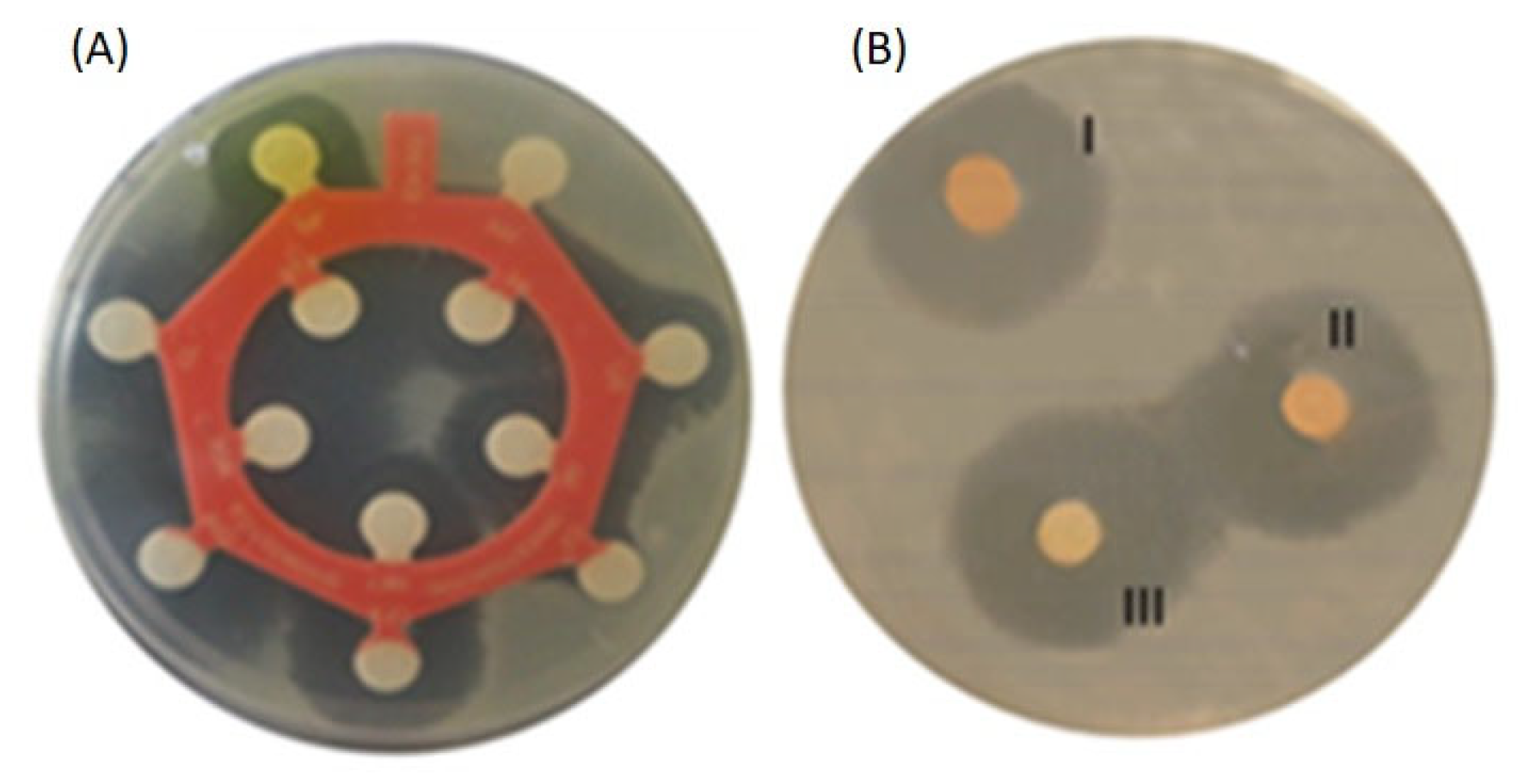
2.2. Essential Oils (EOs) Disc Diffusion Test (DDt)

The EO that had the biggest inhibition zone was thyme, while tea tree was the least effective because it was significantly lower in all tests. The bacteria with the lower results were V. cholerae and A. Iwoffii. The results of the measurement of inhibition halos and their means are shown in Table 2. The inhibition zones observed in CEO ranged from 5.33 to 18.00 mm; 0.93 to 13.67 mm was the range for TTEO; and 4.67 to 19.67 mm was the range for TEO. Cinnamon showed higher mean inhibition halos in Aeromonas sp., Aeromonas veronii, Aeromonas sobria, Shewanella, and Comamonas thiooxydans; in contrast, Aeromonas dhakensis, Pseudomonas, and Empedobacter showed higher mean inhibition halos for thyme DDt. Vibrio cholerae and Acinetobacter Iwoffii shared similar results using TEO and CEO. Comparative images of DDt and antibiogram testing are shown in Figure 1.
Principal Component Analysis (PCA) was performed, which showed distinct differences in the antibacterial activity of cinnamon, thyme, and tea tree essential oils against various bacterial species (Figure 2).
Aeromonas veronii, Shewanella putrefaciens, and Comamonas thiooxydans are highly associated with thyme essential oil, suggesting that this compound significantly impacts their antibacterial activity. Pseudomonas anguilliseptica, Enterobacter falsenii, and Acinetobacter lwoffii are located near the cinnamon vector, suggesting that the compounds in this essential oil may be key in inhibiting bacterial growth. Vibrio cholerae and Aeromonas sobria are closer to the tea tree oil vector, suggesting that compounds such as terpinene-4-ol and α-terpineol may play a key role in the antibacterial activity of these species.

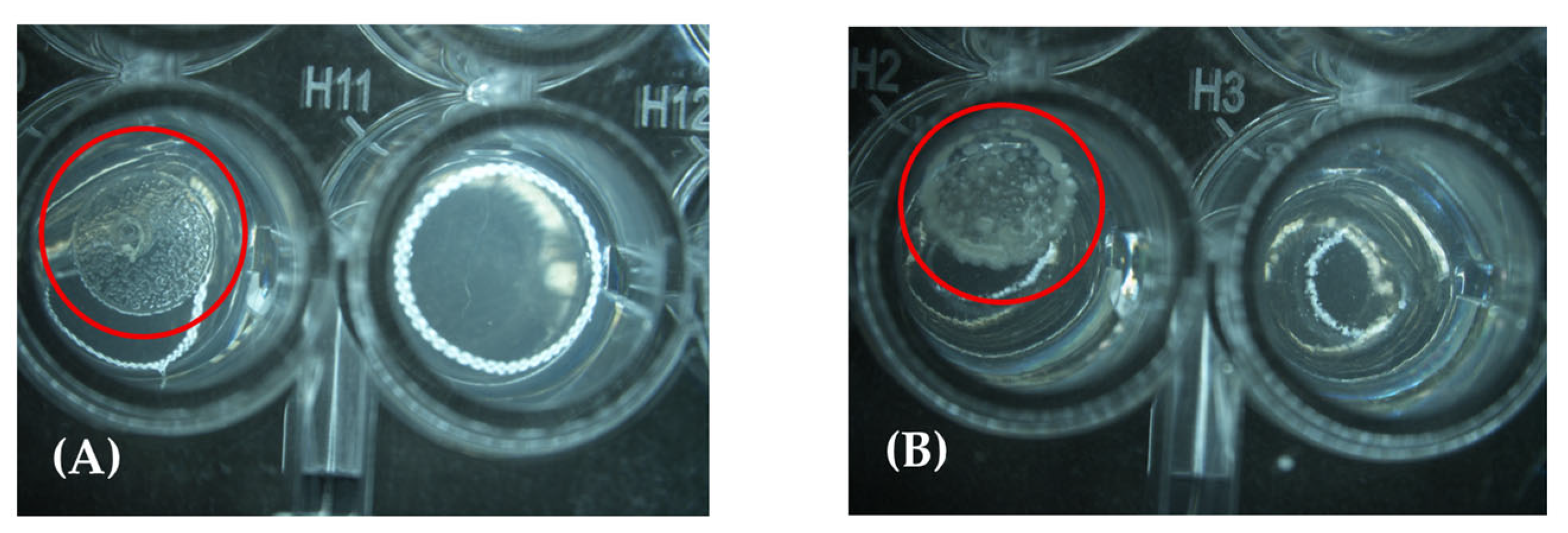
2.3. Minimal Inhibitory Concentrations (MICs)

All strains tested (100%) showed sensitivity to thyme at the lowest concentration (0.12); similar results were found in cinnamon, where the sensitivity ranges were 0.12 to 2%. Tea tree was the least effective, with the widest range, at 0.12 to 4%.
All Aeromonas strains were sensitive to the lowest concentration of thyme, followed by cinnamon (Table 3); only two strains needed a higher concentration of 1% to inhibit the development. The range in the tea tree case was even higher, from 1.0 to 4.0%. The lowest concentration of TEO inhibited Pseudomonas (Figure 3), while cinnamon and tea tree were at 0.5%, like Shewanella, where a higher concentration of cinnamon and tea tree EOs was needed (2%). Comamonas strain was inhibited by cinnamon at 1% and by tea tree at 0.5%; Vibrio needed the same concentration of cinnamon oil, while 2% was used for tea tree. Acinetobacter needed the same amount of cinnamon and tea tree EOs (1%). Empedobacter had no development at (0.12%) for thyme and tea tree, while 0.25% was needed for cinnamon.

3. Discussion

The composition of bioactive molecules varies depending on several plant characteristics such as location, age, climate, temperature, and the part of the plant sampled, with the collection and obtaining of essential oils also being important [23]. Many can be found in different EOs; Table 4 summarizes the main components in each EO used and some molecules reported in the manufacturer’s datasheet (doTERRA Holdings, LLC. Pleasant Grove, UT, USA). Tea tree essential oil (TTEO) and thyme essential oil (TEO) both showed the presence of γ-terpinene and p-cymene; in TEO and cinnamon essential oil (CEO), linalool was found; and TTEO and CEO shared the presence of α-pinene. P-cymene is a common ingredient in food flavoring and in the production of fungicides and pesticides [24]. Gamma-terpinene is the precursor of thymol and p-cymene and possesses low toxicity in animal models; along with its isomer, α-terpinene shares an antioxidative effect [25]. The monoterpene α-pinene displays antioxidant, antifungal, antibacterial, and anti-inflammatory activity [26]. Linalool is a monoterpene that represents up to 90.6% of the oil constituents of over 200 aromatic plant species of different families [27]; combinations with florfenicol or oxytetracycline against A. hydrophila demonstrated a synergic effect [28]. Terpinen-4-ol, the main active constituent of tea tree oil, possesses antiseptic, antioxidative, and sedative activity, along with α-terpineol, which has been pointed to as responsible for its antimicrobial activity [29]. Its addition in diets containing tilapia proved to increase nutrient absorption and growth rates, and even helping in the immune response against bacteria like Aeromonas [24,25,26,27,28,29,30]. The favorable hydrophobic/hydrophilic character of terpinen-4-ol is thought to be the basis for its antimicrobial activity [31]. Thymol, the main component in TEO, is a monoterpenoid phenolic compound that exists naturally along with its isomer carvacrol; both exhibit a distinct set of biological activities, including antioxidant, antitumor, antibacterial, antifungal, anesthetic, and insecticidal properties [27]. Trans-cinnamaldehyde is a natural product from cinnamon oils; its cis counterpart, cinnamaldehyde, cannot be found in nature; thus, the word “trans” is usually omitted. Its multiple mechanisms of action against bacteria have been studied because of its ability to cause alterations in membrane cells and inhibition of cell division, motility, biofilm formation, and quorum sensing [32]. The carbonyl group of cinnamaldehyde is considered responsible for antimicrobial action by binding to cellular proteins, preventing them from functioning properly [33]. Eugenol can be found in many EOs, possesses bacteriostatic activity, and has a major spectrum of action against Gram-negative bacteria [34].
Bioactive constituents of EOs have many mechanisms of action. Gram-positive bacteria are more sensitive to plant extracts than Gram-negative bacteria because the cell wall of Gram-negative bacteria is more complex due to the outer membrane and lipopolysaccharides (LPS), conferring greater resistance to hydrophobic compounds [32]. This could be a disadvantage because most bacteria that affect fish and those used in our test are Gram-negative. The mechanisms of action against these bacteria are described as binding to the surface of bacterial cells and subsequently disrupting the integrity of the cell membrane’s phospholipid bilayer; inhibition of ATP; inhibition of peptidoglycan synthesis or denaturation of proteins; and disturbing the structural integrity of the cell membrane, hence exerting detrimental effects on cellular metabolism, and ultimately leading to cell death [35]. However, the effectiveness of antimicrobial activity has been attributed to the interactions between major and minor bioactive components in each oil [36].
Antimicrobial resistance against EOs is a concern, but it can be considered a minor threat since the multi-component nature of EOs may reduce the potential for resistance to occur spontaneously, and it would require multiple simultaneous mutations to overcome each component’s antimicrobial actions [29]. In some trials, repetition up to 50 times did not show development of resistance [37,38].
There have been different strategies in aquaculture to incorporate the benefits of bioactive compounds present in plants. Some involve the supplementation in diets of extracts like garlic, which, when tested against A. hydrophila, proved to reduce mortalities [39]. Öntas et al. [40] added sweet orange peel (a cheap source of orange essential oil) to fish feed, and it conferred resistance to Streptococcus iniae infection in Mozambique tilapia. Sönmez et al. [41] observed positive effects from the addition of thyme and sage EOs to the diet of rainbow trout.
As pointed out before, the antimicrobial activity of EOs is effective because of the action of their combined constituents. CEO has proven to inhibit a wide range of bacteria. Anandhi et al. [42] evaluated its performance against bacteria isolated from wounds. It showed high antibacterial activity attributed to cinnamaldehyde, benzoic acid, benzaldehyde, and cinnamic acid. Sousa et al. [43] observed synergistic effects in the combination of carvacrol and p-cymene, caused by the interaction of p-cymene with the lipidic membrane of cells, followed by the expansion of the membrane, which facilitated and increased the transportation of carvacrol into the cells. The effectiveness of TEO in the MIC test could be attributed to this type of interaction, since carvacrol and p-cymene can be found among the bioactive components. Our results with respect to the DDt for CEO differ slightly from those observed by Rattanachaikunsopon and Phumkhachorn [33]. Their assays resulted in inhibition halos of 21.5 mm using 30 µL of CEO, while our results showed 16.67 mm at 15 and 10 µL. Their analysis of CEO showed a relatively higher concentration of cinnamaldehyde (90.24%), along with the presence of limonene (2.42%), linalool (1.16%), α-terpineol (0.84%), and cinnamyl acetate (2.03%), hinting that, while cinnamaldehyde has elevated antimicrobial activity, its synergy with other components like cinnamyl acetate or linalool can affect its effectiveness. Hudecová [44] observed bigger inhibition zones for Aeromonas veronii isolated from trout for TEO (43.0mm) and TTEO (39.33 mm) at a dose of 10 µL. Similar results were observed by Kacániová et al. [45] when evaluating CEO and TEO against several strains of Pseudomonas isolated from fish, with results ranging from 15.00 mm to 9.67 mm, respectively.
Cinnamon and thyme EOs showed better performance in comparison with TTEO, with the latter seeming to need to be used at a higher concentration. Mumu and Hossain [46] observed this effect in their evaluation of EOs from eucalypt, lemongrass, and tea tree at 50 µL against pathogenic bacteria, where TTEO was the most effective and had the biggest inhibition zones (36.33 mm against A. hydrophila). The MICs we observed in Shewanella putrefaciens required a bigger dose in CEO than the results declared by Lyu et al. [47]. Golus et al. [48] found MICs of 0.06 (%v/v) in CEO and 0.25 (%v/v) in TTEO tested against Pseudomonas aeruginosa, differing from the 0.5 (%v/v) we obtained when testing against Pseudomonas anguilliseptica. Nonetheless, many authors suggest the differences could be attributed to the testing methods, variability of EOs, and microbial strains.
In our study, CEO and TEO showed the best results in terms of DDt in the majority of the microorganisms tested. TEO inhibited all strains at 0.12 (%v/v), and all strains were sensitive to the action of EOs. However, the smallest inhibition zones were observed in Vibrio cholera and Acinetobacter Iwoffii, the first one a major microorganism of concern and the second one an emerging pathogen; hence, further studies should be carried out to explore other EOs. Aeromonas strains emerge as pathogens of both human and animal health, but our results showed a good effect with respect to CEO against these bacteria. The results suggest that thyme strongly affects Aeromonas veronii and Shewanella putrefaciens, while cinnamon influences Pseudomonas anguilliseptica and Enterobacter falsenii. Conversely, tea tree oil appears to be more effective against Vibrio cholerae and Aeromonas sobria. These findings align with previous studies and demonstrate the selective antibacterial activities of these essential oils against specific bacterial strains. The variations between studies indicate the need for standardized procedures for substances whose composition is a mix of complex molecules, like EOs, and for emerging microorganisms, as no guidelines are established for these. As we have shown, there is a growing resistance to the effects of antibiotics in aquaculture. Nonetheless, our results on EOs showed that they can be used as future alternatives with respect to improving and treating infections caused by bacteria in tilapia breeding. Moreover, new routes of administration could be explored.
The area of the Mezquital Valley has a growing community of tilapia producers (60.22% of the total state aquaculture production) [49], who receive a mixture of urban, industrial, and rain wastewater from the City of Mexico, which is contaminated with diverse drugs [50]. Lesser [51] and Garduño-Jiménez et al. [52] found carbamazepine, erythromycin, flumequine, ofloxacin, ormetoprim, oxacillin, oxytetracycline, sulfadiazine, sulfadimethoxine, sulfamethazine, sulfamethoxazole, and trimethoprim in Mezquital Valley. This is a current problem for aquaculture, since heavily polluted waters can also have teratogenic effects in fish embryos [53].
All the strains evaluated are Gram-negative bacilli of different groups with wide environmental distribution and emerging antimicrobial activity. Aeromonas spp. are associated with aquatic environments such as lakes, rivers, ponds, seas, estuaries, drinking water, sediments, and wastewater [54]. They have also been isolated in meat, raw milk, poultry, fish, shellfish, and vegetables [55]. They are an opportunistic pathogen of domestic animals, amphibians, reptiles, fish, and even humans [56]. Infected fish develop lesions like ulcers in the skin, ascites, hemorrhagic septicemia, furunculosis, and disorders in the gastrointestinal tract, liver, and kidney [57]. Pseudomonas anguilliseptica, an opportunistic fish pathogen of salt and fresh water, causes septicemia, petechial bleeding lesions, and subcutaneous edema, among others, finally causing death between 1 and 2 days after infection [58]. Vibrio cholerae can be isolated from aquatic environments, especially in hot months, and has been pointed out as an occasional pathogen of fish and shrimp, among other aquatic animals [59]. Consumption of seafood or raw or undercooked fillets causes deep dehydration, dysentery, and, in some cases, extraintestinal diseases such as meningitis or septicemia [60]. Shewanella putrefaciens has great relevance in aquaculture and the food industry, due to the conditions it causes on fish such as skin lesions and ulcers, while provoking mortalities of up to 85% [61]. In addition, it can survive refrigeration temperatures, causing rotting meat of aquaculture, bovine, or poultry origin [62]. Acinetobacter Iwoffii is an emerging aquaculture pathogen that causes lesions such as skin hemorrhages, enteritis, and ascites in bullfrog, catfish, and common carp [63]. Comamonas thiooxydans belongs to the Comamonas genus, whose members have been isolated from sites heavily contaminated with complex organic compounds and heavy metals, and increasingly from clinical environments, thus regarded as a rare opportunistic pathogen [64]. Empedobacter falsenii, formerly known as Wautersiella falsenii, was first described in 2006; it is not a common cause of human infection [65].
Treatments in aquaculture usually mean antibiotics, but as has been heavily implied in this study, constant exposure to these substances has led to antimicrobial resistance. Our results show a heavy contrast to those reported worldwide. Outbreaks of P. anguilliseptica resistant to ampicillin, kanamycin, penicillin, and colistin have been reported [66], similar to our results; thus, our isolate was resistant to aminoglycosides, an antibiotic group where ampicillin, kanamycin, amikacin, and gentamicin belong. Hassan et al. [67] reported strains isolated from fish farms sensitive to norfloxacin, ciprofloxacin, and gentamycin, contrary to our results, where the strain evaluated showed resistance to those antimicrobials. With Aeromonas spp. and Vibrio cholerae, there are alarming reports of antibiotic resistance in species like tilapia, koi carp (Cyprinus carpio koi), and catfish (Ictalurus punctatus) [68]. Elgendy et al. [69] found strains resistant to ampicillin and amoxicillin but sensitive to ciprofloxacin and trimethoprim/sulfamethoxazole in farms in Egypt with mortality episodes suspected as a result of improper use of antibiotics; in our results, Aeromonas and Vibrio were resistant to ciprofloxacin and sulfamethoxazole. Contrary to our results, Jiang et al. [70] found strains of Shewanella isolated from largemouth bass (Micropterus salmoides) susceptible to cefotaxime, sulfamethoxazole, gentamicin, ampicillin, and norfloxacin. Kozińska [71] also reported an A. Iwoffi strain resistant to ampicillin, amoxicillin, and cephalothin, intermediate in terms of cefuroxime and oxytetracycline and sensitive to enrofloxacin, flumequime, norfloxacin, gentamycin, and sulfamethoxazole/trimethoprim. Zhang et al. [72] isolated strains resistant to florfenicol, sulfadiazinum, penicillin, and tetracycline but sensitive to neomycin sulfate, minocycline, doxycycline, polymyxin B, amikacin, gentamycin, and ampicillin. Cao et al. [73] found strains with similar resistance to ampicillin, cefotaxime, and gentamicin, but moderately sensitive to ciprofloxacin and norfloxacin. Comamonas spp. can be resistant to many antibiotic families, including β-lactams (penicillins, cephalosporins, carbapenems), with treatments based upon in vitro assays testing isolates [74]. Guo [75] reported a case of isolation of C. thiooxydans from urine in a coinfection with E. coli from a patient with a urinary tract infection; the strain proved resistant to aztreonam, fluoroquinolones, and aminoglycosides. However, some clinical cases report strains resistant to aztreonam, ampicillin–sulbactam, cephalothin, meropenem, and colistin; intermediate with respect to piperacillin–tazobactam, cefotaxime, ceftazidime, imipenem, and ciprofloxacin; and susceptible to cefepime, amikacin, gentamicin, and trimethoprim–sulfamethoxazole [67,76], but our study demonstrated C. thiooxydans resistant to gentamicin and sulfamethoxazole.

4. Materials and Methods

4.1. Essential Oils

The essential oils (EOs) utilized in this research included cinnamon (Cinnamomum zeylanicum), tea tree (Melaleuca alternifolia), and thyme (Thymus vulgaris) sourced from doTERRA (doTERRA Holdings, LLC; 2021). The analysis for each oil was provided by the manufacturer (Gas Chromatography). The major components of each EO are presented in Table 4. The essential oils were used in their pure form for the disk diffusion method. When conducting Minimal Inhibitory Concentration (MIC) testing, the essential oils were diluted in DMSO (dimethyl sulfoxide; Sigma Aldrich, Saint Louis, MO, USA) at a concentration of 2.0–1.0% (v/v) and added in molten MH Agar (Mueller Hinton, MCD. LAB, Tlalnepantla, México)

4.2. Bacterial Strains

The bacterial strains used in this study were previously isolated from various municipalities in Hidalgo State, Mexico, during the month of August 2021 and identified by PCR for gcaT, rpoD, and 16s rRNA genes following the methodology fully described by Zepeda et al. [77]. Details about the strains are provided in Table 5. Subcultures were prepared from isolates previously preserved in Brain Heart broth (MCD Lab, Mexico) and stored in vials at −20 °C until required [78]. To generate fresh cultures before each test, isolates were grown on Mueller–Hinton agar at 35 °C. All bacterial isolates used in this study were subjected to antibiotic susceptibility testing following EUCAST guidelines. The antibiogram results revealed that the strains exhibited multidrug resistance (MDR), with MAR (Multiple Antibiotic Resistance). The identity of each bacterial strain was confirmed by PCR and Sanger sequencing. Specific genes used for molecular identification included gcaT and rpoD for Aeromonas and 16S rRNA for all strains. The sequences were compared against the NCBI GenBank database using the online version of BLAST (https://blast.ncbi.nlm.nih.gov/Blast.cgi) (accessed on August 2022), and all isolates showed >98% identity with reference sequences. The GenBank accession numbers for each strain are listed in Table 5.

4.3. Inoculum Preparation

The inoculum was prepared using the CLSI (Clinical & Laboratory Standards Institute) direct colony suspension method. Bacterial colonies following 18–24 h of growth were suspended in Mueller–Hinton broth and adjusted to a turbidity of 0.5 MacFarland standard, equivalent to 1–2 × 10⁸ colony forming units (CFUs) mL−1 [79]. For the Minimal Inhibitory Concentration test (MIC), the inoculums were diluted 1:10 in Mueller–Hinton broth to achieve a concentration of 107 CFUs mL−1. Then, 2 µL was applied to the agar surface of each well in the microplate to accomplish a final inoculum density of approximately 104 CFUs per well [49]. All bacterial suspensions were used within 15 min after preparation.

4.4. Antibiogram

The antibiogram was used to evaluate the sensitivity of the isolates to several antibiotics, and it was performed according to the CLSI M02-A11 [79]. The inoculum was prepared as described above. A sterile swab was introduced into the inoculum and then rotated against the tube wall to eliminate excess broth. Next, the swab was used to strike the surface of Mueller–Hinton agar plates and left to dry for 15 min before placing antibiotic discs (PT-35 Multibac I.D. for Gram-negative bacteria, Mexico). The antibiotics used were ampicillin (AM; 10 mcg), amikacin (AK; 30 mcg), carbenicillin (CB; 100 mcg), gentamicin (GE; 10 mcg), cephalothin (CF; 30 mcg), cefotaxime (CFX; 30 mcg), ciprofloxacin (CPF; 5 mcg), norfloxacin (NOF; 10 mcg), chloramphenicol (CL; 30 mcg), sulfamethoxazole (STX; 25 mcg), nitrofurantoin (NF; 300 mcg), and netilmicin (NET; 30 mcg). Antibiotic discs were left on the surface of plates and incubated at 35 °C for 18 h. Afterward, plates were placed against a dark background and photographed, and the diameter of the inhibition halos was measured. According to the standards proposed by CLSI [79] and EUCAST [80], bacteria were classified as Susceptible (S), Susceptible, increased exposure (I), and Resistant (R); for organisms without guidelines, interpretative criteria for similar antimicrobial or organism combinations were used. The MAR index was used, which is the number of antibiotics to which the isolate was resistant divided by the number of antibiotics to which the isolate was exposed [81]

4.5. Essential Oils (EOs) Disc Diffusion Test (DDt)

The CLSI protocol for disc diffusion was adapted to assess the efficacy of EOs against pathogenic bacteria. A similar process to the one mentioned above was performed to inoculate Mueller–Hinton plates with inoculum, previously prepared and adjusted to a turbidity of 0.5 MacFarland standard, equivalent to 1–2 × 10⁸ colony forming units (CFUs) mL−1, using sterilized filter paper discs (6 mm) impregnated with 15, 10, and 5 µL of each oil. These discs were placed on the Petri dishes’ surface and incubated at 35 °C for 24 h. The test was conducted in triplicate, and the results were analyzed using one-factor ANOVA with the Tukey method (means p < 0.05 are significantly different). Additionally, a Principal Component Analysis (PCA) was performed to evaluate differences in the antibacterial activity of cinnamon, thyme, and tea tree essential oils against various bacterial species

4.6. Determination of Minimum Inhibitory Concentration (MIC)

The MIC of EOs was evaluated following the methodology proposed by Golus et al. [48]. Sterile, flat-bottomed 96-well plates (Corning) were utilized. All oils were used in the 8.0–0.125% (v/v) range. These were poured into test tubes to which 2% (v/v) DMSO was added, ensuring the final volume did not exceed this concentration; following this, melted Mueller–Hinton agar was added, achieving 100 µL for each well. The tubes were subjected to vortexing and placed in a digital reactor (Hach DRB200) at 50 °C until use. The melted Mueller–Hinton agar was subsequently added to achieve volumes of 100 µL per well in all plates, except for the first file, where only molten Mueller–Hinton agar was left. The plates were left at room temperature until solidified, and then the bacterial suspensions previously prepared were inoculated in the plate, as shown in Figure 4. For each bacterium, two columns were used: inoculum wells (2, 4, 6, 8, 10) and uncultured wells (3, 5, 7, 9, 11). A plate was used for every EO. The MIC was defined as the lowest dilution with no visible growth of the microorganism (absence of turbidity). Incubation at 35 °C may lead to excessive evaporation, potentially causing agar well desiccation and compromising the hydration status of the culture. Supplementing with sterile water could mitigate this issue, particularly in wells exhibiting rapid evaporation rates, which may otherwise adversely impact bacterial growth dynamics.

5. Conclusions

This study presents a promising alternative for enhancing fish health, specifically in tilapia reared in aquaculture farms, considering findings regarding potential antibiotic resistance in pathogens isolated from these fish. The observed improvements in fish health can be attributed to the antimicrobial properties of essential oils, particularly in cinnamon and thyme, which exhibit effectiveness against microorganisms such as Vibrio cholerae and Aeromonas. These essential oils show potential as natural agents for controlling bacterial proliferation, offering a viable alternative to the unrestricted use of antibiotics in farming practices. Based on these results, there is an opportunity for further, more in-depth studies across additional farms to explore various applications and methods of administration, as well as an evaluation of the effectiveness of EOs against different fish species.

Author Contributions

Conceptualization, L.A.-B., K.A.T.-P. and V.V.-S.; methodology, K.A.T.-P. and J.A.D.-E.; formal analysis, R.M.-E., G.C.-D., V.V.-S., L.A.-B., C.A.A.-O. and F.R.G.-d.A.; investigation, K.A.T.-P.; resources, L.A.-B. and F.R.G.-d.A.; writing—original draft, K.A.T.-P. and J.A.D.-E.; preparation, K.A.T.-P., L.A.-B., C.A.A.-O. and V.V.-S.; writing—review and editing, V.V.-S., L.A.-B., C.A.A.-O. and F.R.G.-d.A.; supervision, L.A.-B., V.V.-S. and F.R.G.-d.A.; project administration, L.A.-B.; funding acquisition, L.A.-B. All authors have read and agreed to the published version of the manuscript.

Funding

This research was funded by the National Polytechnic Institute (Instituto Politécnico Nacional) with projects SIP 20231809 and SIP 20240733.

Institutional Review Board Statement

Not applicable.

Informed Consent Statement

Not applicable.

Data Availability Statement

The original contributions presented in this study are included in the article. Further inquiries can be directed to the corresponding author.

Acknowledgments

Author K.A.T.P. and J.A.D.E. thank CONAHCYT-Mexico and SIP-IPN-Mexico for doctoral study grants. All authors acknowledge the financial support given by SIP-IPN, and appreciate the facilities provided by the Institute of Agricultural Sciences-UAEH to carry out microbiological experiments.

Conflicts of Interest

The authors declare no conflicts of interest.

Abbreviations

The following abbreviations are used in this manuscript:
EOsEssential Oils
CEOCinnamon Essential Oil
TEOThyme Essential Oil
TTEOTea Tree Essential Oil
MAR IndexMultiple Antibiotic Resistance Index
DDtDisc Diffusion Test
MICsMinimal Inhibitory Concentrations
DMSODimethyl Sulfoxide

References

  1. Islam, S.; Bhowmik, S.; Majumdar, P.R.; Srzednicki, G.; Rahman, M.; Hossain, M.A. Nutritional Profile of Wild, Pond-, Gher- and Cage-Cultured Tilapia in Bangladesh. Heliyon 2021, 7, e06968. [Google Scholar] [CrossRef] [PubMed]
  2. González Razo, F.D.J.; Sangerman-Jarquín, D.M.; Omaña Silvestre, J.M.; Rebollar Rebollar, S.; Hernández Martínez, J.; Ayllón Benítez, J.C. Marketing of tilapia (Oreochromis niloticus) in southern state of Mexico. Rev. Mex. De. Cienc. Agric. 2016, 7, 1985–1996. [Google Scholar]
  3. Prabu, E.; Rajagopalsamy, C.B.T.; Ahilan, B.; Jeevagan, I.J.M.A.; Renuhadevi, M. Tilapia—An Excellent Candidate Species for World Aquaculture: A Review. Annu. Res. Rev. Biol. 2019, 31, 1–14. [Google Scholar] [CrossRef]
  4. Vijayakumar, S.; Vaseeharan, B.; Malaikozhundan, B.; Gobi, N.; Ravichandran, S.; Karthi, S.; Ashokkumar, B.; Sivakumar, N. A Novel Antimicrobial Therapy for the Control of Aeromonas hydrophila Infection in Aquaculture Using Marine Polysaccharide Coated Gold Nanoparticle. Microb. Pathog. 2017, 110, 140–151. [Google Scholar] [CrossRef] [PubMed]
  5. Armwood, A.R.; Camus, A.C.; López-Porras, A.; Ware, C.; Griffin, M.J.; Soto, E. Pathologic Changes in Cultured Nile Tilapia (Oreochromis Niloticus) Associated with an Outbreak of Edwardsiella anguillarum. J. Fish. Dis. 2019, 42, 1463–1469. [Google Scholar] [CrossRef]
  6. Monir, S.; Yusoff, S.M.; Mohamad, A.; Ina-Salwany, M.Y. Vaccination of Tilapia against Motile Aeromonas Septicemia: A Review. J. Aquat. Anim. Health 2020, 32, 65–76. [Google Scholar] [CrossRef] [PubMed]
  7. Ruzauskas, M.; Klimiene, I.; Armalyte, J.; Bartkiene, E.; Siugzdiniene, R.; Skerniskyte, J.; Krasauskas, R.; Suziedeliene, E. Composition and Antimicrobial Resistance Profile of Gram-Negative Microbiota Prevalent in Aquacultured Fish. J. Food Saf. 2018, 38, 1–10. [Google Scholar] [CrossRef]
  8. Karunasagar, I. Complexities Involved in Source Attribution of Antimicrobial Resistance Genes Found in Aquaculture Products. Asian Fish. Sci. 2020, 33 (Suppl. S1), 16–21. [Google Scholar] [CrossRef]
  9. Eissa, F.; Ghanem, K.; Al-Sisi, M. Occurrence and Human Health Risks of Pesticides and Antibiotics in Nile Tilapia along the Rosetta Nile Branch, Egypt. Toxicol. Rep. 2020, 7, 1640–1646. [Google Scholar] [CrossRef]
  10. Health for Animals. How Prevention Can Reduce the Need for Antibiotics Pathways to Reduce the Need for Antimicrobials on Farms for Sustainable Agrifood Systems Transformation (RENOFARM); Health for Animals: Brussels, Belgium, 2024. [Google Scholar]
  11. Zaidi, M.B.; Dreser, A.; Figueroa, I.M. A Collaborative Initiative for the Containment of Antimicrobial Resistance in Mexico. Zoonoses Public Health 2015, 62 (Suppl. S1), 52–57. [Google Scholar] [CrossRef]
  12. Matthews Mbokane, E.; Gukuta Moyo, N.A. Use of Medicinal Plants as Feed Additives in the Diets of Mozambique Tilapia (Oreochromis mossambicus) and the African Sharptooth Catfish (Clarias gariepinus) in Southern Africa. Front. Vet. Sci. 2022, 9, 1072369. [Google Scholar] [CrossRef]
  13. Gómez-Rincón, C.; Langa, E.; Murillo, P.; Sofía Valero, M.; Berzosa, C.; López, V. Activity of Tea Tree (Melaleuca alternifolia) Essential Oil against L3 Larvae of Anisakis simplex. BioMed Res. Int. 2014, 2014, 1–6. [Google Scholar] [CrossRef]
  14. Bisht, D.; Pal, A.; Chanotiya, C.S.; Mishra, D.; Pandey, K.N. Terpenoid Composition and Antifungal Activity of Three Commercially Important Essential Oils against Aspergillus flavus and Aspergillus niger. Nat. Prod. Res. 2011, 25, 1993–1998. [Google Scholar] [CrossRef]
  15. Manion, C.R.; Widder, R.M. Essentials of Essential Oils. Am. J. Health-Syst. Pharm. 2017, 74, e153–e162. [Google Scholar] [CrossRef] [PubMed]
  16. Nazzaro, F.; Fratianni, F.; De Martino, L.; Coppola, R.; De Feo, V. Effect of Essential Oils on Pathogenic Bacteria. Pharmaceuticals 2013, 6, 1451–1474. [Google Scholar] [CrossRef] [PubMed]
  17. Abd El-Hack, M.E.; Alagawany, M.; Abdel-Moneim, A.-M.E.; Mohammed, N.G.; Khafaga, A.F.; Bin-Jumah, M.; Othman, S.I.; Allam, A.A.; Elnesr, S.S. Cinnamon (Cinnamomum zeylanicum) Oil as a Potential Alternative to Antibiotics in Poultry. Antibiotics 2020, 9, 210. [Google Scholar] [CrossRef]
  18. López, L.M.T. Tomillo. Ámbito Farm. 2006, 25, 74–77. [Google Scholar]
  19. Sakkas, H.; Papadopoulou, C. Antimicrobial Activity of Basil, Oregano, and Thyme Essential Oils. J. Microbiol. Biotechnol. 2017, 27, 429–438. [Google Scholar] [CrossRef]
  20. Noui Mehidi, I.; Ait Ouazzou, A.; Tachoua, W.; Hosni, K. Investigating the Antimicrobial Properties of Essential Oil Constituents and Their Mode of Action. Molecules 2024, 29, 4119. [Google Scholar] [CrossRef]
  21. Angane, M.; Swift, S.; Huang, K.; Butts, C.A.; Quek, S.Y. Essential Oils and Their Major Components: An Updated Review on Antimicrobial Activities, Mechanism of Action and Their Potential Application in the Food Industry. Foods 2022, 11, 464. [Google Scholar] [CrossRef]
  22. Odeyemi, O.A.; Ahmad, A. Antibiotic Resistance Profiling and Phenotyping of Aeromonas Species Isolated from Aquatic Sources. Saudi J. Biol. Sci. 2017, 24, 65–70. [Google Scholar] [CrossRef] [PubMed]
  23. Assane, I.M.; Valladão, G.M.R.; Pilarski, F. Chemical Composition, Cytotoxicity and Antimicrobial Activity of Selected Plant-Derived Essential Oils against Fish Pathogens. Aquac. Res. 2021, 52, 793–809. [Google Scholar] [CrossRef]
  24. Shaalan, M.; Mahboub, H.H.; Abdelwarith, A.A.; Younis, E.M.; Elnegiry, A.A.; Basher, A.W.; El Houseiny, W.; Shawky, S.M.; Orabi, S.H.; Davies, S.J.; et al. Dietary Tea Tree (Melaleucae aetheroleum) Oil Fortifies Growth, Biochemical, Immune antioxidant Trait, Gene Function, Tissue, and Aeromonas Sobria resistance in Nile Tilapia (Oreochromis niloticus). BMC Vet. Res. 2025, 21, 1–15. [Google Scholar] [CrossRef]
  25. Borotová, P.; Galovičová, L.; Vukovic, N.L.; Vukic, M.; Tvrdá, E.; Kačániová, M. Chemical and Biological Characterization of Melaleuca alternifolia Essential Oil. Plants 2022, 11, 558. [Google Scholar] [CrossRef]
  26. Borges, R.S.; Ortiz, B.L.S.; Pereira, A.C.M.; Keita, H.; Carvalho, J.C.T. Rosmarinus Officinalis Essential Oil: A Review of Its Phytochemistry, Anti-Inflammatory Activity, and Mechanisms of Action Involved. J. Ethnopharmacol. 2019, 229, 29–45. [Google Scholar] [CrossRef]
  27. Dawood, M.A.O.; El Basuini, M.F.; Yilmaz, S.; Abdel-Latif, H.M.R.; Alagawany, M.; Kari, Z.A.; Razab, M.K.A.A.; Hamid, N.K.A.; Moonmanee, T.; Doan, H.V. Exploring the Roles of Dietary Herbal Essential Oils in Aquaculture: A Review. Animals 2022, 10, 823. [Google Scholar] [CrossRef] [PubMed]
  28. Bandeira, J.G.; Sutili, F.J.; Gressler, L.T.; Ely, V.; Silveira, B.; Tasca, C.; Reghelin, M.; Matter, L.; Vargas, A.; Baldisserotto, B.; et al. Antibacterial Potential of Phytochemicals Alone or in Combination with Antimicrobials against Fish Pathogenic Bacteria. J. Appl. Microbiol. 2018, 125, 655–665. [Google Scholar] [CrossRef]
  29. Carson, C.F.; Hammer, K.A.; Riley, T.V. Melaleuca alternifolia (Tea Tree) Oil: A Review of Antimicrobial and Other Medicinal Properties. Clin. Microbiol. Rev. 2006, 19, 50–62. [Google Scholar] [CrossRef]
  30. Baginska, S.; Golonko, A.; Swislocka, R.; Lewandowski, W. Monoterpenes as Medicinal Agents: Exploring the Pharmaceutical Potential of p-Cymene, p-Cymenene, and Γ-Terpinene. Acta Pol. Pharm. Drug Res. 2023, 80, 879–892. [Google Scholar] [CrossRef]
  31. Zheng, X.; Feyaerts, A.F.; Van Dijck, P.; Bossier, P. Inhibitory Activity of Essential Oils against Vibrio campbellii and Vibrio parahaemolyticus. Microorganisms 2020, 8, 1946. [Google Scholar] [CrossRef]
  32. Vasconcelos, N.G.; Croda, J.; Simionatto, S. Antibacterial Mechanisms of Cinnamon and Its Constituents: A Review. Microb. Pathog. 2018, 120, 198–203. [Google Scholar] [CrossRef]
  33. Rattanachaikunsopon, P.; Phumkhachorn, P. Use of Asiatic Pennywort Centella Asiatica Aqueous Extract as a Bath Treatment to Control Columnaris in Nile Tilapia. J. Aquat. Anim. Health 2010, 22, 14–20. [Google Scholar] [CrossRef] [PubMed]
  34. Wickramanayake, M.V.K.S.; Kumarage, P.M.; Majeed, S.; Heo, G.J. An Overview of the Antimicrobial Activity of Some Essential Oils against Fish Pathogenic Bacteria. Vet. Integr. Sci. 2022, 21, 99–119. [Google Scholar] [CrossRef]
  35. Sobhy, M.; Ali, S.S.; Cui, H.; Lin, L.; El-Sapagh, S. Exploring the Potential of 1,8-Cineole from Cardamom Oil against Food-Borne Pathogens: Antibacterial Mechanisms and Its Application in Meat Preservation. Microb. Pathog. 2023, 184, 106375. [Google Scholar] [CrossRef]
  36. Soković, M.; Glamočlija, J.; Marin, P.D.; Brkić, D.; Van Griensven, L.J.L.D. Antibacterial Effects of the Essential Oils of Commonly Consumed Medicinal Herbs Using an In Vitro Model. Molecules 2010, 15, 7532–7546. [Google Scholar] [CrossRef] [PubMed]
  37. Becerril, R.; Nerín, C.; Gómez-Lus, R. Evaluation of Bacterial Resistance to Essential Oils and Antibiotics after Exposure to Oregano and Cinnamon Essential Oils. Foodborne Pathog. Dis. 2012, 9, 699–705. [Google Scholar] [CrossRef]
  38. Zilberg, D.; Tal, A.; Froyman, N.; Abutbul, S.; Dudai, N.; Golan-Goldhirsh, A. Dried Leaves of Rosmarinus officinalis as a Treatment for Streptococcosis in Tilapia. J. Fish. Dis. 2010, 33, 361–369. [Google Scholar] [CrossRef]
  39. Nya, E.J.; Austin, B. Use of Garlic, Allium sativum, to Control Aeromonas Hydrophila Infection in Rainbow Trout, Oncorhynchus Mykiss Rainbow Trout, Oncorhynchus mykiss. J. Fish. Dis. 2009, 32, 963–970. [Google Scholar] [CrossRef]
  40. Öntaş, C.; Baba, E.; Kaplaner, E.; Küçükaydin, S.; Öztürk, M.; Ercan, M.D. Antibacterial Activity of Citrus Limon Peel Essential Oil and Argania Spinosa Oil Against Fish Pathogenic Bacteria. Kafkas Univ. Vet. Fak. Derg. 2016, 22, 741–749. [Google Scholar] [CrossRef]
  41. Sonmez, A.Y.; Bilen, S.; Albayrak, M.; Yilmaz, S.; Biswas, G.; Hisar, O.; Yanik, T. Effects of Dietary Supplementation of Herbal Oils Containing 1,8-Cineole, Carvacrol or Pulegone on Growth Performance, Survival, Fatty Acid Composition, and Liver and Kidney Histology of Rainbow Trout (Oncorhynchus Mykiss) Fingerlings. Turk. J. Fish. Aquat. Sci. 2015, 15, 813–819. [Google Scholar] [CrossRef]
  42. Anandhi, P.; Tharani, M.; Rajeshkumar, S.; Lakshmi, T. Antibacterial Activity of Cinnamon and Clove Oil against Wound Pathogens. J. Popul. Ther. Clin. Pharmacol. 2022, 28, 41–46. [Google Scholar] [CrossRef]
  43. Sousa, L.G.V.; Castro, J.; Cavaleiro, C.; Salgueiro, L.; Tomás, M.; Palmeira-Oliveira, R.; Martinez-Oliveira, J.; Cerca, N. Synergistic Effects of Carvacrol, α-Terpinene, γ-Terpinene, ρ-Cymene and Linalool against Gardnerella Species. Sci. Rep. 2022, 12, 4417. [Google Scholar] [CrossRef]
  44. Rattanachaikunsopon, P.; Phumkhachorn, P. Potential of Cinnamon (Cinnamomum verum) Oil to Control Streptococcus Iniae Infection in Tilapia (Oreochromis niloticus). Fish. Sci. 2010, 76, 287–293. [Google Scholar] [CrossRef]
  45. Hudecová, P.; Koščová, J.; Hajdučková, V.; Király, J.; Hoř Nak, P. Antibacterial and Antibiofilm Activity of Essential Oils Against Aeromonas Spp. Isolated from Rainbow Trout. Animals 2024, 14, 3202. [Google Scholar] [CrossRef]
  46. Kačániová, M.; Terentjeva, M.; Vukovic, N.; Puchalski, C.; Roychoudhury, S.; Kunová, S.; Klūga, A.; Tokár, M.; Kluz, M.; Ivanišová, E. The Antioxidant and Antimicrobial Activity of Essential Oils against Pseudomonas spp. Isolated from Fish. Saudi Pharm. J. 2017, 25, 1108–1116. [Google Scholar] [CrossRef] [PubMed]
  47. Kabir Mumu, S.; Mahboob Hossain, M. Antimicrobial Activity of Tea Tree Oil against Pathogenic Bacteria and Comparison of Its Effectiveness with Eucalyptus Oil, Lemongrass Oil and Conventional Antibiotics. Am. J. Microbiol. Res. 2018, 6, 73–78. [Google Scholar] [CrossRef]
  48. Lyu, F.; Hong, Y.-L.; Cai, J.-H.; Wei, Q.-Q.; Zhou, X.; Ding, Y.-T.; Liu, Z.-F.; Liu, L. Antimicrobial Effect and Mechanism of Cinnamon Oil and Gamma Radiation on Shewanella putrefaciens. J. Food Sci. Technol. 2018, 55, 3353–3361. [Google Scholar] [CrossRef]
  49. Golus, J.; Sawicki, R.; Widelski, J.; Ginalska, G. The Agar Microdilution Method—A New Method for Antimicrobial Susceptibility Testing for Essential Oils and Plant Extracts. J. Appl. Microbiol. 2016, 121, 1291–1299. [Google Scholar] [CrossRef]
  50. Conapesca. Anuario Estadístico de Acuacultura y Pesca de la Comisión Nacional de Acuacultura y Pesca; Conapesca: Mazatlán, México, 2021. [Google Scholar]
  51. Chávez-Mejía, A.C.; Navarro-González, I.; Magaña-López, R.; Uscanga-Roldán, D.; Zaragoza-Sánchez, P.I.; Jiménez-Cisneros, B.E. Presence and Natural Treatment of Organic Micropollutants and Their Risks after 100 Years of Incidental Water Reuse in Agricultural Irrigation. Water 2019, 11, 2148. [Google Scholar] [CrossRef]
  52. Lesser, L.E.; Mora, A.; Moreau, C.; Mahlknecht, J.; Hernández-Antonio, A.; Ramírez, A.I.; Barrios-Piña, H. Survey of 218 Organic Contaminants in Groundwater Derived from the World’s Largest Untreated Wastewater Irrigation System: Mezquital Valley, Mexico. Chemosphere 2018, 198, 510–521. [Google Scholar] [CrossRef]
  53. Garduño-Jiménez, A.L.; Durán-Álvarez, J.C.; Ortori, C.A.; Abdelrazig, S.; Barrett, D.A.; Gomes, R.L. Delivering on Sustainable Development Goals in Wastewater Reuse for Agriculture: Initial Prioritization of Emerging Pollutants in the Tula Valley, Mexico. Water Res. 2023, 238, 119903. [Google Scholar] [CrossRef] [PubMed]
  54. Pérez-Coyotl, I.; Galar-Martínez, M.; García-Medina, S.; Gómez-Oliván, L.M.; Gasca- Pérez, E.; Martínez-Galero, E.; Islas-Flores, H.; Pérez-Pastén, B.R.; Barceló, D.; López de Alda, M.; et al. Polluted Water from an Urban Reservoir (Madín Dam, México) Induces Toxicity and Oxidative Stress in Cyprinus carpio Embryos. Environ. Pollut. 2019, 251, 510–521. [Google Scholar] [CrossRef]
  55. Cortes-Sanchez, A.D.J.; Espinosa-Chaurand, L.D.; Garza-Torres, R.; Diaz-Ramirez, M.; Salgado-Cruz, M.D.L.P.; Sánchez-Minutii, L.; García-Barrientos, R. Foodborne Diseases, Fish and the Case of Aeromonas Spp. Afr. J. Agric. Res. 2019, 14, 617–628. [Google Scholar] [CrossRef]
  56. Hoel, S.; Vadstein, O.; Jakobsen, A.N. The Significance of Mesophilic Aeromonas spp. in Minimally Processed Ready-to-Eat Seafood. Microorganisms 2019, 7, 91. [Google Scholar] [CrossRef]
  57. Martin-Carnahan, A.; Joseph, S.W. Aeromonas. In Bergey’s Manual of Systematics of Archaea and Bacteria; Wiley: Hoboken, NJ, USA, 2015; pp. 1–44. [Google Scholar] [CrossRef]
  58. Zhai, W.; Wang, Q.; Zhu, X.; Jia, X.; Chen, L. Pathogenic Infection and Microbial Composition of Yellow Cat-fish (Pelteobagrus Fulvidraco) Challenged by Aeromonas veronii and Proteus mirabilis. Aquac. Fish. 2023, 8, 166–173. [Google Scholar] [CrossRef]
  59. Phumkhachorn, P.; Rattanachaikunsopon, P. Use of Cassia Alata Aqueous Extract as a Bath Treatment to Control Pseudomonas anguilliseptica Infection in Tilapia (Oreochromis niloticus). Arch. Biol. Sci. 2015, 67, 1165–1172. [Google Scholar] [CrossRef]
  60. Economopoulou, A.; Chochlakis, D.; Almpan, M.A.; Sandalakis, V.; Maraki, S.; Tselentis, Y.; Psaroulaki, A. Environmental Investigation for the Presence of Vibrio Species Following a Case of Severe Gastroenteritis in a Touristic Island. Environ. Sci. Pollut. Res. 2017, 24, 4835–4840. [Google Scholar] [CrossRef]
  61. Yang, Y.; Zhang, H.; Liu, Y.; Dong, J.; Xu, N.; Yang, Q.; Zhou, S.; Ai, X. Identification of Vibrio cholerae as a Bacterial Pathogen of Bluegill Sunfish. Aquac. Rep. 2022, 23, 101092. [Google Scholar] [CrossRef]
  62. Sood, N.; Pradhan, P.K.; Ravindra; Verma, D.K.; Yadav, M.K.; Mishra, R.K.; Kumar, U.; Swaminathan, T.R.; Sood, N.K. Large-Scale Mortality in Cultured Tilapia Oreochromis niloticus Due to Infection with Shewanella putrefaciens in India. J. World Aquac. Soc. 2020, 51, 563–570. [Google Scholar] [CrossRef]
  63. Pȩkala-Safińska, A. Contemporary Threats of Bacterial Infections in Freshwater Fish. J. Vet. Res. 2018, 62, 261–267. [Google Scholar] [CrossRef]
  64. Cao, H.; Ye, W.; He, S.; Li, Y.; Yang, Y. Acinetobacter Lwoffii: An Emerging Pathogen for Red Disease in Farmed Channel Catfish Ictalurus. Isr. J. Aquac. 2016, 68, 20799. [Google Scholar]
  65. Willems, A.; Gillis, M. Comamonas. In Bergey’s Manual of Systematics of Archaea and Bacteria; Wiley: Hoboken, NJ, USA, 2015; pp. 1–17. [Google Scholar] [CrossRef]
  66. Traglia, G.M.; Dixon, C.; Chiem, K.; Almuzara, M.; Barberis, C.; Montaña, S.; Merino, C.; Mussi, M.A.; Tolmasky, M.E.; Iriarte, A.; et al. Draft Genome Sequence of Empedobacter (Formerly Wautersiella) falsenii Comb. Nov. Wf282, a Strain Isolated from a Cervical Neck Abscess. Genome Announc. 2016, 3, e00235-15. [Google Scholar] [CrossRef] [PubMed]
  67. Hossain, M.; Chowdhury, B. Pseudomonas anguilliseptica As a Pathogen of Tilapia (Oreochromis niloticus) Culture in Bangladesh. Bangladesh Res. Pub. J. 2009, 2, 712–721. [Google Scholar]
  68. Hassan, S.; Abdel-Rahman, M.; Mansour, E.S.; Monir, W. Isolation, Phenotypic Characterization and Antibiotic Susceptibility of Prevalent Bacterial Pathogens Implicating the Mortality of Cultured Nile Tilapia, Oreochromis niloticus. Egypt. J. Aquac. 2020, 10, 23–43. [Google Scholar] [CrossRef]
  69. Preena, P.G.; Dharmaratnam, A.; Swaminathan, T.R. Antimicrobial Resistance Analysis of Pathogenic Bacteria Isolated from Freshwater Nile Tilapia (Oreochromis niloticus) Cultured in Kerala, India. Curr. Microbiol. 2020, 77, 3278–3287. [Google Scholar] [CrossRef] [PubMed]
  70. Elgendy, M.Y.; Abdelsalam, M.; Kenawy, M.A.; Shimaa, E.A. Vibriosis Outbreaks in Farmed Nile Tilapia (Oreochromis niloticus) Caused by Vibrio mimicus and V. cholerae. Aquac. Int. 2022, 30, 2661–2677. [Google Scholar] [CrossRef]
  71. Jiang, X.; Wang, X.; Li, L.; Niu, C.; Pei, C.; Zhu, L.; Kong, X.; Yang, Y.; Qi, Z.; Kong xhkong, X. Identification of Shewanella putrefaciens as a Novel Pathogen of the Largemouth Bass (Micropterus salmoides) and Histopathological Analysis of Diseased Fish. Front. Cell Infect. Microbiol. 2022, 12. [Google Scholar] [CrossRef]
  72. Kozińska, A.; Paździor, E.; Pȩkala, A.; Niemczuk, W. Acinetobacter Johnsonii and Acinetobacter Lwoffii—The Emerging Fish Pathogens. Bull. Vet. Inst. Pulawy 2014, 58, 193–199. [Google Scholar] [CrossRef]
  73. Zhang, M.; Dou, Y.; Xiao, Z.; Xue, M.; Jiang, N.; Liu, W.; Xu, C.; Fan, Y.; Zhang, Q.; Zhou, Y. Identification of an Acinetobacter Lwoffii Strain Isolated from Diseased Hybrid Sturgeon (Acipenser Baerii♀ × Acipenser Schrenckii♂). Aquaculture 2023, 574, 739649. [Google Scholar] [CrossRef]
  74. Cao, S.; Geng, Y.; Yu, Z.; Deng, L.; Gan, W.; Wang, K.; Ou, Y.; Chen, D.; Huang, X.; Zuo, Z.; et al. Acinetobacter Lwoffii, an Emerging Pathogen for Fish in Schizothorax Genus in China. Transbound. Emerg. Dis. 2018, 65, 1816–1822. [Google Scholar] [CrossRef]
  75. Ryan, M.P.; Sevjahova, L.; Gorman, R.; White, S. The Emergence of the Genus Comamonas as Important Opportunistic Pathogens. Pathogens 2022, 11, 1032. [Google Scholar] [CrossRef] [PubMed]
  76. Guo, X.; Wang, Q.; Xu, H.; He, X.; Guo, L.; Liu, S.; Wen, P.; Gou, J. Emergence of IMP-8-Producing Comamonas thiooxydans Causing Urinary Tract Infection in China. Front. Microbiol. 2021, 12, 585716. [Google Scholar] [CrossRef] [PubMed]
  77. Martinez, V.; Matabang, M.A.; Miller, D.; Aggarwal, R.; LaFortune, A. First Case Report on Empedobacter Falsenii Bacteremia. IDCases 2023, 33, e01814. [Google Scholar] [CrossRef] [PubMed]
  78. Zepeda-Velazquez, A.P.; Gómez-De-Anda, F.-R.; Aguilar-Mendoza, L.F.; Castrejón-Jiménez, N.S.; Hernán-dez-González, J.C.; Varela-Guerrero, J.A.; de-la-Rosa-Arana, J.-L.; Vega-Sánchez, V.; Reyes-Rodríguez, N.E. Bullfrogs (Lithobates Catesbeianus) as a Potential Source of Foodborne Disease. J. Food Prot. 2023, 86, 100067. [Google Scholar] [CrossRef]
  79. M02-A11; Performance Standards for Antimicrobial Disk Susceptibility Tests; Approved Standard—Eleventh Edition. CLSI: Wayne, PA, USA, 2012.
  80. M100; Performance Standards for Antimicrobial Susceptibility Testing. 34th ed. CLSI Supplement M100. CLSI: Wayne, PA, USA, 2024.
  81. EUCAST—The European Committee on Antimicrobial Susceptibility Testing. Breakpoint Tables for Interpretation of MICs and Zone Diameters; EUCAST—The European Committee on Antimicrobial Susceptibility Testing: Växjö, Sweden, 2024. [Google Scholar]
Figure 1. Comparative results of Aeromonas veronii from antibiogram and disk diffusion testing of essential oils. (A) Antibiogram. (B) Disk diffusion of cinnamon essential oil: (I) corresponds to the disk impregnated with 15 µL, (II) to the disk impregnated with 10 µL, and (III) to the disk impregnated with 5 µL.
Molecules 30 02799 g001
Figure 2. Principal Component Analysis (PCA) shows distinct differences in the antibacterial activity of cinnamon, thyme, and tea tree essential oils against various bacterial species. The first two principal components account for 96.9% of the total variability in the data: Dim1 contributes 63.6% and Dim2 33.3%.
Molecules 30 02799 g002
Figure 3. Comparative images of two strains evaluated in the MIC assay at the lowest concentration (0.12% v/v) against TTEO; (A) corresponds to Vibrio cholerae and (B) to Empedobacter falsenii.
Molecules 30 02799 g003
Figure 4. Minimal Inhibition Concentration (MIC) plate test distribution. Sterile water (Sw) is placed in each corner; Mueller–Hinton agar (MHa) without EO is placed in the top row as a first Grow Control (GC) and Sterile Control (SC). Two columns are used for each pathogen, with paired numbers inoculated and unpaired numbers left as a second sterile control. The concentration of EOs has a descending range of 8.0–0.12% (v/v).
Molecules 30 02799 g004
Table 1. Antibiogram results of bacteria tested against several antibiotics.
MicroorganismResistant toMAR Index
Aeromonas sp.AM, AK, CB, GE, CF, CFX, CPF, NOF, CL, STX, NF, NET1.00
AeromonasdhakensisAM, AK, CB, GE, CF, CFX, CPF, NOF, CL, STX, NF, NET1.00
Aeromonas veroniiAM, AK, CB, GE, CF, CFX, CPF, NOF, CL, STX, NF, NET1.00
Aeromonas sobriaAM, AK, CB, GE, CF, CFX, CPF, NOF, CL, STX, NF, NET1.00
Pseudomonas anguillisepticaAM, AK, CB, GE, CF, CFX, CPF, NOF, CL, STX, NF, NET1.00
Shewanella putrefaciensAM, AK, CB, GE, CF, CFX, CPF, NOF, CL, STX, NF, NET1.00
Comamonas thiooxydansAM, AK, CB, GE, CF, CFX, CPF, NOF, CL, STX, NF, NET1.00
Vibrio choleraeAM, AK, CB, GE, CF, CFX, CPF, NOF, CL, STX, NF, NET1.00
Acinetobacter IwoffiiAM, AK, CB, GE, CF, CFX, CPF, NOF, CL, NF, NET0.83
Empedobacter falseniiAK, GE, CPF, STX, NET0.50
Antibiotics: AM, ampicillin; AK, amikacin; CB, carbenicillin; GE, gentamicin; CF, cephalothin; CFX, cefotaxime; CPF, ciprofloxacin; NOF, norfloxacin; CL, chloramphenicol; STX, sulfamethoxazole; NF, nitrofurantoin; and NET, netilmicin.
Table 2. Inhibition halos of bacteria tested against EOs in the disk diffusion test.
MicroorganismsVol EOs
(µL)
CinnamonThymeTea tree
Aeromonas sp.5
10
15
7.33 ± 0.57 Aa
8.33 ± 1.15 ABc
9.67 ± 0.57 Be
5.33 ± 1.55 Cab
6.67 ± 1.55 Ccd
8.67 ± 2.31 Ce
3.33 ± 0.57 Db
4.67 ± 0.57 Dd
8.00 ± 1.00 Ee
Aeromonasdhakensis5
10
15
9.33 ± 0.57 Ff
9.67 ± 0.57 Fg
10.67 ± 0.57 Fh
6.67 ± 1.15 Gf
8.33 ± 2.08 Gg
11.00 ± 2.65 Gh
5.67 ± 2.89 Hf
6.67 ± 2.08 Hg
10.33 ± 2.08 Hh
Aeromonas veronii5
10
15
9.00 ± 3.46 Iij
9.33 ± 3.21 Ikl
10.67 ± 2.89 Imn
15.33 ± 4.16 Ji
16.00 ± 4.00 Jk
19.67 ± 7.51 Jm
2.67 ± 0.57 Kj
4.67 ± 0.57 Ll
6.33 ± 0.57 Mn
Aeromonas veronii5
10
15
10.00 ± 0.57
10.67 ± 0.57 Ño
14.67 ± 0.57 Ñp
4.67 ± 1.5
5.00 ± 1.73 Oo
6.33 ± 2.31 Oq
6.67 ± 2.36
7.33 ± 4.04 Po
5.17 ± 2.36 Pq
Aeromonas veronii5
10
15
9.67 ± 1.52 Qr
10.67 ± 0.57 Qt
13.33 ± 2.31 Qu
5.00 ± 1.00 Rrs
7.00 ± 2.65 Rt
7.67 ± 2.52 Ruv
3.00 ± 3.61 Ss
5.33 ± 3.51 St
5.67 ± 2.31 Sv
Aeromonas sobria5
10
15
18.00 ± 1.73 Tx
16.00 ± 1.00 Tz
17.00 ± 0.00 Tb’
5.67 ± 0.57 Uy
7.00 ± 1.73 UVa’
10.00 ± 1.73 Vc’
9.67 ± 3.51 Wy
9.00 ± 1.00 Wa’
9.67 ± 1.52 Wc’
Pseudomonas anguilliseptica5
10
15
9.33 ± 2.89 Xd
10.00 ± 2.00 Xe’
11.33 ± 3.51 Xf’
10.67 ± 6.03 Yd’
11.00 ± 4.58 Ye’
12.33 ± 4.04 Yf’
5.00 ± 2.00 Zd’
10.00 ± 1.00 A’e’
13.67 ± 1.528 A’f’
Shewanella putrefaciens5
10
15
13.67 ± 1.15 B’g’
13.67 ± 0.57 B’i’
16.67 ± 1.528 C’k’
11.33 ± 2.08 D’g’h’
12.33 ± 3.06 D’i’
14.00 ± 3.61 D’k’l’
7.67 ± 2.52 E’h’
6.33 ± 1.52 E’j’
7.67 ± 3.06 E’l’
Vibrio cholerae5
10
15
5.33 ± 0.57 F’m’
6.67 ± 0.57 F’G’n’
8.33 ± 1.15 G’ñ’
5.33 ± 4.04 H’m’
7.33 ± 2.08 H’n’
8.33 ± 2.08 H’ñ’
1.333 ± 0.57  I’m’
3.33 ± 2.08 I’n’
4.00 ± 2.65 I’ñ’
Acinetobacter Iwoffii5
10
15
8.33 ± 1.528  J’o’
10.00 ± 2.65 J’q’
11.67 ± 2.89 J’s’
8.67 ± 1.15 K’o’
9.67 ± 0.57 K’q’
11.67 ± 2.89 K’s’
1.60 ± 0.693 L’p’
1.30 ± 0.608 L’r’
0.93 ± 0.115 L’t’
Comamonas thiooxydans5
10
15
10.00 ± 0.00 M’u’
16.67 ± 0.577 N’w’
16.67 ± 2.08 N’y’
6.67 ± 2.89 Ñ’u’v’
7.67 ± 1.528 Ñ’x’
9.67 ± 2.08 Ñ’z’
3.00 ± 1.00 O’v’
8.00 ± 1.73 P’x’
9.67 ± 2.31 P’z’
Empedobacter falsenii5
10
15
9.00 ± 0.00 Q’a”
10.00 ± 1.00 Q’c”d”
10.67 ± 1.528 Q’e”f
12.33 ± 2.52 R’a”
12.33 ± 2.08 R’c”
16.00 ± 4.36 R’e”
4.17 ± 2.02 S’b”
5.33 ± 3.21 S’d”
6.33 ± 3.06 S’f”
Expressed values, expressed in mm, are the mean diameter of inhibition halos measured after being tested at different concentrations (15, 10, and 5 μL). Each species was evaluated separately. Uppercase letters express comparisons between EOs and bacterium individually; lowercase letters, and lowercase letters with apostrophes and quotation marks, express comparisons between bacterium and the EOs at the same vol. (µL). Bold numbers indicate the most effective dose at the lowest volume for each case; underlined numbers express the minimum dose with the higher inhibition halo compared in terms of the most effective dose per assay. Means that do not share a letter are significantly different (p < 0.05).
Table 3. Minimal Inhibitory Concentrations (MICs) of EOs (% v/v) determined by the agar microdilution method.
StrainCinnamon
MIC
Thyme
MIC
Tea Tree
MIC
Aeromonas sp.1<0.122
Aeromonas dhakensis<0.12<0.122
Aeromonas veronii1<0.121
Aeromonas veronii<0.12<0.122
Aeromonas sobria<0.12<0.124
Pseudomonas anguilliseptica0.5<0.120.5
Shewanella putrefaciens2<0.122
Comamonas thiooxydans1<0.120.5
Vibrio cholerae1<0.122
Acinetobacter Iwoffii1<0.121
Empedobacter falsenii0.25<0.12>0.12
These results are derived from the MICs of essential oils; bold numbers indicate the lowest concentration at which each strain was sensitive to the different EOs.
Table 4. Chemical composition of EOs.
Essential OilChemical Composition
Cinnamon oil
(Cinnamomum zeylanicum)
trans-cinnamaldehyde (53.79%)
trans-cinnamyl acetate (9.83%)
β-phellandrene (5.29%)
β-caryophyllene (4.17%)
linalool (3.01%)
α-pinene (2.51%)
para-cymene (2.33%)
eugenol (2.12%)
α-phellandrene (1.72%)
limonene (1.51%)
α-terpinene (1.37%)
camphene (1.10%)
Tea tree
(Melaleuca alternifolia)
terpinen-4-ol (38.26%)
γ-terpinene (17.01%)
α-terpinene (8.59%)
α-terpineol (4.69%)
terpinolene (3.15%)
α-pinene (2.23%)
delta-cadinene (2.22%)
p-cymene (2.10%)
1,8-Cineole (1.97%)
viridiflorene (1.90%)
bicyclogermacrene (1.88%)
Thyme
(Thymus vulgaris)
Thymol (54.88%)
p-cymene (17.30%)
carvacrol (3.33%)
γ-terpinene (9.80%)
Linalool (3.87%)
myrcene (1.29%)
β-caryophyllene (1.17%)
borneol (1.13%)
α-terpinene (1.12%)
Source: manufacturer’s datasheet. Bold text points to the main component of each EO.
Table 5. Bacterial strains previously isolated and accession numbers.
MicroorganismAccession Number
Aeromonas sp. EF491849.1
Aeromonas aquariorumFN796727.1
Aeromonas veroniiCP014774.1
Aeromonas veroniiJF490068.1
Aeromonas veroniiFR682763.1
Aeromonas sobriaAB526508.1
Pseudomonas anguillisepticaMH185879.1
Shewanella putrefaciensCP046329.1
Vibrio choleraeCP026531.1
Acinetobacter IwoffiiPP762073.1
Comamonas thiooxydansAP026738.1
Empedobacter falseniiMH712956.1
Disclaimer/Publisher’s Note: The statements, opinions and data contained in all publications are solely those of the individual author(s) and contributor(s) and not of MDPI and/or the editor(s). MDPI and/or the editor(s) disclaim responsibility for any injury to people or property resulting from any ideas, methods, instructions or products referred to in the content.

