Review

The Multifaceted Health Benefits of Broccoli—A Review of Glucosinolates, Phenolics and Antimicrobial Peptides

by
Celia María Curieses Andrés
1,
José Manuel Pérez de la Lastra
2,*,
Elena Bustamante Munguira
1,
Celia Andrés Juan
3,* and
Eduardo Pérez-Lebeña
4
1
Hospital Clínico Universitario de Valladolid, Avenida de Ramón y Cajal, 3, 47003 Valladolid, Spain
2
Institute of Natural Products and Agrobiology, CSIC-Spanish Research Council, Avda. Astrofísico Francisco Sánchez, 3, 38206 San Cristóbal de La Laguna, Tenerife, Spain
3
Cinquima Institute, Department of Organic Chemistry, Faculty of Sciences, Valladolid University, Paseo de Belén, 7, 47011 Valladolid, Spain
4
Sistemas de Biotecnología y Recursos Naturales, 47625 Valladolid, Spain
*
Authors to whom correspondence should be addressed.
Molecules 2025, 30(11), 2262; https://doi.org/10.3390/molecules30112262
Submission received: 9 April 2025 / Revised: 8 May 2025 / Accepted: 13 May 2025 / Published: 22 May 2025
(This article belongs to the Special Issue Natural Antioxidants in Functional Food)

Abstract

:
Broccoli, a highly valued Brassica vegetable, is renowned for its rich content of bioactive substances, including glucosinolates, phenolic compounds, vitamins, and essential minerals. Glucosinolates (GSLs), secondary plant metabolites, are particularly abundant in broccoli. The global consumption of broccoli has increased due to its high nutritional value. This review examines the essential bioactive compounds in broccoli and their biological properties. Numerous in vitro and in vivo studies have demonstrated that broccoli exhibits various biological activities, including antioxidant, anticancer, antimicrobial, anti-inflammatory, anti-obesity and antidiabetic effects. This review analyzes several aspects of the chemical and biological activity of GSLs and their hydrolysis products, isothiocyanates such as sulforaphane, as well as phenolic compounds. Particular emphasis is placed on sulforaphane’s chemical structure, the reactivity of its isothiocyanate fraction (-NCS), and given the different behavior of SFN enantiomers, a wide and detailed review of the chemical synthesis methods described, by microbial oxidation, or using a chiral ruthenium catalyst and more widely using chiral auxiliaries for synthesizing sulforaphane enantiomers. In addition, the methods of chiral resolution of racemates by HPLC are reviewed, explaining the different chiral fillers used for this resolution and a third section on resolution using the formation of diastereomeric complexes and subsequent separation on achiral columns. Additionally, this review highlights the presence of antimicrobial peptides in broccoli, which have shown potential applications in food preservation and as natural alternatives to synthetic antibiotics. The antimicrobial peptides (AMPs) derived from broccoli target bacterial membranes, enzymes, oxidative stress pathways and inflammatory mediators, contributing to their effectiveness against a wide range of pathogens and with potential therapeutic applications.

1. Introduction

Brassica oleracea L. var. italica, commonly known as broccoli, is an edible plant and represents one of the most important horticultural crops of the Brassicaceae family, to which other species of interest in agriculture such as cabbage, Brussels sprouts, cauliflower, radish, cabbage and arugula, among others, also belong. Broccoli can reach a height between 60 and 90 cm, 60 cm in diameter and a weight of 700 g [1]. The leaves make up the majority of the mature plant (47%), followed by the stems (21%), roots (17%) and inflorescences (15%) [2] (Figure 1).
The edible parts of broccoli are its immature flower heads (inflorescences) and the adjacent short, tender stems. This highly nutritious vegetable, originating from Western Asia and Europe, is widely consumed for its health benefits. Broccoli is an increasingly popular vegetable. According to FAO data, the greatest worldwide production of broccoli is centered in Asia, a continent that accounts for 79% of the production of this vegetable, with China being the main broccoli-producing nation in the world, producing an average of 9 million tons per year. This is followed by India as the second largest producer on the same continent, with an annual average of 8 million tons. The largest contributor to broccoli and cauliflower production in America is the United States, with a production of 1 million tons of this vegetable group in 2020. The same position in Europe is held by Spain, with an annual production in 2020 of 746,000 tons of these vegetables. Broccoli can be eaten fresh or prepared using various methods, both at home and industrially, including blanching, steaming, sautéing, baking and traditional boiling [3].
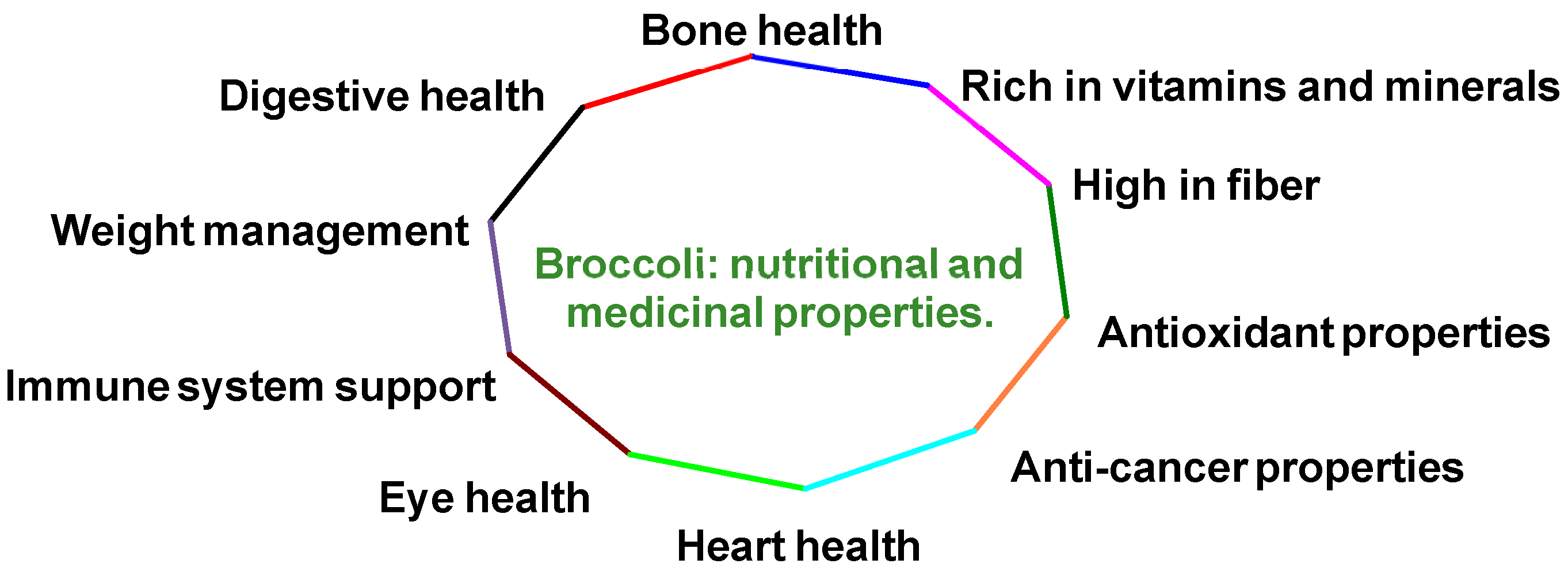
Many species in broccoli’s plant family are crucial components of human diets globally [4]. Regular consumption of these vegetables offers numerous health advantages and specifically provides several benefits (Figure 2):
  • It contains abundant vitamins C, K and A, along with minerals such as potassium, calcium and iron. These nutrients act as antioxidants, helping to protect the body from oxidative stress and reducing inflammation [5,6].
  • It is rich in dietary fiber, which supports digestion, increases feelings of fullness and contributes to a healthy digestive system [6].
  • It provides various antioxidants, including vitamins C and E, β-carotene and several flavonoids, which help shield cells from free radical damage [7].
  • It possesses potential anti-cancer properties due to its glucosinolate content, which the body can transform into cancer-fighting compounds [8,9,10].
  • It supports heart health by potentially lowering cholesterol levels and helping maintain healthy blood pressure [9,11].
  • It promotes eye health through its high vitamin A content and the presence of antioxidants [12].
  • It boosts the immune system while supporting collagen production, wound healing and iron absorption [13].
  • It contributes to bone health as it is a significant source of calcium and contains vitamin K [14].
  • It aids in weight management due to its low-calorie content and high fiber levels [15].
  • It supports digestive health, with its high fiber content promoting regular bowel movements [16].
Broccoli boasts an impressive nutritional profile and contains a significant number of bioactive compounds, also known as secondary metabolites, phytochemicals or phytonutrients. These bioactive compounds offer health benefits beyond basic nutrition. While not essential for human health, these phytochemicals can positively influence various disease processes and play a crucial role in long-term well-being [17].
The cultivation of broccoli is mainly focused on food consumption; the co-edible part comprises only 15% of the whole plant. This portion includes the florets, often accompanied by tender stems and sprouts. Broccoli production generates a substantial number of by-products, such as leaves and stems, which are generally discarded because they are considered unusable or commercially under-utilized; therefore, it is necessary to consider the revalorization of these by-products. These strategies can be focused on obtaining new ingredients for the food, cosmetics and pharmaceutical industries [18,19].
The primary bioactive compounds found in broccoli include phenols, glucosinolates, carotenoids, tocopherols, ascorbic acid and peptides. These compounds are renowned for their antioxidant properties and are associated with the health benefits of broccoli consumption. However, it is important to note that broccoli has a limited shelf life of only a few days. Without proper storage conditions, the phytochemical content can decrease rapidly [20].
The United Nations’ Food and Agriculture Organization (FAO) has recognized broccoli as one of the most nutritionally valuable vegetables. This classification is based on broccoli’s high content of health-promoting compounds (such as glucosinolates and phenolic compounds) and its significant contribution of vitamins, minerals and fiber to the diet (FAO, 2021) [21].

2. Bioactive Compounds in Broccoli

The health benefits of broccoli extend beyond its primary nutrients to include secondary plant metabolites known as bioactive phytochemicals. These compounds include phenolic compounds (which possess antioxidant properties and help neutralize free radicals, such as flavonoids, hydroxycinnamic acids and their derivatives), carotenoids (lutein, zeaxanthin, β-carotene, violaxanthin and neoxanthin, which act as powerful antioxidants and support eye health), chlorophylls (which contribute to the green color of broccoli and have potential health-promoting effects), glucosinolates (sulfur-containing compounds that can be converted into cancer-fighting substances in the body, such as sulforaphane (SFN)), vitamins (particularly vitamins A, C and K, which contribute to various health benefits), essential minerals (including selenium, potassium and manganese, which play crucial roles in various bodily functions) and antimicrobial peptides (which have shown potential applications in food preservation and as natural alternatives to synthetic antibiotics) [22].
These bioactive phytochemicals work synergistically to provide various health benefits, including antioxidant, anti-inflammatory, anticancer and antimicrobial activities. The concentration and efficacy of these compounds in broccoli can vary depending on factors such as genotype, growing conditions, harvest time and plant part (e.g., florets, leaves, or stems) [23].

2.1. Glucosinolates

Glucosinolates, also known as thioglycosides, are nitrogen–sulfur-containing secondary metabolites found almost exclusively in plants of the Brassicaceae family, with broccoli being one of the richest sources. These bioactive compounds are considered the hallmark of broccoli due to their high concentration. Glucosinolates are particularly abundant in broccoli seeds and sprouts during their early developmental stages, where their levels can be up to ten times higher than those found in mature broccoli. These compounds are responsible for the distinctive pungent aroma and bitter taste of Brassicaceae vegetables and serve as natural defense mechanisms for the plants [24].
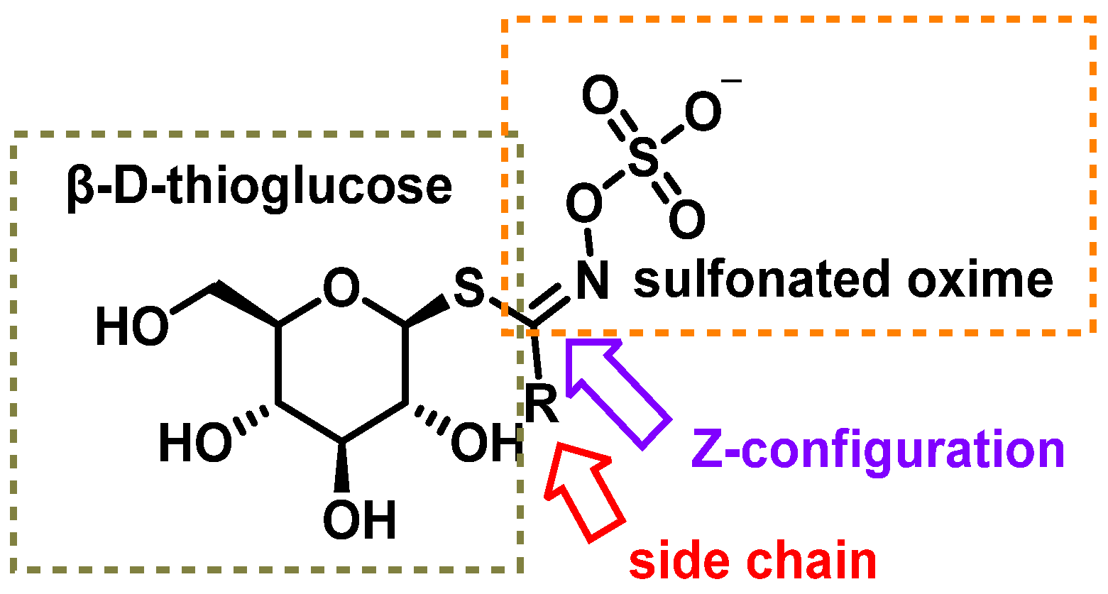
The chemical structure of glucosinolates (β-D-thioglycoside-N-hydroxysulfate) consists of three main components: (i) a β-D-thioglucose group, (ii) a sulfonated oxime group with a C=N double bond in the Z configuration, confirmed through X-ray crystallography and (iii) a R side chain derived from amino acids such as methionine, phenylalanine, tryptophan, or branched-chain amino acids [25,26].
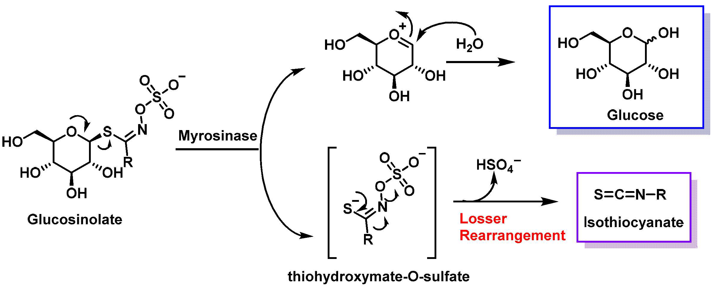
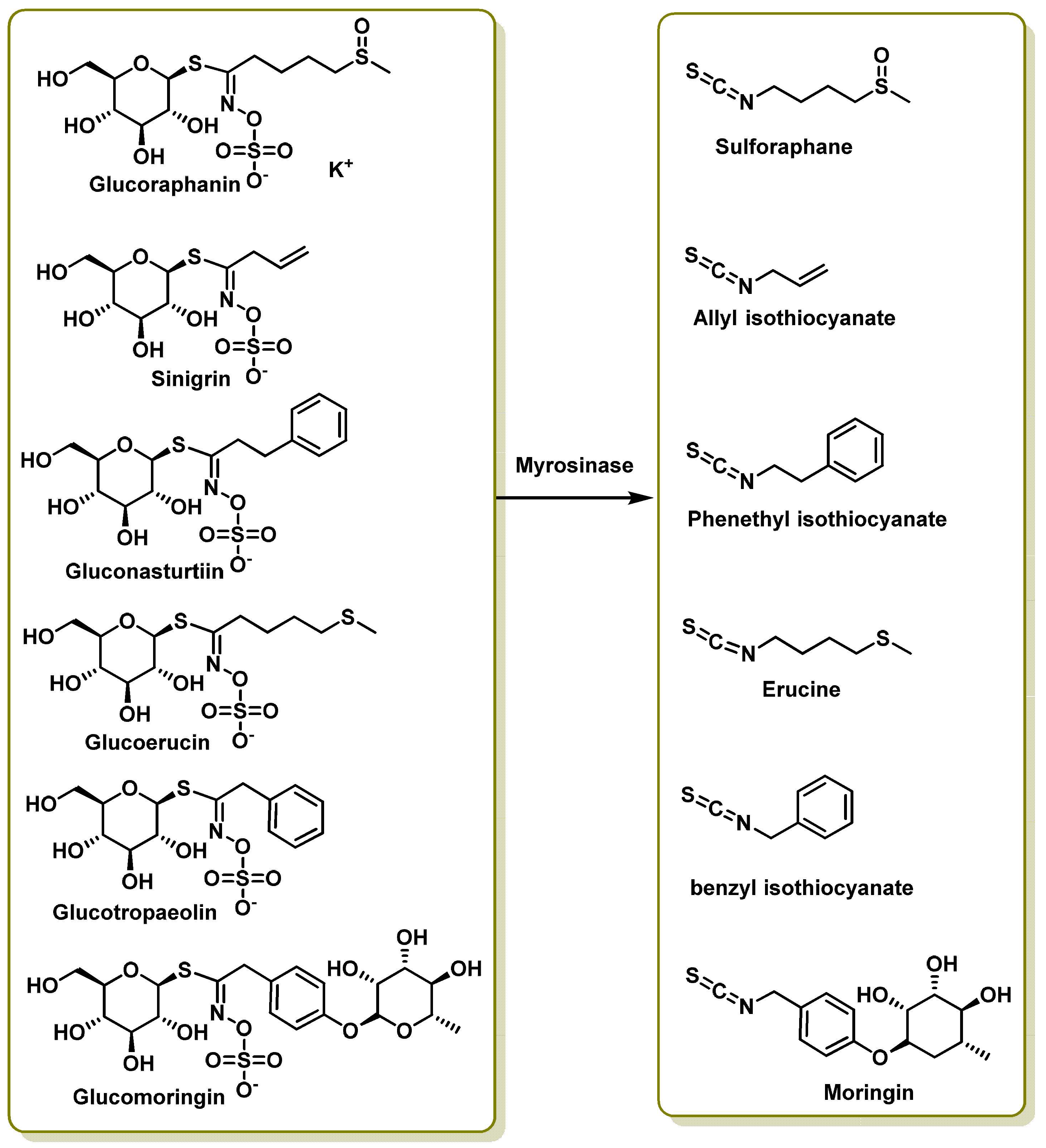
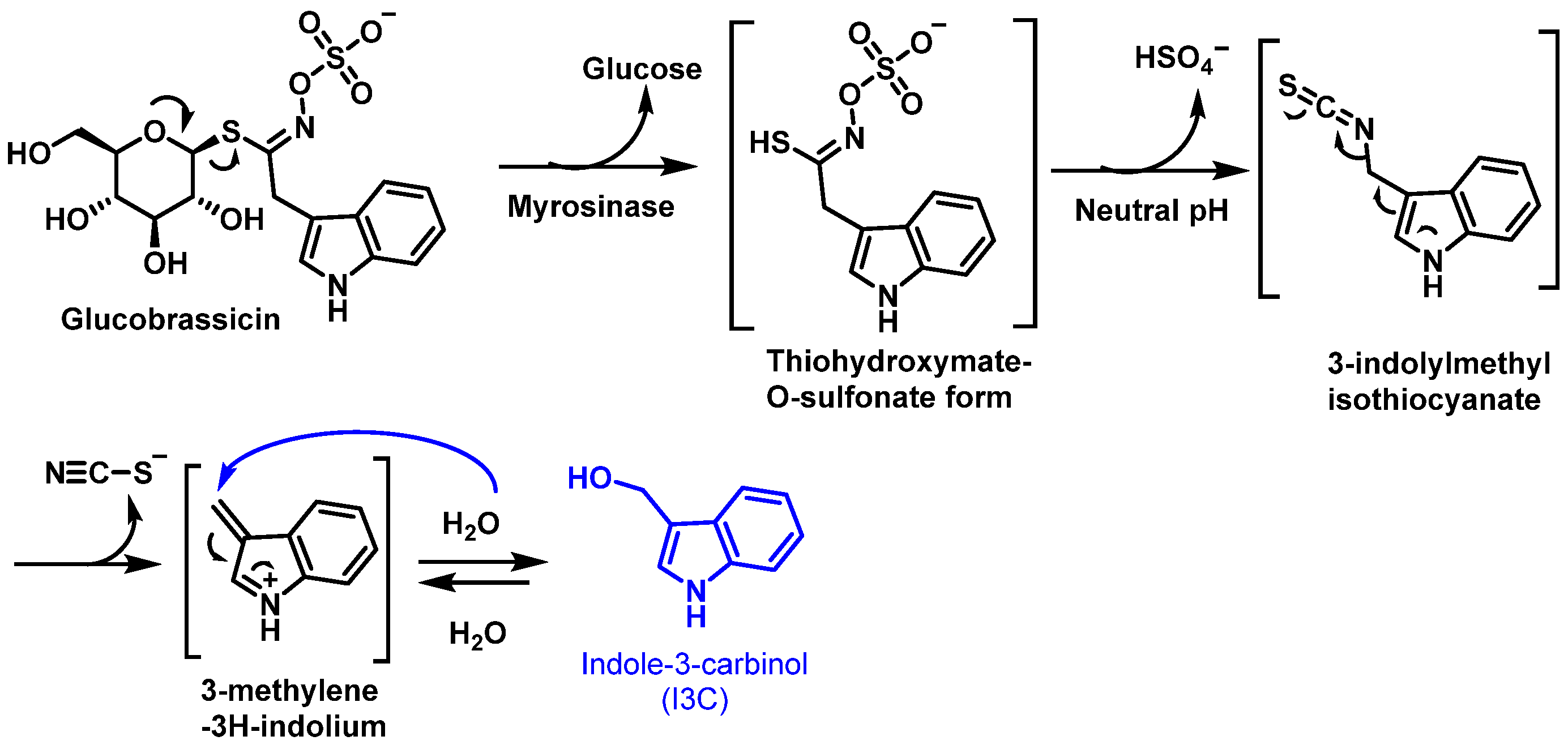
When glucosinolates are hydrolyzed by the enzyme myrosinase, glucose and thiohydroxymate-O-sulfate are released. This reaction is followed by an intermolecular Lossen rearrangement, resulting in the formation of isothiocyanate (RN=C=S), a compound known for its potential health-promoting properties, including anticancer effects [27] (Figure 3).
Glucosinolates (GSLs) are chemically stable molecules under standard conditions, with their breakdown primarily occurring through enzymatic action. These compounds coexist with their hydrolyzing enzyme, myrosinase (a β-thioglucosidase), within plant cells but remain segregated in distinct cellular compartments. GSLs remain inert until contact with myrosinase occurs via physical disruption, such as chewing, food processing, or pest damage [28].
Humans lack endogenous myrosinase to convert GSLs into bioactive isothiocyanates (ITCs); however, the gut microbiota—particularly certain Bifidobacterium species—can perform this conversion 2–3 h after consuming Brassica vegetables. Hydrolysis occurs via plant-derived myrosinase, which activates GSLs in the small intestine, or bacterial myrosinase, which facilitates hydrolysis in the colon [29].
GSLs only become biologically active when hydrolyzed to ITCs. These metabolites are absorbed in the small intestine and colon, with detectable levels appearing in human urine within 2–3 h of Brassica consumption.
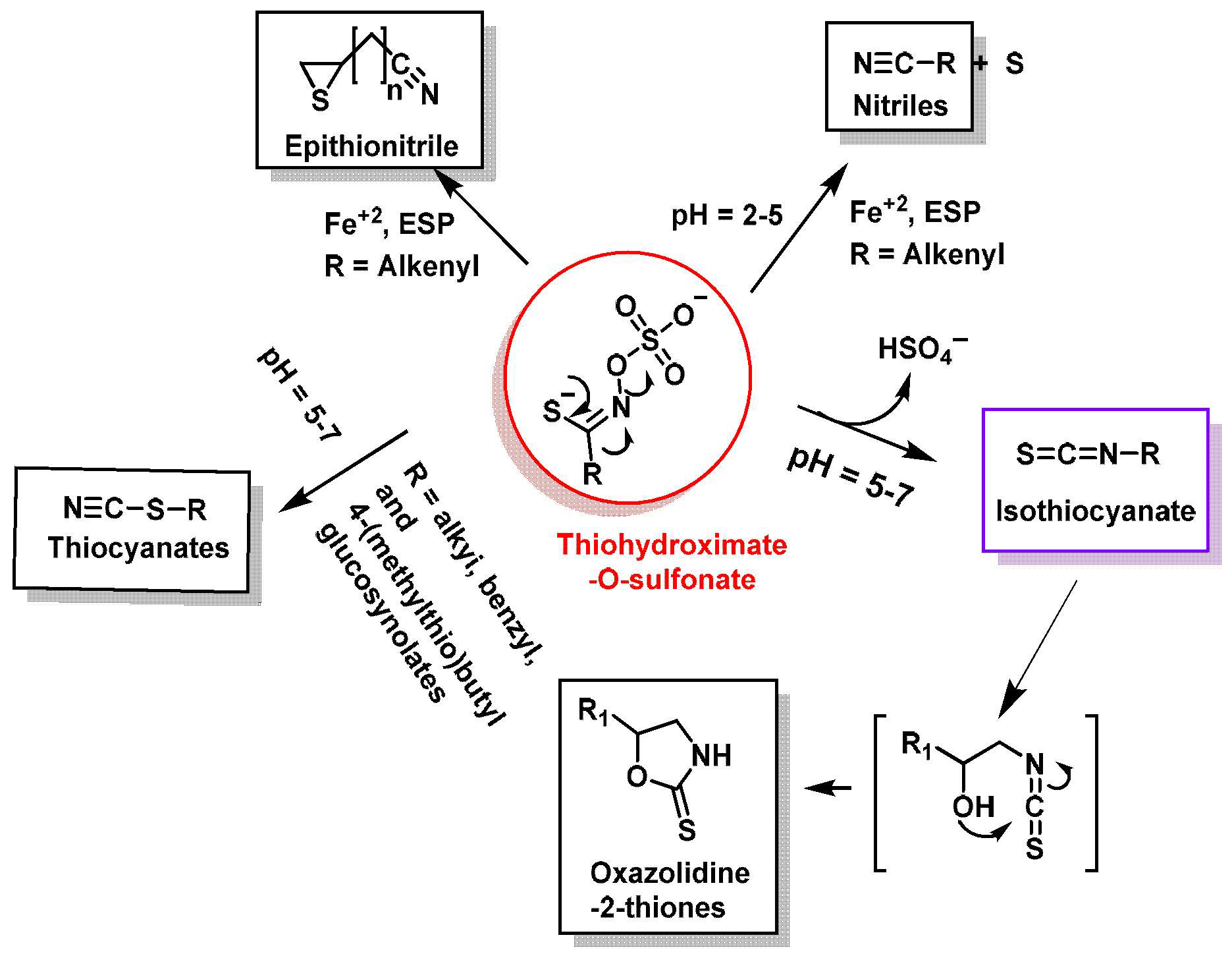
In the hydrolysis mechanism (Figure 4), myrosinase cleaves the thioglycosidic bond, releasing β-D-glucose and forming an unstable thiohydroxymate-O-sulfonate intermediate. This compound undergoes pH-dependent transformations: at neutral conditions (pH~7), the Lossen rearrangement produces ITCs (R-NCS), and at acidic conditions (pH~4), decomposition yields nitriles instead of ITCs. At low pH, the Lossen rearrangement is inhibited, favoring nitrile formation over ITCs [30].
Thiohydroxymate-O-sulfate—the unstable organic aglycone derived from glucosinolate hydrolysis—undergoes structural rearrangements influenced by parent glucosinolate structure, hydrolysis conditions (pH, temperature), cofactor presence (e.g., Fe2+ ions) and regulatory proteins (e.g., epithiospecifier protein (ESP), thiocyanate-forming protein (TFP)). These factors determine the final degradation products, which fall into isothiocyanates (ITCs), thiocyanates, nitriles, epithionitriles (EPT) and oxazolidine-2-thiones [31].
ESP-mediated pathways direct the conversion of alkenyl glucosinolates (GLs) to epithionitriles and non-alkenyl GLs to nitriles. This protein acts as a myrosinase cofactor, steering reactions toward specific byproducts (Figure 5). In ESP’s presence, intermediates like thiocyanate (RSC≡N) or oxazolidin-thione transform into epithionitriles (alkenyl GLs) or nitriles (non-alkenyl GLs) [31,32,33].
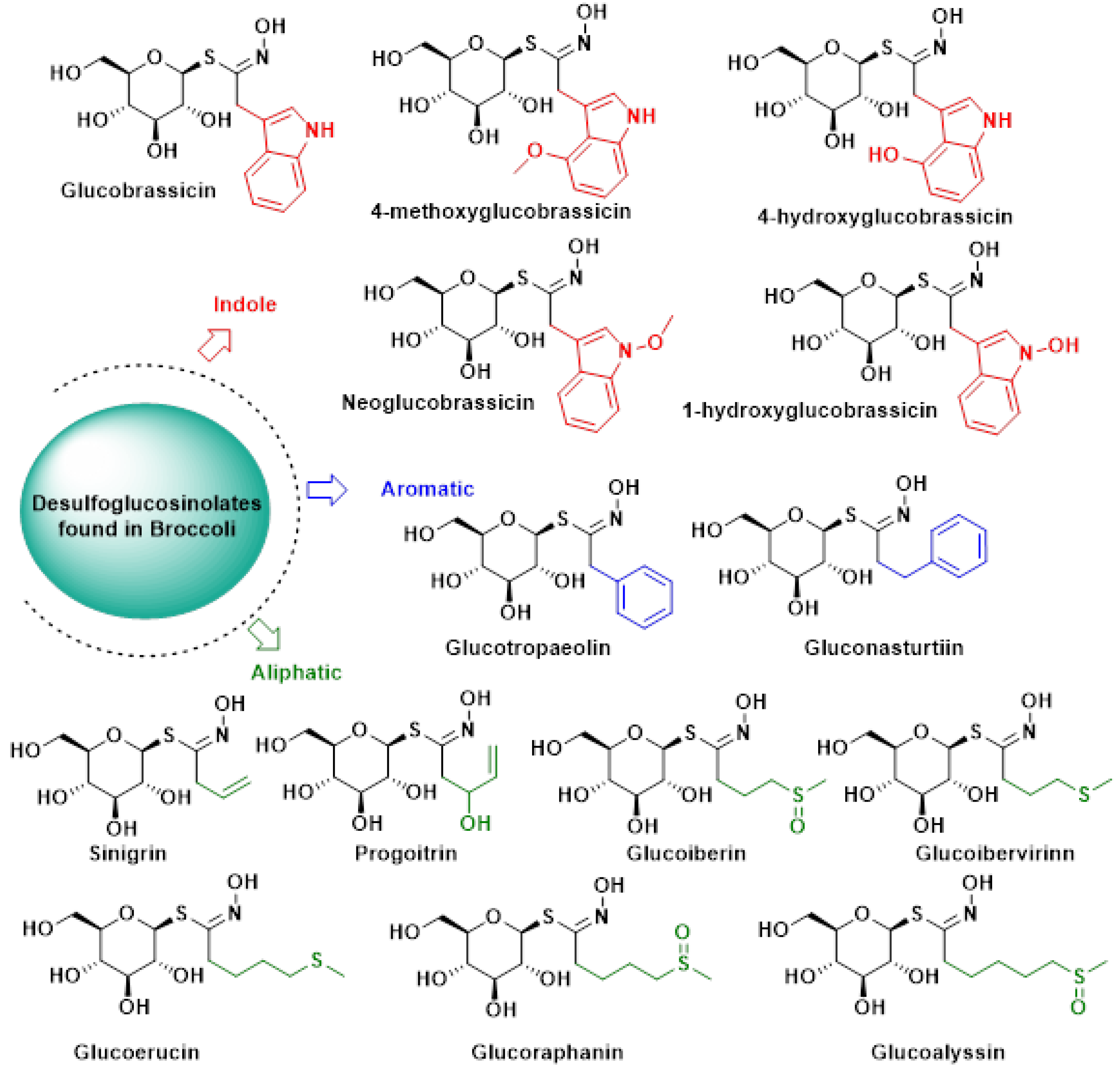
Over 130 distinct glucosinolates (GSLs) have been identified and characterized. These compounds are typically classified based on the chemical structure of their R side chains [34,35]. For instance, GSLs isolated from broccoli varieties are grouped into three categories according to their precursor amino acids:
(i)
Aliphatic GSLs, derived from methionine, isoleucine, leucine, or valine [36,37,38,39,40,41,42,43,44,45,46].
(ii)
Aromatic GSLs, originating from phenylalanine or tyrosine [47,48,49].
(iii)
Indole GSLs, synthesized from tryptophan [50,51].
A more recent classification system, proposed by Blaževic et al., categorizes GSLs based on the presence or absence of aromatic motifs in their molecular structure [35] (Figure 6).
Of the more than 100 identified glucosinolates (GSLs), approximately 50 are commonly found in Brassica genus vegetables. Typically, each plant contains 3 to 4 predominant GSLs, though up to 15 different types have been observed in some species [52]. The diversity in GSL side chains leads to a wide array of sulfur-containing derivatives through enzymatic breakdown [53].
In broccoli, the prevalent glucosinolates include glucoraphanin (4-methylsulfinylbutyl), sinigrin, progoitrin (2-hydroxy-3-butenyl), gluconapine (3-butenyl), glucobrassicin, neoglucobrassicin (1-methoxy-3-indolylmethyl) and glucoraphanin (the most abundant, comprising over 50% of total glucosinolates, while sinigrin is present in comparatively lower amounts) [54].
Key glucosinolates and their corresponding hydrolysis products include glucoraphanin (GRA), sulforaphane (SFN), sinigrin (SIN), allyl isothiocyanate (AITC), gluconasturtiin (GST), phenethyl isothiocyanate (PEITC), glucoerucin (GER), erucin (ER), glucotropaeolin (GTL), benzyl isothiocyanate (BITC), glucomoringin (GMG) and moringin (MG). Figure 7 illustrates these transformations [55,56].

2.1.1. Bioavailability of Glucosinolates

In cruciferous vegetables, the composition and content of glucosinolates are relatively stable, but depend on the genus and species, and may vary with plant growth, with different parts of a plant (e.g., in seeds, roots or leaves), post-harvest storage conditions and culinary processing [52,57]. Taking these variables into account, the amounts of isothiocyanates formed from glucosinolates in foods are variable.
Most cruciferous vegetables are cooked before consumption; the bacterial myrosinase in the intestine, rather than vegetable myrosinase, is responsible for the initial process of glucosinolate degradation. The intensity of heat denaturation is important when the temperature applied is high and the cooking time is long [58]. Inactivation may occur due to cooking at 60 °C [59], sautéing [60], microwaving [61] or other forms of heating or processing of cruciferous vegetables [58]. When myrosinase is inactivated, glucosinolates transit to the colon, due to their hydrophilic nature (thioglucose and sulphate group), and are metabolized by the intestinal microbiota. For example, intact glucosinolates may be partially absorbed in the stomach, and the remaining glucosinolates will transit through the gastrointestinal tract to reach the small intestine where they may be hydrolyzed by plant myrosinase and the degradation products may be absorbed. The remaining unhydrolyzed glucosinolates will then transit to reach the colon where they may be hydrolyzed by bacterial myrosinase, and the degradation molecules generated will be absorbed and/or excreted.
Johnson et al., 2002, reported that isothiocyanates were absorbed through the small intestine and colon, where their metabolites were detected 2–3 h after consumption [62].
Detailed studies of the effect of cooking Brassica vegetables on the absorption of isothiocyanates have been carried out [52]. Vermeulen et al., 2008, found that the consumption of raw broccoli resulted in faster absorption, higher bioavailability and higher maximum plasma amounts of sulforaphane compared to cooked broccoli [63]. Higher amounts of sulforaphane were found in blood and urine when broccoli was eaten raw (bioavailability 37%) compared to cooked (3.4%, p = 0.002). Sulforaphane absorption was delayed when cooked broccoli was consumed (peak plasma time = 6 h) compared to raw broccoli (1.6 h, p = 0.001). Excretion half-lives were comparable, 2.6 and 2.4 h on average, for raw and cooked broccoli, respectively (p = 0.5) [63].
Conaway et al., 2000, observed that isothiocyanate levels were three times higher in urine after consumption of raw broccoli compared to steamed broccoli (32.3% vs. 10.2%). These authors compared the metabolic fate of glucosinolates after the ingestion of fresh and steamed broccoli [64].
Fahey et al., 2015, also demonstrated that active myrosinase was critical for the bioavailability of glucoraphanin in broccoli extracts as sulforaphane was three to four times more bioavailable in extracts with an active myrosinase enzyme than in extracts without [65].
Ghawi et al., 2013, found that the addition of mustard seeds, which contain a myrosinase isoform that is much more heat stable than other brassicas, to thermally processed broccoli increased sulforaphane formation by three to five times [66].

2.1.2. Biosynthesis of Glucosinolates in Plants

Glucosinolate (GSL) biosynthesis originates from α-amino acid metabolism and can be divided into three phases [34,67].
(1)
Elongation of the side chain of amino acids.
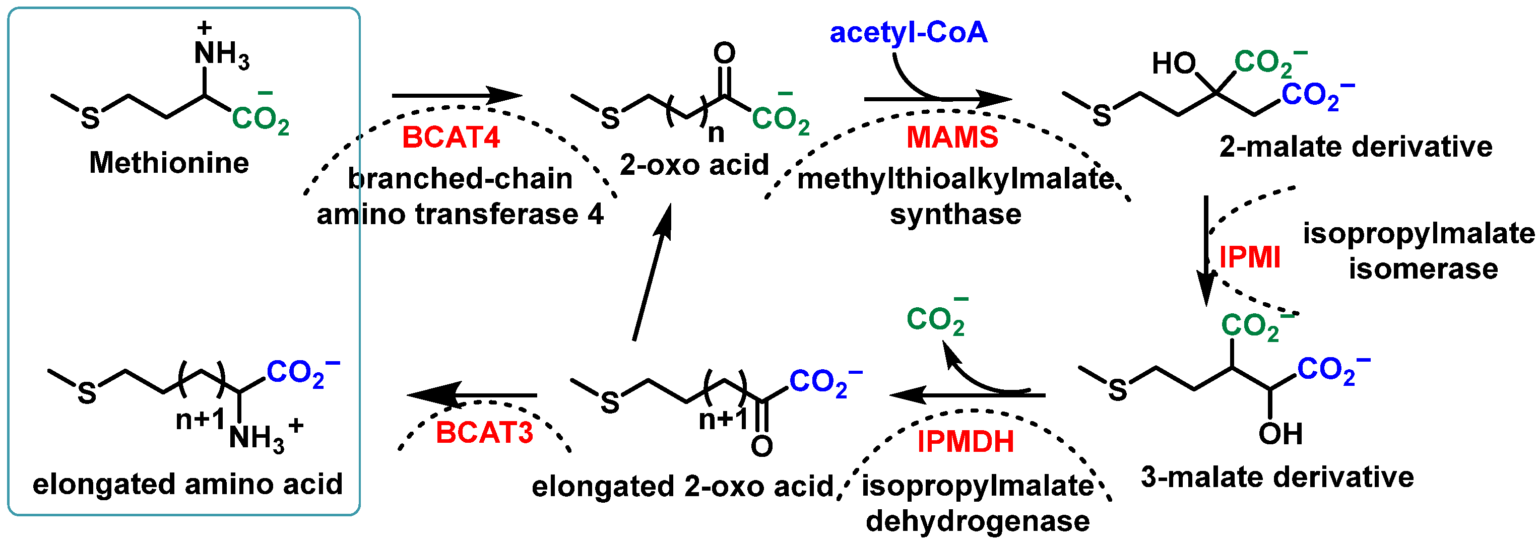
The initial reaction occurs in the cytosol and the subsequent elongation steps take place in chloroplasts. The process unfolds as follows:
  • Deamination of aliphatic or aromatic amino acids, yielding 2-oxo acids;
  • Condensation of 2-oxo acids with Acetyl-CoA, forming 2-malate derivatives;
  • Isomerization of 2-malate to 3-malate;
  • Oxidation and decarboxylation, resulting in the loss of the initial amino acid carboxyl group and the formation of an elongated 2-oxo acid molecule.
The elongated 2-oxo acid can then re-enter the elongation cycle or undergo transamination to form an elongated amino acid. Figure 8 illustrates this process [49].
(2)
Synthesis of GSL from the modified amino acid
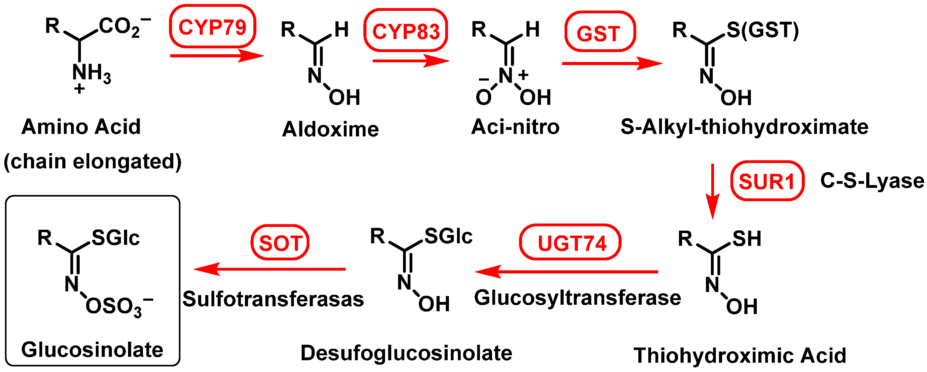
The biosynthesis of glucosinolates (GSLs) involves a multi-stage enzymatic pathway that transforms amino acids into diverse sulfur-containing secondary metabolites. This process includes chain elongation, core structure assembly, and secondary modifications, with specific enzymes catalyzing each step. The key stages are as follows: (i) conversion of amino acids to aldoximes, cytochrome P450 monooxygenases oxidize amino acids (e.g., methionine, phenylalanine) into aldoximes (CYP79 family, catalyzing the oxidation of parent amino acids or their chain-elongated derivatives); (ii) formation of reactive intermediates, due to CYP83 family enzymes further oxidize aldoximes to generate reactive aci-nitro or nitrile oxide compounds, critical for sulfur conjugation in the next step; (iii) sulfur conjugation with glutathione (GSH), where GSH, not cysteine, donates sulfur for conjugation, in a process in which activated aldoximes bind to GSH via enzymatic action, forming S-alkylthiohydroxymate conjugates; (iv) cleavage (the C-S lyase enzyme SUR1 cleaves the GSH conjugate to produce thiohydroxymates) and glycosylation (UDP-glucose:thiohydroxymic acid S-glucosyltransferase (UGT74B1) adds a glucose moiety to form desulfoglucosinolates); and (v) final sulfation, where desulfoglucosinolate sulfotransferases catalyze sulfation, converting desulfoglucosinolates into bioactive GSLs [24,68,69,70].
Transferases involved in forming the GSL core structure have been identified and extensively documented in the scientific literature [71,72] (Figure 9).
(3)
GSL sidechain modifications
The third stage of glucosinolate (GSL) biosynthesis is crucial for increasing the diversity of GSLs found in nature. After the formation of parent glucosinolates, a wide range of side chain modifications occur, including oxidation, esterification, hydroxylation, methoxylation, alkenylation and benzoylation. These modifications give rise to the various GSLs observed in plants. The biological activity of GSLs is largely determined by their side chain structure, making these secondary modifications particularly interesting for potential applications [73].
For aliphatic glucosinolates, secondary modifications include oxygenations, hydroxylations, alkenylations, and benzoylations. Indole glucosinolates commonly undergo hydroxylations and methoxylations. A common modification, the S-oxygenation of aliphatic glucosinolates, is carried out by flavin monooxygenases (FMO GS-OXs). Alkenyl glucosinolates, such as sinigrin, are produced from S-oxygenated glucosinolates by 2-oxoglutarate-dependent dioxygenases (AOPs).
In Arabidopsis thaliana, the GSL-ALK locus controls the conversion of methylsulfinyl glucosinolates to alkenyl glucosinolates, which is related to the production of sinigrin and gluconapin. The BoGSL-ALK gene, which has been cloned, has been shown to influence side-chain modifications in glucosinolate biosynthesis. These secondary modifications play a key role in determining the final structure and biological activity of glucosinolates, contributing to their diverse functions in plant defense and human nutrition [74].

3. Most Studied Broccoli Glucosinolate Hydrolysis Compounds

The hydrolysis of glucosinolates (GSLs) in broccoli primarily produces isothiocyanates (ITCs), which are more bioactive than their GSL precursors. Among these, glucoraphanin (GR), a precursor to the isothiocyanate SFN, is the most extensively studied glucosinolate in broccoli. Indole glucosinolates, on the other hand, yield unstable thioketone intermediates (TKIs), which predominantly break down into indole-3-carbinol (I3C) and 3,3′-diindolylmethane [75,76].

3.1. Sulforaphane

SFN, chemically known as 1-isothiocyan-4-(methylsulfinyl) butane, is a low-toxicity phytochemical derived from its inactive precursor glucoraphanin, which constitutes 35–50% of the total glucosinolates in broccoli. SFN has garnered significant attention for its potential use in clinical applications and as a dietary supplement [77].
The conversion of glucoraphanin to SFN is catalyzed by myrosinase, an enzyme present in broccoli tissues. Broccoli sprouts are particularly rich in SFN content. Research has demonstrated that SFN acts as a potent natural antioxidant and free radical scavenger. It also activates the nuclear factor erythroid 2-related factor 2 (Nrf2) signaling pathway, which plays a critical role in cellular defense mechanisms against oxidative stress [78].
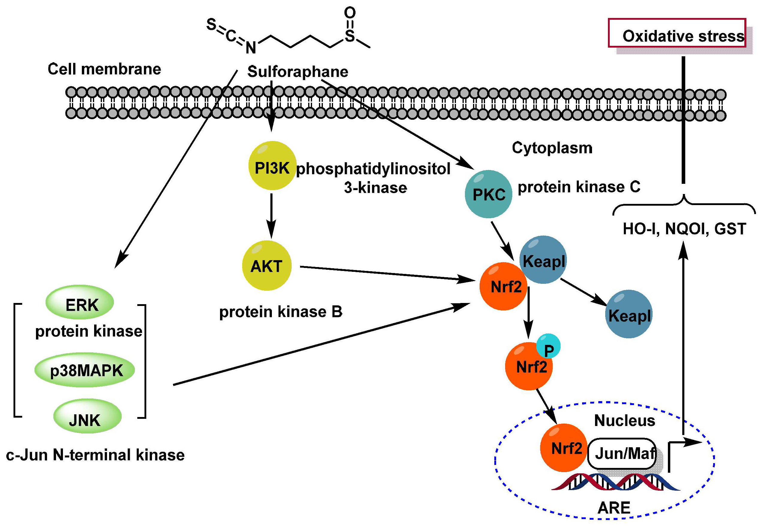
Key mechanisms of SFN action involve the activation of MAPKs (SFN activates three mitogen-activated protein kinases (MAPKs): extracellular signal-regulated protein kinase (ERK), c-Jun N-terminal kinase (JNK), and p38 MAPK), activation of PKC (SFN stimulates protein kinase C (PKC), which directly phosphorylates Nrf2) and PI3K/AKT pathway, because SFN activates phosphatidylinositol 3-kinase (PI3K) and protein kinase B (AKT), leading to Nrf2 activation [79].
Upon activation, Nrf2 undergoes phosphorylation, translocates to the nucleus, and binds to the antioxidant response element (ARE). This interaction induces the transcription of cytoprotective genes that reduce oxidative stress. Exposure to ARE inducers inhibits Nrf2 ubiquitination, allowing it to accumulate in the nucleus. Here, Nrf2 dimerizes with small Maf proteins and binds to ARE sequences, stimulating the expression of genes encoding enzymes such as heme oxygenase 1 (HO-1), quinone oxidoreductase 1 (NQO1), and glutathione S-transferases (GST) [79,80].
Keap1, a regulatory protein that normally sequesters Nrf2 in the cytoplasm, releases it under oxidative stress conditions, enabling Nrf2 to bind to AREs and initiate gene transcription. This cascade significantly enhances cellular protection against oxidative damage [81,82] (Figure 10).
SFN exhibits a wide range of beneficial effects beyond its anti-inflammatory and antimicrobial properties [83]. These include anticancer activity, antidiabetic effects [84,85], anti-obesogenic and anti-obesity properties, and cardioprotective effects [81,86,87,88]. The SFN content in broccoli has been quantified as follows: broccoli seeds contain 396.18 mg/100 g fresh weight, while broccoli sprouts contain 391.81 mg/100 g dry weight [19,89].
SFN’s anticancer mechanisms operate simultaneously on multiple cellular targets. It protects DNA by inhibiting mutagenic factors (phase I) and activating phase II detoxification enzymes. Additionally, SFN inhibits cancer cell proliferation and activates apoptosis, limiting the multiplication of mutated cancer cells and inhibiting neogenesis and metastasis. SFN has demonstrated the ability to prevent, remove, and reverse preneoplastic lesions. Researchers have investigated the primary anticancer mechanisms of SFN and determined the concentrations required for their activation, as outlined in Table 1 [90].
SFN’s anticancer activity is multifaceted, involving the inhibition of cancer cell proliferation, arrest of the cell cycle, and enhancement of apoptosis. It alters several epigenetic and non-epigenetic mechanisms, including the inhibition of histone deacetylase (HDAC) activity, halting histone phosphorylation, downregulation of DNA methyltransferases (DNMTs), and regulation of noncoding RNAs [91].
Beyond its anticancer effects, SFN has shown promise in addressing other health concerns. For instance, it has antidiabetic effects, improving blood sugar levels by suppressing hepatic glucose production and enhancing glucose tolerance. SFN also exhibits anti-obesogenic and anti-obesity properties by improving glucose tolerance and reducing fat accumulation, partly through its ability to activate Nrf2, a transcription factor regulating oxidative stress and inflammation in cells [92].
Furthermore, SFN has demonstrated cardioprotective properties. It improves the viability of cardiomyocytes, diminishes apoptotic cells, suppresses caspase-3 activity, alleviates damage to mitochondrial membrane potential, and decreases the expression of ER stress-related apoptosis proteins. Additionally, SFN inhibits foam cell formation and atherosclerosis by regulating macrophage cholesterol transport and accumulation [93].
These diverse effects highlight SFN’s potential as a therapeutic agent for various health conditions, including cancer, diabetes, obesity and cardiovascular disease.
Fimognari et al., 2007, studied the main anticancer mechanisms of SFN and the concentrations required for their activation (Table 1) [90].
According to the U.S. National Cancer Institute, SFN is one of the 40 most promising anticancer compounds. There are several excellent reviews on the anticancer activities as well as the mechanisms of action of SFN [94,95,96,97]. In the review conducted by Lukasz Janczewski, different methods of SFN synthesis are described [98].

3.1.1. Sulforaphane Reactivity

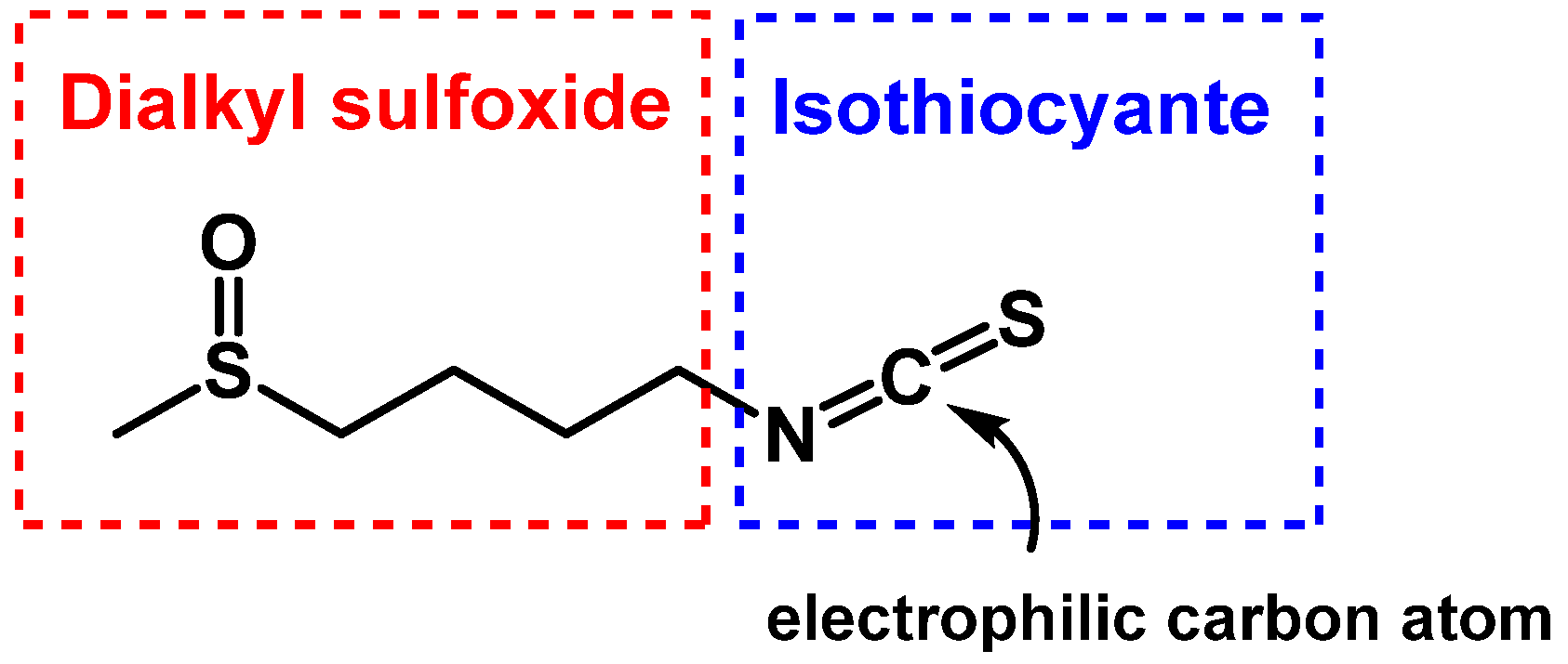
SFN has two reactive functional groups, a dikazkyl-sulfoxide and an isothiocyanate (Figure 11).
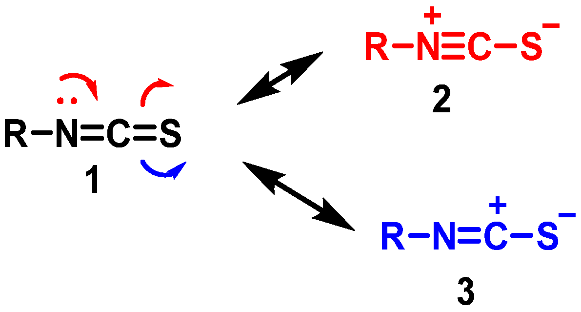
The group –N=C=S has a linear structure that was proposed as early as 1929 by Perschke [99]. The isothiocyanate group presents the canonical structures 1–3 shown in the figure containing accumulated double bonds (1) and ionic structures with a triple (2) or double bond N-C (3) and a negative charge located in the sulfur atom (Figure 12).
SFN structure is influenced by the dipole moments of its canonical forms, specifically, structures 1 and 3 contribute most significantly to the -NCS group, while structure 2’s contribution is negligible. This structural aspect is important for understanding the reactivity and stability of SFN.
SFN is highly reactive and unstable due to its functional groups. Its production and stability are affected by several factors, including temperature, pH, and enzyme activity. These factors limit SFN’s practical applications, which is why there are no marketed SFN drugs. Instead, the SFN-related market primarily consists of broccoli extracts, anti-aging creams, dietary concentrates, and other herbal products.
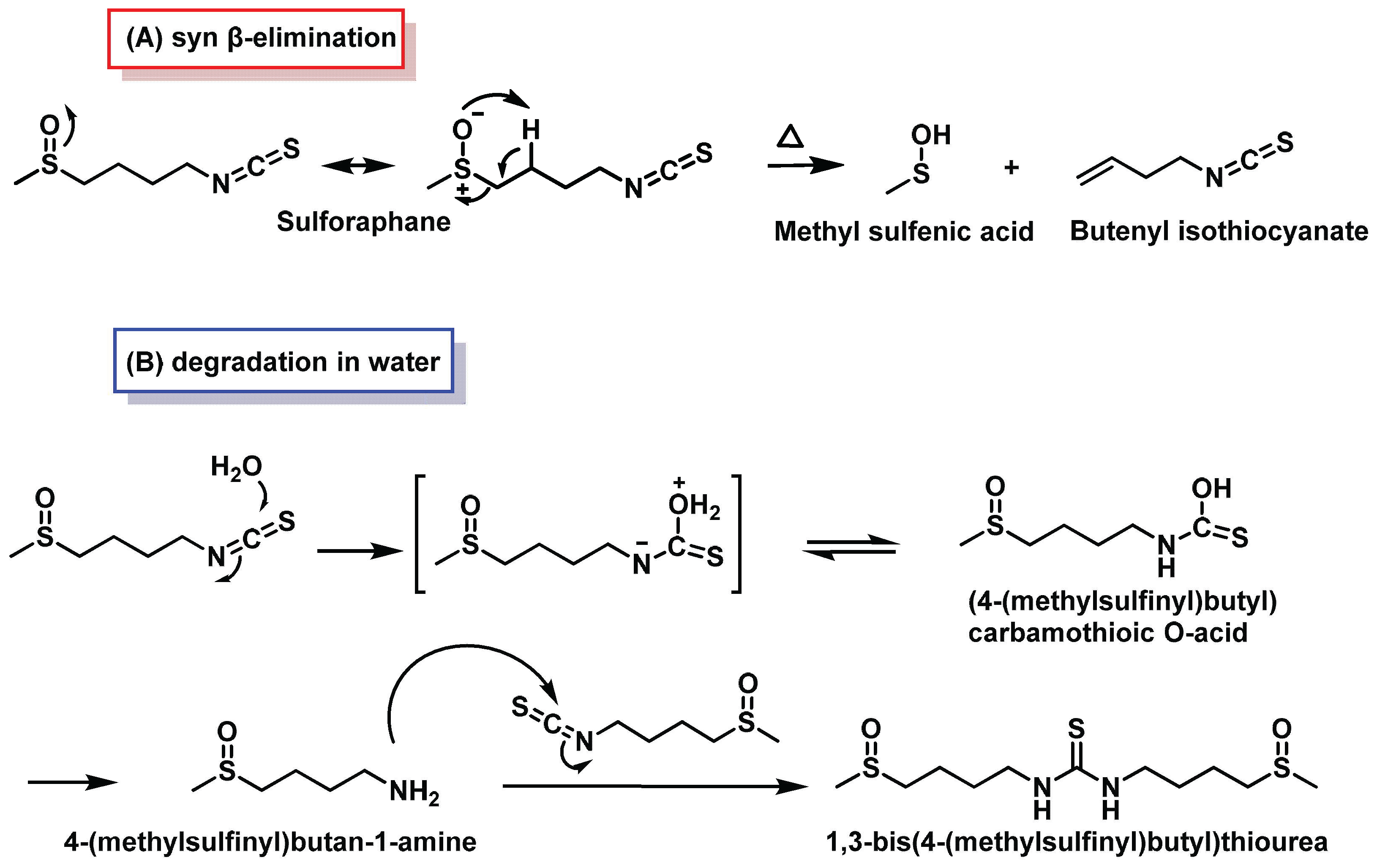
At high temperatures, SFN’s sulfoxide group can undergo β-elimination, transforming SFN into butenyl isothiocyanate and methylsulfenic acid (Figure 13A) [100]. The isothiocyanate group in SFN is highly reactive towards nucleophiles due to its electrophilic central carbon. It readily reacts with sulfur, nitrogen, and oxygen nucleophiles. Common reactions include the formation of thiocarbamates with cysteine residues in proteins and glutathione (GSH), as well as alkylation reactions with α-amino groups in N-terminal protein residues, ε-amino groups of lysine, and secondary amines like proline. These reactions form thiourea products.
In aqueous and biological media, isothiocyanates react to form inactive, non-volatile organic compounds. Under physiological conditions, they react reversibly with thiols and irreversibly with amines. Notably, isothiocyanates react 1000 times faster with thiols than with amines. Their hydrolysis produces amines (Figure 13B) [101]. This reactivity profile highlights the complex interactions of SFN with biological molecules, which is crucial for understanding its potential health benefits and limitations [102].

3.1.2. Interaction of Sulforaphane with Keap1

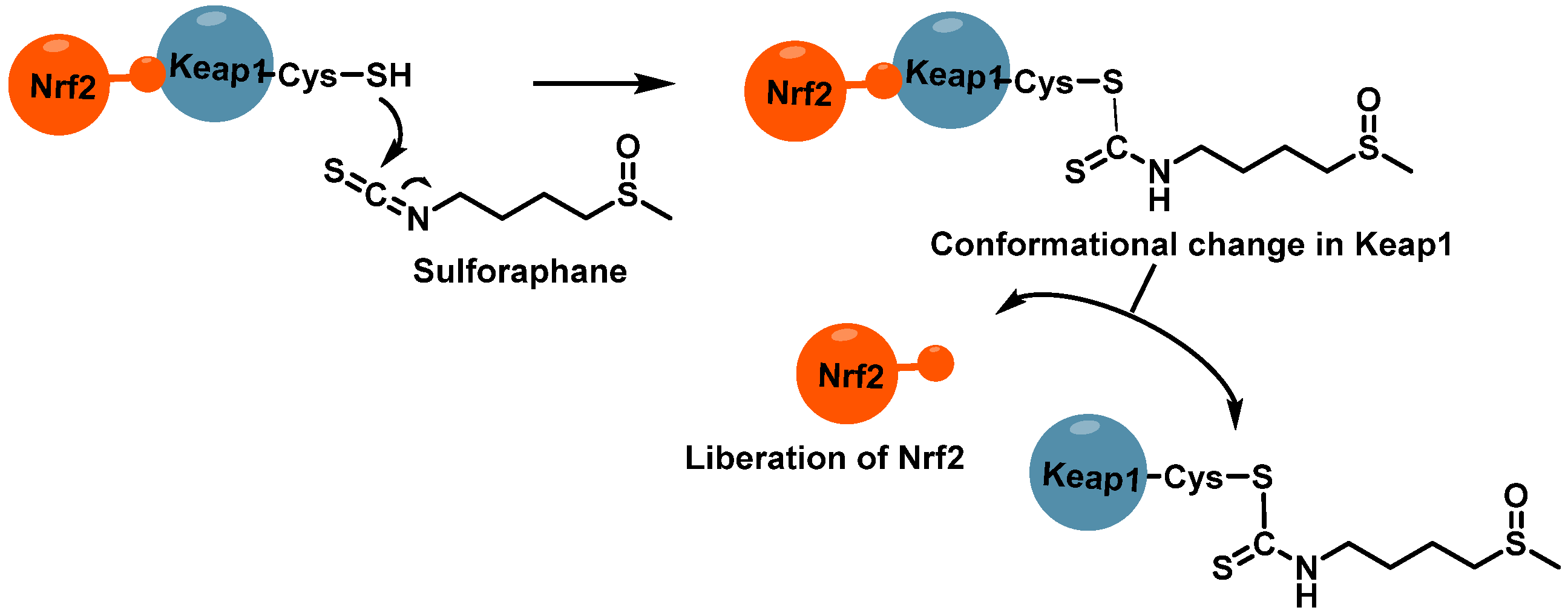
SFN demonstrates a high affinity for cysteine residues in proteins, forming thionoacyl adducts through its electrophilic properties. This interaction is particularly notable with Cys-151 in human Keap1, a key protein in the Nrf2 signaling pathway. However, SFN’s reactivity extends beyond this single residue, also modifying other sensing cysteines such as Cys-38, Cys-368, and Cys-489 in Keap1 [103,104,105].
The isothiocyanate group of SFN and its analogues exhibits rapid but reversible reactions with the sulfhydryl groups of Keap1 cysteines. This chemical interaction has significant implications for the Nrf2/Keap1 complex. By modifying these critical cysteine residues, SFN disrupts the normal functioning of the complex, effectively protecting Nrf2 from ubiquitination. This mechanism is illustrated in Figure 14, highlighting how SFN’s interaction with Keap1 leads to Nrf2 activation and subsequent cellular protective responses [106,107].
Interestingly, SFN’s ability to modify cysteine residues extends beyond its effects on the Nrf2/Keap1 pathway. Research has shown that SFN can also interact with cysteine residues in other proteins, such as those in the AP-1 transcription factor. Specifically, SFN has been found to inhibit AP-1 activity by interacting with Cys154 in cFos and Cys272 in cJun, which are part of the DNA-binding domain of these proteins [108].
This interaction contributes to SFN’s chemopreventive properties, particularly in reducing UVB-induced squamous cell carcinomas in animal models [109].
The versatility of SFN’s interactions with various cysteine-containing proteins underscores its potential as a multifaceted compound in cellular regulation and disease prevention. These interactions not only affect transcription factor activity but also influence various cellular processes, including apoptosis, cell migration, and invasion [110]. As research continues, the full scope of SFN’s cysteine-mediated effects on cellular function and its therapeutic potential are likely to be further elucidated.

3.1.3. Interaction of Sulforaphane with GSH

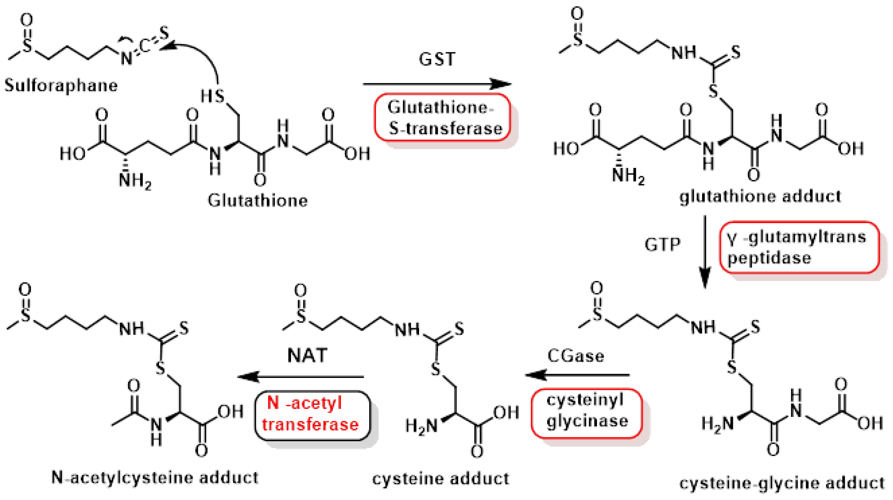
SFN undergoes a complex metabolic transformation in the body through the mercapturic acid pathway, a process that is crucial for its eventual elimination and potential therapeutic effects. This pathway begins immediately after SFN ingestion and involves several enzymatic steps. The initial step in this metabolic process is the conjugation of SFN with glutathione (GSH). This reaction is catalyzed by glutathione transferase (GST), an enzyme that plays a key role in cellular detoxification processes. The electrophilic isothiocyanate group of SFN reacts with the nucleophilic -SH group of the cysteine residue in GSH, forming a dithiocarbamate. This reaction marks the beginning of the isothiocyanate metabolism, which is commonly referred to as the mercapturic acid pathway [111].
Following this initial conjugation, the SFN-GSH complex undergoes a series of successive cleavage reactions. These reactions are catalyzed by specific enzymes: γ-glutamyltranspeptidase, cysteinylglycinase, and N-acetyltransferase. The final product of this enzymatic cascade is SFN-N-acetylcysteine, also known as a mercapturic acid.
The formation of this mercapturic acid primarily occurs in the liver, which is the main site of drug metabolism in the body. Once formed, the SFN-N-acetylcysteine is transported to the kidneys. From there, it is eliminated from the body through urinary excretion. This entire process, from ingestion to excretion, is illustrated in Figure 15, providing a visual representation of SFN’s metabolic journey through the body [112].
Understanding this metabolic pathway is crucial for appreciating how SFN exerts its biological effects and how it is processed and eliminated by the body. This knowledge can inform future research into SFN’s potential therapeutic applications and help in developing strategies to enhance its bioavailability and efficacy [113].
Most SFN undergoes metabolic transformation through the mercapturic acid pathway before excretion. The main urinary metabolite is sulforaphane-N-acetylcysteine (SF-NAC). The excretion rate and percentage of SFN eliminated through urine vary depending on the form of administration. When consumed as fresh broccoli sprouts, about 60–65% of the ingested dose is excreted as SF-NAC within 24 h. For SFN-rich formulations, approximately 70% of the administered SFN is eliminated in 24 h. Overall, urinary excretion of SFN metabolites can range from 70% to 90% of the ingested dose. The elimination of SFN and its metabolites is indeed rapid, with most excretion occurring within the first 12–24 h after administration. Urinary concentrations of SFN equivalents are typically 2–4 orders of magnitude higher than those in plasma. This rapid metabolism and excretion pose challenges for maintaining therapeutic levels of SFN in the body [114,115].
Given these pharmacokinetic characteristics, developing appropriate drug delivery systems to increase SFN’s bioavailability and concentration at target sites is a valid consideration. Some approaches being explored include nanoencapsulation using broccoli membrane vesicles, which has shown improved cellular uptake and metabolism of SFN in cancer cells. Another promising approach involves gold-coated iron oxide nanoparticles functionalized with folic acid for targeted delivery to cancer cells [116].
These delivery systems aim to enhance SFN’s stability, absorption and therapeutic efficacy, potentially addressing the limitations posed by its rapid metabolism and excretion. By improving the pharmacokinetic profile of SFN, these novel delivery methods could enhance its potential as a therapeutic agent in various health conditions [117].

3.2. The Risks of Excessive Intake of Sulforaphane

Sulforaphane is consumed as a component of cruciferous vegetables, and also as a dietary supplement or drug. Possible side effects, especially when consumed in large amounts or by supplementation, are summarized below:
-
Gastrointestinal discomfort: Some people report digestive symptoms such as diarrhea, gas or stomach upset after consuming sulforaphane supplements or large amounts of broccoli and other cruciferous vegetables. These symptoms may be more pronounced if the digestive system is not accustomed to the fiber and compounds present in these vegetables. Therefore, sufferers of gastrointestinal disorders, such as irritable bowel syndrome, may experience worsening symptoms due to the fermentation of fiber in the intestinal tract [118].
-
Allergic reactions. Although rare, some people may experience allergic reactions to sulforaphane or its food sources. In rare cases, symptoms can range from mild skin rashes to more severe reactions [119].
-
Thyroid interaction: There are indications that sulforaphane may interfere with thyroid function, especially in people with hypothyroidism. This is due to the goitrogenic compounds present in cruciferous vegetables, which may affect iodine absorption and thus thyroid hormone production. These vegetables also contain 2-hydroxy-3-butenyl glucosinolate (progoitrin) and indole glucosinolate, which can be converted to goitrin and thiocyanate, which act as biocogens in animal models. The conversion can occur either by spontaneous cyclization, as in Figure 16, or activated by the enzyme myrosinase [120,121].
These dioxins can block the transport of iodine into the thyroid gland and inhibit the incorporation of iodine into thyroglobulin, which impairs the synthesis of active thyroid hormone. Some methods of food preparation, such as washing, soaking and boiling, help to reduce bocytogens in food by destroying myrosinase and preventing the release of bocytrin from progoitrin [120,121,122].
-
Potential drug interactions. Sulforaphane potentiates the anticonvulsant efficacy of carbamazepine in a seizure test, indicating possible pharmacokinetic interactions. Sulforaphane may interact with drugs that are metabolized in the liver, particularly those that are substrates of cytochrome P450 enzymes, such as CYP3A4 and CYP1A2. The interaction between SFN and fu-rosemide, verapamil and ketoprofen modifies the activity of the enzyme system involved in drug metabolism and transport. This can lead to altered drug effectiveness and also to the development of multidrug resistance [123].
-
Variable effects on tumorigenesis. Several studies with higher doses of sulforaphane in mice describe toxicities that require careful attention to risk-benefit analyses and determination of therapeutic or prophylactic indices. Shorey et al., 2013, observed increased morbidity and no reduction in lung tumorigenesis in offspring born to mothers receiving transplacental and lactational exposure to the carcinogen dibenzo[def,p]chrysene and supplemented with dietary sulforaphane (400 ppm) in contrast to many reports of chemoprotection in adult animal models [124].
-
Tao et al., 2018, used a vinyl carbamate chemical carcinogenesis model (A/J mice) and a genetic model (LSL-K-rasG12D/+ mice) to induce lung cancers [125]. In the genetic model, pretreatment with SF had no effect on the number of tumors, but post-treatment increased the number and size of tumors. Kombairaju et al. reported that prolonged treatment with sulforaphane (0.5 mg, 5 days/week for 3 months did not improve tumorigenesis in the same LSL-K-rasG12D/+ murine model [125].
-
A toxicity study of SFN supplementation carried out in mouse models showed that ingestion of high doses of sulforaphane produced: marked sedation (at 150–300 mg/kg), hypothermia (at 150–300 mg/kg), impaired motor coordination (at 200–300 mg/kg), decreased skeletal muscle strength (at 250–300 mg/kg) and deaths (at 200–300 mg/kg). In addition, blood analysis showed leukopenia in mice injected with sulforaphane at 200 mg/kg [118].
-
Yagishita et al., 2019, provide an excellent review assessing the current state of knowledge on the relationships between formulation (e.g., plants, sprouts, drinks, supplements), bioavailability and efficacy, and the doses of glucoraphanin and/or sulforaphane that have been used in preclinical and clinical studies, paying particular attention to better integration of animal models and clinical studies, particularly with regard to dose selection and route of administration [126].

3.3. Optical Isomers of Sulforaphane

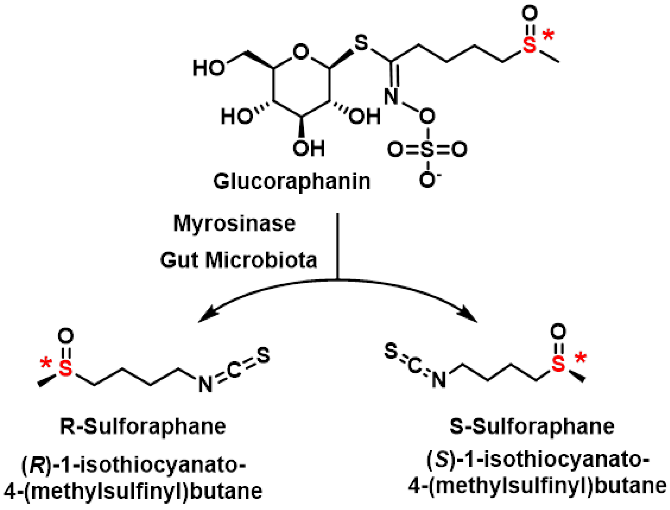
Glucoraphanin, the precursor of SFN found in broccoli, exists as a pure optical isomer with its sulfoxide group in the R configuration. This stereochemistry is preserved during the enzymatic hydrolysis by myrosinase, resulting in the formation of R-SFN. The sulfoxide group in SFN’s side chain contains a stereogenic sulfur atom that is configurationally stable, allowing for the separation of the two enantiomers (R and S) at room temperature [127], Figure 17.
SFN’s asymmetric sulfur atom gives rise to two optical isomers: R-SFN and S-SFN. However, in nature, specifically in broccoli sprouts, SFN exists exclusively as the R enantiomer. This natural occurrence of a single enantiomeric form is illustrated in Figure 16.
Research comparing these isomers has revealed significant differences in their biological activities. The naturally occurring R-SFN has demonstrated higher biological activity compared to its S counterpart. This finding has important implications for the potential therapeutic applications of SFN, as the R enantiomer may offer enhanced benefits [128].
The stereochemical aspects of SFN are crucial for understanding its biological effects and for developing potential pharmaceutical applications [129]. The higher activity of R-SFN suggests that maintaining this specific configuration could be important in the development of SFN-based therapies or supplements. Furthermore, this stereochemical preference in nature highlights the importance of chirality in biological systems and how subtle structural differences can lead to significant variations in biological activity [127].
Research by Vanduchova et al., 2019, highlights the distinct biological activities of SFN enantiomers, with the natural (R)-SFN showing significantly greater efficacy compared to the (S)-SFN enantiomer. Specifically, (R)-SFN enhances the activity of phase II detoxifying enzymes, such as quinone reductase and glutathione S-transferase, while (S)-SFN is either ineffective or much less active in this regard. This enantiomeric difference underscores the importance of stereochemistry in SFN’s biological activity [128].
Natural SFN predominantly exists as the (R)-enantiomer in broccoli, with its prevalence varying depending on the part of the plant and the variety of broccoli used for extraction. The content of (S)-SFN ranges from 1.5% to 41.8% of total SFN, depending on whether florets, leaves or stems are analyzed. For example, broccoli florets typically contain a higher proportion of (R)-SFN compared to stems and leaves [129].
The higher biological activity of (R)-SFN compared to its (S) counterpart has significant implications for its therapeutic potential. The ability of (R)-SFN to induce detoxifying enzymes makes it a promising candidate for cancer prevention and other health applications. These findings also emphasize the need for precise extraction and characterization methods to maximize the presence of the biologically active (R)-enantiomer in SFN-based products [130].
This variability in enantiomer content across different parts of broccoli highlights the importance of selecting specific plant parts and varieties for optimal SFN extraction. Additionally, advanced analytical techniques such as high-performance liquid chromatography (HPLC) are essential for accurately separating and quantifying SFN enantiomers, ensuring the efficacy and consistency of SFN-containing supplements and therapeutic formulations [131].

3.3.1. Obtaining Sulforaphane Enantiomers

The production of both enantiomers of SFN has been explored using a variety of methods, although only a few are described in the literature (Figure 18).
SFN naturally exists as a single enantiomer, specifically the (R)-SFN form, in broccoli and other Brassica plants. This enantiomer is responsible for the majority of SFN’s biological activity, including its antioxidant, anti-inflammatory, and anticancer properties. However, despite the prevalence of the natural (R)-SFN enantiomer, many studies investigating SFN’s biological activities have been conducted using the racemic form, which contains both (R)-SFN and (S)-SFN in equal proportions [132].
The use of racemic SFN in research introduces variability in results due to differences in biological activity between the two enantiomers. Studies have shown that (R)-SFN is significantly more effective at inducing phase II detoxification enzymes, such as quinone reductase and glutathione S-transferase, compared to (S)-SFN. The (S)-SFN enantiomer is either less active or completely inactive in these processes. As a result, the findings from studies using racemic SFN may not fully reflect the potency and efficacy of the naturally occurring (R)-SFN [98].
This discrepancy underscores the importance of conducting research specifically on the natural (R)-SFN enantiomer to better understand its unique biological effects. By focusing on the single enantiomer found in nature, researchers can more accurately evaluate its therapeutic potential and optimize its use in clinical applications. Additionally, isolating and studying (R)-SFN could lead to more targeted and effective formulations for dietary supplements and pharmaceutical products [133].

3.3.2. Chemical Synthesis of Sulforaphane Enantiomers

Holland’s group achieved the production of (R)-SFN through microbial oxidation using Helminthosporium sp. to oxidize a prochiral sulfide precursor. This method resulted in an enantiomeric excess (ee) of 86% and a chemical yield of 54% [134].
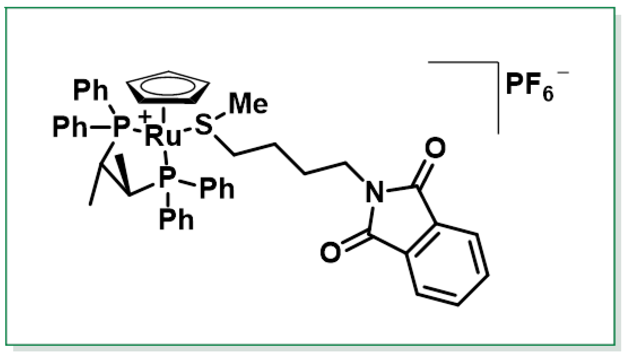
Another approach was described by Schenk et al., 1997, who developed an enantioselective synthesis of (R)-SFN with 80% ee. This method utilized a chiral ruthenium complex, specifically [CpRu((R,R)-CHIRAPHOS)]+, as a chiral catalyst. The process involved diastereoselective oxidation of a cationic ruthenium complex, providing an efficient route to the desired enantiomer [135], Figure 19.
The synthesis of SFN has been explored through various advanced methods, each aiming to achieve high yields, stereoselectivity and efficiency. One such method involves the oxygen transfer of dimethyldioxirane (DMD), which produces a sulfoxide complex with high throughput and excellent diastereoselectivity. In this approach, the cleavage of the phthaloyl group using aqueous hydrazine, followed by a reaction with thiophosgene, results in the formation of the SFN complex [CpRu(L–L)(MeS(O)C4H8NCS)]PF6. Finally, treatment of this complex with sodium iodide releases SFN without racemization, ensuring the retention of its enantiomeric purity [136,137].
These methods highlight the challenges and advancements in synthesizing enantiopure SFN. While microbial oxidation offers a biocatalytic approach, the use of chiral metal complexes enables precise control over stereochemistry. However, both methods have limitations in terms of yield and scalability, which may impact their practical application for large-scale production.
Further research into novel synthetic strategies, such as the development of sulfinate ester intermediates or DAG (diacetone-d-glucofuranose) methodologies, could provide more efficient and versatile routes for producing SFN and its analogues. These approaches aim to improve yields, enantiopurity and scalability, making them promising candidates for future exploration in SFN synthesis [138].
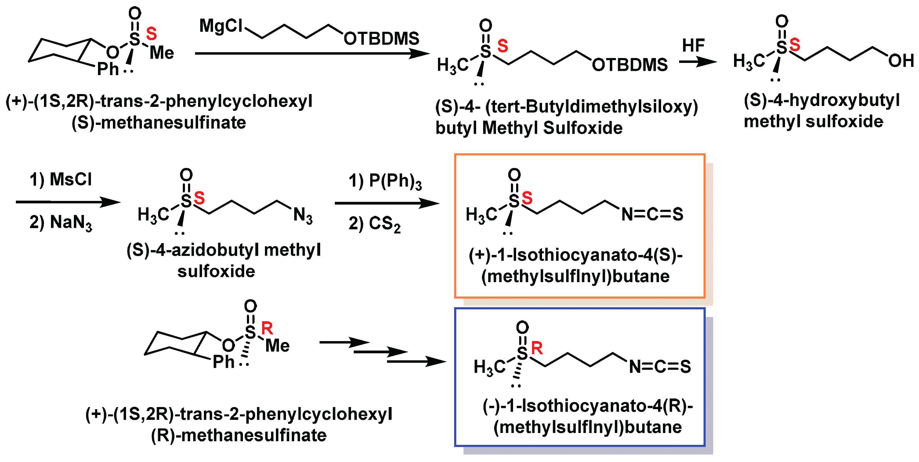
Another innovative method was developed by Whitesell et al., 1994, who used trans-2-phenylhexanol as a chiral auxiliary. The reaction of this chiral auxiliary with thionyl chloride produced an equimolar mixture of two diastereomeric chlorosulfite esters. Subsequent treatment of this mixture with dimethylzinc yielded both chlorosulfite esters with a favorable diastereomeric ratio and high chemical yields [139]. This process demonstrates the utility of chiral auxiliaries in achieving precise stereochemical control during SFN synthesis (Figure 20).
These methodologies highlight the advancements in synthesizing SFN with high stereoselectivity and chemical efficiency. The use of DMD for oxygen transfer ensures high diastereoselectivity, while Whitesell’s approach leverages chiral auxiliaries to optimize enantiomeric outcomes. Both methods contribute to the growing repertoire of techniques for producing SFN and its analogues, paving the way for their potential applications in pharmaceuticals and nutraceuticals.The method described was used to synthesize both enantiomers of SFN. The process for synthesizing (S)-SFN involves the following steps:
  • Reaction of (+)-(1S,2R)-trans-2-phenylcyclohexyl(S)-methanesulfinate with a Grignard reagent derived from 4-chlorobutyl tert-butyldimethylsilyl ether.
  • Removal of the tert-butyldimethylsiloxy protective group to yield (S)-4-hydroxybutyl methyl sulfoxide.
  • Conversion of the alcohol to a mesylate.
  • Reaction with sodium azide to form (S)-4-azidobutyl methylsulfoxide.
  • Reaction of the azide with triphenylphosphine to form an iminophosphorane.
  • Reaction of the iminophosphorane with carbon disulfide to produce enantiomerically pure (S)-SFN.
The same procedure was applied to the diastereoisomer (+)-(1S,2R)-trans-2-phenylcyclohexyl (R)-methanesulfinate to synthesize (R)-SFN. This enantio-divergent approach allows for the synthesis of both enantiomers of SFN using a single chiral auxiliary, demonstrating the versatility of the method. The process is illustrated in Figure 21.
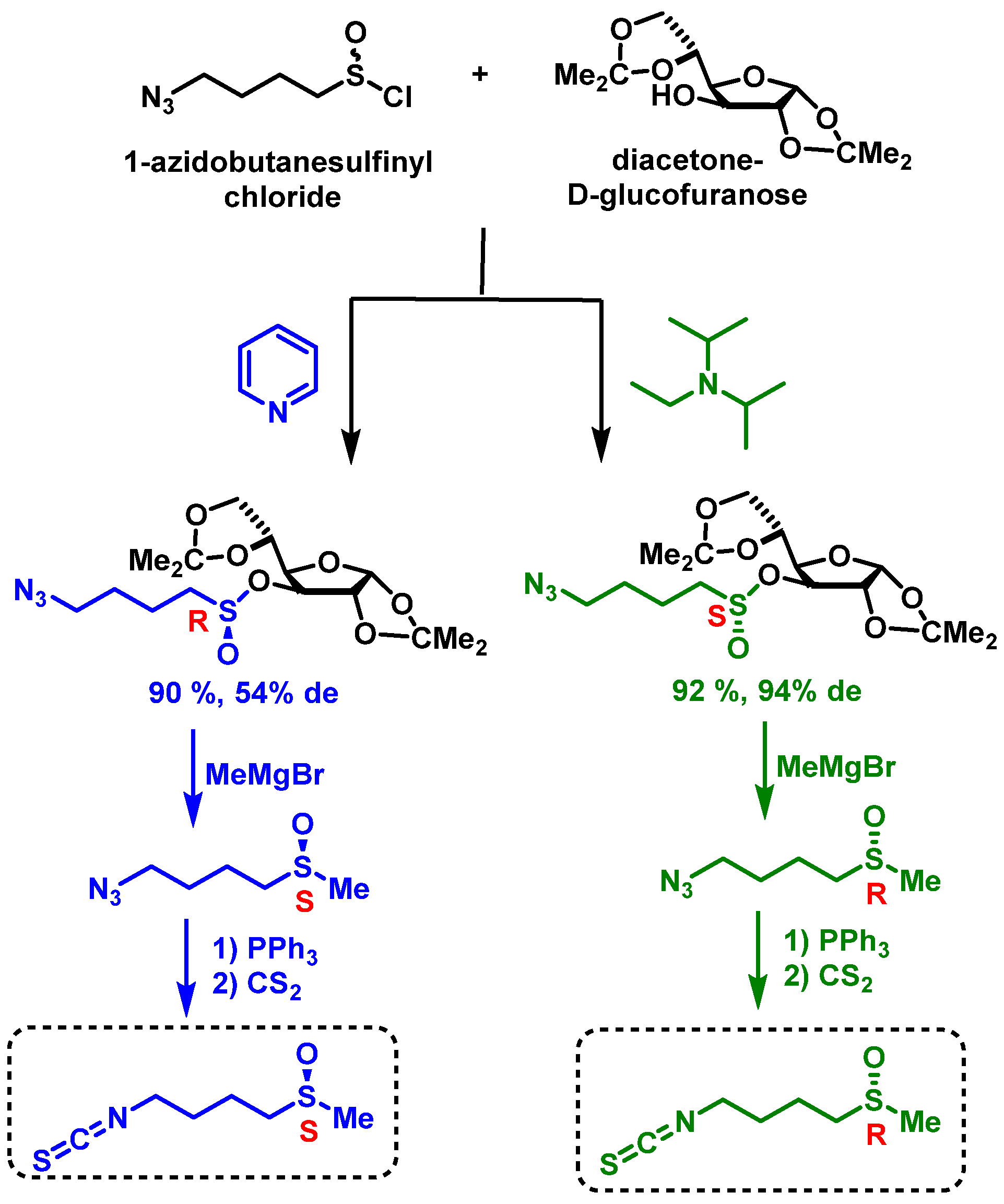
The synthetic route provides a valuable method for producing both enantiomers of SFN, which is crucial for studying the biological activities of each isomer separately. Such enantioselective syntheses are important for developing potential therapeutic applications of SFN and its analogues. Khiar et al. developed an efficient method for synthesizing both enantiomers of SFN using diacetone-D-glucofuranose (DAG) as a chiral auxiliary. This approach, known as the “DAG methodology”, enables the stereoselective synthesis of both SFN enantiomers with high yields and enantiopurity. The process involves several key steps [140], Figure 22.
First, azidoalkanesulfinate chlorides react with DAG under different conditions to produce either (S)- or (R)-sulfinate esters. Using DIPEA as a base yields (S)-sulfinate esters with high yield and good diastereomeric excess, while pyridine as a base produces (R)-sulfinate esters with high yield but lower diastereomeric excess. The Grignard reaction is then employed to replace the chiral auxiliary in sulfinates, inverting the configuration at the sulfinyl sulfur. Finally, a Staudinger/aza-Wittig tandem reaction involving triphenylphosphine and carbon disulfide generates either R or S enantiopure SFN.
A notable feature of this method is its enantiodivergent nature. By simply changing the base used to catalyze the reaction, both epimers at sulfur become accessible through a dynamic kinetic transformation of the starting sulfinyl chloride. This flexibility makes the DAG methodology particularly versatile for producing SFN enantiomers [141].
The DAG methodology offers several advantages. It provides high yields and enantiopurity for both SFN enantiomers, allows for the synthesis of various sulfoxide stereoisomers, and can be adapted to produce SFN homologues and analogues. These features make it a valuable tool for researchers studying the biological activities of SFN and its derivatives or developing potential therapeutic applications [142,143].
This synthetic approach represents a significant advancement in SFN synthesis, offering researchers a powerful method for exploring its diverse biological functions and therapeutic potential.

3.3.3. HPLC Separation of Sulforaphane Enantiomers in Broccoli

Enantioselective high-performance liquid chromatography (HPLC) has become the method of choice for analyzing natural and synthetic chiral compounds like SFN. This direct method employs chiral stationary phases to separate and quantify the R and S enantiomers of SFN [144].
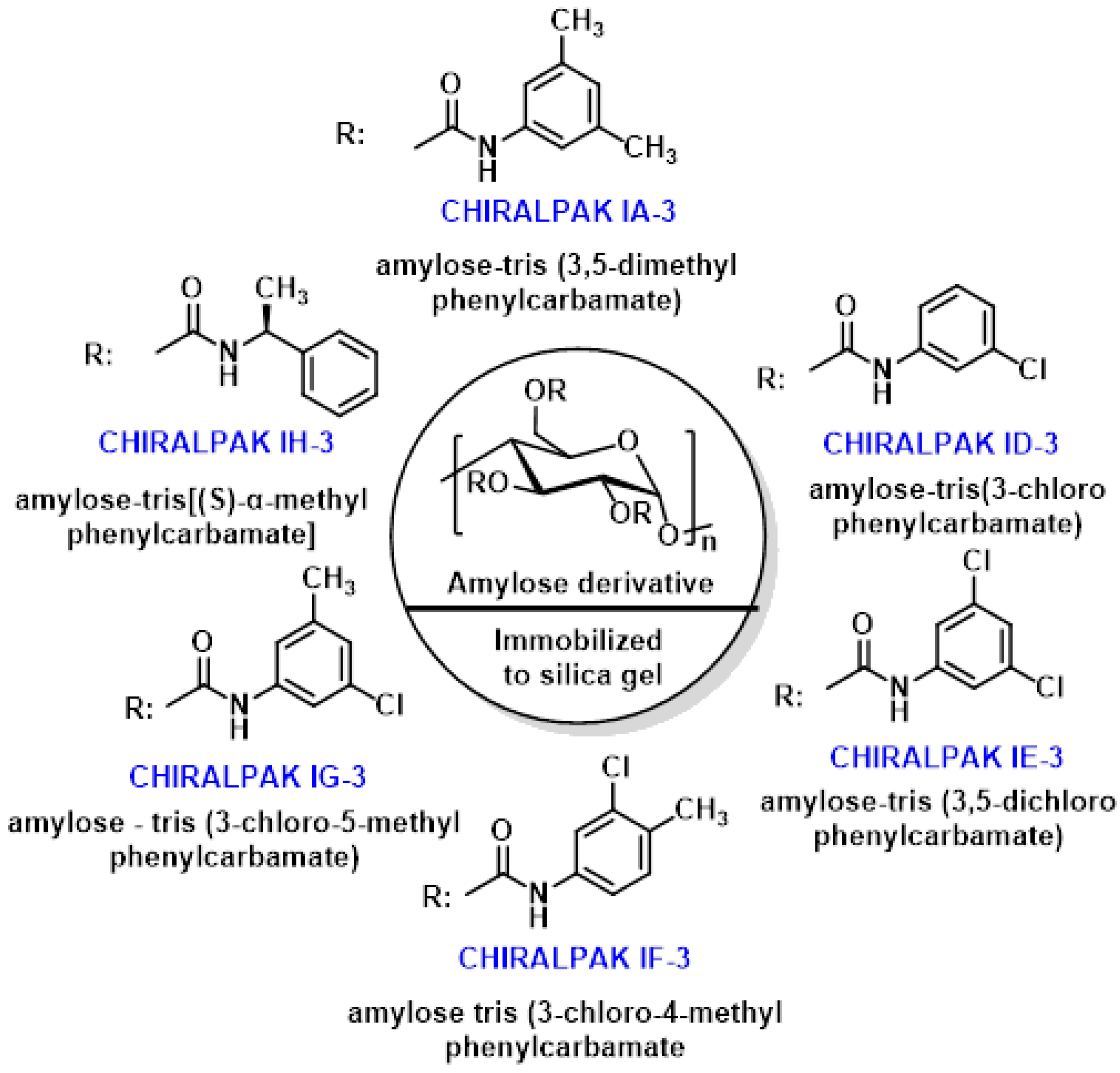
Recent studies have demonstrated HPLC’s effectiveness in resolving racemic SFN. Mammone et al., 2024 [144], used the Chiralpak IH-3 chiral column containing amylose tris-[(S)-methylbenzylcarbamate] as the chiral selector. Using various mobile phases of pure alcoholic solvents and hydroalcoholic mixtures, they observed that (R)-SFN elutes before its S enantiomer. Panusa et al., 2020, investigated five immobilized-type amylose-based chiral stationary phases: Chiralpak IA-3, ID-3, IE-3, IF-3, and IG-3. With these columns, the elution order was reversed, with the S enantiomer eluting before the R enantiomer [145].
The varying chiral resolution capabilities of these chiral stationary phases (CSPs) are attributed to the different substitution patterns in the aromatic ring of the amylose backbone (Figure 23).
These studies underscore the importance of selecting appropriate chiral columns and mobile phase conditions for accurate separation and quantification of SFN enantiomers. The ability to distinguish between R and S forms is crucial, as the R enantiomer is generally considered more biologically active.
The choice of chiral stationary phase significantly impacts the separation and elution order of SFN enantiomers. Researchers must carefully consider these factors when developing HPLC methods for SFN analysis to ensure accurate results and reliable quantification of the biologically active R enantiomer [146].
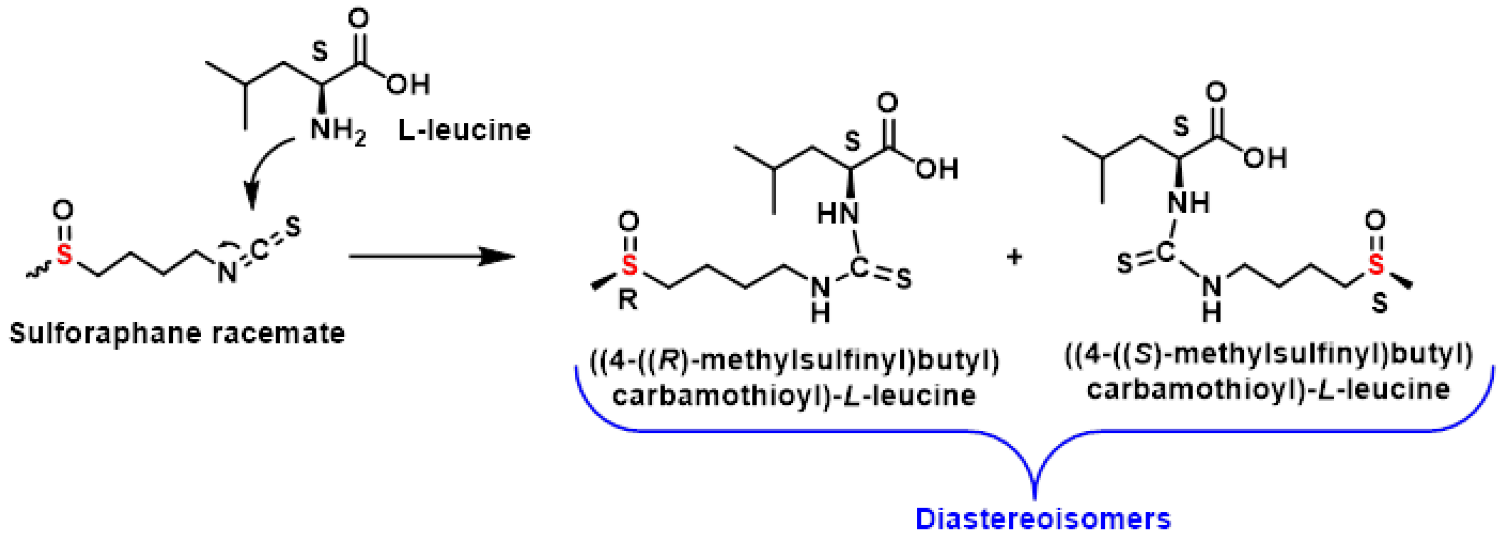
An alternative approach to separating SFN enantiomers involves an indirect method based on the formation of diastereomers. This technique relies on reacting the SFN enantiomers with a chiral derivatization reagent, creating diastereomers that can be separated using chromatography with achiral stationary phases [129].
Okada et al., 2017, demonstrated this method by derivatizing racemic SFN with (S)-leucine. The resulting diastereomers were successfully resolved using reversed-phase high-performance liquid chromatography (HPLC) with UV detection. This approach provides a practical solution for determining the proportion of R and S enantiomers in SFN samples [129], Figure 24.
The indirect method offers several advantages over direct enantioselective techniques, such as chiral stationary phase-based HPLC. By forming diastereomers, this approach enables the use of standard achiral columns, which are often more accessible and cost-effective.
Additionally, it allows for precise quantification of trace amounts of one enantiomer in the presence of the other, making it particularly useful for studying enantiomeric purity. This method was used by Okada et al., 2017, who derived racemic SFN with (S)-leucine, and was resolved by reverse-phase HPLC with UV detection [129] (Figure 22).
This method highlights the versatility of chiral derivatization reagents in resolving racemic mixtures and underscores their importance in the analysis of SFN and other chiral compounds. As research continues to explore both direct and indirect methods, these techniques will remain essential tools for understanding the stereochemistry and biological activity of SFN enantiomers.
High-performance liquid chromatography (HPLC) represents the most popular and widely applicable technology in the field of chiral analysis of various chiral mixtures. Indirect separations are based on the formation of diastereoisomeric complexes between the enantiomers and suitable chiral derivatizing agents, and their subsequent separation by an achiral liquid chromatographic method. This method is not very practical, as derivatization represents an additional step that can lead to undesirable side reactions, the formation of decomposition products and racemization. In addition, the chiral derivatization reagent must be of high enantiomeric purity and the presence of derivatizable groups in the analyte is a prerequisite, and finally, once the diastereomers have been separated, the derivatization agent must be removed.

3.3.4. Importance of Sulfur Chirality in the Biological Activity of SFN

Naturally occurring SFN exists exclusively as the R-enantiomer in terms of its sulfoxide sulfur configuration. However, most studies investigating its anti-inflammatory and antioxidant activities have been conducted using racemic mixtures. This discrepancy has led to evolving insights into the importance of SFN’s stereochemistry.
Initial findings suggested that sulfur chirality had no significant impact on SFN’s biological activity. However, recent discoveries have revealed important differences between the R and S enantiomers. In terms of enzyme induction, natural (R)-SFN has been shown to be a more potent inducer of carcinogen-detoxifying enzyme systems in rat liver and lung compared to the (S)-isomer [130].
Furthermore, (R)-SFN demonstrated superior ability in regulating two key enzyme systems involved in carcinogen metabolism. It increased microsomal epoxide hydrolase levels more effectively than the (S)-isomer and enhanced glucuronosyl transferase activity, while the (S)-enantiomer impaired it [147].
These findings highlight the importance of stereochemistry in SFN’s biological activities and suggest that the natural R-enantiomer may offer superior chemopreventive and detoxifying properties compared to its S counterpart or racemic mixtures. This understanding underscores the need for further research focusing on the specific biological activities of the R-enantiomer, which more closely resembles the naturally occurring form of SFN in broccoli and other cruciferous vegetables [147].
The evolving knowledge about SFN’s stereochemistry and its impact on biological activity has important implications for future research and potential therapeutic applications. It suggests that studies and interventions using pure (R)-SFN may yield more accurate and potentially more potent results than those using racemic mixtures. This insight could guide the development of more effective SFN-based therapies and dietary interventions [98].

4. Indole-3-Carbinol (I3C)

Glucosinolate glucobrassicin, found in broccoli, is metabolized by the enzyme myrosinase into its corresponding isothiocyanate. This isothiocyanate undergoes thiocyanate removal, forming the intermediate 3-methylene-3H-indoleum. The intermediate then reacts with water to produce 3-methylene-3-carbinol, a phytochemical with a wide range of biological activities [59], Figure 25.
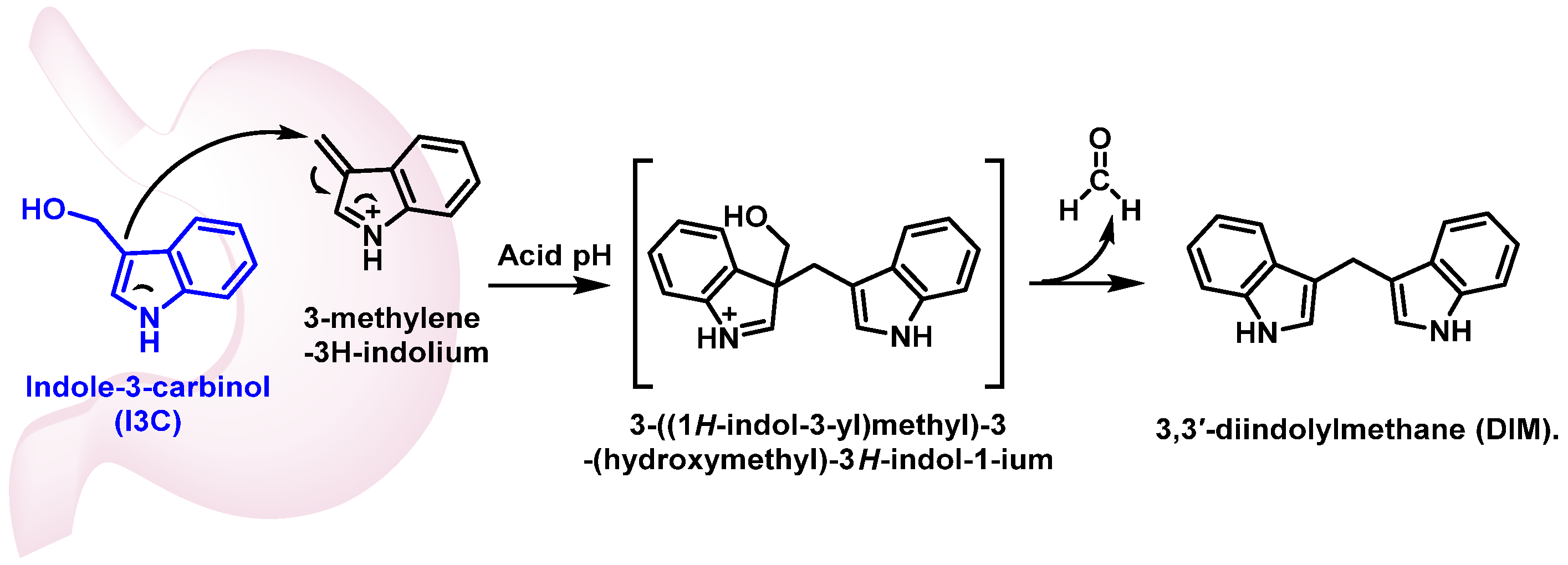
3-Methylene-3-carbinol exhibits cardioprotective, antioxidant, anti-inflammatory, antiangiogenic, and antimicrobial properties. Additionally, it promotes apoptosis in tumor cells, making it a compound of interest for potential therapeutic applications. These diverse effects highlight the importance of glucobrassicin-derived metabolites in health-promoting and disease-preventing mechanisms [148,149]. In vivo, 20 to 40% of this carbinol is transformed into 3,3′-diindolylmethane (DIM) by condensation in an acidic medium [150] (Figure 26).
Indole-3-carbinol (I3C) and its derivative diindolylmethane (DIM) exhibit anti-inflammatory properties through multiple mechanisms:
  • Nrf2 activation: I3C positively stimulates the transcription factor Nrf2, which plays a crucial role in cellular defense against oxidative stress. Activation of the Nrf2-ARE signaling pathway by I3C and DIM leads to increased expression of cytoprotective genes, reducing inflammation and oxidative damage [151].
  • NF-κB pathway inhibition: DIM’s main target is the NF-κB signaling pathway. By inhibiting this pathway, DIM decreases the production of proinflammatory cytokines such as TNF-α and IL-6, as well as prostaglandins. This results in a reduced inflammatory response [152].
  • Modulation of inflammatory mediators: I3C treatment has been shown to decrease the expression of pro-inflammatory factors like IL-1β and IL-6 while increasing anti-inflammatory factors such as IL-4 and IL-10. This modulation of inflammatory mediators contributes to the overall anti-inflammatory effect [153].
  • Regulation of microglia: I3C has been found to reduce the number of activated microglia and increase the number of M2-type microglia, which have anti-inflammatory properties. This regulation of microglia populations further contributes to the anti-inflammatory effects of I3C [154].
  • Cancer chemoprevention: both I3C and DIM have been extensively studied for their chemopreventive properties against various types of cancer [155].
Their mechanisms of action include modulation of aryl hydrocarbon receptor (AHR) signaling, impact on estrogen receptor (ER)-dependent signaling in breast cancer cells, antagonism of the androgen receptor in prostate cancer, epigenetic regulation of gene expression and modulation of DNA methylation and histone modifications [156].
These diverse mechanisms highlight the potential of I3C and DIM as promising compounds for reducing inflammation and preventing cancer development.

5. Other Compounds in Broccoli

5.1. Polyphenols

Phenolic compounds are a major class of secondary metabolites in Brassica vegetables, including broccoli, where they play critical physiological roles. These compounds contribute to plant growth and reproduction and serve as a defense mechanism against pathogens, predators and ultraviolet radiation. Their high antioxidant capacity is attributed to their ability to act as hydrogen and electron donors, which enables them to neutralize reactive oxygen species (ROS). Additionally, phenolic compounds function as metal ion chelators, inhibiting the Fenton reaction and thereby reducing oxidative stress [157].
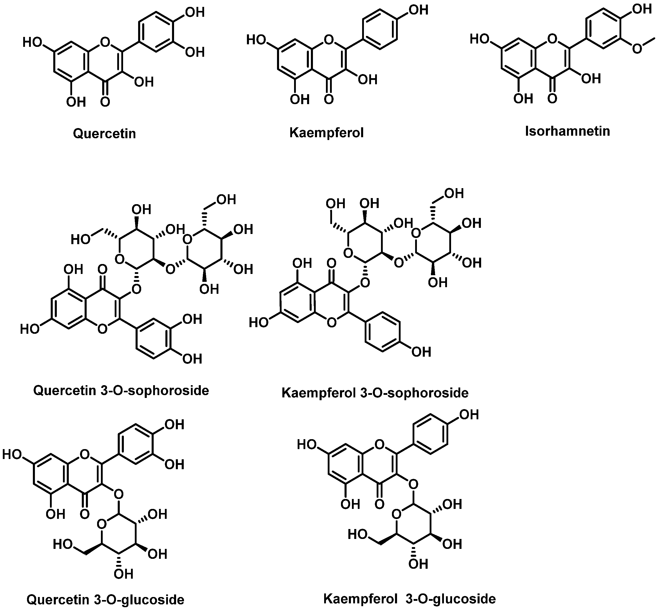
The flavonoid composition of broccoli inflorescences has been extensively studied using advanced techniques such as high-performance liquid chromatography coupled with mass spectrometry (LC/UV-DAD/ESI-MS). Broccoli contains derivatives of flavonols like quercetin, kaempferol, and, to a lesser extent, isorhamnetin. These flavonols are typically conjugated with sugars and organic acids, with glycosylation most frequently occurring at position 3 of the C-ring. However, substitutions can also occur at positions 5, 7, 3′, 4′, and 5′. The sugars involved in glycosylation are usually glucose or rhamnose, most commonly attached to position C3 and less frequently to C7 of ring A [158,159].
In Brassica crops, the most abundant glycosylated flavonols are quercetin 3-O-sophoroside and kaempferol 3-O-sophoroside. Other derivatives such as quercetin 3-O-glucoside and kaempferol 3-O-glucoside have also been identified. These compounds contribute significantly to the antioxidant properties of broccoli and other Brassica vegetables. Their ability to mitigate oxidative stress highlights their potential health benefits for humans while also playing crucial roles in the plant’s defense mechanisms [28,160] (Figure 27).
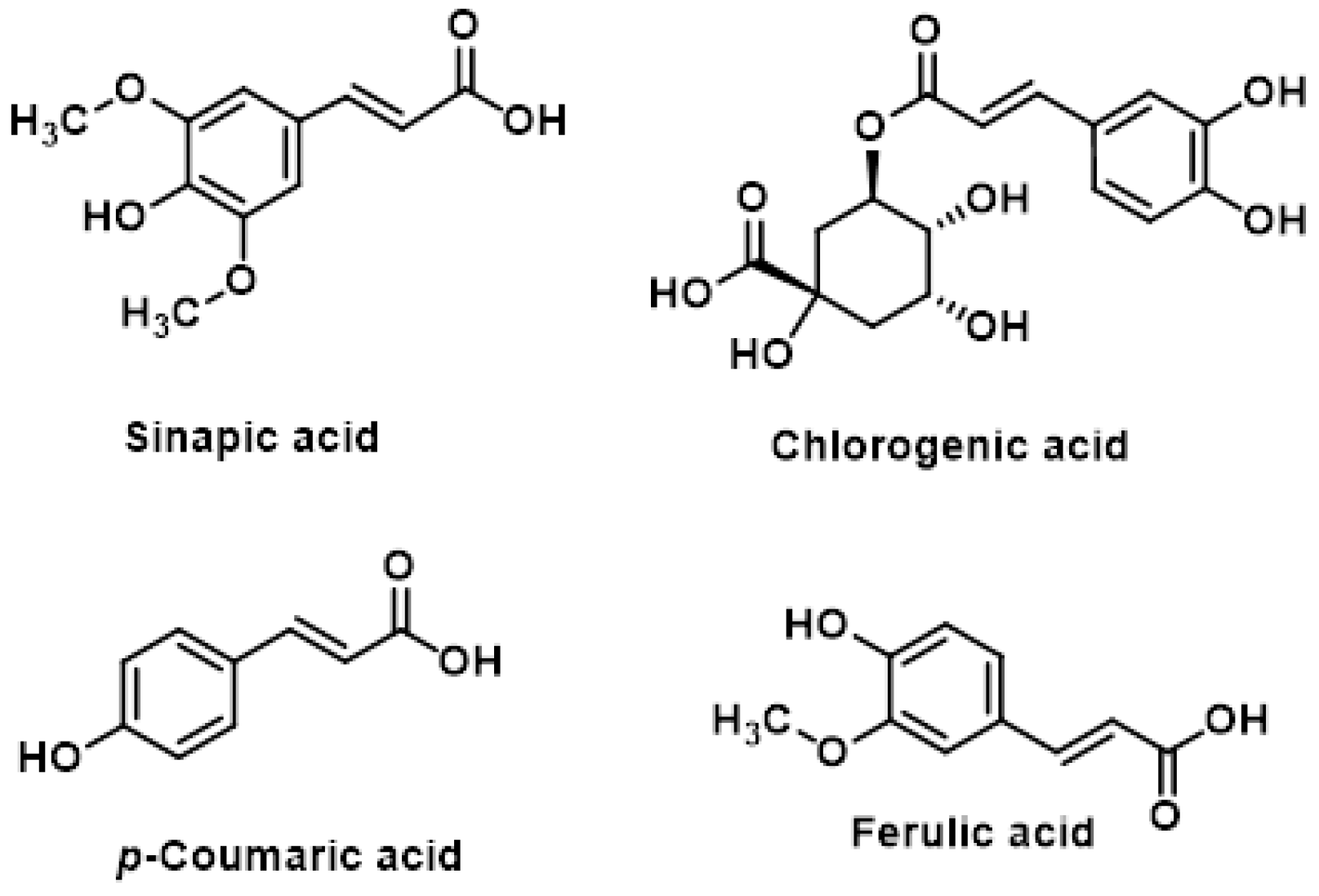
Hydroxycinnamic acids are a class of non-flavonoid phenolic compounds found in broccoli. These compounds and their derivatives include sinapic acid, chlorogenic acid, p-coumaric acid and ferulic acid [161] (Figure 28).
Recent studies have demonstrated that hydroxycinnamic acids and their derivatives exhibit anti-metastatic properties, particularly in inhibiting the spread of cancer cells to lungs and bones [162].
The anti-metastatic effects of hydroxycinnamic acids have been observed in various studies. p-hydroxycinnamic acid (HCA) has shown anticancer effects on MDA-MB-231 human breast cancer bone metastatic cells in vitro. HCA was found to suppress cancer cell proliferation, induce apoptosis in confluent cancer cells and prevent the negative effects of cancer cells on osteoblastogenesis and osteoclastogenesis. HCA’s anticancer effects are mediated through multiple signaling pathways, including NF-κB, extracellular signal-regulated kinase (ERK), protein kinase C, calcium signaling, phosphatidylinositol 3-kinase (PI3K) and nuclear transcription activity [163,164]. The apoptotic effects of HCA on cancer cells were found to be caspase-3 dependent, suggesting a mechanism involving the activation of this key apoptotic enzyme.
HCAs have demonstrated antioxidant properties, which may contribute to their anti-cancer effects. For example, quercetin 3-O-sophoroside, a flavonol glycoside found in broccoli, was shown to be a potent inhibitor of lipid peroxidation. Recent in silico studies have explored the potential of 4-hydroxycinnamic acid, found in broccoli, to interact with estrogen receptor beta protein, suggesting a possible role in hormone-related therapies [165].
These findings highlight the potential of hydroxycinnamic acids and their derivatives as promising candidates for the prevention and treatment of cancer metastasis, particularly in breast cancer bone metastasis [166].

5.2. Carotenoids

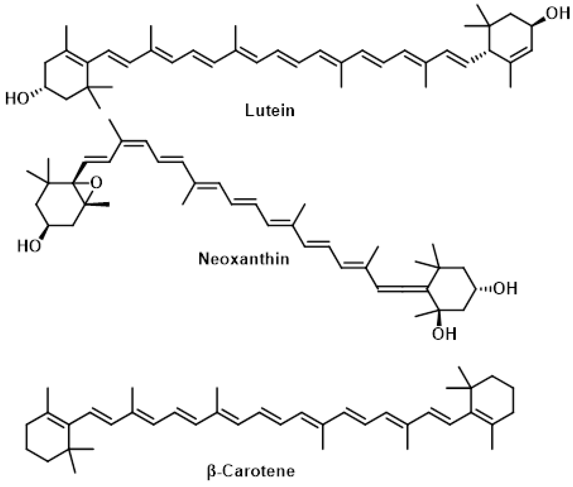
Carotenoids are tetraterpene fat-soluble pigments composed of 40 carbon atoms arranged in conjugated polyenic chains. Humans cannot synthesize carotenoids, so they must be obtained through diet or supplements. The major carotenoids found in the genus Brassica, including broccoli, are β-carotene, lutein, and neoxanthin. Broccoli sprouts can contain up to 1.16 mg of carotenoids per 100 g of fresh weight [167]. These compounds act as potent antioxidants, helping to protect cells from damage caused by free radicals, particularly in tissues such as the eyes and skin [168]. Carotenoids exhibit several important functions:
  • In eye health, lutein and zeaxanthin form the macular pigment in the retina, protecting photoreceptor cells from oxidative stress caused by sunlight exposure. This may help reduce the risk of age-related macular degeneration [169].
  • In skin protection, dietary carotenoids accumulate in the skin and offer measurable photo-protective benefits against UV-induced damage. Carotenoids can quench singlet oxygen and scavenge toxic free radicals, preventing or reducing oxidative stress [170].
  • Some carotenoids, like β-carotene, serve as precursors to vitamin A, which is essential for various biological processes, including vision [171].
Recent research has shown that the antioxidant properties of carotenoids can vary depending on oxygen concentration, potentially switching from antioxidant to pro-oxidant behavior. This highlights the complexity of carotenoid function in the body and the need for further research to fully understand their health benefits (Figure 29) [172].
Carotenoids, including lutein, neoxanthin and β-carotene, are important compounds found in broccoli that exhibit lipophilic antioxidant and immunomodulatory activities. Their biological effects stem from the conjugated double bonds in their long polyene chain, which enable them to inhibit reactive oxygen species and reduce oxidative damage [173]. The distribution of carotenoids varies across different parts of the broccoli plant [174]:
  • Florets and leaves, that contain high levels of lutein, neoxanthin, and β-carotene.
  • Stems, that contain lutein and neoxanthin, but lack β-carotene.
Lutein is particularly abundant in broccoli, and its concentration can increase when various cooking methods are applied, compared to fresh broccoli. This suggests that certain cooking techniques may enhance the bioavailability of some carotenoids. The carotenoid content in broccoli contributes to its health benefits due to antioxidant activity, neutralizing harmful free radicals and reduce oxidative stress. Lutein is important for eye health and may help prevent age-related macular degeneration [175].
Interestingly, broccoli leaves, which are often discarded, contain the highest concentrations of carotenoids, especially β-carotene. This finding suggests potential applications for broccoli by-products in enhancing the nutritional value of food products or as sources of bioactive compounds [176].

5.3. Tocopherols

In broccoli, tocopherols, which are forms of vitamin E, play an important role as lipid-soluble antioxidants that protect cell membranes from oxidative damage. Among the tocopherols present in broccoli, α-tocopherol is the most abundant, accounting for 78.5% of the total tocopherol content. This makes α-tocopherol the primary contributor to broccoli’s vitamin E activity, as it is the most biologically active form of tocopherol in humans and is essential for maintaining cellular health and preventing oxidative stress [177].
The remaining tocopherols are present in smaller proportions. γ-tocopherol constitutes 19.3% of the total tocopherols and, while less biologically active than α-tocopherol, it has unique antioxidant properties, particularly in scavenging reactive nitrogen species. δ-tocopherol, which accounts for 2.2% of the total tocopherols in broccoli, also contributes to antioxidant defense but is present in much lower concentrations [178].
The predominance of α-tocopherol in broccoli highlights its potential role as a dietary source of vitamin E, which is crucial for protecting lipids, proteins, and DNA from oxidative damage. The presence of γ- and δ-tocopherol adds to the diversity of antioxidant compounds in broccoli, further enhancing its health-promoting properties. The balance of these tocopherols may vary depending on factors such as broccoli variety, growing conditions, and post-harvest handling, but their combined presence underscores broccoli’s value as a nutrient-dense vegetable with significant antioxidant capacity [178].

5.4. Vitamin C

Broccoli stands out as an excellent source of vitamin C, boasting approximately 100 mg per 100 g fresh weight. This impressive content significantly surpasses that of other cruciferous vegetables like cabbage or cauliflower, which contain between 35 and 68 mg per 100 g fresh weight [15].
The vitamin C in broccoli serves as a potent antioxidant, effectively scavenging free radicals and shielding cells from oxidative damage. It also plays a crucial role in regenerating other antioxidants in the body, such as vitamin E [179,180,181,182,183]. Notably, the antioxidant activity of vitamin C is dose-dependent, with higher doses exhibiting more pronounced antioxidant effects [184].
Several factors influence the vitamin C content in broccoli. Genetic variability can lead to up to 71% variation in vitamin C levels among different broccoli varieties. Environmental factors, such as sunlight exposure also impact vitamin C content. Additionally, harvesting and storage processes can affect the final vitamin C levels in the vegetable [38].
Cooking methods significantly influence the retention of vitamin C in broccoli. Steaming emerges as the best method for preserving vitamin C, while boiling and stir-frying can result in substantial losses. Despite these potential losses during cooking, broccoli remains an excellent source of vitamin C, providing up to 101% of the daily recommended value in a 100 g serving [185].
In conclusion, broccoli’s high vitamin C content, coupled with its resilience to some degree of processing, makes it a valuable addition to a healthy diet, offering substantial antioxidant benefits even after cooking.

5.5. Phytosterols

Phytosterols, natural compounds found in plants, exhibit anti-inflammatory properties and have been associated with various health benefits, particularly in cardiovascular health. These compounds have been linked to reduced risks of cardiovascular disease and heart attacks, as well as helping maintain healthy blood cholesterol levels [186].
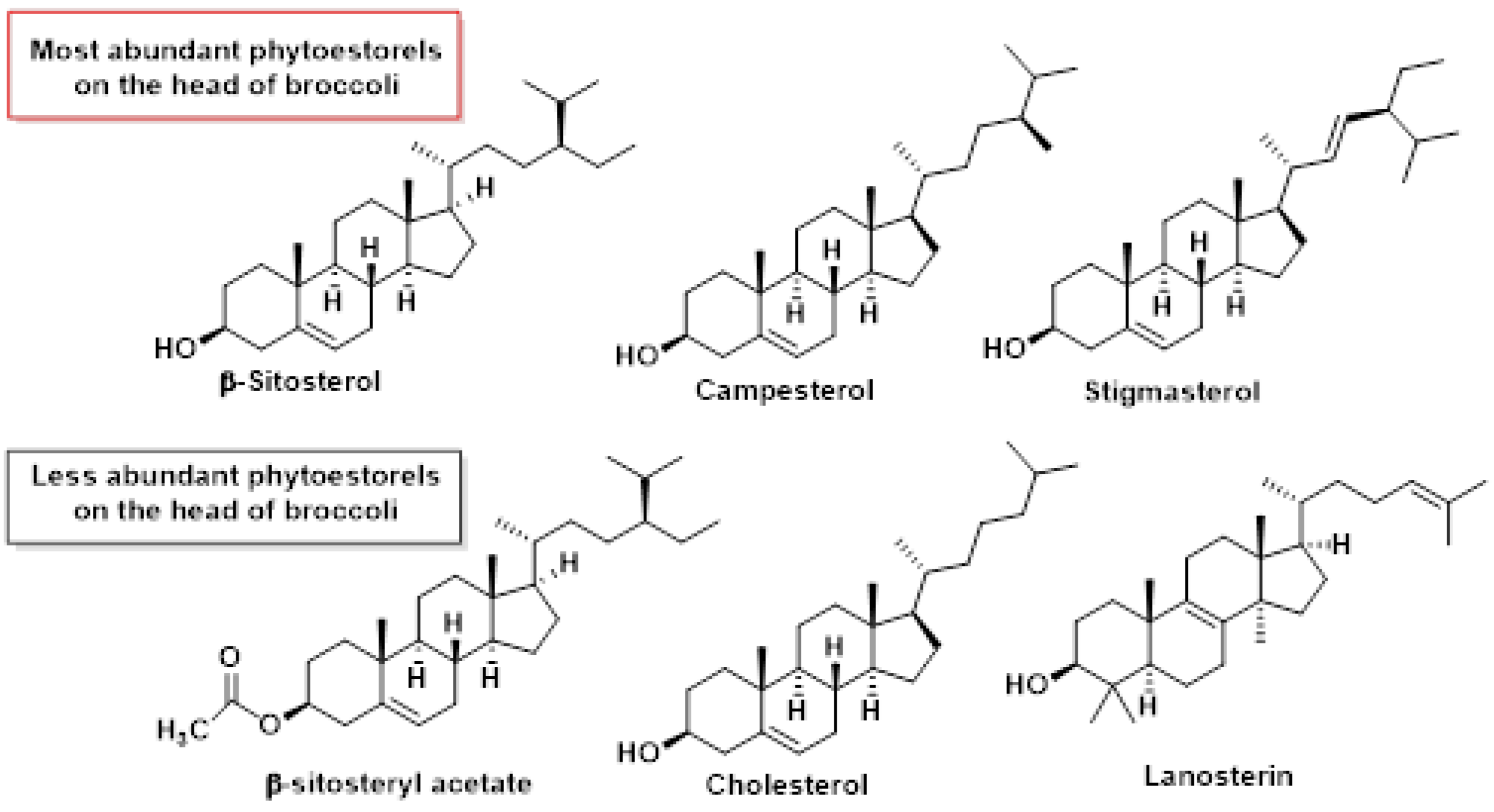
A study conducted by Shi et al., 2024, investigated the composition of bioactive compounds in broccoli heads, considering different organ sizes and growing seasons. Their research identified six phytosterols present in broccoli heads: beta-sitosterol, stigmasterol, campesterol, beta-sitosterol acetate, lanosterol, and cholesterol [187].
Among these phytosterols, beta-sitosterol was found to be the most abundant, with content ranging from 5.67 to 20.78 mg per 100 g of broccoli. Campesterol was the second most prevalent, with concentrations between 0.7 and 6.37 mg per 100 g, followed by stigmasterol, ranging from 0.48 to 2.38 mg per 100 g. Interestingly, beta-sitosterol acetate, lanosterol, and cholesterol were present in such small quantities that they were nearly undetectable in the broccoli heads [187] (Figure 30).
The presence of these phytosterols, particularly beta-sitosterol, contributes to broccoli’s potential health benefits. Phytosterols have been shown to help lower LDL cholesterol levels, with some studies suggesting that consuming 2 g of phytosterols daily may reduce LDL cholesterol by up to 10% [188].
Phytosterol content in broccoli can vary depending on factors such as growing conditions, plant part, and preparation methods. For instance, cooked broccoli may have different phytosterol concentrations compared to raw broccoli [187].
These findings highlight broccoli’s role as a valuable source of phytosterols in the diet, potentially contributing to its overall health-promoting properties. The presence of these beneficial compounds further underscores the importance of including broccoli in a balanced diet for maintaining cardiovascular health and managing cholesterol levels [189].

6. Antimicrobial Peptides

Antimicrobial peptides (AMPs) derived from broccoli (Brassica oleracea var. italica) have emerged as promising bioactive molecules, offering natural solutions to combat microbial infections and address various health challenges [190,191]. These small, proteinaceous molecules exhibit broad-spectrum activity against bacteria, fungi, and yeasts, making them valuable candidates for therapeutic applications [192].

6.1. Distribution and Sources of Broccoli AMPs

Broccoli contains a diverse array of AMPs distributed across its various parts, including seeds, stems, leaves, and fermented extracts. These peptides are integral to the plant’s defense system, protecting it from microbial attack while also offering significant potential for human health applications [193].
Recent studies have identified a range of peptides from different parts of broccoli, each with unique properties and mechanisms of action (Table 2).
The bioactivity of these peptides extends beyond antimicrobial effects to include anti-inflammatory, antioxidant, and enzyme inhibitory properties [15]. Pacheco-Cano et al. (2020) identified Class I defensins (BraDef) from broccoli seeds, which demonstrated potent antimicrobial activity. These small, cysteine-rich peptides play a crucial role in plant defense mechanisms and show promise for applications in human health [192].
Focusing on broccoli stems, Nicolas-Espinosa et al. (2022) explored bioactive peptides that enhance wound healing and keratinocyte proliferation. Their comprehensive study identified 1256 peptide sequences in protein extracts from broccoli stems, highlighting the rich diversity of bioactive compounds in this often-overlooked part of the plant [194]. Similarly, Chen et al. (2020) investigated peptides from broccoli stems and leaves, concentrating on their antioxidant and hypolipidemic properties. Their findings suggest potential applications in managing oxidative stress and lipid metabolism, expanding the therapeutic potential of broccoli-derived peptides beyond antimicrobial applications [193].
The antimicrobial activities of peptides isolated from cruciferous vegetables, including broccoli, were explored by Favela-González et al., emphasizing their potential as natural alternatives to synthetic antimicrobials [198]. This research underscores the growing interest in plant-derived AMPs as sustainable solutions to combat microbial resistance and promote human health. Furthermore, Li et al. (2023) identified novel anti-inflammatory peptides from broccoli fermented by Lactobacillus strains, employing virtual screening and molecular docking techniques [196]. This innovative approach to identifying bioactive peptides opens new avenues for discovering and developing broccoli-derived compounds with specific therapeutic properties.

6.2. Mechanisms of Action

The antimicrobial peptides (AMPs) derived from broccoli exhibit diverse mechanisms of action, targeting bacterial membranes, enzymes, oxidative stress pathways, and inflammatory mediators. These multifaceted approaches contribute to their effectiveness against a wide range of pathogens and their potential therapeutic applications.
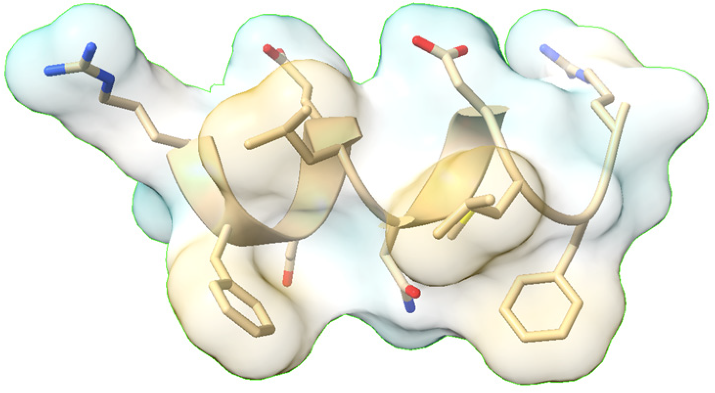
Membrane disruption is a primary mechanism employed by several broccoli-derived AMPs. Pacheco-Cano et al. (2020) identified that peptides like ARFEELNMDLFR, isolated from broccoli stems, create pores in bacterial cell membranes, leading to ion leakage and cell lysis [192]. This mechanism is particularly effective against gram-negative bacteria such as Escherichia coli. Additionally, SFN disrupts biofilm formation in pathogens like Helicobacter pylori, preventing microbial adhesion to host tissues and enhancing the overall antimicrobial effect [199].
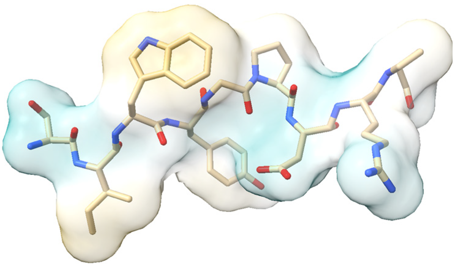
Enzyme inhibition represents another crucial mechanism through which broccoli-derived peptides exert their effects. Anti-inflammatory peptides like SIWYGPDRP bind to inducible nitric oxide synthase (iNOS), effectively reducing NO production in macrophages and contributing to their anti-inflammatory properties.
The anti-inflammatory effects of broccoli-derived AMPs extend beyond oxidative stress modulation. Li et al. (2023) isolated the peptide RFR from fermented broccoli, which demonstrated a remarkable 75% inhibition of TNF-α release [196]. This potent anti-inflammatory action suggests potential applications in managing chronic inflammatory diseases such as rheumatoid arthritis or inflammatory bowel disease.
Recent studies have also revealed that some AMPs can target intracellular components of pathogens. While specific examples from broccoli are yet to be fully elucidated, this mechanism involves AMPs binding to nucleic acids and proteins within microbial cells, impeding replication and other essential cellular processes [200]. This intracellular targeting adds another layer to the antimicrobial arsenal of broccoli-derived peptides.

6.3. Potential Applications

The antimicrobial peptides (AMPs) derived from broccoli hold transformative potential across multiple sectors, offering eco-friendly solutions to pressing global challenges. In the food industry, broccoli extracts are emerging as natural preservatives to combat foodborne pathogens. Studies demonstrate that peptides like ARFEELNMDLFR (Figure 31) inhibit Salmonella and Listeria monocytogenes, reducing spoilage and extending shelf life [194,195].
Pacheco-Cano et al. (2020) showed that broccoli seed defensins (BraDef) effectively suppress Bacillus cereus [192], a common contaminant in dairy and meat products. Innovators are now integrating antimicrobial peptides into edible films and packaging materials, creating “active packaging” that passively releases antimicrobial agents to protect perishable goods. This approach not only enhances food safety but also aligns with consumer demand for clean-label, synthetic-free products [201,202].
In agriculture, broccoli AMPs offer a sustainable alternative to chemical pesticides. Nicolas-Espinosa et al., 2022, highlighted that stem-derived peptides suppress phytopathogenic fungi such as Aspergillus niger and Colletotrichum gloeosporioides, which devastate crops like tomatoes and citrus [194]. Additionally, these peptides are biodegradable, minimizing environmental runoff and soil toxicity compared to conventional fungicides [203].
There has been a growing interest in the health benefits of plants, driven by their safety and versatility in developing pharmaceuticals and cosmetics [204]. The healthcare sector benefits from broccoli AMPs’ dual antimicrobial and regenerative properties. For example, peptides from fermented broccoli stems accelerate wound closure by stimulating keratinocyte migration, as shown in murine models where treated wounds healed 40% faster than controls [194]. SFN has shown promise against antibiotic-resistant pathogens like MRSA and Helicobacter pylori, the latter being a major cause of gastric ulcers [205]. Li et al. (2023) identified anti-inflammatory peptides such as SIWYGPDRP (Figure 32), which inhibits nitric oxide (NO) production by 52%, offering potential therapies for chronic inflammation and autoimmune disorders [196].
Beyond these fields, cosmeceuticals are leveraging broccoli peptides for skincare. Peptide mixtures from stems enhance collagen synthesis and protect against UV-induced oxidative stress, making them ideal for anti-aging formulations. Chen et al. (2020) further reported hypolipidemic effects in broccoli leaf peptides, suggesting applications in functional foods targeting metabolic syndrome [193]. Broccoli extract is also a potential anti-photoaging agent [206]. As research in this field progresses, a deeper understanding of these mechanisms will likely lead to novel therapeutic strategies and applications in both human health and agriculture.

7. Use of Broccoli By-Products as a Source of Bioactive Products

An important trend in the future will be to consider the revaluation of broccoli by-products. Only a small proportion of broccoli is used in food, the remaining stems, leaves, roots and inflorescences are discarded, which leads to an increase in broccoli by-products that are mostly used for composting or incorporated into the soil [207]. These by-products can also be rich in bioactive compounds and present beneficial properties [208,209,210]. Current research on the functionality of broccoli by-products, such as leaves and stems, has provided valuable information on the use of these waste materials as new ingredients for the food, cosmetic and/or pharmaceutical industry.
As ingredients in functional foods [176], to improve the nutritional properties of foods [211] or as natural preservatives to prolong their shelf life, it is of great importance to consider the best use of plant foods at a time when the hectares of arable land are decreasing and the world’s population is increasing [212].
In the pharmaceutical industry, these by-products offer considerable health benefits due to their richness in bioactive compounds, similar to the edible parts. Studies have shown that by-products such as stems and leaves contain high levels of phenolic compounds, glucosinolates, vitamins and fiber [213,214,215].
In the cosmetics industry, consumers are more inclined to choose products of natural origin, so the use of by-products from broccoli by-products would be a great source of bioactive compounds for these products. This is why more and more people are looking to incorporate these elements into the development of new cosmetic products and the cosmetic industry may represent a cost-effective solution on how to recycle disposable by-products [216].

8. Conclusions

Broccoli, a widely consumed vegetable from the Brassica genus, has been extensively studied for its health benefits, with new advantages continually being discovered. The nutritional and health-promoting properties of broccoli are largely attributed to its rich content of glucosinolates (GSLs), secondary metabolites abundant in nitrogen and sulfur. GSLs have demonstrated protective properties against various diseases, particularly cancer. These compounds contribute to improving human health through several mechanisms, such as anti-inflammatory effects, antioxidant activity (broccoli is rich in antioxidants, including vitamin C, carotenoids, and anthocyanins, which help combat oxidative stress), cardiovascular health and metabolic regulation of glucose and lipid metabolism, potentially reducing the risk of type 2 diabetes and metabolic syndrome. SFN, a key isothiocyanate derived from broccoli, has garnered significant attention for its health-promoting properties: anticancer activity against various types of cancer, including prostate, pancreatic, leukemia, and colon cancer; enhancing the body’s antioxidant defenses by activating the Nrf2 pathway and NF-κB inhibition. Despite the promising health benefits of SFN, several challenges and areas for future research remain: chemical instability, the clinical use of SFN has been hampered by its chemical instability, necessitating the development of more stable formulations. SFN has optical isomers due to the chirality of sulfur, clinical and toxicological studies are necessary to fully evaluate the effects of SFN enantiomers on different organs of the human body. Appropriate enantio-selective synthesis methods and environmentally friendly solvent HPLC enantio-separation methods are needed.
The antimicrobial peptides (AMPs) derived from broccoli exhibit diverse mechanisms of action, targeting bacterial membranes, enzymes, oxidative stress pathways, and inflammatory mediators. These multifaceted approaches contribute to their effectiveness against a wide range of pathogens and their potential therapeutic applications.
In conclusion, while broccoli and its bioactive compounds, particularly GSLs and SFN, show great promise in promoting human health and preventing chronic diseases, further research is needed to overcome the challenges associated with their stability, synthesis, and clinical application. Additionally, more comprehensive human trials are required to establish optimal consumption patterns and to fully elucidate the mechanisms by which these compounds exert their beneficial effects.

Author Contributions

Conceptualization, C.M.C.A. and C.A.J.; investigation, C.M.C.A. and C.A.J.; writing—review and editing, C.M.C.A., C.A.J., E.B.M., J.M.P.d.l.L. and E.P.-L.; supervision, C.M.C.A., C.A.J., E.B.M., J.M.P.d.l.L. and E.P.-L. All authors have read and agreed to the published version of the manuscript.

Funding

This research received no external funding.

Institutional Review Board Statement

Not applicable.

Informed Consent Statement

Not applicable.

Data Availability Statement

No new data were created or analyzed in this study. Data sharing is not applicable to this article.

Conflicts of Interest

The authors declare no conflict of interest. The funders had no role in the design of the study; in the collection, analyses, or interpretation of data; in the writing of the manuscript; or in the decision to publish the results.

References

  1. Li, H.; Xia, Y.; Liu, H.-Y.; Guo, H.; He, X.-Q.; Liu, Y.; Wu, D.-T.; Mai, Y.-H.; Li, H.-B.; Zou, L.; et al. Nutritional values, beneficial effects, and food applications of broccoli (Brassica oleracea var. italica Plenck). Trends Food Sci. Technol. 2022, 119, 288–308. [Google Scholar] [CrossRef]
  2. Liu, B.; Tao, Y.; Manickam, S.; Li, D.; Han, Y.; Yu, Y.; Liu, D. Influence of sequential exogenous pretreatment and contact ultrasound-assisted air drying on the metabolic pathway of glucoraphanin in broccoli florets. Ultrason. Sonochem. 2022, 84, 105977. [Google Scholar] [CrossRef] [PubMed]
  3. Zhong, X.; Dolan, K.D.; Almenar, E. Effect of steamable bag microwaving versus traditional cooking methods on nutritional preservation and physical properties of frozen vegetables: A case study on broccoli (Brassica oleracea). Innov. Food Sci. Emerg. Technol. 2015, 31, 116–122. [Google Scholar] [CrossRef]
  4. Quizhpe, J.; Ayuso, P.; Rosell, M.d.l.Á.; Peñalver, R.; Nieto, G. Brassica oleracea var italica and Their By-Products as Source of Bioactive Compounds and Food Applications in Bakery Products. Foods 2024, 13, 3513. [Google Scholar] [CrossRef]
  5. Jacques, P.F.; Lyass, A.; Massaro, J.M.; Vasan, R.S.; D’Agostino, R.B., Sr. Relationship of lycopene intake and consumption of tomato products to incident CVD. Br. J. Nutr. 2013, 110, 545–551. [Google Scholar] [CrossRef]
  6. Barber, T.M.; Kabisch, S.; Pfeiffer, A.F.H.; Weickert, M.O. The Health Benefits of Dietary Fibre. Nutrients 2020, 12, 3209. [Google Scholar] [CrossRef]
  7. Kim, J.S.; Cuong, D.M.; Bae, Y.B.; Cho, S.K. Antioxidant and antiproliferative activities of solvent fractions of broccoli (Brassica oleracea L.) sprout. Appl. Biol. Chem. 2022, 65, 34. [Google Scholar] [CrossRef]
  8. Wang, T.T.; Schoene, N.W.; Milner, J.A.; Kim, Y.S. Broccoli-derived phytochemicals indole-3-carbinol and 3,3′-diindolylmethane exerts concentration-dependent pleiotropic effects on prostate cancer cells: Comparison with other cancer preventive phytochemicals. Mol. Carcinog. 2012, 51, 244–256. [Google Scholar] [CrossRef]
  9. Kapusta-Duch, J.; Kopeć, A.; Piatkowska, E.; Borczak, B.; Leszczyńska, T. The beneficial effects of Brassica vegetables on human health. Rocz. Panstw. Zakl. Hig. 2012, 63, 389–395. [Google Scholar]
  10. Le, T.N.; Chiu, C.H.; Hsieh, P.C. Bioactive Compounds and Bioactivities of Brassica oleracea L. var. Italica Sprouts and Microgreens: An Updated Overview from a Nutraceutical Perspective. Plants 2020, 9, 946. [Google Scholar] [CrossRef]
  11. Tang, G.Y.; Meng, X.; Li, Y.; Zhao, C.N.; Liu, Q.; Li, H.B. Effects of Vegetables on Cardiovascular Diseases and Related Mechanisms. Nutrients 2017, 9, 857. [Google Scholar] [CrossRef] [PubMed]
  12. Rasmussen, H.M.; Johnson, E.J. Nutrients for the aging eye. Clin. Interv. Aging 2013, 8, 741–748. [Google Scholar] [CrossRef]
  13. Mahn, A.; Castillo, A. Potential of Sulforaphane as a Natural Immune System Enhancer: A Review. Molecules 2021, 26, 752. [Google Scholar] [CrossRef]
  14. Akbari, S.; Rasouli-Ghahroudi, A.A. Vitamin K and Bone Metabolism: A Review of the Latest Evidence in Preclinical Studies. Biomed. Res. Int. 2018, 2018, 4629383. [Google Scholar] [CrossRef] [PubMed]
  15. Syed, R.U.; Moni, S.S.; Break, M.K.B.; Khojali, W.M.A.; Jafar, M.; Alshammari, M.D.; Abdelsalam, K.; Taymour, S.; Alreshidi, K.S.M.; Elhassan Taha, M.M.; et al. Broccoli: A Multi-Faceted Vegetable for Health: An In-Depth Review of Its Nutritional Attributes, Antimicrobial Abilities, and Anti-inflammatory Properties. Antibiotics 2023, 12, 1157. [Google Scholar] [CrossRef]
  16. Kaczmarek, J.L.; Liu, X.; Charron, C.S.; Novotny, J.A.; Jeffery, E.H.; Seifried, H.E.; Ross, S.A.; Miller, M.J.; Swanson, K.S.; Holscher, H.D. Broccoli consumption affects the human gastrointestinal microbiota. J. Nutr. Biochem. 2019, 63, 27–34. [Google Scholar] [CrossRef] [PubMed]
  17. Guijarro-Real, C.; Fita, A.; Prohens, J.; Moreno, D.A. Conventional and Innovative Processing in the Stability of Glucosinolates. Nutraceutical Funct. Food Compon. 2022, 411–460. [Google Scholar] [CrossRef]
  18. Liu, M.; Zhang, L.; Ser, S.L.; Cumming, J.R.; Ku, K.M. Comparative Phytonutrient Analysis of Broccoli By-Products: The Potentials for Broccoli By-Product Utilization. Molecules 2018, 23, 900. [Google Scholar] [CrossRef]
  19. Lv, X.; Meng, G.; Li, W.; Fan, D.; Wang, X.; Espinoza-Pinochet, C.A.; Cespedes-Acuña, C.L. Sulforaphane and its antioxidative effects in broccoli seeds and sprouts of different cultivars. Food Chem. 2020, 316, 126216. [Google Scholar] [CrossRef]
  20. Ramirez, D.; Abellán-Victorio, A.; Beretta, V.; Camargo, A.; Moreno, D.A. Functional Ingredients From Brassicaceae Species: Overview and Perspectives. Int. J. Mol. Sci. 2020, 21, 1998. [Google Scholar] [CrossRef]
  21. Gudiño, I.; Casquete, R.; Martín, A.; Wu, Y.; Benito, M.J. Comprehensive Analysis of Bioactive Compounds, Functional Properties, and Applications of Broccoli By-Products. Foods 2024, 13, 3918. [Google Scholar] [CrossRef] [PubMed]
  22. Ares, A.M.; Nozal, M.J.; Bernal, J. Extraction, chemical characterization and biological activity determination of broccoli health promoting compounds. J. Chromatogr. A 2013, 1313, 78–95. [Google Scholar] [CrossRef]
  23. Santana-Gálvez, J.; Cisneros-Zevallos, L.; Jacobo-Velázquez, D.A. A practical guide for designing effective nutraceutical combinations in the form of foods, beverages, and dietary supplements against chronic degenerative diseases. Trends Food Sci. Technol. 2019, 88, 179–193. [Google Scholar] [CrossRef]
  24. Halkier, B.A.; Gershenzon, J. Biology and biochemistry of glucosinolates. Annu. Rev. Plant Biol. 2006, 57, 303–333. [Google Scholar] [CrossRef]
  25. Jaki, B.; Sticher, O.; Veit, M.; Fröhlich, R.; Pauli, G.F. Evaluation of glucoiberin reference material from Iberis amara by spectroscopic fingerprinting. J. Nat. Prod. 2002, 65, 517–522. [Google Scholar] [CrossRef] [PubMed]
  26. Waser, J.; Watson, W.H. Crystal structure of sinigrin. Nature 1963, 198, 1297–1298. [Google Scholar] [CrossRef]
  27. Sánchez-Pujante, P.J.; Borja-Martínez, M.; Pedreño, M.; Almagro, L. Biosynthesis and bioactivity of glucosinolates and their production in plant in vitro cultures. Planta 2017, 246, 19–32. [Google Scholar] [CrossRef]
  28. Moreno, D.A.; Carvajal, M.; López-Berenguer, C.; García-Viguera, C. Chemical and biological characterisation of nutraceutical compounds of broccoli. J. Pharm. Biomed. Anal. 2006, 41, 1508–1522. [Google Scholar] [CrossRef]
  29. Baldelli, S.; Lombardo, M.; D’Amato, A.; Karav, S.; Tripodi, G.; Aiello, G. Glucosinolates in Human Health: Metabolic Pathways, Bioavailability, and Potential in Chronic Disease Prevention. Foods 2025, 14, 912. [Google Scholar] [CrossRef]
  30. Uda, Y.; Kurata, T.; Arakawa, N. Effects of pH and ferrous ion on the degradation of glucosinolates by myrosinase. Agric. Biol. Chem. 1986, 50, 2735–2740. [Google Scholar]
  31. Rungapamestry, V.; Duncan, A.J.; Fuller, Z.; Ratcliffe, B. Changes in glucosinolate concentrations, myrosinase activity, and production of metabolites of glucosinolates in cabbage (Brassica oleracea Var. capitata) cooked for different durations. J. Agric. Food Chem. 2006, 54, 7628–7634. [Google Scholar] [CrossRef] [PubMed]
  32. Burow, M.; Losansky, A.; Müller, R.; Plock, A.; Kliebenstein, D.J.; Wittstock, U. The genetic basis of constitutive and herbivore-induced ESP-independent nitrile formation in Arabidopsis. Plant Physiol. 2009, 149, 561–574. [Google Scholar] [CrossRef]
  33. Wittstock, U.; Burow, M. Glucosinolate breakdown in Arabidopsis: Mechanism, regulation and biological significance. In The Arabidopsis Book; BioOne: Washington, DC, USA, 2010; Volume 2010, p. e0134. [Google Scholar] [CrossRef]
  34. Nguyen, V.P.T.; Stewart, J.; Lopez, M.; Ioannou, I.; Allais, F. Glucosinolates: Natural Occurrence, Biosynthesis, Accessibility, Isolation, Structures, and Biological Activities. Molecules 2020, 25, 4537. [Google Scholar] [CrossRef] [PubMed]
  35. Blažević, I.; Montaut, S.; Burčul, F.; Olsen, C.E.; Burow, M.; Rollin, P.; Agerbirk, N. Glucosinolate structural diversity, identification, chemical synthesis and metabolism in plants. Phytochemistry 2020, 169, 112100. [Google Scholar] [CrossRef]
  36. Avato, P.; Argentieri, M. Brassicaceae: A rich source of health improving phytochemicals. Phytochem. Rev. 2015, 14, 1019–1033. [Google Scholar] [CrossRef]
  37. Verhoeven, D.T.; Verhagen, H.; Goldbohm, R.A.; van den Brandt, P.A.; van Poppel, G. A review of mechanisms underlying anticarcinogenicity by brassica vegetables. Chem. Biol. Interact. 1997, 103, 79–129. [Google Scholar] [CrossRef]
  38. Jeffery, E.H.; Araya, M. Physiological effects of broccoli consumption. Phytochem. Rev. 2009, 8, 283–298. [Google Scholar] [CrossRef]
  39. Ambrosone, C.B.; McCann, S.E.; Freudenheim, J.L.; Marshall, J.R.; Zhang, Y.; Shields, P.G. Breast cancer risk in premenopausal women is inversely associated with consumption of broccoli, a source of isothiocyanates, but is not modified by GST genotype. J. Nutr. 2004, 134, 1134–1138. [Google Scholar] [CrossRef]
  40. Brennan, P.; Hsu, C.C.; Moullan, N.; Szeszenia-Dabrowska, N.; Lissowska, J.; Zaridze, D.; Rudnai, P.; Fabianova, E.; Mates, D.; Bencko, V.; et al. Effect of cruciferous vegetables on lung cancer in patients stratified by genetic status: A mendelian randomisation approach. Lancet 2005, 366, 1558–1560. [Google Scholar] [CrossRef]
  41. Wang, L.I.; Giovannucci, E.L.; Hunter, D.; Neuberg, D.; Su, L.; Christiani, D.C. Dietary intake of Cruciferous vegetables, Glutathione S-transferase (GST) polymorphisms and lung cancer risk in a Caucasian population. Cancer Causes Control 2004, 15, 977–985. [Google Scholar] [CrossRef]
  42. Rungapamestry, V.; Duncan, A.J.; Fuller, Z.; Ratcliffe, B. Effect of cooking brassica vegetables on the subsequent hydrolysis and metabolic fate of glucosinolates. Proc. Nutr. Soc. 2007, 66, 69–81. [Google Scholar] [CrossRef] [PubMed]
  43. Verhoeven, D.T.; Goldbohm, R.A.; van Poppel, G.; Verhagen, H.; van den Brandt, P.A. Epidemiological studies on brassica vegetables and cancer risk. Cancer Epidemiol. Biomark. Prev. 1996, 5, 733–748. [Google Scholar]
  44. Joseph, M.A.; Moysich, K.B.; Freudenheim, J.L.; Shields, P.G.; Bowman, E.D.; Zhang, Y.; Marshall, J.R.; Ambrosone, C.B. Cruciferous vegetables, genetic polymorphisms in glutathione S-transferases M1 and T1, and prostate cancer risk. Nutr. Cancer 2004, 50, 206–213. [Google Scholar] [CrossRef] [PubMed]
  45. Lin, H.J.; Probst-Hensch, N.M.; Louie, A.D.; Kau, I.H.; Witte, J.S.; Ingles, S.A.; Frankl, H.D.; Lee, E.R.; Haile, R.W. Glutathione transferase null genotype, broccoli, and lower prevalence of colorectal adenomas. Cancer Epidemiol. Biomark. Prev. 1998, 7, 647–652. [Google Scholar]
  46. Fowke, J.H.; Chung, F.L.; Jin, F.; Qi, D.; Cai, Q.; Conaway, C.; Cheng, J.R.; Shu, X.O.; Gao, Y.T.; Zheng, W. Urinary isothiocyanate levels, brassica, and human breast cancer. Cancer Res. 2003, 63, 3980–3986. [Google Scholar]
  47. Dinkova-Kostova, A.T.; Kostov, R.V. Glucosinolates and isothiocyanates in health and disease. Trends Mol. Med. 2012, 18, 337–347. [Google Scholar] [CrossRef]
  48. Ishida, M.; Hara, M.; Fukino, N.; Kakizaki, T.; Morimitsu, Y. Glucosinolate metabolism, functionality and breeding for the improvement of Brassicaceae vegetables. Breed. Sci. 2014, 64, 48–59. [Google Scholar] [CrossRef]
  49. Fahey, J.W.; Zalcmann, A.T.; Talalay, P. The chemical diversity and distribution of glucosinolates and isothiocyanates among plants. Phytochemistry 2001, 56, 5–51. [Google Scholar] [CrossRef]
  50. Bell, L.; Wagstaff, C. Glucosinolates, myrosinase hydrolysis products, and flavonols found in rocket (Eruca sativa and Diplotaxis tenuifolia). J. Agric. Food Chem. 2014, 62, 4481–4492. [Google Scholar] [CrossRef]
  51. Rollin, P.; Tatibouët, A. Glucosinolates: The synthetic approach. Comptes Rendus Chim. 2011, 14, 194–210. [Google Scholar] [CrossRef]
  52. Verkerk, R.; Schreiner, M.; Krumbein, A.; Ciska, E.; Holst, B.; Rowland, I.; De Schrijver, R.; Hansen, M.; Gerhäuser, C.; Mithen, R.; et al. Glucosinolates in Brassica vegetables: The influence of the food supply chain on intake, bioavailability and human health. Mol. Nutr. Food Res. 2009, 53, S219. [Google Scholar] [CrossRef] [PubMed]
  53. Fuentes, F.; Paredes-Gonzalez, X.; Kong, A.N. Dietary Glucosinolates Sulforaphane, Phenethyl Isothiocyanate, Indole-3-Carbinol/3,3′-Diindolylmethane: Anti-Oxidative Stress/Inflammation, Nrf2, Epigenetics/Epigenomics and In Vivo Cancer Chemopreventive Efficacy. Curr. Pharmacol. Rep. 2015, 1, 179–196. [Google Scholar] [CrossRef] [PubMed]
  54. Kushad, M.M.; Brown, A.F.; Kurilich, A.C.; Juvik, J.A.; Klein, B.P.; Wallig, M.A.; Jeffery, E.H. Variation of glucosinolates in vegetable crops of Brassica oleracea. J. Agric. Food Chem. 1999, 47, 1541–1548. [Google Scholar] [CrossRef]
  55. Jaafaru, M.S.; Abd Karim, N.A.; Enas, M.E.; Rollin, P.; Mazzon, E.; Abdull Razis, A.F. Protective Effect of Glucosinolates Hydrolytic Products in Neurodegenerative Diseases (NDDs). Nutrients 2018, 10, 580. [Google Scholar] [CrossRef]
  56. Jaafaru, M.S.; Abd Karim, N.A.; Mohamed Eliaser, E.; Maitalata Waziri, P.; Ahmed, H.; Mustapha Barau, M.; Kong, L.; Abdull Razis, A.F. Nontoxic Glucomoringin-Isothiocyanate (GMG-ITC) Rich Soluble Extract Induces Apoptosis and Inhibits Proliferation of Human Prostate Adenocarcinoma Cells (PC-3). Nutrients 2018, 10, 1174. [Google Scholar] [CrossRef]
  57. Fahey, J.W.; Wehage, S.L.; Holtzclaw, W.D.; Kensler, T.W.; Egner, P.A.; Shapiro, T.A.; Talalay, P. Protection of humans by plant glucosinolates: Efficiency of conversion of glucosinolates to isothiocyanates by the gastrointestinal microflora. Cancer Prev. Res. 2012, 5, 603–611. [Google Scholar] [CrossRef] [PubMed]
  58. Sikorska-Zimny, K.; Beneduce, L. The glucosinolates and their bioactive derivatives in Brassica: A review on classification, biosynthesis and content in plant tissues, fate during and after processing, effect on the human organism and interaction with the gut microbiota. Crit. Rev. Food Sci. Nutr. 2021, 61, 2544–2571. [Google Scholar] [CrossRef]
  59. Abdel-Massih, R.M.; Debs, E.; Othman, L.; Attieh, J.; Cabrerizo, F.M. Glucosinolates, a natural chemical arsenal: More to tell than the myrosinase story. Front. Microbiol. 2023, 14, 1130208. [Google Scholar] [CrossRef]
  60. Wu, Y.; Shen, Y.; Wu, X.; Zhu, Y.; Mupunga, J.; Bao, W.; Huang, J.; Mao, J.; Liu, S.; You, Y. Hydrolysis before Stir-Frying Increases the Isothiocyanate Content of Broccoli. J. Agric. Food Chem. 2018, 66, 1509–1515. [Google Scholar] [CrossRef]
  61. Sarvan, I.; Verkerk, R.; Dekker, M. Modelling the fate of glucosinolates during thermal processing of Brassica vegetables. LWT-Food Sci. Technol. 2012, 49, 178–183. [Google Scholar] [CrossRef]
  62. Johnson, I.T. Glucosinolates: Bioavailability and importance to health. Int. J. Vitam. Nutr. Res. 2002, 72, 26–31. [Google Scholar] [CrossRef] [PubMed]
  63. Vermeulen, M.; Klöpping-Ketelaars, I.W.; van den Berg, R.; Vaes, W.H. Bioavailability and kinetics of sulforaphane in humans after consumption of cooked versus raw broccoli. J. Agric. Food Chem. 2008, 56, 10505–10509. [Google Scholar] [CrossRef]
  64. Conaway, C.C.; Getahun, S.M.; Liebes, L.L.; Pusateri, D.J.; Topham, D.K.; Botero-Omary, M.; Chung, F.L. Disposition of glucosinolates and sulforaphane in humans after ingestion of steamed and fresh broccoli. Nutr. Cancer 2000, 38, 168–178. [Google Scholar] [CrossRef]
  65. Fahey, J.W.; Holtzclaw, W.D.; Wehage, S.L.; Wade, K.L.; Stephenson, K.K.; Talalay, P. Sulforaphane Bioavailability from Glucoraphanin-Rich Broccoli: Control by Active Endogenous Myrosinase. PLoS ONE 2015, 10, e0140963. [Google Scholar] [CrossRef] [PubMed]
  66. Ghawi, S.K.; Methven, L.; Niranjan, K. The potential to intensify sulforaphane formation in cooked broccoli (Brassica oleracea var. italica) using mustard seeds (Sinapis alba). Food Chem. 2013, 138, 1734–1741. [Google Scholar] [CrossRef] [PubMed]
  67. Kitainda, V.; Jez, J.M. Structural Studies of Aliphatic Glucosinolate Chain-Elongation Enzymes. Antioxidants 2021, 10, 1500. [Google Scholar] [CrossRef]
  68. Grubb, C.D.; Abel, S. Glucosinolate metabolism and its control. Trends Plant Sci. 2006, 11, 89–100. [Google Scholar] [CrossRef]
  69. Bednarek, P.; Pislewska-Bednarek, M.; Svatos, A.; Schneider, B.; Doubsky, J.; Mansurova, M.; Humphry, M.; Consonni, C.; Panstruga, R.; Sanchez-Vallet, A.; et al. A glucosinolate metabolism pathway in living plant cells mediates broad-spectrum antifungal defense. Science 2009, 323, 101–106. [Google Scholar] [CrossRef]
  70. Schlaeppi, K.; Bodenhausen, N.; Buchala, A.; Mauch, F.; Reymond, P. The glutathione-deficient mutant pad2-1 accumulates lower amounts of glucosinolates and is more susceptible to the insect herbivore Spodoptera littoralis. Plant J. 2008, 55, 774–786. [Google Scholar] [CrossRef]
  71. Piotrowski, M.; Schemenewitz, A.; Lopukhina, A.; Müller, A.; Janowitz, T.; Weiler, E.W.; Oecking, C. Desulfoglucosinolate sulfotransferases from Arabidopsis thaliana catalyze the final step in the biosynthesis of the glucosinolate core structure. J. Biol. Chem. 2004, 279, 50717–50725. [Google Scholar] [CrossRef]
  72. Grubb, C.D.; Zipp, B.J.; Ludwig-Müller, J.; Masuno, M.N.; Molinski, T.F.; Abel, S. Arabidopsis glucosyltransferase UGT74B1 functions in glucosinolate biosynthesis and auxin homeostasis. Plant J. 2004, 40, 893–908. [Google Scholar] [CrossRef] [PubMed]
  73. Hopkins, R.J.; van Dam, N.M.; van Loon, J.J. Role of glucosinolates in insect-plant relationships and multitrophic interactions. Annu. Rev. Entomol. 2009, 54, 57–83. [Google Scholar] [CrossRef] [PubMed]
  74. Iwamoto, Y.; Saito, S.; Teramoto, T.; Maruyama-Nakashita, A.; Kakuta, Y. Crystal structure of Arabidopsis thaliana sulfotransferase SOT16 involved in glucosinolate biosynthesis. Biochem. Biophys. Res. Commun. 2023, 677, 149–154. [Google Scholar] [CrossRef]
  75. Prieto, M.A.; López, C.J.; Simal-Gandara, J. Glucosinolates: Molecular structure, breakdown, genetic, bioavailability, properties and healthy and adverse effects. Adv. Food Nutr. Res. 2019, 90, 305–350. [Google Scholar] [CrossRef]
  76. Wittstock, U.; Kurzbach, E.; Herfurth, A.-M.; Stauber, E. Glucosinolate breakdown. In Advances in Botanical Research; Elsevier: Amsterdam, The Netherlands, 2016; Volume 80, pp. 125–169. [Google Scholar]
  77. Houghton, C.A.; Fassett, R.G.; Coombes, J.S. Sulforaphane: Translational research from laboratory bench to clinic. Nutr. Rev. 2013, 71, 709–726. [Google Scholar] [CrossRef]
  78. Houghton, C.A. Sulforaphane: Its “Coming of Age” as a Clinically Relevant Nutraceutical in the Prevention and Treatment of Chronic Disease. Oxid. Med. Cell. Longev. 2019, 2019, 2716870. [Google Scholar] [CrossRef] [PubMed]
  79. Bai, Y.; Wang, X.; Zhao, S.; Ma, C.; Cui, J.; Zheng, Y. Sulforaphane protects against cardiovascular disease via Nrf2 activation. Oxid. Med. Cell. Longev. 2015, 2015, 407580. [Google Scholar] [CrossRef]
  80. Liu, Y.; Liu, P.; Wang, Q.; Sun, F.; Liu, F. Sulforaphane Attenuates H2O2-induced Oxidant Stress in Human Trabecular Meshwork Cells (HTMCs) via the Phosphatidylinositol 3-Kinase (PI3K)/Serine/Threonine Kinase (Akt)-Mediated Factor-E2-Related Factor 2 (Nrf2) Signaling Activation. Med. Sci. Monit. 2019, 25, 811–818. [Google Scholar] [CrossRef]
  81. Greaney, A.J.; Maier, N.K.; Leppla, S.H.; Moayeri, M. Sulforaphane inhibits multiple inflammasomes through an Nrf2-independent mechanism. J. Leukoc. Biol. 2016, 99, 189–199. [Google Scholar] [CrossRef]
  82. Houghton, C.A.; Fassett, R.G.; Coombes, J.S. Sulforaphane and Other Nutrigenomic Nrf2 Activators: Can the Clinician’s Expectation Be Matched by the Reality? Oxid. Med. Cell. Longev. 2016, 2016, 7857186. [Google Scholar] [CrossRef]
  83. Johansson, N.L.; Pavia, C.S.; Chiao, J.W. Growth inhibition of a spectrum of bacterial and fungal pathogens by sulforaphane, an isothiocyanate product found in broccoli and other cruciferous vegetables. Planta Med. 2008, 74, 747–750. [Google Scholar] [CrossRef] [PubMed]
  84. Kaiser, A.E.; Baniasadi, M.; Giansiracusa, D.; Giansiracusa, M.; Garcia, M.; Fryda, Z.; Wong, T.L.; Bishayee, A. Sulforaphane: A Broccoli Bioactive Phytocompound with Cancer Preventive Potential. Cancers 2021, 13, 4796. [Google Scholar] [CrossRef] [PubMed]
  85. Liu, P.; Zhang, B.; Li, Y.; Yuan, Q. Potential mechanisms of cancer prevention and treatment by sulforaphane, a natural small molecule compound of plant-derived. Mol. Med. 2024, 30, 94. [Google Scholar] [CrossRef]
  86. Li, Z.; Guo, H.; Li, J.; Ma, T.; Zhou, S.; Zhang, Z.; Miao, L.; Cai, L. Sulforaphane prevents type 2 diabetes-induced nephropathy via AMPK-mediated activation of lipid metabolic pathways and Nrf2 antioxidative function. Clin. Sci. 2020, 134, 2469–2487. [Google Scholar] [CrossRef] [PubMed]
  87. Men, X.; Han, X.; Lee, S.J.; Oh, G.; Park, K.T.; Han, J.K.; Choi, S.I.; Lee, O.H. Anti-Obesogenic Effects of Sulforaphane-Rich Broccoli (Brassica oleracea var. italica) Sprouts and Myrosinase-Rich Mustard (Sinapis alba L.) Seeds In Vitro and In Vivo. Nutrients 2022, 14, 3814. [Google Scholar] [CrossRef]
  88. Men, X.; Han, X.; Lee, S.J.; Park, K.T.; Han, J.K.; Choi, S.I.; Lee, O.H. Anti-adipogenic Effects of Sulforaphane-rich Ingredient with Broccoli Sprout and Mustard Seed in 3T3-L1 Preadipocytes. Planta Med. 2023, 89, 526–538. [Google Scholar] [CrossRef]
  89. Bricker, G.V.; Riedl, K.M.; Ralston, R.A.; Tober, K.L.; Oberyszyn, T.M.; Schwartz, S.J. Isothiocyanate metabolism, distribution, and interconversion in mice following consumption of thermally processed broccoli sprouts or purified sulforaphane. Mol. Nutr. Food Res. 2014, 58, 1991–2000. [Google Scholar] [CrossRef]
  90. Fimognari, C.; Hrelia, P. Sulforaphane as a promising molecule for fighting cancer. Mutat. Res. 2007, 635, 90–104. [Google Scholar] [CrossRef]
  91. Kelloff, G.J.; Crowell, J.A.; Steele, V.E.; Lubet, R.A.; Malone, W.A.; Boone, C.W.; Kopelovich, L.; Hawk, E.T.; Lieberman, R.; Lawrence, J.A.; et al. Progress in cancer chemoprevention: Development of diet-derived chemopreventive agents. J. Nutr. 2000, 130, 467s–471s. [Google Scholar] [CrossRef]
  92. Tian, S.; Li, X.; Wang, Y.; Lu, Y. The protective effect of sulforaphane on type II diabetes induced by high-fat diet and low-dosage streptozotocin. Food Sci. Nutr. 2021, 9, 747–756. [Google Scholar] [CrossRef]
  93. Li, Y.P.; Wang, S.L.; Liu, B.; Tang, L.; Kuang, R.R.; Wang, X.B.; Zhao, C.; Song, X.D.; Cao, X.M.; Wu, X.; et al. Sulforaphane prevents rat cardiomyocytes from hypoxia/reoxygenation injury in vitro via activating SIRT1 and subsequently inhibiting ER stress. Acta Pharmacol. Sin. 2016, 37, 344–353. [Google Scholar] [CrossRef] [PubMed]
  94. Clarke, J.D.; Dashwood, R.H.; Ho, E. Multi-targeted prevention of cancer by sulforaphane. Cancer Lett. 2008, 269, 291–304. [Google Scholar] [CrossRef] [PubMed]
  95. Tomczyk, J.; Olejnik, A. Sulforaphane—A possible agent in prevention and therapy of cancer. Postep. Hig. Med. Dosw. 2010, 64, 590–603. [Google Scholar]
  96. Jiang, X.; Liu, Y.; Ma, L.; Ji, R.; Qu, Y.; Xin, Y.; Lv, G. Chemopreventive activity of sulforaphane. Drug Des. Devel Ther. 2018, 12, 2905–2913. [Google Scholar] [CrossRef] [PubMed]
  97. Leone, A.; Diorio, G.; Sexton, W.; Schell, M.; Alexandrow, M.; Fahey, J.W.; Kumar, N.B. Sulforaphane for the chemoprevention of bladder cancer: Molecular mechanism targeted approach. Oncotarget 2017, 8, 35412–35424. [Google Scholar] [CrossRef]
  98. Janczewski, Ł. Sulforaphane and its bifunctional analogs: Synthesis and biological activity. Molecules 2022, 27, 1750. [Google Scholar] [CrossRef]
  99. Perschke, W. On the structure of the triatomic radicals of rhodan hydrogen and nitrogen hydroacid. Ber. Dtsch. Chem. Ges. 1929, 62, 3054–3056. [Google Scholar] [CrossRef]
  100. Cubbage, J.W.; Guo, Y.; McCulla, R.D.; Jenks, W.S. Thermolysis of alkyl sulfoxides and derivatives: A comparison of experiment and theory. J. Org. Chem. 2001, 66, 8722–8736. [Google Scholar] [CrossRef]
  101. Dubois, J.; Marchal, A.; Lacroix, D.; Cabou, J. Sulforaphane Stabilization. U.S. Patent No. 9,254,331, 2 September 2016. [Google Scholar]
  102. Drobnica, Ľ.; Kristian, P.; Augustin, J. The chemistry of the—NCS group. Cyanates Their Thio Deriv. 1977, 2, 1003–1221. [Google Scholar]
  103. Kensler, T.W.; Wakabayashi, N.; Biswal, S. Cell survival responses to environmental stresses via the Keap1-Nrf2-ARE pathway. Annu. Rev. Pharmacol. Toxicol. 2007, 47, 89–116. [Google Scholar] [CrossRef]
  104. Keum, Y.S. Regulation of the Keap1/Nrf2 system by chemopreventive sulforaphane: Implications of posttranslational modifications. Ann. N. Y. Acad. Sci. 2011, 1229, 184–189. [Google Scholar] [CrossRef] [PubMed]
  105. Dinkova-Kostova, A.T.; Holtzclaw, W.D.; Cole, R.N.; Itoh, K.; Wakabayashi, N.; Katoh, Y.; Yamamoto, M.; Talalay, P. Direct evidence that sulfhydryl groups of Keap1 are the sensors regulating induction of phase 2 enzymes that protect against carcinogens and oxidants. Proc. Natl. Acad. Sci. USA 2002, 99, 11908–11913. [Google Scholar] [CrossRef] [PubMed]
  106. Hong, F.; Freeman, M.L.; Liebler, D.C. Identification of sensor cysteines in human Keap1 modified by the cancer chemopreventive agent sulforaphane. Chem. Res. Toxicol. 2005, 18, 1917–1926. [Google Scholar] [CrossRef] [PubMed]
  107. Hu, C.; Eggler, A.L.; Mesecar, A.D.; van Breemen, R.B. Modification of keap1 cysteine residues by sulforaphane. Chem. Res. Toxicol. 2011, 24, 515–521. [Google Scholar] [CrossRef] [PubMed]
  108. Dickinson, S.E.; Melton, T.F.; Olson, E.R.; Zhang, J.; Saboda, K.; Bowden, G.T. Inhibition of activator protein-1 by sulforaphane involves interaction with cysteine in the cFos DNA-binding domain: Implications for chemoprevention of UVB-induced skin cancer. Cancer Res. 2009, 69, 7103–7110. [Google Scholar] [CrossRef]
  109. Li, S.; Dina Kuo, H.-C.; Wang, L.; Wu, R.; Sargsyan, D.; Kong, A.-N. UVB drives metabolic rewiring and epigenetic reprograming and protection by sulforaphane in human skin keratinocytes. Chem. Res. Toxicol. 2022, 35, 1220–1233. [Google Scholar] [CrossRef]
  110. Hoch, C.C.; Shoykhet, M.; Weiser, T.; Griesbaum, L.; Petry, J.; Hachani, K.; Multhoff, G.; Bashiri Dezfouli, A.; Wollenberg, B. Isothiocyanates in medicine: A comprehensive review on phenylethyl-, allyl-, and benzyl-isothiocyanates. Pharmacol. Res. 2024, 201, 107107. [Google Scholar] [CrossRef]
  111. Rajendran, P.; Williams, D.E.; Ho, E.; Dashwood, R.H. Metabolism as a key to histone deacetylase inhibition. Crit. Rev. Biochem. Mol. Biol. 2011, 46, 181–199. [Google Scholar] [CrossRef]
  112. Kim, B.R.; Hu, R.; Keum, Y.S.; Hebbar, V.; Shen, G.; Nair, S.S.; Kong, A.N. Effects of glutathione on antioxidant response element-mediated gene expression and apoptosis elicited by sulforaphane. Cancer Res. 2003, 63, 7520–7525. [Google Scholar]
  113. Otoo, R.A.; Allen, A.R. Sulforaphane’s Multifaceted Potential: From Neuroprotection to Anticancer Action. Molecules 2023, 28, 6902. [Google Scholar] [CrossRef]
  114. Clarke, J.D.; Hsu, A.; Williams, D.E.; Dashwood, R.H.; Stevens, J.F.; Yamamoto, M.; Ho, E. Metabolism and tissue distribution of sulforaphane in Nrf2 knockout and wild-type mice. Pharm. Res. 2011, 28, 3171–3179. [Google Scholar] [CrossRef] [PubMed]
  115. Tarozzi, A.; Angeloni, C.; Malaguti, M.; Morroni, F.; Hrelia, S.; Hrelia, P. Sulforaphane as a potential protective phytochemical against neurodegenerative diseases. Oxid. Med. Cell. Longev. 2013, 2013, 415078. [Google Scholar] [CrossRef] [PubMed]
  116. Yepes-Molina, L.; Carvajal, M. Nanoencapsulation of sulforaphane in broccoli membrane vesicles and their in vitro antiproliferative activity. Pharm. Biol. 2021, 59, 1490–1504. [Google Scholar] [CrossRef] [PubMed]
  117. Zhang, Y.; Zhang, W.; Zhao, Y.; Peng, R.; Zhang, Z.; Xu, Z.; Simal-Gandara, J.; Yang, H.; Deng, J. Bioactive sulforaphane from cruciferous vegetables: Advances in biosynthesis, metabolism, bioavailability, delivery, health benefits, and applications. Crit. Rev. Food Sci. Nutr. 2024, 2024, 2354937. [Google Scholar] [CrossRef]
  118. Socała, K.; Nieoczym, D.; Kowalczuk-Vasilev, E.; Wyska, E.; Wlaź, P. Increased seizure susceptibility and other toxicity symptoms following acute sulforaphane treatment in mice. Toxicol. Appl. Pharmacol. 2017, 326, 43–53. [Google Scholar] [CrossRef]
  119. Scott, O.; Galicia-Connolly, E.; Adams, D.; Surette, S.; Vohra, S.; Yager, J.Y. The safety of cruciferous plants in humans: A systematic review. J. Biomed. Biotechnol. 2012, 2012, 503241. [Google Scholar] [CrossRef]
  120. Petroski, W.; Minich, D.M. Is There Such a Thing as “Anti-Nutrients”? A Narrative Review of Perceived Problematic Plant Compounds. Nutrients 2020, 12, 2929. [Google Scholar] [CrossRef]
  121. Bajaj, J.K.; Salwan, P.; Salwan, S. Various Possible Toxicants Involved in Thyroid Dysfunction: A Review. J. Clin. Diagn. Res. 2016, 10, Fe01–Fe03. [Google Scholar] [CrossRef]
  122. Oloyede, O.O.; Wagstaff, C.; Methven, L. The Impact of Domestic Cooking Methods on Myrosinase Stability, Glucosinolates and Their Hydrolysis Products in Different Cabbage (Brassica oleracea) Accessions. Foods 2021, 10, 2908. [Google Scholar] [CrossRef]
  123. Lubelska, K.; Milczarek, M.; Modzelewska, K.; Krzysztoń-Russjan, J.; Fronczyk, K.; Wiktorska, K. Interactions between drugs and sulforaphane modulate the drug metabolism enzymatic system. Pharmacol. Rep. 2012, 64, 1243–1252. [Google Scholar] [CrossRef]
  124. Shorey, L.E.; Madeen, E.P.; Atwell, L.L.; Ho, E.; Löhr, C.V.; Pereira, C.B.; Dashwood, R.H.; Williams, D.E. Differential modulation of dibenzo[def,p]chrysene transplacental carcinogenesis: Maternal diets rich in indole-3-carbinol versus sulforaphane. Toxicol. Appl. Pharmacol. 2013, 270, 60–69. [Google Scholar] [CrossRef]
  125. Tao, S.; Rojo de la Vega, M.; Chapman, E.; Ooi, A.; Zhang, D.D. The effects of NRF2 modulation on the initiation and progression of chemically and genetically induced lung cancer. Mol. Carcinog. 2018, 57, 182–192. [Google Scholar] [CrossRef]
  126. Yagishita, Y.; Fahey, J.W.; Dinkova-Kostova, A.T.; Kensler, T.W. Broccoli or Sulforaphane: Is It the Source or Dose That Matters? Molecules 2019, 24, 3593. [Google Scholar] [CrossRef] [PubMed]
  127. Vergara, F.; Wenzler, M.; Hansen, B.G.; Kliebenstein, D.J.; Halkier, B.A.; Gershenzon, J.; Schneider, B. Determination of the absolute configuration of the glucosinolate methyl sulfoxide group reveals a stereospecific biosynthesis of the side chain. Phytochemistry 2008, 69, 2737–2742. [Google Scholar] [CrossRef] [PubMed]
  128. Vanduchova, A.; Anzenbacher, P.; Anzenbacherova, E. Isothiocyanate from Broccoli, Sulforaphane, and Its Properties. J. Med. Food 2019, 22, 121–126. [Google Scholar] [CrossRef]
  129. Okada, M.; Yamamoto, A.; Aizawa, S.I.; Taga, A.; Terashima, H.; Kodama, S. HPLC Separation of Sulforaphane Enantiomers in Broccoli and Its Sprouts by Transformation into Diastereoisomers Using Derivatization with (S)-Leucine. J. Agric. Food Chem. 2017, 65, 244–250. [Google Scholar] [CrossRef] [PubMed]
  130. Abdull Razis, A.F.; Iori, R.; Ioannides, C. The natural chemopreventive phytochemical R-sulforaphane is a far more potent inducer of the carcinogen-detoxifying enzyme systems in rat liver and lung than the S-isomer. Int. J. Cancer 2011, 128, 2775–2782. [Google Scholar] [CrossRef]
  131. Mithen, R.; Faulkner, K.; Magrath, R.; Rose, P.; Williamson, G.; Marquez, J. Development of isothiocyanate-enriched broccoli, and its enhanced ability to induce phase 2 detoxification enzymes in mammalian cells. Theor. Appl. Genet. 2003, 106, 727–734. [Google Scholar] [CrossRef]
  132. Alcarranza, M.; Villegas, I.; Muñoz-García, R.; Recio, R.; Fernández, I.; Alarcón-de-la-Lastra, C. Immunomodulatory Effects of (R)-Sulforaphane on LPS-Activated Murine Immune Cells: Molecular Signaling Pathways and Epigenetic Changes in Histone Markers. Pharmaceuticals 2022, 15, 966. [Google Scholar] [CrossRef]
  133. Mthembu, S.X.; Mazibuko-Mbeje, S.E.; Silvestri, S.; Orlando, P.; Nkambule, B.B.; Muller, C.J.; Tiano, L.; Dludla, P.V. Dietary Supplements as Modulators of the Nrf2 Pathway to Enhance Intracellular Antioxidant Responses and Protect Against Dyslipidemia-Associated Cardiovascular Complications. Food Rev. Int. 2025, 1–29. [Google Scholar] [CrossRef]
  134. Holland, H.L.; Brown, F.M.; Larsen, B.G.; Zabic, M. Biotransformation of organic sulfides. Part 7. Formation of chiral isothiocyanato sulfoxides and related compounds by microbial biotransformation. Tetrahedron Asymmetry 1995, 6, 1569–1574. [Google Scholar] [CrossRef]
  135. Schenk, W.A.; Dürr, M. Synthesis of (R)-Sulforaphane Using [cpru(R, R)-CHIRAPHOS)]+ as Chiral Auxiliary. Chem.—A Eur. J. 1997, 3, 713–716. [Google Scholar] [CrossRef]
  136. Pal, S.; Badireenath Konkimalla, V. Hormetic potential of sulforaphane (SFN) in switching cells’ fate towards survival or death. Mini Rev. Med. Chem. 2016, 16, 980–995. [Google Scholar] [CrossRef] [PubMed]
  137. Vaughn, S.F.; Berhow, M.A. Glucosinolate hydrolysis products from various plant sources: pH effects, isolation, and purification. Ind. Crops Prod. 2005, 21, 193–202. [Google Scholar] [CrossRef]
  138. Yoshida, S. Recent advances in the synthesis and transformations of sulfinate esters. Chem. Commun. 2025, 61, 5084–5093. [Google Scholar] [CrossRef]
  139. Whitesell, J.K.; Wong, M.-S. Asymmetric synthesis of chiral sulfinate esters and sulfoxides. Synthesis of sulforaphane. J. Org. Chem. 1994, 59, 597–601. [Google Scholar] [CrossRef]
  140. Khiar, N.; Werner, S.; Mallouk, S.; Lieder, F.; Alcudia, A.; Fernández, I. Enantiopure sulforaphane analogues with various substituents at the sulfinyl sulfur: Asymmetric synthesis and biological activities. J. Org. Chem. 2009, 74, 6002–6009. [Google Scholar] [CrossRef]
  141. Elhalem, E.; Recio, R.; Werner, S.; Lieder, F.; Calderón-Montaño, J.M.; López-Lázaro, M.; Fernández, I.; Khiar, N. Sulforaphane homologues: Enantiodivergent synthesis of both enantiomers, activation of the Nrf2 transcription factor and selective cytotoxic activity. Eur. J. Med. Chem. 2014, 87, 552–563. [Google Scholar] [CrossRef]
  142. Balcells, D.; Ujaque, G.; Fernández, I.; Khiar, N.; Maseras, F. How does the achiral base decide the stereochemical outcome in the dynamic kinetic resolution of sulfinyl chlorides? A computational study. Adv. Synth. Catal. 2007, 349, 2103–2110. [Google Scholar] [CrossRef]
  143. Khiar, N.; Alcudia, F.; Espartero, J.-L.; Rodríguez, L.; Fernández, I. Dynamic Kinetic Resolution of Bis-Sulfinyl Chlorides:  A General Enantiodivergent Synthesis of C2-Symmetric Bis-Sulfinate Esters and Bis-Sulfoxides. J. Am. Chem. Soc. 2000, 122, 7598–7599. [Google Scholar] [CrossRef]
  144. Mammone, F.R.; Panusa, A.; Risoluti, R.; Cirilli, R. Green HPLC Enantioseparation of Chemopreventive Chiral Isothiocyanates Homologs on an Immobilized Chiral Stationary Phase Based on Amylose tris-[(S)-α-Methylbenzylcarbamate]. Molecules 2024, 29, 2895. [Google Scholar] [CrossRef] [PubMed]
  145. Panusa, A.; Rosetti, A.; Villani, C.; Cirilli, R. Direct HPLC enantioseparation of chemopreventive chiral isothiocyanates sulforaphane and iberin on immobilized amylose-based chiral stationary phases under normal-phase, polar organic and aqueous conditions. Talanta 2020, 218, 121151. [Google Scholar] [CrossRef] [PubMed]
  146. Ong, C.; Elbarbry, F. A new validated HPLC method for the determination of sulforaphane: Application to study pharmacokinetics of sulforaphane in rats. Biomed. Chromatogr. 2016, 30, 1016–1021. [Google Scholar] [CrossRef]
  147. Abdull Razis, A.F.; Bagatta, M.; De Nicola, G.R.; Iori, R.; Ioannides, C. Induction of epoxide hydrolase and glucuronosyl transferase by isothiocyanates and intact glucosinolates in precision-cut rat liver slices: Importance of side-chain substituent and chirality. Arch. Toxicol. 2011, 85, 919–927. [Google Scholar] [CrossRef]
  148. Li, Q.; Xia, B.; Wu, J.; Yuan, X.; Lu, X.; Huang, C.; Gu, H.; Zheng, K.; You, Q.; Liu, K. Indole-3-Carbinol (I3C) Protects the Heart From Ischemia/Reperfusion Injury by Inhibiting Oxidative Stress, Inflammation, and Cellular Apoptosis in Mice. Front. Pharmacol. 2022, 13, 924174. [Google Scholar] [CrossRef] [PubMed]
  149. Prado, N.J.; Ramirez, D.; Mazzei, L.; Parra, M.; Casarotto, M.; Calvo, J.P.; Cuello Carrión, D.; Ponce Zumino, A.Z.; Diez, E.R.; Camargo, A.; et al. Anti-inflammatory, antioxidant, antihypertensive, and antiarrhythmic effect of indole-3-carbinol, a phytochemical derived from cruciferous vegetables. Heliyon 2022, 8, e08989. [Google Scholar] [CrossRef]
  150. Xue, L.; Pestka, J.J.; Li, M.; Firestone, G.L.; Bjeldanes, L.F. 3,3′-Diindolylmethane stimulates murine immune function in vitro and in vivo. J. Nutr. Biochem. 2008, 19, 336–344. [Google Scholar] [CrossRef]
  151. Singh, A.A.; Yadav, D.; Khan, F.; Song, M. Indole-3-Carbinol and Its Derivatives as Neuroprotective Modulators. Brain Sci. 2024, 14, 674. [Google Scholar] [CrossRef]
  152. Takada, Y.; Andreeff, M.; Aggarwal, B.B. Indole-3-carbinol suppresses NF-kappaB and IkappaBalpha kinase activation, causing inhibition of expression of NF-kappaB-regulated antiapoptotic and metastatic gene products and enhancement of apoptosis in myeloid and leukemia cells. Blood 2005, 106, 641–649. [Google Scholar] [CrossRef]
  153. Jiang, J.; Kang, T.B.; do Shim, W.; Oh, N.H.; Kim, T.J.; Lee, K.H. Indole-3-carbinol inhibits LPS-induced inflammatory response by blocking TRIF-dependent signaling pathway in macrophages. Food Chem. Toxicol. 2013, 57, 256–261. [Google Scholar] [CrossRef]
  154. Peng, L.; Zhu, X.; Wang, C.; Jiang, Q.; Yu, S.; Song, G.; Liu, Q.; Gong, P. Indole-3-carbinol (I3C) reduces apoptosis and improves neurological function after cerebral ischemia-reperfusion injury by modulating microglia inflammation. Sci. Rep. 2024, 14, 3145. [Google Scholar] [CrossRef] [PubMed]
  155. Reyes-Hernández, O.D.; Figueroa-González, G.; Quintas-Granados, L.I.; Gutiérrez-Ruíz, S.C.; Hernández-Parra, H.; Romero-Montero, A.; Del Prado-Audelo, M.L.; Bernal-Chavez, S.A.; Cortés, H.; Peña-Corona, S.I.; et al. 3,3′-Diindolylmethane and indole-3-carbinol: Potential therapeutic molecules for cancer chemoprevention and treatment via regulating cellular signaling pathways. Cancer Cell Int. 2023, 23, 180. [Google Scholar] [CrossRef]
  156. Safe, S.; Wormke, M.; Samudio, I. Mechanisms of inhibitory aryl hydrocarbon receptor-estrogen receptor crosstalk in human breast cancer cells. J. Mammary Gland. Biol. Neoplasia 2000, 5, 295–306. [Google Scholar] [CrossRef]
  157. Ahmad, P.; Jaleel, C.A.; Salem, M.A.; Nabi, G.; Sharma, S. Roles of enzymatic and nonenzymatic antioxidants in plants during abiotic stress. Crit. Rev. Biotechnol. 2010, 30, 161–175. [Google Scholar] [CrossRef]
  158. Vallejo, F.; Tomás-Barberán, F.A.; Ferreres, F. Characterisation of flavonols in broccoli (Brassica oleracea L. var. italica) by liquid chromatography–UV diode-array detection–electrospray ionisation mass spectrometry. J. Chromatogr. A 2004, 1054, 181–193. [Google Scholar] [CrossRef]
  159. Koh, E.; Wimalasiri, K.M.S.; Chassy, A.W.; Mitchell, A.E. Content of ascorbic acid, quercetin, kaempferol and total phenolics in commercial broccoli. J. Food Compos. Anal. 2009, 22, 637–643. [Google Scholar] [CrossRef]
  160. Cartea, M.E.; Francisco, M.; Soengas, P.; Velasco, P. Phenolic compounds in Brassica vegetables. Molecules 2010, 16, 251–280. [Google Scholar] [CrossRef] [PubMed]
  161. Vallejo, F.; Gil-Izquierdo, A.; Pérez-Vicente, A.; García-Viguera, C. In Vitro Gastrointestinal Digestion Study of Broccoli Inflorescence Phenolic Compounds, Glucosinolates, and Vitamin C. J. Agric. Food Chem. 2004, 52, 135–138. [Google Scholar] [CrossRef]
  162. Afnan; Saleem, A.; Akhtar, M.F.; Sharif, A.; Akhtar, B.; Siddique, R.; Ashraf, G.M.; Alghamdi, B.S.; Alharthy, S.A. Anticancer, Cardio-Protective and Anti-Inflammatory Potential of Natural-Sources-Derived Phenolic Acids. Molecules 2022, 27, 7286. [Google Scholar] [CrossRef]
  163. Yamaguchi, M.; Murata, T.; El-Rayes, B.F.; Shoji, M. The flavonoid p-hydroxycinnamic acid exhibits anticancer effects in human pancreatic cancer MIA PaCa-2 cells in vitro: Comparison with gemcitabine. Oncol. Rep. 2015, 34, 3304–3310. [Google Scholar] [CrossRef]
  164. Yamaguchi, M.; Murata, T.; Shoji, M.; Weitzmann, M.N. The flavonoid p-hydroxycinnamic acid mediates anticancer effects on MDA-MB-231 human breast cancer cells in vitro: Implications for suppression of bone metastases. Int. J. Oncol. 2015, 47, 1563–1571. [Google Scholar] [CrossRef] [PubMed]
  165. Plumb, G.W.; Price, K.R.; Rhodes, M.J.; Williamson, G. Antioxidant properties of the major polyphenolic compounds in broccoli. Free Radic. Res. 1997, 27, 429–435. [Google Scholar] [CrossRef] [PubMed]
  166. Burhanoğlu, T.; Halbutoğulları, Z.S.; Turhal, G.; Demiroglu-Zergeroglu, A. Evaluation of the anticancer effects of hydroxycinnamic acid isomers on breast cancer stem cells. Med. Oncol. 2025, 42, 73. [Google Scholar] [CrossRef]
  167. Kapusta-Duch, J.; Borczak, B.; Kopeć, A.; Filipiak-Florkiewicz, A.; Leszczyńska, T. The Influence of Packaging Type and Time of Frozen Storage on Antioxidative Properties of Brussels Sprouts. J. Food Process. Preserv. 2014, 38, 1089–1096. [Google Scholar] [CrossRef]
  168. Saini, R.K.; Nile, S.H.; Park, S.W. Carotenoids from fruits and vegetables: Chemistry, analysis, occurrence, bioavailability and biological activities. Food Res. Int. 2015, 76, 735–750. [Google Scholar] [CrossRef] [PubMed]
  169. Bian, Q.; Gao, S.; Zhou, J.; Qin, J.; Taylor, A.; Johnson, E.J.; Tang, G.; Sparrow, J.R.; Gierhart, D.; Shang, F. Lutein and zeaxanthin supplementation reduces photooxidative damage and modulates the expression of inflammation-related genes in retinal pigment epithelial cells. Free Radic. Biol. Med. 2012, 53, 1298–1307. [Google Scholar] [CrossRef]
  170. Anbualakan, K.; Tajul Urus, N.Q.; Makpol, S.; Jamil, A.; Mohd Ramli, E.S.; Md Pauzi, S.H.; Muhammad, N. A Scoping Review on the Effects of Carotenoids and Flavonoids on Skin Damage Due to Ultraviolet Radiation. Nutrients 2022, 15, 92. [Google Scholar] [CrossRef] [PubMed]
  171. Grune, T.; Lietz, G.; Palou, A.; Ross, A.C.; Stahl, W.; Tang, G.; Thurnham, D.; Yin, S.A.; Biesalski, H.K. Beta-carotene is an important vitamin A source for humans. J. Nutr. 2010, 140, 2268s–2285s. [Google Scholar] [CrossRef]
  172. Fiedor, J.; Burda, K. Potential role of carotenoids as antioxidants in human health and disease. Nutrients 2014, 6, 466–488. [Google Scholar] [CrossRef]
  173. Namitha, K.K.; Negi, P.S. Chemistry and biotechnology of carotenoids. Crit. Rev. Food Sci. Nutr. 2010, 50, 728–760. [Google Scholar] [CrossRef]
  174. Jeffery, E.; Brown, A.; Kurilich, A.; Keck, A.; Matusheski, N.; Klein, B.; Juvik, J. Variation in content of bioactive components in broccoli. J. Food Compos. Anal. 2003, 16, 323–330. [Google Scholar] [CrossRef]
  175. Ochoa Becerra, M.; Mojica Contreras, L.; Hsieh Lo, M.; Mateos Díaz, J.; Castillo Herrera, G. Lutein as a functional food ingredient: Stability and bioavailability. J. Funct. Foods 2020, 66, 103771. [Google Scholar] [CrossRef]
  176. Krupa-Kozak, U.; Drabińska, N.; Bączek, N.; Šimková, K.; Starowicz, M.; Jeliński, T. Application of Broccoli Leaf Powder in Gluten-Free Bread: An Innovative Approach to Improve Its Bioactive Potential and Technological Quality. Foods 2021, 10, 819. [Google Scholar] [CrossRef]
  177. Shahidi, F.; de Camargo, A.C. Tocopherols and Tocotrienols in Common and Emerging Dietary Sources: Occurrence, Applications, and Health Benefits. Int. J. Mol. Sci. 2016, 17, 1745. [Google Scholar] [CrossRef]
  178. Jiang, Q.; Im, S.; Wagner, J.G.; Hernandez, M.L.; Peden, D.B. Gamma-tocopherol, a major form of vitamin E in diets: Insights into antioxidant and anti-inflammatory effects, mechanisms, and roles in disease management. Free Radic. Biol. Med. 2022, 178, 347–359. [Google Scholar] [CrossRef]
  179. Chambial, S.; Dwivedi, S.; Shukla, K.K.; John, P.J.; Sharma, P. Vitamin C in disease prevention and cure: An overview. Indian J. Clin. Biochem. 2013, 28, 314–328. [Google Scholar] [CrossRef] [PubMed]
  180. Pham-Huy, L.A.; He, H.; Pham-Huy, C. Free radicals, antioxidants in disease and health. Int. J. Biomed. Sci. 2008, 4, 89–96. [Google Scholar] [CrossRef] [PubMed]
  181. Anand David, A.V.; Arulmoli, R.; Parasuraman, S. Overviews of Biological Importance of Quercetin: A Bioactive Flavonoid. Pharmacogn. Rev. 2016, 10, 84–89. [Google Scholar] [CrossRef]
  182. Zehiroglu, C.; Ozturk Sarikaya, S.B. The importance of antioxidants and place in today’s scientific and technological studies. J. Food Sci. Technol. 2019, 56, 4757–4774. [Google Scholar] [CrossRef]
  183. Soundararajan, P.; Kim, J.S. Anti-Carcinogenic Glucosinolates in Cruciferous Vegetables and Their Antagonistic Effects on Prevention of Cancers. Molecules 2018, 23, 2983. [Google Scholar] [CrossRef]
  184. Lauer, A.-C.; Groth, N.; Haag, S.F.; Darvin, M.E.; Lademann, J.; Meinke, M.C. Dose-Dependent Vitamin C Uptake and Radical Scavenging Activity in Human Skin Measured with in vivo Electron Paramagnetic Resonance Spectroscopy. Ski. Pharmacol. Physiol. 2013, 26, 147–154. [Google Scholar] [CrossRef] [PubMed]
  185. Yuan, G.F.; Sun, B.; Yuan, J.; Wang, Q.M. Effects of different cooking methods on health-promoting compounds of broccoli. J. Zhejiang Univ. Sci. B 2009, 10, 580–588. [Google Scholar] [CrossRef]
  186. AbuMweis, S.S.; Marinangeli, C.P.; Frohlich, J.; Jones, P.J. Implementing phytosterols into medical practice as a cholesterol-lowering strategy: Overview of efficacy, effectiveness, and safety. Can. J. Cardiol. 2014, 30, 1225–1232. [Google Scholar] [CrossRef]
  187. Shi, L.; Li, Y.; Lin, M.; Liang, Y.; Zhang, Z. Profiling the Bioactive Compounds in Broccoli Heads with Varying Organ Sizes and Growing Seasons. Plants 2024, 13, 1329. [Google Scholar] [CrossRef]
  188. Armah, C.N.; Derdemezis, C.; Traka, M.H.; Dainty, J.R.; Doleman, J.F.; Saha, S.; Leung, W.; Potter, J.F.; Lovegrove, J.A.; Mithen, R.F. Diet rich in high glucoraphanin broccoli reduces plasma LDL cholesterol: Evidence from randomised controlled trials. Mol. Nutr. Food Res. 2015, 59, 918–926. [Google Scholar] [CrossRef] [PubMed]
  189. Ağagündüz, D.; Şahin, T.; Yılmaz, B.; Ekenci, K.D.; Duyar Özer, Ş.; Capasso, R. Cruciferous Vegetables and Their Bioactive Metabolites: From Prevention to Novel Therapies of Colorectal Cancer. Evid. Based Complement. Altern. Med. 2022, 2022, 1534083. [Google Scholar] [CrossRef] [PubMed]
  190. Pacheco-Cano, R.; Salcedo-Hernández, R.; López-Meza, J.; Bideshi, D.; Barboza-Corona, J. Antimicrobial activity of broccoli (Brassica oleracea var. italica) cultivar Avenger against pathogenic bacteria, phytopathogenic filamentous fungi and yeast. J. Appl. Microbiol. 2018, 124, 126–135. [Google Scholar] [CrossRef]
  191. Le, T.N.; Luong, H.Q.; Li, H.-P.; Chiu, C.-H.; Hsieh, P.-C. Broccoli (Brassica oleracea L. var. italica) Sprouts as the Potential Food Source for Bioactive Properties: A Comprehensive Study on In Vitro Disease Models. Foods 2019, 8, 532. [Google Scholar] [CrossRef]
  192. Pacheco-Cano, R.D.; Salcedo-Hernández, R.; Casados-Vázquez, L.E.; Wrobel, K.; Bideshi, D.K.; Barboza-Corona, J.E. Class I defensins (BraDef) from broccoli (Brassica oleracea var. italica) seeds and their antimicrobial activity. World J. Microbiol. Biotechnol. 2020, 36, 1–12. [Google Scholar] [CrossRef]
  193. Chen, H.; Xia, L.-S.; Zhang, X.-D.; Wei, Z.-J. Antioxidant and Hypolipidemic Potential of Peptides from Broccoli Stems and Leaves. Curr. Top. Nutraceutical Res. 2020, 18, 16–21. [Google Scholar]
  194. Nicolas-Espinosa, J.; Lucía, Y.-M.; Carvajal, M. Bioactive peptides from broccoli stems strongly enhance regenerative keratinocytes by stimulating controlled proliferation. Pharm. Biol. 2022, 60, 235–246. [Google Scholar] [CrossRef] [PubMed]
  195. Taniguchi, M.; Ochiai, A. Characterization and production of multifunctional cationic peptides derived from rice proteins. Biosci. Biotechnol. Biochem. 2017, 81, 634–650. [Google Scholar] [CrossRef] [PubMed]
  196. Li, Y.; Gao, X.; Pan, D.; Liu, Z.; Xiao, C.; Xiong, Y.; Du, L.; Cai, Z.; Lu, W.; Dang, Y. Identification and virtual screening of novel anti-inflammatory peptides from broccoli fermented by Lactobacillus strains. Front. Nutr. 2023, 9, 1118900. [Google Scholar] [CrossRef]
  197. Thery, T.; Lynch, K.M.; Zannini, E.; Arendt, E.K. Isolation, characterisation and application of a new antifungal protein from broccoli seeds—New food preservative with great potential. Food Control 2020, 117, 107356. [Google Scholar] [CrossRef]
  198. Favela-González, K.M.; Hernández-Almanza, A.Y.; De la Fuente-Salcido, N.M. The value of bioactive compounds of cruciferous vegetables (Brassica) as antimicrobials and antioxidants: A review. J. Food Biochem. 2020, 44, e13414. [Google Scholar] [CrossRef]
  199. Fahey, J.W.; Haristoy, X.; Dolan, P.M.; Kensler, T.W.; Scholtus, I.; Stephenson, K.K.; Talalay, P.; Lozniewski, A. Sulforaphane inhibits extracellular, intracellular, and antibiotic-resistant strains of Helicobacter pylori and prevents benzo[a]pyrene-induced stomach tumors. Proc. Natl. Acad. Sci. USA 2002, 99, 7610–7615. [Google Scholar] [CrossRef] [PubMed]
  200. Gagandeep, K.R.; Balenahalli Narasingappa, R.; Vishnu Vyas, G. Unveiling mechanisms of antimicrobial peptide: Actions beyond the membranes disruption. Heliyon 2024, 10, e38079. [Google Scholar] [CrossRef]
  201. Liu, Y.; Sameen, D.E.; Ahmed, S.; Dai, J.; Qin, W. Antimicrobial peptides and their application in food packaging. Trends Food Sci. Technol. 2021, 112, 471–483. [Google Scholar] [CrossRef]
  202. Agrillo, B.; Balestrieri, M.; Gogliettino, M.; Palmieri, G.; Moretta, R.; Proroga, Y.T.; Rea, I.; Cornacchia, A.; Capuano, F.; Smaldone, G. Functionalized polymeric materials with bio-derived antimicrobial peptides for “active” packaging. Int. J. Mol. Sci. 2019, 20, 601. [Google Scholar] [CrossRef]
  203. Ghanbarzadeh, Z.; Mohagheghzadeh, A.; Hemmati, S. The Roadmap of Plant Antimicrobial Peptides Under Environmental Stress: From Farm to Bedside. Probiotics Antimicrob. Proteins 2024, 16, 2269–2304. [Google Scholar] [CrossRef]
  204. Michalak, M. Plant Extracts as Skin Care and Therapeutic Agents. Int. J. Mol. Sci. 2023, 24, 15444. [Google Scholar] [CrossRef] [PubMed]
  205. Guo, K.; Wang, L.; Mahe, J.; Li, L.; Jiao, S.; Wang, H.; Xie, Y.; Liu, X.; Zeng, X.; Hu, X. Effect of aqueous extract of seed of broccoli on inflammatory cytokines and Helicobacter pylori infection: A randomized, double-blind, controlled trial in patients without atrophic gastritis. Inflammopharmacology 2022, 30, 1659–1668. [Google Scholar] [CrossRef] [PubMed]
  206. Mechchate, H.; El Allam, A.; El Omari, N.; El Hachlafi, N.; Shariati, M.A.; Wilairatana, P.; Mubarak, M.S.; Bouyahya, A. Vegetables and Their Bioactive Compounds as Anti-Aging Drugs. Molecules 2022, 27, 2316. [Google Scholar] [CrossRef] [PubMed]
  207. Zhang, Y.; Jiang, Z.; Wang, L.; Xu, L. Extraction optimization, antioxidant, and hypoglycemic activities in vitro of polysaccharides from broccoli byproducts. J. Food Biochem. 2017, 41, e12387. [Google Scholar] [CrossRef]
  208. Ferreira, S.S.; Passos, C.P.; Cardoso, S.M.; Wessel, D.F.; Coimbra, M.A. Microwave assisted dehydration of broccoli by-products and simultaneous extraction of bioactive compounds. Food Chem. 2018, 246, 386–393. [Google Scholar] [CrossRef] [PubMed]
  209. Shi, M.; Hlaing, M.M.; Ying, D.; Ye, J.; Sanguansri, L.; Augustin, M.A. New food ingredients from broccoli by-products: Physical, chemical and technological properties. Int. J. Food Sci. Technol. 2019, 54, 1423–1432. [Google Scholar] [CrossRef]
  210. Castelão-Baptista, J.P.; Barros, A.; Martins, T.; Rosa, E.; Sardão, V.A. Three in One: The Potential of Brassica By-Products against Economic Waste, Environmental Hazard, and Metabolic Disruption in Obesity. Nutrients 2021, 13, 4194. [Google Scholar] [CrossRef]
  211. Rafael Audino, Z.; Bruna Caroline Venceslau, P.; Evellheyn Reboucas, P.; Marina Lisboa, S.; Edilberto Cordeiro dos Santos, J.; Luan Icaro Freitas, P.; Cicera Alyne Lemos, M.; Maryana Monteiro, F.; da Cristiano Silva, C.; da Ana Caroline, S. Broccoli and Carrot Industrial Solid Waste Characterization and Application in the Bread Food Matrix. Int. J. Nutr. Food Sci. 2017, 6, 9–15. [Google Scholar] [CrossRef]
  212. Angiolillo, L.; Spinelli, S.; Conte, A.; Del Nobile, M.A. Extract from Broccoli Byproducts to Increase Fresh Filled Pasta Shelf Life. Foods 2019, 8, 621. [Google Scholar] [CrossRef]
  213. Thomas, M.; Badr, A.; Desjardins, Y.; Gosselin, A.; Angers, P. Characterization of industrial broccoli discards (Brassica oleracea var. italica) for their glucosinolate, polyphenol and flavonoid contents using UPLC MS/MS and spectrophotometric methods. Food Chem. 2018, 245, 1204–1211. [Google Scholar] [CrossRef]
  214. Rivas Muñoz, M.Á.; Benito Bernáldez, M.J.; Martín González, A.; Córdoba Ramos, M.G.; Ruiz-Moyano Seco de Herrera, S.; Casquete Palencia, R. Improve the functional properties of dietary fibre isolated from broccoli by-products by using different technologies. Innov. Food Sci. Emerg. Technol. 2022, 80, 103075. [Google Scholar] [CrossRef]
  215. Gudiño, I.; Martín, A.; Casquete, R.; Prieto, M.H.; Ayuso, M.C.; Córdoba, M.G. Evaluation of broccoli (Brassica oleracea var. italica) crop by-products as sources of bioactive compounds. Sci. Hortic. 2022, 304, 111284. [Google Scholar] [CrossRef]
  216. Borja-Martínez, M.; Lozano-Sánchez, J.; Borrás-Linares, I.; Pedreño, M.A.; Sabater-Jara, A.B. Revalorization of Broccoli By-Products for Cosmetic Uses Using Supercritical Fluid Extraction. Antioxidants 2020, 9, 1195. [Google Scholar] [CrossRef] [PubMed]
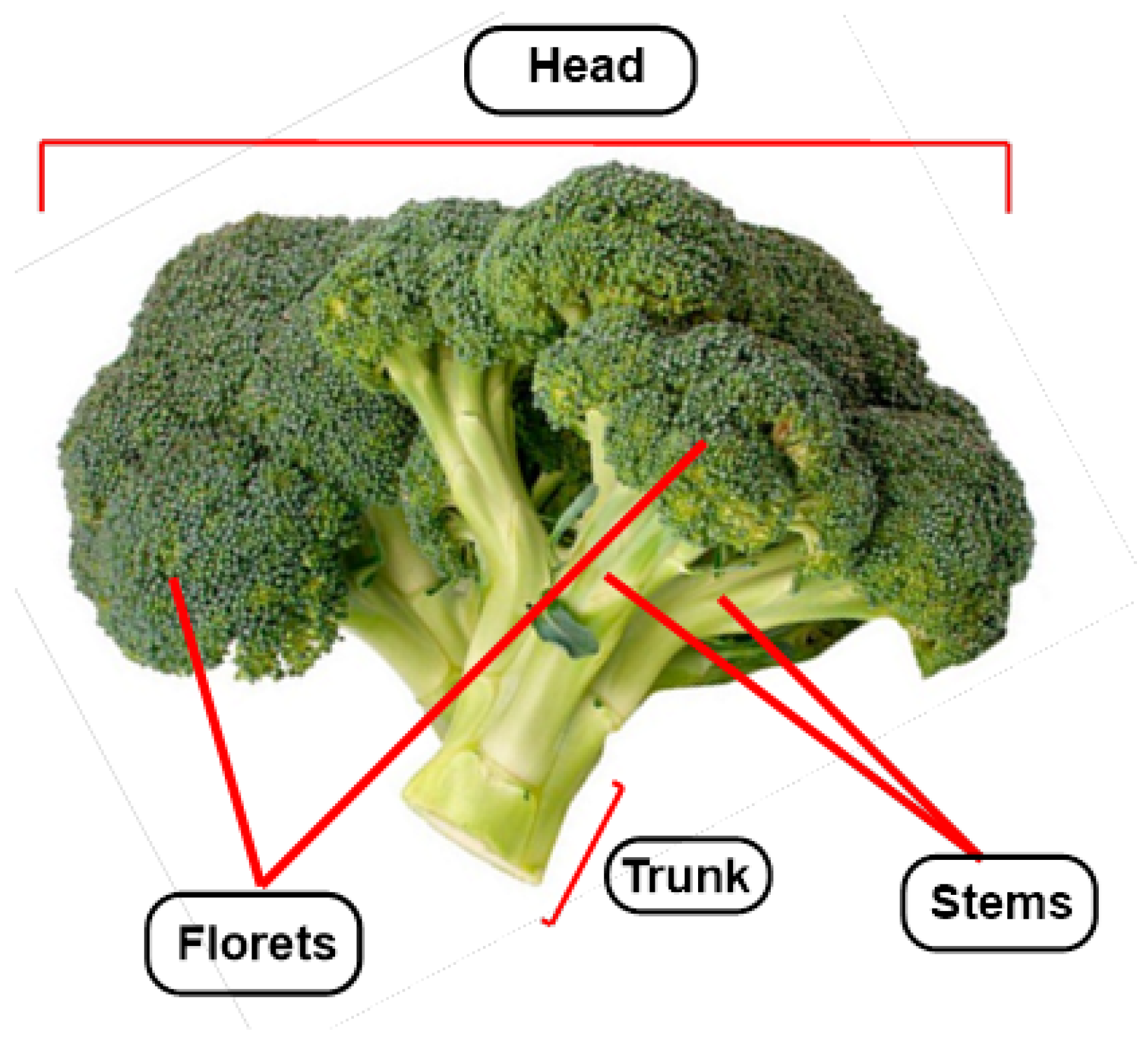
Figure 1. Broccoli (Brassica oleracea L. var. italica) anatomy.
Molecules 30 02262 g001
Figure 2. Summary of nutritional and health benefits of broccoli.
Molecules 30 02262 g002
Figure 3. Glucosinolate structure.
Molecules 30 02262 g003
Figure 4. Hydrolysis of glucosinolates by myrosinase (an enzyme found in plants and intestinal microflora) to form isothiocyanates and glucose.
Molecules 30 02262 g004
Figure 5. Chemical structure of the different thiohydroxymate-O-sulphate decomposition compounds.
Molecules 30 02262 g005
Figure 6. Structures of desulfoglucosinolates found in Broccoli.
Molecules 30 02262 g006
Figure 7. Examples of isothiocyanates and their glucosinolate precursors.
Molecules 30 02262 g007
Figure 8. Aliphatic glucosinolate chain elongation.
Molecules 30 02262 g008
Figure 9. Glucosinolate core biosynthesis.
Molecules 30 02262 g009
Figure 10. SFN activation of Nrf2 signaling.
Molecules 30 02262 g010
Figure 11. Chemical structure of the functional groups of SFN.
Molecules 30 02262 g011
Figure 12. Mesomeric structures proposed for –NCS group.
Molecules 30 02262 g012
Figure 13. (A) β-elimination without, transforming SFN into a butenyl isothiocyanate and methylsulfenic acid. (B) Reaction with SFN and water.
Molecules 30 02262 g013
Figure 14. Interaction of SFN with Keap1.
Molecules 30 02262 g014
Figure 15. SFN is metabolized via the mercapturic acid pathway upon conjugation with glutathione and undergoes further biotransformation to produce different metabolites.
Molecules 30 02262 g015
Figure 16. Progoitrin is biologically inactive, but upon hydrolysis by the enzyme myrosinase it is transformed into goitrin.
Molecules 30 02262 g016
Figure 17. Chemical structures of glucoraphanin and enantiomers (R y S) of SFN. The stereogenic sulfur atom is indicated as S* and highlighted in red.
Molecules 30 02262 g017
Figure 18. Methods for obtaining sulforaphane enantiomers.
Molecules 30 02262 g018
Figure 19. Diasteromeric intermediate used for the synthesis of enantioenriched R-SFN.
Molecules 30 02262 g019
Figure 20. General procedures for the synthesis of chiral sulfinate esters via chlorosulfinates. In red, sulfur configuration in chiral sulfonate esters.
Molecules 30 02262 g020
Figure 21. Synthesis of both enantiomers of SFN using (+)-(1S,2R)-trans-2-phenylcyclohexanol as a chiral auxiliary.
Molecules 30 02262 g021
Figure 22. Enantio-divergent synthesis of both enantiomers of SFN.
Molecules 30 02262 g022
Figure 23. Amylose-based chiral stationary phases used in HPLC to discriminate SFN enantiomers.
Molecules 30 02262 g023
Figure 24. Formation of SFN diastereoisomers by (S)-Leucine reaction.
Molecules 30 02262 g024
Figure 25. Biosynthesis of I3C starting from glucobrassicin.
Molecules 30 02262 g025
Figure 26. Formation of DIM in vivo by dimerization in acidic medium of indole-3-carbinol.
Molecules 30 02262 g026
Figure 27. Most abundant flavanols in broccoli.
Molecules 30 02262 g027
Figure 28. Some of the most common hydroxycinnamic acids in broccoli.
Molecules 30 02262 g028
Figure 29. Chemical structure of carotenoids in broccoli.
Molecules 30 02262 g029
Figure 30. Chemical structure of phytosterols in the head of broccoli.
Molecules 30 02262 g030
Figure 31. Broccoli antimicrobial peptide ARFEELNMDLFR. Structure was modeled using Alphafold2. Image was generated using the software ChimeraX 1.9. The peptide is displayed in stick representation, with a transparent hydrophobic surface overlay colored using a gradient from blue (hydrophilic) to yellow (hydrophobic).
Molecules 30 02262 g031
Figure 32. Broccoli anti-inflammatory peptide SIWYGPDRP. Structure was modeled using Alphafold2. Image was generated using the software ChimeraX 1.9. The peptide is displayed in stick representation, with a transparent hydrophobic surface overlay colored using a gradient from blue (hydrophilic) to yellow (hydrophobic).
Molecules 30 02262 g032
Table 1. Major anticancer mechanisms of SFN and concentrations needed for triggering them.
MechanismConcentrationConcentration
In VitroIn Vivo
Modulation of phase I and phase II enzymes0.5–25 μM6 μmol/day to 1 mmol/kg
DNA protection from chemical insult0.06–20 μM10–450 μmol/kg
Induction of apoptosis0.1–300 μM2.4 μmol to 15 nmol/day
Cell-cycle modulation<1 μM–2 mM10–100 μmol/day
Inhibition of angiogenesis0.1–50 μM
Inhibition of metastasis formation6–28 μM2.8 μmol/kg
Table 2. Antimicrobial and antioxidant peptides and proteins from broccoli.
Peptide Name/SequenceActivityTarget Pathogens/EffectsReference
ARFEELNMDLFRAntimicrobial activityPorphyromonas gingivalis, Candida albicans[194,195]
SIWYGPDRPAnti-inflammatory (52% NO inhibition)Reduces inflammation in macrophages[196]
RFRTNF-α inhibition (75%)Chronic inflammatory diseases[196]
KASFAFAGLIL-6 inhibition (30%)Autoimmune disorders[196]
KSVLLKFAntioxidant, hypolipidemicOxidative stress, lipid metabolism[193]
BoNapAntifungal inhibitionFusarium culmorum, Penicillium expansum[197]
BraDef (Class I)Broad-spectrum antimicrobialB. cereus, C. albicans[192]
Disclaimer/Publisher’s Note: The statements, opinions and data contained in all publications are solely those of the individual author(s) and contributor(s) and not of MDPI and/or the editor(s). MDPI and/or the editor(s) disclaim responsibility for any injury to people or property resulting from any ideas, methods, instructions or products referred to in the content.