1. Introduction
In the modern era of personalized medicine, implant material development has become an important issue which requires defining the specific balance of multiple parameters. These include composition, shape, structure, mechanical properties, biocompatibility, the ability to stimulate angiogenesis or bone formation, and antimicrobial activity. In implants or wound treatment materials, all these factors define the material ability to interact with surrounding tissue. Efficiently used bioactive materials can serve as a frame for new tissue growth [
1].
On the other hand, while developing materials interacting with the milieu interieur, it is highly important to minimize their toxicity for body cells and tissues. However, many substances with required antibacterial properties are too toxic for the milieu interieur, and high antibacterial activity goes hand in hand with low biocompatibility. Due to this, the creation of materials combining both antibacterial properties and high biocompatibility is a difficult but important objective.
Hemocompatibility is one of the important aspects of biocompatibility. It includes thromboresistance—a biomaterial’s ability to prevent biomaterial thromb formation. This is a key feature of biocompatibility since foreign material interaction with blood may lead to coagulation or thromb formation. Hemocompatibility implies that the material does not negatively affect blood functions, composition changes and thromb formation [
2].
The hemocompatibility of biomaterials depends on their physicochemical properties such as surface tension, free surface energy, roughness and hydrophilic capacity [
2].
Another approach to studying hemocompatibility is related to the material surface electric charge. According to this model, blood protein adsorption occurs on abiotic surfaces, and the character of this adsorbed layer depends on the surface electric potential values and voltage difference. If the positive potential difference between the material and the blood is significant, thromb formation risk increases [
3].
One of the materials that might possess the mentioned features is diatomite modified with bioactive compounds (
DA). Diatomite is petrified residue of the diatomic plankton algae living in all bodies of water. Generally, diatomite almost completely consists of silicon dioxide (SiO
2) [
4].
DA is non-toxic and odorless, is present in nature in large quantities, is easily cleaned and is relatively cheap [
5]. Diatomite has some unique features compared to other natural materials, including high porosity (10–100 nm), permeability, small particle size, large surface area (29 m
2/g), high pore volume (0.09 cm
3/g), low heat conductivity and chemical inertness [
6]. Due to this, diatomite is used in various areas, for example, in construction, water filtration, agriculture, etc. The most well-known way of using it is as a contact insecticide in dry climates or as an ameliorant for soil and livestock or human food products [
5].
However, the most promising prospect for diatomite is medicinal use as the base for potential systems of medicinal compound address delivery or as a component for bone implant creation. Diatomite has features stimulating bone tissue regeneration, such as large surface area, due to its porous structure and optimal roughness. These features are important factors for osteoblast adhesion and proliferation [
4,
7].
One of the necessary features of biomaterials is the ability to suppress the growth and development of bacteria entering a wound during surgery [
8]. Bacteria entering a wound during surgical manipulations in the process of osteosynthesis may cause intrahospital and paraimplant infections. Such infections significantly decrease a patient’s quality of life and lead to more surgical operations with unpredictable outcomes.
However,
DA itself does not possess antibacterial activity [
4]. This is why in this study diatomite has been modified by coating its surface with organic compounds of natural and synthetic origin displaying antibacterial features. The high sorption capacity of diatomite allows it to provide long antibacterial compound emission from the biomaterial and, consequently, suppression of pathogenic wound microflora development. Due to this fact, diatomite was chosen as the base for biocomposites with antibacterial properties.
As mentioned above, the aim of this study was to research the antibacterial activity level of newly obtained biocompatible composite materials by modifying them with compositions of various nitrogen-containing compounds of natural and synthetic origin. In this study, the toxic effect of the obtained biocomposites was evaluated by studying their hemocompatibility, plasma protein adsorption and antibacterial activity. In this study, the impact of the biological effects of the composition of a number of synthetic and natural organic compounds applied onto diatomite in order to create promising biocomposite materials was researched. The frame base for the researched materials in the experiments was natural
DA in two forms—intact (
IDA) and cleaned (
CDA), the latter obtained by boiling
DA in hydrochloric acid [
4].
2. Research Materials and Methods
Glycoluril was purchased from Novochem (Tomsk, Russia). All other chemicals were from Merck/Sigma–Aldrich (Darmstadt, Germany).
2.1. Diatomite (DA) Synthesis
This research used diatomite in two forms—intact diatomite (
IDA) and diatomite that was additionally cleaned of involved cations and anions (
CDA). The diatomite’s specific surface area was 29 m
2/g, with SiO
2 content approx. 90% by mass. Primary impurities were Al (2.53% by mass) and Fe (1.81% by mass), as well as Mg, Ca, K and Na which combined were approx. 2% by mass. This sort of diatomite is a promising material for obtaining various composites due to its surface and properties [
9].
Diatomite cleaning was performed by boiling in 18% hydrochloric acid solution using the standard procedure [
9]. To do this, a diatomite aliquot (m = 40 g) was submerged into hydrochloric acid solution (V = 300 mL). Then, the solution was brought to a boil and exposed for 24 h, and then the diatomite was filtered and dried at room temperature until reaching constant mass. The diatomites (
IDA and
CDA) were pressed into cylindrical pellets approx. 1 mm thick and calcinated at 600 °C.
2.2. Tetrahydroxymethylglycoluril (TGMGU) Synthesis
Tetrahydroxymethylglycoluril (
TGMGU) is a white powder highly soluble in water and dimethylformamide, but poorly soluble in most organic solvents. It was synthesized using the method given in [
10] by condensing formaldehyde with glycoluril in an alkaline environment as shown in
Figure 1:
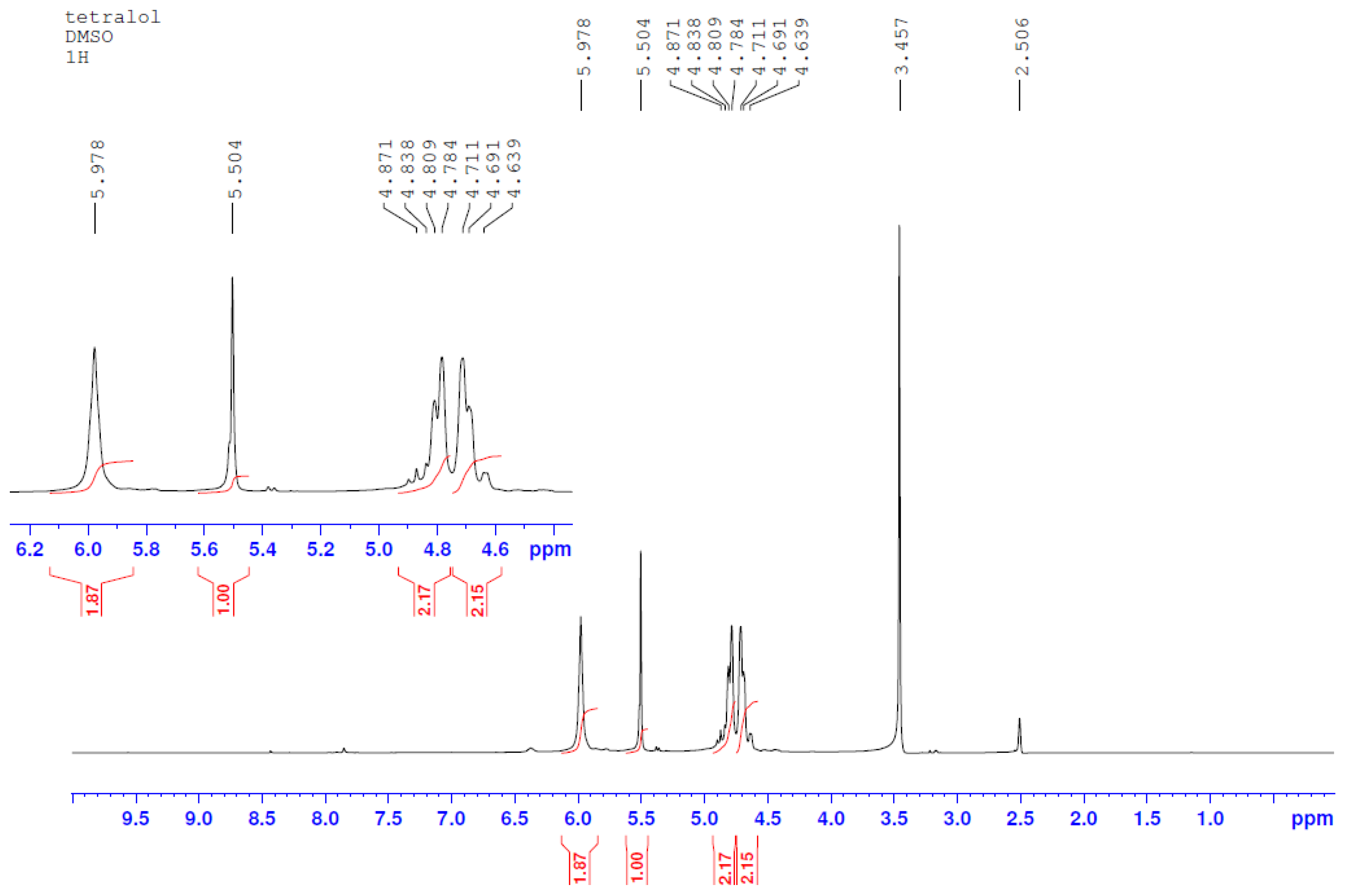
The obtained substance was characterized using NMR spectroscopy:
Specter NMR
1H (
Figure 2) (400 MHz, DMSO-d
6), δ ppm Hz: 5.978 (c. 4H, OH), 5.504 (c. 2H, CH-CH), 4.80 (p. 2H, J = 11.2, N-CH
2), 4.70 (p. 2H, J = 11.2, N-CH
2).
Specter NMR
13C (
Figure 3) (100 MHz, DMSO-d
6), 8 ppm: 157.48 (C=O), 87.03 (CH-CH), 65.10 (N-CH
2).
IR specter, v, cm−1: 1703 (C-O), 3273 (OH).
2.3. Betulin (BET) Separation
Betulin (
BET) was separated through extraction from birch tree bark using the standard procedure [
11]. The obtained substance was recrystallized from isopropyl alcohol and characterized using IR and NMR spectroscopy.
The IR specter (KBr, ν, cm–1) of betulin is characterized by absorption bands in the 3368 cm–1 area, which is typical for –OH groups, 1646 cm–1 (C=C) and others: 2968–2864 (C–H), 1450, 1372, 1105, 1029, 878.
1H NMR (400,17 MHz, CDCl3, δ, ppm): the weaker fields area has isopropenyl group proton signals (4.51 and 4.61 ppm); 3.26 and 3.73 (p, 2H, C28H2-OH); 3.10–3.14 (m, 1H, C3H-OH); 2.28–2.35 (m, 1H, C19–H); in the 0.79–2.00 ppm area there are lupane skeleton proton signals, where C-H group proton signals are found in betulin structure (6CH3, 10CH2, 5CH).
13C NMR (100,63 MHz, CDCl3, δ, m.f.): 150,51 (C-20); 109,71 (C-29); 78,98 (C-3); 64,46 (C-17); 60,51 (C-28); 55,28 (C-5); 50,38 (C-9); 48,74 (C-18); 47,79 (C-19); 42,71 (C-14); 40,91 (C-8); 38,87 (C-4); 38,69 (C-1); 37,30 (C-13); 37,15 (C-10); 34,22 (C-22); 33,98 (C-7); 29,74 (C-16); 29,16 (C-21); 27,99 (C-15); 27,38 (C-2); 27,04 (C-12); 25,37 (C-23); 25,19 (C-24); 20,83 (C-11); 19,09 (C-30); 18,30 (C-6); 16,12 (C-25); 15,98 (C-26); 15,38 (C-27).
2.4. Obtained Materials Characterization Methods
2.4.1. Nuclear Magnetic Resonance (NMR) Spectroscopy
NMR specters of synthesized compounds were registered on an NMR spectrometer Bruker Avance 400 III HD in DMSO-d6 solution at 25 °C, working frequency at hydrogen nuclei—400 MHz, at 13C—100 MHz.
2.4.2. Infrared (IR) Spectroscopy
Sample research was performed using IR spectroscopy with Fourier transform with an IR spectrometer Nicolet 6700, Thermo Fisher Scientific. Sample research was performed using the attenuated total reflectance method within a specter range between 400 and 4000 cm–1 with resolution 4 cm–1. Obtained reflectance specters were transformed into absorption bands using Kubelka–Munk transformation.
2.4.3. Scanning Electron Microscopy (SEM) and Energy Dispersive X-ray Spectroscopy (EDS)
The JSM-IT200 is an easy-to-use compact scanning electron microscope, which enables the user to quickly and easily perform SEM analysis. The electron optics system, achieving a resolution of 3.0 nm at an accelerating voltage of 30 kV, is useful for a variety of applications, from high-resolution observation to EDS analysis. Specimen Exchange Navi, a beginner-friendly function, offers guided operation from sample loading to area search, and SEM image observation. Fully integrated EDS (silicon drift detector, resolution: 130 eV) includes “live EDS analysis”, displaying the chemical composition of the specimen while imaging. With the “Zeromag” function, sample navigation is easier than ever before. “Zeromag”, which links the SEM image with Holder Graphics or an optical CCD image, enables you to quickly locate areas for imaging and analysis.
2.4.4. X-ray Photoelectron Spectroscopy (XPS) Analysis
For XPS analysis, we used a Nexsa X-Ray Photoelectron Spectrometer (XPS) by Thermo Scientific, which is a multi-technique surface chemistry analysis system. The main specifications of the Nexsa XPS system are as follows: sampling area: 3600 mm2 (samples must be smaller than a 60 × 60 mm sample holder); max. sample thickness: 20 mm; X-ray spot size: 10–400 µm (adjustable in 5 µm steps); analyzer type: 180°, double-focusing, hemispherical analyzer with 128-channel detector; depth profiling: EX06 monatomic ion source; and X-ray source type: monochromated low-power Al Ka X-ray source—1486.6 eV.
2.5. Biocomposite Hemocompatibility Evaluation
One of the ways to evaluate general cytotoxicity is to study hemolytical activity. Hemolysis is a process of hemoglobin release as a result of erythrolysis. The test for material hemolytical activity is based on erythrolysis and the hemoglobin dissociation degree during material contact with erythrocytes in vitro [
12].
In order to evaluate hemocompatibility, whole hemostated blood from a healthy donor was used. After centrifuging, the blood was separated and erythromass was isolated. Obtained erythromass was diluted with sterile 1X PBS solution at 37 °C to a 1:9 ratio. Then, samples were placed into a standard 12-slot tray for cell cultivation and the obtained blood solution with PBS was poured at the ratio of 1 mln per 1 cm2 of sample surface area. As a positive control to define reaching 100% hemolysis, deionized water was used; as negative control, 1X PBS solution was used, which provides 0% hemolysis. During the experiment, IDA and CDA were used as control materials. Then, the tray with the samples was incubated in thermostat at 37 °C for 60 min. Next, the contents of the tray slots were transferred to centrifugal tubes and remaining erythrocytes were spun down by centrifugation for 5 min at 3000 rpm. Then, the supernatant was carefully removed and transferred to a standard 96-slot tray for further spectroscopy. The samples’ optical density was measured using the EIA reader TecanInfiniteF50 (Tecan Inc., Morrisville, NC, USA) at 492 nm.
The hemolysis percentage was the average value of three replications and was calculated using the formula given in GOST [
12]:
2.6. Evaluation of Plasma Protein Adsorption by Biocomposites
In order to analyze plasma protein adsorption in studied samples, a modified method of solution depletion was used. This method includes two quantitative definitions of finding the protein concentration in blood plasma—before and after sample incubation [
13].
We used the biuret reaction method to determine the amount of protein in solution. This method was chosen because of its simplicity and relative accuracy [
14,
15].
Initial plasma was separated from whole hemostated blood of a healthy donor by centrifugating. Then, the protein content in the plasma was found using the biuret reaction. Samples were filled with 2 mL of plasma and incubated at 37 °C in thermostat for 24 h. After incubation, the protein concentration was found a second time. The difference between the protein concentration of the initial plasma and that after incubation allows us to evaluate the protein adsorption rate—the greater this difference, the more protein that was adsorbed on the sample surface.
The interaction of blood serum proteins with cupric sulphate in a basic environment leads to the formation of complex compounds of purple color due to the proteins’ peptide bonds with copper ions. The intensity of the solution color depends on its protein concentration.
After the samples’ incubation and separation from the solution, the biuret reaction was carried out, followed by optical density measurement. To do this, 5.0 mL of working biuret reagent solution was added to 0.1 mL of blood plasma and carefully stirred, avoiding foam formation. The control experiment was performed simultaneously and 0.1 mL of 0.9% sodium chloride solution was used as control, then adding 5 mL of working biuret reagent solution. Then, 30 min later (but no later than 1 h), the solution optical density was measured using the EIA reader Tecan Infinite F50 (Tecan Inc., Morrisville, NC, USA) at 492 nm wavelength against control.
2.7. Biocomposite Antimicrobial Activity Evaluation
To evaluate the antibacterial activity of created samples, a standard disc diffusion method modified by the authors of this study was used. According to this method, the samples were placed on the surface of a high-density agar environment. CHL, BET and TGMGU (chloramphenicol, betulin and tetrahydroxymethylglycoluril) modifiers diffused into the environment, creating areas of bacteria growth suppression.
To study samples’ impact on the microflora, the Escherichia coli ATCC 25922 strain was used as test object. Escherichia coli was chosen as a model object because of the high importance of this microorganism for agriculture and medicine. The media used for E. coli cultivation were the standard LB media for these bacteria, containing peptone (10 g/L), yeast extract (5 g/L), sodium chloride (10 g/L) and microbiological agar (1.5–2% by volume). Incubation was carried out in thermostat at 37–38 °C for 24 h.
Each Petri dish with 15 mL of concentrated nutritious environment was inoculated with E. coli using lawn inoculation and pure bacteria culture. The volume of inoculum applied was 0.1 mL (0.5 McFarland’s standard, 108 microorganisms/mL). Then, the sample was placed in the center of the dish. After incubation, the diameter of the zone with suppressed bacteria growth was measured to an accuracy of 0.1 mm. A large area of bacteria growth suppression implied a higher antibacterial activity of the sample. A bacteria growth inhibition area was considered one where growth was completely suppressed.
Samples without CHL in IDA and CDA were used as negative control. Samples modified by submersion into chloramphenicol solution (IDA + CHL and CDA + CHL) were used as positive control.
2.8. Statistical Analysis
Statistical analysis was performed using STATISTICA 8.0 for Windows (STATISTICA, RRID: SCR_014213). The Mann–Whitney test and t-test for independent groups were implemented. The data were checked for normality of distribution using the Shapiro–Wilk statistical criterion. Results were considered to be significant with *** p < 0.001, ** p < 0.01 and * p < 0.05.
3. Results and Discussion
Chloramphenicol (
CHL) is an antibiotic used for treating bacterial infections. However, its use has been limited in recent decades due to its high systemic toxicity [
16]. In this study, CHL was chosen as a model antibacterial compound because the test strain
E. coli is highly sensitive to it.
Another substance used as a model antibacterial compound was tetrahydroxymethylglycoluril (
TGMGU)—a nitrogen-containing heterocycle, one of bicyclic bis-ureas. As more bacteria strains become resistant to anti-bacterial compounds, those with biocidal properties become particularly interesting.
TGMGU gradually releases formaldehyde, which causes its antibacterial properties. Also,
TGMGU is used in medicine as a biomaterial component to improve fixation, keeping tissues’ and cells’ structure [
13].
The third model compound was betulin (
BET)—pentacyclic triterpenoid extracted from birch tree bark. Betulin and its derivatives possess various bioactive properties, including wound-healing, antibacterial, antitumoral, hypolipidemic, hepatoprotective and antiviral properties [
17]. Betulin (
BET) also can overcome resistance and induce apoptosis in malignized cells in various human cancer diseases. It displays antibacterial activity against
Bacillus subtilis and
Escherichia coli, and highly inhibits urease activity of
Helicobacter pylori [
17,
18].
Moreover,
BET wound-healing properties are caused by its ability to increase cell migration to damaged areas, increase their survival ability and stimulate collagen synthesis [
19]. This wide range of biological activity suggests that
BET can compensate for the toxic impact of antibacterial compounds, and combined with other antibacterial agents it can display synergy and increase their activity.
According to the stated objective, we studied the hemocompatibility and synergetic antibacterial activity of the composite materials. The following were used in this study as surface modifiers: chloramphenicol (
CHL), betulin (
BET) and tetrahydroxymethylglycoluril (
TGMGU); their structures are shown in
Figure 4.
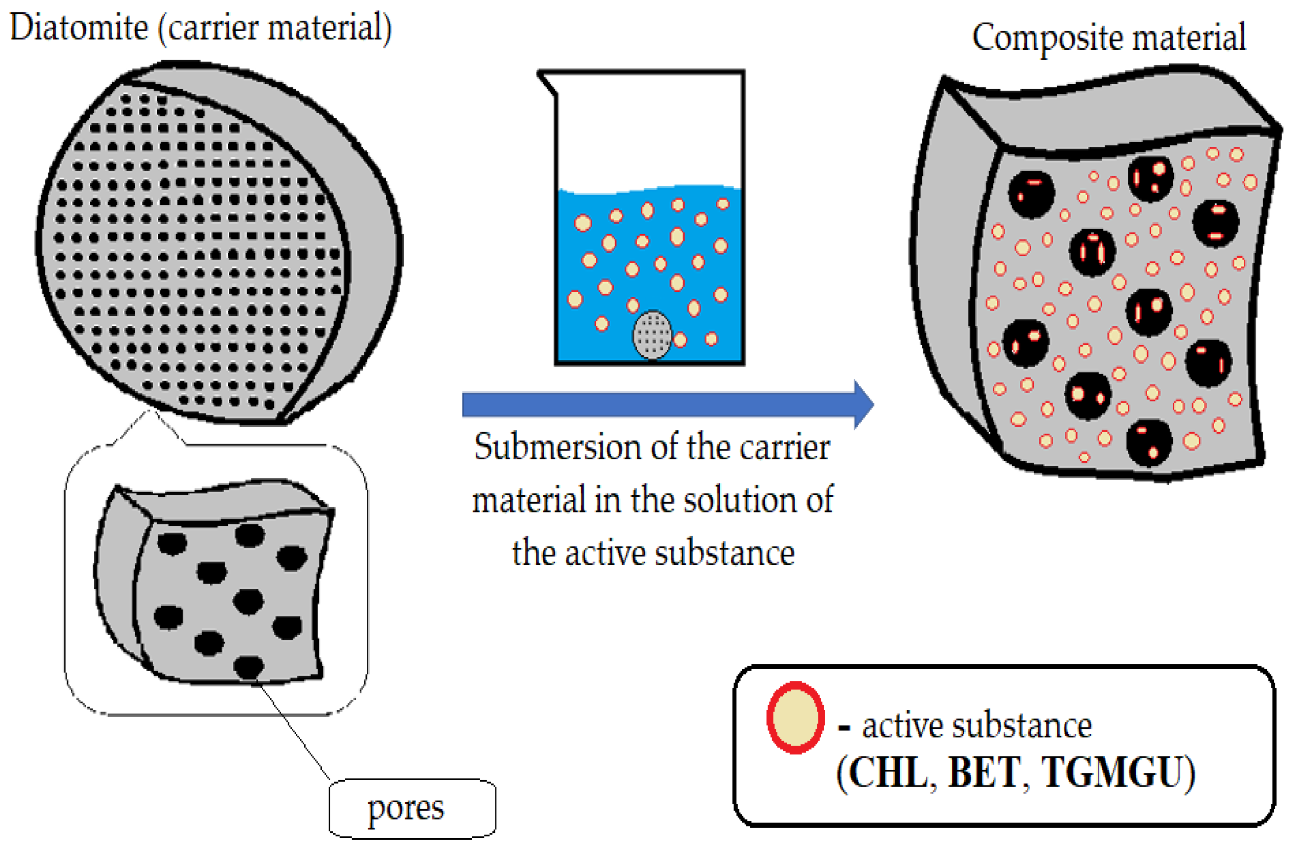
In order to add modifiers, a
DA sample was submerged into a working solution for 40 min (
Figure 5). Working solutions were prepared as follows. For
CHL, a 3% solution in ethyl alcohol was used to obtain the
IDA + CHL and
CDA + CHL composites. To obtain the
IDA + CHL + BET and
CDA + CHL + BET composites, 15 mg of
BET was added to a 3% solution of
CHL in ethanol. Solutions for obtaining composites with
TGMGU were prepared in an ethanol solution with a concentration of each substance (
TGMGU and
BET) of 1 mg/1 mL. The resulting solutions were taken in an amount of 15 mL per sample. There also was a control experiment, where distilled water was used instead of bioactive solution. Obtained materials were completely dried at room temperature and studied for their biological activity.
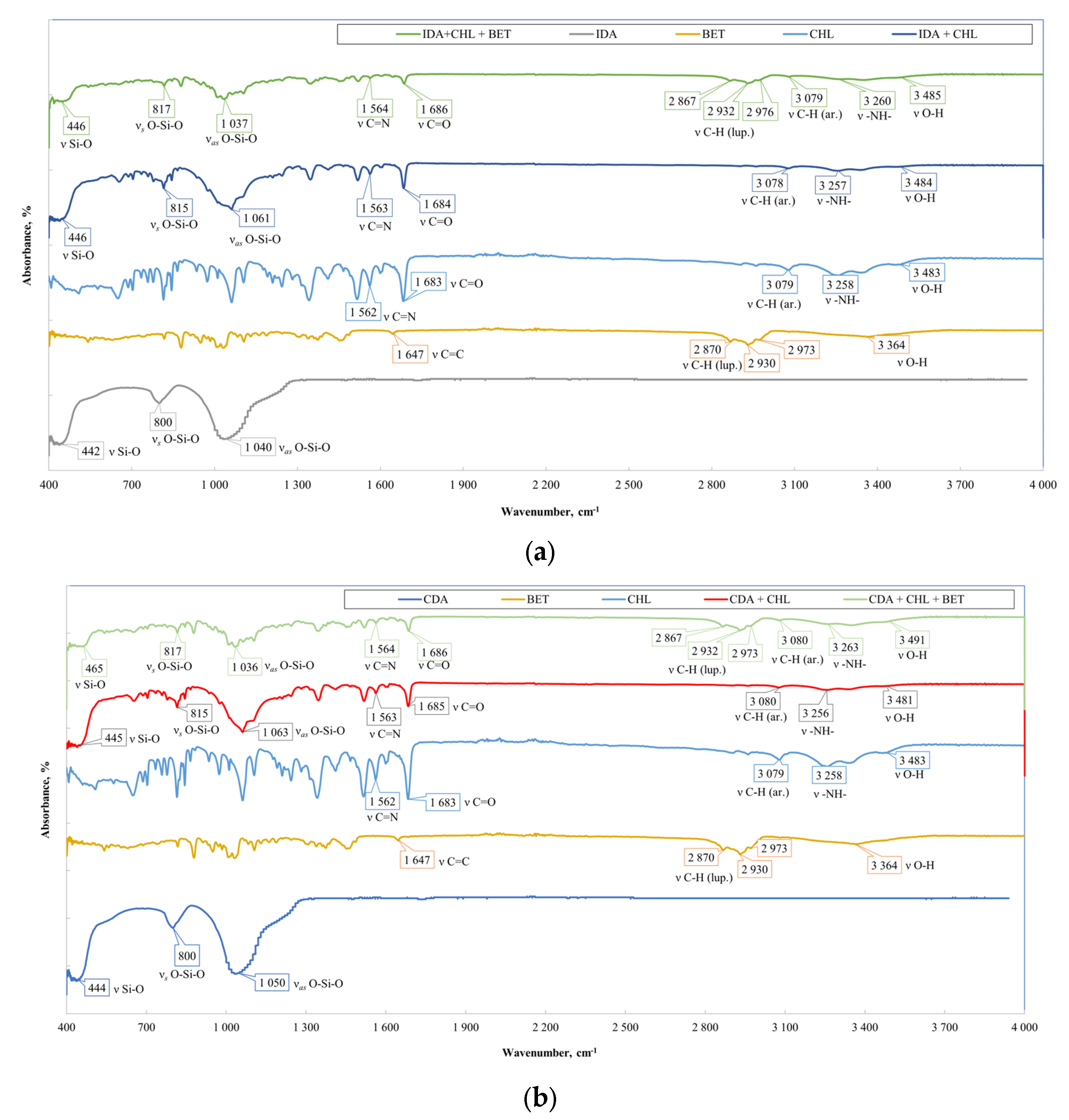
For each obtained sample (IDA with CHL, CHL + BET, TGMGU, TGMGU + BET and CDA with CHL, CHL + BET, TGMGU, TGMGU + BET), the IR specter was measured. IR specters of substance (complex compounds) absorption were obtained using the IR Fourier spectrometer Nicolet 6700 with FTIR console on a diamond crystal (resolution 4 cm−1, 64 scans, range—4000–450 cm−1); no visible sample decomposition was observed. To obtain IR spectra, the samples were dried to a constant weight, and scanning was from the surface of the composites.
The
IDA with
CHL sample specter (
Figure 6a) showed the following absorption bands related to diatomite stretching vibrations—451, 816 and 1060 cm
−1. The rest of the specter showed
CHL absorption bands: stretching vibrations of OH-group were seen at 3480 cm
−1, NH-groups of secondary amine at 3260 cm
−1, C-H aromatic ring at 3080 cm
−1 and C=O at 1680 cm
−1, as well as nitrogroup stretching vibration absorption bands at 1560 cm
−1. All the IR spectroscopy data set suggested
CHL presence on
IDA surface.
When analyzing the IR specter of
IDA with
CHL and
BET fracture plane (
Figure 6a), there were additional absorption bands typical for betulin—C-H stretching vibrations of the lupane skeleton at 2870, 2930 and 2970 cm
−1. At 3480 cm
−1, there was absorption band broadening as there was interference of the
BET,
CHL and
DA OH-groups. All the IR spectroscopy data set proved
BET presence in the composite. The IR spectra of the
CDA samples (
Figure 6b) showed a similar pattern.
In addition to IR spectroscopy, these samples were analyzed using SEM, EMF and XPS. To carry out the analysis, the samples were dried at room temperature to constant weight. The analysis was carried out from the surface of the scaffolds, which are circular in shape, 2 cm in diameter and 2 mm thick. The surface of the
IDA is visible on the SEM images (
Figure 7). An EDS was carried out from this surface (
Figure 8), which showed that carbon is present on the surface in addition to the constituent elements of
DA (silicon and oxygen).
The X-ray photoelectron spectroscopy (XPS) analysis of these composite materials provided information about the elemental composition and chemical bonding of the molecules. Since
IDA and
CDA contain only silicon and oxygen (other than metals), atoms will appear in the presence of organic substances (
Figure 9), for example, nitrogen. The combination of these factors confirms the presence of
CHL in the sample. Common acquisition parameters of the experiments were as follows: source gun type—Al K Alpha; spot size—400 µm; analyzer mode—CAE: Pass Energy 50.0 eV; and energy step size—0.100 eV.
Samples with cleaned diatomite provided a similar picture. All the typical absorption bands for the
CHL and
BET functional groups had the same positions (
Figure 10).
Samples with
TGMGU and
TGMGU/BET mixtures were also analyzed using IR spectroscopy and X-ray photoelectron spectroscopy. Absorption maximums earlier identified for
DA were also seen in the IR specters of these obtained materials. In the
IDA + TGMGU sample section, there were no absorption bands typical for
TGMGU (
Figure 10a), but the samples still displayed a distinctive antibacterial activity (Table 6). The IR specter also had absorption bands at 1638 cm
−1, which might be related to formaldehyde presence in the sample. The IR spectra of the
CDA samples (
Figure 6b) showed a similar pattern [
20].
The experiment observations were obviously related to the fact that
TGMGU can extendedly separate formaldehyde in water solution [
15]. Formed formaldehyde tends to bind with the
DA surface and provide antibacterial effect, while the rest of the
TGMGU does not remain due to high water solubility [
21,
22]. Another fact that suggests this is that in the SEM scan (
Figure 11) no
TGMGU or fragments thereof can be seen. The presence of free formaldehyde on the
IDA + TGMGU sample surface can also be suggested from X-ray photoelectron spectroscopy scans (
Figure 12). Considering the data (
Figure 12) for carbon distribution on the
IDA surface, a carbon-containing organic substance can be seen. Moreover, according to the SEM data, there were no nitrogen atoms on the
IDA + TGMGU surface, as could be expected due to the use of
TGMGU.
In the X-ray photoelectron spectroscopy (XPS) analysis of these composite materials, in addition to the basic atoms (carbon and oxygen), the binding energy with nitrogen is observed (
Figure 13).
Samples with cleaned diatomite produced a similar picture. The IR specters (
Figure 10) did not show absorption bands; however, similar to the
IDA + TGMGU composites, there were absorption bands typical for
DA.
These facts were also proven by SEM scan (
Figure 14). Considering the X-ray photoelectron spectroscopy scans (
Figure 15), carbon distribution on the surface can be seen, which suggests the presence of a carbon-containing organic substance (
Figure 15), similar to the
IDA + TGMGU composites.
The X-ray photoelectron spectroscopy (XPS) analysis of this composite produced a similar picture (
Figure 16).
Based on the obtained data, a
TGMGU and diatomite surface interaction pattern was produced (
Figure 17).
The presence of
BET on the composite surfaces can be suggested from SEM scans of
IDA + TGMGU + BET (
Figure 18a) and
CDA + TGMGU + BET (
Figure 18b). In these cases,
BET is present as a dispersion in water solution and its crystals can be seen in the SEM scans—needle-shaped crystals on the surface marked with red rectangles.
After having studied the obtained composites IDA + CHL, IDA + CHL + BET, IDA + TGMGU, IDA + TGMGU + BET, CDA + CHL, CDA + CHL + BET, CDA + TGMGU and CDA + TGMGU + BET, we studied the toxic impact of the obtained biocomposites, which was estimated by studying their hemocompatibility, plasma protein adsorption and antibacterial activity.
One of the ways to estimate general material cytotoxicity is to study hemolytical activity.
Table 1 shows the hemolytical activity level estimation results for the sample groups
DA + CHL + BET, which suggested that the modified
CHL and
BET samples did not display any hemolytical activity.
The absence of hemolytical activity in the studied composite samples may be related to the fact that during the
DA treatment with
CHL and
BET, the base component’s porosity decreases. According to reference data, hemolysis on inert biomaterial surfaces is directly connected to plasma protein adsorption, and particularly to fibrinogen on the surface of the material being in contact with blood [
23]. In other words, the more adsorption of plasma proteins there is, the greater the amount of hemolysis. Diatomite is a porous material with small pore size, at least they are smaller than the size of protein molecules, so plasma protein adsorption is insignificant on it [
24]. Considering the abovementioned, it can be suggested that this is the reason for the low hemolysis level on these composites.
In order to determine whether the hemolytical activity level was connected to the plasma protein adsorption level in the studied samples, we studied the degree of the decrease in plasma proteins concentration after incubation with the samples obtained during the research using a modified solution depletion method.
As
Table 2 suggests, all samples, including control ones (
IDA and
CDA), showed decreased protein in plasma after incubation, which was proven statistically (
p < 0.05). Samples from the group
CDA + CHL, as well as
IDA and
CDA, displayed the highest adsorption, but statistically no proven differences were found between them (
p > 0.05). Other samples (
IDA + CHL,
IDA + CHL + BET and
CDA + CHL + BET) also showed decreased protein content in solution (
p < 0.05).
Supposedly, the low levels of the studied composites’ adsorption was related to the low hemolysis level and, consequently, low protein adsorption, the level of which was beyond the sensitivity of the method used. It was found that the betulin presence in the samples IDA + CHL + BET and CDA + CHL + BET also did not influence adsorption. Probably, the decrease in protein concentration after incubation may be connected not to surface adsorption but more to high porosity and the generally high sorption capacity of the studied samples.
As
Table 3 suggests, the not modified with chloramphenicol (
CHL) samples of the negative controls (
IDA and
CDA) did not display antibacterial effect in this method. Positive control group samples (
IDA + CHL and
CDA + CHL) displayed the highest antibacterial activity among all samples (51,0 and 52,1 mm, respectively). The antibacterial activity of the
CDA + CHL samples was higher, but there were no statistically proven differences between them (
p > 0.05). Betulin presence in samples did not influence the general antibacterial activity (
p > 0.05).
According to our hypothesis, due to the high content of other natural mineral compounds and higher porosity, intact diatomite (IDA) would display higher antibacterial activity; however, no difference was found between intact and cleaned diatomite. Probably, this is connected to the high sensitivity to chloramphenicol (CHL) of the test strain used. The high background activity of chloramphenicol (CHL) did not allow us to find the fine differences in antibacterial activity in the created biocomposites due to the limitations of the disc diffusion method used.
Diatomites modified with
TGMGU and
BET displayed different levels of activity (
Table 4).
As
Table 4 suggests, the highest hemolysis level was shown by the
CDA + TGMGU + BET samples (2.1122 ± 0.0026%) (
p < 0.05). Other group samples did not display hemolytical activity (
Table 4). Probably, the absence of hemolytical activity in these groups was connected to the high initial biocompatibility of diatomite, which is proven by reference data [
4].
It was also found that a slightly higher hemolysis level was displayed by the
CDA sample containing betulin (
CDA + TGMGU + BET). Probably, this happened due to higher protein adsorption on the sample surface under betulin influence. However, it should be noted that the hemolysis level of biomaterials contacting the milieu interieur should not exceed 5% [
12]. As the experiment shows, none of the studied samples exceeded this level, which shows that all the composite samples were hemocompatible.
It should be noted that the problem of unwanted blood clotting at contact with implanted materials and devices is still unsolved. The cause of this is the fact that healthy vessel endothelium has mechanisms resisting thromb formation, but foreign objects (i.e., foreign to the human body) do not have such defense mechanisms. Instead, biomaterials promote blood clotting through activating a number of interconnected processes including protein adsorption, thrombocyte and leukocyte adhesion, the production of thrombin and complement activation [
23]. Due to this, the search for antibacterial substances that modify biocompatible materials but do not increase the hemolysis percentage is of high importance.
It should also be noted that the cytocompatibility control method tested in this study was successful as a reliable express method for general material cytotoxicity control. None of the samples exceeded the hemolysis level for medical materials contacting blood, according to ISO 10993-4:2017 [
12].
As
Table 5 suggests, the lowest adsorption level was displayed by the
CDA + TGMGU + BET and
IDA + TGMGU + BET samples, but no statistically proven differences between them were found (
p > 0.05).
According to experiments previously performed in our study for hemocompatibility, the research sample CDA + TGMGU + BET displayed the highest hemolysis in the series of samples. However, the hemolysis level of this sample was 2.1122 ± 0.0026%, which suggests low hemolytical activity. However, the protein adsorption level for this sample was low. Probably, the treatment of the porous materials clogged their pores and reduced the sorption capacity, and the differences in adsorption level were connected directly to the samples’ physical structure.
Supposedly, a low adsorption level was related to a low hemolysis level and, consequently, low protein adsorption, the level of which was beyond the used method’s sensitivity. The presence of betulin (BET) in a sample also influences adsorption. Probably, the reduction in protein concentration after incubation was connected not to surface adsorption but more to high porosity and the generally high sorption capacity of the studied samples.
With respect to the antibacterial activity of composite
DA + TGMGU, as
Table 6 suggests, samples
IDA + TGMGU and
CDA + TGMGU also displayed antibacterial activity (27.9 and 31,9 mm, respectively), but no statistically proven differences between them were found (
p > 0.05). But it can be noted that in the case of samples’ modification with betulin, the levels of activity changed. Samples
IDA + TGMGU + BET and
CDA + TGMGU + BET had growth suppression area diameters of 33,8 and 27,2 mm, respectively. The activity of sample
IDA + TGMGU + BET was slightly higher, which was statistically proven (
p < 0.05).


 ,
, 


