
Dual/Multi-Modal Image-Guided Diagnosis and Therapy Based on Luminogens with Aggregation-Induced Emission


Abstract

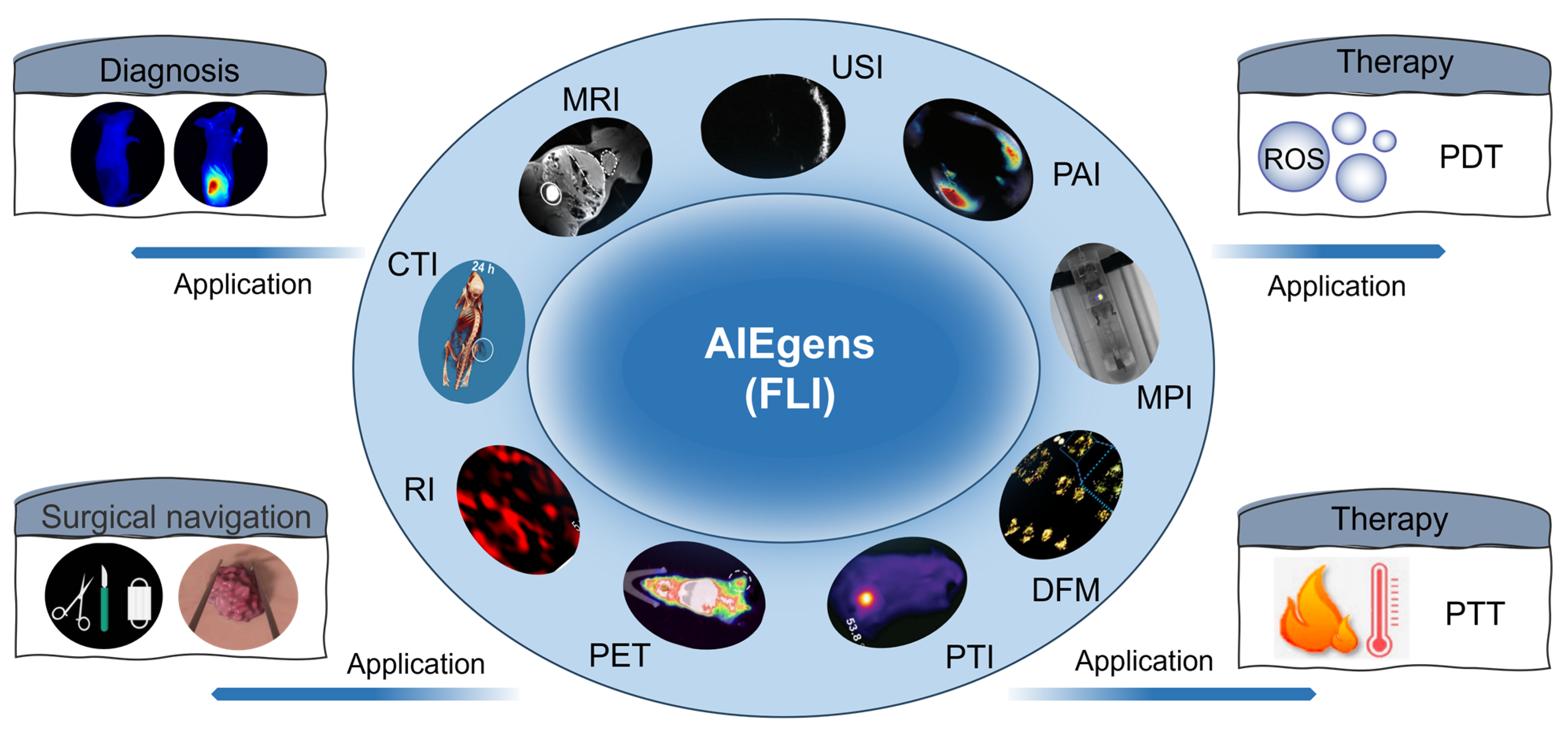
1. Introduction
2. AIEgen-Based Dual-Modal Imaging
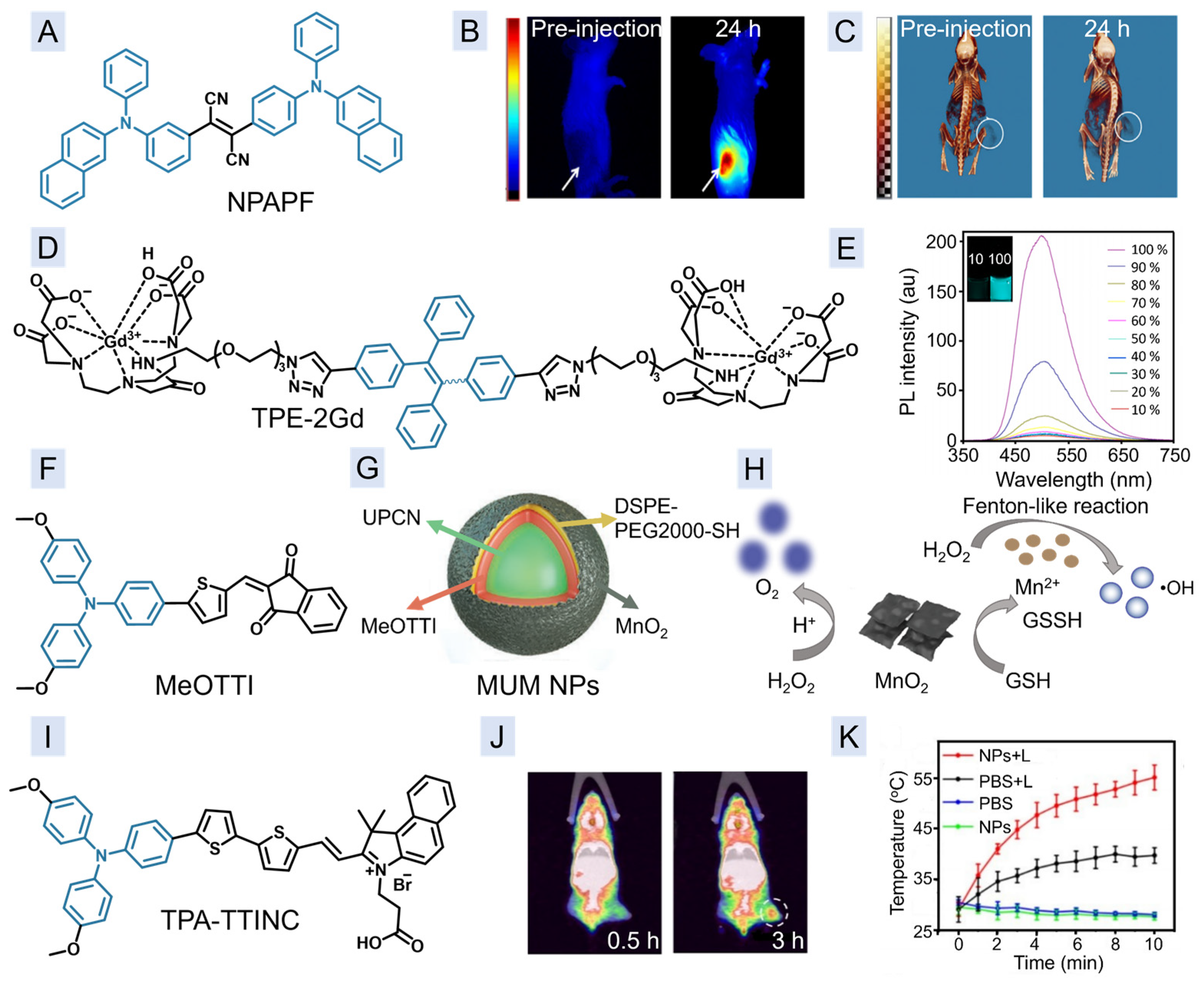
2.1. Dual-Modal Imaging of FLI and CTI
2.2. Dual-Modal Imaging of FLI and MRI
2.3. Dual-Modal Imaging of FLI and PET Imaging
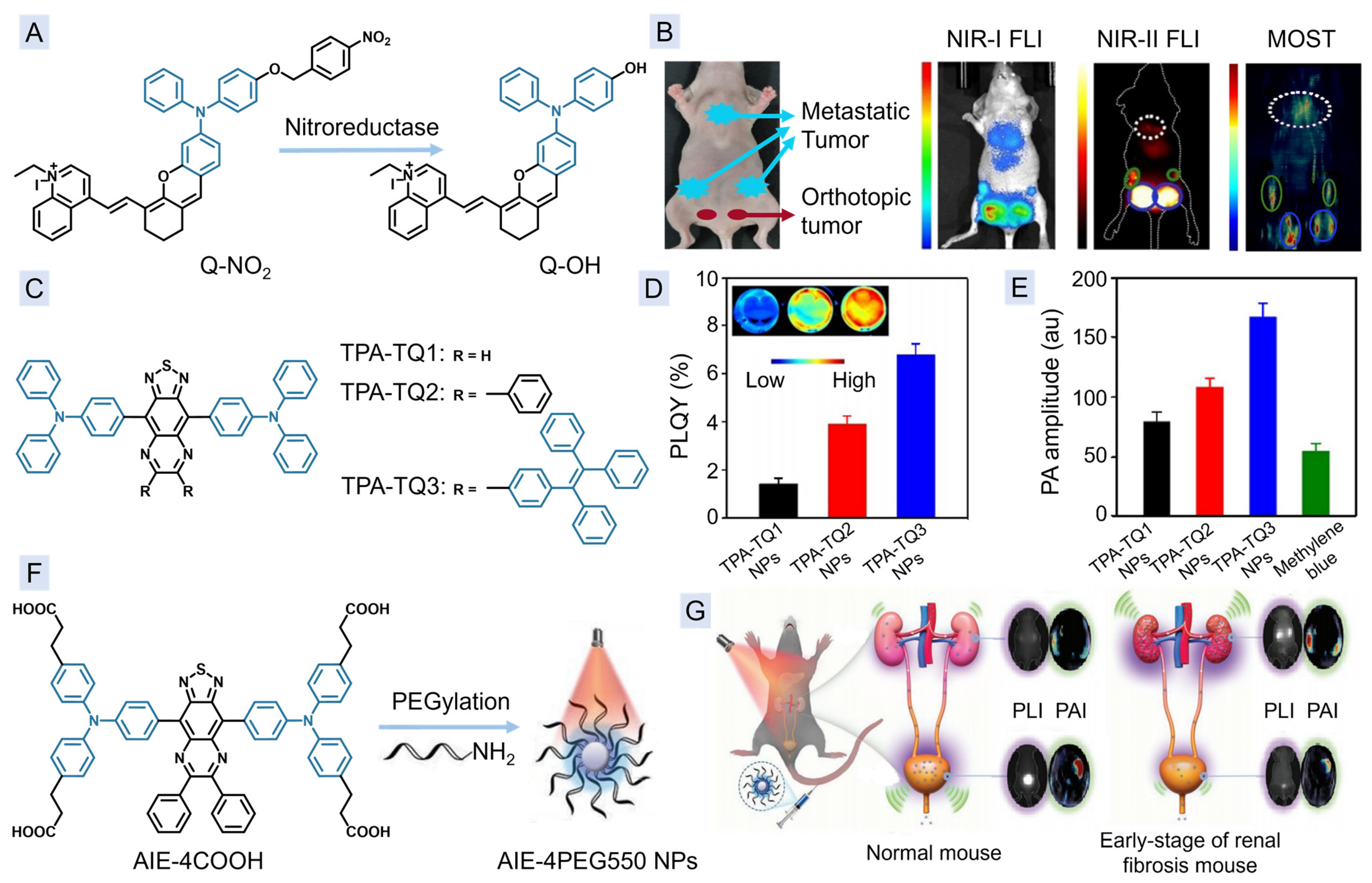
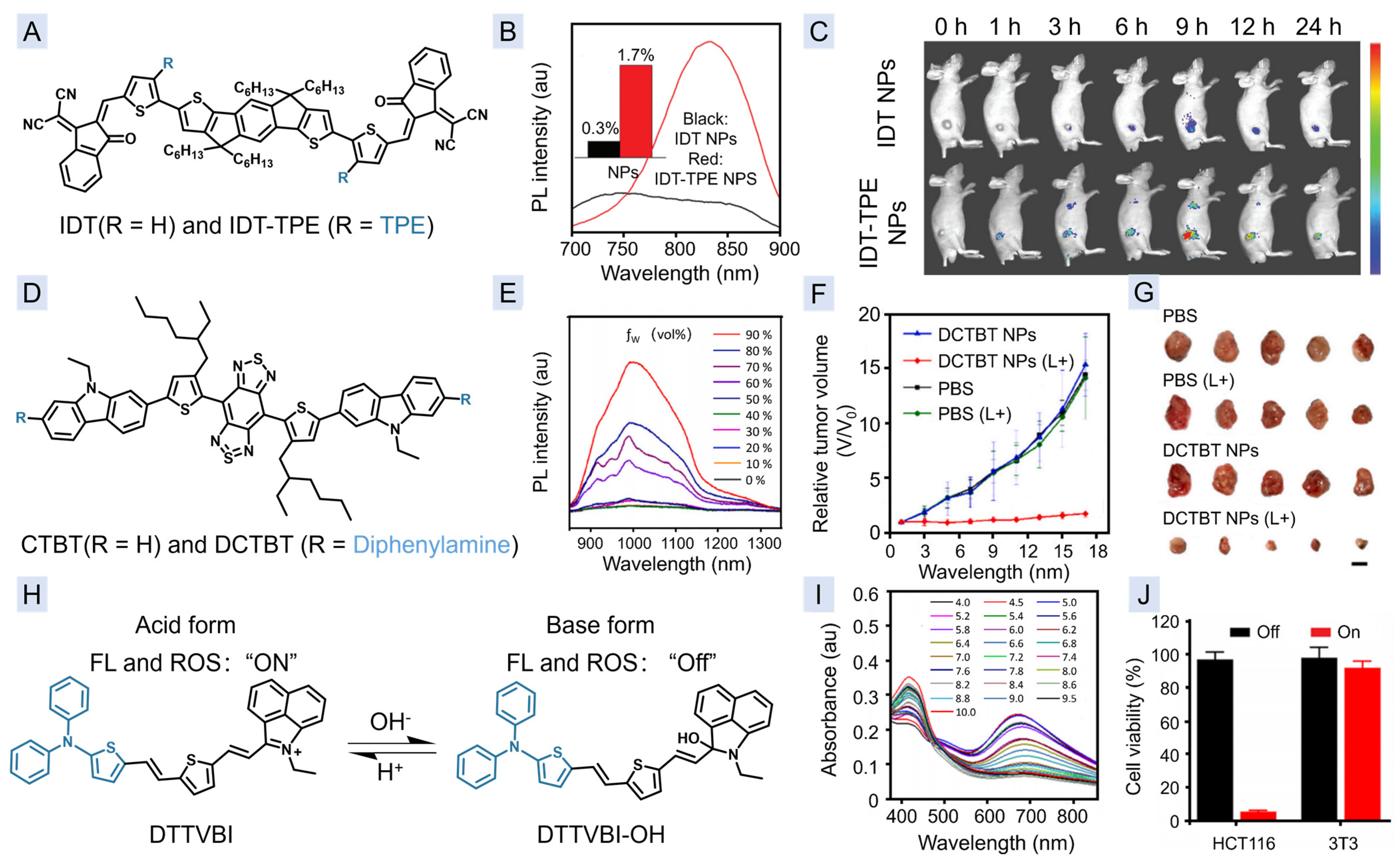
2.4. Dual-Modal Imaging of FLI and PAI
2.5. Dual-Modal Imaging of FLI and PTI
3. AIEgen-Based Multi-Modal Imaging
3.1. Multi-Modal Imaging of FLI, CTI, and DFM Imaging
3.2. Multi-Modal Imaging of FLI, PAI, and RI
3.3. Multi-Modal Imaging of FLI, PAI, and PTI
3.4. Multi-Modal Imaging of FLI, MPI, CTI, and MRI
4. Conclusions
Author Contributions
Funding
Institutional Review Board Statement
Informed Consent Statement
Data Availability Statement
Conflicts of Interest
References
- Weissleder, R. Molecular Imaging in Cancer. Science 2006, 312, 1168–1171. [Google Scholar] [CrossRef]
- Jokerst, J.V.; Gambhir, S.S. Molecular Imaging with Theranostic Nanoparticles. Acc. Chem. Res. 2011, 44, 1050–1060. [Google Scholar] [CrossRef] [PubMed]
- Lin, V.S.; Chen, W.; Xian, M.; Chang, C.J. Chemical probes for molecular imaging and detection of hydrogen sulfide and reactive sulfur species in biological systems. Chem. Soc. Rev. 2015, 44, 4596–4618. [Google Scholar] [CrossRef]
- Gao, M.; Yu, F.; Lv, C.; Choo, J.; Chen, L. Fluorescent chemical probes for accurate tumor diagnosis and targeting therapy. Chem. Soc. Rev. 2017, 46, 2237–2271. [Google Scholar] [CrossRef] [PubMed]
- Komljenovic, D.; Wiessler, M.; Waldeck, W.; Ehemann, V.; Pipkorn, R.; Schrenk, H.H.; Debus, J.; Braun, K. NIR-Cyanine Dye Linker: A Promising Candidate for Isochronic Fluorescence Imaging in Molecular Cancer Diagnostics and Therapy Monitoring. Theranostics 2016, 6, 131–141. [Google Scholar] [CrossRef] [PubMed]
- Guo, Z.; Park, S.; Yong, J.; Shin, I. Recent progress in the development of near-infrared fluorescent probes for bioimaging applications. Chem. Soc. Rev. 2014, 43, 16–29. [Google Scholar] [CrossRef]
- Xu, Y.; Li, B.; Xiao, L.; Ouyang, J.; Sun, S.; Pang, Y. A colorimetric and near-infrared fluorescent probe with high sensitivity and selectivity for acid phosphatase and inhibitor screening. Chem. Commun. 2014, 50, 8677–8680. [Google Scholar]
- Jiao, H.; Wang, B.; Chen, J.; Liao, D.; Li, W.; Yu, C. Label free fluorescence turn-on detection of polynucleotide kinase activity with a perylene probe. Chem. Commun. 2012, 48, 7862–7864. [Google Scholar] [CrossRef]
- Wang, B.; Yu, C. Fluorescence Turn-On Detection of a Protein through the Reduced Aggregation of a Perylene Probe. Angew. Chem. 2010, 122, 1527–1530. [Google Scholar] [CrossRef]
- Luo, J.; Xie, Z.; Lam, J.W.Y.; Chen, L.; Chen, H.; Qiu, C.; Kwok, H.S.; Zhan, X.; Liu, Y.; Zhu, D.; et al. Aggregation-induced emission of 1-methyl-1,2,3,4,5-pentaphenylsilole. Chem. Commun. 2001, 18, 1740–1741. [Google Scholar] [CrossRef]
- Hong, Y.; Lam, J.W.Y.; Tang, B.Z. Aggregation-induced emission: Phenomenon, mechanism and applications. Chem. Commun. 2009, 29, 4332–4353. [Google Scholar] [CrossRef] [PubMed]
- Ni, J.-S.; Liu, H.; Liu, J.; Jiang, M.; Zhao, Z.; Chen, Y.; Kwok, R.T.K.; Lam, J.W.Y.; Peng, Q.; Tang, B. The unusual aggregation-induced emission of coplanar organoboron isomers and their lipid droplet-specific applications. Mater. Chem. Front. 2018, 2, 1498–1507. [Google Scholar] [CrossRef]
- Maeda, H.; Wu, J.; Sawa, T.; Matsumura, Y.; Hori, K. Tumor vascular permeability and the EPR effect in macromolecular therapeutics: A review. J. Control. Release 2000, 65, 271–284. [Google Scholar] [CrossRef]
- Kwok, R.T.K.; Leung, C.W.T.; Lam, J.W.Y.; Tang, B. Biosensing by luminogens with aggregation-induced emission characteristics. Chem. Soc. Rev. 2015, 44, 4228–4238. [Google Scholar] [CrossRef]
- Ding, D.; Li, K.; Liu, B.; Tang, B. Bioprobes Based on AIE Fluorogens. Acc. Chem. Res. 2012, 46, 2441–2453. [Google Scholar] [CrossRef] [PubMed]
- Wang, D.; Tang, B.Z. Aggregation-Induced Emission Luminogens for Activity-Based Sensing. Acc. Chem. Res. 2019, 52, 2559–2570. [Google Scholar] [CrossRef] [PubMed]
- Hong, Y.; Lam, J.W.Y.; Tang, B.Z. Aggregation-induced emission. Chem. Soc. Rev. 2011, 40, 5361–5388. [Google Scholar] [CrossRef]
- Mei, J.; Leung, N.L.C.; Kwok, R.T.K.; Lam, J.W.Y.; Tang, B. Aggregation-Induced Emission: Together We Shine, United We Soar! Chem. Rev. 2015, 115, 11718–11940. [Google Scholar] [CrossRef]
- Feng, G.; Liu, B. Multifunctional AIEgens for Future Theranostics. Small 2016, 12, 6528–6535. [Google Scholar] [CrossRef]
- Qi, J.; Li, J.; Liu, R.; Li, Q.; Zhang, H.; Lam, J.W.Y.; Kwok, R.T.K.; Liu, D.; Ding, D.; Tang, B. Boosting Fluorescence-Photoacoustic-Raman Properties in One Fluorophore for Precise Cancer Surgery. Chem 2019, 5, 2657–2677. [Google Scholar] [CrossRef]
- Wang, Q.; Xu, J.; Geng, R.; Cai, J.; Xie, C.; Tang, W.; Sheng, Q.; Huang, W.; Fan, Q. High performance one-for-all phototheranostics: NIR-II fluorescence imaging guided mitochondria-targeting phototherapy with a single-dose injection and 808 nm laser irradiation. Biomaterials 2020, 231, 119671. [Google Scholar] [CrossRef]
- Li, D.; Li, Y.; Wu, Q.; Xiao, P.; Wang, L.; Wang, D.; Tang, B. Add the Finishing Touch: Molecular Engineering of Conjugated Small Molecule for High-Performance AIE Luminogen in Multimodal Phototheranostics. Small 2021, 17, 2102044. [Google Scholar] [CrossRef] [PubMed]
- Chen, C.; Ni, X.; Tian, H.-W.; Liu, Q.; Guo, D.S.; Ding, D. Calixarene-Based Supramolecular AIE Dots with Highly Inhibited Nonradiative Decay and Intersystem Crossing for Ultrasensitive Fluorescence Image-Guided Cancer Surgery. Angew. Chem. Int. Ed. 2020, 59, 10008–10012. [Google Scholar] [CrossRef] [PubMed]
- Li, X.; Kim, J.; Yoon, J.; Chen, X. Cancer-Associated, Stimuli-Driven, Turn on Theranostics for Multimodality Imaging and Therapy. Adv. Mater. 2017, 29, 1606857. [Google Scholar] [CrossRef] [PubMed]
- Qi, J.; Chen, C.; Ding, D.; Tang, B. Aggregation-Induced Emission Luminogens: Union Is Strength, Gathering Illuminates Healthcare. Adv. Healthc. Mater. 2018, 7, 1800477. [Google Scholar] [CrossRef]
- Wang, J.; Li, J.; Wang, L.; Han, T.; Wang, D.; Tang, B. AIEgen-Based Polymer Nanocomposites for Imaging-Guided Photothermal Therapy. ACS Appl. Polym. Mater. 2020, 2, 4306–4318. [Google Scholar] [CrossRef]
- Yan, D.; Qin, Y.; Yan, S.; Sun, P.; Wang, Y.; Wang, D.; Tang, B. Near-infrared emissive AIE nanoparticles for biomedical applications: From the perspective of different nanocarriers. Particuology 2023, 74, 103–118. [Google Scholar] [CrossRef]
- Kak, A.C.; Slaney, M. Principles of Computerized Tomographic Imaging; IEEE: New York, NY, USA, 1988. [Google Scholar]
- Schima, W.; Mukerjee, A.; Saini, S. Contrast-enhanced MR imaging. Clin. Radiol. 1996, 51, 235–244. [Google Scholar] [CrossRef]
- Wells, P.N.T. Ultrasound imaging. Phys. Med. Biol. 2006, 51, R83–R98. [Google Scholar] [CrossRef]
- Muehllehner, G.; Karp, J.S. Positron emission tomography. Phys. Med. Biol. 2006, 51, R117–R137. [Google Scholar] [CrossRef]
- Engels, S.A.; Klinteberg, C.; Svanberg, K. In vivo fluorescence imaging for tissue diagnostics. Phys. Med. Biol. 1997, 42, 815–824. [Google Scholar] [CrossRef]
- Schultz, S.; Smith, D.R.; Mock, J.J.; Schultz, D.A. Single-target molecule detection with nonbleaching multicolor optical immunolabels. Proc. Natl. Acad. Sci. USA 2000, 97, 996–1001. [Google Scholar] [CrossRef] [PubMed]
- Xu, M.; Wang, L.V. Photoacoustic imaging in biomedicine. Rev. Sci. Instrum. 2006, 77, 041101. [Google Scholar] [CrossRef]
- Stewart, S.; Priore, R.J.; Nelson, M.P.; Treado, P.J. Raman Imaging. Annu. Rev. Anal. Chem. 2012, 5, 337–360. [Google Scholar] [CrossRef] [PubMed]
- Panagiotopoulos, N.; Duschka, R.L.; Ahlborg, M.; Bringout, G.; Debbeler, C.; Graeser, M.; Kaethner, C.; Buzude, K.; Medimagh, H.; Stelzner, J.; et al. Magnetic particle imaging: Current developments and future directions. Int. J. Nanomed. 2015, 10, 3097–3114. [Google Scholar] [CrossRef]
- Aamodt, L.C.; Murphy, J.C. Thermal effects in photothermal spectroscopy and photothermal imaging. J. Appl. Phys. 1983, 54, 581–591. [Google Scholar] [CrossRef]
- Cheng, P.; Pu, K. Activatable Phototheranostic Materials for Imaging-Guided Cancer Therapy. ACS Appl. Mater. Interfaces 2020, 12, 5286–5299. [Google Scholar] [CrossRef]
- Kumar, R.; Shin, W.S.; Sunwoo, K.; Kim, W.Y.; Koo, S.; Bhuniya, S.; Kim, J.S. Small conjugate-based theranostic agents: An encouraging approach for cancer therapy. Chem. Soc. Rev. 2015, 44, 6670–6683. [Google Scholar] [CrossRef]
- Smith, B.R.; Gambhir, S.S. Nanomaterials for In Vivo Imaging. Chem. Rev. 2017, 117, 901–986. [Google Scholar] [CrossRef]
- Zhao, J.; Chen, J.; Ma, S.; Liu, Q.; Huang, L.; Chen, X.; Luo, K.; Wang, W. Recent developments in multimodality fluorescence imaging probes. Acta Pharm. Sin. B 2018, 8, 320–338. [Google Scholar] [CrossRef]
- Wu, Y.; Zeng, F.; Zhao, S.; Zhao, Y.; Wu, S. Emerging contrast agents for multispectral optoacoustic imaging and their biomedical applications. Chem. Soc. Rev. 2021, 50, 7924–7940. [Google Scholar] [CrossRef] [PubMed]
- Graziotto, M.E.; Kidman, C.J.; Adair, L.D.; James, S.A.; Harris, H.H.; New, E.J. Towards multimodal cellular imaging: Optical and X-ray fluorescence. Chem. Soc. Rev. 2023, 52, 8295–8318. [Google Scholar] [CrossRef]
- Yun, W.S.; Cho, H.; Jeon, S.I.; Lim, D.-K.; Kim, K. Fluorescence-Based Mono- and Multimodal Imaging for In Vivo Tracking of Mesenchymal Stem Cells. Biomolecules 2023, 13, 1787. [Google Scholar] [CrossRef] [PubMed]
- Li, J.; Pu, K. Development of organic semiconducting materials for deep-tissue optical imaging, phototherapy and photoactivation. Chem. Soc. Rev. 2019, 48, 38–71. [Google Scholar] [CrossRef]
- Miao, Q.; Pu, K. Organic Semiconducting Agents for Deep-Tissue Molecular Imaging: Second Near-Infrared Fluorescence, Self-Luminescence, and Photoacoustics. Adv. Mater. 2018, 30, 1801778. [Google Scholar] [CrossRef] [PubMed]
- Ji, C.; Cheng, W.; Yuan, Q.; Müllen, K.; Yin, M. From Dyestuff Chemistry to Cancer Theranostics: The Rise of Rylenecarboximides. Acc. Chem. Res. 2019, 52, 2266–2277. [Google Scholar] [CrossRef]
- Lemaster, J.E.; Chen, F.; Kim, T.; Hariri, A.; Jokerst, J.V. Development of a Trimodal Contrast Agent for Acoustic and Magnetic Particle Imaging of Stem Cells. ACS Appl. Nano Mater. 2018, 1, 1321–1331. [Google Scholar] [CrossRef] [PubMed]
- Lee, N.; Choi, S.H.; Hyeon, T. Nano-Sized CT Contrast Agents. Adv. Mater. 2013, 25, 2641–2660. [Google Scholar] [CrossRef]
- Lusic, H.; Grinstaff, M.W. X-ray-Computed Tomography Contrast Agents. Chem. Rev. 2012, 113, 1641–1666. [Google Scholar] [CrossRef] [PubMed]
- Zhang, J.; Li, C.; Zhang, X.; Huo, S.; Jin, S.; An, F.-F.; Wang, X.; Xue, X.; Okeke, C.I.; Duan, G. In vivo tumor-targeted dual-modal fluorescence/CT imaging using a nanoprobe co-loaded with an aggregation-induced emission dye and gold nanoparticles. Biomaterials 2015, 42, 103–111. [Google Scholar] [CrossRef]
- Chen, Y.; Li, M.; Hong, Y.; Lam, J.W.Y.; Zhwng, Q.; Tang, B. Dual-Modal MRI Contrast Agent with Aggregation-Induced Emission Characteristic for Liver Specific Imaging with Long Circulation Lifetime. ACS Appl. Mater. Interfaces 2014, 6, 10783–10791. [Google Scholar] [CrossRef] [PubMed]
- Wang, Y.; Li, Y.; Zhang, Z.; Wang, L.; Wang, D.; Tang, B. Triple-Jump Photodynamic Theranostics: MnO2 Combined Upconversion Nanoplatforms Involving a Type-I Photosensitizer with Aggregation-Induced Emission Characteristics for Potent Cancer Treatment. Adv. Mater. 2021, 33, 2103748. [Google Scholar] [CrossRef]
- Yu, K.; Huang, H.; Zhang, H.; He, Q.; Fu, Z.; Zhang, H.; Mao, Z.; Tian, M. Radiolabeled AIE Probes as Dual-modality Imaging Agents for PET/NIR-II Fluorescence-Guided Photothermal Therapy. Chem.-Asian J. 2023, 18, e202300189. [Google Scholar] [CrossRef] [PubMed]
- Caravan, P. Strategies for increasing the sensitivity of gadolinium based MRI contrast agents. Chem. Soc. Rev. 2006, 35, 512–523. [Google Scholar] [CrossRef]
- Tian, S.; Liu, G.; Wang, X.; Zhang, G.; Hu, J. pH-Responsive Tumor-Targetable Theranostic Nanovectors Based on Core Crosslinked (CCL) Micelles with Fluorescence and Magnetic Resonance (MR) Dual Imaging Modalities and Drug Delivery Performance. Polymers 2016, 8, 226. [Google Scholar] [CrossRef] [PubMed]
- Li, K.; Ding, D.; Prashant, C.; Qin, W.; Yang, C.-T.; Tang, B.; Liu, B. Gadolinium-Functionalized Aggregation-Induced Emission Dots as Dual-Modality Probes for Cancer Metastasis Study. Adv. Healthc. Mater. 2013, 2, 1600–1605. [Google Scholar] [CrossRef] [PubMed]
- Yang, H.; He, Y.; Wang, Y.; Yang, R.; Wang, N.; Zhang, L.-M.; Gao, M.; Jiang, X. Theranostic Nanoparticles with Aggregation-Induced Emission and MRI Contrast Enhancement Characteristics as a Dual-Modal Imaging Platform for Image-Guided Tumor Photodynamic Therapy. Int. J. Nanomed. 2020, 15, 3023–3038. [Google Scholar] [CrossRef]
- Wang, L.V.; Hu, S. Photoacoustic Tomography: In Vivo Imaging from Organelles to Organs. Science 2012, 335, 1458–1462. [Google Scholar] [CrossRef] [PubMed]
- Pu, K.; Shuhendler, A.J.; Jokerst, J.V.; Mei, J.; Gambhir, S.V.; Bao, Z.; Rao, J. Semiconducting polymer nanoparticles as photoacoustic molecular imaging probes in living mice. Nat. Nanotechnol. 2014, 9, 233–239. [Google Scholar] [CrossRef]
- Zhou, C.; Zhang, L.; Sun, T.; Zhang, Y.; Liu, Y.; Gong, M.; Xu, Z.; Du, M.; Liu, Y.; Liu, G.; et al. Activatable NIR-II Plasmonic Nanotheranostics for Efficient Photoacoustic Imaging and Photothermal Cancer Therapy. Adv. Mater. 2020, 3, 2006532. [Google Scholar] [CrossRef]
- Zha, M.; Lin, X.; Ni, J.-S.; Li, Y.; Zhang, Y.; Zhang, X.; Wang, L.; Li, K. An Ester-Substituted Semiconducting Polymer with Efficient Nonradiative Decay Enhances NIR-II Photoacoustic Performance for Monitoring of Tumor Growth. Angew. Chem. Int. Ed. 2020, 59, 23268–23276. [Google Scholar] [CrossRef]
- Ouyang, J.; Sun, L.; Zeng, Z.; Zeng, F.; Wu, S. Nanoaggregate Probe for Breast Cancer Metastasis through Multispectral Optoacoustic Tomography and Aggregation-Induced NIR-I/II Fluorescence Imaging. Angew. Chem. Int. Ed. 2019, 59, 10111–10121. [Google Scholar] [CrossRef] [PubMed]
- Gao, H.; Duan, X.; Jiao, D.; Zeng, Y.; Zheng, X.; Zhang, J.; Ou, H.; Qi, J.; Ding, D. Boosting Photoacoustic Effect via Intramolecular Motions Amplifying Thermal-to-Acoustic Conversion Efficiency for Adaptive Image-Guided Cancer Surgery. Angew. Chem. Int. Ed. 2021, 60, 21047–21055. [Google Scholar] [CrossRef] [PubMed]
- Yan, D.; Li, T.; Yang, Y.; Niu, N.; Wang, D.; Ge, J.; Wang, L.; Zhang, R.; Wang, D.; Tang, B. A Water-Soluble AIEgen for Noninvasive Diagnosis of Kidney Fibrosis via SWIR Fluorescence and Photoacoustic Imaging. Adv. Mater. 2022, 34, 2206643. [Google Scholar] [CrossRef] [PubMed]
- Li, M.; Li, H.; Wu, Q.; Niu, N.; Huang, J.; Zhang, L.; Li, Y.; Wang, D.; Tang, B. Hypoxia-activated probe for NIR fluorescence and photoacoustic dual-mode tumor imaging. iScience 2021, 24, 102261. [Google Scholar] [CrossRef] [PubMed]
- Qi, J.; Chen, C.; Zhang, X.; Hu, X.; Ji, S.; Kwok, R.T.K.; Lam, J.W.Y.; Ding, D.; Tang, B. Light-driven transformable optical agent with adaptive functions for boosting cancer surgery outcomes. Nat. Commun. 2018, 9, 1848. [Google Scholar] [CrossRef] [PubMed]
- Sun, L.; Ouyang, J.; Ma, Y.; Zeng, Z.; Zeng, C.; Zeng, F.; Wu, S. An Activatable Probe with Aggregation-Induced Emission for Detecting and Imaging Herbal Medicine Induced Liver Injury with Optoacoustic Imaging and NIR-II Fluorescence Imaging. Adv. Healthc. Mater. 2021, 10, 2100867. [Google Scholar] [CrossRef] [PubMed]
- Geng, J.; Liao, L.-D.; Qin, W.; Tang, B.; Thakor, N.; Liu, B. Fluorogens with Aggregation Induced Emission: Ideal Photoacoustic Contrast Reagents Due to Intramolecular Rotation. J. Nanosci. Nanotechnol. 2015, 15, 1864–1868. [Google Scholar] [CrossRef]
- Wang, L.V.; Wu, H.-I. Biomedical Optics, Principles and Imaging. J. Biomed. Opt. 2008, 13, 049902. [Google Scholar] [CrossRef]
- Chen, Y.-S.; Frey, W.; Kim, S.; Kruizinga, P.; Homan, K.; Emelianov, S. Silica-Coated Gold Nanorods as Photoacoustic Signal Nanoamplifiers. Nano Lett. 2011, 11, 348–354. [Google Scholar] [CrossRef] [PubMed]
- Aoki, H.; Nojiri, M.; Mukai, R.; Ito, S. Near-infrared absorbing polymer nano-particle as a sensitive contrast agent for photo-acoustic imaging. Nanoscale 2015, 7, 337–343. [Google Scholar] [CrossRef] [PubMed]
- Zhen, X.; Feng, X.; Xie, C.; Zheng, Y.; Pu, K. Surface engineering of semiconducting polymer nanoparticles for amplified photoacoustic imaging. Biomaterials 2017, 127, 97–106. [Google Scholar] [CrossRef] [PubMed]
- Li, K.; Liu, B. Polymer-encapsulated organic nanoparticles for fluorescence and photoacoustic imaging. Chem. Soc. Rev. 2014, 43, 6570–6597. [Google Scholar] [CrossRef] [PubMed]
- Ai, X.; Ho, C.J.H.; Aw, J.; Attia, A.B.E.; Mu, J.; Wang, Y.; Wang, X.; Wang, Y.; Liu, X.; Chen, H.; et al. In vivo covalent cross-linking of photon-converted rare-earth nanostructures for tumour localization and theranostics. Nat. Commun. 2016, 7, 10432. [Google Scholar] [CrossRef]
- Xie, L.; Pang, X.; Yan, X.; Dan, Q.; Lin, H.; Ye, J.; Cheng, Y.; Zhao, Q.; Ma, X.; Zhang, X.; et al. Photoacoustic Imaging-Trackable Magnetic Microswimmers for Pathogenic Bacterial Infection Treatment. ACS Nano 2020, 14, 2880–2893. [Google Scholar] [CrossRef] [PubMed]
- Li, X.; Park, E.-Y.; Kang, Y.; Kwon, N.; Yang, M.; Lee, S.; Kim, W.J.; Kim, C.; Yoon, J. Supramolecular Phthalocyanine Assemblies for Improved Photoacoustic Imaging and Photothermal Therapy. Angew. Chem. Int. Ed. 2020, 59, 8630–8634. [Google Scholar] [CrossRef]
- Li, D.; Chen, X.; Wang, D.; Wu, H.; Wen, H.; Wang, L.; Jin, Q.; Wang, D.; Ji, J.; Tang, B. Synchronously boosting type-I photodynamic and photothermal efficacies via molecular manipulation for pancreatic cancer theranostics in the NIR-II window. Biomaterials 2022, 283, 121476. [Google Scholar] [CrossRef]
- Xiao, P.; Xie, W.; Zhang, J.; Wu, Q.; Shen, Z.; Guo, C.; Wu, Y.; Wang, F.; Tang, B.; Wang, D. De Novo Design of Reversibly pH-Switchable NIR-II Aggregation-Induced Emission Luminogens for Efficient Phototheranostics of Patient-Derived Tumor Xenografts. J. Am. Chem. Soc. 2023, 145, 334–344. [Google Scholar] [CrossRef]
- Huang, J.; He, B.; Zhang, Z.; Li, Y.; Kang, M.; Wang, Y.; Li, K.; Wang, D.; Tang, B. Aggregation-Induced Emission Luminogens Married to 2D Black Phosphorus Nanosheets for Highly Efficient Multimodal Theranostics. Adv. Mater. 2020, 32, 2003382. [Google Scholar] [CrossRef]
- Jiang, R.; Dai, H.; Dong, X.; Wang, Q.; Meng, Z.; Guo, J.; Yu, Y.; Wang, S.; Xia, F.; Zhao, Z.; et al. Improving Image-Guided Surgical and Immunological Tumor Treatment Efficacy by Photothermal and Photodynamic Therapies Based on a Multifunctional NIR AIEgen. Adv. Mater. 2021, 33, 2101158. [Google Scholar] [CrossRef]
- Wang, Y.; Sun, Y.; Ran, J.; Yang, H.; Xiao, S.; Yang, J.; Yang, C.; Wang, H.; Liu, Y. Utilization of Nonradiative Excited-State Dissipation for Promoted Phototheranostics Based on an AIE-Active Type I ROS Generator. ACS Appl. Mater. Interfaces 2022, 14, 225–235. [Google Scholar] [CrossRef] [PubMed]
- Zhu, W.; Kang, M.; Wu, Q.; Zhang, Z.; Wu, Y.; Li, C.; Li, K.; Wang, L.; Wang, D.; Tang, B. Zwitterionic AIEgens: Rational Molecular Design for NIR-II Fluorescence Imaging-Guided Synergistic Phototherapy. Adv. Funct. Mater. 2020, 31, 2007026. [Google Scholar] [CrossRef]
- Gatenby, R.A.; Gillies, R.J. Why do cancers have high aerobic glycolysis? Nat. Rev. Cancer 2004, 4, 891–899. [Google Scholar] [CrossRef] [PubMed]
- Lee, K.; Cui, Y.; Lee, L.P.; Irudayaraj, J. Quantitative imaging of single mRNA splice variants in living cells. Nat. Nanotechnol. 2014, 9, 474–480. [Google Scholar] [CrossRef] [PubMed]
- Liu, Y.; Huang, C.Z. Real-Time Dark-Field Scattering Microscopic Monitoring of the in Situ Growth of Single Ag@Hg Nanoalloys. ACS Nano 2013, 7, 11026–11034. [Google Scholar] [CrossRef] [PubMed]
- Gao, P.F.; Gao, M.X.; Zou, H.Y.; Li, R.S.; Zhou, J.; Ma, J.; Wang, Q.; Liu, F.; Li, N.; Li, Y.F.; et al. Plasmon-induced light concentration enhanced imaging visibility as observed by a composite-field microscopy imaging system. Chem. Sci. 2016, 7, 5477–5483. [Google Scholar] [CrossRef] [PubMed]
- Anker, J.N.; Hall, W.P.; Lyandres, O.; Shan, N.C.; Zhao, J.; Duyue, R.P.V. Biosensing with plasmonic nanosensors. Nat. Mater. 2008, 7, 442–453. [Google Scholar] [CrossRef]
- He, X.; Zhao, Z.; Xiong, L.-H.; Gao, P.F.; Peng, C.; Li, R.S.; Xiong, Y.; Li, Z.; Sung, H.H.-Y.; Williams, I.D.; et al. Redox-Active AIEgen-Derived Plasmonic and Fluorescent Core@Shell Nanoparticles for Multimodality Bioimaging. J. Am. Chem. Soc. 2018, 140, 6904–6911. [Google Scholar] [CrossRef]
- Wei, L.; Hu, F.; Chen, Z.; Shen, Y.; Zhang, L.; Min, W. Live-Cell Bioorthogonal Chemical Imaging: Stimulated Raman Scattering Microscopy of Vibrational Probes. Acc. Chem. Res. 2016, 49, 1494–1502. [Google Scholar] [CrossRef]
- Song, Z.-L.; Chen, Z.; Bian, X.; Zhou, L.-Y.; Ding, D.; Liang, H.; Zou, Y.-X.; Wang, S.-S.; Chen, L.; Yang, C.; et al. Alkyne-Functionalized Superstable Graphitic Silver Nanoparticles for Raman Imaging. J. Am. Chem. Soc. 2014, 136, 13558–13561. [Google Scholar] [CrossRef]
- Li, S.; Chen, T.; Wang, Y.; Lv, F.; Li, Z.; Huang, Y.; Schanze, K.S.; Wang, S. Conjugated Polymer with Intrinsic Alkyne Units for Synergistically Enhanced Raman Imaging in Living Cells. Angew. Chem. Int. Ed. 2017, 56, 13455–13458. [Google Scholar] [CrossRef] [PubMed]
- Zhang, T.; Zhang, J.; Wang, F.-B.; Cao, H.; Zhu, D.; Chen, X.; Xu, C.; Yang, X.; Huang, W.; Wang, Z.; et al. Mitochondria-Targeting Phototheranostics by Aggregation-Induced NIR-II Emission Luminogens: Modulating Intramolecular Motion by Electron Acceptor Engineering for Multi-Modal Synergistic Therapy. Adv. Funct. Mater. 2022, 32, 2110526. [Google Scholar] [CrossRef]
- Yang, S.; Zhang, J.; Zhang, Z.; Zhang, R.; Ou, X.; Xu, W.; Kang, M.; Li, X.; Yan, D.; Kwok, R.T.K.; et al. More Is Better: Dual-Acceptor Engineering for Constructing Second Near-Infrared Aggregation-Induced Emission Luminogens toBoost Multimodal Phototheranostics. J. Am. Chem.Soc. 2023, 145, 22776–22787. [Google Scholar] [CrossRef] [PubMed]
- Zhang, Z.; Xu, W.; Kang, M.; Wen, H.; Guo, H.; Zhang, P.; Xi, L.; Li, K.; Wang, L.; Wang, D.; et al. An All-Round Athlete on the Track of Phototheranostics: Subtly Regulating the Balance between Radiative and Nonradiative Decays for Multimodal Imaging-Guided Synergistic Therapy. Adv. Mater. 2020, 32, 2003210. [Google Scholar] [CrossRef] [PubMed]
- Yan, D.; Xie, W.; Zhang, J.; Wang, L.; Wang, D.; Tang, B.Z. Donor/π-Bridge Manipulation for Constructing a Stable NIR-II Aggregation-Induced Emission Luminogen with Balanced Phototheranostic Performance. Angew. Chem. 2021, 133, 26973–26980. [Google Scholar] [CrossRef]
- Yan, D.; Wang, M.; Wu, Q.; Niu, N.; Li, M.; Song, R.; Rao, J.; Kang, M.; Zhang, Z.; Zhou, F.; et al. Multimodal Imaging-Guided Photothermal Immunotherapy Based on a Versatile NIR-II Aggregation-Induced Emission Luminogen. Angew. Chem. 2022, 134, e202202614. [Google Scholar] [CrossRef]
- Huang, C.; Shi, T.; Zhang, J.; Sun, Y.; Ma, T.; Li, W.; Li, Y. An NIR-II-absorbing photothermal agent containing multiple rotors with enhanced photothermal conversion capacity for multimodal-imaging-guided photothermal therapy. Dye. Pigm. 2023, 210, 110932. [Google Scholar] [CrossRef]
- Xu, W.; Zhang, Z.; Kang, M.; Guo, H.; Li, Y.; Wen, H.; Lee, M.M.S.; Wang, Z.; Kwok, R.T.K.; Lam, J.W.Y.; et al. Making the Best Use of Excited-State Energy: Multimodality Theranostic Systems Based on Second Near-Infrared (NIR-II) Aggregation-Induced Emission Luminogens (AIEgens). ACS Mater. Lett. 2020, 2, 1033–1040. [Google Scholar] [CrossRef]
- Xu, Y.; Zhang, Y.; Li, J.; An, J.; Li, C.; Bai, S.; Sharma, A.; Deng, G.; Kim, J.S.; Sun, Y. NIR-II emissive multifunctional AIEgen with single laser-activated synergistic photodynamic/photothermal therapy of cancers and pathogens. Biomaterials 2020, 259, 120315. [Google Scholar] [CrossRef]
- Liu, L.; Wang, X.; Wang, L.-J.; Guo, L.; Li, Y.; Bai, B.; Fu, F.; Lu, H.; Zhao, X. One-for-All Phototheranostic Agent Based on Aggregation-Induced Emission Characteristics for Multimodal Imaging-Guided Synergistic Photodynamic/Photothermal Cancer Therapy. ACS Appl. Mater. Interfaces 2021, 13, 19668–19678. [Google Scholar] [CrossRef]
- Wang, X.; Liu, L.; Wang, L.-J.; Guo, L.; Li, Y.; Bai, B.; Fu, F.; Lu, H.; Zhao, X. Optimizing Comprehensive Performance of Aggregation-Induced Emission Nanoparticles through Molecular Packing Modulation for Multimodal Image-Guided Synergistic Phototherapy. Adv. Healthc. Mater. 2021, 10, 2100360. [Google Scholar] [CrossRef]
- Wen, H.; Zhang, Z.; Kang, M.; Li, H.; Xu, W.; Guo, H.; Li, Y.; Tan, Y.; Wen, Z.; Wu, Q.; et al. One-for-all phototheranostics: Single component AIE dots as multi-modality theranostic agent for fluorescence-photoacoustic imaging-guided synergistic cancer therapy. Biomaterials 2021, 274, 120892. [Google Scholar] [CrossRef] [PubMed]
- Song, N.; Zhang, Z.; Liu, P.; Dai, D.; Chen, C.; Li, Y.; Wang, L.; Han, T.; Yang, Y.-W.; Wang, D.; et al. Pillar [5]arene-Modified Gold Nanorods as Nanocarriers for Multi-Modal Imaging-Guided Synergistic Photodynamic-Photothermal Therapy. Adv. Funct. Mater. 2021, 31, 2009924. [Google Scholar] [CrossRef]
- Zhang, Y.; Wang, J.; Wang, S.; Zhu, X.; Yu, Z.; Wu, Z.; Yu, J.; Zhou, H. Simultaneous one-step regulation of planarization and donor rotation to enhance multi-modal imaging guided therapy. Mater. Chem. Front. 2022, 6, 2921–2928. [Google Scholar] [CrossRef]
- Gu, H.; Liu, W.; Sun, W.; Du, J.; Fan, J.; Peng, X. Single-molecule photosensitizers for NIR-II fluorescence and photoacoustic imaging guided precise anticancer phototherapy. Chem. Sci. 2022, 13, 9719–9726. [Google Scholar] [CrossRef]
- Wang, Y.; Niu, N.; Huang, Y.; Song, S.; Tan, H.; Wang, L.; Wang, D.; Tang, B. Three-Pronged Attack by Hybrid Nanoplatform Involving MXenes, Upconversion Nanoparticle and Aggregation-Induced Emission Photosensitizer for Potent Cancer Theranostics. Small Methods 2022, 6, 2200393. [Google Scholar] [CrossRef] [PubMed]
- Meng, L.; Ma, X.; Jiang, S.; Ji, G.; Han, W.; Xu, B.; Tian, J.; Tian, W. High-efficiency fluorescent and magnetic multimodal probe for long-term monitoring and deep penetration imaging of tumors. J. Mater. Chem. B 2019, 7, 5345–5351. [Google Scholar] [CrossRef]
- Gleich, B.; Weizenecker, J. Tomographic imaging using the nonlinear response of magnetic particles. Nature 2005, 435, 1214–1217. [Google Scholar] [CrossRef]
- Yu, E.Y.; Bishop, M.; Zheng, B.; Ferguson, R.M.; Khandlhar, A.P.; Kemp, S.C.; Krishnan, K.M.; Goodwill, P.W.; Conolly, S.M. Magnetic Particle Imaging: A Novel in Vivo Imaging Platform for Cancer Detection. Nano Lett. 2017, 17, 1648–1654. [Google Scholar] [CrossRef]
- Song, G.; Chen, M.; Zhang, Y.; Cui, L.; Qu, H.; Zheng, X.; Wintermark, M.; Liu, Z.; Rao, J. Janus Iron Oxides @ Semiconducting Polymer Nanoparticle Tracer for Cell Tracking by Magnetic Particle Imaging. Nano Lett. 2018, 18, 182–189. [Google Scholar] [CrossRef]

Disclaimer/Publisher’s Note: The statements, opinions and data contained in all publications are solely those of the individual author(s) and contributor(s) and not of MDPI and/or the editor(s). MDPI and/or the editor(s) disclaim responsibility for any injury to people or property resulting from any ideas, methods, instructions or products referred to in the content. |
© 2024 by the authors. Licensee MDPI, Basel, Switzerland. This article is an open access article distributed under the terms and conditions of the Creative Commons Attribution (CC BY) license (https://creativecommons.org/licenses/by/4.0/).
Share and Cite
Zhu, L.; Wu, W. Dual/Multi-Modal Image-Guided Diagnosis and Therapy Based on Luminogens with Aggregation-Induced Emission. Molecules 2024, 29, 371. https://doi.org/10.3390/molecules29020371
Zhu L, Wu W. Dual/Multi-Modal Image-Guided Diagnosis and Therapy Based on Luminogens with Aggregation-Induced Emission. Molecules. 2024; 29(2):371. https://doi.org/10.3390/molecules29020371
Chicago/Turabian StyleZhu, Linlin, and Wenbo Wu. 2024. "Dual/Multi-Modal Image-Guided Diagnosis and Therapy Based on Luminogens with Aggregation-Induced Emission" Molecules 29, no. 2: 371. https://doi.org/10.3390/molecules29020371
APA StyleZhu, L., & Wu, W. (2024). Dual/Multi-Modal Image-Guided Diagnosis and Therapy Based on Luminogens with Aggregation-Induced Emission. Molecules, 29(2), 371. https://doi.org/10.3390/molecules29020371