Review

An Examination of Chemical Tools for Hydrogen Selenide Donation and Detection

by
Rynne A. Hankins
and
John C. Lukesh
*
Department of Chemistry, Wake Forest University, Wake Downtown Campus, 455 Vine Street, Winston-Salem, NC 27101, USA
*
Author to whom correspondence should be addressed.
Molecules 2024, 29(16), 3863; https://doi.org/10.3390/molecules29163863
Submission received: 26 July 2024 / Revised: 12 August 2024 / Accepted: 13 August 2024 / Published: 15 August 2024
(This article belongs to the Special Issue Organosulfur and Organoselenium Chemistry)

Abstract

:
Hydrogen selenide (H2Se) is an emerging biomolecule of interest with similar properties to that of other gaseous signaling molecules (i.e., gasotransmitters that include nitric oxide, carbon monoxide, and hydrogen sulfide). H2Se is enzymatically generated in humans where it serves as a key metabolic intermediate in the production of selenoproteins and other selenium-containing biomolecules. However, beyond its participation in biosynthetic pathways, its involvement in cellular signaling or other biological mechanisms remains unclear. To uncover its true biological significance, H2Se-specific chemical tools capable of functioning under physiological conditions are required but lacking in comparison to those that exist for other gasotransmitters. Recently, researchers have begun to fill this unmet need by developing new H2Se-releasing compounds, along with pioneering methods for selenide detection and quantification. In combination, the chemical tools highlighted in this review have the potential to spark groundbreaking explorations into the chemical biology of H2Se, which may lead to its branding as the fourth official gasotransmitter.

Graphical Abstract

1. Introduction

The discovery of elemental selenium can be traced back to a Swedish sulfuric acid plant in the early 19th Century where chemist Jöns Jakob Berzelius first observed a reddish-brown sediment in the acid being produced there [1]. Berzelius initially mistook this new substance for tellurium due to its odor and appearance. Famed for his advancement of modern chemical notation, the principle of stoichiometry, and the determination of atomic weights of most known elements at the time, Berzelius chemically compared the red-brown byproduct with a known sample of tellurium and determined that the two were, in fact, different elements with the new substance having the properties of a metal combined with that of sulfur. In the words of Berzelius, he had discovered “a new kind of sulfur”.
Today, selenium is regarded as an essential micronutrient that is acquired through dietary means with an optimal daily dose of 55 µg for adults [2]. There are at least 25 selenoproteins [3,4,5], many of which play a central role in cellular redox homeostasis, including glutathione peroxidases (GPx) and thioredoxin reductases (TrxR), and require dietary selenium for their production. Low selenium levels in humans are associated with a myriad of illnesses [6,7,8,9]. Keshan Disease [10,11], a potentially fatal form of cardiomyopathy, is primarily observed in selenium-deficient regions in China. The same is true for Kashin–Beck Disease [12,13], a chronic joint disease predominantly found in parts of the world where selenium is scarce. Weakened immune function [14,15], cardiovascular diseases [16,17], and certain cancers [18,19,20,21] are also strongly associated with selenium deficiency. These correlative studies suggest that the role of selenium in human health and biology could extend beyond its incorporation into selenium-containing proteins and point towards its possible involvement in other cellular processes.
In terms of its chemistry, selenium, like sulfur, can exist in numerous oxidation states, including selenate (SeO42−, +6), selenite (SeO32−, +4), and selenide (Se2−, −2) [22]. Selenide, the most reduced form of selenium, is the heavier chalcogen counterpart to sulfide (S2−). Both sulfide and selenide exist in different protonation states, dependent on environmental pH, with the fully protonated forms of both—hydrogen sulfide (H2S, pKa1: 6.9) [23] and hydrogen selenide (H2Se, pKa1: 3.9) [24]—being formerly dismissed as highly toxic gases with little biological relevance [25,26]. However, in the case of hydrogen sulfide, this malodorous gas has recently experienced a rebirth as an important, endogenous signaling molecule (or gasotransmitter) in mammals [27,28,29,30].
H2S is primarily produced by three principal enzymes—cystathionine β-synthase (CBS) [31], cystathionine γ-lyase (CSE) [32], and 3-mercaptopyruvate sulfur transferase (3-MST) [33]—providing exquisite control over its production. As such, endogenous H2S is known to be involved in numerous signaling processes throughout the body, including the brain and central nervous system [34,35], and within specific cellular compartments (i.e., mitochondria) [36,37]. Much of what is known about H2S pharmacology stems from the advent of donor compounds, synthetic small molecules designed to slowly liberate H2S in a controlled fashion that mimics its natural biosynthesis, and the use of said compounds in various cellular and animal models of disease [38,39,40,41].
Alongside nitric oxide (NO) and carbon monoxide (CO), hydrogen sulfide is the most recent, widely recognized member of the gasotransmitter family [42,43,44]. Its inclusion was suggested in the early 2000s and was based on five key observations [45]. Namely, H2S is (i) a small molecule gas that (ii) freely permeates cellular membranes. It is (iii) endogenously and enzymatically generated, with (iv) well-defined biological functions that stem from its (v) action at specific cellular targets.
It is interesting to note that H2Se (Predominantly HSe at physiological pH) already checks several of these boxes. It exists as a lipophilic gas in its diprotic form and is expressed enzymatically in mammals where it serves as a key intermediate in the production of selenium-containing biomolecules (Figure 1) [46]. H2Se was shown to act on at least some protein targets, including inhibition of cytochrome c oxidase, which modulates aerobic respiration [46,47]. Moreover, it is the reduced selenide form of selenium, and not the often administrated selenite (or other oxidized forms), that is believed to be responsible for the observed biological activity of selenium, including its anticancer effects and noted protection against myocardial ischemia reperfusion injury [48,49,50].
Still, compared to H2S, very little is known about the (patho)physiological effects of H2Se. This is due, at least in part, to a lack of refined donor compounds that can increase the bioavailability of selenide and be used to effectively modulate cellular concentrations. We and others have begun to fill this unmet need by developing new H2Se-releasing compounds, which we will highlight below, along with current methods for selenide detection and quantification. In combination, the chemical tools outlined in this review have the potential to serve as invaluable exploratory tools for uncovering H2Se biology and its potential in medicine.

2. Chemical Tools for H2Se Donation

2.1. Selenotrisulfides

A key intermediate in the biosynthesis of H2Se is believed to be glutathione selenotrisulfide (GSSeSG), which forms in vitro from the reduction of selenite with four equivalents of glutathione (GSH) [51,52,53]. While the isolation of GSSeSG is difficult due to its chemical instability, the reduction of selenite is not specific to GSH, meaning the preparation and evaluation of alternative selenotrisulfides (RSSeSR) as potential H2Se-donating motifs could be a viable option (Figure 2A).
In a pioneering study by Nakayama and co-workers, the more robust penicillamine selenotrisulfide (PenSSeSPen) was prepared, which is isolable due to increased steric bulk near the S–Se–S motif, and its bioavailability was compared with that of selenite, an established dietary source of selenium, in Se-deficient mice [54]. Following oral administration, the selenium content in selected organs was quantified fluorometrically using 2,3-diaminonaphthalene. Similar to selenite-treated mice, the administration of PenSSeSPen led to a significant increase in selenium in the heart, liver and blood. Additionally, PenSSeSPen-fed mice exhibited similar GPx activity to that of selenite-fed mice, indicating that the selenium content from PenSSeSPen was available for selenoprotein production.
In a later study by Nakayama, it was proposed that human serum albumin (HSA), the most abundant plasma protein, serves as a selenium carrier via a selenotrisulfide linkage that enables it to distribute selenide to peripheral tissues and organs throughout the body [55]. The authors observed that treatment of red blood cells (RBCs) with selenite led to selenium efflux that was dependent on HSA concentration. Moreover, pretreatment of HSA with iodoacetamide, a thiol-blocking agent, appeared to inhibit selenium transfer from RBCs to HSA, confirming that the thiol functional group on HSA played a key role in the transfer event. When selenium-bound HSA was treated with penicillamine (Pen), selenotrisulfide PenSSeSPen was produced. Additionally, the same PenSSeSPen product was formed when the selenium efflux experiment was conducted in the presence of Pen rather than HSA. These observations led to the conclusion that selenium is likely to be exported from RBCs as a selenotrisulfide (RSSeSR), which forms from the reaction between selenite and an RBC thiol. The ensuing selenotrisulfide then binds to HSA via a thiol exchange reaction, enabling the transport of selenium throughout the body (Figure 2B).
In a very recent study by Wang, Xu, Xie, and co-workers, it was reported that a stable selenotrisulfide (AcidSSeSAcid) could be formed by treating 2-mercaptoacetic acid with selenium dioxide [56]. However, when the same reaction was run with 2-mercaptoethanol, the resultant selenotrisulfide (HydSSeSHyd) behaved more like GSSeSG and proved difficult to characterize. Thus, only the reactivity of the more robust AcidSSeSAcid was assessed further in the presence of glutathione. Using ESI-MS, the byproducts of this reaction were found to be GSSG, AcidSH, AcidSSeSG, AcidSSG, and AcidSSeH, providing some indirect evidence of glutathione-promoted H2Se release. At the outset of this study, the authors hypothesized that H2Se might function as an “H2S disguiser” and thereby assist in overcoming H2S-indcued antibiotic resistance. This theory was tested with AcidSSeSAcid in an H2S-induced antibiotic-resistant MRSA model (MRSAS+). While the antibiotic gentamicin alone proved ineffective against MRSAS+ it, in combination with AcidSSeSAcid, displayed impressive bactericidal activity. The authors attributed this reduction in antibiotic resistance to the release of H2Se, which increases bacterial membrane permeability and reactivates bacterial respiratory flux.
Although the direct liberation of H2Se was not confirmed in any of these studies with trapping experiments, they do underscore the biological relevance of the RSSeSR motif and its likely ability to function as an endogenous selenide delivery agent. The general instability of this framework, however, may limit its overall utility as an exogenous source of H2Se, as the ability to generate a large library of selenotrisulfides with tunable rates of release could prove difficult. Thus, the search continues for alternative frameworks with the potential to supply selenide in a controlled and sustained fashion.

2.2. Selenide Salts

Logically, many H2Se donors were inspired by previously reported H2S-releasing compounds. To this end, selenide salts, which serve as a convenient H2Se equivalent in buffer, were examined for convenience [47,50,57], drawing parallels to sulfide salts being used in initial studies aimed at exploring H2S chemical biology [58].
Using sodium hydroselenide (NaHSe), the Dyson group was among the first to explore in detail the pharmacology and therapeutic utility of H2Se [47]. Given its instability and absence of a reliable commercial source, the group elected to generate NaHSe in-house by reducing elemental selenium with an aqueous solution of sodium borohydride [59]. Once in hand, they evaluated the metabolic effects of NaHSe ex vivo using dissected rat soleus muscle and homogenized liver tissue. In these models, NaHSe was shown to inhibit oxygen consumption in a concentration-dependent manner, albeit to a lesser extent than sodium hydrosulfide (NaHS) and potassium cyanide (KCN), which were used as positive controls. The authors also investigated the mechanism of inhibition of O2 consumption and found its inhibition of cytochrome C oxidase to be a likely candidate, similar to NaHS.
The influence of NaHSe on selenoprotein expression in HepG2 (human hepatocyte) cells was also inspected [47]. Using a Western blot analysis, a significant increase in the production of glutathione peroxidase-1 (GPx-1) was observed upon treatment with NaHSe. Interestingly, the addition of selenite (SeO32−), a common dietary source of selenium, had the opposite effect with a notable reduction in GPx-1 expression compared to nontreated controls. Moreover, the Dyson group also demonstrated that the addition of DL-propargylglycine (PAG), an established CSE inhibitor, led to significant reductions in both GPx-1 and thioredoxin reductase-1 (TrxR), presumably due to its inhibition of the H2Se producing enzyme selenocysteine lyase (SCLY, Figure 1). However, the expression of both proteins was restored by the addition of exogenous NaHSe, suggesting that endogenous selenoprotein expression can be regulated by H2Se administration.
The therapeutic value of H2Se supplementation was also examined in HepG2 cells exposed to hydrogen peroxide (H2O2) insult [47]. Indeed, incubation with NaHSe (0.3 and 1 µM) for 1 h conferred cellular protection in a dose-dependent manner. The authors noted that multiple mechanisms could be in play to account for the improved ROS management by NaHSe-treated cells, including its direct ROS scavenging, its functioning as a metabolic modulator, and/or its serving as the catalytic component of antioxidant selenoproteins.
Finally, the in vivo pharmacological effects of NaHSe were also investigated in this study [47]. Anesthetized rats were given an escalated dose of 0.01 mg/kg to 10 mg/kg of selenide. Both blood pressure and heart rate decreased notably at the highest dose level but, overall, cardiac output remained relatively unaffected. The authors also noted significant hyperlactatemia (inhibition of oxidative phosphorylation) at the highest dose of NaHSe.
To date, this remains one of the most comprehensive studies, detailing the chemical biology and pharmacological effects of H2Se [47]. Still, a severe limitation, even noted by the authors, was the use of NaHSe as an H2Se source. As cited above, the use of selenide salts is analogous to early work on H2S which employed sulfide salts as a convenient method for H2S delivery. Their use, however, creates a bolus effect that poorly mimics the endogenic production of H2S. The same is likely true for the use of selenide salts as a research tool for examining the pharmacology and medicinal value of H2Se. Thus, refined selenium-containing compounds with suitable pharmacokinetics and exquisite control over their selenide release are highly desirable and early attempts to access such compounds are summarized below.

2.3. Selenoanhydrides

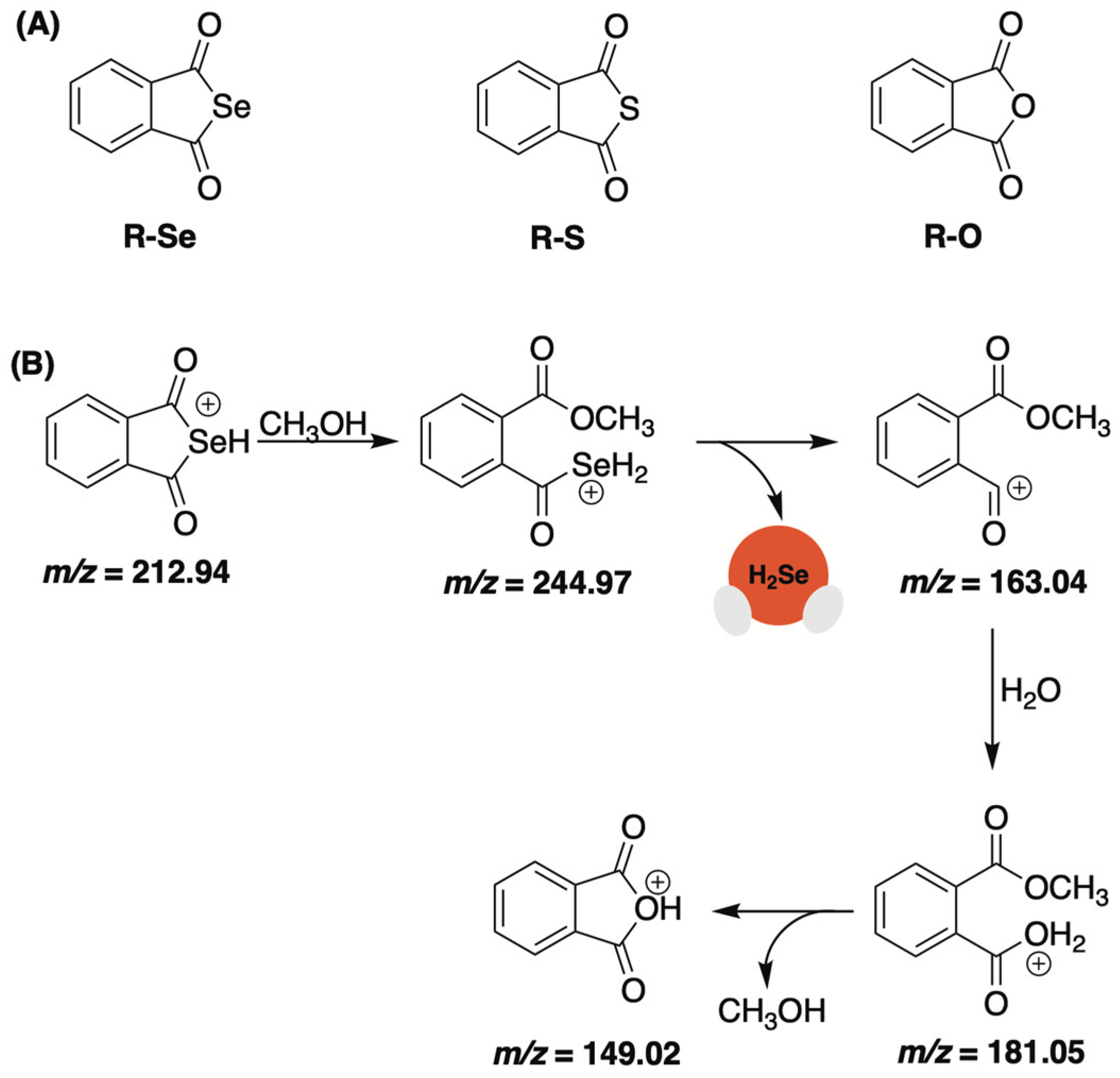
Selenoanhydrides were among the first small molecules assessed for their ability to deliver selenide in a controlled fashion and in response to biologically pertinent molecules [60]. Domínguez-Álvarez and co-workers reported on the impressive anticancer effects of selenoanhydride R-Se (Figure 3A) in earlier studies and suspected it might be due to its release of H2Se [61,62,63]. To examine this, they monitored the fragmentation pattern of R-Se in a 50% methanol/water solution. Under electrospray ionization conditions (ESI), initial attack by methanol led to the observed fragmentation products with the loss of H2Se (Figure 3B). Moreover, the addition of Na2S appeared to amplify the decomposition of the donor with selenium-containing fragments being observed but with a significantly lower abundance. While experimental conditions were not biologically relevant, this simple ESI-MS analysis did provide some insight into the propensity of selenoanhydrides to expel selenide upon nucleophilic exposure.
The reducing capacity of R-Se and related chalcogens (R-S and R-O, Figure 3A) was assessed using cPTIO, a nitric oxide radical scavenger [60]. The authors noted that the addition of H2S potentiated the radical scavenging ability of R-Se and R-S (albeit to a lesser extent) but not R-O. The same trend was observed with the antioxidant glutathione (GSH). While the addition of GSH alone did not effectively quench cPTIO, it in combination with R-Se was found to significantly reduce cPTIO radicals. Similar results were obtained with superoxide (O2•−). Using BMPO as an EPR spin trap reagent, the authors found that H2S/R-Se and H2S/R-S (but not H2S/R-O) could effectively scavenge BMPO-OOH/OH adducts and, perhaps, O2•− directly.
In total, these observations indicate that the reducing potency of thiols (i.e., GSH and H2S) are significantly boosted upon their interaction with R-Se (and to a lesser extent R-S), which is likely to liberate reactive selenium (or sulfur) species, including hydrogen selenide. Although these studies never provided any direct evidence of H2Se release from R-Se, the radical scavenging activity of this purported donor provides a strong indication.

2.4. P=Se Motifs

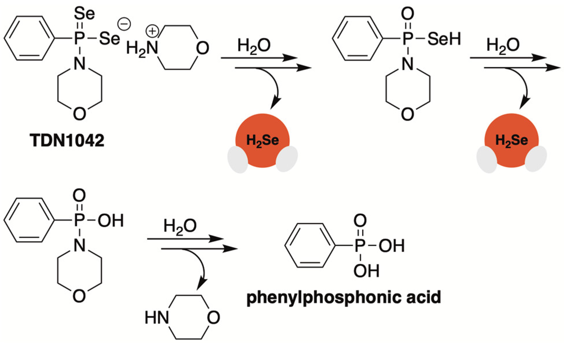
With inspiration from GYY4137, an early and extensively studied H2S donor that gradually decomposes to release H2S via P=S bond cleavage [64], Pluth and co-workers developed an analogous H2Se donor, TDN1042, that delivered selenide (H2S/H2Se) via a similar hydrolytic pathway (Figure 4) [65]. Synthetically, TD1042 was accessed upon treatment of Woollins’ reagent with excess morpholine, akin to the preparation of GYY4137.
Once prepared and structurally characterized by NMR and single crystal X-ray diffraction, H2Se release from TDN1042 in wet DMSO was evaluated using 31P NMR spectroscopy. These studies revealed the clean conversation of TD1042 to phenylphosphonic acid, as expected (Figure 4). This clean transformation to phenylphosphonic acid was also observed in citrate buffer (pH 3.0 to 6.0), with higher rates of release occurring at more acidic pH values, which is consistent with a hydrolysis-based mechanism.
The authors also confirmed the direct liberation of H2Se from TD1042, a key experiment that was omitted from previous reports of H2Se donating motifs. To accomplish this, an aqueous solution of TD1042 was acidified with HCl and sparged with N2 to assist in volatilizing any released H2Se into the vial headspace where it was then bubbled through a separate trapping solution of dinitrofluorobenzene (DNFB). Using HPLC, Pluth and co-workers observed the formation of both di(2,4-dinitrophenyl) selenide ((DNP)2Se) and the related diselenide ((DNP)2Se2) in the trapping solution.
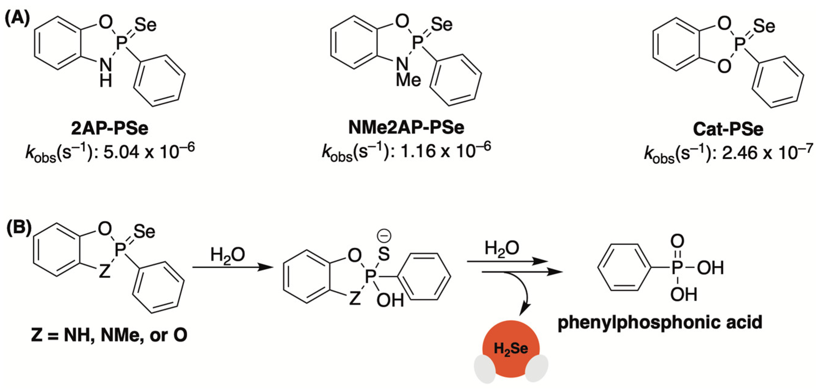
In a later study, the Pluth group sought to augment the rate of H2Se donation from this platform through the introduction of cyclic-PSe donors [66]. To accomplish this, Woollins’ reagent was treated with various ortho-substituted phenols to generate a small library of donors with a single P=Se motif (Cat-PSe, 2AP-PSe, and NMe2AP-PSe, Figure 5A).
By introducing a second electronegative heteroatom (i.e., oxygen or nitrogen), it was suspected that the rate of hydrolysis would increase due to the enhanced electrophilic character of the phosphorous center. This was confirmed by 31P NMR, which was used to monitor donor hydrolysis in PIPES buffer (pH 7.4). Like TD1042, cyclic-PSe compounds were found to release selenide while cleanly forming phenylphosphonic acid as a byproduct (Figure 5B). However, unlike TD1042, these donors were shown to operate at neutral pH due to their enhanced hydrolytic lability. The rates of hydrolysis among cyclic-PSe donors varied significantly with Cat-PSe displaying the slowest rate of hydrolysis and 2AP-PSe hydrolyzing the quickest. This result was somewhat unexpected and suggests that factors other than electronic effects influence the rates of hydrolysis.
Cell permeability studies were also conducted using time-of-flight secondary ion mass spectrometry (TOF-SIMS). A dose-dependent increase in intracellular selenium levels was observed in HeLa cells with increasing concentrations of 2AP-PSe, the most efficient H2Se donor identified in this study.
The in cellulo antioxidant activity of 2AP-PSe was also assessed. When live HeLa cells were treated with exogenous H2O2 (500 µM) and peroxide sensor DCFH-DA, a significant fluorescence response was observed due to the formation of 2′,7′-dichlorofluorescein (DCF) [67]. However, when cells were first pre-treated with 2AP-PSe (5–25 µM) a notable decrease in ROS-generated fluorescence was observed. Due to the instability of H2Se, the authors stress that the observed effects are unlikely due to the buildup of H2Se, but rather an increase in selenocompounds (likely antioxidant proteins) that effectively scavenge ROS.

2.5. Selenocarbonyls

In a comprehensive study by Yi and co-workers, both selenocyclopropenones and arylselenoamides were found to provide highly tunable rates of H2Se delivery in the presence of supraphysiological concentrations of cysteine [68].
Initially, the authors generated a selenium analogue of Michler’s ketone but found that it quickly hydrolyzed in a buffer, producing a red residue (Se0), presumably due to its rapid discharge of selenide (Figure 6A). Searching for a selenocarbonyl with a more tractable H2Se-releasing profile, selenocyclopropenones were then investigated. A small library was generated by treating the corresponding ketone with Woollins’ reagent (Figure 6B). Compound 1 was chosen for initial studies, evaluating its reactivity and selectivity as a selenide donor. HPLC analysis confirmed that 1 was stable in a 50% PBS/CH3CN (pH 7.4) mixture. The introduction of cysteine (2–10 mM), however, led to the expulsion of selenide and the subsequent formation of a red solid (Se0). This was further corroborated by an H2Se-selective gas detector. Donors 2 and 3, with electron-withdrawing substituents at the para position, were found to liberate even more H2Se than 1 in the presence of cysteine, implying that the rates of donation from this platform can be easily tuned via simple structural modifications that alter the electrophilicity of the selenocarbonyl. The reaction between 1 and cysteine was further scrutinized by high-resolution mass spectrometry (HRMS) and identifiable byproducts were uncovered. Based on these observations, a mechanism for cys/thiol-triggered H2Se release from selenocyclopropenones was put forward by the authors (Figure 6B).
Aryl thioamides were previously shown to function as an advantageous platform for the controlled delivery of H2S under biologically relevant conditions [69,70,71]. Yi and co-workers suspected that selenoanalogues would provide an avenue for H2Se donation under similar conditions (Figure 6C) [68]. To test their hypothesis, a small library of selenoamides was generated by treating benzamide derivatives with Woollins’ reagent or by subjecting 4-hydroxybenzonitrile to a mixture of selenium powder and NaBH4. Selenobenzamide (4) was shown to be stable in PBS (pH 7.4), but the addition of cysteine led to the formation of red Se0. This was further substantiated with an H2Se gas detector. As expected, the rate of H2Se release from 4 was found to be significantly slower than 1. However, it was noted that the selenoamide platform could be structurally modified to alter reaction kinetics. For example, the introduction of an electron-donating hydroxyl group at the para position on the phenyl ring (5, Figure 6C) appeared to amplify the speed of selenide release. However, when the hydroxyl group was converted to a methyl ester (6, Figure 6C), its donating efficiency diminished somewhat, even relative to 4. On the other hand, amide N-alkylation (7, Figure 6C) was shown to suppress the rate of selenide delivery even further. HPLC analysis and DFT calculations were used to establish a mechanism for cysteine-triggered H2Se donation (Figure 6C). This proposed pathway aligns with the observation that treatment of arylselenoamides with other thiols that lack a nucleophilic amine (i.e., N-acetylcysteine, glutathione, and β-mercaptoethanol) fail to generate H2Se.

2.6. Selenocarbamates

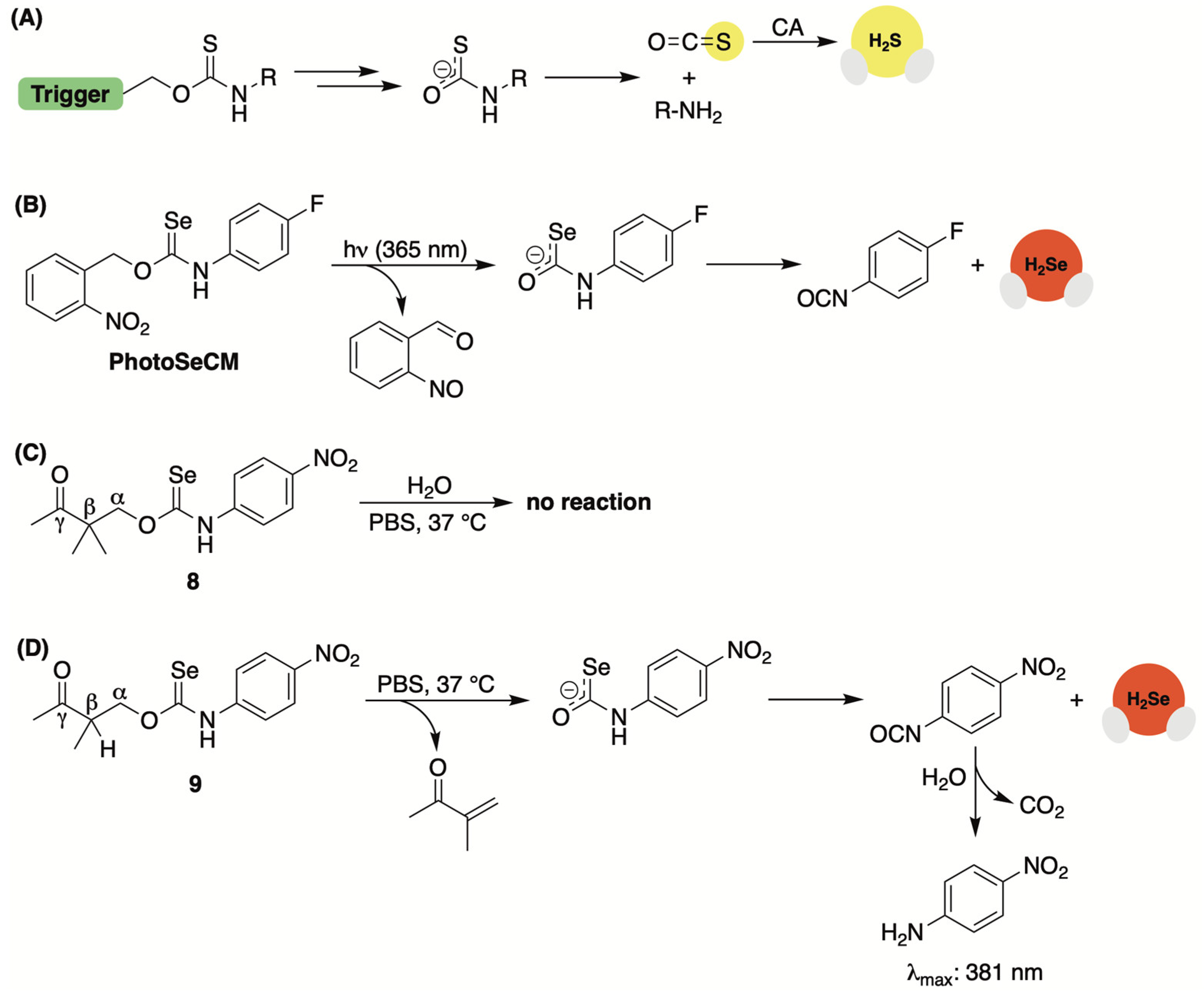
Caged thiocarbamates have offered a reliable avenue for H2S delivery (Figure 7A). They also provide an opportunity to selectivity tune the release of H2S to a specific biological trigger (i.e., ROS [72,73,74,75,76], light [77,78], pH [79,80], and enzymes [81,82]). However, the production of H2S from this donor class is multi-layered, with the triggering event causing the donor to undergo a self-immolating process that first generates carbonyl sulfide (COS) prior to its quick conversation to H2S by the ubiquitous enzyme carbonic anhydrase (Figure 7A) [83].
Continuing with the common theme of prior H2S-releasing motifs laying the foundation for H2Se donor development, Pluth and co-workers were curious whether this framework could be reengineered to release COSe, providing a new means for the controlled delivery of selenide [84]. To test their hypothesis, the authors treated p-fluorophenyl isoselenocyanate with 2-nitrobenzyl alcohol in the presence of NaH to generate light-activated PhotoSeCM (Figure 7B). To analyze product formation upon photoactivation, the authors used 19F NMR to streamline the process. Using this method, consumption of PhotoSeCM (−117 ppm) was confirmed but the expected p-fluoroaniline product (due to COSe release) at −130 ppm was not observed. Instead, a signal matching p-fluoroisocyanate was detected, suggesting the direct liberation of selenide rather than COSe serving as an intermediary (Figure 7B).
Interested in assessing a second COSe/H2Se-releasing system but without the requirement of photoactivation to simplify the overall mechanism, Pluth and team also examined γ-ketoselenocarbamates, which were reconstituted from their earlier work on γ-ketothiocarbamates [80]. This H2S donor class undergoes an enol-mediated self-immolating process that delivers COS/H2S alongside p-nitroaniline, which can be used to track reaction progress due to its UV–Vis signature at 381 nm. γ-Ketoselenocarbamates were constructed from p-nitrophenyl isoselenocyanate and their activation at different pH values was monitored by UV–Vis. While compound 8, which lacks deprotonatable hydrogens at the β position, failed to generate p-nitroaniline (Figure 7C), compound 9, as expected, showed a pH-dependent rate of release, increasing at higher pH values (Figure 7D). In buffer, the authors were unable to provide direct experimental evidence of selenide release from this donor system as the ensuing p-nitrophenyl isocyanate byproduct is unstable and likely to hydrolyze quickly to p-nitroaniline and CO2. Nevertheless, computational studies corroborate the likelihood of direct selenide liberation as it was calculated to be by far the lowest energy decomposition pathway, underscoring the fundamental differences in thiocarbamate/selenocarbamate reactivity.

2.7. γ-Ketoselenides

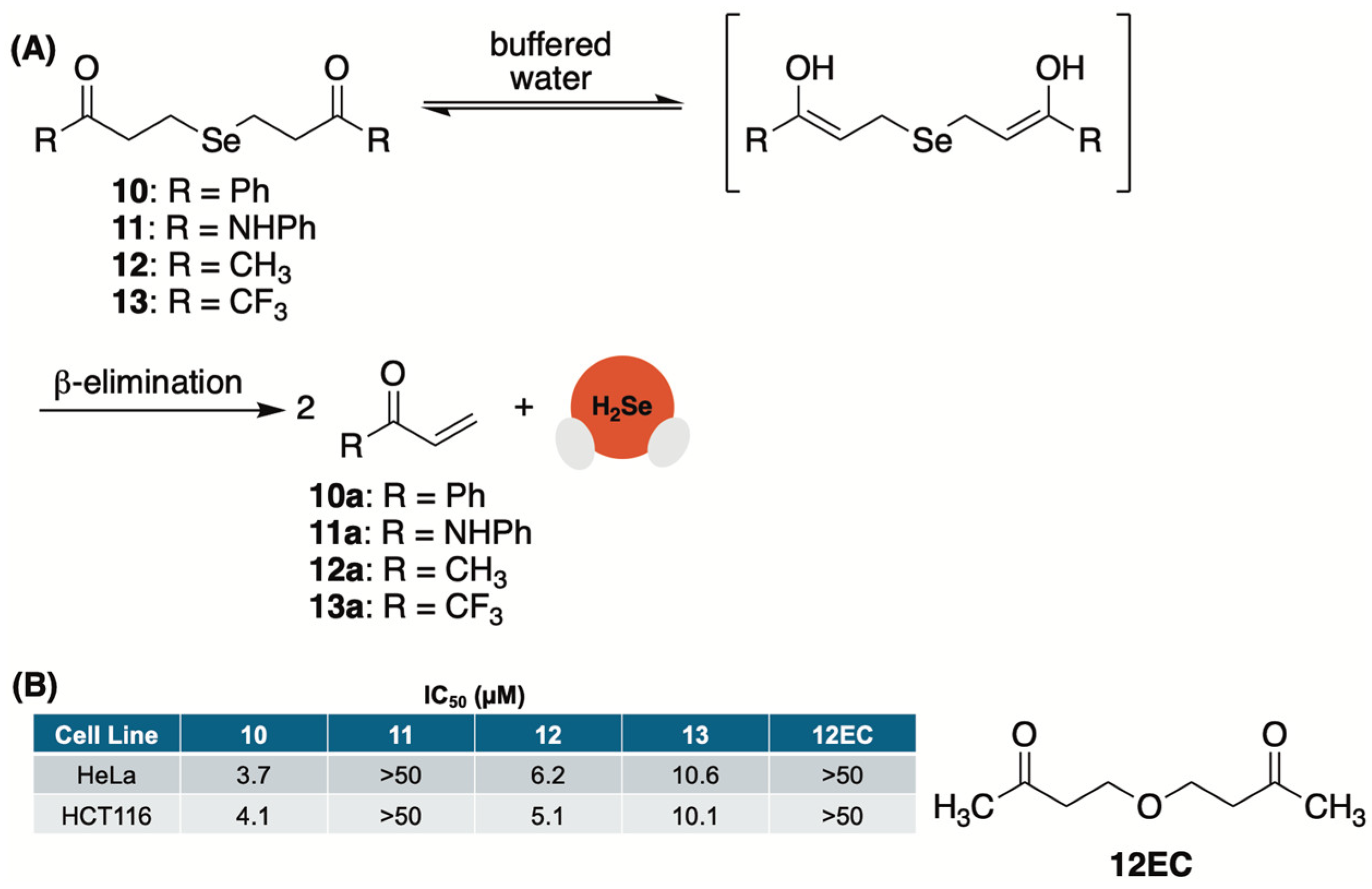
An analogous base-mediated selenide delivery system was first reported by us in 2022 [85]. In this study, we prepared a library of γ-ketoselenides (10–13, Figure 8A) upon treatment of the corresponding γ-ketohalide/tosylate with a solution of sodium selenide that was generated in situ by reducing elemental selenium with NaBH4.
Once in hand, we first monitored the release of selenide from 10 (Figure 8A) in a 1:1 mixture of CD3CN and deuterated phosphate buffer (50 mM, pD 7.4) by tracking the formation of byproduct 10a (Figure 8A) using 1H NMR. During the experiment, we observed a decrease in the intensity of the protons alpha to selenium while the signal of the terminal enone protons simultaneously increased. The addition of 1,4-dioxane as an internal standard allowed us to track the concentration of both and confirm that the consumption of 10 correlated well with the production of 10a. During these studies, we also observed the formation of a red film in our NMR tube, which we attributed to the rapid oxidation of H2Se in an aqueous buffer and its conversion to Se0. This was confirmed by 31P NMR and the formation of triphenylphosphine selenide upon treatment with triphenylphosphine.
Appearing to proceed through an α-deprotonation/β-elimination sequence, we suspected that an enhanced rate of H2Se donation from γ-ketoselenides would be observed at both higher pH values and with donors that possess more acidic alpha protons. To test our hypotheses, the same kinetic studies were repeated at pH 6 and 8.5. As anticipated, the rate of selenide donation was found to be intensified under more basic conditions (pH 8.5) and hindered under more acidic conditions (pH 6). Similarly, alpha proton acidity appeared to play a significant role in donor reactivity with 13 undergoing rapid selenide liberation (confirmed with trapping experiments using benzyl bromide), while 11 was found to be considerably more stable than both 10 and 12. In total, these experiments not only verified the release of selenide from γ-ketoselenides but also the mechanism through which it occurs.
The anticancer activity of these compounds was also assessed in HeLa (human cervical cancer) and HCT116 (human colon cancer) cells in culture (Figure 8B). Predictably, 11, which was found to be stable in buffer for months at a time, liberating only trace amounts of selenide, was found to be completely inactive in both cell lines. Conversely, 10, 12, and 13 exhibited low micromolar activity. As a key control, compound 12EC, an oxygen conger of 12, was also tested and found to be completely inactive, confirming that the activity of donors is due to their release of selenide and not the other components of the reaction. It is also worth mentioning that 11, which liberates selenide at an order of magnitude faster than both 10 and 12, was found to be two-fold less potent in both cell lines. This implies that greater antiproliferative activity may be achieved through continuous exposure to low levels of H2Se for a prolonged period as opposed to a rapid surge in selenide concentration that is likely afforded by 11.

2.8. 5′-O-Selenophosphate Nucleosides

It had been previously shown by Kaczmarek and colleagues that 5′-O-thiophosphate nucleosides function as an H2S source in the presence of histidine triad nucleotide-binding protein 1 (HINT1) [86,87]. Consistent with the common theme throughout this review, Kaczmarek speculated that a selenium congener might function as a HINT1 substrate [88]. If so, then perhaps H2Se, like H2S, would be afforded as a byproduct of the enzyme’s hydrolase activity.
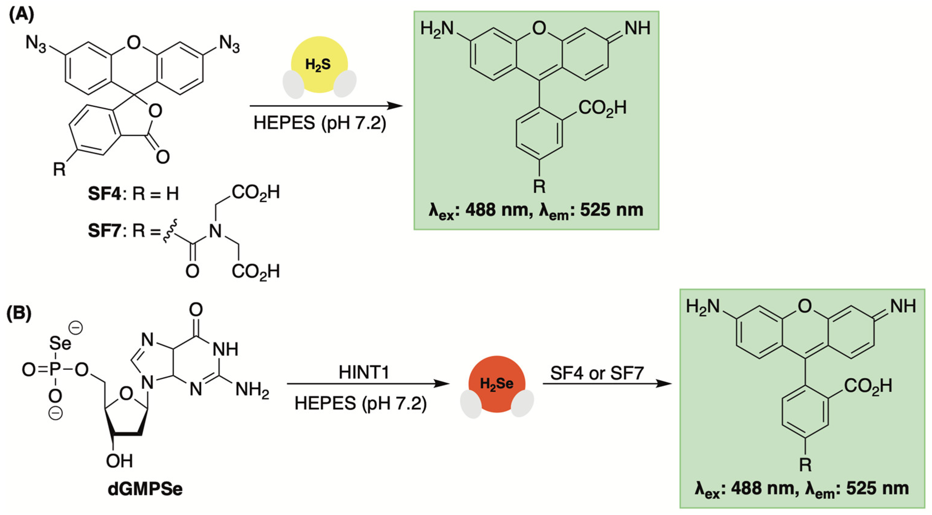
To test this premise, 2′-deoxyguanosine-5′-O-selenophosphate (dGMPSe) was produced using a known method for generating phosphoroselenoates [89]. After confirming the stability of dGMPSe in the buffer alone, its HINT1-catalyzed hydrolysis was assessed. This selenophosphate derivative was found to be a substrate for HINT1 with both deoxyguanosine monophosphate (dGMP), confirmed by HPLC, and H2Se, detected by fluorescence spectroscopy and the use of the reaction-based probe SF7 [90], were observed as products of the enzyme-catalyzed hydrolysis (Figure 9).
After confirming the HINT1-promoted release of H2Se in vitro, the cytotoxicity of dGMPSe was evaluated in HeLa cells. Compared to dGMP, which was found to be nontoxic at all concentrations tested, dGMPSe exhibited dose-dependent cytotoxicity with an IC50 value of 8 µM after a 24 h incubation period. Furthermore, dead cells were shown to exhibit higher fluorescence in the presence of SF7, further supporting the notion that released H2Se from dGMPSe is responsible for cell death.

3. Chemical Tools for H2Se Detection

3.1. Nonspecific Electrophilic Traps: Dinitrofluorobenzene, Benzyl Bromide, and Iodoacetamide

While selective microsensors and a plethora of reaction-based fluorescent probes offer reliable methods for sulfide detection, analogous chemical tools with selenide specificity are lacking. Furthermore, the methylene blue assay, a ubiquitous method for sulfide quantification in buffer, is unlikely to translate to accurate selenide detection due to its rapid oxidation and overall chemical instability compared to that of sulfide. As such, many researchers have turned to the use of nonspecific electrophilic traps as a quick and dirty method for selenide sensing and validation of H2Se donor activation.
As mentioned earlier, Pluth and co-workers relied on dinitrofluorobenzene (DNFB) for confirmation of H2Se donation from TDN1042 (Figure 10A) [65]. However, the high electrophilicity of DNFB, coupled with the augmented nucleophilic character of selenium, made this a challenging endeavor. The authors had previously observed the P=Se moiety of the donor reacting directly with other electrophiles, such as benzyl bromide, making it impossible to distinguish donor alkylation from the alkylation of H2Se. Thus, to unequivocally establish selenide release from TDN1042, the authors placed the DNFB trapping solution in a vial separate from the donor. Under this setup, when TDN1042 was acidified with HCl and sparged with N2, the liberated H2Se was volatilized into the headspace and bubbled through the separate trapping solution containing an excess of DNFB. The contents of the trapping solution were then analyzed by HPLC with the expected mono and diselenide products being clearly visible (Figure 10A).
Likewise, Yi and colleagues first utilized iodoacetamide for quick confirmation of selenide donation from their selenocyclopropenone-based donors (Figure 10B) [68]. This analysis was further complicated by the fact that cysteine, which can be consumed by the added iodoacetamide, was used to trigger H2Se release from this donor class. Nevertheless, using HRMS the authors observed the corresponding mono and diselenide products, which is consistent with the release of selenide and the autooxidation process.
We, too, relied on nonspecific electrophiles, both benzyl bromide and iodoacetamide, for confirmation of H2Se liberation from our γ-ketoselenide-based system [85]. Like the Pluth group, we also sought to unambiguously confirm H2Se donation from 10 by trapping the gas in a separate vial (Figure 10C). We elected to use iodoacetamide for these experiments as the ensuing product from its trapping of volatilized H2Se would be easily recognizable by HRMS. Indeed, when 10 was acidified and sparged with argon, released H2Se was transferred through a cannula needle and into a separate trapping solution where the expected selenide product was clearly visible by HRMS.

3.2. Fluorescent Sensors Based on Benzoselenadiazole Se–N Bond Cleavage

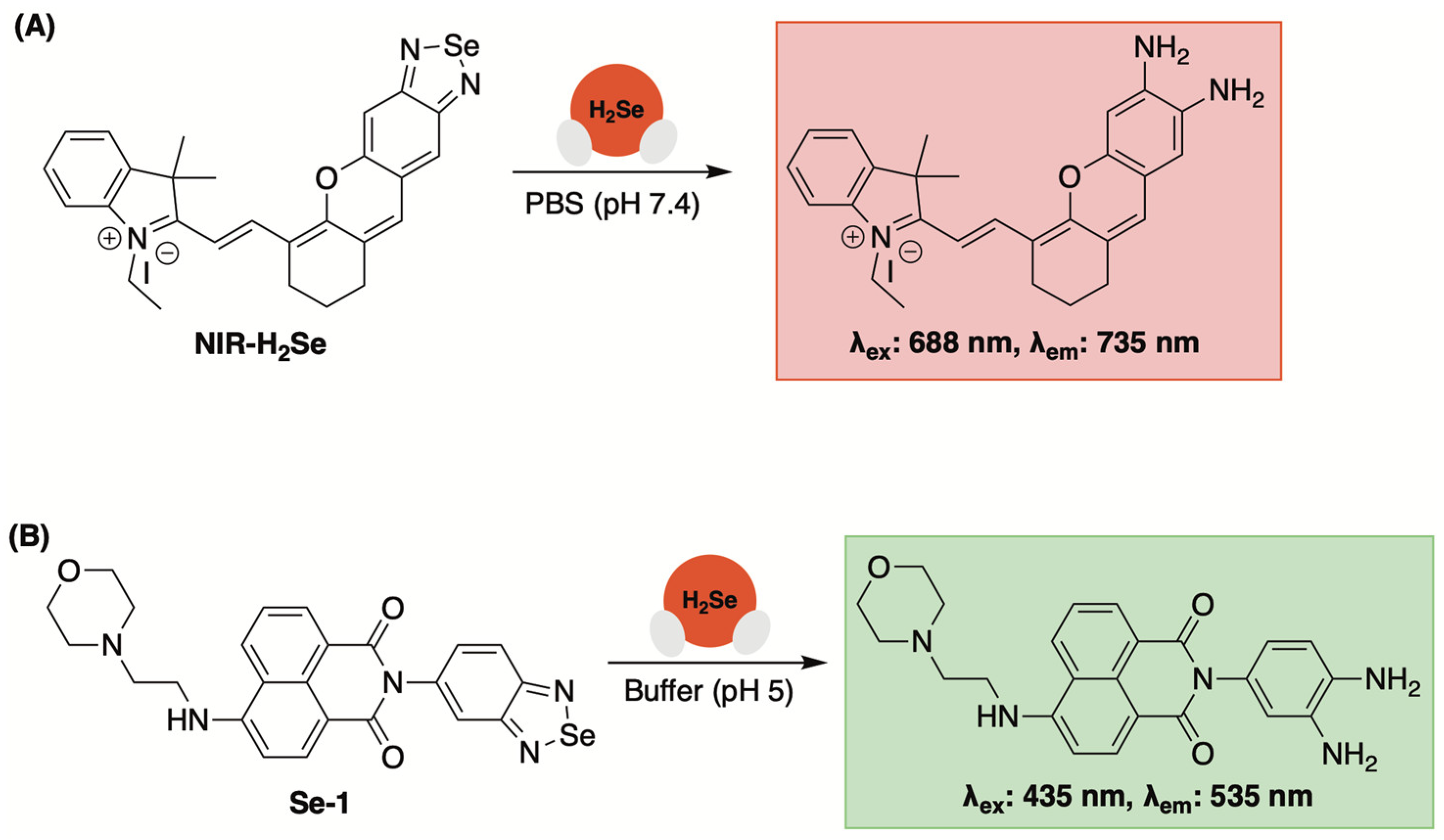
A small molecule fluorescent probe with high selectivity towards H2Se was first reported by Tang and colleagues in 2016 (NIR-H2Se, Figure 11A) [91]. By fabricating a benzoselenadiazole moiety onto a mercaptan dye, the authors found that Se–N bond cleavage occurred quickly in the presence of H2Se, but not when exposed to other selenols or thiols, to afford to a fluorescent diamine reporter (λex: 688 nm, λem: 735 nm).
With an H2Se-selective sensor in hand, the authors then used NIR-H2Se to monitor cellular H2Se levels in HepG2 cells using Na2SeO3 as a metabolic precursor. In this experiment, a dose and time-dependent increase in fluorescence was observed, as expected. Moreover, these investigations were conducted under both hypoxic (1% pO2) and normoxic (20% O2) conditions, with a significant reduction in fluorescence being observed during the latter. This was attributed to rapid H2Se oxidation and the production of superoxide and other reactive oxygen species while exposed to higher O2 levels. Based on these observations, it was concluded that the anticancer activity of Na2SeO3 in HepG2 cells under normoxic conditions can be attributed to ROS-induced cell death. Conversely, under hypoxic conditions, which is a hallmark of solid tumors, a non-oxidative stress mechanism is likely in play due to a notable buildup in cellular H2Se. This hypothesis was further validated in a solid tumor mouse model using NIR-H2Se.
A lysosomal-specific H2Se sensor based on this same framework was reported by Zhang and Jing in 2019 (Se-1, Figure 11B) [92]. In addition to a benzoselenadiazole moiety for selective H2Se detection, a morpholino group was appended to the sensor for lysosomal-targeting [93]. Under simulated lysosomal conditions (acetate buffer, pH 5), a notable increase in fluorescence at 535 nm (λex: 435 nm) was observed when a solution of Se-1 was exposed to H2Se (but not other biologically relevant analytes). Moreover, the fluorescent enhancement of Se-1 in the presence of H2Se was found to be consistent within a pH range of 4.5–7, confirming its compatibility with lysosomal conditions.
After establishing its reactivity and photophysical properties in buffer, lysosomal-targeting of Se-1 was confirmed in HepG2 cells using LysoTracker Blue (Pearson’s correlation coefficient of 0.91). Additionally, using Se-1, the authors observed elevated levels of lysosomal H2Se in hypoxic HepG2 cells, whereas little lysosomal fluorescence was observed under normoxic conditions.
To date, benzoselenadiazoles have not been used as a recognition subunit to validate selenide delivery from novel donor scaffolds. However, its reported high reactivity towards H2Se could prove useful in future studies as the transient nature of H2Se requires rapid detection for accurate monitoring.

3.3. Fluorescent Sensors Based on Disulfide Bond Cleavage

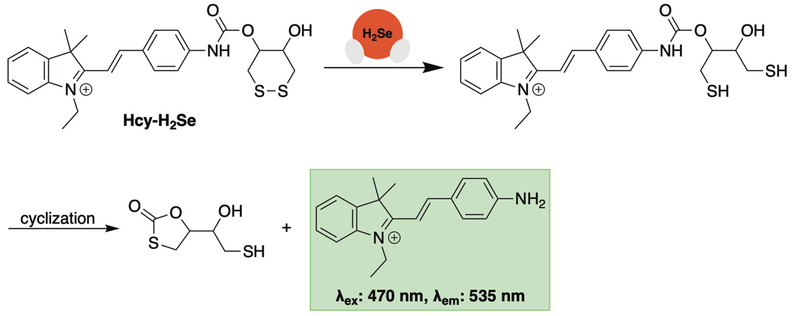
A second-generation sensor from Tang and co-workers employed disulfide bond reduction as a recognition mechanism for H2Se-initiated turn-on fluorescence (Hcy-H2Se, Figure 12) [94]. Thiol-activated prodrugs and chemosensors that utilize disulfide bond reduction as their initiation step have been widely reported [95]. Based on these accounts, it was suspected that a more stable cyclic disulfide (i.e., a six-membered ring) would respond much more quickly to H2Se, given its heightened nucleophilicity, thereby imparting selectivity over other selenols and thiols.
Hcy-H2Se was generated by combining oxidized dithiothreitol [96] with a masked hemicyanine dye via a carbamate linker. When exposed to H2Se, a maximum fluorescence intensity (λex: 470 nm, λem: 535 nm) was reached almost immediately, indicating rapid H2Se-initiated disulfide bond reduction and cyclization (Figure 12). Selectivity studies indicated that the addition of glutathione, H2S, selenocysteine, dithiothreitol, bovine serum albumin, and thioredoxin reductase yielded little fluorescence compared to H2Se. Moreover, the emission intensity at 535 nm correlated well with increasing concentrations of H2Se, affording a good linear relationship between the two.
Bioimaging of H2Se in live human cells was accomplished with Hcy-H2Se [94]. HepG2 cells exposed to Na2SeO3 and Hcy-H2Se under hypoxic conditions exhibited fluorescence that was both dose- and time-dependent. The authors also demonstrated that hypoxic tumor regions in mice injected with sodium selenite could be imaged with Hcy-H2Se. This further supports the anticancer effects of selenium in hypoxic solid tumors being due to an increase in reductive rather an oxidative stress.

3.4. Spectroscopic Sensors Based on Nucleophilic Substitution

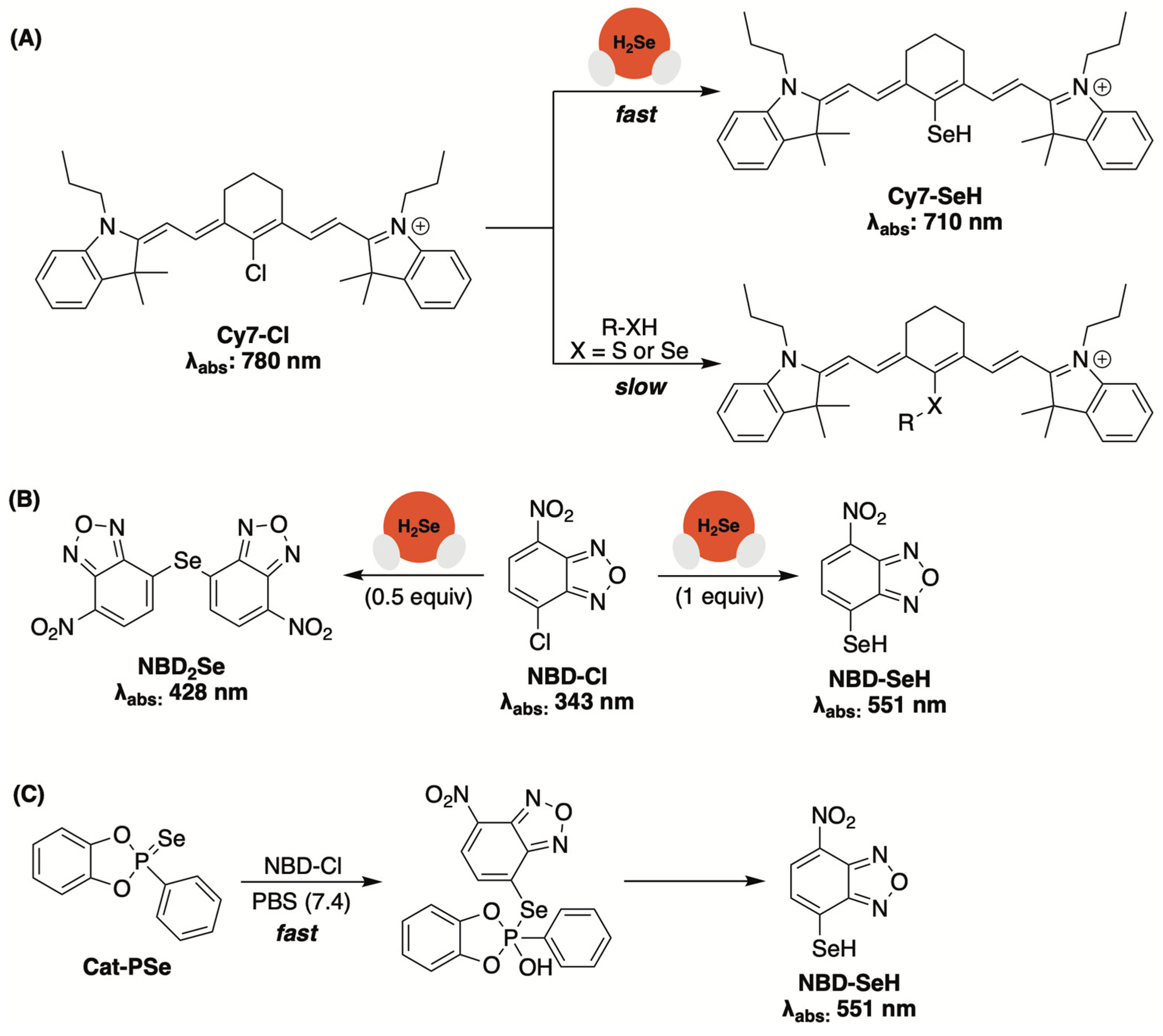
While examining selenocyclopropenones and selenoamides as potential cysteine-activated donors (Figure 6), Yi and collaborators developed a quantitative assay for H2Se based on its nucleophilic substitution with a carefully chosen electrophilic species [68]. Upon testing several potential candidates, the authors discovered that commercially available Cy7-Cl reacted much more quickly with selenide than other selenols and thiols, to form Cy7-SeH (Figure 13A). As the reaction progressed, it was noted that the starting absorbance of 780 nm (Cy7-Cl) was shifted to 710 nm (Cy7-SeH), offering a convenient colorimetric method for monitoring reaction progress. The reaction between Cy7-Cl (500 µM) and various amounts of Na2Se (25–350 µM) was also monitored HPLC, with a plot of Cy7-SeH peak area vs. selenide concentration yielding a straight line. This calibration curve was then used to effectively determine the H2Se-releasing efficiency from selenocyclopropenones in the presence of cysteine.
While evaluating the H2Se-releasing efficiencies of cyclic-PSe donors (Figure 5), the Pluth group found that commercially available 4-chloro-7-nitrobenzofurazan (NBD-Cl), which had been previously used to detect H2S [97], could also be used to trap selenide via a nucleophilic aromatic substitution reaction (Figure 13B) [66]. Initially, the authors analyzed the reaction between NBD-Cl and tetrabutylammonium hydroselenide (NBu4SeH) in PBS (pH 7.4). They found that substoichiometric amounts of selenide provided NBD2Se (λabs: 428 nm), while stoichiometric HSe yielded NBD-SeH as the primary product (λabs: 551 nm). With confirmation of a colorimetric response, NBD-Cl was then used to monitor the hydrolysis of Cat-PSe in PBS (Figure 13C). While the formation of NBD-SeH was clearly visible (λabs: 551 nm), the rate of apparent H2Se liberation appeared to occur much faster when compared to earlier 31P NMR hydrolysis experiments. The authors attributed this notable rate enhancement to donor alkylation, rather than hydrolysis, while in the presence of a strong electrophile, such as NBD-Cl. Therefore, while the use of NBD-Cl appears to provide a convenient colorimetric method for monitoring donor progress, additional trapping experiments are likely necessary to untangle donor alkylation from the alkylation of released H2Se in solution.

3.5. Fluorescent Sensors Based on Azide Reduction

Like H2S-responsive NBD-Cl being reintroduced for H2Se detection, H2S-sensitive fluorescent sensors that rely on aryl azide reduction were reexamined for their capacity to detect H2Se [89]. Probes SF4 and SF7 (Figure 14) were previously engineered by Chang and co-workers for monitoring H2S levels under physiological conditions [90,98]. While stable towards other sulfur-containing molecules, H2S initiates aryl azide reduction on the masked rhodamine dye, resulting in a turn-fluorescent response (Figure 14A).
Given the increased reactivity of H2Se, Kaczmarek and co-workers suspected that these same sensors would be responsive towards H2Se and used them to confirm selenide release from dGMPSe in both in a buffer and in live HeLa cells (Figure 14B) [88]. In fact, when using SF7 and SF4, higher fluorescence values were observed with dGMPSe compared to its sulfur congener, which likely stems from the higher reactivity and reductive properties of H2Se compared to that of H2S. Thus, while not selective for H2Se, aryl azide reduction appears to be a potential method for the real-time monitoring of donor progress.

4. Conclusions and Outlook

The continued evolution of H2Se-specific chemical tools is a prime objective for those interested in uncovering the (patho)physiological effects of hydrogen selenide. While donor compounds capable of providing the slow and sustained release of H2Se have begun to emerge, the introduction of additional stimulus-responsive donors (i.e., enzyme or bioanalyte-triggered) with greater spatiotemporal control over their biological delivery of H2Se will serve as a great advancement in the field. H2Se detection and quantification, especially when liberated from a donor scaffold, remains a great challenge. Most current methods rely on electrophiles to trap the released selenide, which often leads to interference from the donor itself given the intensified nucleophilic character of selenium. New approaches for detection that avoid the introduction of strong electrophiles should continue to be explored, including additional reaction-based fluorescent sensors that can be used in cellular and in vivo imaging experiments. Together, these compounds will continue to unearth the unique aspects of H2Se chemical biology, while providing new evidence that further supports (or refutes) the addition of H2Se as the fourth gasotransmitter.

Funding

This research was funded by the National Science Foundation (Grant No. 2143826).

Institutional Review Board Statement

Not applicable.

Informed Consent Statement

Not applicable.

Data Availability Statement

Not applicable.

Conflicts of Interest

The authors declare no conflict of interest.

References

  1. Weeks, M.E. The Discovery of the Elements. VI. Tellurium and Selenium. J. Chem. Educ. 1932, 9, 474. [Google Scholar] [CrossRef]
  2. Levander, O.A. Scientific Rationale for the 1989 Recommended Dietary Allowance for Selenium. J. Am. Diet. Assoc. 1991, 91, 1572–1576. [Google Scholar] [CrossRef]
  3. Labunskyy, V.M.; Hatfield, D.L.; Gladyshev, V.N. Selenoproteins: Molecular Pathways and Physiological Roles. Physiol. Rev. 2014, 94, 739–777. [Google Scholar] [CrossRef]
  4. Atkins, J.F.; Gesteland, R.F. The Twenty-First Amino Acid. Nature 2000, 407, 463–464. [Google Scholar] [CrossRef]
  5. Böck, A.; Forchhammer, K.; Heider, J.; Leinfelder, W.; Sawers, G.; Veprek, B.; Zinoni, F. Selenocysteine: The 21st Amino Acid. Mol. Microbiol. 1991, 5, 515–520. [Google Scholar] [CrossRef]
  6. Brown, K.M.; Arthur, J.R. Selenium, Selenoproteins and Human Health: A Review. Public Health Nutr. 2001, 4, 593–599. [Google Scholar] [CrossRef]
  7. Kieliszek, M.; Błażejak, S. Current Knowledge on the Importance of Selenium in Food for Living Organisms: A Review. Molecules 2016, 21, 609. [Google Scholar] [CrossRef]
  8. Rayman, M.P. Selenium and Human Health. Lancet 2012, 379, 1256–1268. [Google Scholar] [CrossRef]
  9. Schomburg, L. Dietary Selenium and Human Health. Nutrients 2016, 9, 22. [Google Scholar] [CrossRef] [PubMed]
  10. Chen, J. An Original Discovery: Selenium Deficiency and Keshan Disease (an Endemic Heart Disease). Asia Pac. J. Clin. Nutr. 2012, 21, 320–326. [Google Scholar] [PubMed]
  11. Shi, Y.; Yang, W.; Tang, X.; Yan, Q.; Cai, X.; Wu, F. Keshan Disease: A Potentially Fatal Endemic Cardiomyopathy in Remote Mountains of China. Front. Pediatr. 2021, 9, 576916. [Google Scholar] [CrossRef] [PubMed]
  12. Wang, L.; Yin, J.; Yang, B.; Qu, C.; Lei, J.; Han, J.; Guo, X. Serious Selenium Deficiency in the Serum of Patients with Kashin–Beck Disease and the Effect of Nano-Selenium on Their Chondrocytes. Biol. Trace Elem. Res. 2020, 194, 96–104. [Google Scholar] [CrossRef]
  13. Yao, Y.; Pei, F.; Kang, P. Selenium, Iodine, and the Relation with Kashin-Beck Disease. Nutrition 2011, 27, 1095–1100. [Google Scholar] [CrossRef] [PubMed]
  14. Arthur, J.R.; McKenzie, R.C.; Beckett, G.J. Selenium in the Immune System. J. Nutr. 2003, 133, 1457S–1459S. [Google Scholar] [CrossRef]
  15. Hoffmann, P.R.; Berry, M.J. The Influence of Selenium on Immune Responses. Mol. Nutr. Food Res. 2008, 52, 1273–1280. [Google Scholar] [CrossRef] [PubMed]
  16. Al-Mubarak, A.A.; van der Meer, P.; Bomer, N. Selenium, Selenoproteins, and Heart Failure: Current Knowledge and Future Perspective. Curr. Heart Fail. Rep. 2021, 18, 122–131. [Google Scholar] [CrossRef] [PubMed]
  17. Bomer, N.; Grote Beverborg, N.; Hoes, M.F.; Streng, K.W.; Vermeer, M.; Dokter, M.M.; IJmker, J.; Anker, S.D.; Cleland, J.G.F.; Hillege, H.L.; et al. Selenium and Outcome in Heart Failure. Eur. J. Heart Fail. 2020, 22, 1415–1423. [Google Scholar] [CrossRef]
  18. Rataan, A.O.; Geary, S.M.; Zakharia, Y.; Rustum, Y.M.; Salem, A.K. Potential Role of Selenium in the Treatment of Cancer and Viral Infections. Int. J. Mol. Sci. 2022, 23, 2215. [Google Scholar] [CrossRef]
  19. Rayman, M.P. Selenium in Cancer Prevention: A Review of the Evidence and Mechanism of Action. Proc. Nutr. Soc. 2005, 64, 527–542. [Google Scholar] [CrossRef]
  20. Sayehmiri, K.; Azami, M.; Mohammadi, Y.; Soleymani, A.; Tardeh, Z. The Association between Selenium and Prostate Cancer: A Systematic Review and Meta-Analysis. Asian Pac. J. Cancer Prev. 2018, 19, 1431–1437. [Google Scholar] [CrossRef]
  21. Vinceti, M.; Filippini, T.; Del Giovane, C.; Dennert, G.; Zwahlen, M.; Brinkman, M.; Zeegers, M.P.; Horneber, M.; D’Amico, R.; Crespi, C.M. Selenium for Preventing Cancer. Cochrane Database Syst. Rev. 2018, 1, CD005195. [Google Scholar] [CrossRef]
  22. Barceloux, D.G.; Barceloux, D. Selenium. J. Toxicol. Clin. Toxicol. 1999, 37, 145–172. [Google Scholar] [CrossRef]
  23. Hughes, M.N.; Centelles, M.N.; Moore, K.P. Making and Working with Hydrogen Sulfide. Free Radic. Biol. Med. 2009, 47, 1346–1353. [Google Scholar] [CrossRef]
  24. Haynes, W.M.; Lide, D.R.; Bruno, T.J. (Eds.) CRC Handbook of Chemistry and Physics 2016–2017: A Ready-Reference Book of Chemical and Physical Data, 97th ed.; CRC Press: Boca Raton, FL, USA, 2017; ISBN 978-1-4987-5429-3. [Google Scholar]
  25. Jiang, J.; Chan, A.; Ali, S.; Saha, A.; Haushalter, K.J.; Lam, W.-L.M.; Glasheen, M.; Parker, J.; Brenner, M.; Mahon, S.B.; et al. Hydrogen Sulfide—Mechanisms of Toxicity and Development of an Antidote. Sci. Rep. 2016, 6, 20831. [Google Scholar] [CrossRef] [PubMed]
  26. National Research Council (U.S.) (Ed.) Acute Exposure Guideline Levels for Selected Airborne Chemicals; The Compass Series; National Academy Press: Washington, DC, USA, 2000; ISBN 978-0-309-07294-6. [Google Scholar]
  27. Filipovic, M.R.; Zivanovic, J.; Alvarez, B.; Banerjee, R. Chemical Biology of H2S Signaling through Persulfidation. Chem. Rev. 2018, 118, 1253–1337. [Google Scholar] [CrossRef]
  28. Li, L.; Rose, P.; Moore, P.K. Hydrogen Sulfide and Cell Signaling. Annu. Rev. Pharmacol. Toxicol. 2011, 51, 169–187. [Google Scholar] [CrossRef]
  29. Wang, R. Hydrogen Sulfide: The Third Gasotransmitter in Biology and Medicine. Antioxid. Redox Signal. 2010, 12, 1061–1064. [Google Scholar] [CrossRef]
  30. Wang, R. The Gasotransmitter Role of Hydrogen Sulfide. Antioxid. Redox Signal. 2003, 5, 493–501. [Google Scholar] [CrossRef]
  31. Miles, E.W.; Kraus, J.P. Cystathionine β-Synthase: Structure, Function, Regulation, and Location of Homocystinuria-Causing Mutations. J. Biol. Chem. 2004, 279, 29871–29874. [Google Scholar] [CrossRef]
  32. Pan, L.L.; Liu, X.H.; Gong, Q.H.; Yang, H.B.; Zhu, Y.Z. Role of Cystathionine γ-Lyase/Hydrogen Sulfide Pathway in Cardiovascular Disease: A Novel Therapeutic Strategy? Antioxid. Redox Signal. 2012, 17, 106–118. [Google Scholar] [CrossRef]
  33. Shibuya, N.; Tanaka, M.; Yoshida, M.; Ogasawara, Y.; Togawa, T.; Ishii, K.; Kimura, H. 3-Mercaptopyruvate Sulfurtransferase Produces Hydrogen Sulfide and Bound Sulfane Sulfur in the Brain. Antioxid. Redox Signal. 2009, 11, 703–714. [Google Scholar] [CrossRef] [PubMed]
  34. Zhang, X.; Bian, J.-S. Hydrogen Sulfide: A Neuromodulator and Neuroprotectant in the Central Nervous System. ACS Chem. Neurosci. 2014, 5, 876–883. [Google Scholar] [CrossRef]
  35. Abe, K.; Kimura, H. The Possible Role of Hydrogen Sulfide as an Endogenous Neuromodulator. J. Neurosci. 1996, 16, 1066–1071. [Google Scholar] [CrossRef]
  36. Guo, W.; Kan, J.; Cheng, Z.; Chen, J.; Shen, Y.; Xu, J.; Wu, D.; Zhu, Y. Hydrogen Sulfide as an Endogenous Modulator in Mitochondria and Mitochondria Dysfunction. Oxidative Med. Cell. Longev. 2012, 2012, 878052. [Google Scholar] [CrossRef]
  37. Murphy, B.; Bhattacharya, R.; Mukherjee, P. Hydrogen Sulfide Signaling in Mitochondria and Disease. FASEB J. 2019, 33, 13098–13125. [Google Scholar] [CrossRef]
  38. Powell, C.R.; Dillon, K.M.; Matson, J.B. A Review of Hydrogen Sulfide (H2S) Donors: Chemistry and Potential Therapeutic Applications. Biochem. Pharmacol. 2018, 149, 110–123. [Google Scholar] [CrossRef]
  39. Xu, S.; Hamsath, A.; Neill, D.L.; Wang, Y.; Yang, C.; Xian, M. Strategies for the Design of Donors and Precursors of Reactive Sulfur Species. Chem. A Eur. J. 2019, 25, 4005–4016. [Google Scholar] [CrossRef] [PubMed]
  40. Levinn, C.M.; Cerda, M.M.; Pluth, M.D. Activatable Small-Molecule Hydrogen Sulfide Donors. Antioxid. Redox Signal. 2020, 32, 96–109. [Google Scholar] [CrossRef]
  41. Hu, Q.; Lukesh, J.C. H2S Donors with Cytoprotective Effects in Models of MI/R Injury and Chemotherapy-Induced Cardiotoxicity. Antioxidants 2023, 12, 650. [Google Scholar] [CrossRef] [PubMed]
  42. Papapetropoulos, A.; Foresti, R.; Ferdinandy, P. Pharmacology of the ‘Gasotransmitters’ NO, CO and H2S: Translational Opportunities. Br. J. Pharmacol. 2015, 172, 1395–1396. [Google Scholar] [CrossRef]
  43. Wang, R. Shared Signaling Pathways among Gasotransmitters. Proc. Natl. Acad. Sci. USA 2012, 109, 8801–8802. [Google Scholar] [CrossRef]
  44. Yang, G.; Sener, A.; Ji, Y.; Pei, Y.; Pluth, M.D. Gasotransmitters in Biology and Medicine: Molecular Mechanisms and Drug Targets. Oxidative Med. Cell. Longev. 2016, 2016, 4627308. [Google Scholar] [CrossRef] [PubMed]
  45. Wang, R. Two’s Company, Three’s a Crowd: Can H2S Be the Third Endogenous Gaseous Transmitter? FASEB J. 2002, 16, 1792–1798. [Google Scholar] [CrossRef]
  46. Kuganesan, M.; Samra, K.; Evans, E.; Singer, M.; Dyson, A. Selenium and Hydrogen Selenide: Essential Micronutrient and the Fourth Gasotransmitter? Intensive Care Med. Exp. 2019, 7, 71. [Google Scholar] [CrossRef]
  47. Samra, K.; Kuganesan, M.; Smith, W.; Kleyman, A.; Tidswell, R.; Arulkumaran, N.; Singer, M.; Dyson, A. The Pharmacology and Therapeutic Utility of Sodium Hydroselenide. Int. J. Mol. Sci. 2021, 22, 3258. [Google Scholar] [CrossRef]
  48. Tarze, A.; Dauplais, M.; Grigoras, I.; Lazard, M.; Ha-Duong, N.-T.; Barbier, F.; Blanquet, S.; Plateau, P. Extracellular Production of Hydrogen Selenide Accounts for Thiol-Assisted Toxicity of Selenite against Saccharomyces Cerevisiae. J. Biol. Chem. 2007, 282, 8759–8767. [Google Scholar] [CrossRef] [PubMed]
  49. Pan, X.; Song, X.; Wang, C.; Cheng, T.; Luan, D.; Xu, K.; Tang, B. H2Se Induces Reductive Stress in HepG2 Cells and Activates Cell Autophagy by Regulating the Redox of HMGB1 Protein under Hypoxia. Theranostics 2019, 9, 1794–1808. [Google Scholar] [CrossRef]
  50. Iwata, A.; Morrison, M.L.; Blackwood, J.E.; Roth, M.B. Selenide Targets to Reperfusing Tissue and Protects It from Injury. Crit. Care Med. 2015, 43, 1361–1367. [Google Scholar] [CrossRef]
  51. Painter, E.P. The Chemistry and Toxicity of Selenium Compounds, with Special Reference to the Selenium Problem. Chem. Rev. 1941, 28, 179–213. [Google Scholar] [CrossRef]
  52. Ganther, H.E. Selenotrisulfides. Formation by the Reaction of Thiols with Selenious Acid. Biochemistry 1968, 7, 2898–2905. [Google Scholar] [CrossRef] [PubMed]
  53. Nakagawa, T.; Aoyama, E.; Kobayashi, N.; Tanaka, H.; Chikuma, M.; Sakurai, H.; Nakayama, M. Thiol Exchange Reactions Involving Selenotrisulfides. Biochem. Biophys. Res. Commun. 1988, 150, 1149–1154. [Google Scholar] [CrossRef]
  54. Haratake, M.; Ono, M.; Nakayama, M. Penicillamine Selenotrisulfide as a Selenium-Source in Mice. J. Health Sci. 2004, 50, 366–371. [Google Scholar] [CrossRef]
  55. Haratake, M.; Hongoh, M.; Miyauchi, M.; Hirakawa, R.; Ono, M.; Nakayama, M. Albumin-Mediated Selenium Transfer by a Selenotrisulfide Relay Mechanism. Inorg. Chem. 2008, 47, 6273–6280. [Google Scholar] [CrossRef]
  56. Liu, M.; Bu, F.; Li, G.; Xie, W.; Xu, H.; Wang, X. S-Se-S Type Molecule: A Bactericidal Promoter against H2S-Induced Antibiotic Resistance. Innov. Life 2024, 2, 100076. [Google Scholar] [CrossRef]
  57. Asfar, P.; Radermacher, P. Drug-Induced “Suspended Animation”: Can a Dream Become True? Crit. Care Med. 2015, 43, 1528–1530. [Google Scholar] [CrossRef] [PubMed]
  58. Szabo, C. Gaseotransmitters: New Frontiers for Translational Science. Sci. Transl. Med. 2010, 2, 59ps54. [Google Scholar] [CrossRef]
  59. Klayman, D.L.; Griffin, T.S. Reaction of Selenium with Sodium Borohydride in Protic Solvents. A Facile Method for the Introduction of Selenium into Organic Molecules. J. Am. Chem. Soc. 1973, 95, 197–199. [Google Scholar] [CrossRef]
  60. Kharma, A.; Misak, A.; Grman, M.; Brezova, V.; Kurakova, L.; Baráth, P.; Jacob, C.; Chovanec, M.; Ondrias, K.; Domínguez-Álvarez, E. Release of Reactive Selenium Species from Phthalic Selenoanhydride in the Presence of Hydrogen Sulfide and Glutathione with Implications for Cancer Research. New J. Chem. 2019, 43, 11771–11783. [Google Scholar] [CrossRef]
  61. Domínguez-Álvarez, E.; Gajdács, M.; Spengler, G.; Palop, J.A.; Marć, M.A.; Kieć-Kononowicz, K.; Amaral, L.; Molnár, J.; Jacob, C.; Handzlik, J.; et al. Identification of Selenocompounds with Promising Properties to Reverse Cancer Multidrug Resistance. Bioorg. Med. Chem. Lett. 2016, 26, 2821–2824. [Google Scholar] [CrossRef] [PubMed]
  62. Domínguez-Álvarez, E.; Plano, D.; Font, M.; Calvo, A.; Prior, C.; Jacob, C.; Palop, J.A.; Sanmartín, C. Synthesis and Antiproliferative Activity of Novel Selenoester Derivatives. Eur. J. Med. Chem. 2014, 73, 153–166. [Google Scholar] [CrossRef]
  63. Gajdács, M.; Spengler, G.; Sanmartín, C.; Marć, M.A.; Handzlik, J.; Domínguez-Álvarez, E. Selenoesters and Selenoanhydrides as Novel Multidrug Resistance Reversing Agents: A Confirmation Study in a Colon Cancer MDR Cell Line. Bioorg. Med. Chem. Lett. 2017, 27, 797–802. [Google Scholar] [CrossRef]
  64. Li, L.; Whiteman, M.; Guan, Y.Y.; Neo, K.L.; Cheng, Y.; Lee, S.W.; Zhao, Y.; Baskar, R.; Tan, C.-H.; Moore, P.K. Characterization of a Novel, Water-Soluble Hydrogen Sulfide–Releasing Molecule (GYY4137): New Insights into the Biology of Hydrogen Sulfide. Circulation 2008, 117, 2351–2360. [Google Scholar] [CrossRef] [PubMed]
  65. Newton, T.D.; Pluth, M.D. Development of a Hydrolysis-Based Small-Molecule Hydrogen Selenide (H2Se) Donor. Chem. Sci. 2019, 10, 10723–10727. [Google Scholar] [CrossRef]
  66. Newton, T.D.; Bolton, S.G.; Garcia, A.C.; Chouinard, J.E.; Golledge, S.L.; Zakharov, L.N.; Pluth, M.D. Hydrolysis-Based Small-Molecule Hydrogen Selenide (H2Se) Donors for Intracellular H2Se Delivery. J. Am. Chem. Soc. 2021, 143, 19542–19550. [Google Scholar] [CrossRef]
  67. Myhre, O.; Andersen, J.M.; Aarnes, H.; Fonnum, F. Evaluation of the Probes 2′,7′-Dichlorofluorescin Diacetate, Luminol, and Lucigenin as Indicators of Reactive Species Formation. Biochem. Pharmacol. 2003, 65, 1575–1582. [Google Scholar] [CrossRef]
  68. Kang, X.; Huang, H.; Jiang, C.; Cheng, L.; Sang, Y.; Cai, X.; Dong, Y.; Sun, L.; Wen, X.; Xi, Z.; et al. Cysteine-Activated Small-Molecule H2Se Donors Inspired by Synthetic H2S Donors. J. Am. Chem. Soc. 2022, 144, 3957–3967. [Google Scholar] [CrossRef]
  69. Martelli, A.; Testai, L.; Citi, V.; Marino, A.; Pugliesi, I.; Barresi, E.; Nesi, G.; Rapposelli, S.; Taliani, S.; Da Settimo, F.; et al. Arylthioamides as H2S Donors: L-Cysteine-Activated Releasing Properties and Vascular Effects In Vitro and In Vivo. ACS Med. Chem. Lett. 2013, 4, 904–908. [Google Scholar] [CrossRef]
  70. Hu, Q.; Suarez, S.I.; Hankins, R.A.; Lukesh, J.C. Intramolecular Thiol- and Selenol-Assisted Delivery of Hydrogen Sulfide. Angew. Chem. Int. Ed. 2022, 61, e202210754. [Google Scholar] [CrossRef]
  71. Hu, Q.; Zhu, C.; Hankins, R.A.; Murmello, A.R.; Marrs, G.S.; Lukesh, J.C. An ROS-Responsive Donor That Self-Reports Its H2S Delivery by Forming a Benzoxazole-Based Fluorophore. J. Am. Chem. Soc. 2023, 145, 25486–25494. [Google Scholar] [CrossRef] [PubMed]
  72. Zhao, Y.; Pluth, M.D. Hydrogen Sulfide Donors Activated by Reactive Oxygen Species. Angew. Chem. Int. Ed. 2016, 55, 14638–14642. [Google Scholar] [CrossRef]
  73. Zhao, Y.; Henthorn, H.A.; Pluth, M.D. Kinetic Insights into Hydrogen Sulfide Delivery from Caged-Carbonyl Sulfide Isomeric Donor Platforms. J. Am. Chem. Soc. 2017, 139, 16365–16376. [Google Scholar] [CrossRef] [PubMed]
  74. Chauhan, P.; Jos, S.; Chakrapani, H. Reactive Oxygen Species-Triggered Tunable Hydrogen Sulfide Release. Org. Lett. 2018, 20, 3766–3770. [Google Scholar] [CrossRef]
  75. Hu, Y.; Li, X.; Fang, Y.; Shi, W.; Li, X.; Chen, W.; Xian, M.; Ma, H. Reactive Oxygen Species-Triggered off-on Fluorescence Donor for Imaging Hydrogen Sulfide Delivery in Living Cells. Chem. Sci. 2019, 10, 7690–7694. [Google Scholar] [CrossRef] [PubMed]
  76. Zhu, C.; Suarez, S.I.; Lukesh, J.C. Illuminating and Alleviating Cellular Oxidative Stress with an ROS-Activated, H2S-Donating Theranostic. Tetrahedron Lett. 2021, 69, 152944. [Google Scholar] [CrossRef]
  77. Zhao, Y.; Bolton, S.G.; Pluth, M.D. Light-Activated COS/H2S Donation from Photocaged Thiocarbamates. Org. Lett. 2017, 19, 2278–2281. [Google Scholar] [CrossRef] [PubMed]
  78. Sharma, A.K.; Nair, M.; Chauhan, P.; Gupta, K.; Saini, D.K.; Chakrapani, H. Visible-Light-Triggered Uncaging of Carbonyl Sulfide for Hydrogen Sulfide (H2S) Release. Org. Lett. 2017, 19, 4822–4825. [Google Scholar] [CrossRef] [PubMed]
  79. Gilbert, A.K.; Zhao, Y.; Otteson, C.E.; Pluth, M.D. Development of Acid-Mediated H2S/COS Donors That Respond to a Specific pH Window. J. Org. Chem. 2019, 84, 14469–14475. [Google Scholar] [CrossRef]
  80. Zhao, Y.; Steiger, A.K.; Pluth, M.D. Colorimetric Carbonyl Sulfide (COS)/Hydrogen Sulfide (H2S) Donation from γ-Ketothiocarbamate Donor Motifs. Angew. Chem. Int. Ed. 2018, 57, 13101–13105. [Google Scholar] [CrossRef] [PubMed]
  81. Chauhan, P.; Bora, P.; Ravikumar, G.; Jos, S.; Chakrapani, H. Esterase Activated Carbonyl Sulfide/Hydrogen Sulfide (H2S) Donors. Org. Lett. 2017, 19, 62–65. [Google Scholar] [CrossRef]
  82. Steiger, A.K.; Marcatti, M.; Szabo, C.; Szczesny, B.; Pluth, M.D. Inhibition of Mitochondrial Bioenergetics by Esterase-Triggered COS/H2S Donors. ACS Chem. Biol. 2017, 12, 2117–2123. [Google Scholar] [CrossRef]
  83. Steiger, A.K.; Zhao, Y.; Pluth, M.D. Emerging Roles of Carbonyl Sulfide in Chemical Biology: Sulfide Transporter or Gasotransmitter? Antioxid. Redox Signal. 2018, 28, 1516–1532. [Google Scholar] [CrossRef] [PubMed]
  84. Newton, T.D.; Li, K.; Sharma, J.; Champagne, P.A.; Pluth, M.D. Direct Hydrogen Selenide (H2Se) Release from Activatable Selenocarbamates. Chem. Sci. 2023, 14, 7581–7588. [Google Scholar] [CrossRef] [PubMed]
  85. Hankins, R.A.; Carter, M.E.; Zhu, C.; Chen, C.; Lukesh, J.C. Enol-Mediated Delivery of H2Se from γ-Keto Selenides: Mechanistic Insight and Evaluation. Chem. Sci. 2022, 13, 13094–13099. [Google Scholar] [CrossRef] [PubMed]
  86. Ozga, M.; Dolot, R.; Janicka, M.; Kaczmarek, R.; Krakowiak, A. Histidine Triad Nucleotide-Binding Protein 1 (HINT-1) Phosphoramidase Transforms Nucleoside 5′-O-Phosphorothioates to Nucleoside 5′-O-Phosphates. J. Biol. Chem. 2010, 285, 40809–40818. [Google Scholar] [CrossRef] [PubMed]
  87. Krakowiak, A.; Kaczmarek, R.; Baraniak, J.; Wieczorek, M.; Stec, W.J. Stereochemistry of rHint1 Hydrolase Assisted Cleavage of P–N Bond in Nucleoside 5′-O-Phosphoramidothioates. Chem. Commun. 2007, 2163–2165. [Google Scholar] [CrossRef] [PubMed]
  88. Krakowiak, A.; Czernek, L.; Pichlak, M.; Kaczmarek, R. Intracellular HINT1-Assisted Hydrolysis of Nucleoside 5′-O-Selenophosphate Leads to the Release of Hydrogen Selenide That Exhibits Toxic Effects in Human Cervical Cancer Cells. Int. J. Mol. Sci. 2022, 23, 607. [Google Scholar] [CrossRef] [PubMed]
  89. Guga, P.; Maciaszek, A.; Stec, W.J. Oxathiaphospholane Approach to the Synthesis of Oligodeoxyribonucleotides Containing Stereodefined Internucleotide Phosphoroselenoate Function. Org. Lett. 2005, 7, 3901–3904. [Google Scholar] [CrossRef]
  90. Lin, V.S.; Lippert, A.R.; Chang, C.J. Cell-Trappable Fluorescent Probes for Endogenous Hydrogen Sulfide Signaling and Imaging H2O2-Dependent H2S Production. Proc. Natl. Acad. Sci. USA 2013, 110, 7131–7135. [Google Scholar] [CrossRef] [PubMed]
  91. Kong, F.; Ge, L.; Pan, X.; Xu, K.; Liu, X.; Tang, B. A Highly Selective Near-Infrared Fluorescent Probe for Imaging H2Se in Living Cells and In Vivo. Chem. Sci. 2016, 7, 1051–1056. [Google Scholar] [CrossRef]
  92. Tian, Y.; Xin, F.; Jing, J.; Zhang, X. Fluorescence Imaging of Lysosomal Hydrogen Selenide under Oxygen-Controlled Conditions. J. Mater. Chem. B 2019, 7, 2829–2834. [Google Scholar] [CrossRef]
  93. Choi, N.-E.; Lee, J.-Y.; Park, E.-C.; Lee, J.-H.; Lee, J. Recent Advances in Organelle-Targeted Fluorescent Probes. Molecules 2021, 26, 217. [Google Scholar] [CrossRef]
  94. Kong, F.; Zhao, Y.; Liang, Z.; Liu, X.; Pan, X.; Luan, D.; Xu, K.; Tang, B. Highly Selective Fluorescent Probe for Imaging H2Se in Living Cells and In Vivo Based on the Disulfide Bond. Anal. Chem. 2017, 89, 688–693. [Google Scholar] [CrossRef]
  95. Lee, M.H.; Yang, Z.; Lim, C.W.; Lee, Y.H.; Dongbang, S.; Kang, C.; Kim, J.S. Disulfide-Cleavage-Triggered Chemosensors and Their Biological Applications. Chem. Rev. 2013, 113, 5071–5109. [Google Scholar] [CrossRef] [PubMed]
  96. Cleland, W.W. Dithiothreitol, a New Protective Reagent for SH Groups. Biochemistry 1964, 3, 480–482. [Google Scholar] [CrossRef] [PubMed]
  97. Montoya, L.A.; Pearce, T.F.; Hansen, R.J.; Zakharov, L.N.; Pluth, M.D. Development of Selective Colorimetric Probes for Hydrogen Sulfide Based on Nucleophilic Aromatic Substitution. J. Org. Chem. 2013, 78, 6550–6557. [Google Scholar] [CrossRef] [PubMed]
  98. Lin, V.S.; Lippert, A.R.; Chang, C.J. Azide-Based Fluorescent Probes. In Methods in Enzymology; Elsevier: Amsterdam, The Netherlands, 2015; Volume 554, pp. 63–80. ISBN 978-0-12-801512-4. [Google Scholar]
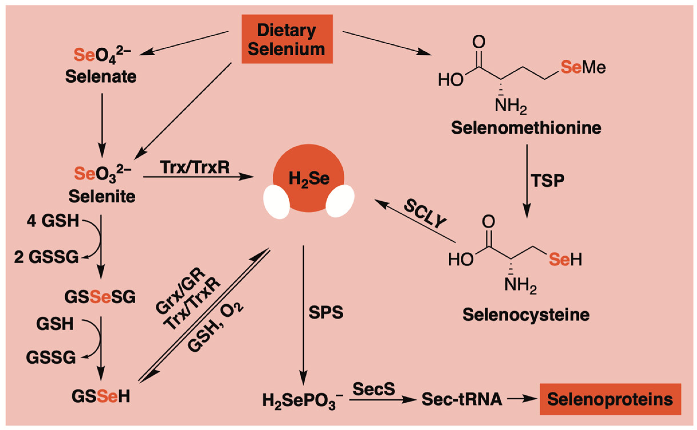
Figure 1. A simplified schematic of H2Se production in mammalian systems. Glutathione (GSH), glutathione disulfide (GSSG), glutaredoxin (Grx), glutathione reductase (GR), thioredoxin (Trx), thioredoxin reductase (TrxR), transsulfuration pathway (TSP), selenocysteine lyase (SCLY), selenophosphate synthetase (SPS), and selenocysteine synthase (SecS).
Molecules 29 03863 g001
Figure 2. (A) Selenotrisulfides as H2Se donors. (B) Peripheral delivery of H2Se via human serum albumin. Glutathione (GSH), glutaredoxin (Grx), glutathione reductase (GR), thioredoxin (Trx), thioredoxin reductase (TrxR), and human serum albumin (HSA).
Molecules 29 03863 g002
Figure 3. (A) Chalcogen anhydrides (R-Se, R-S, and R-O) under examination; (B) Proposed fragmentation pattern for R-Se in 50% methanol/water under ESI conditions.
Molecules 29 03863 g003
Figure 4. Proposed hydrolytic pathway of TDN1042, which results in the release of two equivalents of H2Se and the formation of phenylphosphonic acid.
Molecules 29 03863 g004
Figure 5. (A) Cyclic-PSe donors arranged in order of decreasing rates of hydrolysis; (B) Proposed hydrolytic pathway of cyclic-PSe donors, which results in the release of one equivalent of H2Se and the formation of phenylphosphonic acid.
Molecules 29 03863 g005
Figure 6. (A) Rapid hydrolysis of SelenoMichler’s ketone generates H2Se in PBS (pH 7.4). (B) Proposed mechanism for cysteine/thiol-triggered H2Se donation from selenocyclopropenones. (C) Proposed mechanism for cysteine-triggered H2Se donation from arylselenoamides.
Molecules 29 03863 g006
Figure 7. (A) General strategy for generating stimuli-responsive COS/H2S donors. (B) Proposed mechanism for the direct release of H2Se from PhotoSeCM upon irradiation at 365 nm. (C) Control compound used to highlight the stability of γ-ketoselenocarbamates in water. (D) Proposed mechanism for the direct release of H2Se from γ-ketoselenocarbamates containing a deprotonatable hydrogen at the β position.
Molecules 29 03863 g007
Figure 8. (A) A library of γ-ketoselenides that undergo base-promoted α-deprotonation/β-elimination to release H2Se. (B) Cell growth inhibition of HeLa and HCT116 cells in culture. IC50 values were determined after a 24 h incubation period with donor.
Molecules 29 03863 g008
Figure 9. 5′-O-Selenophosphates (dGMPSe) undergo HINT1-catalyzed hydrolysis to generate H2Se.
Molecules 29 03863 g009
Figure 10. Use of nonspecific electrophilic traps to confirm H2Se release from donors ((A) TDN1042, (B) 1, (C) 10) by forming stable selenide/diselenide products that can be easily identified by spectroscopic methods.
Molecules 29 03863 g010
Figure 11. (A) Reaction-based fluorescent sensor with selectivity towards H2Se based on benzoselenadiazole Se–N bond cleavage. (B) A lysosomal-targeting fluorescent sensor for H2Se bioimaging.
Molecules 29 03863 g011
Figure 12. Proposed mechanism for Hcy-H2Se turn-on fluorescence initiated by H2Se-promoted disulfide reduction.
Molecules 29 03863 g012
Figure 13. (A,B) Commercially available electrophilic traps that provide a colorimetric readout for monitoring H2Se donor progress. (C) Cautioning researchers that complementary H2Se measurements should be employed to avoid confusing donor alkylation with the alkylation of released H2Se in solution.
Molecules 29 03863 g013
Figure 14. (A) Aryl azide reduction for H2S-initiated turn-on fluorescence. (B) The use of aryl azides as fluorescent probes for tracking H2Se liberation from donor compounds.
Molecules 29 03863 g014
Disclaimer/Publisher’s Note: The statements, opinions and data contained in all publications are solely those of the individual author(s) and contributor(s) and not of MDPI and/or the editor(s). MDPI and/or the editor(s) disclaim responsibility for any injury to people or property resulting from any ideas, methods, instructions or products referred to in the content.