Communication

Qualitative and Quantitative Analysis of the Major Bioactive Components of Juniperus chinensis L. Using LC-QTOF-MS and LC-MSMS and Investigation of Antibacterial Activity against Pathogenic Bacteria

1
Department of Research and Development, Center for Industrialization of Agricultural and Livestock Microorganisms, Jeongeup-si 56212, Republic of Korea
2
Biological Resources Assessment Division, National Institute of Biological Resources, Incheon 22689, Republic of Korea
*
Authors to whom correspondence should be addressed.
Molecules 2023, 28(9), 3937; https://doi.org/10.3390/molecules28093937
Submission received: 17 April 2023 / Revised: 3 May 2023 / Accepted: 5 May 2023 / Published: 7 May 2023

Abstract

:
Plants in the genus Juniperus have been reported to produce a variety of chemical components, such as coumarins, flavonoids, lignans, sterols, and terpenoids. Here, ultra-high-performance liquid chromatography coupled with quadrupole time-of-flight mass spectrometry (UPLC-QTOF-MS) and ultra-high-performance liquid chromatography-tandem mass spectrometry (UPLC-MS/MS) were applied to qualitatively and quantitatively analyze the major bioactive components in an ethanolic crude extract from the leaves of Juniperus chinensis L., which grows naturally in Korea. In addition, the antibacterial activity of the crude extract against pathogenic bacteria was investigated. Using LC-QTOF-MS analysis, we identified ten compounds, of which six were confirmed to be flavonoid and lignan-based components as the major bioactive components, i.e., isoquercetin, quercetin-3-O-α-l-rhamnoside, hinokiflavone, amentoflavone, podocarpusflavone A, and matairesinoside. Among them, a quantitative analysis performed using LC-MS/MS revealed that the levels of quercetin-3-O-α-l-rhamnoside and amentoflavone in the crude extract were 203.78 and 69.84 mg/g, respectively. Furthermore, the crude extract exhibited potential antibacterial activity against 10 pathogenic bacteria, with the highest antibacterial activity detected against Bordetella pertussis. Thus, further studies of the leaf extract of J. chinensis L. must be carried out to correlate the compounds present in the extract with the antibacterial activity and elucidate the mechanisms of action of this extract against bacteria.

1. Introduction

Juniperus chinensis L. is known as Chinese juniper and is commonly found in many regions in Asia, including China, Taiwan, Myanmar, Japan, Malaysia, and Korea [1]. The genus Juniperus is considered as an important source of medicinal plants that are rarely used in traditional medicine. Various Juniperus species have been reported to produce a variety of chemical components, such as coumarins, flavonoids, lignans, sterols, and terpenoids [2], with a wide range of medicinal properties, such as antimicrobial, antioxidant, antitumor, and anticancer effects [3,4,5,6].
Flavonoids or bioflavonoids are a class of polyphenolic secondary metabolites that are found in plants and fulfill many functions [7]. The basic structure of these compounds consists of a general structure of 15 carbons with two phenyl rings and a heterocyclic ring, termed diphenylpropane (C6-C3-C6) skeleton [8]. Flavonoids are especially well known as having antibacterial properties against many pathogenic microorganisms. Recently, several investigations were performed regarding the antimicrobial activities, also summarizing the probable relationships between the chemical structures and antimicrobial activities [8]. These compounds exert their antibacterial effects via a mechanism mainly targeting the cell membrane, which likely involves the inhibition of attachment and biofilm formation, the inhibition of phospholipid bilayer formation, the inhibition of the respiratory chain, ATP-synthesis-mediated damage to the energy metabolism, etc. [7,9,10]. Lignans are a class of polyphenolic secondary metabolites that are found in plants and are precursors to phytoestrogens. These compounds have a general basic structure of nine carbons consisting of two phenylpropane (C6-C3) skeletons [11] and have been reported to have anticancer, antioxidant, antimicrobial, anti-inflammatory, and immunosuppressive activities [12,13,14].
In the present study, we qualitatively analyzed the bioactive components of a crude extract of J. chinensis L. using ultra-high-performance liquid chromatography coupled with quadrupole time-of-flight mass spectrometry (UPLC-QTOF-MS). Among them, quantitative analysis was performed for those with a mass spectrum, fragmentation pattern, and retention time which matched the reference compounds based on liquid chromatography-tandem mass spectrometry (LC-MS/MS). Furthermore, the antibacterial activity of the crude extract against 10 pathogenic bacteria was evaluated.

2. Results and Discussion

2.1. Chemical Profiling of Juniperus chinensis L.

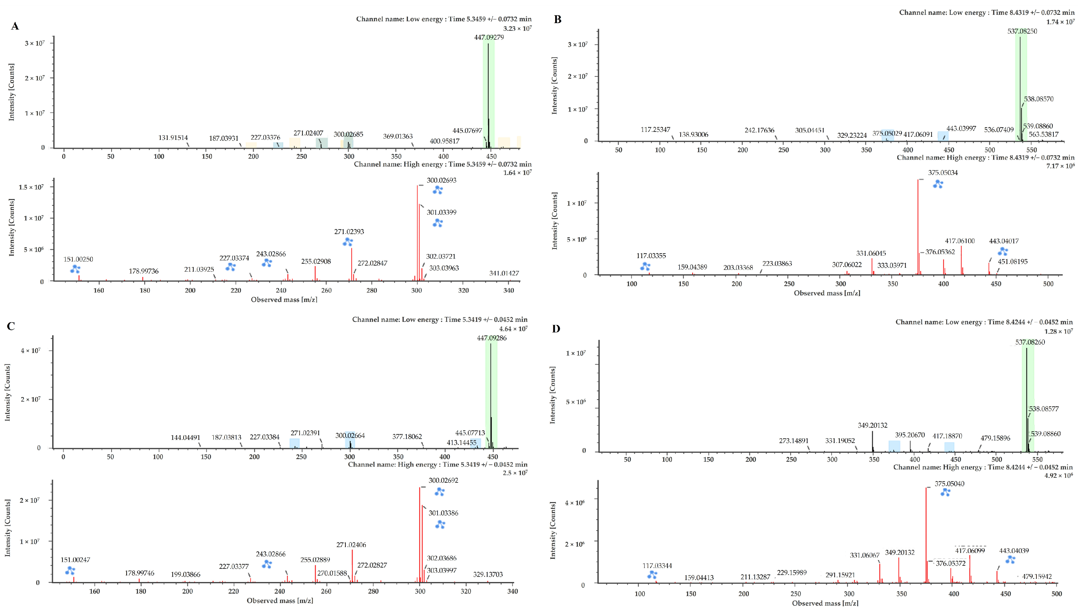
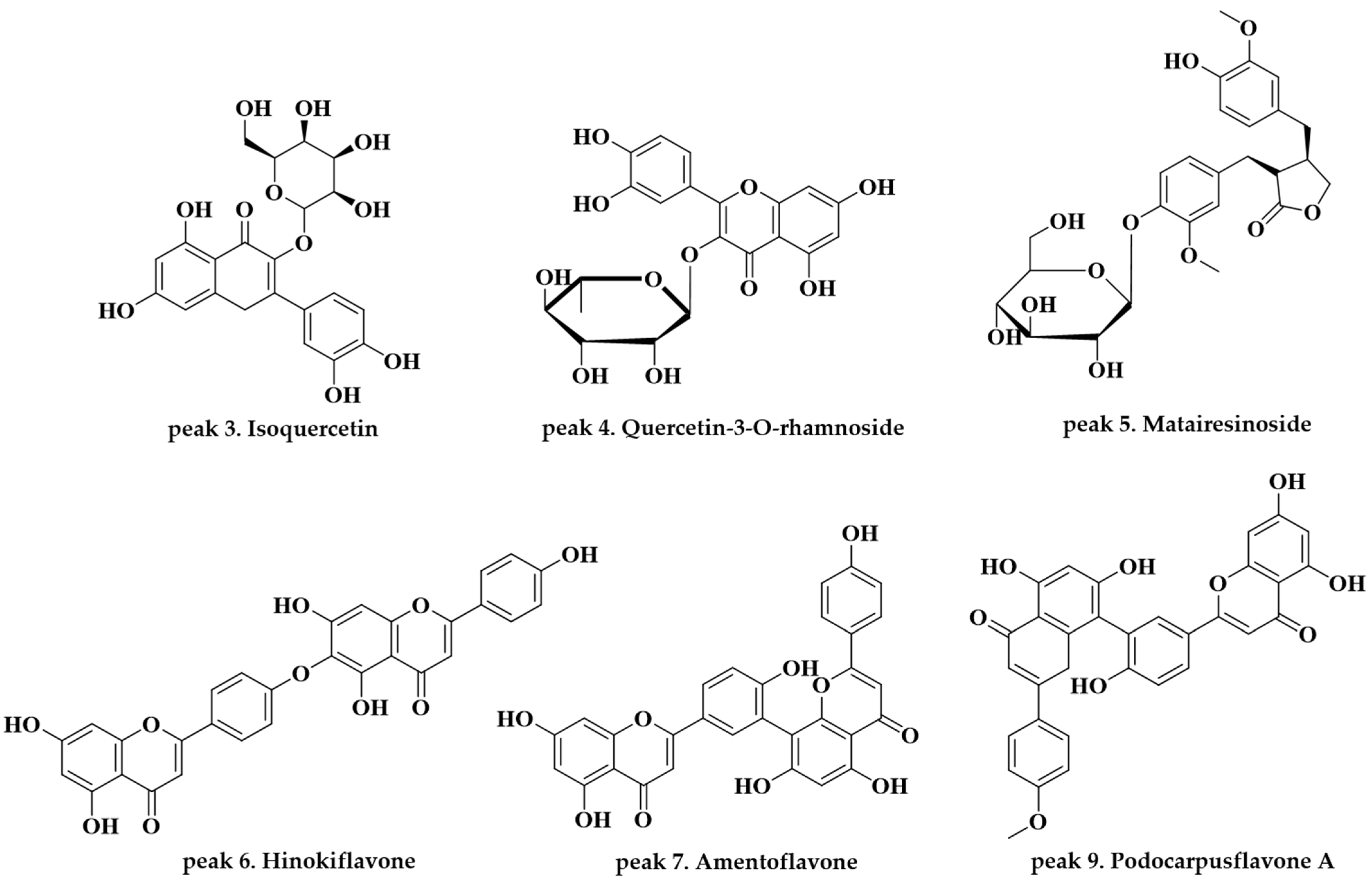
An ethanolic crude extract of the leaves of J. chinensis L. was analyzed and characterized using UPLC-QTOF-MS in electrospray ionization (ESI) negative ion mode, because more information and a higher fragmentation could be obtained in this ion mode compared with positive ion mode. We investigated various physiologically active component peaks in the base peak ion chromatogram, as shown in Figure 1. Ten components were tentatively identified after a library search using Waters’ UNIFI software (version 1.9, Milford, MA, USA) and the ChemSpider online database of isolated components under chromatographic conditions. All components exhibited a mass error below 5 mg/kg (Table 1, Supplementary Figure S1). Among them, the components corresponding to peaks 4 and 7 were identified as quercetin-3-O-α-l-rhamnoside and amentoflavone, respectively, based on reference compounds with mass spectra with identical patterns (Figure 2). Peak 1 (observed RT: 2.31 min; formula: C21H24O11), Peak 2 (observed RT: 3.19 min, formula: C15H14O6), Peak 8 (observed RT: 9.76 min; formula: C20H16O7), and Peak 10 (observed RT: 11.12 min; formula: C17H26O4) detected in the crude extract were identified via a tentative investigation based solely on formulas. In contrast, other components were unambiguously identified by comparison with reference substances. The structures of those components are shown in Figure 3.
Peak 3 (observed RT: 4.84 min; formula: C21H20O12) was identified as isoquercetin, as reported in [15,16]. The precursor ion of this component was detected at m/z 463.0878 [M − H] ion, with a fragment ion at m/z 300.0267 [463–C6H12O5] with the main peak. Isoquercetin has been reported to inhibit the oxidative stress effect of multiple carcinogens [21,22]. Peak 4 (observed RT: 5.34 min; formula: C21H20O11) was identified as quercetin-3-O-α-l-rhamnoside, as reported in [16,17,18]. The precursor ion of this component was detected at m/z 447.0929 [M − H] with fragment ions at m/z 300.0266 [447–C6H12O4] and 243.0299 [447–C8H13O6] and the main peak at m/z 300.0266. Quercetin-3-O-α-l-rhamnoside has been reported to have cytotoxic effects and antibacterial and antioxidant activities [23,24,25]. Peak 5 (observed RT: 5.94 min; formula: C26H32O11) was identified as matairesinoside, as reported in [19]. The precursor ion of this component was detected at m/z 519.1870 [M − H] with fragment ions at m/z 357.1340 [519–C6H11O5] and 343.1104 [519–C7H14O5] and the main peak at m/z 357.1340. Matairesinoside has been reported to have cytotoxic effects and antibacterial activity [26]. Peak 6 (observed RT: 7.78 min; formula: C30H18O10) was identified as hinokiflavone, as reported in [20]. The precursor ion of this component was detected at m/z 537.0826 [M − H] with fragment ions at m/z 443.0489 [537–C6H7O], 375.0510 [537–C9H7O3], and 117.0346 [537–C22H13O9] and the main peak at m/z 375.0510. Hinokiflavone has been reported to have cytotoxic effects and anti-inflammatory and antioxidant activity [27,28]. Peak 7 (observed RT: 8.42 min; formula: C30H18O10) was identified as amentoflavone, as reported in [16,18,20]. The precursor ion of this component was detected at m/z 537.0826 [M − H] with fragment ions at m/z 443.0409 [537–C6H7O] and 375.0510 [537–C9H7O3] and the main peak at m/z 375.0510. Amentoflavone has been reported to have various bioactivities, including antioxidant, anti-inflammatory, anti-senescence, antitumor, anti-virus, and anti-fungal effects [29,30,31,32]. Peak 9 (observed RT: 9.97 min; formula: C31H20O10) was identified as podocarpusflavone A, as reported in [20]. The precursor ion of this component was detected at m/z 551.0984 [M − H] with fragment ions at m/z 519.0713 [551–CH5O] and 375.0506 [551–C21H11O7] and the main peak at m/z 375.0506. Podocarpusflavone A has been reported to have cytotoxic effects and antimicrobial activity [33,34]. Of the 10 compounds identified in this study, six were confirmed to be flavonoid and lignan-based components which have diverse physiological activities according to the literature.

2.2. Quantitative Analysis of the Reference Compounds

The two reference compounds, quercetin-3-O-α-l-rhamnoside and amentoflavone, were analyzed using UPLC-Xevo TQ-S micro MS/MS in ESI mode. The analytical conditions of the instrument were set so that the retention time of the analyte was the same on the chromatograms of the standard solutions and the sample solutions. The ion ratio was calculated as the peak area ratio between a less intense ion and a more intense ion. The reference ion ratio value was calculated as the average of the ion ratios of the calibration solutions. All samples met the instrument analytical conditions described above (not shown). Moreover, the quercetin-3-O-α-l-rhamnoside standard calibration revealed a coefficient of determination (R2) of 0.9991 with good linearity, ranging from 0.05 to 1 ng. The amentoflavone standard calibration revealed a coefficient of determination (R2) of 0.9955 with good linearity, ranging from 0.25 to 2.5 ng. Overall, these results indicated that the methods were sufficiently validated to identify the two reference compounds in the samples, as a result of the standard curves of quercetin-3-O-α-l-rhamnoside and amentoflavone and the measurement of the total compound content (Table 2). The total contents (weight) in the ethanolic extract of quercetin-3-O-α-l-rhamnoside and amentoflavone were 203.78 and 69.84 mg/g, respectively.

2.3. Antibacterial Analysis

The antibacterial activities of the ethanolic crude extract of the leaves of J. chinensis L. were evaluated against pathogenic strains. The ethanolic crude extract of J. chinensis L. exhibited antibacterial activity against all strains tested here (Table 3). The inhibition zones were similar to or lower than those of antibiotics. Our results showed activity for both gram-positive and gram-negative pathogens similar to those observed in the literature [2,35,36]. For E. coli and S. aureus, antibacterial activity was observed among various Juniperus leaves and fruits [35,36]. Ennajar M et al. showed intense activity for K. pneumoniae, while our results showed only low activity [37]. On the other hand, the crude extract of J. chinensis L. exhibited strong antimicrobial activity against Bordetella pertussis NCCP13671.

3. Materials and Methods

3.1. Plant Material

The leaves of Juniperus chinensis L. were collected on 6 November 2018 from Imhyeon-ri, Eosangcheon-myeon, Danyang-gun, Chungcheongbuk-do, South Korea. The National Institute of Biological Resources (NIBR) performed the botanical identification of the plant materials. Specimens and materials of J. chinensis L. were deposited in the Wildlife Natural Products Bank at the NIBR, with NIBR numbers NIBRVP0000725557 and NIBRGR0000611643, respectively.

3.2. Preparation of the Plant Extract

The leaves of J. chinensis L. were collected to prepare the ethanolic extract. The leaves were dried in an oven (HB-503SF, HANBAEK) at 40 °C for 48 h and then ground to a fine powder to pass through a 20-mesh sieve. The powder was extracted with 70% ethanol (Sigma-Aldrich, Saint Louis, MO, USA) at four times the volume (1:4, v/v). The mixture was left at room temperature for 72 h, centrifuged at 4000× g for 10 min (VARISPIN 15R, CRYSTE), and filtered using filter paper (Whatman No. 2, 4.25 cm diameter). Moreover, the solvent was concentrated by rotary evaporation (Rotavapor R-100, Buchi, Flawil, Switzerland). The crude extract was stored at −80 °C in a freezer until it was used for analysis of the major bioactive components and antibacterial activity.

3.3. Qualitative Analysis Using Ultra-High-Performance Liquid Chromatography-Quadrupole Time-of-Flight Mass Spectrometry

The chemical profiling analysis of the crude extract from J. chinensis L. was performed using Ultra-High-Performance Liquid Chromatography-Quadrupole Time-of-Flight Mass Spectrometry (UPLC-QTOF-MS) in negative modes using an ACQUITYTM UPLC-Xevo GS-XS Q-TOF (Waters) instrument equipped with an ACQUITY UPLC® BEH C18 column (100 × 2.1 mm, 1.7 µm; Waters, Milford, MA, USA) at 40 °C. The mobile phase consisted of 0.1% formic acid in water (A) and acetonitrile (B) at a flow rate of 0.4 mL/min, and the injection volume was set at 1 µL. At this time, the injected sample was diluted to 2500 mg/L level with LC grade methanol. The mobile phase was a gradient: 0–1 min, 5% solvent B; 1–20 min, 100% solvent B; 20–20.30 min, 100% solvent B; 22.30–22.40 min, 5% solvent B; and 22.40–25 min, 5% solvent B. Full-scan mass spectra were acquired to detect the mass-to-charge ratio (m/z) in the range of 50–1500. The detector conditions were optimized as follows: the source temperature was set at 120 °C with a capillary voltage of 2.5 kV. Data acquisition and analysis were controlled using the traditional medicine library in the Waters’ UNIFI software (version 1.9, Milford, MA, USA). First, the identification of the components was performed by checking the allowable chemical formula calculated for the mass error range ± 5 ppm and setting it to fragment tolerance 10 mDa to check the mass pattern. In addition, if the fragment ion pattern did not match, it was finally identified using the Chemspider (http://www.chemspider.com/, accessed on 27 September 2022) online database.

3.4. Quantitative Analysis Using Ultra-Performance Liquid Chromatography-Tandem Mass Spectrometry

A quantitative analysis was performed using an ACQUITYTM UPLC-Xevo TQ-S micro MS/MS instrument (Waters). Among the major chemical compounds identified through LC-TOF-MS, two chemical compounds, peak 4 (quercetin-3-O-α-l-rhamnoside) and peak 7 (amentoflavone), which had the same mass spectra, fragmentation patterns, and retention times as the reference compounds, were quantitatively analyzed. Peak 4 had the following analytical conditions: the mobile phase consisted of 0.1% formic acid and 5 mM ammonium acetate in water (A) and methanol (B) at a flow rate of 0.3 mL/min, with the injection volume set at 2 µL. The mobile phase was a gradient: 0–1 min, 30% solvent B; 1–2 min, 70% solvent B; 3.5 min, 70% solvent B; 3.5–4 min, 30% solvent B; and 4–5 min, 30% solvent B using a CORTECSTM UPLC C18+ column (100 × 2.1 mm, 1.6 µm; Waters) at 35 °C. The MS conditions were optimized as follows: capillary voltage (0.8 kV), source temperature (150 °C), desolvation temperature (350 °C), desolvation gas flow (650 L/h), and cone gas flow (50 L/h), and the system was operated in electrospray ionization (ESI) positive ion mode. Peak 7 had the following analytical conditions: the mobile phase consisted of 0.1% formic acid in water (A) and methanol (B) at a flow rate of 0.5 mL/min, with the injection volume set at 5 µL. The mobile phase was a gradient: 0–0.5 min, 30% solvent B; 0.5–1 min, 70% solvent B; 2 min, 70% solvent B; 2–2.5 min, 100% solvent B; 3.5 min, 100% solvent B; 3.5–4 min, 30% solvent B; and 5 min, 30% solvent B using a CAPCELL CORE C18 column (150 × 2.1 mm, 2.7 µm; Osaka Soda, Shiseido) at 35 °C. The MS conditions were optimized as follows: capillary voltage (2.5 kV), source temperature (150 °C), desolvation temperature (300 °C), desolvation gas flow (450 L/h), and cone gas flow (10 L/h), and the system was operated in ESI negative ion mode. The multiple reaction monitoring for the quantitative mass ion analysis is presented in Table 4. Data acquisition and analysis were controlled using Waters’ Mass Lynx (version 4.2, Milford, MA, USA).

3.5. Reference Compounds and Preparation of the Standard Solution

Reference compound quercetin-3-O-α-l-rhamnoside (98% purity) was purchased from LGC standard, whereas amentoflavone (98% purity) was purchased from Sigma-Aldrich. The reference compounds were prepared by dissolving them in methanol at a final concentration of 1000 mg/L. The standard solutions were diluted to obtain calibration curves with five points in the concentration range of 0.025–0.5 and 0.05–0.5 mg/L, respectively.

3.6. Antibacterial Analysis

The antibacterial activity of the crude ethanol extract from J. chinensis L. was evaluated against various pathogenic bacterial strains using a modified disk diffusion method [38]. This study used 10 pathogenic bacteria strains deposited in NCCP or ATCC to conduct the antibacterial analysis: Escherichia coli KCTC 2617, Salmonella enterica serovar Enteritidis NCCP 14546, Streptococcus mutans KCTC 3065, Staphylococcus aureus NCCP 14560, Acinetobacter baylyi ATCC 33305, Klebsiella pneumoniae NCCP 16052, Bordetella pertussis NCCP13671, Moraxella catarrhalis ATCC 43628, Staphylococcus pyrogenes NCCP14783, and Streptococcus pneumoniae NCCP 14774. Each bacterial pathogenic strain was grown on suitable media at 30 °C–37 °C for 20 h. E. coli, S. aureus, and K. pneumoniae were grown on nutrient agar; S. enteritidis on tryptic soy agar; S. mutans, A. baylyi, M. catarrhalis, and S. pyrogenes on brain heart infusion agar; B. pertussis on Bordet–Gengou agar; and S. pneumoniae on sheep blood agar. To carry out the disc diffusion test as an antimicrobial disc susceptibility test according to the guidelines of the Clinical & Laboratory Standard Institute, each agar plate medium was inoculated with pathogenic bacteria at a concentration of 1–2 × 10⁸ CFU/mL. Sterilized paper discs (8 mm) were placed on the agar, and 10 µL of the crude plant extract was used to impregnate the discs. The extracts were dissolved in DMSO and ethanol (1:1, v/v) at a concentration of 20 mg/mL. The culture was incubated at 30 °C–37 °C for 24 h, and the diameters of the inhibition zones that formed around each disk were measured in millimeters. The positive control consisted of the appropriate antibiotic for each pathogen, i.e., ampicillin (2.5 mg/mL) for E. coli, S. enteritidis, S. mutans, K. pneumoniae, B. pertussis, and S. pneumoniae; gentamycin (2.5 mg/mL) for A. baylyi; penicillin (2.5 mg/mL) for M. catarrhalis and S. pyrogenes; and kanamycin (50 mg/mL) for S. aureus. In contrast, the negative control consisted of DMSO and ethanol (1:1, v/v). Each experiment was performed in triplicate.

4. Conclusions

In the present study, various chemical components were identified in an ethanolic crude extract of the leaves of J. chinensis L., which grows naturally in Korea. We identified 10 flavonoid and lignan-based molecules via LC-QTOF-MS analysis as the major bioactive components, including isoquercetin, quercetin-3-O-α-l-rhamnoside, hinokiflavone, amentoflavone, podocarpusflavone A, and matairesinoside. Among them, quercetin-3-O-α-l-rhamnoside and amentoflavone were quantitatively analyzed using LC-MS/MS; they existed in the crude extract at 203.78 and 69.84 mg/g, respectively. Moreover, the ethanolic crude extracts showed overall inhibitory activities against all Gram-positive and Gram-negative pathogenic bacteria tested, with the highest activity observed against Bordetella pertussis. Based on these results, we concluded that the J. chinensis L. extract showed antibacterial activity against various pathogens. However, we did not directly check if the 10 components identified here had antibacterial activity. Therefore, further studies are required to assess the antibacterial activity of the compounds isolated from J. chinensis L.

Supplementary Materials

The following supporting information can be downloaded at: https://www.mdpi.com/article/10.3390/molecules28093937/s1, Figure S1: Fragment ion pattern isoquercetin (A), quercetin-3-O-α-l-rhamnoside (B), matairesinoside (C), hinokiflavone (D), amentoflavone (E), and podocarpusflavone A (F) using mass spectra obtained by UPLC-QTOF-MS.

Author Contributions

Conceptualization, Y.K. and Y.K.S.; methodology, Y.K., D.J.L., J.-S.S. and B.-H.L. software, D.J.L. and J.-S.S.; validation, D.J.L., J.-S.S. and Y.K.; formal analysis, D.J.L., J.-S.S. and Y.K.; investigation, D.J.L., J.-S.S., B.-H.L., Y.K.S. and Y.K.; resources, Y.K., Y.K.S. and B.-H.L.; data curation, D.J.L. and Y.K.; writing—original draft preparation, D.J.L.; writing—review and editing, D.J.L., Y.K. and Y.K.S.; visualization, D.J.L. and Y.K.; supervision, Y.K. and Y.K.S.; project administration, Y.K. and Y.K.S.; funding acquisition, Y.K. and Y.K.S. All authors have read and agreed to the published version of the manuscript.

Funding

This research was funded by grants from the National Institute of Biological Resources (NIBR), funded by the Ministry of Environment(MOE) of the Republic of Korea (NIBR202325101), Korea Institute of Planning and Evaluation for Technology in Food, and Agriculture and Forestry (IPET) through Agricultural Machinery/Equipment Localization Technology Development Program, funded by Ministry of Agriculture, Food and Rural Affairs (MAFRA) (122022-3), and INNOPOLIS FOUNDATION through Science and Technology Project Opens the Future of the Region, funded by Ministry of Science and ICT (2022-DD-UP-0333).

Institutional Review Board Statement

Not applicable.

Informed Consent Statement

Not applicable.

Data Availability Statement

Not applicable.

Acknowledgments

We thank the anonymous reviewers for their careful reviewing of this manuscript and for providing insightful comments.

Conflicts of Interest

The authors declare no conflict of interest.

Sample Availability

Not applicable.

References

  1. Farjon, A. Juniperus chinensis. In The IUCN Red List of Threatened Species; IUCN Global Species Programme Red List Unit: Cambridge, UK, 2013. [Google Scholar]
  2. Seca, A.M.; Silva, A.M. The chemical composition of the Juniperus genus (1970–2004). Recent Prog. Med. Plants 2006, 16, 401–522. [Google Scholar]
  3. Asili, J.; Emami, S.A.; Rahimizadeh, M.; Fazly-Bazzaz, B.S.; Hassanzadeh, M.K. Chemical and antimicrobial studies of Juniperus excelsa subsp. excelsa and Juniperus excelsa subsp. polycarpos essential oils. J. Essent. Oil Bear. Plants 2008, 11, 292–302. [Google Scholar] [CrossRef]
  4. Taviano, M.F.; Marino, A.; Trovato, A.; Bellinghieri, V.; La Barbera, T.M.; Güvenç, A.; Hürkul, M.M.; De Pasquale, R.; Miceli, N. Antioxidant and antimicrobial activities of branches extracts of five Juniperus species from Turkey. Pharm. Biol. 2011, 49, 1014–1022. [Google Scholar] [CrossRef] [PubMed]
  5. Salaritabar, A.; Darvishi, B.; Hadjiakhoondi, F.; Manayi, A.; Sureda, A.; Nabavi, S.F.; Fitzpatrick, L.R.; Bishayee, A. Therapeutic potential of flavonoids in inflammatory bowel disease: A comprehensive review. World J. Gastroenterol. 2017, 23, 5097. [Google Scholar] [CrossRef] [PubMed]
  6. Keskes, H.; Mnafgui, K.; Hamden, K.; Damak, M.; El Feki, A.; Allouche, N. In vitro anti-diabetic, anti-obesity and antioxidant proprieties of Juniperus phoenicea L. leaves from Tunisia. Asian Pac. J. Trop. Biomed. 2014, 4, S649–S655. [Google Scholar] [CrossRef]
  7. Xie, Y.; Yang, W.; Tang, F.; Chen, X.; Ren, L. Antibacterial activities of flavonoids: Structure-activity relationship and mechanism. Curr. Med. Chem. 2015, 22, 132–149. [Google Scholar] [CrossRef]
  8. Farhadi, F.; Khameneh, B.; Iranshahi, M.; Iranshahy, M. Antibacterial activity of flavonoids and their structure–activity relationship: An update review. Phytother. Res. 2019, 33, 13–40. [Google Scholar] [CrossRef]
  9. Yuan, G.; Guan, Y.; Yi, H.; Lai, S.; Sun, Y.; Cao, S. Antibacterial activity and mechanism of plant flavonoids to gram-positive bacteria predicted from their lipophilicities. Sci. Rep. 2021, 11, 10471. [Google Scholar] [CrossRef]
  10. Cushnie, T.T.; Lamb, A.J. Antimicrobial activity of flavonoids. Int. J. Antimicrob. Agents 2005, 26, 343–356. [Google Scholar] [CrossRef]
  11. Saleem, M.; Kim, H.J.; Ali, M.S.; Lee, Y.S. An update on bioactive plant lignans. Nat. Prod. Rep. 2005, 22, 696–716. [Google Scholar] [CrossRef]
  12. Kitts, D.D.; Yuan, Y.V.; Wijewickreme, A.; Thompson, L.U. Antioxidant activity of the flaxseed lignan secoisolariciresinol diglycoside and its mammalian lignan metabolites enterodiol and enterolactone. Mol. Cell. Biochem. 1999, 202, 91–100. [Google Scholar] [CrossRef]
  13. MacRae, W.D.; Towers, G.N. Biological activities of lignans. Phytochemistry 1984, 23, 1207–1220. [Google Scholar] [CrossRef]
  14. Céspedes, C.L.; Avila, J.G.; Garcıá, A.M.; Becerra, J.; Flores, C.; Aqueveque, P.; Bittner, M.; Hoeneisen, M.; Martinez, M.; Silva, M. Antifungal and antibacterial activities of Araucaria araucana (Mol.) K. Koch heartwood lignans. Z. Nat. C 2006, 61, 35–43. [Google Scholar] [CrossRef] [PubMed]
  15. Dai, Y.; Dou, Z.; Zhou, R.; Luo, L.; Bian, L.; Chen, Y.; Tao, J.; Chen, Z. Quality evaluation of artemisia capillaris thunb. Based on qualitative analysis of the HPLC fingerprint and UFLC-Q-TOF-MS/MS combined with quantitative analysis of multicomponents. J. Anal. Methods Chem. 2021, 2021, 5546446. [Google Scholar] [CrossRef]
  16. Zeliou, K.; Koui, E.-M.; Papaioannou, C.; Koulakiotis, N.S.; Iatrou, G.; Tsarbopoulos, A.; Papasotiropoulos, V.; Lamari, F.N. Metabolomic fingerprinting and genetic discrimination of four Hypericum taxa from Greece. Phytochemistry 2020, 174, 112290. [Google Scholar] [CrossRef] [PubMed]
  17. Gao, Q.; Ma, R.; Chen, L.; Shi, S.; Cai, P.; Zhang, S.; Xiang, H. Antioxidant profiling of vine tea (Ampelopsis grossedentata): Off-line coupling heart-cutting HSCCC with HPLC–DAD–QTOF-MS/MS. Food Chem. 2017, 225, 55–61. [Google Scholar] [CrossRef]
  18. Wu, B.; Song, H.-P.; Zhou, X.; Liu, X.G.; Gao, W.; Dong, X.; Li, H.-J.; Li, P.; Yang, H. Screening of minor bioactive compounds from herbal medicines by in silico docking and the trace peak exposure methods. J. Chromatogr. A 2016, 1436, 91–99. [Google Scholar] [CrossRef]
  19. Avula, B.; Katragunta, K.; Wang, Y.H.; Ali, Z.; Khan, I.A. Simultaneous determination and characterization of flavonoids, sesquiterpene lactone, and other phenolics from Centaurea benedicta and dietary supplements using UHPLC-PDA-MS and LC-DAD-QToF. J. Pharm. Biomed. Anal. 2022, 216, 114806. [Google Scholar] [CrossRef]
  20. Wang, G.; Yao, S.; Zhang, X.-X.; Song, H. Rapid screening and structural characterization of antioxidants from the extract of Selaginella doederleinii Hieron with DPPH-UPLC-Q-TOF/MS method. Int. J. Anal. Chem. 2015, 2015, 849769. [Google Scholar] [CrossRef]
  21. Ran, J.; Wang, Y.; Zhang, W.; Ma, M.; Zhang, H. Research on the bioactivity of isoquercetin extracted from marestail on bladder cancer EJ cell and the mechanism of its occurrence. Artif. Cells Nanomed. Biotechnol. 2016, 44, 859–864. [Google Scholar] [CrossRef]
  22. Jayachandran, M.; Wu, Z.; Ganesan, K.; Khalid, S.; Chung, S.; Xu, B. Isoquercetin upregulates antioxidant genes, suppresses inflammatory cytokines and regulates AMPK pathway in streptozotocin-induced diabetic rats. Chem.-Biol. Interact. 2019, 303, 62–69. [Google Scholar] [CrossRef]
  23. Aljubiri, S.M.; Mahmoud, K.; Mahgoub, S.A.; Almansour, A.I.; Shaker, K.H. Bioactive compounds from Euphorbia schimperiana with cytotoxic and antibacterial activities. S. Afr. J. Bot. 2021, 141, 357–366. [Google Scholar] [CrossRef]
  24. Ismail, A.S.; Rizal, Y.; Armenia, A.; Kasim, A. Identification of bioactive compounds in gambier (Uncaria gambir) liquid by-product in West Sumatra, Indonesia. Biodiversitas J. Biol. Divers. 2021, 22, 1474–1480. [Google Scholar]
  25. Srinivasan, R.; Natarajan, D.; Shivakumar, M.S. Antioxidant Compound Quercetin-3-O-α-l-rhamnoside (1 → 6)-β-D-glucose (Rutin) isolated from ethyl acetate leaf extracts of Memecylon edule Roxb (Melastamataceae). Free Radic. Antioxid. 2015, 5, 35–42. [Google Scholar] [CrossRef]
  26. Kumarasamy, Y.; Nahar, L.; Cox, P.J.; Dinan, L.N.; Ferguson, C.A.; Finnie, D.A.; Jaspars, M.; Sarker, S.D. Biological activity of lignans from the seeds of Centaurea scabiosa. Pharm. Biol. 2003, 41, 203–206. [Google Scholar] [CrossRef]
  27. Lin, Y.-M.; Chen, F.-C.; Lee, K.-H. Hinokiflavone, a Cytotoxic Principle from Rhus succedanea and the Cytotoxicity of the Related Biflavonoids1. Planta Med. 1989, 55, 166–168. [Google Scholar] [CrossRef] [PubMed]
  28. Chen, Y.; Feng, X.; Li, L.; Zhang, X.; Song, K.; Diao, X.; Sun, Y.; Zhang, L. UHPLC-Q-TOF-MS/MS method based on four-step strategy for metabolites of hinokiflavone in vivo and in vitro. J. Pharm. Biomed. Anal. 2019, 169, 19–29. [Google Scholar] [CrossRef]
  29. Yu, S.; Yan, H.; Zhang, L.; Shan, M.; Chen, P.; Ding, A.; Li, S.F.Y. A review on the phytochemistry, pharmacology, and pharmacokinetics of amentoflavone, a naturally-occurring biflavonoid. Molecules 2017, 22, 299. [Google Scholar] [CrossRef]
  30. Okigawa, M.; Hwa, C.W.; Kawano, N.; Rahman, W. Biflavones in Selaginella species. Phytochemistry 1971, 10, 3286–3287. [Google Scholar] [CrossRef]
  31. Park, N.-H.; Lee, C.-W.; Bae, J.-H.; Na, Y.J. Protective effects of amentoflavone on Lamin A-dependent UVB-induced nuclear aberration in normal human fibroblasts. Bioorganic Med. Chem. Lett. 2011, 21, 6482–6484. [Google Scholar] [CrossRef]
  32. Hwang, I.-S.; Lee, J.; Jin, H.-G.; Woo, E.-R.; Lee, D.G. Amentoflavone stimulates mitochondrial dysfunction and induces apoptotic cell death in Candida albicans. Mycopathologia 2012, 173, 207–218. [Google Scholar] [CrossRef] [PubMed]
  33. Miura, H.; Kihara, T.; Kawano, N. Studies on bisflavones in the leaves of Podocarpus macrophylla and P. nagi. Chem. Pharm. Bull. 1969, 17, 150–154. [Google Scholar] [CrossRef]
  34. Bagla, V.P.; McGaw, L.J.; Elgorashi, E.E.; Eloff, J.N. Antimicrobial activity, toxicity and selectivity index of two biflavonoids and a flavone isolated from Podocarpus henkelii (Podocarpaceae) leaves. BMC Complement. Altern. Med. 2014, 14, 383. [Google Scholar] [CrossRef]
  35. Miceli, N.; Marino, A.; Köroğlu, A.; Cacciola, F.; Dugo, P.; Mondello, L.; Taviano, M.F. Comparative study of the phenolic profile, antioxidant and antimicrobial activities of leaf extracts of five Juniperus L.(Cupressaceae) taxa growing in Turkey. Nat. Prod. Res. 2020, 34, 1636–1641. [Google Scholar] [CrossRef]
  36. Manel, M.; Nouzha, H.; Rim, M.; Imane, M.; Sana, A.; Yasmine, O.; Ammar, A. Antibacterial and antioxidant activity of Juniperus thurifera L. leaf extracts growing in East of Algeria. Vet. World 2018, 11, 373. [Google Scholar] [CrossRef] [PubMed]
  37. Ennajar, M.; Bouajila, J.; Lebrihi, A.; Mathieu, F.; Abderraba, M.; Raies, A.; Romdhane, M. Chemical composition and antimicrobial and antioxidant activities of essential oils and various extracts of Juniperus phoenicea L. (Cupressacees). J. Food Sci. 2009, 74, M364–M371. [Google Scholar] [CrossRef] [PubMed]
  38. Vandenbossche, I.; Vaneechoutte, M.; Vandevenne, M.; De Baere, T.; Verschraegen, G. Susceptibility testing of fluconazole by the NCCLS broth macrodilution method, E-test, and disk diffusion for application in the routine laboratory. J. Clin. Microbiol. 2002, 40, 918–921. [Google Scholar] [CrossRef]
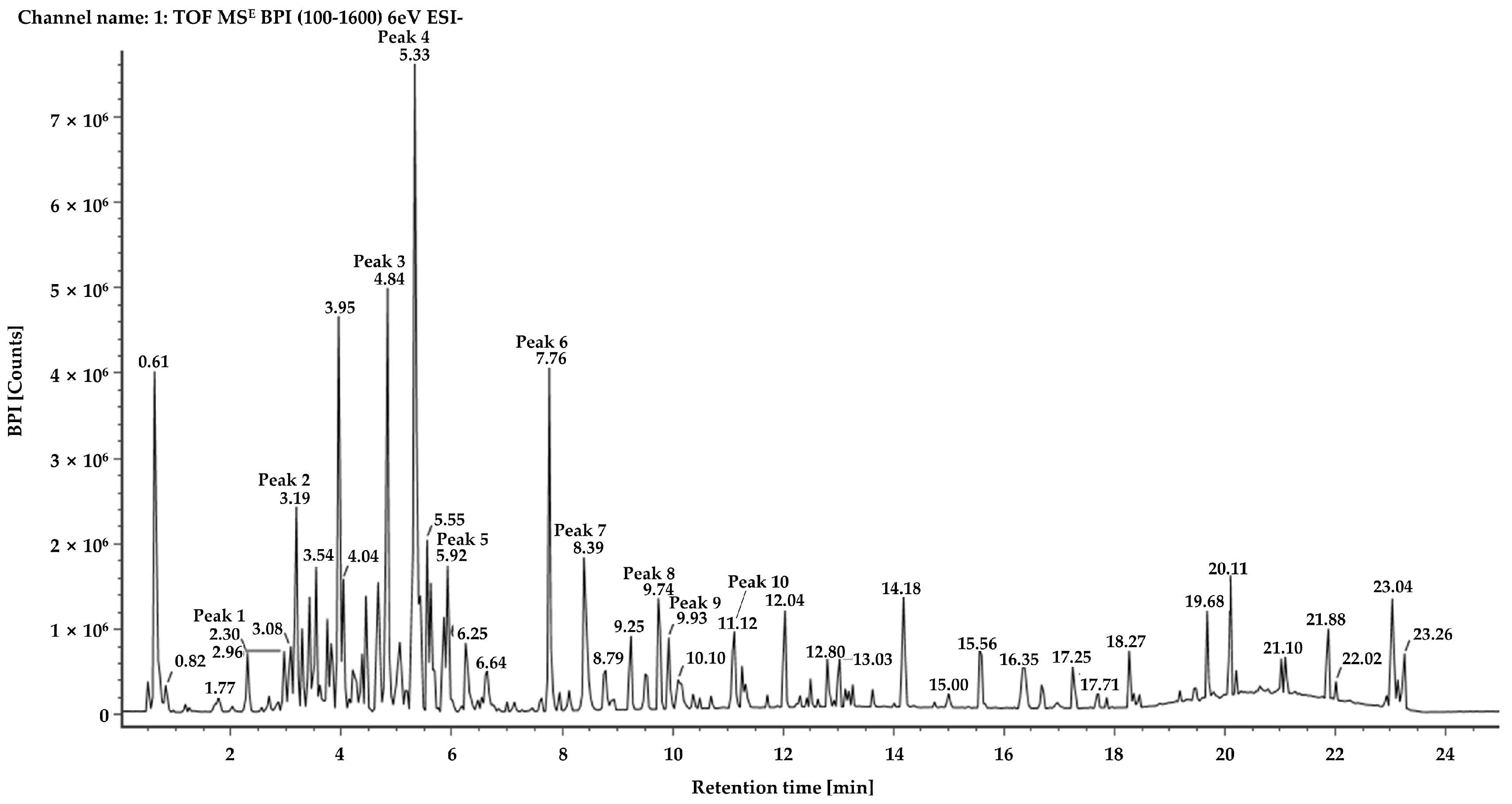
Figure 1. Base peak ion chromatogram of the J. chinensis L. extract obtained by UPLC-QTOF-MS analysis.
Molecules 28 03937 g001
Figure 2. Comparison of reference materials quercetin-3-O-α-l-rhamnoside (A) and amentoflavone (B) to identify the components of peak 4 (C) and peak 7 (D) using mass spectra obtained by UPLC-QTOF-MS.
Molecules 28 03937 g002
Figure 3. Chemical structures of the components identified in the ethanolic extract of J. chinensis L. based on the UPLC-QTOF-MS analysis.
Molecules 28 03937 g003
Table 1. Tentative identification of the chemical components of the ethanolic extract of J. chinensis L. obtained from the UPLC-QTOF-MS analysis.
PeakTentative
Chemical
Component
FormulaObserved RT
(min)
Neutral Mass
(Da)
Observed
[M − H]
(m/z)
Mass Error
(ppm)
Fragmentation Peaks
m/z
(% Base Peak)
References
1Unknown 1C21H24O112.31452.1319451.1242−0.9289.0709(100); 137.0232(100)-
2Unknown 2C15H14O63.19290.0790289.0712−2245.0810(78); 137.0232(100)-
3IsoquercetinC21H20O124.84464.0955463.0878−0.9300.0267(100)[15,16]
4Quercetin-3-O-α-l-rhamnosideC21H20O115.34448.1006447.0929−1300.0266(100); 243.0299(7)Standard [16,17,18]
5MatairesinosideC26H32O115.94520.1945519.1870−0.3357.1340(100); 342.1104(7)[19]
6HinokiflavoneC30H18O107.78538.0900537.0826−0.2443.0489(13); 375.0510(100); 117.0346(2)[20]
7AmentoflavoneC30H18O108.42538.0900537.0826−0.2443.0409(14); 375.0510(100)Standard [16,18,20]
8Unknown 3C20H16O79.76368.0896367.0819−1.3323.0925(85); 294.0884(100); 159.0452(97)-
9Podocarpusflavone AC31H20O109.97552.1056551.09840519.0713(2); 375.0506(100)[20]
10Unknown 4C17H26O411.12294.1831293.1752−2.1249.1853(69); 193.1598(100)-
Table 2. Results of the investigation of linear relationships and quantification based on UPLC-MS/MS analysis.
ParameterQuercetin-3-O-α-l-rhamnosideAmentoflavone
Coefficients of regression equationy = 34,990x − 205.23y = 3991.5x + 404.2
Coefficients of determination (R2)0.99910.9955
Linear range (ng)0.05–1.00.25–2.5
Concentration
(mg/g of dry ethanolic extract weight)
203.78 ± 5.4269.84 ± 1.94
Table 3. Antibacterial activity of the ethanolic crude extract of J. chinensis L. against pathogenic bacterial strains.
Test Pathogenic StrainAntibacterial Activity *
Ethanolic Crude Extract of
J. chinensis
Positive Control
Escherichia coli KCTC 2617++++ampicillin
Salmonella enterica serovar Enteritidis NCCP 14546++++ampicillin
Streptococcus mutans KCTC 3065+++++ampicillin
Staphylococcus aureus NCCP 14560+++++kanamycin
Acinetobacter baylyi ATCC 33305+++gentamycin
Klebsiella pneumoniae NCCP 16052+++ampicillin
Bordetella pertussis NCCP13671++++++ampicillin
Moraxella catarrhalis ATCC 43628++++penicillin
Staphylococcus pyrogenes NCCP14783++++penicillin
Streptococcus pneumoniae NCCP 14774++++ampicillin
* The inhibition zone (mm) around the paper disc containing the microbial cell-free supernatant was classified as follows: +++, >13 mm; ++, 10–12 mm; +, less than 9 mm. All microbial pathogens showed no inhibition against the negative control (DMSO:EtOH = 1:1, v/v).
Table 4. UPLC-MS/MS instrument conditions for the analysis of compounds in multiple reaction mode.
ComponentIon ModePrecursor Ion
m/z, (Cone Voltage, V)
Product Ions
m/z, (Collision Energy, V)
QuantitationReference
Quercetin-3-O-α-l-rhamnosidepositive449.18 (20)303.11 (9)84.94 (9)
Amentoflavonenegative537.48 (9)375.31 (33)443.34 (33)
Disclaimer/Publisher’s Note: The statements, opinions and data contained in all publications are solely those of the individual author(s) and contributor(s) and not of MDPI and/or the editor(s). MDPI and/or the editor(s) disclaim responsibility for any injury to people or property resulting from any ideas, methods, instructions or products referred to in the content.