Article

Phytochemical Characterization and Antimicrobial Activity of Several Allium Extracts

by
Ioana Andreea Barbu
1,2,3,
Alexandra Ciorîță
1,4,
Rahela Carpa
1,3,5,*,
Augustin Catalin Moț
6,
Anca Butiuc-Keul
1,2,3 and
Marcel Pârvu
1,3
1
Faculty of Biology and Geology, Babeș-Bolyai University, 1 M. Koganiceanu Str., 400084 Cluj-Napoca, Romania
2
Doctoral School of Integrative Biology, Babeș-Bolyai University, 400015 Cluj-Napoca, Romania
3
Center for Systems Biology, Biodiversity and Bioresources, Babeş-Bolyai University, Clinicilor Str., 400006 Cluj-Napoca, Romania
4
National Institute for Research and Development of Isotopic and Molecular Technologies, 400293 Cluj-Napoca, Romania
5
Institute for Research-Development-Innovation in Applied Natural Sciences, Babes-Bolyai University, 30 Fântânele Str., 400294 Cluj-Napoca, Romania
6
Faculty of Chemistry and Chemical Engineering, Babes-Bolyai University, 11 Arany Janos Str., 400028 Cluj-Napoca, Romania
*
Author to whom correspondence should be addressed.
Molecules 2023, 28(10), 3980; https://doi.org/10.3390/molecules28103980
Submission received: 20 March 2023 / Revised: 5 May 2023 / Accepted: 7 May 2023 / Published: 9 May 2023

Abstract

:
Microbial infections affect both the human population and animals. The appearance of more and more microbial strains resistant to classical treatments led to the need to develop new treatments. Allium plants are known for their antimicrobial properties due to their high content of thiosulfinates, especially allicin, polyphenols or flavonoids. The hydroalcoholic extracts of six Allium species obtained by cold percolation were analyzed regarding their phytochemical compounds and antimicrobial activity. Among the six extracts, Allium sativum L. and Allium ursinum L. have similar contents of thiosulfinates (approx. 300 μg allicin equivalents/g), and the contents of polyphenols and flavonoids were different between the tested species. The HPLC-DAD method was used to detail the phytochemical composition of species rich in thiosulfinates. A. sativum is richer in allicin (280 μg/g) than A. ursinum (130 μg/g). The antimicrobial activity of A. sativum and A. ursinum extracts against Escherichia coli, Staphylococcus aureus, Candida albicans and Candida parapsilosis can be correlated with the presence of large amounts of thiosulfinates. Both extracts have shown results against Candida species (inhibition zones of 20–35 mm) and against Gram-positive bacteria, Staphylococcus aureus (inhibition zones of 15–25 mm). These results demonstrate the antimicrobial effect of the extracts and suggest their use as an adjuvant treatment for microbial infections.

1. Introduction

The uncontrolled use of antibiotics in treatment and antimicrobial chemical preservatives in food preservation is the major cause of the emergence of antibiotic resistance [1,2]. Since the identification of microorganisms that cause various infections is not rigorously carried out, broad-spectrum antibiotics are used to treat these conditions, most of the time unnecessarily. This has led to the difficult treatment of some infections associated with the emergence of resistance to antibiotics that affect both the human population and various animals [3,4]. The emergence of multiresistant bacteria to antimicrobial agents and the ineffectiveness of common antibiotic treatments are increasing at an alarming rate. Among the multiresistant bacteria that affect animals are Salmonella spp., S. aureus, Campylobacter spp. and E. coli, which cause urinary, respiratory and reproductive tract infections [3]. This prompts researchers to look for new ways to fight microbial infections.
Plants can synthesize several active compounds that can have antibacterial [5], antifungal [6,7], antiparasitic [8] and antitumor effects [9], making them perfect candidates for the development of new antimicrobial agents.
Plants of the Allium genus are perennial and herbaceous and belong to the Liliaceae family. They have been known for their antimicrobial properties since ancient times, due to the presence of allicin in large amounts in their chemical composition [10,11,12].
In recent years, many studies have shown that garlic and its bioactive constituents (polyphenols, flavonoids and thiosulfinates), especially allicin, exhibit antioxidant, anti-inflammatory, antibacterial, antifungal and antitumor properties [13]. Plants of the Allium genus can also be used in the treatment and prevention of hypertension. Due to garlic’s anti-inflammatory properties, it could treat neurodegenerative diseases such as Alzheimer disease [14]. Extracts from these plants can have an important role in the adjuvant treatment of various microbial infections, due to the bioactive substances they contain. That is why the polarity of the solvent used to obtain the extracts influences their content, with the ethanolic extracts being richer in polyphenols and flavonoids than the methanolic or aqueous ones [15,16]. Garlics and onions have antibacterial compounds with effects against Gram-positive (S. aureus, S. spp.) and Gram-negative bacteria, (Escherichia coli, Salmonella spp., Klebsiella spp., Pseudomonas aeruginosa and Helicobacter pylori) [17,18,19,20,21]. Several studies demonstrate that A. ursinum can be used as an antimicrobial agent. Extracts prepared from its fresh flowers and leaves exert antifungal and antibacterial activities, directed predominantly on Gram-positive bacteria and less on Gram-negative. The antimicrobial potential of this plant is attributed to sulfur compounds, especially allicin [22].
The aim of this study is the analysis of the chemical composition of the hydroalcoholic extracts of two forms of Allium cepa L.—the Arieș red cultivar of Allium cepa and white variety of A. cepa, which were not previously investigated—and other common species: Allium sativum L., Allium ursinum L., Allium fistulosum L. and Allium senescens L. subsp. montanum (F. W. Schmidt) Holub. Investigating the antimicrobial activity of these extracts against Escherichia coli, Staphylococcus aureus, Candida albicans and Candida parapsilosis, common microorganisms involved in human and animal infections, was also one of our objectives.

2. Results

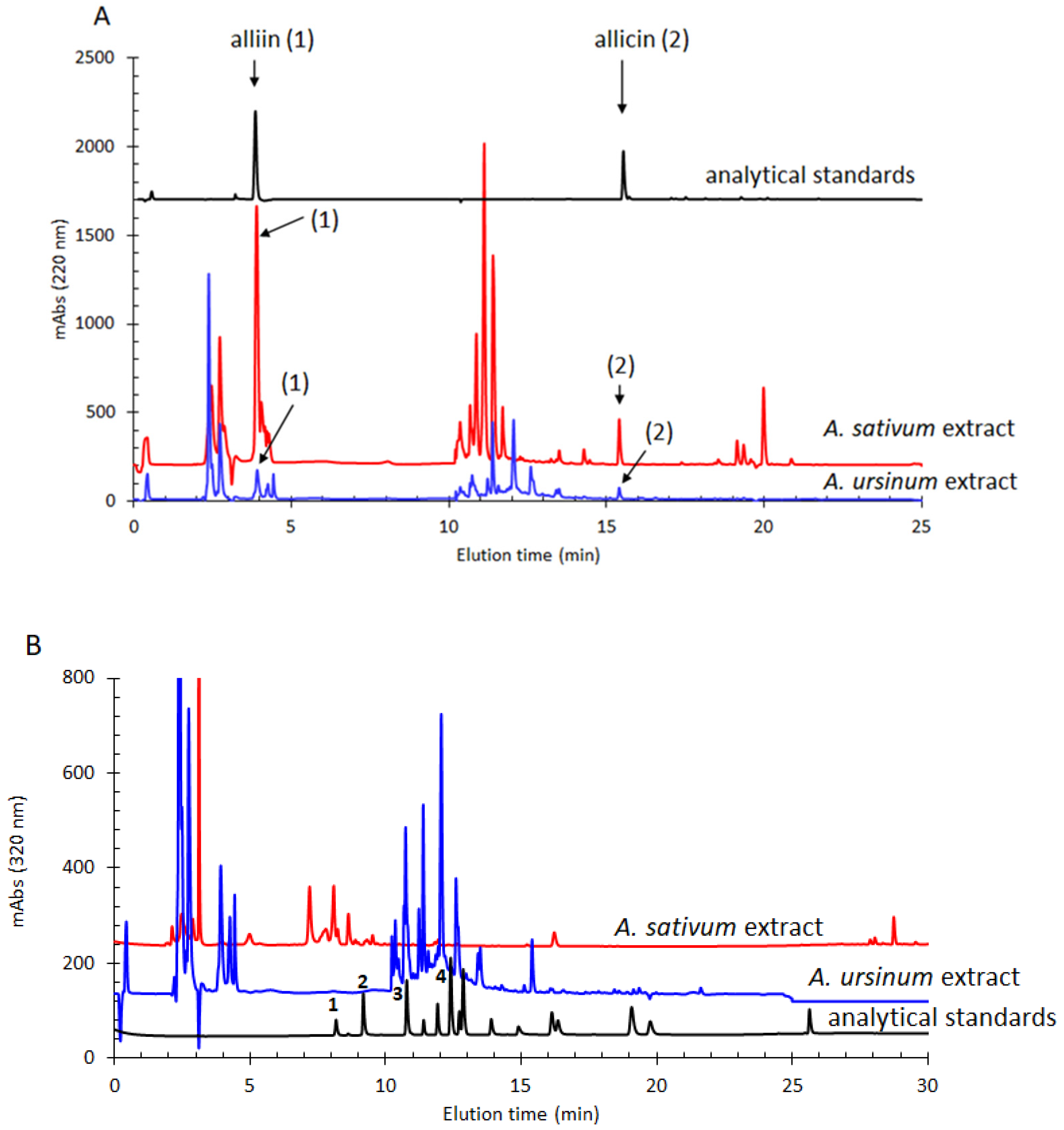
2.1. Phytochemical Composition of Allium Extracts

The total content of polyphenols, flavonoids and thiosulfinates in the Allium extracts is shown in Table 1. The total flavonoids content proved to be double in the extract of A. sativum compared to that of A. ursinum. The highest concentration of flavonoids was in the extracts of A. senescens subsp. montanum (F. W. Schmidt) Holub (52.6 ± 1.2 μg QE/g) and the Arieș red cultivar of Allium cepa (54.9 ± 2.4 μg QE/g), and the lowest in the extract of the white variety of A. cepa (18.6 ± 1.5 μg QE/g). The total polyphenols content was similar in the case of the extracts of Allium senescens L. subsp. montanum (F. W. Schmidt) Holub and the Arieș red cultivar of Allium cepa, which had the highest concentrations (423 ± 35 μg GAE/g and 418 ± 7 μg GAE/g). A. fistulosum and A. ursinum also had similar contents, and the lowest concentration was in the extract of the white variety of A. cepa (175 ± 11 μg GAE/g). The extracts of A. sativum and A. ursinum had a high content of thiosulfinates (333 ± 5 μg AE/g and 312 ± 7 μg AE/g, respectively).
Using the HPLC-DAD method, the most important phenolic and thiosulfinate compounds present in the A. sativum and A. ursinum extracts that showed antimicrobial activity were determined. The two extracts showed different levels of alliin and allicin, with A. sativum being richer in these compounds than A. ursinum, as shown in Figure 1. Chlorogenic acid was present in a similar amount in the two extracts, while p-coumaric acid was found in a significantly higher amount in the A. ursinum extract (102 ± 10 μg/g). Gentisic acid and 4-hydroxybenzoic acid were evident in notable quantities only in the extract of A. sativum, as can be seen in Table 2.

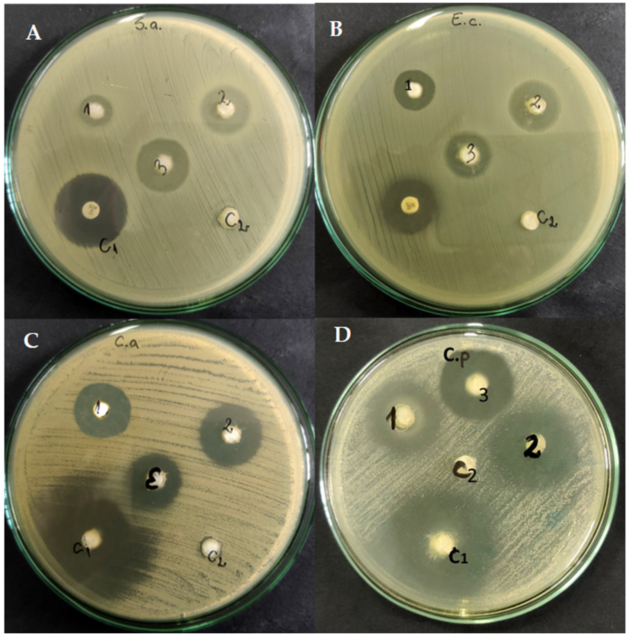
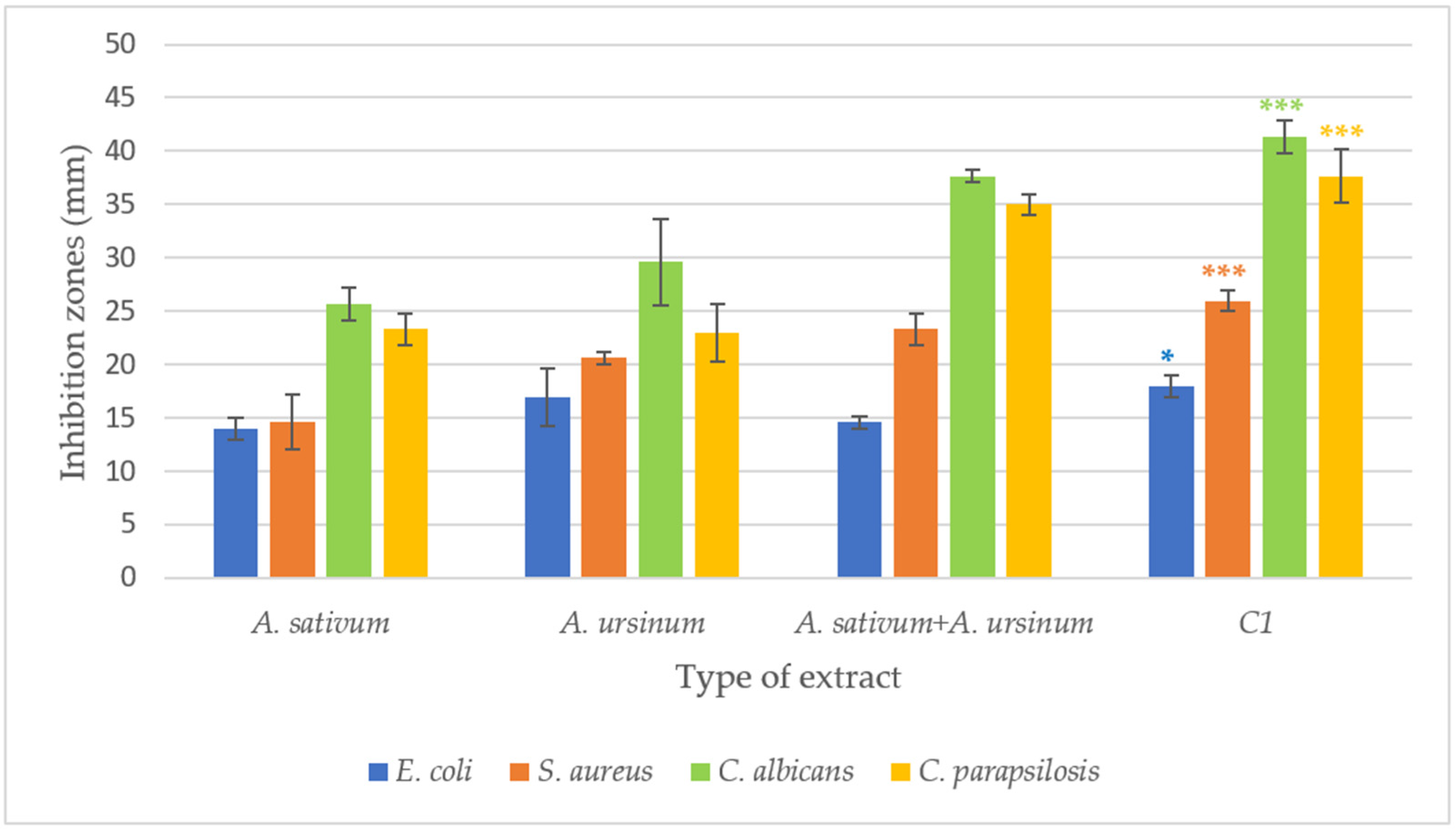
2.2. Antimicrobial Activity

Out of the six Allium extracts tested, only two showed antimicrobial effects: A. sativum and A. ursinum (Figure S1). Both extracts were more effective against Candida species compared to bacterial ones. S. aureus proved to be more sensitive to the tested extracts than E. coli (zones of inhibition of 15–22 mm vs. 15–20 mm). A. ursinum extract compared to that of A. sativum had larger inhibition zones against S. aureus than E. coli (Figure S1 and Figure 2). For the fungal species tested, the extract of A. ursinum (inhibition zones of 20–32 mm) was more effective than that of A. sativum (inhibition zones of 22–27 mm) (Figure 2). The effects against C. albicans and C. parapsilosis were more promising compared to the bacteria tested. At the same time, the 1:1 combination of the two extracts was also tested (Figure 2 and Figure 3). For E. coli, no differences were observed between the combination of extracts and the individual extracts. In the case of S. aureus, a slight increase in the inhibition zone (22–25 mm) was observed, and for Candida strains, an increase in the inhibition zones was observed compared to the individually tested extracts. For all microorganisms tested, the diameters of the inhibition zones of the antibiotics and antifungals used are close to those of the extracts. The 1:1 combination of the two extracts, tested on C. parapsilosis, had an effect close to that of Fluconazole (32 mm vs. 36 mm). For the agar diffusion method, three independent experiments were conducted and the mean was calculated (Figure 3). According to one-way ANOVA statistical analysis, all values are statistically significant at p value < 0.05, as compared to the control.
The microdilution method confirmed the results obtained by antibiograms and antifungigrams using the diffusion method (Table 3). The extracts of A. sativum and A. ursinum had antimicrobial effects on the strains E. coli ATCC 25922 and S. aureus ATCC 25923 and two Candida species: C. albicans ATCC 10231, C. parapsilosis ATCC 22019. The 1:1 combination proved to be more effective than the 1:2 (A. sativum:A. ursinum) and 2:1 (A. ursinum:A. sativum) combinations.
The antimicrobial activity of A. sativum, A. ursinum, and mixed A. sativum and A. ursinum 1:1 extracts was tested on four microbial strains. In addition, for each strain, we also used a control of 30% ethanol (C2) and a control of an antimicrobial substance (C1): ciprofloxacin for S. aureus, sulfamethoxazole for E. coli and fluconazole for Candida species. Compared to the control (C1 = antimicrobial substances), the antimicrobial activity of Allium extracts was good. For some experimental variants, it exhibits an inhibition of almost half of the inhibition value given by the control substance and for other experimental variants it reaches almost the level of inhibition of the chosen antimicrobial substance (Figure 3).
In these experiments, the bacteriostatic (MIC) and bactericidal effects (MBC) of Allium extracts against E. coli, S. aureus, C. albicans and C. parapsilosis were also determined.
The minimum inhibitory concentration (MIC) of the A. sativum and A. ursinum extracts tested on bacteria and fungi varied depending on the species and the plant extract (Table 3). It can be observed that the MIC of the extracts is higher for Gram-positive bacteria (12.5) compared to Gram-negative (6.25).
The minimum bactericidal concentration (MBC) was determined by diluting each well in tenfold dilutions. From each dilution, aliquots were transferred on agar plates and incubated for 24 h. The first concentration at which no colony forming units were observed was considered the MBC.
The MICs for the tested microbial species were similar to the MBCs for some Allium sp. extracts, but the tested microbial species generally showed a lower concentration for MBC (Table 3). There were also discrepancies between MICs and MBCs, e.g., for the, A. ursinum extract on C. parapsilosis (MIC = 6.25 and MBC = 25) and for the A. sativum extract on E. coli (MIC = 6.25 and MBC = 25).

3. Discussion

Allium plants have been known for their antimicrobial properties since ancient times. Recent studies have shown that the leaves and bulbs of these plants are rich in phenols, flavonoids and thiosulfinates, compounds that have proven antimicrobial, antioxidant or antitumor activity [23,24,25]. Naturally, in Allium bulbs or leaves allicin is formed after crushing or cutting the plant organs under the action of the allinase enzyme [24].
The chemical composition of the extracts depends on the species. In our study, we compared five Allium species, two of which are A. cepa varieties, which have not been previously analyzed, in order to correlate the phytochemical composition with the antimicrobial activity. Although the Arieș red cultivar of Allium cepa and Allium senescens L. subsp. montanum (F. W. Schmidt) Holub had the highest content of flavonoids and phenols, respectively, according to Table 1, Allium sativum and Allium ursinum had the highest content of thiosulfinates among the tested species. It is known that allicin is a compound with high antimicrobial activity, so we can correlate the antimicrobial activity of the two species with the presence of the high content of thiosulfinates [26]. Moreover, extracts of A. sativum and A. ursinum had a high content of polyphenols, indicating a synergistic effect of the two classes of compounds [5,25], which was observed in our study as well. In addition, the total content of polyphenols varies depending on the species, the organ of the plant from which the extract is obtained and its maturity. Plants in the flowering period have a higher polyphenol content than in the growing period, and the leaves are the richest in these compounds [27,28,29]. Nhut et al. (2020) also analyzed the phytochemical composition of an Allium tuberosum Rottler ex Spreng extract and their results were similar with ours regarding the total content of flavonoids (31.10 ± 1.12 mg QE/g) [30] but the total content of polyphenols is higher in the extracts used for this study. Allium schoenoprasum and Allium vineale also have a similar content of polyphenols, flavonoids and sulfur compounds to the tested extracts in our study [31,32]. The extract of A. scorodoprasum L. subsp. rotundum instead has a lower total content of polyphenols (25.60 ± 0.48 mg GAE/g) and flavonoids (2.21 ± 0.04 mg QE/g) than the extracts analyzed in this study [33]. In addition, the solvent used to obtain the extracts is an important factor that influences the content of polyphenols and flavonoids due to their different solubility [34]. It was proven that the ethanolic onion extract was the richest in polyphenols and flavonoids compared to the methanol, acetone or aqueous extracts [34,35].
The antimicrobial activity of A. ursinum and A. sativum extracts is also documented in the literature [26,36], and the results obtained are correlated with the already existing ones. The A. ursinum extract was more effective against Gram-positive than Gram-negative bacteria both in our study and in previous studies [5]. The antimicrobial properties of the A. cepa extract were previously examined against S. aureus, E. coli, Bacillus cereus, Listeria innocua and Pseudomonas aeruginosa. The extracts at different concentrations showed antibacterial activity against all tested bacterial strains. The extract at the highest concentration was more effective against E. coli and least effective against P. aeruginosa [20,21,37]. Although previous studies showed antimicrobial effects produced by extracts of Allium cepa, our results did not show antimicrobial activity. Allium fistulosum was also not effective against Pseudomonas aeruginosa at any concentrations tested [38]. This fact may be due to the method of obtaining the extracts and the solvents used. The Allium sativum petroleum ether extract was the most effective against Staphylococcus spp., followed by E. coli and Proteus spp. Likewise, the chloroform extract was more active against Pseudomonas spp. than the aqueous extract. The methanol and aqueous extracts presented some of the smallest zones of inhibition against the pathogens tested [39].
Due to the antimicrobial activity of the extracts of A. sativum and A. ursinum, we performed an analysis of extract combination to demonstrate their synergistic activity. The results obtained showed an additive effect of inhibiting the tested microorganisms but without a high increase in the inhibition zones. Moreover, the combination of extracts was more effective against S. aureus than E. coli and proved to be more effective against Candida spp., for which they also had larger inhibition zones individually. To our knowledge, the combination of these two extracts has not been tested yet but could be of interest for future research.

4. Materials and Methods

4.1. Plant Material and Extract Preparation

The plant extracts of Allium sativum, Allium cepa cv. Arieș red and the white variety of Allium cepa were obtained from bulbs collected from the private garden of Cluj-Napoca and kept in optimal conditions until November and December 2021. For Allium fistulosum, Allium senescens subsp. montanum and Allium ursinum extracts, the leaves collected in April 2022 and September 2021 from the “Alexandru Borza” Botanical Garden of Cluj-Napoca (46°45′36″ N and 23°35′13″ E) were used.
The plants were taxonomically identified and authenticated and voucher specimens (CL 666161 for A. sativum; CL 663978 for Allium cepa cv. Arieș red; CL 659761 for A. fistulosum; CL 659563 for A. senescens subsp. montanum; and CL 659750 for A. ursinum) were deposited in the Herbarium of “Alexandru Borza” Botanical Garden, “Babeș-Bolyai” University, Cluj-Napoca, Romania.
Fresh Allium plant material was washed with distilled water and cut into 1 cm fragments then put in the percolator, where it was extracted with 70% ethanol (Merck, Bucuresti, Romania) by cold repercolation method, at room temperature, for 3 days [40,41]. After filtration, the Allium extracts had a final concentration of 30% ethanol, except A. cepa cv. Arieș red, for which the ethanol concentration was 25% ethanol. The weight-to-volume ratios (w:v or g:mL) of the extracts were the following: 1:1 (A. sativum, A. cepa cv. Aries red, white variety of A. cepa), 1:1.1 (A. senescens subsp. montanum), 1:1.2 (A. ursinum) and 1:1.5 (A. fistulosum). The extraction yields were, thus, 1%, 1.1%, 1.2% and 1.5%, respectively.

4.2. Phytochemical Analyses of the Allium Extracts

4.2.1. Total Polyphenolic Content Procedure (TPC)

Before carrying out the protocols, the extracts were centrifuged for 5 min at 16,500 rpm.
The total content of polyphenols was determined using a Folin–Ciocâlteu reducing capacity assay: 50 µL of each extract together with 30 µL of Folin–Ciocâlteu reagent (Merck) were added to a volume of 1320 µL of distilled water in a PP 2 mL tube. A volume of 100 µL of 10% Na2CO3 was then added to this mixture. The mixture was incubated in the dark for one hour and then the absorbances were measured at 735 nm. Gallic acid (Merck) was used as the standard for the calibration curve with the following final concentrations: 50, 100, 150, 200, 300, 400 and 500 μg/mL (Figure S3). The TPC was calculated in each sample based on the calibration curve and was expressed as gallic acid equivalents (μg GAEs/g). The analysis was performed in triplicate and the results are expressed as mean ± standard error [42].

4.2.2. Total Flavonoid Content Procedure (TFC)

The total flavonoid content (TFC) was determined by AlCl3 complexation. Thus, 250 µL of distilled water, 600 µL of 1 M sodium acetate pH 4.5, 300 µL of AlCl3 and 350 µL of each extract were mixed. After 20 min of incubation, the absorbances were measured at 452 nm. For the calibration curve (Figure S4), quercetin (Sigma) was used as the standard (final cuvette concentrations of 0, 3, 7, 10, 13, 20, 27 and 33 μg/mL). Using this calibration curve, the TFC was expressed as quercetin equivalents (μg QEs/g). The analysis was performed in triplicate and the results are expressed as mean ± standard error [43].

4.2.3. Total Thiosulfinate Content Procedure (TTC)

To determine the total content of thiosulfinates, the 4-mercaptopyridine assay was used. A stock solution of 4-mercaptopyridine 90 mM in 70% ethanol was prepared. A volume of 125 µL of 4-mercaptopyridine stock solution was mixed in 50 mL of phosphate buffer pH 7.2, yielding a concentration of 0.225 mM of 4-mercaptopyridine. A volume of 950 µL from this buffer containing 4-mercaptopyridine was mixed with 50 µL of each extract. The solutions were incubated for 60 min and the 200–900 nm spectrum was measured at time intervals of 60, 75, 90, 105 and 120 min. The calculated analytical signal was the difference of the absorbances at time zero and after 120 min (A0min − A120min) at 324 nm. A calibration curve (Figure S5) was constructed using a pure allicin standard (prepared as previously described by Pârvu et al., 2019) [44], using exactly the sample procedure. The concentrations of the allicin standards were as follows: 0, 5, 10, 20, 50, 100 and 150 μg/mL [45]. The analysis was performed in triplicate and the results are expressed as mean ± standard error [46].

4.3. Chromatographic Analysis of the Allium Extracts

The extracts that exhibited antimicrobial analysis, namely A. sativum and A. ursinum, have been analyzed in more detail using HPLC-DAD instrumentation. For this purpose, the extracts were filtered with a 0.22 μm filter. Two chromatographic procedures that were previously validated were used for the analysis of the two Allium extracts [44]. Briefly, the chromatographic analysis was performed on an Agilent 1200 HPLC instrument that was equipped with a quaternary pump and vacuum degasser. During the analysis, the extracts were kept in a temperature-controlled sample tray at 5 °C and were automatically injected into a Zorbax SB-C18 column (250 mm × 4.6 mm, 5 µm particle size, Agilent) that was held in a thermostat compartment at 30 °C. The detection was performed using a DAD detector. The flow rate was 1 mL/min and the injected sample volume was 15 µL. For the analysis of allicin and alliin (method 1), the protocol that was used employed 10 mM ammonium formate pH 2.5 as solvent A and acetonitrile as solvent B and a multistep gradient for elution as follows: isocratic at 0% B for 0–5 min, then the gradient increased linearly from 0 to 70% B for 5–14 min and from 70% to 90% B for 14–15 min and from 90% to 100% B for 15–18 min followed by an isocratic step at 100% B for 18–22 min and back to 0% B for 22–22.1 min, where it was kept for equilibration until 25 min. For this method, the chromatograms were monitored at 220 nm and the entire spectrum was measured every 2 s in the 210–260 nm region. Calibration curves were generated at six levels of concentration (39, 78, 156, 313, 625 and 1250 µg/mL) for both allicin (R2 = 0.9999) and alliin (R2 = 0.9994, Supplementary Material Figure S5) determination. For the analysis of phenolic acids, a distinct approach was optimized (method 2) that employed 10 mM ammonium acetate pH 5.5 as solvent A and acetonitrile as solvent B. For this method, the elution gradient was as follows: isocratic at 5% B for 0–2 min followed by a linear gradient from 5 to 35% B for 2–10 min, from 35% to 45% B for 10–20 min, from 45% to 95% B for 20–25 min, from 95% to 100% B for 25–28 min and an isocratic step at 100% B for 28–32 min, and then the column was re-equilibrated back to 5% B for 32–32.1 min and kept for complete re-equilibration until 35 min. For this second method, the chromatograms were monitored at 320 nm and the calibrations were conducted at seven levels of concentration (35, 53, 70, 105, 140, 210 and 280 µg/mL) for each standard (gentisic acid, chlorogenic acid, 4-hydroxybenzoic acid, p-coumaric acid, R2 > 0.9959, Supplementary Material Figure S6). The stock solution of the individual standard was created at 2 mg/mL in pure ethanol and diluted accordingly so that a mixture of the standards at the above-indicated concentration was obtained, also in pure ethanol. All the analyses were performed in duplicates and the results are expressed as mean ± standard error. The standards used for HPLC analysis were procured from commercial sources (detailed information in Supplementary Material Table S1 and Figure S7), except for allicin, which was synthesized and purified in our laboratory as previously described [40], and the purity was assessed by 1H NMR to at least 94% using the relative 100% method. Another allicin batch synthesis led to a purity of 95% after both 1H NMR and HPLC-MS assessment, thus supporting the reproducibility of allicin synthesis and purity analysis [47].

4.4. Disk Diffusion Method

The antimicrobial activity of the extracts was studied on E. coli ATCC 25922, S. aureus ATCC 25923 and two Candida species: C. albicans ATCC 10231 and C. parapsilosis ATCC 22019.
A Mueller–Hinton-agar (MH) medium was poured into Petri dishes. After solidification, 6 mm wells were created using a sterile cut tip. A suspension of pure culture microorganisms with a concentration of 0.5 on the MacFarland scale was inoculated onto these plates using a sterile equivion. A piece of sterile cotton with a diameter of 6 mm was placed in each well, over which 100 µL of each extract and 100 µL of 30% alcohol were pipetted. The plates were left to incubate for 24 h, at 37 °C, then the inhibition zones were measured [37,48]. The analysis was performed in triplicate and the results are expressed as mean ± standard error.

4.5. The Microdilution Method (MIC and MBC)

For the microdilution method, 96-well plates were prepared. An MH medium (100 µL) was inoculated into each well. In the first row of wells, a 100 µL extract was loaded, and for the following ones, serial dilutions were made with concentrations of 50, 25, 12.5 and 6.25% extract in the medium. The microbial suspension of 0.5 MacFarland turbidity was inoculated into each well (20 µL). The plates were incubated for 24 h at 35 °C, then the absorbances were read at 600 nm, using the BioTech Epoch plate reader by BioTek Instruments, Winooski, VT, USA and Gen5 Software (version 1.09) [38,49]. The MIC dilution wells were without visible growth and the control wells were subcultured on culture media and incubated at 37 °C, then colonies were counted after 12 and 24 h. Organisms grown from the control wells were compared to those grown from MIC test wells without visible growth. The same concentrations were used to determine the minimum bactericidal concentration (MBC). The lowest concentration of Allium species that killed 99% of the bacteria was considered the minimum bactericidal concentration (MBC) [50,51].

4.6. Statistical Analyses

Each analysis and antimicrobial activity was performed in triplicate, and the mean and standard deviation were then calculated. For our experiments, one-way ANOVA statistical analyses was performed using a free site (https://goodcalculators.com/one-way-anova-calculator/, accessed on 3 May 2023). Values of p ≤ 0.05 were considered statistically significant.

5. Conclusions

The chemical compositions of A. sativum, A. ursinum, the white variety of A. cepa, the Arieș red cultivar of A. cepa, A. fistulosum and A. senescens subsp. montanum differ depending on the species; this fact also influences their antimicrobial activity. It was observed that the species with the highest contents of thiosulfinates also have the best antimicrobial activity against E. coli, S. aureus, C. albicans and C. parapsilosis. Moreover, A. sativum and A. ursinum had a high content of polyphenols, which potentiated their antimicrobial effect. Although these two species had effects against the bacteria and fungi tested, their combination had an additive effect, without highly increasing the zones of inhibition.
Due to their antimicrobial activity, these extracts could be used as adjuvant treatments for microbial infections or for the development of new drugs and could be further tested in combination with other compounds against various pathogens infecting humans and animals.

Supplementary Materials

The following supporting information can be downloaded at: https://www.mdpi.com/article/10.3390/molecules28103980/s1, Figure S1. Antimicrobial activity of (1) A. cepaRoșie de Arieș”, (2) A. cepa var. alba, (3) A. senescens subsp. montanum (F. W. Schmidt) Holub, (4) A. sativum, (5) A. fistulosum, C1 = ethanol 30%, C2 = ethanol 25% against (A) C. albicans, (B) C. parapsilosis (C) E. coli, (D) S. aureus. Figure S2. UV-vis molecular absorbance spectra of the standard solution and the calibration curve used for TPC determination, according to the described protocol. Figure S3. UV-vis molecular absorbance spectra of the standard solution and the calibration curve used for TFC determination, according to the described protocol. Figure S4. Time-dependence UV-vis molecular absorbance spectra for a standard solution and A. sativum sample and the calibration curve used for TTC determination, according to the described protocol. Figure S5. Calibration curve for allicin and alliin determination. Figure S6. Calibration curve for phenolic acids determination. Table S1. Supplementary information and classification for the analytical standards used in this study. The chromatograms of all the standards at different wavelength are indicated in Figure S7. Figure S7. Chromatograms monitored at different wavelengths for all the polyphenolic analytes that assessed in samples. Only four phenolic acids were detected in the analysed samples and are indicated with blue colour (here numbers 1, 3, 5 and 9 and their corresponding name is in Table S1).

Author Contributions

Conceptualization, I.A.B., A.C., A.C.M. and R.C.; methodology, I.A.B., A.C., R.C., and A.C.M.; validation, I.A.B., A.C., R.C., A.C.M., A.B.-K. and M.P.; formal analysis, I.A.B., A.C. and A.C.M.; data curation, I.A.B., A.C. and A.C.M.; writing—original draft preparation, I.A.B.; writing—review and editing, I.A.B., A.C., A.B.-K., R.C., M.P. and A.C.M.; supervision, A.B.-K. and M.P.; funding acquisition, I.A.B. All authors have read and agreed to the published version of the manuscript.

Funding

This research received no external funding.

Institutional Review Board Statement

Not applicable.

Informed Consent Statement

Not applicable.

Data Availability Statement

Not applicable.

Acknowledgments

This research was funded by Babes-Bolyai University, under the doctoral grant of Ioana Andreea Barbu.

Conflicts of Interest

The authors declare no conflict of interest.

Sample Availability

Not applicable.

References

  1. Banik, A.; Abony, M.; Zerin, T.; Datta, S. Antibacterial Activity of Allium Sativum, Syzygium Aromaticum, and Cinnamomum Zeylanicum against Food Borne Pathogens in vitro. IOSR J. Pharm. Biol. Sci. 2018, 13, 68–73. [Google Scholar]
  2. Klein, E.Y.; Van Boeckel, T.P.; Martinez, E.M.; Pant, S.; Gandra, S.; Levin, S.A.; Goossens, H.; Laxminarayan, R. Global increase and geographic convergence in antibiotic consumption between 2000 and 2015. Proc. Natl. Acad. Sci. USA 2018, 115, E3463–E3470. [Google Scholar] [CrossRef] [PubMed]
  3. Rzewuska, M.; Czopowicz, M.; Kizerwetter-Swida, M.; Chrobak, D.; Błaszczak, B.; Binek, M. Multidrug resistance in Escherichia coli strains isolated from infections in dogs and cats in Poland (2007–2013). Sci. World J. 2015, 2015, 408205. [Google Scholar] [CrossRef] [PubMed]
  4. Frieri, M.; Kumar, K.; Boutin, A. Antibiotic resistance. J. Infect. Public Health 2017, 10, 369–378. [Google Scholar] [CrossRef]
  5. Sapunjieva, T.; Alexieva, I.; Mihaylova, D. Antimicrobial and antioxidant activity of extracts of Allium ursinum L. In Proceedings of the National Youth Conference “Biological Sciences for a Better Future”, Plovdiv, Bulgaria, 19–20 October 2012; pp. 143–145. [Google Scholar]
  6. Diba, A.; Alizadeh, F. In vitro and in vivo antifungal activity of Allium hirtifolium and Allium sativum. Avicenna J. Phytomed. 2018, 8, 465–474. [Google Scholar]
  7. Umamaheswari, K.; Abirami, M. Assessment of antifungal action mechanism of green synthesized gold nanoparticles (AuNPs) using Allium sativum on Candida species. Mater. Lett. 2023, 333, 133616. [Google Scholar] [CrossRef]
  8. Krstin, S.; Sobeh, M.; Braun, M.S.; Wink, M. Tulbaghia violacea and Allium ursinum Extracts Exhibit Anti-Parasitic and Antimicrobial Activities. Molecules 2018, 23, 313. [Google Scholar] [CrossRef]
  9. Gomaa, E.Z. Antimicrobial, antioxidant and antitumor activities of silver nanoparticles synthesized by Allium cepa extract: A green approach. J. Genet. Eng. Biotechnol. 2017, 15, 49–57. [Google Scholar] [CrossRef]
  10. Harris, J.C.; Cottrell, S.L.; Plummer, S.; Lloyd, D. Antimicrobial properties of Allium sativum (garlic). Appl. Microbiol. Biotechnol. 2001, 57, 282–286. [Google Scholar] [CrossRef]
  11. Leyva, J.M.; Ortega-Ramirez, L.A.; Ayala-Zavala, J.F. Garlic (Allium sativum Linn.) oils. In Essential Oils in Food Preservation, Flavor and Safety, 1st ed.; Academic Press: London, UK, 2015; pp. 441–446. [Google Scholar]
  12. Sobolewska, D.; Podolak, I.; Makowska-Wąs, J. Allium ursinum: Botanical, phytochemical and pharmacological overview. Phytochem. Rev. 2015, 14, 81–97. [Google Scholar] [CrossRef]
  13. Shang, A.; Cao, S.Y.; Xu, X.Y.; Gan, R.Y.; Tang, G.Y.; Corke, H.; Mavumengwana, V.; Li, H. Bioactive compounds and biological functions of garlic (Allium sativum L.). Foods 2019, 8, 246. [Google Scholar] [CrossRef]
  14. Choudhary, S.; Noor, M.U.; Hussain, M.S.; Mishra, M.; Tyagi, S. Pharmacological properties and phytoconstituents of garlic (Allium sativum L.): A review. Biol. Sci. 2022, 2, 338–346. [Google Scholar] [CrossRef]
  15. Abdul Qadir, M.; Shahzadi, S.K.; Bashir, A.; Munir, A.; Shahzad, S.H. Evaluation of phenolic compounds and antioxidant and antimicrobial activities of some common herbs. Int. J. Anal. Chem. 2017, 2017, 3475738. [Google Scholar] [CrossRef]
  16. Bar, M.; Binduga, U.E.; Szychowski, K.A. Methods of Isolation of Active Substances from Garlic (Allium sativum L.) and Its Impact on the Composition and Biological Properties of Garlic Extracts. Antioxidants 2022, 11, 1345. [Google Scholar] [CrossRef]
  17. Goncagul, G.; Ayaz, E. Antimicrobial effect of garlic (Allium sativum). Recent Pat. Antiinfect Drug Discov. 2010, 5, 91–93. [Google Scholar] [CrossRef]
  18. Khashan, A.A. Antibacterial Activity of garlic extract (Allium sativum) against Staphylococcus aureus in vitro. Glob. J. Bio-Sci. Biotechnol. 2014, 3, 346–348. [Google Scholar]
  19. Jain, I.; Jain, P.; Bisht, D.; Sharma, A.; Srivastava, B.; Gupta, N. Comparative evaluation of antibacterial efficacy of six indian plant extracts against Streptococcus mutans. J. Clin. Diagn. Res. JCDR 2015, 9, 50–53. [Google Scholar] [CrossRef]
  20. Induja, M.P.; Geetha, R.V. Antimicrobial activity of Allium cepa against bacteria causing enteric infection. Drug Invent. Today 2018, 10, 2489–2492. [Google Scholar]
  21. Yasmin, H.; Anbumalarmathi, J.; Aruna Sharmili, S. Phytochemical analysis and antimicrobial activity of garlic (Allium sativum L.) and onion (Allium cepa L.). Res. Crops 2018, 19, 245–248. [Google Scholar] [CrossRef]
  22. Krivokapić, M.; Bradić, J.; Petković, A.; Popović, M. Phytochemical and pharmacological properties of Allium ursinum. Ser. J. Exp. Clin. Res. 2018, 22, 357–362. [Google Scholar] [CrossRef]
  23. Chang, T.-C.; Chang, H.-T.; Chang, S.-T.; Lin, S.-F.; Chang, Y.-H.; Jang, H.-D. A Comparative Study on the Total Antioxidant and Antimicrobial Potentials of Ethanolic Extracts from Various Organ Tissues of Allium spp. Food Nutr. Sci. 2013, 4, 182–190. [Google Scholar]
  24. Lanzotti, V.; Scala, F.; Bonanomi, G. Compounds from Allium species with cytotoxic and antimicrobial activity. Phytochem. Rev. 2014, 13, 769–791. [Google Scholar] [CrossRef]
  25. Stupar, A.; Šaric, L.; Vidovic, S.; Bajic, A.; Kolarov, V.; Šaric, B. Antibacterial Potential of Allium ursinum Extract Prepared by the Green Extraction Method. Microorganisms 2022, 10, 1358. [Google Scholar] [CrossRef] [PubMed]
  26. Batiha, G.E.S.; Beshbishy, A.M.; Wasef, L.G.; Elewa, Y.H.A.; Al-Sagan, A.A.; El-Hack, M.E.A.; Taha, A.E.; Abd-Elhakim, Y.M.; Devkota, H.P. Chemical Constituents and Pharmacological Activities of Garlic (Allium sativum L.): A Review. Nutrients 2020, 12, 872. [Google Scholar] [CrossRef]
  27. Nencini, C.; Cavallo, F.; Capasso, A.; Franchi, G.G.; Giorgio, G.; Micheli, L. Evaluation of antioxidative properties of Allium species growing wild in Italy. Phytother. Res. 2007, 21, 874–878. [Google Scholar] [CrossRef]
  28. Mihaylova, D.S.; Lante, A.; Tinello, F.; Krastanov, A.I. Study on the antioxidant and antimicrobial activities of Allium ursinum L. pressurised-liquid extract. Nat. Prod. Res. 2014, 28, 2000–2005. [Google Scholar] [CrossRef]
  29. Lachowicz, S.; Kolniak-Ostek, J.; Oszmiański, J.; Wiśniewski, R. Comparison of Phenolic Content and Antioxidant Capacity of Bear Garlic (Allium ursinum L.) in Different Maturity Stages. J. Food Process. Preserv. 2017, 41, e12921. [Google Scholar] [CrossRef]
  30. Nhut, P.T.; An, T.N.T.; Minh, L.V.; Truc, T.T.; Anh, N.H.T. Phytochemical screening of Allium tuberosum Rottler. ex Spreng as food spice. IOP Conf. Ser. Mater. Sci. Eng. 2020, 991, 012021. [Google Scholar] [CrossRef]
  31. Singh, V.; Chauhan, G.; Krishan, P.; Shri, R. Allium schoenoprasum L.: A review of phytochemistry, pharmacology and future directions. Nat. Prod. Res. 2018, 32, 2202–2216. [Google Scholar] [CrossRef]
  32. Cakmakci, O.; Sensoy, S.; Alan, A.R. Bioactive constituents of Allium vineale L. accessions from Eastern Turkey. Sci. Hortic. 2022, 303, 111203. [Google Scholar] [CrossRef]
  33. Demir, T.; Akpınar, Ö.; Kara, H.; Güngör, H. Phenolic profile and investigation of biological activities of Allium scorodoprasum L. subsp. rotundum. Food Biosci. 2022, 46, 101548. [Google Scholar] [CrossRef]
  34. Khalili, S.; Saeidi Asl, M.R.; Khavarpour, M.; Vahdat, S.M.; Mohammadi, M. Comparative study on the effect of extraction solvent on total phenol, flavonoid content, antioxidant and antimicrobial properties of red onion (Allium cepa). Food Meas. 2022, 16, 3578–3588. [Google Scholar] [CrossRef]
  35. Moo-Huchin, V.M.; Canto-Pinto, J.C.; Cuevas-Glory, L.F.; Sauri-Duch, E.; Perez-Pacheco, E.; Betancur-Ancona, D. Effect of extraction solvent on the phenolic compounds content and antioxidant activity of ramon nut (Brosimum alicastrum). Chem. Pap. 2019, 73, 1647–1657. [Google Scholar] [CrossRef]
  36. Cahayani, W.A.; Tanuwijaya, C.; Chi, L.X.; Mulyastuti, Y. Antibacterial activity of garlic (Allium sativum) extract and molecular docking studies of allicin. In Proceedings of the International Conference on Bioinformatics and Nano-Medicine from Natural Resources for Biomedical Research—3rd Annual Scientific Meeting for Biomedical Sciences, Malang, Indonesia, 21–23 November 2018; Volume 2108. [Google Scholar]
  37. Liguori, L.; Giuseppina, A.; Filomena, N.; Fratianni, F.; Di Mateo, M.; Albanese, D. Biochemical, antioxidant properties and antimicrobial activity of different onion varieties in the Mediterranean area. J. Food Meas. Charact. 2019, 13, 1232–1241. [Google Scholar]
  38. Limpahong, S.J.; Moya, N.D.; Sapungan, R.A. Antibacterial effect of welsh onion (Allium fistulosum) leaf extract against Pseudomonas aeruginosa. Biol. Life Sci. 2019, 38, 7. [Google Scholar]
  39. Packia Lekshmi, N.C.J.; Viveka, S.; Jeeva, S.; Raja Brindha, J. Efficacy of crude extracts of Allium sativum and Allium cepa against human pathogens. Adv. Appl. Sci. Res. 2015, 6, 72–78. [Google Scholar]
  40. Pârvu, M.; Pârvu, A.E.; Vlase, L.; Rosca-Casian, O.; Pârvu, O. Antifungal properties of Allium ursinum L. ethanol extract. J. Med. Plant Res. 2011, 5, 2041–2046. [Google Scholar]
  41. Pârvu, M.; Vlase, L.; Fodorpataki, L.; Pârvu, O.; Bartha, C.; Rosca-Casian, O.; Barbu-Tudoran, L.; Pârvu, A.E. Chemical composition of celandine (Chelidonium majus L.) extract and its effects on Botrytis tulipae (Lib.) lind fungus and the tulip. Not. Bot. Hort. Agrobot. 2013, 41, 414–426. [Google Scholar] [CrossRef]
  42. Mot, A.C.; Damian, G.; Sarbu, C.; Silaghi-Dumitrescu, R. Redox reactivity in propolis: Direct detection of free radicals in basic medium and interaction with hemoglobin. Redox Rep. 2009, 14, 267–274. [Google Scholar] [CrossRef]
  43. Orăşan, O.; Oprean, R.; Saplonţai-Pop, A.; Filip, M.; Carpa, R.; Saroşi, C.; Moldovan, M.; Man, S.C. Antimicrobial activity and thiosulfinates profile of a formulation based on Allium cepa L. extract. Open Chem. 2017, 15, 175–181. [Google Scholar] [CrossRef]
  44. Pârvu, M.; Moţ, C.A.; Pârvu, A.E.; Mircea, C.; Stoeber, L.; Roşca-Casian, O.; Ţigu, A.B. Allium sativum extract chemical composition, antioxidant activity and antifungal effect against Meyerozyma guilliermondii and Rhodotorula mucilaginosa causing onychomycosis. Molecules 2019, 24, 3958. [Google Scholar] [CrossRef] [PubMed]
  45. Miron, T.; Shin, I.; Feigenblat, G.; Weiner, L.; Mirelman, D.; Wilchek, M.; Rabinkov, A. A spectrophotometric assay for allicin, alliin, and alliinase (alliin lyase) with a chromogenic thiol: Reaction of 4-mercaptopyridine with thiosulfinates. Anal. Biochem. 2002, 307, 76–83. [Google Scholar] [CrossRef] [PubMed]
  46. Saplonţai-Pop, A.; Moţ, A.; Moldovan, M.; Oprean, R.; Silaghi-Dumitrescu, R.; Orășan, O.; Pârvu, M.; Gal, E.; Ionescu, C. Testing antiplatelet and antioxidant activity of the extract of seven varieties of Allium cepa L. Open Life Sci. 2015, 10, 89–98. [Google Scholar] [CrossRef]
  47. Bosca, B.; Mot, A.C. Novel simultaneous determination of alliin and allicin in Allium sp. using digital subtraction HPTLC. J. Chromatogr. B 2023, 1222, 123700. [Google Scholar] [CrossRef] [PubMed]
  48. Carpa, R.; Dragan-Bularda, M.; Muntean, V. General Microbiology—Practical Guide; Presa Universitara Clujeana: Cluj-Napoca, Romania, 2014; pp. 58–61. [Google Scholar]
  49. Ciorîță, A.; Zăgrean-Tuza, C.; Moț, A.C.; Carpa, R.; Pârvu, M. The phytochemical analysis of Vinca L. species leaf extracts is correlated with the antioxidant, antibacterial, and antitumor effects. Molecules 2021, 26, 3040. [Google Scholar] [CrossRef]
  50. Bienvenue, D.N.; Bong, D.A.; Brian, N.Z.; Staphane, J.; Hortense, G.; Charles, M.B. In Vitro Evaluation of the Efficacy of an Aqueous Extract of Allium Sativum as an Antibacterial Agent on Three Major Periodontal Pathogens. J Oral Dent. Health Res. 2021, 3, 121. [Google Scholar]
  51. Bin, C.; Al-Dhabi, N.A.; Esmail, G.A.; Arokiyaraj, S.; Arasu, M.V. Potential effect of Allium sativum bulb for the treatment of biofilm forming clinical pathogens recovered from periodontal and dental caries. Saudi J. Biol. Sci. 2020, 27, 1428–1434. [Google Scholar] [CrossRef]
Figure 1. (A) Chromatographic profiles for alliin and allicin evaluation (220 nm) in the A. sativum and A. ursinum extracts. (B) Chromatographic profiles for phenolics evaluation (320 nm) in the A. sativum and A. ursinum extracts. Analytes determined: 1—gentisic acid, 2—chlorogenic acid, 3—4-hydroxybenzoic acid, 4—p-coumaric acid. For clarity, only the detected analytes are indicated in this figure. The other analytes used in this chromatographic method that were not detected in the analyzed sample are indicated in the Supplementary Materials (Table S1).
Molecules 28 03980 g001
Figure 2. Antimicrobial activity of Allium sativum (1), A. ursinum (2), A. sativum and A. ursinum 1:1 extracts (3), Ciprofloxacin/Sulfamethoxazole/Fluconazole (c1AC1/c1B/c1C,D) and ethanol 30% (c2) on S. aureus (A), E. coli (B) C. albicans (C) and C. parapsilosis (D).
Molecules 28 03980 g002
Figure 3. The antimicrobial effect of Allium sativum, A. ursinum, A. sativum and A. ursinum 1:1 on S. aureus E. coli, C. albicans and C. parapsilosis. The values represent the mean of three measurements ± standard deviation; *** p < 0.0005, * p < 0.05, according to one-way ANOVA.
Molecules 28 03980 g003
Table 1. Phytochemical compounds of the analyzed Allium species. The values are indicated as μg analyte per g of wet plant material.
SpeciesTFC
(μg QE/g)
TPC
(μg GAE/g)
TTC
(μg AE/g)
A. sativum44.5± 4.1278 ± 15333 ± 5
A. senescens L. subsp. montanum52.6 ± 1.2423 ± 3521 ± 2
A. fistulosum27.6 ± 0.3327 ± 836 ± 2
A. cepa cv. Arieș red 54.9 ± 2.4418 ± 712 ± 4
White variety of A. cepa18.6 ± 1.5175 ± 115 ± 1
A. ursinum22.9 ± 0.2354 ± 13312 ± 7
TFC—total flavonoid content (QEs—quercetin equivalents); TPC—total phenolic content (GAEs—gallic acid equivalents); TTC—total thiosulfinates content (AEs—allicin equivalents).
Table 2. Phytochemical analysis (HPLC-DAD) for the identified analytes in the two analyzed Allium species. The values are indicated as μg analyte per g of wet plant material.
Analyte (μg/g)A. sativumA. ursinum
Gentisic acid 38 ± 5<LOD
Chlorogenic acid36 ± 340 ± 5
4-hydroxybenzoic acid16 ± 3<LOD
p-coumaric acid 26 ± 4102 ± 10
Alliin1580 ± 30260 ± 15
Allicin280 + 15130 ± 10
Table 3. The minimum inhibitory concentration (MIC) and minimum bactericidal concentration (MBC) (%) of Allium extracts.
ExtractsE. coliS. aureusC. albicansC. parapsilosis
MICMBCMICMBCMICMBCMICMBC
A. sativum6.252512.52512.512.56.2512.5
A. ursinum6.2512.525506.2512.56.2525
A. sativum + A. ursinum 1:16.2512.56.2512.56.2512.56.2512.5
A. sativum + A. ursinum 1:26.2512.512.512.550506.2512.5
A. sativum + A. ursinum 2:15050505012.5255050
Disclaimer/Publisher’s Note: The statements, opinions and data contained in all publications are solely those of the individual author(s) and contributor(s) and not of MDPI and/or the editor(s). MDPI and/or the editor(s) disclaim responsibility for any injury to people or property resulting from any ideas, methods, instructions or products referred to in the content.

