Article

Discovery of N,4-Di(1H-pyrazol-4-yl)pyrimidin-2-amine-Derived CDK2 Inhibitors as Potential Anticancer Agents: Design, Synthesis, and Evaluation

Drug Discovery and Development, Clinical and Health Sciences, University of South Australia, Adelaide, SA 5000, Australia
*
Authors to whom correspondence should be addressed.
Molecules 2023, 28(7), 2951; https://doi.org/10.3390/molecules28072951
Submission received: 3 March 2023 / Revised: 16 March 2023 / Accepted: 19 March 2023 / Published: 25 March 2023
(This article belongs to the Special Issue Enzyme Inhibitors: Design, Synthesis and Biological Evaluation)

Abstract

:
Cyclin-dependent kinase 2 (CDK2) has been garnering considerable interest as a target to develop new cancer treatments and to ameliorate resistance to CDK4/6 inhibitors. However, a selective CDK2 inhibitor has yet to be clinically approved. With the desire to discover novel, potent, and selective CDK2 inhibitors, the phenylsulfonamide moiety of our previous lead compound 1 was bioisosterically replaced with pyrazole derivatives, affording a novel series of N,4-di(1H-pyrazol-4-yl)pyrimidin-2-amines that exhibited potent CDK2 inhibitory activity. Among them, 15 was the most potent CDK2 inhibitor (Ki = 0.005 µM) with a degree of selectivity over other CDKs tested. Meanwhile, this compound displayed sub-micromolar antiproliferative activity against a panel of 13 cancer cell lines (GI50 = 0.127–0.560 μM). Mechanistic studies in ovarian cancer cells revealed that 15 reduced the phosphorylation of retinoblastoma at Thr821, arrested cells at the S and G2/M phases, and induced apoptosis. These results accentuate the potential of the N,4-di(1H-pyrazol-4-yl)pyrimidin-2-amine scaffold to be developed into potent and selective CDK2 inhibitors for the treatment of cancer.

Graphical Abstract

1. Introduction

Cyclin-dependent kinase 2 (CDK2) is a serine/threonine protein kinase that is activated by binding to cyclin A or E and phosphorylation at its Thr160 residue by the CDK-activating kinase (CAK, i.e., CDK7-cyclin H-MAT1). The activity of CDK2 is also negatively regulated by phosphorylation at its Thr14 and Tyr15 by Wee1/Myt1 as well as by binding to CDK inhibitory proteins such as the CDK-interacting protein (Cip)/kinase inhibitory protein (Kip) family members (i.e., p21Cip1, p27Kip1, and p57Kip2) [1]. CDK2 plays important roles in overseeing various facets of the cell division cycle, including regulation of the G1-to-S transition, centrosome duplication, DNA replication and repair, and activation of CDK1/cyclin B for the G2-to-M transition [2,3]. Aberrant activity of CDK2 is linked with a myriad of human cancer types, including those originating from the ovary [4], breast [5], lung [6], and brain [1]. Moreover, this kinase is associated with resistance to CDK4/6 inhibitors [7,8]. Therefore, CDK2 has been an attractive target for the development of new anticancer treatments and the amelioration of resistance to CDK4/6 inhibitors [9]. Indeed, several studies have demonstrated the potential of CDK2 inhibition in cancer treatments both as a monotherapy and as part of combination therapies [9,10,11,12].
Structurally-diverse small-molecule CDK2 inhibitors have been developed, including AT7519, CYC065, dinaciclib, PF-06873600, and PF-07104091, all of which are currently at different stages of clinical trials, however, there is not yet any clinically-approved selective CDK2 inhibitor [1,13]. Consequently, it is imperative to develop novel, potent, and selective CDK2 inhibitors.
With our continued aspiration to discover potent and selective CDK inhibitors [14,15,16,17,18,19,20], we employed a bioisosteric replacement strategy to develop a new chemotype of CDK2 inhibitors from our previously-identified compound, 3-((5-fluoro-4-(1-methyl-1H-pyrazol-4-yl)pyrimidin-2-yl)amino)benzenesulfonamide 1 (Figure 1). This compound inhibited multiple CDKs potently, particularly CDKs 1, 2, and 9. Pyrazole is a privilege scaffold that features in many drugs, including the anticancer kinase inhibitors such as crizotinib, erdafitinib, encorafenib, and pralsetinib [21]. This scaffold has also been commonly used as a bioisostere during lead optimization [22,23,24]. A search of the SwissBioisostere database returned that pyrazole has been utilized as a bioisosteric replacement of the benzene ring 263 times, and >80% (214 times) of instances resulted in positive outcomes (i.e., better or similar biological activities for 70 and 144 times, respectively) (http://www.swissbioisostere.ch accessed on 27 November 2022) [25,26]. Encouraged by these outcomes, we designed and synthesized N,4-di(1H-pyrazol-4-yl)pyrimidin-2-amine derivatives by substituting the phenylsulfonamide moiety of 1 with a pyrazole motif. Herein, we report the preparation and evaluation of N,4-di(1H-pyrazol-4-yl)pyrimidin-2-amine derivatives as potential CDK2 inhibitors.

2. Results and Discussion

2.1. Inhibitor Design

With the desire to discover potent and selective CDK2 inhibitors with drug-like properties, we used a bioisosteric replacement strategy, i.e., substitution of the phenylsulfonamide moiety of 1, our recently-reported lead compound [20], with a wide range of pyrazole-derived groups to conceive a series of N,4-di(1H-pyrazol-4-yl)pyrimidin-2-amine analogues (Figure 1). We envisioned that this replacement might improve the potency, selectivity, and physiochemical properties. Moreover, the N-NH motif of the newly introduced pyrazole ring could serve as both a hydrogen-bond acceptor and donor to interact with CDK2, and such an interaction might contribute positively to the potency of the entire molecule [27,28].
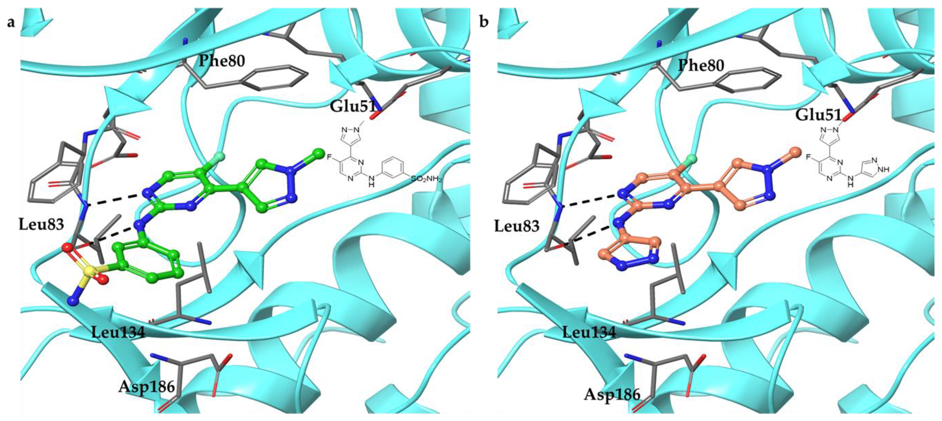
To understand the potential of these newly designed compounds to inhibit CDK2, a molecular modeling study on 14, an exemplar of the proposed series (Scheme 1), or 1 in complex with CDK2/cyclin E was performed, and their respective plausible binding modes are shown in Figure 2. Both compounds are predicted to engage the hinge region, forming hydrogen bonds through their common pyrimidinyl-N1-C2-NH motif with Leu83 of CDK2. These highly similar CDK2-binding features between 14 and 1 encouraged us to synthesize the N-pyrazole analogues.

2.2. Synthesis

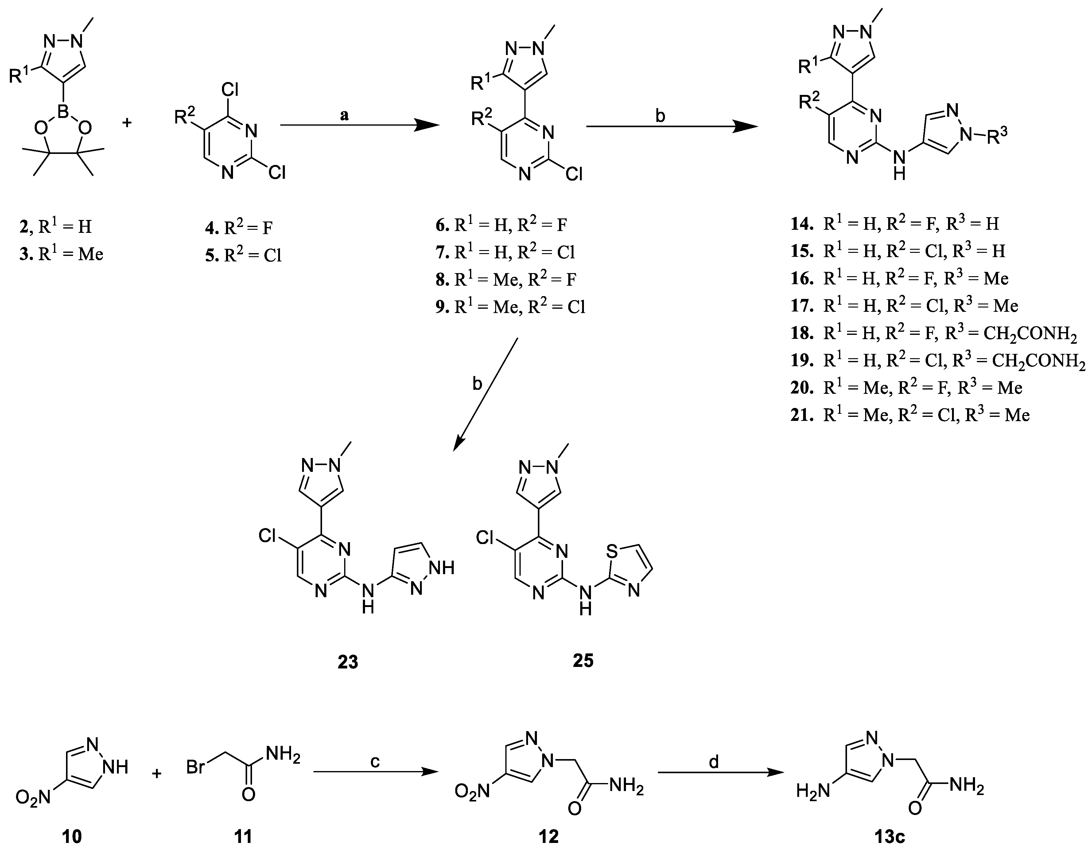
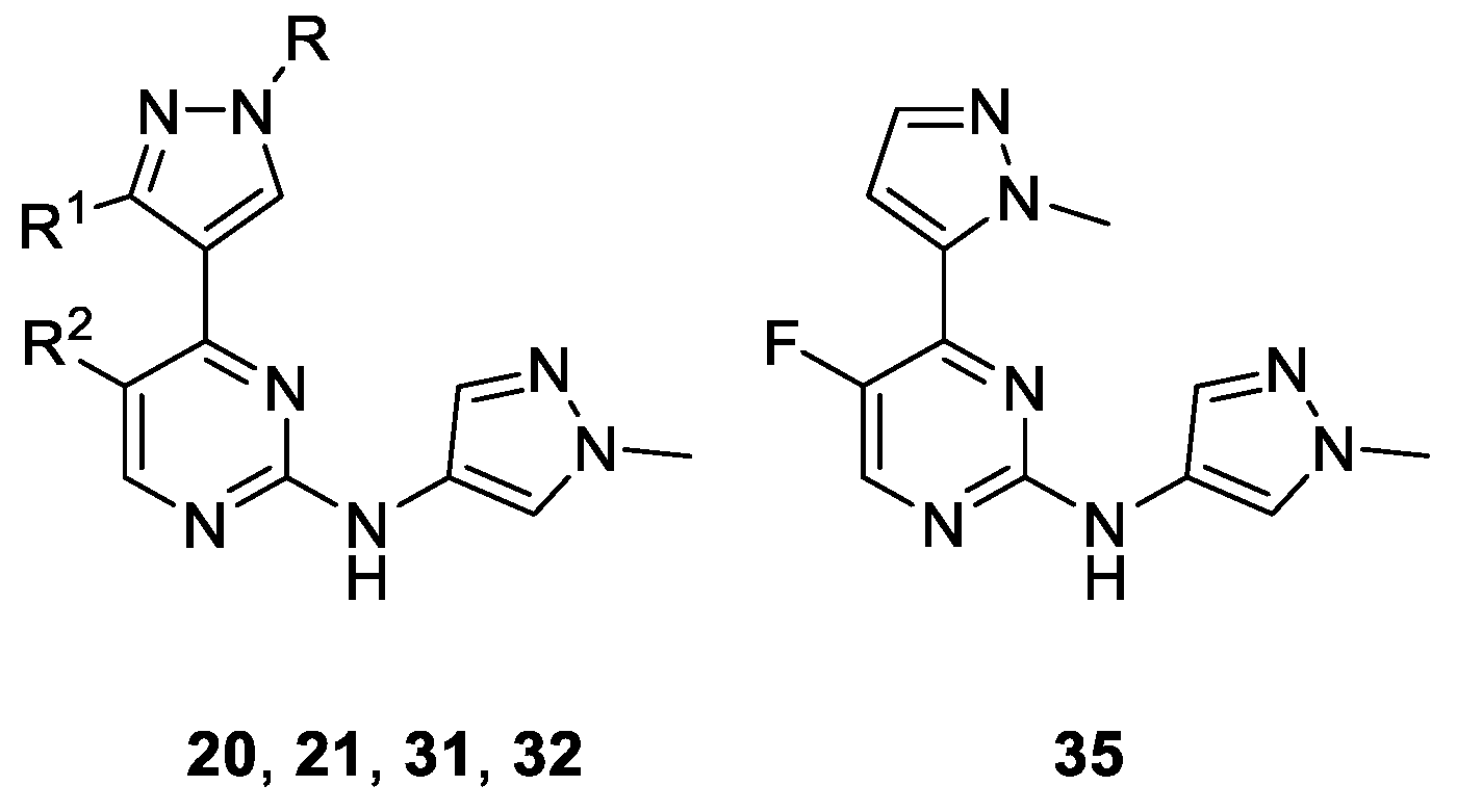
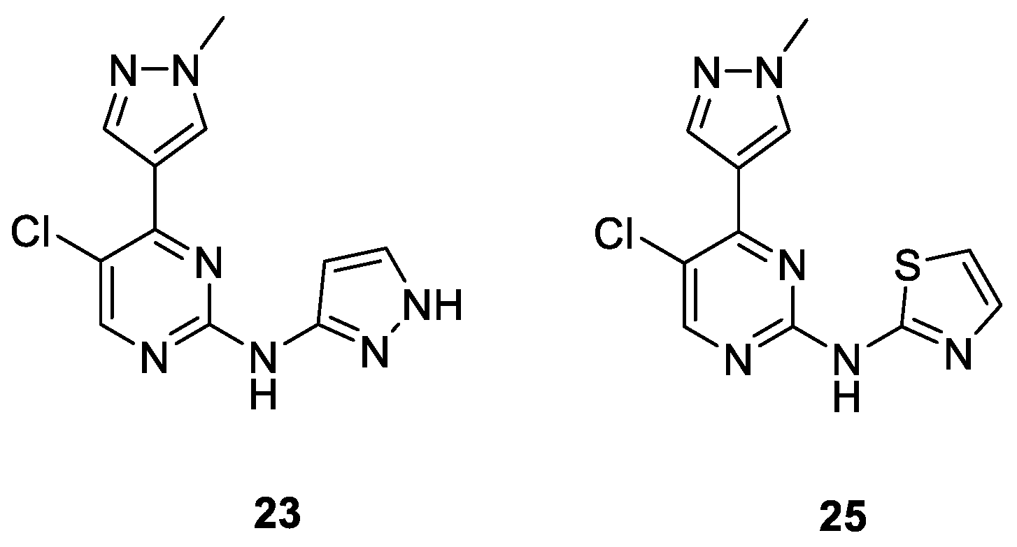
Scheme 1 depicts the synthesis of N,4-di(1H-pyrazolyl)pyrimidin-2-amines 14–21 and 23 and N-(5-chloro-4-(1-methyl-1H-pyrazol-4-yl)pyrimidin-2-yl)thiazol-2-amine 25. Suzuki coupling reactions of either (1-methyl-1H-pyrazol-4-yl)boronic acid pinacol ester 2 or its pyrazolyl-C3-methylated counterpart 3 with a 5-substituted-2,4-dichloropyrimidine 4 or 5 in the presence of PdCl2(dppf)·DCM and K2CO3 in 1,4-dioxane/ethanol/water afforded 2-chloro-4-(1-methyl-1H-pyrazol-4-yl)pyrimidines 6–9 in 59–69% yields. Buchwald–Hartwig amination of these chlorides with appropriate aminopyrazoles 13a–13c and 22 or thiazol-2-amine 24 in the presence of Pd2(dba)3, xantphos, and Cs2CO3, yielded the desired compounds (14–21, 23, and 25) in varying yields (13–28%). Compounds 13a and 13b were purchased, whereas 13c was prepared in-house (Scheme 1).
Scheme 2 illustrates the preparation of 31, 32, and 35. Compound 28 was prepared in moderate yield (57%) from (1H-pyrazol-4-yl)boronic acid pinacol ester 26 and 4-(2-chloroethyl)morpholine 27 in the presence of Cs2CO3 in acetonitrile at reflux. Suzuki coupling reactions of 28 with 4 or 5 afforded 29 and 30 which each were subsequently aminated with 1-methyl-1H-pyrazol-4-amine 13b under Buchwald–Hartwig coupling conditions to afford 31 and 32 in yields of 17% and 16%, respectively.
To explore the effect of regioisomerism of the pyrazole ring at the pyrimidinyl-C4 position on the CDK2 inhibition, 5-fluoro-N-(1-methyl-1H-pyrazol-4-yl)-4-(1-methyl-1H-pyrazol-5-yl)pyrimidin-2-amine 35 was synthesized (Scheme 2). Suzuki coupling reaction of (1-methyl-1H-pyrazol-5-yl)boronic acid pinacol ester 33 with 2,4-dichloro-5-fluoropyrimidine 4 gave 2-chloro-5-fluoro-4-(1-methyl-1H-pyrazol-5-yl)pyrimidine 34 in 42% yield. Subsequent Buchwald–Hartwig amination of 34 with 13b produced 35 in a yield of 26%.

2.3. Structure-Activity Relationships

With the desire to improve the potency and selectivity of our previously-identified lead compound 1 [20], its phenylsulfonamide moiety was replaced with pyrazole-derived groups, affording a series of N,4-di(1H-pyrazol-4-yl)pyrimidin-2-amine derivatives. These new analogues were then evaluated for their activities against CDK2E; in order to understand their selectivity profiles, they were also tested against other CDKs, namely CDK1B, CDK5p25, and CDK9T1, all of which share high amino acid sequence similarity with CDK2E. Additionally, the antiproliferative activities of these compounds were screened against A2780 ovarian cancer cells.
As shown in Table 1, substitution of the phenylsulfonamide moiety of 1 with an unsubstituted pyrazol-4-yl ring gave 14, which displayed more potent CDK2 and CDK5 inhibition with Ki values of 0.007 and 0.003 µM, respectively. Compound 14 also improved the selectivity of 1 for CDK2 over CDKs 1 and 9. However, 14 showed about 28-fold lower antiproliferative activity against A2780 ovarian cancer cells when compared to 1. Replacement of the fluorine atom in 14 (R2 = F) with chlorine afforded 15 (R2 = Cl), which maintained the inhibitory potencies towards CDK2 (Ki = 0.005 µM) and CDK5 (Ki = 0.003 µM) and slightly enhanced the selectivity for CDK2 over CDK1 and CDK9. Furthermore, in comparison with 1, 15 showed improved potency against and selectivity for CDK2 but reduced antiproliferative activity against A2780 cells (15: GI50 = 0.158 µM versus 1: GI50 = 0.018 µM). Encouraged by these results (i.e., enhanced CDK2 inhibitory activity and selectivity), further SAR studies were carried out by varying R1 and R3 substituents while retaining R2 as chlorine or fluorine.
Substitution of the N-pyrazol-4-yl-NH in 14 and 15 (R3 = H) by a methyl, affording 16 and 17 (R3 = Me), respectively, led to a reduction in the inhibitory potencies across all the CDKs tested and, consequently, resulted in diminished antiproliferative activities against A2780 cells. An acetamide group was then introduced at the same position to test whether the terminal amide could interact with Asp86 and/or Lys89 residues of CDK2. However, 18 and 19 (R3 = CH2CONH2) thus obtained had detrimental effects on the inhibition of all four CDKs and A2780 cells. Taken together, these results suggest the importance of the unmasked pyrazole ring (R3 = H) at the pyrimidinyl-C2-NH position for inhibitory activities towards both CDK2 and A2780 cells.
We next investigated the SAR around the other pyrazole ring occupying the pyrimidinyl-C4 position (i.e., R and R1). As shown in Table 2, methylation of the pyrazolyl-C3 position as in 20 and 21 (R1 = Me) led to significant decreases in inhibitory activities against all the CDKs tested with concomitant reductions in the cellular activities. Similarly, substitution of the pyrazolyl-N1-methyl group by a larger 2-morpholinoethyl as in 31 and 32 (R = 2-morpholinoethyl) had detrimental effects on both kinase and cellular inhibition (e.g., 32: Ki(CDK2) = 0.252 µM and %GI(A2780) = 3 at 1 µM versus 17: Ki(CDK2) = 0.011 µM and %GI(A2780) = 87 at 1 µM (not shown in Table 1)), thus signifying that diversification of the pyrazole ring by introduction of a larger substituent at the N1 or C3 position may not be tolerated. To explore the effect of regioisomerism regarding the pyrazole ring at the pyrimidinyl-C4 position on the CDK2 inhibition, 35 with a 1-methyl-1H-pyrazol-5-yl moiety was synthesized and found to be less active towards CDK2 than its 1-methyl-1H-pyrazol-4-yl counterpart 16 (% CDK2 inhibition = 64 and 97 (not shown in Table 1), respectively), suggesting the dependency of the kinase inhibition on the topology of the pyrazole ring at the pyrimidinyl-C4 position.
Having identified the dependence of the CDK2 inhibitory activity on the topology of the 1-methyl-1H-pyrazol-4-yl group at the pyrimidinyl-C4 position, it seemed logical to investigate if a similar dependence existed on the other pyrazole ring at the pyrimidinyl-C2-NH position. As a result, 5-chloro-4-(1-methyl-1H-pyrazol-4-yl)-N-(1H-pyrazol-5-yl)pyrimidin-2-amine 23 was synthesized. As Table 3 shows, the compound displayed greatly diminished CDK inhibitory activities, particularly with an 18-fold reduced CDK2 inhibition when compared to its 1H-pyrazol-4-yl counterpart 15 (Ki = 0.090 and 0.005 µM, respectively). Consequently, 23 showed 47-fold less potent antiproliferative activity against A2780 cells than did 15 (GI50 = 7.350 and 0.158 µM, respectively), indicating the dependency of the CDK2 inhibitory activity on the orientation of the 1H-pyrazolyl ring at the pyrimidinyl-C2-NH position.
Next, we investigated the effect of a related heteroaromatic system—thiazol-2-yl—at the pyrimidinyl-C2-NH position (i.e., 25) on the CDK and A2780 inhibitory activities. As shown in Table 3, the introduction of such a ring led to a significant reduction of the inhibitory activities toward all the CDKs tested as well as A2780 cells.
Taken together, compounds with chlorine at the pyrimidinyl-C5 position (R2 = Cl) were generally more potent CDK2 inhibitors than their fluorine counterparts (R2 = F), which may be attributed to a stronger hydrophobic interaction between the chlorine and Phe80.

2.4. Docking Study on 15

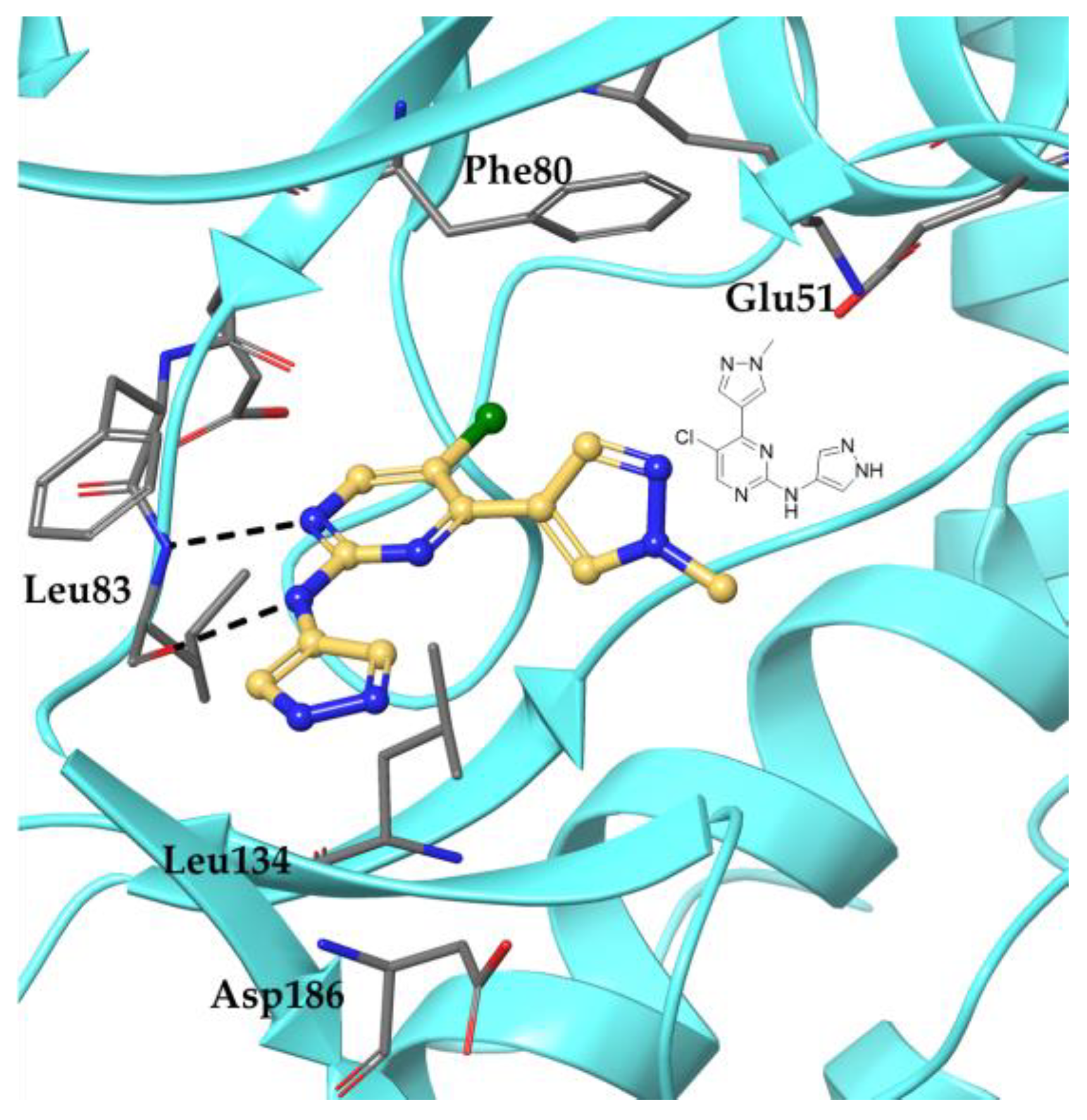
To understand the likely binding mode of 15, the most potent CDK2 inhibitor in the series, with CDK2 and to provide a rationale for the observed potency, the compound was docked into the crystal structure of CDK2/cyclin E (PDB ID: 7KJS) using OEDocking 4.1.2.1 from OpenEye Scientific Software. The result revealed that 15 may form two hydrogen bonds with Leu83 in the ATP binding pocket of CDK2 (Figure 3). Specifically, while the 2-amino group of the pyrimidine ring could develop a hydrogen bond with the carbonyl of Leu83, the pyrimidinyl-N1 is likely to form a second hydrogen bond with the NH of the same amino acid residue. This binding pose is similar to those of 1 or 14 in complex with CDK2/cyclin E (Figure 2) and those of other inhibitors co-crystallized with CDK2 [15,27,29].

2.5. Antiproliferative Activities of 15

Based on the most potent CDK2 inhibition displayed by 15 (Ki = 0.005 µM), it was selected for further antiproliferative studies using various types of cancer cell lines, including leukemia (MV4-11 and U937), breast (MCF7 (Rb-positive) and MDA-MB-231(Rb-negative)), and ovarian (OVCAR5, OAW28, OV90, Cov318, and Cov504) cancers, and glioblastoma (U87, T98G, and U251). As Table 4 shows, 15 inhibited the proliferation of all the cancer cell lines tested at submicromolar concentrations, with GI50 values ranging from 0.127 to 0.560 µM. The most sensitive cell line to 15 (GI50 = 0.127 µM) was MV4-11, which was followed by the ovarian cancer cell line OVCAR5 (GI50 = 0.150 µM).

2.6. Cellular Mechanistic Studies of 15

Given that A2780 and OVCAR5 were two of the most sensitive cancer cell lines to 15 in the above cancer cell panel screen, both ovarian cell lines were selected for cellular mechanistic studies. The effects of 15 on cell cycle progression and the level of phosphorylated Rb were determined by flow cytometry and Western blotting, respectively. In addition, the impacts of 15 on colony formation and apoptosis were analyzed by staining with crystal violet and annexin V/propidium iodide (PI), respectively. CYC065, a CDK2/9 inhibitor and a clinical candidate [30], was used as a control in all mechanistic studies conducted.

2.6.1. Effect of 15 on the Cell Cycle Progression

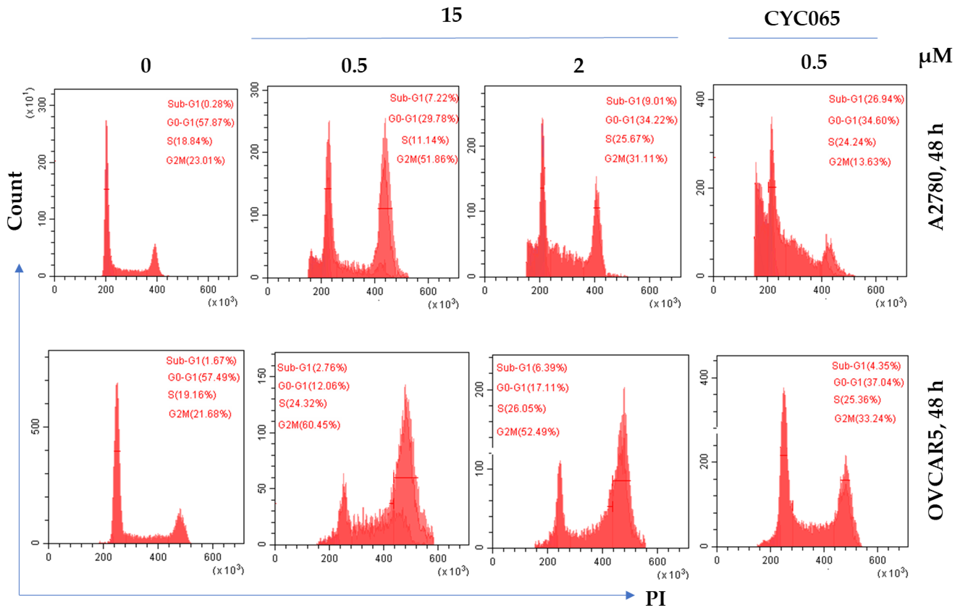
A2780 cells were treated with 15 at concentrations of 0.5 or 2 µM for a period of 48 h. The flow cytometric analysis (Figure 4; top) showed that the compound at 0.5 µM arrested the cells at the G2/M phase (about 52%) when compared to the untreated cells (23%). An increased subpopulation of S-phase cells was also observed with the treatment of 2 µM of 15 (26% versus 19% in untreated cells). At 0.5 µM, CYC065 caused the cell to arrest in sub-G1 (27% versus 0.3% in untreated cells) and S-phase (24% versus 19% in untreated cells).
Similar cell-cycle effects on OVCAR5 cells were also observed (Figure 4, bottom). At the concentration of 0.5 µM, 15 resulted in a 39% increment in the subpopulation of G2/M cells when compared to the vehicle control. At 2 µM, the compound also showed a similar effect (53% G2/M-phase cells versus 22% in untreated cells) with an increased S-phase subpopulation (26% versus 19% in untreated cells). CYC065 caused a similar cell-cycle profile with an increased percentage of G2/M cells, but to a lesser extent (a 12% increase when compared to the vehicle control).

2.6.2. Effect of 15 on Apoptosis in Cancer Cells

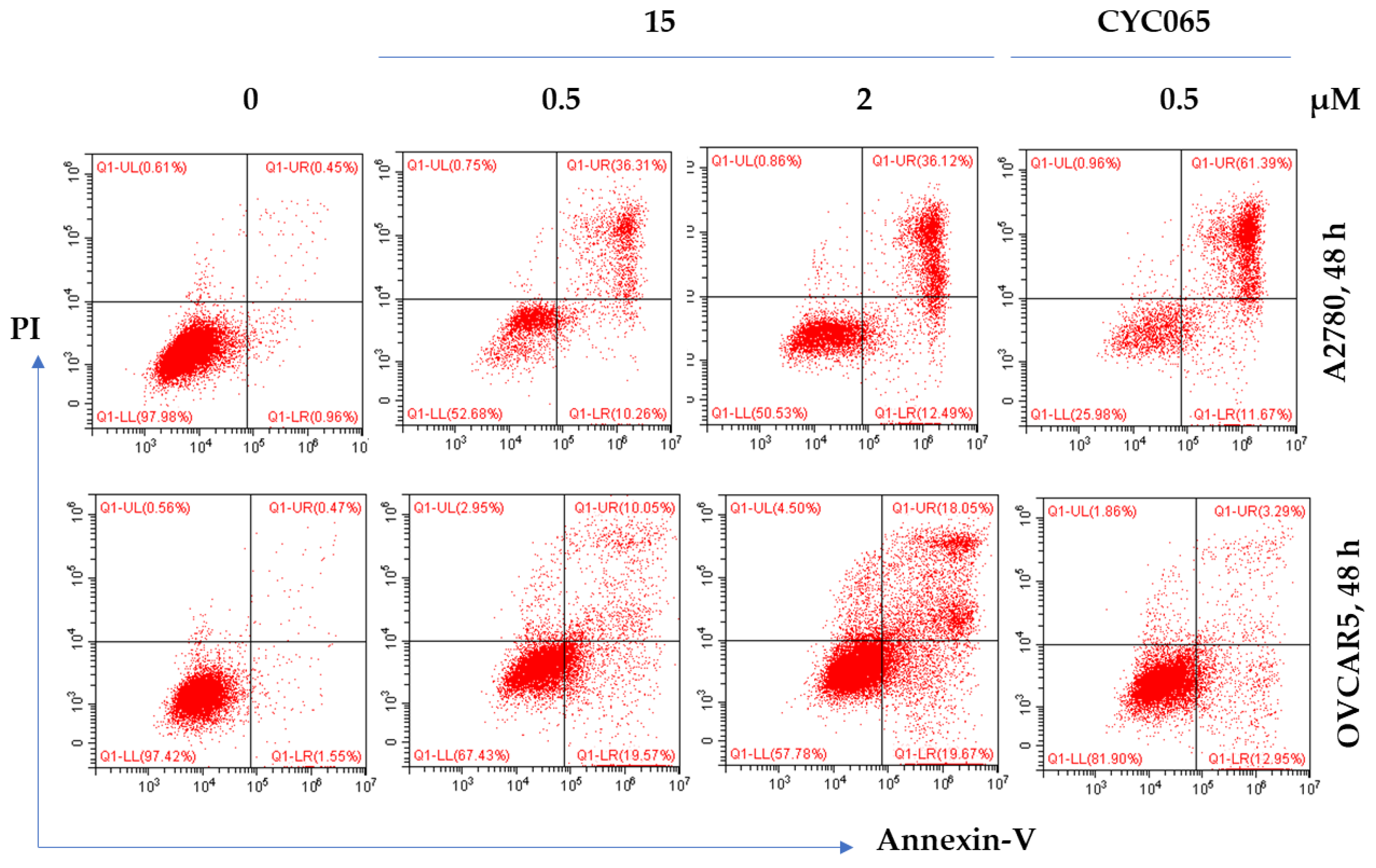
To probe whether the antiproliferative activities of 15 were due to the induction of apoptosis, A2780 and OVCAR5 cells were incubated with 15 at concentrations of 0.5 and 2 µM for 48 h, and apoptosis was determined by annexin V and PI double staining. As Figure 5 shows, in A2780 cells, 15 induced 47% and 49% apoptosis at 0.5 and 2 µM, respectively, while only 1% was found in untreated cells. In contrast, CYC065 at 0.5 µM showed 73% apoptosis. In OVCAR5 cells, 15 caused 30% and 38% apoptosis at 0.5 and 2 µM, respectively, whereas 2% was observed in untreated cells. On the other hand, CYC065 induced 16% apoptosis. CYC065 induced more apoptosis in A2780 cells than did 15, and the opposite held true in OVCAR5 cells.

2.6.3. Effect of 15 on Colony Formation

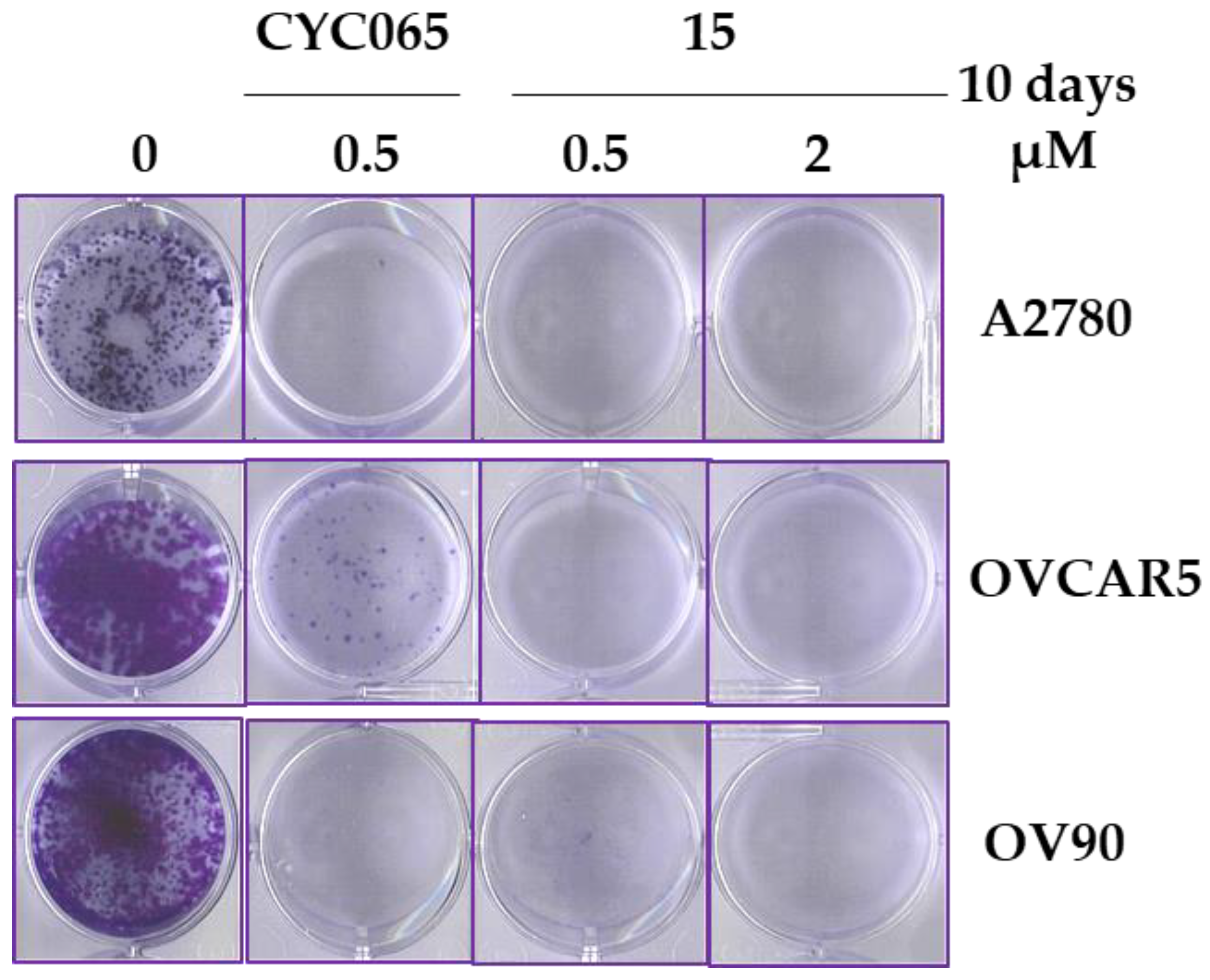
As Figure 6 illustrates, 15 inhibited the ability of three ovarian cancer cell lines (A2780, OVCAR5, and OV90) to form viable colonies at 0.5 and 2 µM. However, partial inhibition of colony formation was observed at 0.5 µM in OV90 cells. These results are in agreement with the observed antiproliferative effects of 15 (Table 1 and Table 4). Similarly, CYC065 reduced the clonogenic growth of all three cell lines, with the lowest effect on OVCAR5.

2.6.4. Western Blot Analysis

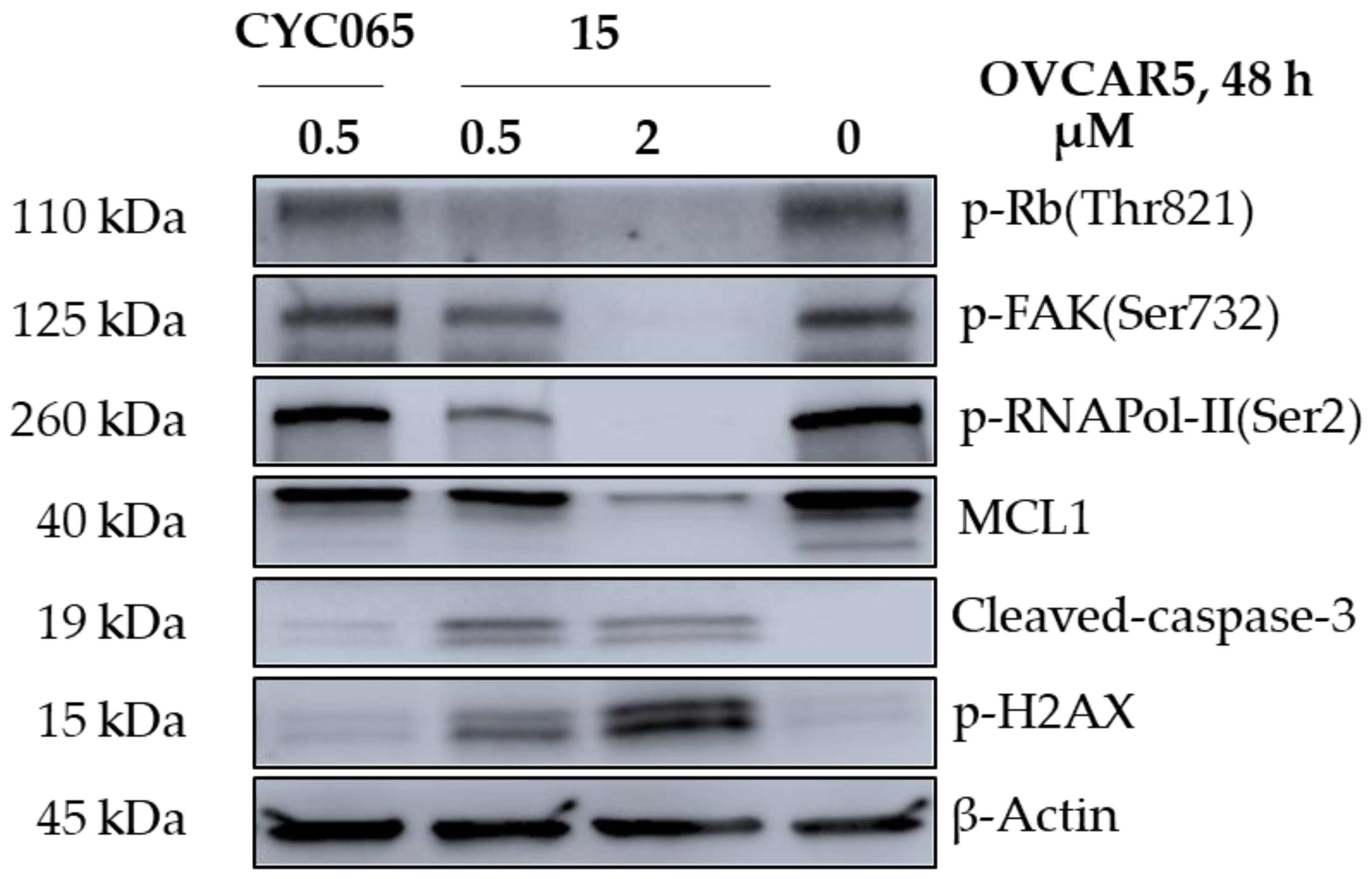
To explore whether 15 could inhibit its target kinases within cancer cells and to understand the molecular mechanism underlying its anticancer effects, OVCAR5 cells were incubated with 0.5 or 2 µM of 15 or 0.5 µM of CYC065 for 48 h, and the phosphorylation levels of the physiological substrates of CDK2, CDK5, and CDK9 (i.e., Rb, FAK, and RNAPol-II, respectively) were determined by Western blotting. As shown in Figure 7, 15 completely abolished the kinase activity of CDK2, CDK5, and CDK9 at 2 µM as demonstrated by the disappearance of p-Rb(Thr821), p-FAK(Ser732), and p-RNAPol-II(Ser2), respectively. Additionally, 15 also caused double-strand DNA breaks, showcased by the appearance of phosphorylated histone H2AX (p-H2AX), which is an independent marker of DNA damage. The DNA damage resulted in apoptotic cell death which was evidenced by the cleavage of caspase-3 and the downregulation of the anti-apoptotic protein MCL1.

3. Conclusions

The bioisosteric replacement of the phenylsulfonamide moiety of our previous lead compound 1 with pyrazole-derived groups led to the discovery of a new chemotype of CDK2 inhibitors, namely 4-(1-methyl-1H-pyrazol-4-yl)-N-(1H-pyrazol-4-yl)pyrimidin-2-amines. Among them, 14 and 15 inhibited CDK2 at single-digit nanomolar concentrations (Ki = 0.007 and 0.005 µM, respectively). The SAR analysis revealed that N-alkylation or topological change of the pyrazol-4-yl ring at the pyrimidinyl-C2-NH position had a detrimental effect on both the CDK2 inhibition and the antiproliferative activity. Similar effects were also observed upon derivatization of the other pyrazol-4-yl moiety at the pyrimidinyl-C4 position. Mechanistically, the antiproliferative activity of 15 was shown to be associated with inhibition of the phosphorylation of Rb, FAK, and RNAPol-II, cell cycle arrest at the S and G2/M phases, and induction of apoptosis. Thus, 15 can be considered a lead compound that could be further optimized into a potent and selective CDK2 inhibitor with potential anticancer activity.

4. Experimental Section

4.1. Method for Computational Modeling

Computational modeling was carried out using the crystal structure of CDK2/cyclin E in complex with a pyridopyrimidinone inhibitor (i.e., PF-06873600) (PDB ID: 7KJS). OMEGA was used for ligand preparation starting from the SMILES string, generating the lowest energy conformer of the ligand, which was retained for docking (OMEGA 4.2.1.2: OpenEye Scientific Software, Santa Fe, NM. http://www.eyesopen.com, accessed on 27 November 2022) [31]. Docking was undertaken using OEDocking and the chemgauss4 scoring function (OEDocking 4.1.2.1 OpenEye Scientific docking program, (http://www.eyesopen.com, accessed on 27 November 2022). The visualization was generated by Maestro Schrödinger Release 2021-1 as described elsewhere [32].

4.2. Chemistry

Chemicals and solvents were obtained from commercial sources and were used without further purification. A CEM Discover SP and Explorer 48/72/96 microwave system (CEM corporation, Matthews, NC, USA) controlled by the SynergyTM software was used for microwave-assisted synthesis. The progression of a reaction was monitored by thin-layer chromatography using Merck 60 F254 silica gel-precoated aluminum plates and visualized by UV irradiation (254 nm). Flash column chromatography was carried out using a glass column packed with silica gel (230–400 mesh). An AB SCIEX TripleTOF® 5600 mass spectrometer (Concord, Ontario, Canada) with ESI ionization mode was used to determine the high-resolution mass spectra. 1H and 13C NMR spectra were determined by a Bruker Avance III HD spectrometer (Faellanden, Switzerland) at 500.16 and 125.76 MHz, respectively, and were analyzed using the Bruker Topspin 4.1 program. Chemical shifts are reported in parts per million (ppm) and referenced to 1H signals of residual nondeuterated solvents and 13C signals of the deuterated solvents. The multiplicity of 1H NMR signals was reported as: s = singlet, d = doublet, and t = triplet. Coupling constant (J) values are reported in Hz. A Shimadzu Prominence UltraFast Liquid Chromatograph System (Kyoto, Japan) equipped with a CBM-20A communications bus module, a DGU-20A5R degassing unit, an LC-20AD liquid chromatograph pump, an SIL-20AHT auto-sampler, an SPD-M20A photodiode array detector, a CTO-20A column oven, and a Phenomenex Kinetex 5u C18 100A 250 mm × 4.60 mm column was used to determine the purity of the final compounds. All the final compounds used for biological assays had a purity greater than 95%. Method A (gradient 5%–95% methanol containing 0.1% formic acid (FA) over 20 min at a flow rate of 1 mL/min, followed by 95% methanol containing 0.1% FA over 5 min) and method B (gradient 5%–95% MeCN containing 0.1% FA over 20 min at a flow rate of 1 mL/min, followed by 95% MeCN containing 0.1% FA over 5 min) were used for analytic HPLC. The melting points (m.p.) of final compounds were determined using a Stuart SMP10 melting point apparatus and are uncorrected.

4.2.1. General Synthetic Procedure A: Suzuki Coupling [33]

A mixture of a pyrazole boronic acid pinacol ester (1.30 equivalent (eqv.)), a 5-substituted-2,4-dichloropyrimidine (1.00 eqv.), PdCl2(dppf)·DCM (0.10 eqv.), and potassium carbonate (2.50 eqv.) in 1,4-dioxane:ethanol:water (7:3:4, v/v/v) was degassed, purged with nitrogen (3×), and heated under nitrogen at 80 °C for 12 h. The reaction mixture was cooled down and concentrated under reduced pressure, and the residue was partitioned between water and ethyl acetate (1:2, v/v). The aqueous layer was extracted with ethyl acetate (3×). The organic layer and extracts were combined, washed with brine, dried over Na2SO4, and filtered; the filtrate was concentrated under reduced pressure. The residue was purified by flash column chromatography (silica gel) eluting with 20–40% ethyl acetate in petroleum ether (unless otherwise stated) to afford the desired product.

4.2.2. General Synthetic Procedure B: Buchwald–Hartwig Amination [33]

A mixture of a 2-chloropyrimidine (1.20 eqv.), an arylamine (1.00 eqv.), Cs2CO3 (2.50 eqv.), Pd2(dba)3 (0.05 eqv.), and Xantphos (0.10 eqv.) in 1,4-dioxane was degassed, purged with nitrogen (3×), and subjected to microwave irradiation at 140 °C for 1 h. The reaction mixture was concentrated under reduced pressure, and the residue was partitioned between water and ethyl acetate (1:2, v/v). The aqueous layer was extracted with ethyl acetate (3×), and the organic layer and extracts were combined, washed with brine, dried over Na2SO4, and filtered. The filtrate was concentrated under reduced pressure, and the residue was purified by flash column chromatography (silica gel) to give the desired product. 1H & 13C NMR spectra, HRMS & HPLC chromatograms of compounds see Supplementary Materials.
2-Chloro-5-fluoro-4-(1-methyl-1H-pyrazol-4-yl)pyrimidine (6): Prepared following the general synthetic procedure A using 2,4-dichloro-5-fluoropyrimidine (4) (5.00 g, 30.0 mmol) and (1-methyl-1H-pyrazol-4-yl)boronic acid pinacol ester (2) (5.20 g, 25.0 mmol). White solid (3.70 g, 69%). 1H NMR (DMSO-d6): δ 3.96 (s, 3H, CH3); 8.11 (s, 1H, pyrazolyl-H); 8.57 (s, 1H, pyrazolyl-H); and 8.79 (d, 1H, J = 2.4, pyrimidinyl-H). HRMS (ESI-TOF) 213.0345 [M(35Cl)+H]+ and 215.0316 [M(37Cl)+H]+; calcd. for C8H7ClFN4+ 213.0338 [M(35Cl)+H]+ and 215.0309 [M(37Cl)+H]+.
2,5-Dichloro-4-(1-methyl-1H-pyrazol-4-yl)pyrimidine (7): Prepared following the general synthetic procedure A using 2,4,5-trichloropyrimidine (5) (5.50 g, 30.0 mmol) and (1-methyl-1H-pyrazol-4-yl)boronic acid pinacol ester (2) (5.20 g, 25.0 mmol). White solid (3.70 g, 65%). 1H NMR (CDCl3): δ 4.02 (s, 3H, CH3); 8.37 (s, 1H, pyrazolyl-H); 8.39 (s, 1H, pyrazolyl-H); and 8.52 (s, 1H, pyrimidinyl-H). HRMS (ESI-TOF) 229.0051 [M(2 × 35Cl)+H]+, 231.0018 [M(35Cl and 37Cl)+H]+ and 232.9987 [M(2 × 37Cl)+H]+, calcd. For C8H7Cl2N4+ 229.0043 [M(2 × 35Cl)+H]+, 231.0013 [M(35Cl and 37Cl)+H]+, and 232.9984 [M(2 × 37Cl)+H]+.
2-Chloro-4-(1,3-dimethyl-1H-pyrazol-4-yl)-5-fluoropyrimidine (8): Prepared following the general synthetic procedure A using (1,3-dimethyl-1H-pyrazol-4-yl)boronic acid pinacol ester (3) (5.60 g, 25.0 mmol) and 2,4-dichloro-5-fluoropyrimidine (4) (5.00 g, 30.0 mmol). Off-white solid (3.60 g, 63%). 1H NMR (DMSO-d6): δ 2.54 (s, 3H, CH3); 3.84 (s, 3H, CH3); 7.93 (s, 1H, pyrazolyl-H); 8.27 (d, 1H, J = 2.7, pyrimidinyl-H). HRMS (ESI-TOF) 227.0502 [M(35Cl)+H]+ and 229.0470 [M(37Cl)+H]+; calcd. For C9H9ClFN4+ 227.0495 [M(35Cl)+H]+, and 229.0465 [M(37Cl)+H]+.
2,5-Dichloro-4-(1,3-dimethyl-1H-pyrazol-4-yl)pyrimidine (9): Prepared following the general synthetic procedure A using 2,4,5-trichloropyrimidine (5) (5.50 g, 30.0 mmol) and (1,3-dimethyl-1H-pyrazol-4-yl)boronic acid pinacol ester (3) (5.60 g, 25.0 mmol). Off-white solid (3.60 g, 59%). 1H NMR (DMSO-d6): δ 2.42 (s, 3H, CH3); 3.87 (s, 3H, CH3); 8.63 (s, 1H, pyrazolyl-H); 8.76 (s, 1H, pyrimidinyl-H). HRMS (ESI-TOF) 243.0213 [M(2 × 35Cl)+H]+, 245.0180 [M(35Cl and 37Cl)+H]+ and 247.0151 [M(2 × 37Cl)+H]+; calcd. For C9H9ClFN4+ 243.0199 [M(2 × 35Cl)+H]+, 245.0170 [M(35Cl and 37Cl)+H]+, and 247.0140 [M(2 × 37Cl)+H]+.
2-(4-Nitro-1H-pyrazol-1-yl)acetamide (12): To a solution of 4-nitro-1H-pyrazole (10) (1.70 g, 15.0 mmol) in acetonitrile (150 mL) were added K2CO3 (4.10 g, 30.0 mmol) and 2-bromoacetamide (11) (2.10 g, 15.0 mmol), and the mixture was heated at 60 °C overnight, cooled down, filtered, and washed with acetonitrile (50 mL). The filtrate and washing were concentrated under reduced pressure, and the residue was triturated with diethyl ether:petroleum ether (2:1) to yield 12 as a white solid (2.40 g, 95%). 1H NMR (DMSO-d6): δ 4.88 (s, 2H, CH2), 7.41 (s, 1H, acetamide-NH), 7.68 (s, 1H, acetamide-NH), 8.27 (s, 1H, pyrazolyl-H), 8.83 (s, 1H, pyrazolyl-H). HRMS (ESI-TOF) 171.0523 [M+H]+; calcd. for C5H7N4O3+ 171.0513 [M+H]+.
2-(4-Amino-1H-pyrazol-1-yl)acetamide (13c): To a suspension of 2-(4-nitro-1H-pyrazol-1-yl)acetamide (12) (2.40 g, 14.1 mmol) in methanol (100 mL) was added 10% palladium on carbon (150 mg, 141 µmol), and the reaction mixture was stirred at room temperature under hydrogen overnight. The reaction product was filtered through a pad of Celite, and the residue was washed with methanol (100 mL). The filtrate and washing were combined and concentrated under reduced pressure to afford 13c as a dark brown solid, which was used in the next step without further purification (1.80 g, 90%). 1H NMR (DMSO-d6): δ 3.81 (s, 2H, CH2); 4.51 (s, 2H, NH2); 6.89 (s, 1H, pyrazolyl-H); 6.99 (s, 1H, pyrazolyl-H); and 7.14 (s, 2H, acetamide-NH2). HRMS (ESI-TOF) 141.0778 [M+H]+; calcd. for C5H9N4O+ 141.0771 [M+H]+.
5-Fluoro-4-(1-methyl-1H-pyrazol-4-yl)-N-(1H-pyrazol-4-yl)pyrimidin-2-amine (14): Prepared from 2-chloro-5-fluoro-4-(1-methyl-1H-pyrazol-4-yl)pyrimidine (6) (213 mg, 1.00 mmol) and 4-amino-1H-pyrazole (13a) (100 mg, 1.20 mmol) following the general synthetic procedure B. The residue was purified by flash column chromatography, eluting with 0–5% methanol in ethyl acetate to afford 14 as a yellow solid (34 mg, 13%). m.p. 253.8–254.8 °C. 1H NMR (DMSO-d6): δ 3.94 (s, 3H, CH3); 7.71 (s, 2H, pyrazolyl-H); 8.04 (s, 1H, pyrazolyl-H), 8.38 (s, 1H, pyrazolyl-H), 8.41 (d, 1H, J = 2.9, pyrimidinyl-H), 9.38 (s, 1H, NH); and 12.43 (s, 1H, pyrazolyl-NH). 13C NMR (DMSO-d6): δ 39.4, 116.2 (d, JC–F = 5.5), 117.9, 123.5, 130.3, 132.8 (d, JC–F = 8.0), 139.1 (d, JC–F = 5.2), 146.4, 148.3 (d, JC–F = 246.0), and 156.8 (one carbon signal overlapping or obscured). HRMS (ESI-TOF): 260.1064 [M+H]+; calcd. for C11H11FN7+ 260.1054 [M+H]+. Anal. RP/HPLC Method A: tR 15.53 min, purity 100%; Method B: tR 11.35 min, purity > 99%.
5-Chloro-4-(1-methyl-1H-pyrazol-4-yl)-N-(1H-pyrazol-4-yl)pyrimidin-2-amine (15): Prepared from 2,5-dichloro-4-(1-methyl-1H-pyrazol-4-yl)pyrimidine (7) (229 mg, 1.00 mmol) and 4-amino-1H-pyrazole (13a) (100 mg, 1.20 mmol) following the general synthetic procedure B. The residue was purified by flash column chromatography, eluting with 0–5% methanol in ethyl acetate to afford 15 as a yellow solid (52 mg, 19%). m.p. 269.1 °C. 1H NMR (DMSO-d6): δ 3.97 (s, 3H, CH3); 7.63 (s, 1H, pyrazolyl-H); 7.92 (s, 1H, pyrazolyl-H); 8.19 (s, 1H, pyrazolyl-H); 8.43 (s, 1H, pyrazolyl-H); 8.59 (s, 1H, pyrimidinyl-H); 9.56 (s, 1H, NH); and 12.51 (s, 1H, pyrazolyl-NH). 13C NMR (DMSO-d6): δ 40.5, 114.2, 118.8, 118.9, 122.9, 130.9, 133.3, 140.1, 155.4, 158.1, and 158.7. HRMS (ESI-TOF) 276.0758 [M(35Cl)+H]+ and 278.0728 [M(37Cl)+H]+; calcd. for C11H11ClN7+ 276.0759 [M(35Cl)+H]+ and 278.0730 [M(37Cl)+H]+. Anal. RP/HPLC Method A: tR 16.84 min, purity ˃ 95%; Method B: tR 12.34 min, purity ˃ 95%.
5-Fluoro-N,4-bis(1-methyl-1H-pyrazol-4-yl)pyrimidin-2-amine (16): Prepared from 2-chloro-5-fluoro-4-(1-methyl-1H-pyrazol-4-yl)pyrimidine (6) (213 mg, 1.00 mmol) and 1-methyl-1H-pyrazole-4-amine (13b) (117 mg, 1.20 mmol) following the general synthetic procedure B. The residue was purified by flash column chromatography, eluting with 0–2.5% methanol in ethyl acetate to give 16 as a white solid (74 mg, 27%). m.p. 202.8–203.7 °C. 1H NMR (DMSO-d6): δ 3.84 (s, 3H, CH3); 3.97 (s, 3H, CH3); 7.49 (s, 1H, pyrazolyl-H); 7.91 (s, 1H, pyrazolyl-H); 8.09 (s, 1H, pyrazolyl-H); 8.41 (s, 1H, pyrazolyl-H); 8.43 (d, 1H, J = 3.1, pyrimidinyl-H); and 9.42 (s, 1H, NH). 13C NMR (DMSO-d6): δ 39.2, 39.4, 116.2 (d, JC–F = 5.4), 120.4, 123.9, 130.0, 132.8 (d, JC–F = 8.3), 139.3 (d, JC–F = 4.8), 146.3, 148.4 (d, JC–F = 246.0), and 156.6 (one carbon signal overlapping or obscured). HRMS (ESI-TOF) 274.1210 [M+H]+; calcd. for C12H13FN7+ 274.1211 [M+H]+. Anal. RP/HPLC Method A: tR 16.63 min, purity 100%; Method B: tR 12.55 min, purity 100%.
5-Chloro-N,4-bis(1-methyl-1H-pyrazol-4-yl)pyrimidin-2-amine (17): Prepared from 2,5-dichloro-4-(1-methyl-1H-pyrazol-4-yl)pyrimidine (7) (229 mg, 1.00 mmol) and 1-methyl-1H-pyrazole-4-amine (13b) (117 mg, 1.20 mmol) following the general synthetic procedure B. The residue was purified by flash column chromatography, eluting with 0–2.5% methanol in ethyl acetate to give 17 as a white solid (67 mg, 23%). m.p. 201.2–202.3 °C. 1H NMR (DMSO-d6): δ 3.84 (s, 3H, CH3); 3.97 (s, 3H, CH3); 7.53 (s, 1H, pyrazolyl-H); 7.87 (s, 1H, pyrazolyl-H); 8.21 (s, 1H, pyrazolyl-H); 8.40 (s, 1H, pyrazolyl-H); 8.55 (s, 1H, pyrimidinyl-H); and 9.42 (s, 1H, NH). 13C NMR (DMSO-d6): δ 40.6, 40.7, 114.4, 118.9, 121.0, 123.4, 130.5, 133.2, 140.1, 155.5, 158.1, and 158.5. HRMS (ESI-TOF): 290.0918 [M(35Cl)+H]+ and 292.0893 [M(37Cl)+H]+; calcd. for C12H13ClN7+ 290.0916 [M(35Cl)+H]+ and 292.0886 [M(37Cl)+H]+. Anal. RP/HPLC Method A: tR 11.27 min, purity 100%; Method B: tR 13.52 min, purity 100%.
2-(4-((5-Fluoro-4-(1-methyl-1H-pyrazol-4-yl)pyrimidin-2-yl)amino)-1H-pyrazol-1-yl)acetamide (18): Prepared from 2-chloro-5-fluoro-4-(1-methyl-1H-pyrazol-4-yl)pyrimidine (6) (213 mg, 1.00 mmol) and 2-(4-amino-1H-pyrazol-1-yl)acetamide (13c) (140 mg, 1.00 mmol) following the general synthetic procedure B. The residue was purified by flash column chromatography, eluting with 0–4% methanol in ethyl acetate to give 18 as an off-white solid (54 mg, 17%). m.p. 283.1–284.1 °C. 1H NMR (DMSO-d6): δ 4.02 (s, 3H, CH3); 4.82 (s, 2H, CH2); 7.29 (s, 1H, pyrazolylacetamide-NH); 7.45 (s, 1H, pyrazolylacetamide-NH); 7.58 (s, 1H, pyrazolyl-H); 8.05 (s, 1H, pyrazolyl-H); 8.17 (s, 1H, pyrazolyl-H), 8.47 (s, 1H, pyrazolyl-H); 8.50 (d, 1H, J = 2.4, pyrimidinyl-H); and 9.52 (s, 1H, NH). 13C NMR (DMSO-d6): δ 39.4, 54.5, 116.2, 121.2, 123.9, 130.7, 132.9 (d, JC–F = 8.3), 139.3, 146.5, 148.4 (d, JC–F = 246.3), 156.6, and 169.5 (one carbon signal overlapping or obscured). HRMS (ESI-TOF) 317.1273 [M + H]+; calcd. for C13H14FN8O+ 317.1270 [M + H]+. Anal. RP/HPLC: Method A, tR 14.43 min, purity 100%; Method B, tR 8.14 min, purity ˃ 99%.
2-(4-((5-Chloro-4-(1-methyl-1H-pyrazol-4-yl)pyrimidin-2-yl)amino)-1H-pyrazol-1-yl)acetamide (19): Prepared from 2,5-dichloro-4-(1-methyl-1H-pyrazol-4-yl)pyrimidine (7) (229 mg, 1.00 mmol) and 2-(4-amino-1H-pyrazol-1-yl)acetamide (13c) (168 mg, 1.20 mmol) following the general synthetic procedure B. The residue was purified by flash column chromatography, eluting with 0–5% methanol in ethyl acetate to yield 19 as a pale orange solid (63 mg, 19%). m.p. 267.6–268.1 °C. 1H NMR (DMSO-d6): δ 3.96 (s, 3H, CH3); 4.77 (s, 2H, CH2); 7.25 (s, 1H, pyrazolylacetamide-NH); 7.44 (s, 1H, pyrazolylacetamide-NH); 7.54 (s, 1H, pyrazolyl-H); 7.97 (s, 1H, pyrazolyl-H); 8.24 (s, 1H, pyrazolyl-H); 8.43 (s, 1H, pyrazolyl-H); 8.61 (s, 1H, pyrimidinyl-H); and 9.61 (s, 1H, NH). 13C NMR (DMSO-d6): δ 39.4, 54.5, 114.3, 118.9, 121.7, 123.4, 130.9, 133.4, 140.2, 157.9, 158.6, and 169.4 (one carbon signal overlapping or obscured). HRMS (ESI-TOF) 333.0975 [M(35Cl)+H]+ and 335.0944 [M(37Cl)+H]+; calcd. for C13H14ClN8O+ 333.0974 [M(35Cl)+H]+ and 335.0945 [M(37Cl)+H]+. Anal. RP/HPLC Method A: tR 15.55 min, purity ˃ 98%; Method B: tR 11.46 min, purity ˃ 98%.
4-(1,3-Dimethyl-1H-pyrazol-4-yl)-5-fluoro-N-(1-methyl-1H-pyrazol-4-yl)pyrimidin-2-amine (20): Prepared from 2-chloro-4-(1,3-dimethyl-1H-pyrazol-4-yl)-5-fluoropyrimidine (8) (226 mg, 1.00 mmol) and 1-methyl-1H-pyrazole-4-amine (13b) (117 mg, 1.20 mmol) following the general synthetic procedure B. The residue was purified by flash column chromatography eluting, with 0–2.5% methanol in ethyl acetate to yield 20 as a pale orange solid (66 mg, 23%). m.p. 189.1–190.1 °C. 1H NMR (DMSO-d6): δ 2.49 (s, 3H, CH3); 3.79 (s, 3H, CH3); 3.84 (s, 3H, CH3); 7.46 (s, 1H, pyrazolyl-H); 7.83 (s, 1H, pyrazolyl-H); 8.22 (d, 1H, J = 3.7, pyrazolyl-H); 8.38 (d, 1H, J = 3.3, pyrimidinyl-H); and 9.21 (s, 1H, NH). 13C NMR (DMSO-d6): δ 15.0, 39.5, 111.9, 121.2, 123.8, 130.7, 134.3 (d, JC–F = 13.5), 145.3, 148.3, 148.7 (d, JC-F = 246.0), 148.8, and 156.7 (one carbon signal overlapping or obscured). HRMS (ESI-TOF) 288.1378 [M+H]+; calcd. for C13H15FN7+ 288.1367. Anal. RP/HPLC Method A: tR 17.31 min, purity ˃ 99%; Method B: tR 13.10 min, purity ˃ 99%.
5-Chloro-4-(1,3-dimethyl-1H-pyrazol-4-yl)-N-(1-methyl-1H-pyrazol-4-yl)pyrimidin-2-amine (21): Prepared from 2,5-dichloro-4-(1,3-dimethyl-1H-pyrazol-4-yl)pyrimidine (9) (243 mg, 1.00 mmol) and 1-methyl-1H-pyrazole-4-amine (13b) (117 mg, 1.20 mmol) following the general synthetic procedure B. The residue was purified by flash column chromatography eluting, with 75% ethyl acetate in petroleum ether and ramping up to 2.5% methanol in ethyl acetate to afford 21 as an orange solid (67 mg, 22%). m.p. 182.3–183.3 °C. 1H NMR (DMSO-d6): δ 2.41 (s, 3H, CH3); 3.80 (s, 3H, CH3); 3.85 (s, 3H, CH3); 7.48 (s, 1H, pyrazolyl-H); 7.83 (s, 1H, pyrazolyl-H); 8.37 (s, 1H, pyrazolyl-H); 8.41 (s, 1H, pyrimidinyl-H); and 9.44 (s, 1H, NH). 13C NMR (DMSO-d6): δ 14.5, 38.9, 39.5, 114.7, 115.5, 121.4, 123.3, 130.8, 134.0, 148.3, 157.5, 157.9, and 158.1. HRMS (ESI-TOF) 304.1080 [M(35Cl)+H]+ and 306.1051 [M(37Cl)+H]+; calcd. For C13H15ClN7+ 304.1072 [M(35Cl)+H]+ and 306.1043 [M(37Cl)+H]+. Anal. RP/HPLC Method A: tR 18.22 min, purity 100%; Method B: tR 13.79 min, purity ˃ 99%.
5-Chloro-4-(1-methyl-1H-pyrazol-4-yl)-N-(1H-pyrazol-5-yl)pyrimidin-2-amine (23): Prepared from 2,5-dichloro-4-(1-methyl-1H-pyrazol-4-yl)pyrimidine (7) (229 mg, 1.00 mmol) and 1H-pyrazole-3-amine (22) (100 mg, 1.20 mmol) following the general synthetic procedure B. The residue was purified by flash column chromatography eluting, with 75–100% ethyl acetate in petroleum ether to afford 23 as a white solid (77 mg, 28%). m.p. 245.4–246.4 °C. 1H NMR (DMSO-d6): δ 3.96 (s, 3H, CH3); 6.63 (s, 1H, pyrazolyl-H); 7.62 (s, 1H, pyrazolyl-H); 8.19 (s, 1H, pyrazolyl-H); 8.44 (s, 1H, pyrazolyl-H); 8.58 (s, 1H, pyrimidinyl-H); 9.86 (s, 1H, NH); and 12.25 (s, 1H, pyrazolyl-NH). 13C NMR (DMSO-d6): δ 39.4, 96.6, 115.0, 118.9, 128.8, 133.3, 140.1, 155.3, 158.2, and 158.6 (one carbon signal overlapping or obscured). HRMS (ESI-TOF) 276.0758 [M(35Cl)+H]+ and 278.0728 [M(37Cl)+H]+; calcd. For C11H11ClN7+ 276.0759 [M(35Cl)+H]+ and 278.0730 [M(37Cl)+H]+. Anal. RP/HPLC Method A: tR 17.48 min, purity 100%, Method B: tR 12.57 min, purity > 98%.
N-(5-Chloro-4-(1-methyl-1H-pyrazol-4-yl)pyrimidin-2-yl)thiazol-2-amine (25): Prepared from 2,5-dichloro-4-(1-methyl-1H-pyrazol-4-yl)pyrimidine (7) (229 mg, 1.00 mmol) and thiazole-2-amine (24) (120 mg, 1.20 mmol) following the general synthetic procedure B. The residue was purified by flash column chromatography eluting with 75–100% ethyl acetate in petroleum ether to give 25 as an off-white solid (79 mg, 27%). m.p. 243.6–244.5 °C. 1H NMR (DMSO-d6): δ 4.00 (s, 3H, CH3); 7.18 (d, 1H, J = 3.5, thiazolyl-H); 7.47 (d, 1H, J = 3.5, thiazolyl-H); 8.42 (s, 1H, pyrazolyl-H); 8.65 (s, 1H, pyrazolyl-H); 8.72 (s, 1H, pyrimidinyl-H); and 11.86 (s, 1H, NH). 13C NMR (DMSO-d6): δ 39.4, 112.9, 116.8, 118.4, 133.8, 138.4, 140.7, 155.1, 155.5, 159.1, and 160.0. HRMS (ESI-TOF) 293.0380 [M(35Cl)+H]+ and 295.0349 [M(37Cl)+H]+; calcd. For C11H10ClN6S+ 293.0371 [M(35Cl)+H]+ and 295.0342 [M(37Cl)+H]+. Anal. RP/HPLC Method A: tR 19.54 min, purity 100%, method B: tR 14.52 min, purity ˃ 99%.
4-(2-(4-(4,4,5,5-Tetramethyl-1,3,2-dioxaborolan-2-yl)-1H-pyrazol-1-yl)ethyl)morpholine (28): To a solution of 1H-pyrazole-4-boronic acid pinacol ester (26) (4.90 g, 25.0 mmol) in acetonitrile (20 mL) were added caesium carbonate (28.5 g, 87.5 mmol) and 4-(2-chloroethyl)morpholine (27) (5.60 g, 37.5 mmol), and the reaction mixture was heated at reflux for 24 h, cooled down, and partitioned between water (100 mL) and ethyl acetate (100 mL). The organic layer was separated, and the aqueous layer was extracted with ethyl acetate (2 × 100 mL). The organic layer and washings were combined, washed with brine (100 mL), and concentrated under reduced pressure to afford 28 as a brown solid that was used in the next step without further purification and full characterization (4.40 g, 57%). HRMS (ESI-TOF) 308.2138 [M+H]+; calcd. For C15H27BN3O3+ 308.2140 [M+H]+.
4-(2-(4-(2-Chloro-5-fluoropyrimidin-4-yl)-1H-pyrazol-1-yl)ethyl)morpholine (29): Prepared from 4-(2-(4-(4,4,5,5-tetramethyl-1,3,2-dioxaborolan-2-yl)-1H-pyrazol-1-yl)ethyl)morpholine (28) (4.40 g, 14.0 mmol) and 2,4-dichloro-5-fluoropyrimidine (4) (1.60 g, 9.30 mmol) following the general synthetic procedure A. The residue was purified by flash column chromatography, eluting with 0–4% methanol in dichloromethane to yield 29 as a violet gummy solid (1.30 g, 45%). 1H NMR (DMSO-d6): δ 2.43 (t, 4H, J = 4.5, morpholinyl-H); 2.75 (t, 2H, J = 6.4, ethyl-CH2); 3.54 (t, 4H, J = 4.5, morpholinyl-H); 4.37 (t, 2H, J = 6.4, ethyl-CH2); 6.99 (s, 1H, pyrazolyl-H); and 7.35 (s, 1H, pyrazolyl-H), 8.63 (d, 1H, J = 2.8, pyrimidinyl-H). HRMS (ESI-TOF) 312.1024 [M(35Cl)+H]+ and 314.0995 [M(37Cl)+H]+; calcd. For C13H16ClFN5O+ 312.1022 [M(35Cl)+H]+ and 314.0993 [M(37Cl)+H]+.
4-(2-(4-(2,5-Dichloropyrimidin-4-yl)-1H-pyrazol-1-yl)ethyl)morpholine (30): Prepared from 4-(2-(4-(4,4,5,5-tetramethyl-1,3,2-dioxaborolan-2-yl)-1H-pyrazol-1-yl)ethyl)morpholine (28) (4.40 g, 14.0 mmol) and 2,4,5-trichloropyrimidine (5) (1.70 g, 9.30 mmol) following the general synthetic procedure A. The residue was purified by flash column chromatography eluting, with 0–2.5% methanol in dichloromethane to give 30 as a violet gummy solid (1.20 g, 40%). 1H NMR (DMSO-d6): δ 2.44 (t, 4H, J = 4.3, morpholinyl-H); 2.76 (t, 2H, J = 6.3, ethyl-CH2); 3.55 (t, 4H, J = 4.3, morpholinyl-H); 4.38 (t, 2H, J = 6.3, ethyl-CH2); 7.01 (s, 1H, pyrazolyl-H); 8.40 (s, 1H, pyrazolyl-H); and 8.73 (s, 1H, pyrimidinyl-H). HRMS (ESI-TOF) 328.0729 [M(2 × 35Cl)+H]+, 330.0700 [M(35Cl and 37Cl)+H]+ and 332.0670 [M(2 × 37Cl)+H]+; calcd. For C20H25ClN7O3S+ 328.0727 [M(2 × 35Cl)+H]+, 330.0697 [M(35Cl and 37Cl)+H]+, and 332.0668 [M(2 × 37Cl)+H]+.
5-Fluoro-N-(1-methyl-1H-pyrazol-4-yl)-4-(1-(2-morpholinoethyl)-1H-pyrazol-4-yl)pyrimidin-2-amine (31): Prepared from 4-(2-(4-(2-Chloro-5-fluoropyrimidin-4-yl)-1H-pyrazol-1-yl)ethyl)morpholine (29) (312 mg, 1.00 mmol) and 1-methyl-1H-pyrazole-4-amine (13b) (117 mg, 1.20 mmol) following the general synthetic procedure B. The residue was purified by flash column chromatography, eluting with 0–7.5% methanol in ethyl acetate to afford 31 as an orange solid (63 mg, 17%). m.p. 198.3–199.3 °C. 1H NMR (DMSO-d6): δ 2.44 (t, 4H, J = 4.5, morpholinyl-H); 2.76 (t, 2H, J = 6.4, ethylCH2); 3.55 (t, 4H, J = 4.5, morpholinyl-H); 3.83 (s, 3H, CH3); 4.36 (t, 2H, J = 6.4, ethylCH2); 7.50 (s, 1H, pyrazolyl-H), 7.90 (s, 1H, pyrazolyl-H); 8.11 (s, 1H, pyrazolyl-H); 8.43 (d, 1H, J = 3.0, pyrazolyl-H); 8.45 (s, 1H, NH); and 9.42 (s, 1H, pyrimidinyl-H). 13C NMR (DMSO-d6): δ 39.2, 49.2, 53.5, 57.9, 66.7, 115.9, 120.4, 124.0, 130.0, 132.7 (d, JC–F = 8.4), 139.2, 146.5, 148.4 (d, JC–F = 246.4), and 156.6 (three carbon signals overlapping or obscured). HRMS (ESI-TOF) 373.1894 [M + H]+; calcd. for C17H22FN8O+ 373.1895 [M + H]+. Anal. RP/HPLC Method A: tR 12.12 min, purity 100%; Method B tR 9.31 min, purity ˃ 99%.
5-Chloro-N-(1-methyl-1H-pyrazol-4-yl)-4-(1-(2-morpholinoethyl)-1H-pyrazol-4-yl)pyrimidin-2-amine (32): Prepared from 4-(2-(4-(2,5-dichloropyrimidin-4-yl)-1H-pyrazol-1-yl)ethyl)morpholine (30) (328 mg, 1.00 mmol) and 1-methyl-1H-pyrazole-4-amine (13b) (117 mg, 1.20 mmol) following the general synthetic procedure B. The residue was purified by flash column chromatography, eluting with 0–7.5% methanol in ethyl acetate to afford 32 as a pale violet solid (62 mg, 16%). m.p. 128.5–129.5 °C. 1H NMR (DMSO-d6): δ 2.42 (t, 4H, J = 4.5, morpholinyl-H); 2.74 (t, 2H, J = 6.4, ethyl-CH2); 3.54 (t, 4H, J = 4.5, morpholinyl-H); 3.82 (s, 3H, CH3); 4.35 (t, 2H, J = 6.4, ethyl-CH2); 7.51 (s, 1H, pyrazolyl-H); 7.87 (s, 1H, pyrazolyl-H); 8.22 (s, 1H, pyrazolyl-H); 8.41 (s, 1H, pyrazolyl-H); 8.64 (s, 1H, pyrimidinyl-H); and 9.56 (s, 1H, NH). 13C NMR (DMSO-d6): δ 39.2, 49.3, 53.6, 57.9, 66.7, 114.3, 116.2, 118.6, 120.9, 123.4, 127.6, 130.3, 133.2, 140.1, 141.9, 157.9, and 158.6. HRMS (ESI-TOF) 389.1595 [M(35Cl)+H]+ and 391.1566 [M(37Cl)+H]+; calcd. for C17H22ClN8O+ 389.1600 [M(35Cl)+H]+ and 391.1571 [M(37Cl)+H]+. Anal. RP/HPLC Method A: tR 13.47 min, purity ˃ 99%; Method B: tR 9.89 min, purity ˃ 98%.
2-Chloro-5-fluoro-4-(1-methyl-1H-pyrazol-5-yl)pyrimidine (34): Prepared from 2,4-dichloro-5-fluoropyrimidine (4) (2.50 g, 15.0 mmol) and 1-methyl-1H-pyrazole-5-boronic acid pinacol ester (33) (2.10 g, 10.0 mmol) following the general synthetic procedure A. The residue was purified by flash column chromatography, eluting with 20–40% ethyl acetate in petroleum ether to afford 34 as a white solid (893 mg, 42%). 1H NMR (DMSO-d6): δ 4.32 (s, 3H, CH3); 7.00 (t, 1H, J = 2.2, pyrazolyl-H); 7.58 (s, 1H, pyrazolyl-H); and 8.52 (s, 1H, pyrimidinyl-H). HRMS (ESI-TOF) 213.0335 [M(35Cl)+H]+ and 215.0316 [M(37Cl)+H]+; calcd. for C8H7ClFN4+ 213.0338 [M(35Cl)+H]+ and 215.0319 [M(37Cl)+H]+.
5-Fluoro-N-(1-methyl-1H-pyrazol-4-yl)-4-(1-methyl-1H-pyrazol-5-yl)pyrimidin-2-amine (35): Prepared from 2-Chloro-5-fluoro-4-(1-methyl-1H-pyrazol-5-yl)pyrimidine (34) (213 mg, 1.00 mmol) and 1-methyl-1H-pyrazole-4-amine (13b) (117 mg, 1.20 mmol) following the general synthetic procedure B. The residue was purified by flash column chromatography, eluting with 0–2.5% methanol in ethyl acetate to give 35 as a white solid (71 mg, 26%). m.p. 174.3–175.5 °C. 1H NMR (DMSO-d6): δ 3.82 (s, 3H, CH3); 4.16 (s, 3H, CH3); 6.84 (t, 1H, J = 2.0, pyrazolyl-H); 7.48 (s, 1H, pyrazolyl-H); 7.61 (d, 1H, J = 2.0, pyrazolyl-H); 7.86 (s, 1H, pyrazolyl-H); 8.59 (d, 1H, J = 2.9, pyrimidinyl-H); and 9.56 (s, 1H, NH). 13C NMR (DMSO-d6): δ 39.2, 39.4, 110.5 (d, JC–F = 9.1), 121.4, 123.4, 130.7, 133.8, 138.5, 148.7 (d, JC–F = 246.4), and 156.5 (two carbon signals overlapping or obscured). HRMS (ESI-TOF) 274.1209 [M + H]+; calcd. for C12H13FN7+ 274.1211 [M+H]+. Anal. RP/HPLC Method A: tR 11.08 min, purity 100%; Method B: tR 13.32 min, purity 100%.

4.3. Biology

4.3.1. Kinase Assays

The luminescent ADP-Glo™ assay, which measures the amount of ADP formed in a kinase reaction as described previously [34], was used to determine the kinase inhibition profile of the compounds. Initially, the kinase inhibitory activity of the compounds was evaluated at 1 µM, and the compounds that displayed ≥ 80% kinase inhibition were selected for the determination of their apparent inhibition constant (Ki) values. Ki values were calculated from their corresponding IC50 values (the concentrations of inhibitors that inhibit the kinase activity by 50%) and the Michaelis–Menten constant (Km) of ATP for each kinase using the Cheng–Prusoff equation as previously stated [34]. In brief, 1 μL of each concentration of the test compounds was added to the respective wells, and to it were added 1 μL of the kinases and their respective substrates, diluted to the desired concentration in kinase reaction buffer (40 nM tris base pH 7.5, 20 mM MgCl2, 0.4 µM dithiothreitol, and 0.1 mg/mL bovine serum albumin), and the pH was adjusted by adding 1 μL of a two-fold diluted kinase reaction buffer. To initiate the kinase activity, 1 μL of ATP solution (the concentration varies depending on the type of kinase under investigation) was added to each well, and the mixture was incubated at room temperature (20 to 22 °C) for 30–60 min depending on the kinase being tested. The reaction was then halted, 5 μL of the ADP-Glo reagent was added to each well (removing any unreacted ATP), and it was incubated at room temperature for 40 min. Then, to each well, 10 μL of the kinase detection reagent containing luciferin was added, which was incubated once more at room temperature in the dark for 30 min and the luminescence was measured by an EnVision® multi-label plate reader with an integration time of 1.5 s per well. Positive controls (the wells without the inhibitor) and negative or blank controls (the wells without the inhibitor and the enzyme, but with 1 × kinase reaction buffer) were also determined in 2.5% DMSO.
The percentage of kinase inhibition was determined using the following equation:
%   R e s i d u a l   k i n a s e   a c t i v i t y = L u m i n e s c e n c e   i n   t h e   p r e s e n c e   o f   i n h i b i t o r L u m i n e s c e n c e   o f   b l a n k   c o n t r o l L u m i n e s c e n c e   i n   t h e   a b s e n c e   o f   i n h i b i t o r L u m i n e s c e n c e   o f   b l a n k   c o n t r o l × 100 % % k i n a s e   i n h b i t i o n = 100 % % R e s i d u a l   k i n a s e   a c t i v i t y
To determine the IC50 values, each compound was serially diluted in 2.5% DMSO, and the kinase inhibitory activities at eight different concentrations were determined and analyzed by a four-parameter logistic non-linear regression model using GraphPad Prism version 7.03 (San Diego, CA, USA). Km is defined as the concentration of ATP or the substrate required to produce 50% of the maximal reaction rate in the kinase reaction. Therefore, since we used the concentration of ATP and the substrate at their Km values, Ki simply equals IC50/2 assuming these inhibitors are ATP-competitive based on the Cheng–Prusoff equation: Ki = IC50/(1 + ([ATP]/Km(ATP))) where [ATP] is the ATP concentration used for the IC50 determination and Km(ATP) for each kinase was determined experimentally.

4.3.2. Cell Lines

Cancer cell lines derived from leukemia (MV4-11 and U937), breast (MCF7 (Rb-positive) and MDA-MB-231(Rb-negative)), and ovarian (A2780, OVCAR5, OAW28, OV90, Cov318, and Cov504) cancers, and glioblastoma (U87, T98G and U251) were acquired from the cell bank at Drug Discovery and Development, University of South Australia, and cultured according to the American Type Culture Collection (ATCC) recommendation in Roswell Park Memorial Institute (RPMI)-1640, Dulbecco’s Modified Eagle’s Medium (DMEM), or Minimum Essential Media (MEM), with 10% fetal bovine serum (Sigma-Aldrich, Castle Hill, NSW, Australia) within a humidified incubator at 37 °C in the presence of 5% CO2.

4.3.3. Cell Viability Assays

The MTT (Life Technologies, Mulgrave, VIC, Australia) and resazurin (Sigma-Aldrich) assays were used to determine the antiproliferative activities of the compounds initially at 1 µM against the cancer cell lines stated above. Those compounds that inhibited the growth by ≥80% were subjected to the determination of GI50 using nonlinear regression analysis.

4.3.4. Cell Cycle Analysis

The effect of the compounds on cell cycle distribution was evaluated following a previously reported method [34]. Briefly, six-well plates (Sigma-Aldrich, NSW, Australia) were seeded with about 1–2 × 10⁵ cells/well in 2 mL growth medium, incubated at 37 °C and 5% CO2 overnight, and treated with various concentrations of the test compounds and incubated for 48 h. The wells were collected by trypsinization, transferred into fluorescence-activating cell sorting (FACS) tubes, washed with 1 × phosphate-buffered saline (1 × PBS) twice, fixed in 70% (v/v) ice-cold ethanol for 15 min, and centrifuged at 300× g for 5 min. The cell pellets were then collected, stained with 200 µL of the cell cycle solution (comprising 50.0 μg/mL PI, 0.1 mg/mL RNase A, and 0.05% Triton X-100), and incubated for 1.5 h at room temperature in the dark. Finally, the DNA content was determined by a flow cytometer (CytoFLEX) and analyzed by CytExpert 2.1 software (Beckman Coulter, Brea, CA, USA).

4.3.5. Apoptosis Assay

Annexin V-fluorescein isothiocyanate (FITC) apoptosis detection kit I (consisting of a ten-time stock solution of binding buffer, FITC–annexin V, and PI staining solution) was used to determine the effect of the compounds on cancer cell survival as previously reported [34]. Briefly, six-well plates (Sigma-Aldrich, NSW, Australia) were seeded with about 1–2 × 10⁵ cells/well in 2 mL growth medium, incubated at 37 °C and 5% CO2 overnight, treated with various concentrations of the test compounds, and incubated for 48 h. The wells were collected by trypsinization, transferred into FACS tubes, washed twice with 1 × PBS, and centrifuged at 300× g for 5 min. The pellets were then suspended in 100 µL of 1 × binding buffer and incubated in the dark with 3 µL of PI and 3 µL of FITC-conjugated annexin V solutions for 15 min. Finally, 200 µL of binding buffer was added to each sample and analyzed by the CytoFLEX cytometer. The extent of apoptotic cell death was quantified as the sum of the percentages of early apoptotic cells (annexin V-positive and PI-negative) and late apoptotic cells (both annexin–V- and PI-positive) and presented graphically as a contour diagram relative to the untreated control.

4.3.6. Colony Formation

The effect of compound 15 on colony formation was investigated as previously reported [34]. Briefly, ovarian cancer cells (i.e., A2780, OVCAR5, and OV90) seeded in six-well plates were adhered for 6 h and treated with 15. The medium that contained the compound was then changed every 72 h. Ten days after treatment, the cells were washed with PBS, fixed, and stained with crystal violet staining solution (0.05% crystal violet, 1% formaldehyde, 1% PBS, and 1% ethanol), and colonies were quantified using ImageJ software (National Institute of Health, Maryland, USA), with data presented as percentages of the control cells.

4.3.7. Western Blot Analysis

Western blot analysis of the compounds was carried out as previously reported [35]. The antibodies were purchased from Cell Signaling Technology (Danvers, MA, USA).

Supplementary Materials

The following supporting information can be downloaded at: https://www.mdpi.com/article/10.3390/molecules28072951/s1, The supplementary data (Figures S1–S65) related to this article encompasses the 1H & 13C NMR spectra, HRMS & HPLC chromatograms of 1421, 23, 25, 31, 32 and 35.

Author Contributions

Conceptualization, B.S.F. and S.W.; methodology, B.S.F., J.L., T.T., L.M., S.K.C.B., Y.Y., L.K. and R.H.; formal analysis, B.S.F. and M.Y.; investigation, B.S.F., J.L., T.T., L.M., S.K.C.B., Y.Y., L.K. and R.H.; resources, S.W.; data curation, B.S.F. and M.Y.; writing—original draft preparation, B.S.F.; writing—review and editing, B.S.F. M.J.S. and M.Y.; visualization, B.S.F., J.L., S.K.C.B., T.T., Y.Y. and M.Y.; supervision, S.W., M.Y. and M.J.S.; project administration, S.W.; funding acquisition, S.W. All authors have read and agreed to the published version of the manuscript.

Funding

This research received no external funding.

Institutional Review Board Statement

Not applicable.

Informed Consent Statement

Not applicable.

Data Availability Statement

Not applicable.

Acknowledgments

B.S.F. thanks UniSA for an International Research Tuition Scholarship (IRTS). M.J.S. acknowledges the receipt of a no-cost academic license from Openeye Scientific Software.

Conflicts of Interest

The authors declare that they have no known competing financial interests or personal relationships that could have appeared to influence the work reported in this paper.

References

  1. Tadesse, S.; Caldon, E.C.; Tilley, W.; Wang, S. Cyclin-dependent kinase 2 inhibitors in cancer therapy: An update. J. Med. Chem. 2019, 62, 4233–4251. [Google Scholar] [CrossRef] [PubMed]
  2. Lim, S.; Kaldis, P. CDKs, cyclins and CKIs: Roles beyond cell cycle regulation. Development 2013, 140, 3079–3093. [Google Scholar] [CrossRef] [PubMed] [Green Version]
  3. Malumbres, M. Cyclin-dependent kinases. Genome Biol. 2014, 15, 122. [Google Scholar] [CrossRef] [Green Version]
  4. Yang, L.; Fang, D.; Chen, H.; Lu, Y.; Dong, Z.; Ding, H.F.; Jing, Q.; Su, S.B.; Huang, S. Cyclin-dependent kinase 2 is an ideal target for ovary tumors with elevated cyclin E1 expression. Oncotarget 2015, 6, 20801–20812. [Google Scholar] [CrossRef] [Green Version]
  5. Akli, S.; Van Pelt, C.S.; Bui, T.; Meijer, L.; Keyomarsi, K. CDK2 is required for breast cancer mediated by the low-molecular-weight isoform of cyclin E. Cancer Res. 2011, 71, 3377–3386. [Google Scholar] [CrossRef] [Green Version]
  6. Molenaar, J.J.; Ebus, M.E.; Geerts, D.; Koster, J.; Lamers, F.; Valentijn, L.J.; Westerhout, E.M.; Versteeg, R.; Caron, H.N. Inactivation of CDK2 is synthetically lethal to MYCN over-expressing cancer cells. Proc. Natl. Acad. Sci. USA 2009, 106, 12968–12973. [Google Scholar] [CrossRef] [Green Version]
  7. Guarducci, C.; Bonechi, M.; Benelli, M.; Biagioni, C.; Boccalini, G.; Romagnoli, D.; Verardo, R.; Schiff, R.; Osborne, C.K.; De Angelis, C.; et al. Cyclin E1 and Rb modulation as common events at time of resistance to palbociclib in hormone receptor-positive breast cancer. NPJ Breast Cancer 2018, 4, 38. [Google Scholar] [CrossRef] [Green Version]
  8. Turner, N.C.; Liu, Y.; Zhu, Z.; Loi, S.; Colleoni, M.; Loibl, S.; DeMichele, A.; Harbeck, N.; Andre, F.; Bayar, M.A.; et al. Cyclin E1 expression and palbociclib efficacy in previously treated hormone receptor-positive metastatic breast cancer. J. Clin. Oncol. 2019, 37, 1169–1178. [Google Scholar] [CrossRef]
  9. Tadesse, S.; Anshabo, A.T.; Portman, N.; Lim, E.; Tilley, W.; Caldon, C.E.; Wang, S. Targeting CDK2 in cancer: Challenges and opportunities for therapy. Drug Discov. Today 2020, 25, 406–413. [Google Scholar] [CrossRef] [PubMed]
  10. Pandey, K.; Park, N.; Park, K.S.; Hur, J.; Cho, Y.B.; Kang, M.; An, H.J.; Kim, S.; Hwang, S.; Moon, Y.W. Combined CDK2 and CDK4/6 inhibition overcomes palbociclib resistance in breast cancer by enhancing senescence. Cancers 2020, 12, 3566. [Google Scholar] [CrossRef]
  11. Beale, G.; Haagensen, E.J.; Thomas, H.D.; Wang, L.Z.; Revill, C.H.; Payne, S.L.; Golding, B.T.; Hardcastle, I.R.; Newell, D.R.; Griffin, R.J.; et al. Combined PI3K and CDK2 inhibition induces cell death and enhances in vivo antitumour activity in colorectal cancer. Br. J. Cancer 2016, 115, 682–690. [Google Scholar] [CrossRef] [Green Version]
  12. Rao, S.S.; Stoehr, J.; Dokic, D.; Wan, L.; Decker, J.T.; Konopka, K.; Thomas, A.L.; Wu, J.; Kaklamani, V.G.; Shea, L.D.; et al. Synergistic effect of eribulin and CDK inhibition for the treatment of triple negative breast cancer. Oncotarget 2017, 8, 83925–83939. [Google Scholar] [CrossRef] [PubMed]
  13. Chohan, T.A.; Qian, H.; Pan, Y.; Chen, J.Z. Cyclin-dependent kinase-2 as a target for cancer therapy: Progress in the development of CDK2 inhibitors as anti-cancer agents. Curr. Med. Chem. 2015, 22, 237–263. [Google Scholar] [CrossRef]
  14. Wang, S.D.; Wood, G.; Meades, C.; Griffiths, G.; Midgley, C.; McNae, I.; McInnes, C.; Anderson, S.; Jackson, W.; Mezna, M.; et al. Synthesis and biological activity of 2-anilino-4-(1H-pyrrol-3-yl) pyrimidine CDK inhibitors. Bioorg. Med. Chem. Lett. 2004, 14, 4237–4240. [Google Scholar] [CrossRef]
  15. Wang, S.D.; Meades, C.; Wood, G.; Osnowski, A.; Anderson, S.; Yuill, R.; Thomas, M.; Mezna, M.; Jackson, W.; Midgley, C.; et al. 2-anilino-4-(thiazol-5-yl)pyrimidine CDK inhibitors: Synthesis, SAR analysis, X-ray crystallography, and biological activity. J. Med. Chem. 2004, 47, 1662–1675. [Google Scholar] [CrossRef] [PubMed]
  16. Wang, S.D.; Griffiths, G.; Midgley, C.A.; Barnett, A.L.; Cooper, M.; Grabarek, J.; Ingram, L.; Jackson, W.; Kontopidis, G.; McClue, S.J.; et al. Discovery and characterization of 2-Anilino-4-(Thiazol-5-yl)pyrimidine transcriptional CDK inhibitors as anticancer agents. Chem. Biol. 2010, 17, 1111–1121. [Google Scholar] [CrossRef] [PubMed] [Green Version]
  17. Shao, H.; Shi, S.; Huang, S.; Hole, A.J.; Abbas, A.Y.; Baumli, S.; Liu, X.; Lam, F.; Foley, D.W.; Fischer, P.M.; et al. Substituted 4-(thiazol-5-yl)-2-(phenylamino)pyrimidines are highly active CDK9 inhibitors: Synthesis, X-ray crystal structures, structure-activity relationship, and anticancer activities. J. Med. Chem. 2013, 56, 640–659. [Google Scholar] [CrossRef] [PubMed]
  18. Tadesse, S.; Bantie, L.; Tomusange, K.; Yu, M.F.; Islam, S.; Bykovska, N.; Noll, B.; Zhu, G.; Li, P.; Lam, F.; et al. Discovery and pharmacological characterization of a novel series of highly selective inhibitors of cyclin-dependent kinases 4 and 6 as anticancer agents. Br. J. Pharmacol. 2018, 175, 2399–2413. [Google Scholar] [CrossRef]
  19. Yu, M.F.; Teo, T.; Yang, Y.C.; Li, M.J.; Long, Y.; Philip, S.; Noll, B.; Heinemann, G.K.; Diab, S.; Eldi, P.; et al. Potent and orally bioavailable CDK8 inhibitors: Design, synthesis, structure-activity relationship analysis and biological evaluation. Eur. J. Med. Chem. 2021, 214, 113248. [Google Scholar] [CrossRef]
  20. Fanta, B.S.; Mekonnen, L.; Basnet, S.K.C.; Teo, T.; Lenjisa, J.; Khair, N.Z.; Kou, L.; Tadesse, S.; Sykes, M.J.; Yu, M.; et al. 2-Anilino-4-(1-methyl-1H-pyrazol-4-yl)pyrimidine-derived CDK2 inhibitors as anticancer agents: Design, synthesis & evaluation. Bioorg. Med. Chem. 2023, 80, 117158. [Google Scholar]
  21. Roskoski, R. Properties of FDA-approved small molecule protein kinase inhibitors: A 2020 update. Pharmacol. Res. 2020, 152, 104609. [Google Scholar] [CrossRef]
  22. Bhutani, P.; Joshi, G.; Raja, N.; Bachhav, N.; Rajanna, P.K.; Bhutani, H.; Paul, A.T.; Kumar, R. U.S. FDA approved drugs from 2015-June 2020: A perspective. J. Med. Chem. 2021, 64, 2339–2381. [Google Scholar] [CrossRef] [PubMed]
  23. El-Gamal, M.I.; Zaraei, S.O.; Madkour, M.M.; Anbar, H.S. Evaluation of substituted pyrazole-based kinase inhibitors in one decade (2011–2020): Current status and future prospects. Molecules 2022, 27, 330. [Google Scholar] [CrossRef] [PubMed]
  24. Wang, Y.H.; Zhou, M.Z.; Ye, T.; Wang, P.P.; Lu, R.; Wang, Y.L.; Liu, C.X.; Xiao, W.; Li, J.Y.; Meng, Z.B.; et al. Discovery of a series of 5-amide-1H-pyrazole-3-carboxyl derivatives as potent P2Y(14)R antagonists with anti-inflammatory characters. J. Med. Chem. 2022, 65, 15967–15990. [Google Scholar] [CrossRef]
  25. Cuozzo, A.; Daina, A.; Perez, M.A.S.; Michielin, O.; Zoete, V. SwissBioisostere 2021: Updated structural, bioactivity and physicochemical data delivered by a reshaped web interface. Nucleic Acids Res. 2021, 50, D1382–D1390. [Google Scholar] [CrossRef]
  26. Wirth, M.; Zoete, V.; Michielin, O.; Sauer, W.H.B. SwissBioisostere: A database of molecular replacements for ligand design. Nucleic Acids Res. 2012, 41, D1137–D1143. [Google Scholar] [CrossRef]
  27. Wyatt, P.G.; Woodhead, A.J.; Berdini, V.; Boulstridge, J.A.; Carr, M.G.; Cross, D.M.; Davis, D.J.; Devine, L.A.; Early, T.R.; Feltell, R.E.; et al. Identification of N-(4-piperidinyl)-4-(2,6-dichlorobenzoylamino)-1H-pyrazole-3-carboxamide (AT7519), a novel cyclin dependent kinase inhibitor using fragment-based X-ray crystallography and structure based drug design. J. Med. Chem. 2008, 51, 4986–4999. [Google Scholar] [CrossRef]
  28. Cho, Y.S.; Borland, M.; Brain, C.; Chen, C.H.T.; Cheng, H.; Chopra, R.; Chung, K.; Groarke, J.; He, G.; Hou, Y.; et al. 4-(Pyrazol-4-yl)-pyrimidines as selective inhibitors of cyclin-dependent kinase 4/6. J. Med. Chem. 2010, 53, 7938–7957. [Google Scholar] [CrossRef] [PubMed]
  29. Barvian, M.; Boschelli, D.H.; Cossrow, J.; Dobrusin, E.; Fattaey, A.; Fritsch, A.; Fry, D.; Harvey, P.; Keller, P.; Garrett, M.; et al. Pyrido[2,3-d]pyrimidin-7-one inhibitors of cyclin-dependent kinases. J. Med. Chem. 2000, 43, 4606–4616. [Google Scholar] [CrossRef]
  30. Frame, S.; Saladino, C.; MacKay, C.; Atrash, B.; Sheldrake, P.; McDonald, E.; Clarke, P.A.; Workman, P.; Blake, D.; Zheleva, D. Fadraciclib (CYC065), a novel CDK inhibitor, targets key pro-survival and oncogenic pathways in cancer. PLoS ONE 2020, 15, e0234103. [Google Scholar] [CrossRef]
  31. Hawkins, P.C.; Skillman, A.G.; Warren, G.L.; Ellingson, B.A.; Stahl, M.T. Conformer generation with OMEGA: Algorithm and validation using high quality structures from the Protein Databank and Cambridge Structural Database. J. Chem. Inf. Model. 2010, 50, 572–584. [Google Scholar] [CrossRef]
  32. McGann, M. FRED pose prediction and virtual screening accuracy. J. Chem. Inf. Model. 2011, 51, 578–596. [Google Scholar] [CrossRef] [PubMed]
  33. Yang, T.; Hu, M.S.; Qi, W.Y.; Yang, Z.; Tang, M.H.; He, J.; Chen, Y.; Bai, P.; Yuan, X.; Zhang, C.F.; et al. Discovery of potent and orally effective dual Janus Kinase 2/FLT3 inhibitors for the treatment of acute myelogenous leukemia and myeloproliferative neoplasms. J. Med. Chem. 2019, 62, 10305–10320. [Google Scholar] [CrossRef] [PubMed]
  34. Bantie, L.; Tadesse, S.; Likisa, J.; Yu, M.F.; Noll, B.; Heinemann, G.; Lokman, N.A.; Ricciardelli, C.; Oehler, M.K.; Beck, A.; et al. A first-in-class CDK4 inhibitor demonstrates in vitro, ex-vivo and in vivo efficacy against ovarian cancer. Gynecol. Oncol. 2020, 159, 827–838. [Google Scholar] [CrossRef] [PubMed]
  35. Teo, T.; Yu, M.F.; Yang, Y.C.; Gillam, T.; Lam, F.; Sykes, M.J.; Wang, S.D. Pharmacologic co-inhibition of Mnks and mTORC1 synergistically suppresses proliferation and perturbs cell cycle progression in blast crisis-chronic myeloid leukemia cells. Cancer Lett. 2015, 357, 612–623. [Google Scholar] [CrossRef] [PubMed]
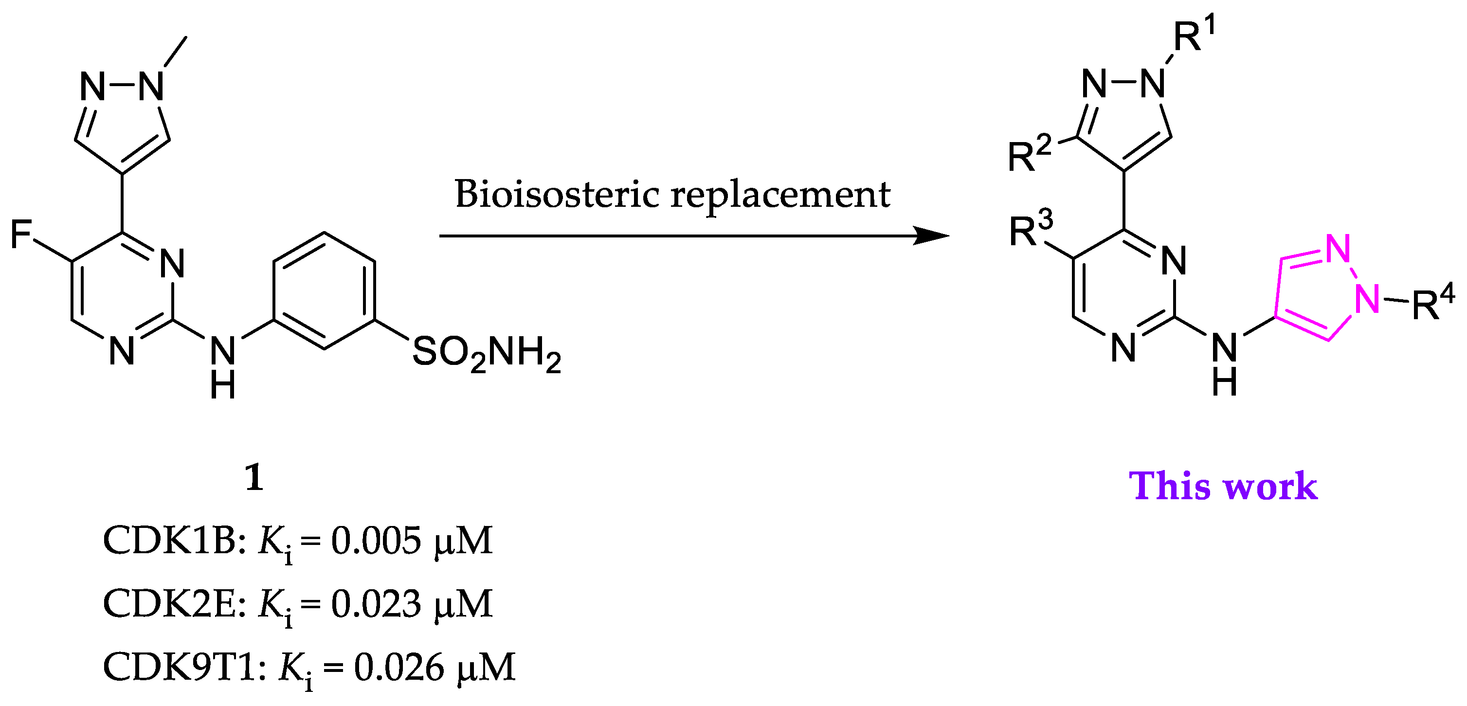
Figure 1. Design of novel CDK2 inhibitors from our previously-identified 1 [20].
Molecules 28 02951 g001
Figure 2. Predicted binding pose of 1 (a) or 14 (b) in CDK2/cyclin E (PDB ID: 7KJS). The kinase is presented in cyan ribbons with selected amino acids annotated, while 1 and 14 are colored green and salmon, respectively. Hydrogen bonds are indicated by dashed black lines. The illustration was generated using Maestro Schrödinger Release 2021-1.
Molecules 28 02951 g002
Scheme 1. Synthesis of N,4-di(1H-pyrazolyl)pyrimidin-2-amines 1421 and 23 and N-(5-chloro-4-(1-methyl-1H-pyrazol-4-yl)pyrimidin-2-yl)thiazol-2-amine 25. Reagents and conditions: (a) PdCl2(dppf)·DCM, K2CO3, 1,4-dioxane/ethanol/water (v/v/v, 7/3/4), 80 °C, 12 h, and 59–69%; (b) appropriate aminopyrazoles (13a13c and 22) or thiazol-2-amine (24), Pd2(dba)3, Xantphos, Cs2CO3, 1,4-dioxane, microwave 200–300 W, 140 °C, 1 h, and 13–28%; (c) K2CO3, acetonitrile, 80 °C, o/n, and 95%; and (d) H2, 10% Pd/C, methanol, rt, o/n, and 90%.
Molecules 28 02951 sch001
Scheme 2. Synthesis of 31, 32, and 35. Reagents and conditions: (a) Cs2CO3, acetonitrile, reflux, 24 h, and 57%; (b) 4 or 5, PdCl2(dppf)·DCM, K2CO3, 1,4-dioxane/ethanol/water (v/v/v, 7/3/4), 80 °C, 12 h, and 40–45%; and (c) 1-methyl-1H-pyrazol-4-amine (13b), Pd2(dba)3, Xantphos, Cs2CO3, 1,4-dioxane, microwave 200–300 W, 140 °C, 1 h, and 16–26%.
Molecules 28 02951 sch002
Figure 3. Compound 15 docked into CDK2/cyclin E (PDB ID: 7KJS). The kinase is presented in cyan ribbons with selected amino acids annotated, while 15 is colored yellow. Hydrogen bonds are indicated by dashed black lines. The illustration was generated using Maestro Schrödinger Release 2021-1.
Molecules 28 02951 g003
Figure 4. Effects of 15 on the cell cycle progression of A2780 and OVCAR5 ovarian cancer cells. Cells were incubated with 15 at 0.5 or 2 µM or CYC065 at 0.5 µM for 48 h. DMSO diluent was employed as a control. The experiments were performed twice, and representative data are presented.
Molecules 28 02951 g004
Figure 5. Effect of 15 on apoptosis in A2780 and OVCAR5 cells. Cells were incubated with 15 at 0.5 or 2 µM or CYC065 at 0.5 µM for 48 h. DMSO diluent was employed as a control. Apoptosis was determined by annexin V/PI staining. Experiments were performed twice, and representative data are presented. Viable cells are shown in the lower left quadrant, and they were not stained by either staining reagent, whereas the early apoptotic cells in the lower right quadrant were stained by annexin V. The late apoptotic cells in the upper right quadrant were stained by both staining reagents, while the necrotic cells in the upper left quadrant were stained by PI. The sum of the percentages of cells undergoing early (annexin V positive/PI negative) and late apoptosis (annexin V positive/PI positive) is taken as the total percentage of the apoptotic cells.
Molecules 28 02951 g005
Figure 6. Effect of 15 on colony formation. Three different ovarian cancer cell lines were incubated with vehicle (DMSO), 0.5 or 2 µM 15, or 0.5 µM CYC065 for 10 days. Colony formation was evaluated by crystal violet staining. Experiments were performed twice, and representative data are presented.
Molecules 28 02951 g006
Figure 7. Western blot analysis of OVCAR5 cells treated with 15. Cells were exposed to 15 at 0.5 or 2 µM or to CYC065 at 0.5 µM for 48 h. DMSO diluent was employed as a control. Experiments were performed twice, and representative data are presented.
Molecules 28 02951 g007
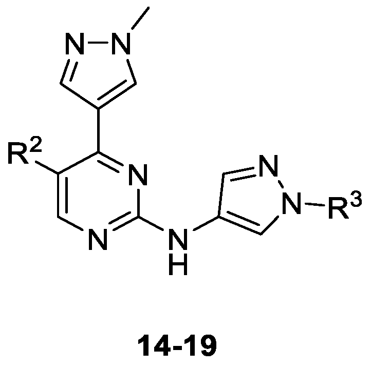
Table 1. Inhibition of CDK kinase activity and A2780 cell viability by 1419.
Molecules 28 02951 i001
CompoundSubstituentKi (µM) or % Enzymatic Inhibition at 1 µM * GI50 (µM) or % Growth Inhibition (GI) at 1 µM *
R2R3CDK1BCDK2ECDK5p25CDK9T1A2780
14FH0.0180.0070.0030.0160.498
15ClH0.0250.0050.0030.0200.158
16FMe0.0920.0230.0180.0989 *
17ClMe0.0340.0110.0060.0561.023
18FCH2CONH20.1120.0850.19368*0 *
19ClCH2CONH20.0180.0250.0520.10144 *
1-0.0050.0230.0770.0260.018
The enzymatic inhibition percentage was determined using a luminescent ADP-Glo™ assay. Apparent inhibition constant (Ki) values were calculated using their corresponding half maximal inhibition (IC50) values and the appropriate Km(ATP) of each kinase as described in the experimental section. The antiproliferative activity was determined by 72 h 3-(4,5-dimethylthiazol-2-yl)-2,5-diphenyltetrazolium bromide (MTT) assays using A2780 cells. The asterisk (*) indicates % for either biochemical or cell-based data. Previously reported data for 1 [20] were included in the table for comparison purposes. CDK1B, CDK2E, CDK5p25, and CDK9T1 denote CDK1/cyclin B, CDK2/cyclin E, CDK5/p25, and CDK9/cyclin T1, respectively. Me: methyl.
Table 2. Inhibition of CDK kinase activity and A2780 cell viability by 20, 21, 31, 32, and 35.
Molecules 28 02951 i002
CompoundSubstituentKi (µM) or % Enzymatic Inhibition at 1 µM * %GI at 1 µM *
RR1R2CDK1BCDK2ECDK5p25CDK9T1A2780
20MeMeF0.1640.0260.0180.3437 *
21MeMeCl0.2250.0230.0350.0628 *
312-morpholinoethylHF26 *48 *43 *24 *0 *
322-morpholinoethylHCl36 *0.2520.12621 *3 *
35-49 *64 *58 *45 *19 *
The enzymatic inhibition percentage was determined using the luminescent ADP-Glo™ assay. Apparent inhibition constant (Ki) values were calculated using their corresponding half maximal inhibition (IC50) values and the appropriate Km (ATP) of each kinase as described in the experimental section. The antiproliferative activity was determined by 72 h MTT assays using A2780 cells. The asterisk (*) indicates % for either biochemical or cell-based data. CDK1B, CDK2E, CDK5p25, and CDK9T1 denote CDK1/cyclin B, CDK2/cyclin E, CDK5/p25, and CDK9/cyclin T1, respectively. Me: methyl.
Table 3. Inhibition of CDK kinase activity and A2780 cell viability by 23 and 25.
Molecules 28 02951 i003
CompoundKi (µM) or % Enzymatic Inhibition at 1 µM *GI50 (µM) or %GI at 1 µM *
CDK1BCDK2ECDK5p25CDK9T1A2780
239 *0.0900.04040 *7.350
2531 *46 *0.17731 *9 *
The enzymatic inhibition percentage was determined using the luminescent ADP-Glo™ assay. Apparent inhibition constant (Ki) values were calculated using their corresponding half maximal inhibition (IC50) values and the appropriate Km (ATP) of each kinase as described in the experimental section. The antiproliferative activity was determined by 72 h MTT assays using A2780 cells. The asterisk (*) indicates % for either biochemical or cell-based data. CDK1B, CDK2E, CDK5p25, and CDK9T1 denote CDK1/cyclin B, CDK2/cyclin E, CDK5/p25, and CDK9/cyclin T1, respectively.
Table 4. Antiproliferative effects of 15.
Cancer TypeCancer Cell LineAntiproliferative Activity, GI50 (µM)
LeukemiaMV4-110.127
U9370.186
BreastMCF70.342
MDA-MB-2310.458
OvarianOVCAR50.150
OAW280.534
OV900.390
Cov3180.488
Cov5040.540
GlioblastomaU870.560
T98G0.380
U2510.350
Disclaimer/Publisher’s Note: The statements, opinions and data contained in all publications are solely those of the individual author(s) and contributor(s) and not of MDPI and/or the editor(s). MDPI and/or the editor(s) disclaim responsibility for any injury to people or property resulting from any ideas, methods, instructions or products referred to in the content.