Next Article in Journal
Could Naringenin Participate as a Regulator of Obesity and Satiety?
Next Article in Special Issue
Study on the Preparation and Properties of Jute Microcrystalline Cellulose Membrane
Previous Article in Journal
Co-Encapsulation of Drugs for Topical Application—A Review
Previous Article in Special Issue
Milk-Derived Carbon Quantum Dots: Study of Biological and Chemical Properties Provides Evidence of Toxicity
 
 
Correction published on 11 May 2024, see Molecules 2024, 29(10), 2253.
Communication

Rational Design of Nitrogen-Doped Carbon Dots for Inhibiting β-Amyloid Aggregation

1
Department of Neurovascular, Shanghai Fourth People’s Hospital, Tongji University School of Medicine, Shanghai 200434, China
2
Department of Neurology, Shanghai East Hospital, Tongji University School of Medicine, Shanghai 200120, China
3
Institute of Nanochemistry and Nanobiology, School of Environmental and Chemical Engineering, Shanghai University, Shanghai 200444, China
*
Authors to whom correspondence should be addressed.
These authors contributed equally to this work.
Molecules 2023, 28(3), 1451; https://doi.org/10.3390/molecules28031451
Submission received: 26 December 2022 / Revised: 12 January 2023 / Accepted: 15 January 2023 / Published: 2 February 2023 / Corrected: 11 May 2024
(This article belongs to the Special Issue Functional Carbon Quantum Dots: Synthesis and Applications)

Abstract

:
The fibrillization and abnormal aggregation of β-amyloid (Aβ) peptides are commonly recognized risk factors for Alzheimer’s disease (AD) brain, and require an effective strategy to inhibit the Aβ deposition and treat AD. Herein, we designed and synthesized nitrogen-doped carbon dots (N-CDs) as an Aβ-targeted probe, which exhibits the capacity of inhibiting the 1–42 Aβ (Aβ1–42) self-assembly in vitro. The N-CDs exhibited orange emission with an emission wavelength of 570 nm, which demonstrates their excellent optical properties with excitation-independent behavior. Meanwhile, the N-CDs have spherical morphologies with an average size of 2.2 nm, whose surface enriches the amino, carboxyl, and hydroxyl groups. These preparties are conducive to improving their biological water solubility and provide a large number of chemical bonds for further interaction with proteins. Contrary to this, the kinetic process, size evolutions, and morphologies changes of Aβ1–42 were inhibited in the presence of N-CDs in the determination of a thioflavin T assay, dynamic light scattering, transmission electron microscope, etc. Finally, the safety application of N-CDs on Aβ1–42-induced cytotoxicity was further demonstrated via in vitro cytotoxicity experiments. This work demonstrates the effective outcome of suppressing Aβ aggregation, which provides a new view into the high-efficiency and low-cytotoxicity strategy in AD theranostics.

Graphical Abstract

1. Introduction

Alzheimer’s disease (AD), a common neurodegenerative disorder, is one of the major causative factors to induce progressive dementia [1]. Notably, AD is reported to affect more than 50 million people, with its prevalence continuing to grow in part because of the aging worldwide population [2]. The symptoms of the disease begin with mildly impaired memory function and evolve towards severe cognitive loss, inevitably terminating in complete incapacity and death [3]. Typically, its pathogenesis is characterized by extracellular aggregates of amyloid β (Aβ) plaques and intracellular neurofibrillary tangles of hyperphosphorylated Tau [4]. As a principal variant of the Aβ peptides in humans [5], Aβ1–42 contains all amino acid sequences and relatively accurately simulates the action of Aβ [6], but also serves as the most common variant in human cerebrospinal fluid [7]. Growing evidence suggests that there are currently no available treatments that can change the course of an illness or the rate of decline [8]. Clinically effective treatment methods include cholinesterase inhibitors for patients with any stage of AD and memantine for people with moderate-to-severe AD [9]. Nevertheless, these medications can only enhance the quality of life when prescribed at the appropriate time during an illness [10]. Thus, there is an urgent need to explore a novel pathway for preventing or treating AD.
Increasing attention has been paid to nanomedicine due to nanomaterials’ great biocompatibility [11,12,13], stable physiochemical properties [14,15,16], photoluminescence properties [17,18,19], edge effect [20], and low cytotoxicity [21,22]. Recent studies have exhibited promising results with regard to the probability of using carbon nanomaterials for amyloid fibrillogenesis [23,24]. The conjugated structure of graphene oxide helps it bind tightly to Aβ through hydrophobic interactions and π-π packing interactions. For instance, thioflavin-S-modified graphene oxide under infrared laser irradiation could dissociate amyloid aggregation due to its high near-infrared absorbance, indicating the possibility of the photothermal treatment of AD [25]. Beyond the graphene oxide, a nano-chaperone based on a mixed-shell polymeric micelle was applicable in selectively capturing Aβ peptides, thus inhibiting Aβ aggregation [26]. However, it cannot be neglected that the hampering effect of the blood-brain barrier (BBB) acts as an obstacle to the transport of these nanomaterials from the vascular compartment to the brain [27]. In addition, as amyloid plaques accompanied by high levels of metal ions (e.g., copper, zinc, iron), coordination compounds [28], gold nanoparticles [29], and metallosupramolecular complexes [30] were synthesized and positive effects were noted on their association of metal ions with β-amyloidosis. Nevertheless, the inhibitors containing metallic elements are toxic to the body at a high dose [31]. Fortunately, carbon dots (CDs) can be an ideal candidate, with greater biocompatibility and a nontoxic nature due to their ability to cross the BBB [32], in addition to the lack of metals [33]. Therefore, amongst all nanoparticle species, the employment of CDs can be inspiring news for inhibiting the aggregation of the Aβ peptide and envisioning its clinical use as an anti-AD drug.
Here, the newly discovered functions of nitrogen-doped carbon dots (N-CDs) inhibited Aβ aggregation was designed and prepared with 1,2,3-benzenetricarboxylic acid and o-phenylenediamine by a one-pot solvothermal method. The N-CDs display excellent optical performance and narrow size distribution. Furthermore, the kinetic process, size evolutions, and morphologies of Aβ1–42 with or without the presence of N-CDs were displayed by a thioflavin T (ThT) assay, dynamic light scattering (DLS), and transmission electron microscope (TEM), respectively. The morphology and spectroscopic characterizations of N-CDs were also shown. Moreover, the safety application of N-CDs on Aβ1–42-induced cytotoxicity was further demonstrated via in vitro cytotoxicity experiments.

2. Results and Discussion

2.1. Characterization of N-CDs

To synthesize CDs, 1,2,3-benzenetricarboxylic acid and o-phenylenediamine were treated as precursors under a solvothermal condition at 180 °C for 12 h. The product solution was removed in large size and solvent, then transferred to the normal saline phase, and the pH was adjusted to 5 (Figure 1). The N-CDs presents an orange emission. As shown in Figure 2a, the UV-vis absorption and photoluminescence (PL) spectroscopy were explored. The N-CDs displayed an absorption peak at 420 nm and their absorption edge extend to 510 nm. The maximum photoluminescence excitation (PLE) and emission wavelength were 380 and 570 nm, respectively. The N-CDs demonstrated stable emission-independent PL behaviors from 360 to 540 nm excitation (Figure 2b). Furthermore, the N-CDs illustrated a monoexponential fluorescence lifetime of 3.21 ns (Figure 2c).
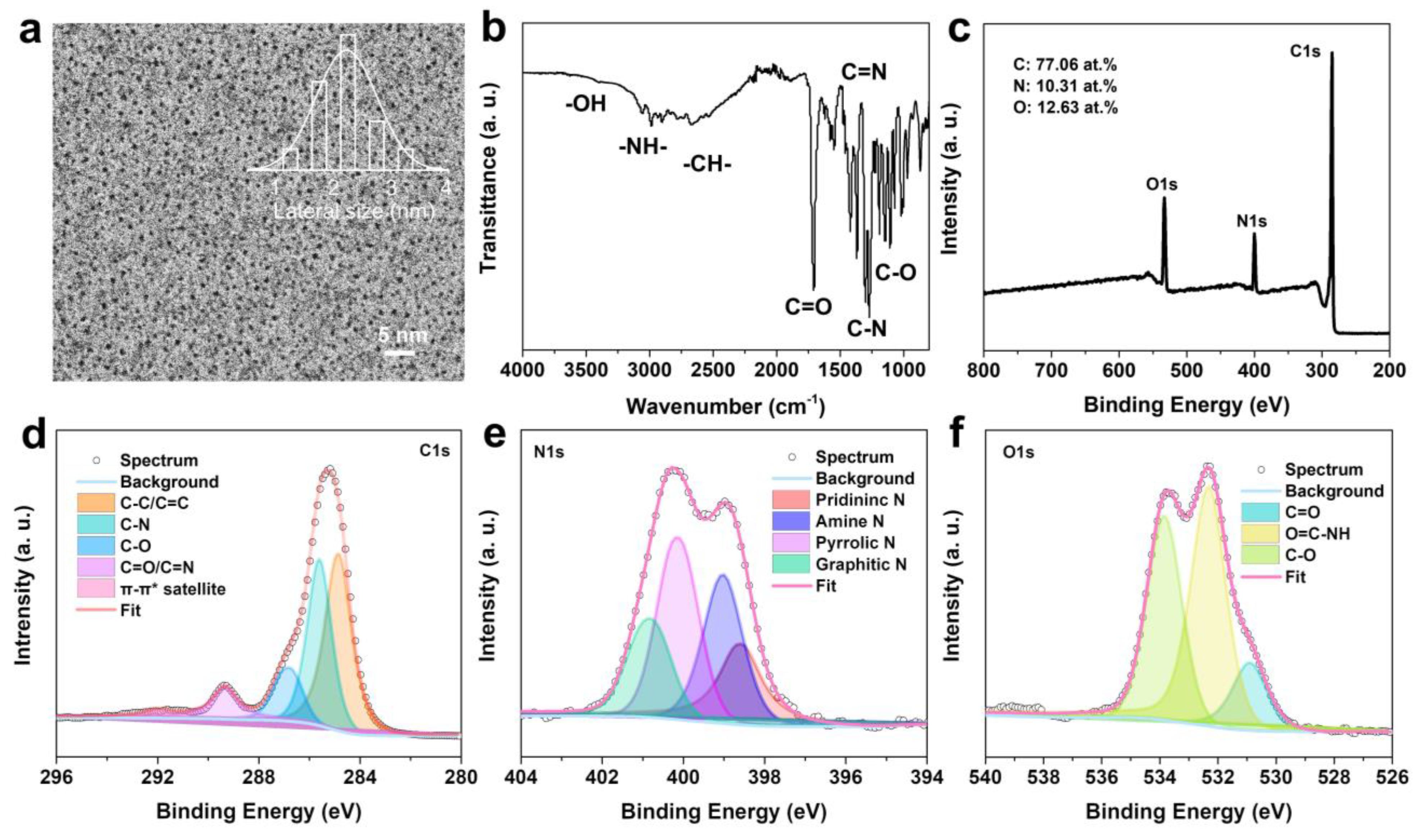
The morphology and structure were further represented by N-CDs. As illustrated in Figure 3a, the TEM image showed that the N-CDs has spherical morphologies with an average size of 2.2 nm. The D band (1362 cm−1) and G band (1580 cm−1) were found in its Raman spectrum (Figure S1), and the intensity ratio ID/IG was 0.73. The ratio indicates that N-CDs consist of a predominantly graphene structure and other disorder structures. [17,34] In addition, the surface properties of the N-CDs were further characterized by FT-IR and XPS spectra. The FT-IR spectrum (Figure 3b) of the N-CDs exhibited several peaks for O-H/N-H/C-H stretches at 2900–3410 cm−1, C=O, C=N, C-N, and C-O stretches at 1710, 1550, 1270, and 1110 cm−1, respectively. The summary of the FT-IR spectrum indicates the presence of N-C and O-C bonds from the amino, carboxyl, hydroxyl, and amide bond groups of N-CDs, which is further verified by its XPS measurement. The XPS survey show the N-CDs’ composition of C (77.06 at.%), N (10.31 at.%), and O (12.63 at.%) in Figure 3c. The XPS C1s spectrum (Figure 3d) displayed sp2 C (C-C/C=C) at 284.8 eV, sp3 C (including C-N and C-O) at 285.58 and 286.76 eV, a carbonyl group C (C=O or C=N) at 289.31 eV, and π-π* satellite at 291.98 eV. As shown in Figure 3e, the XPS N1s spectrum revealed the four N sources, including pyridinic N (398.56 eV), amine N (399.01 eV), pyrrolic N (400.11 eV), and graphitic N (400.81 eV). In Figure 3f, the XPS O1s spectrum was distributed to C=O, O=C-NH, and C-O, at 530.88, 532.30, and 533.82 eV, respectively. The dominant hydroxyl, amino, and carboxyl groups on the N-CDs’ surface support better aqueous solubility and further interact with protein via chemical bonds. Previous reports had demonstrated that inhibition can occur by distinct binding patterns between Aβ monomers and the surfaces of the CDs with hydrophilic/hydrophobic groups (Table S1) [35,36].

2.2. Inhibition on Aβ1–42 Aggregation Self-Assembly with N-CDs

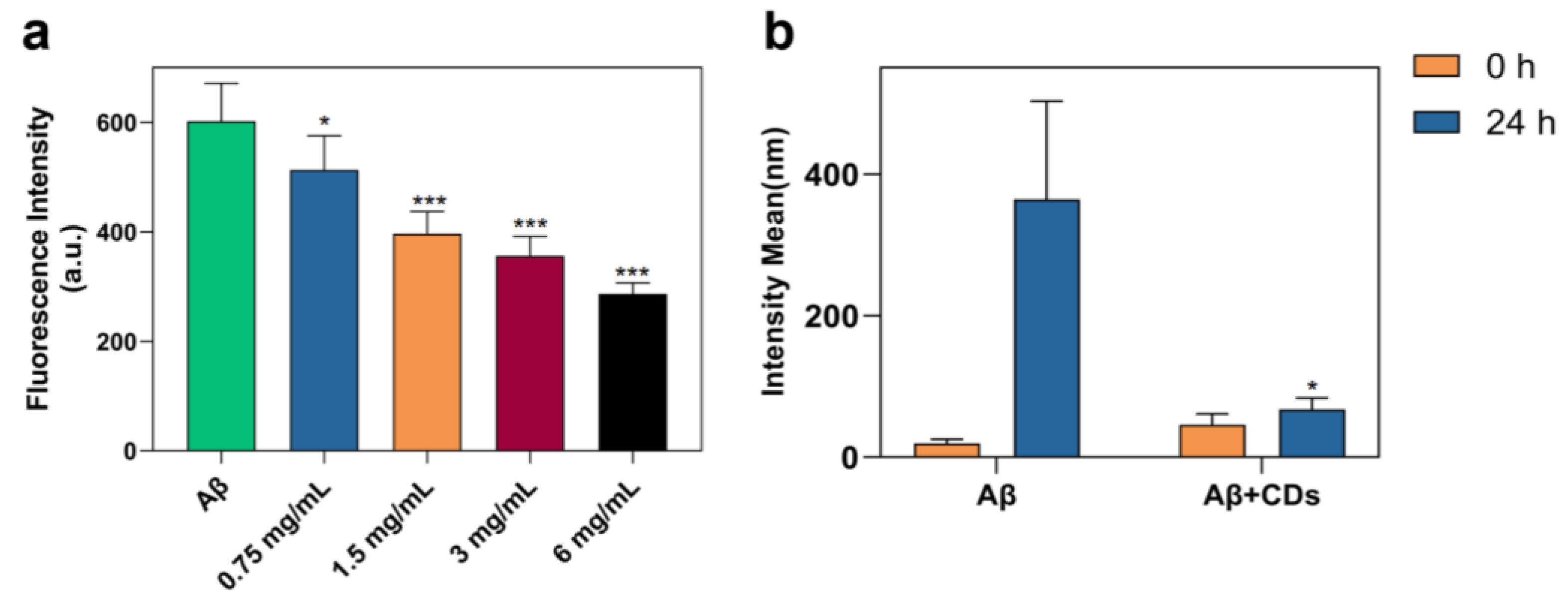
Nanomaterial-based approaches could offer promising directions in addressing the challenges in current therapeutic/diagnostic bio-reagent applications [37,38]. In the present study, the modulation effects of the special N-CDs on the amyloid peptide assemblies are presented according to the ThT assay, DLS, and TEM. As shown in Figure 4a, the effect of N-CDs on the assembly of Aβ1–42 peptides was first employed by a ThT fluorescence assay, which is a widely used method for monitoring amyloid aggregation and the fluorescence intensity increases with the augment of conjugates in the normal condition [39]. Compared to Aβ1–42 alone (the first column), the fluorescence intensity was significantly lower when Aβ1–42 peptides were incubated with N-CDs after 24 h incubation (second to fifth column). Furthermore, the value was even lower after increasing the dosage of N-CDs.
The side effect of CDs on modulating the Aβ1–42 peptide assembly was explored through DLS measurement. As shown in Figure 4b, the solution of Aβ1–42 was incubated with and without N-CDs in a PBS buffer at 37 °C for 24 h. The pure Aβ1–42 has an obvious agglomeration phenomenon after 24 h. However, the aggregation of the Aβ1–42 peptide was inhibited in the presence of N-CDs after 24 h incubation compared with the free Aβ1–42 peptide (67.58 ± 15.84 vs. 364.20 ± 138.90, p < 0.05). Our results clearly show at least a fivefold reduction owing to N-CDs. Such an obvious phenomenon proves that N-CDs acting as an Aβ-targeted probe is feasible.
To further verify the inhibitory effect of N-CDs, the morphology of the Aβ1–42 peptide in the presence or absence of N-CDs was also investigated in Figure 5. Figure 5a shows the initial morphology of the Aβ1–42 peptide. In the absence of N-CDs, Aβ1–42 peptides formed a typical structure for amyloid fibrils (Figure 5b), whereas the formation of the fibril was remarkably lower in the presence of N-CDs compared with the absence group (Figure 5c). The TEM data further supported the results with the ThT fluorescence assay and DLS, indicating that N-CDs could conspicuously influence the aggregation of the Aβ1–42 peptide. Previous studies also showed that various carbon materials including fullerene, carbon nanotubes, and graphene oxide were applied to inhibit the Aβ1–42 peptide aggregation [40,41]. As a newly prepared nano-material, we proved that the inhibiting affectivity of the special N-CDs plays a dominant role in the process of inhibiting the aggregation based on the above analysis in this work.

2.3. Aβ-Induced Cell Viability Was Reversed by N-CDs

To explore whether N-CDs can improve cell viability induced by Aβ1–42, Figure 6 demonstrates that the cellular reduction in the Aβ1–42 group was reversed by N-CDs as measured by MTT assay in a dose-dependent manner. As compared to the control, the cell viability was lower in the purely Aβ1–42-treated group (64.81% ± 3.41%, p < 0.001, third column), indicating that Aβ1–42 causes a noticeable negative effect on N2a cells. Contrary to this, while N-CDs were added into the plates pre-incubated with Aβ1–42 for 24 h, the cell viability was significantly higher (fourth to seventh columns). Notably, the ability of N-CDs to increase cell viability (95.82% ± 0.79% vs. 64.81% ± 3.41%, p < 0.001) suggested that the N-CDs can afford to be an available drug target in blocking Aβ1–42 assembly.

3. Materials and Methods

3.1. Materials

1,2,3-benzenetricarboxylic acid hydrate was purchased from TCI. o-phenylenediamine, ethanol, and hydrochloric acid (HCl) were acquired from Adams (Shanghai Titan Scientific Co., Ltd., Shanghai, China). These chemical reagents were utilized without further purification. Aβ1–42 peptide was purchased from Sigma (Sigma-Aldrich, St. Louis, MO, USA) and re-suspend according to the manufacturer’s protocols.

3.2. Synthesis of N-CDs

Briefly, a 1:1 mass ratio of 1,2,3-benzenetricarboxylic acid (50 mg, 0.24 mmol) and o-phenylenediamine (50 mg, 0.46 mmol) were dispersed in 10 mL ethanol by ultrasound. The mixture solution was then transferred to 25 mL of a poly-(tetrafluoroethylene) (Teflon)-lined autoclave and reacted at 180 °C for 12 h. The primary product was filtered and the solvent was removed. The normal saline was then added to the above, and adjusted to pH = 5 with 0.1 M HCl. The final concentration of the N-CDs solution is 6 mg/mL. Meanwhile, the N-CDs powder was acquired for further characterization after purifying by dialysis (MWCO 3500) for one week.

3.3. Thioflavin T (ThT) Assay

ThT powder (Shanghai Aladdin Biology, Shanghai, China) was dissolved into phosphate buffer saline (PBS) at a concentration of 1 mM and filtered by a 0.22 mm filter membrane before use. The re-suspended Aβ1–42 peptide solution was then re-dissolved into PBS buffer at a concentration of 100 µM. The Aβ1–42 solutions were then incubated with ThT solution and N-CDs at a concentration of 6, 3, 1.5, and 0.75 mg/mL 37 °C for 24 h. Aβ1–42 solutions prepared with the same amount of PBS were used as the control. The ThT fluorescence intensity was recorded at the beginning and end of incubation by a microplate reader (Spectramax M5, San Jose, CA, USA) at an excitation and emission wavelength of 450 and 482 nm, respectively.

3.4. Cell Viability Detected by MTT Assay

Mouse neuroblastoma Neuro-2a cells were first cultured in Dulbecco’s Modified Eagle’s Medium (DMEM) added with 10% fetal bovine serum (FBS) (Shanghai Fuheng Biology, Shanghai, China) at 37 °C in a humidified (5% CO2, 95% air) incubator. Cells were seeded into 96-well microplates with a density of 8000 cells per well, cultured overnight, and then treated with Aβ1–42 (100 µM), N-CDs (6 mg/mL), or Aβ1–42 (100 µM)/N-CDs mixtures at a concentration of 6, 3, 1.5, and 0.75 mg/mL. After being incubated for another 24 h, a 3-(4,5-dimethylthiazol-2-yl)-2,5-diphenyltetrazolium bromide (MTT) assay (Beyotime Institute of Biotechnology, Shanghai, China) was added to assess the cell viability according to the manufacturer’s protocols.

3.5. Characterization

PL and UV-vis absorption spectra were characterized by Horiba Duetta. The time-resolved PL spectra were obtained with a Horiba FluoroMax with a 455 nm laser. A transmission electron microscopy (TEM) image was performed by using a Japan Hitachi HT7700. The Raman spectrum was achieved by an Anton-Paar Cora 5001 with 785 nm of laser wavelength. The Fourier transform infrared (FT-IR) spectrum was characterized with a Thermo Scientific iS50 FT-IR. The X-ray photoelectron spectroscopy (XPS) spectrum was acquired using a Thermo ESCALAB 250Xi spectrometer. Dynamic light scattering (DLS) is a powerful tool used to monitor particle size evolutions. Aβ1–42 peptides were mixed with 6 mg/mL N-CDs incubated for 24 h at 37 °C and then the size distribution was measured by a ZetasizerNano ZS nanoparticle size analyzer (Malvern Instruments Ltd., Malvern, UK). Aβ1–42 solutions incubated with PBS were also utilized as controls. After measuring the effect of N-CDs on Aβ1–42 fibrosis by ThT and DLS as shown previously, the morphologies of the Aβ1–42 aggregates incubated with or without N-CDs were observed and visualized by TEM (Japan Hitachi HT7700, Japan, Tokyo).

3.6. Statistical Analysis

All analyses were performed using the SPSS software (version 26.0; SPSS, Chicago, IL, USA). Data are expressed as the mean ± standard error of the mean of at least three biological replicates for each experiment. Statistical differences were analyzed by Student’s t-test for comparisons between two groups or by ANOVA followed by Tukey’s multiple comparison post hoc test for comparisons among more than two groups. A p-value < 0.05 was considered statistically significant.

4. Conclusions

Prompted by the need to pursue an effective treatment of AD, many inhibitors against Aβ fibration and cytotoxicity were explored [42,43]. As a novel carbon nanomaterial, CDs have received significant attention in materials science [44,45] and biomedicine [46,47,48]. In summary, we designed the N-CDs prepared by 1,2,3-benzenetricarboxylic acid and o-phenylenediamine for inhibiting Aβ aggregation. The N-CDs exhibited orange emission with an emission wavelength of 570 nm, which demonstrates its excellent optical properties with excitation-independent behavior. Meanwhile, the N-CDs has a spherical morphology with an average size of 2.2 nm, whose surface enriches the amino, carboxyl, and hydroxyl groups. These properties are conducive to improving its biological water solubility and providing a large number of chemical bonds for further interaction with proteins. Contrary to this, the kinetic process, size evolutions, and morphology changes of Aβ1–42 were inhibited in the presence of N-CDs in the determination of thioflavin T assay, dynamic light scattering, transmission electron microscope, etc. Finally, the safety application of N-CDs on Aβ1–42 was further demonstrated via in vitro cell viability experiments. Those discoveries make it reasonable to speculate that the N-CDs can inhibit Aβ1–42 aggregation and alleviate Aβ-induced cytotoxicity. Our work improves the probability of the use of the particular nanostructures on Aβ monomers aggregating into fibrils in the future.

Supplementary Materials

The following supporting information can be downloaded at: https://www.mdpi.com/1420-3049/28/3/1451/s1, Figure S1: Raman spectrum of N-CDs.

Author Contributions

H.L. and H.G. contributed equally to investigation, data curation, formal analysis, writing-original draft, and writing, review and editing; conceptualization, review and supervision Y.F., L.W., P.L. All authors have read and agreed to the published version of the manuscript.

Funding

The project was funded by National Natural Science Foundation of China (No. 21901154), Shanghai Pujiang Program (21PJD022).

Institutional Review Board Statement

Not applicable.

Informed Consent Statement

Not applicable.

Data Availability Statement

All data generated or analyzed during this study are included in this published article and its Supplementary Information Files.

Acknowledgments

We thank all participants who contributed to this study. We express our sincere appreciation to the reviewers for their constructive comments.

Conflicts of Interest

The authors declare no conflict of interest.

References

  1. Hodson, R. Alzheimer’s disease. Nature 2018, 559, S1. [Google Scholar] [CrossRef] [PubMed]
  2. Mielke, M.M.; Vemuri, P.; Rocca, W.A. Clinical Epidemiology of Alzheimer’s Disease: Assessing Sex and Gender Differences. Clin. Epidemiol. 2014, 6, 37–48. [Google Scholar] [CrossRef] [PubMed]
  3. Serrano-Pozo, A.; Das, S.; Hyman, B.T. APOE and Alzheimer’s Disease: Advances in Genetics, Pathophysiology, and Therapeutic Approaches. Lancet Neurol. 2021, 20, 68–80. [Google Scholar] [CrossRef] [PubMed]
  4. Graff-Radford, J.; Yong, K.X.X.; Apostolova, L.G.; Bouwman, F.H.; Carrillo, M.; Dickerson, B.C.; Rabinovici, G.D.; Schott, J.M.; Jones, D.T.; Murray, M.E. New Insights into Atypical Alzheimer’s Disease in the Era of Biomarkers. Lancet Neurol. 2021, 20, 222–234. [Google Scholar] [CrossRef]
  5. Fiandaca, M.S.; Kapogiannis, D.; Mapstone, M.; Boxer, A.; Eitan, E.; Schwartz, J.B.; Abner, E.L.; Petersen, R.C.; Federoff, H.J.; Miller, B.L.; et al. Identification of Preclinical Alzheimer’s Disease by a Profile of Pathogenic Proteins in Neurally Derived Blood Exosomes: A Case-Control Study. Alzheimers Dement. 2015, 11, 600–607. [Google Scholar] [CrossRef]
  6. Murakami, K.; Irie, K.; Morimoto, A.; Ohigashi, H.; Shindo, M.; Nagao, M.; Shimizu, T.; Shirasawa, T. Synthesis, Aggregation, Neurotoxicity, and Secondary Structure of Various A Beta 1-42 Mutants of Familial Alzheimer’s Disease at Positions 21–23. Biochem. Biophys. Res. Commun. 2002, 294, 5–10. [Google Scholar] [CrossRef]
  7. Roher, A.E.; Lowenson, J.D.; Clarke, S.; Woods, A.S.; Cotter, R.J.; Gowing, E.; Ball, M.J. Beta-Amyloid-(1-42) is a Major Component of Cerebrovascular Amyloid Deposits: Implications for the Pathology of Alzheimer Disease. Proc. Natl. Acad. Sci. USA 1993, 90, 10836–10840. [Google Scholar] [CrossRef]
  8. Kellar, D.; Craft, S. Brain Insulin Resistance in Alzheimer’s Disease and Related Disorders: Mechanisms and Therapeutic Approaches. Lancet Neurol. 2020, 19, 758–766. [Google Scholar] [CrossRef]
  9. Tatulian, S.A. Challenges and Hopes for Alzheimer’s Disease. Drug Discov. Today 2022, 27, 1027–1043. [Google Scholar] [CrossRef]
  10. Weintraub, D.; Aarsland, D.; Chaudhuri, K.R.; Dobkin, R.D.; Leentjens, A.F.; Rodriguez-Violante, M.; Schrag, A. The Neuropsychiatry of Parkinson’s Disease: Advances and Challenges. Lancet Neurol. 2022, 21, 89–102. [Google Scholar] [CrossRef]
  11. Guo, H.; Zhang, X.; Chen, Z.; Zhang, L.; Wang, L.; Xu, J.; Wu, M. High-Energy Short-Wave Blue Light Conversion Films Via Carbon Quantum Dots for Preventing Retinal Photochemical Damage. Carbon 2022, 199, 431–438. [Google Scholar] [CrossRef]
  12. Xu, Y.; Wang, B.; Zhang, M.; Zhang, J.; Li, Y.; Jia, P.; Zhang, H.; Duan, L.; Li, Y.; Li, Y.; et al. Carbon Dots as a Potential Therapeutic Agent for the Treatment of Cancer-Related Anemia. Adv. Mater. 2022, 34, e2200905. [Google Scholar] [CrossRef] [PubMed]
  13. Hou, L.; Chen, D.; Wang, R.; Wang, R.; Zhang, H.; Zhang, Z.; Nie, Z.; Lu, S. Transformable Honeycomb-Like Nanoassemblies of Carbon Dots for Regulated Multisite Delivery and Enhanced Antitumor Chemoimmunotherapy. Angew. Chem. Int. Ed. Engl. 2021, 60, 6581–6592. [Google Scholar] [CrossRef] [PubMed]
  14. Zhang, X.; Guo, H.; Chen, C.; Quan, B.; Zeng, Z.; Xu, J.; Chen, Z.; Wang, L. Tunable Photoacoustic and Fluorescence Imaging of Nitrogen-Doped Carbon Quantum Dots. Appl. Mater. Today. 2023, 30, 101706. [Google Scholar] [CrossRef]
  15. Guo, H.; Liu, Z.; Shen, X.; Wang, L. One-Pot Synthesis of Orange Emissive Carbon Quantum Dots for All-Type High Color Rendering Index White Light-Emitting Diodes. ACS Sustain. Chem. Eng. 2022, 26, 8289–8296. [Google Scholar] [CrossRef]
  16. Liu, J.; Kong, T.; Xiong, H.M. Mulberry-Leaves-Derived Red-Emissive Carbon Dots for Feeding Silkworms to Produce Brightly Fluorescent Silk. Adv. Mater. 2022, 34, 2200152. [Google Scholar] [CrossRef]
  17. Wang, L.; Li, W.; Yin, L.; Liu, Y.; Guo, H.; Lai, J.; Han, Y.; Li, G.; Li, M.; Zhang, J.; et al. Full-Color Fluorescent Carbon Quantum Dots. Sci. Adv. 2020, 6, eabb6772. [Google Scholar] [CrossRef]
  18. Zhang, L.; Wu, M.; Wang, Z.; Guo, H.; Wang, L.; Wu, M. Phosphorescence Tuning of Fluorine, Oxygen-Codoped Carbon Dots by Substrate Engineering. ACS Sustain. Chem. Eng. 2021, 48, 16262–16269. [Google Scholar] [CrossRef]
  19. Su, W.; Guo, R.; Yuan, F.; Li, Y.; Li, X.; Zhang, Y.; Zhou, S.; Fan, L. Red-Emissive Carbon Quantum Dots for Nuclear Drug Delivery in Cancer Stem Cells. J. Phys. Chem. Lett. 2020, 11, 1357–1363. [Google Scholar] [CrossRef]
  20. Wang, L.; Li, M.; Li, Y.; Wu, B.; Chen, H.; Wang, R.; Xu, T.; Guo, H.; Li, W.; Joyner, J.; et al. Designing a Sustainable Fluorescent Targeting Probe for Superselective Nucleus Imaging. Carbon 2021, 180, 48–55. [Google Scholar] [CrossRef]
  21. Pedone, D.; Moglianetti, M.; De Luca, E.; Bardi, G.; Pompa, P.P. Platinum Nanoparticles in Nanobiomedicine. Chem. Soc. Rev. 2017, 46, 4951–4975. [Google Scholar] [CrossRef] [PubMed]
  22. Li, S.; Su, W.; Wu, H.; Yuan, T.; Yuan, C.; Liu, J.; Deng, G.; Gao, X.; Chen, Z.; Bao, Y.; et al. Targeted Tumour Theranostics in Mice via Carbon Quantum Dots Structurally Mimicking Large Amino Acids. Nat. Biomed. Eng. 2020, 4, 704–716. [Google Scholar] [CrossRef] [PubMed]
  23. Xie, L.; Luo, Y.; Lin, D.; Xi, W.; Yang, X.; Wei, G. The Molecular Mechanism of Fullerene-Inhibited Aggregation of Alzheimer’s β-amyloid Peptide Fragment. Nanoscale 2014, 6, 9752–9762. [Google Scholar] [CrossRef]
  24. Daniyal, M.; Liu, B.; Wang, W. Comprehensive Review on Graphene Oxide for Use in Drug Delivery System. Curr. Med. Chem. 2020, 27, 3665–3685. [Google Scholar] [CrossRef] [PubMed]
  25. Li, M.; Yang, X.; Ren, J.; Qu, K.; Qu, X. Using Graphene Oxide High Near-Infrared Absorbance for Photothermal Treatment of Alzheimer’s Disease. Adv. Mater. 2012, 24, 1722–1728. [Google Scholar] [CrossRef] [PubMed]
  26. Yang, H.; Li, X.; Zhu, L.; Wu, X.; Zhang, S.; Huang, F.; Feng, X.; Shi, L. Heat Shock Protein Inspired Nanochaperones Restore Amyloid-β Homeostasis for Preventative Therapy of Alzheimer’s Disease. Adv. Sci. 2019, 6, 1901844. [Google Scholar] [CrossRef]
  27. Xie, J.; Gonzalez-Carter, D.; Tockary, T.A.; Nakamura, N.; Xue, Y.; Nakakido, M.; Akiba, H.; Dirisala, A.; Liu, X.; Toh, K.; et al. Dual-Sensitive Nanomicelles Enhancing Systemic Delivery of Therapeutically Active Antibodies Specifically into the Brain. ACS Nano 2020, 14, 6729–6742. [Google Scholar] [CrossRef] [PubMed]
  28. He, L.; Wang, X.; Zhao, C.; Wang, H.; Du, W. Ruthenium Complexes as Novel Inhibitors of Human Islet Amyloid Polypeptide Fibril Formation. Metallomics 2013, 5, 1599–1603. [Google Scholar] [CrossRef]
  29. Hsieh, S.; Chang, C.W.; Chou, H.H. Gold Nanoparticles as Amyloid-Like Fibrillogenesis Inhibitors. Colloids Surf. B Biointerfaces 2013, 112, 525–529. [Google Scholar] [CrossRef]
  30. Li, M.; Howson, S.E.; Dong, K.; Gao, N.; Ren, J.; Scott, P.; Qu, X. Chiral Metallohelical Complexes Enantioselectively Target Amyloid β for Treating Alzheimer’s Disease. J. Am. Chem. Soc. 2014, 136, 11655–11663. [Google Scholar] [CrossRef]
  31. Cao, L.; Meziani, M.J.; Sahu, S.; Sun, Y.P. Photoluminescence Properties of Graphene Versus Other Carbon Nanomaterials. Acc. Chem. Res. 2013, 46, 171–180. [Google Scholar] [CrossRef] [PubMed]
  32. Han, G.; Zhao, J.; Zhang, R.; Tian, X.; Liu, Z.; Wang, A.; Liu, R.; Liu, B.; Han, M.Y.; Gao, X.; et al. Membrane-Penetrating Carbon Quantum Dots for Imaging Nucleic Acid Structures in Live Organisms. Angew. Chem. Int. Ed. 2019, 58, 7087–7091. [Google Scholar] [CrossRef] [PubMed]
  33. Zhou, Y.; Peng, Z.; Seven, E.S.; Leblanc, R.M. Crossing the Blood-Brain Barrier with Nanoparticles. J. Control Release 2018, 70, 290–303. [Google Scholar] [CrossRef] [PubMed]
  34. Liu, J.; Geng, Y.; Li, D.; Yao, H.; Huo, Z.; Li, Y.; Zhang, K.; Zhu, S.; Wei, H.; Xu, W.; et al. Deep Red Emissive Carbonized Polymer Dots with Unprecedented Narrow Full width at Half Maximum. Adv. Mater. 2020, 32, 1906641. [Google Scholar] [CrossRef] [PubMed]
  35. Han, X.; Jing, Z.; Wu, W.; Zou, B.; Peng, Z.; Ren, P.; Wikramanayake, A.; Lu, Z.; Leblanc, R.M. Biocompatible and Blood-Brain Barrier Permeable Carbon Dots for Inhibition of Aβ Fibrillation and Toxicity, and BACE1 Activity. Nanoscale 2017, 9, 12862–12866. [Google Scholar] [CrossRef] [PubMed]
  36. Zhou, Y.; Liyanage, P.Y.; Devadoss, D.; Leblanc, R.M. Nontoxic Amphiphilic Carbon Dots as Promising Drug Nanocarriers Across the Blood-Brain Barrier and Inhibitors of β-amyloid. Nanoscale 2019, 11, 22387–22397. [Google Scholar] [CrossRef] [PubMed]
  37. Teleanu, R.I.; Gherasim, O.; Gherasim, T.G.; Grumezescu, V.; Grumezescu, A.M.; Teleanu, D.M. Nanomaterial-Based Approaches for Neural Rregeneration. Pharmaceutics 2019, 11, 266. [Google Scholar] [CrossRef]
  38. Norouzi, M.; Yasamineh, S.; Montazeri, M.; Dadashpour, M.; Sheervalilou, R.; Abasi, M.; Pilehvar-Soltanahmadi, Y. Recent Advances on Nanomaterials-Based Fluorimetric Approaches for MicroRNAs Detection. Mater. Sci. Eng. C. Mater. Biol. Appl. 2019, 104, 110007. [Google Scholar] [CrossRef]
  39. LeVine, H., 3rd. Thioflavine T Interaction with Synthetic Alzheimer’s Disease Beta-Amyloid Peptides: Detection of Amyloid Aggregation in Solution. Protein Sci. 1993, 2, 404–410. [Google Scholar] [CrossRef]
  40. Mahmoudi, M.; Akhavan, O.; Ghavami, M.; Rezaee, F.; Ghiasi, S.M. Graphene Oxide Strongly Inhibits Amyloid Beta Fibrillation. Nanoscale 2012, 4, 7322–7325. [Google Scholar] [CrossRef]
  41. Luo, J.; Wärmländer, S.K.; Yu, C.H.; Muhammad, K.; Gräslund, A.; Pieter Abrahams, J. The Aβ Peptide Forms Non-Amyloid Fibrils in the Presence of Carbon Nanotubes. Nanoscale 2014, 6, 6720–6726. [Google Scholar] [CrossRef] [PubMed]
  42. Röhr, D.; Boon, B.D.C.; Schuler, M.; Kremer, K.; Hoozemans, J.J.M.; Bouwman, F.H.; El-Mashtoly, S.F.; Nabers, A.; Großerueschkamp, F.; Rozemuller, A.J.M. Label-Free Vibrational Imaging of Different Aβ Plaque Types in Alzheimer’s Disease Reveals Sequential Events in Plaque Development. Acta Neuropathol. Commun. 2020, 8, 222. [Google Scholar] [CrossRef] [PubMed]
  43. Kim, Y.M.; Park, S.; Choi, S.Y.; Oh, S.B.; Jung, M.; Pack, C.G.; Hwang, J.J.; Tak, E.; Lee, J.Y. Clusterin Binding Modulates the Aggregation and Neurotoxicity of Amyloid-β(1-42). Mol. Neurobiol. 2022, 59, 6228–6244. [Google Scholar] [CrossRef] [PubMed]
  44. Song, D.; Guo, H.; Huang, K.; Zhang, H.; Chen, J.; Wang, L.; Lian, C.; Wang, Y. Carboxylated Carbon Quantum Dot-induced Binary Metal-organic Framework Nanosheet Synthesis to Boost the Electrocatalytic Performance. Mater. Today. 2022, 54, 42–51. [Google Scholar] [CrossRef]
  45. Han, Y.; Tang, B.; Wang, L.; Bao, H.; Lu, Y.; Guan, C.; Zhang, L.; Le, M.; Liu, Z.; Wu, M. Machine Learning Driven Synthesis of Carbon Dots with Enhanced Quantum Yield. ACS Nano 2020, 14, 14761–14768. [Google Scholar] [CrossRef]
  46. Anand, A.; Unnikrishnan, B.; Wei, S.C.; Chou, C.P.; Zhang, L.Z.; Huang, C.C. Graphene Oxide and Carbon Dots as Broad-Spectrum Antimicrobial Agents-a Minireview. Nanoscale Horiz. 2019, 4, 117–137. [Google Scholar] [CrossRef]
  47. Ge, G.; Li, L.; Wang, D.; Chen, M.; Zeng, Z.; Xiong, W.; Wu, X.; Guo, C. Carbon Dots: Synthesis, Properties and Biomedical Applications. J. Mater. Chem. B. 2021, 9, 6553–6575. [Google Scholar] [CrossRef]
  48. Ding, H.; Zhou, X.X.; Wei, J.S.; Li, X.B.; Qin, B.T.; Chen, X.B.; Xiong, H.M. Carbon Dots with Red/Near-Infrared Emissions and Their Intrinsic Merits for Biomedical Applications. Carbon 2020, 167, 322–344. [Google Scholar] [CrossRef]
Figure 1. Schematic route for the synthesis of N-CDs.
Molecules 28 01451 g001
Figure 2. (a) The UV-vis, PL, and PLE spectra, (b) the PL spectra at different excitation wavelengths, and (c) the time-resolved PL spectrum of N-CDs.
Molecules 28 01451 g002
Figure 3. (a) TEM image and its corresponding lateral size distribution, (b) FT-IR spectrum, (c) survey XPS spectrum, (d) high-resolution C1s spectrum, (e) high-resolution N1s spectrum, and (f) high-resolution O1s spectrum of N-CDs.
Molecules 28 01451 g003
Figure 4. Aβ1–42 aggregation was inhibited by N-CDs. (a) The thioflavin T fluorescence intensity in the presence or absence of N-CDs at the concentration of 0.75, 1.5, 3, and 6 mg/mL after 24 h incubation. *** p < 0.001, * p < 0.05 vs. Aβ1–42 alone (1st column). (b) DLS spectra of Aβ1–42 peptide with or without N-CDs (6 mg/mL) at the beginning and after 24 h incubation. The aggregation of the Aβ1–42 peptide was inhibited in the presence of N-CDs after 24 h incubation compared with the free Aβ1–42 peptide (* p < 0.05).
Molecules 28 01451 g004
Figure 5. TEM images of (a) Aβ1–42 peptide at 0 h, (b) Aβ1–42 peptide incubated for 24 h, and (c) Aβ1–42 peptide incubated with 6 mg/mL N-CDs for 24 h.
Molecules 28 01451 g005
Figure 6. Effect of Aβ1–42 peptide (100 µM), N-CDs (6 mg/mL), or Aβ1–42 (100 µM) mixed with N-CDs at the concentration of 0.75, 1.5, 3, and 6 mg/mL on the viability of N2a cells after 24 h of incubation. (*** p < 0.001 vs. Control; ### p < 0.001 vs. purely Aβ1–42-treated group).
Molecules 28 01451 g006
Disclaimer/Publisher’s Note: The statements, opinions and data contained in all publications are solely those of the individual author(s) and contributor(s) and not of MDPI and/or the editor(s). MDPI and/or the editor(s) disclaim responsibility for any injury to people or property resulting from any ideas, methods, instructions or products referred to in the content.