Article

Impact of Starch Binding Domain Fusion on Activities and Starch Product Structure of 4-α-Glucanotransferase

1
Enzyme and Protein Chemistry, Department of Biotechnology and Biomedicine, Technical University of Denmark, DK-2800 Kongens Lyngby, Denmark
2
School of Food Science and Technology, Jiangnan University, Wuxi 214122, China
3
Protein Chemistry and Enzyme Technology, Department of Biotechnology and Biomedicine, Technical University of Denmark, DK-2800 Kongens Lyngby, Denmark
4
Laboratory of Protein Evolution, Institute of Molecular Biology, Slovak Academy of Sciences, SK-84551 Bratislava, Slovakia
5
Department of Biology, Faculty of Natural Sciences, University of SS. Cyril and Methodius, SK-91701 Trnava, Slovakia
6
Applied Molecular Enzyme Chemistry, Department of Biotechnology and Biomedicine, Technical University of Denmark, DK-2800 Kongens Lyngby, Denmark
*
Authors to whom correspondence should be addressed.
Molecules 2023, 28(3), 1320; https://doi.org/10.3390/molecules28031320
Submission received: 9 January 2023 / Revised: 26 January 2023 / Accepted: 28 January 2023 / Published: 30 January 2023
(This article belongs to the Special Issue Advances in Amylases)

Abstract

:
A broad range of enzymes are used to modify starch for various applications. Here, a thermophilic 4-α-glucanotransferase from Thermoproteus uzoniensis (TuαGT) is engineered by N-terminal fusion of the starch binding domains (SBDs) of carbohydrate binding module family 20 (CBM20) to enhance its affinity for granular starch. The SBDs are N-terminal tandem domains (SBDSt1 and SBDSt2) from Solanum tuberosum disproportionating enzyme 2 (StDPE2) and the C-terminal domain (SBDGA) of glucoamylase from Aspergillus niger (AnGA). In silico analysis of CBM20s revealed that SBDGA and copies one and two of GH77 DPE2s belong to well separated clusters in the evolutionary tree; the second copies being more closely related to non-CAZyme CBM20s. The activity of SBD-TuαGT fusions increased 1.2–2.4-fold on amylose and decreased 3–9 fold on maltotriose compared with TuαGT. The fusions showed similar disproportionation activity on gelatinised normal maize starch (NMS). Notably, hydrolytic activity was 1.3–1.7-fold elevated for the fusions leading to a reduced molecule weight and higher α-1,6/α-1,4-linkage ratio of the modified starch. Notably, SBDGA-TuαGT and-SBDSt2-TuαGT showed Kd of 0.7 and 1.5 mg/mL for waxy maize starch (WMS) granules, whereas TuαGT and SBDSt1-TuαGT had 3–5-fold lower affinity. SBDSt2 contributed more than SBDSt1 to activity, substrate binding, and the stability of TuαGT fusions.

1. Introduction

4-α-glucanotransferases (4αGT, EC 2.4.1.25), belonging to the glycoside hydrolase family 77 (GH77) (http://www.CAZy.org, accessed on 23 December 2022) [1], catalyze four different reactions: cyclization, coupling, hydrolysis, and disproportionation [2]. The disproportionation is attractive as it involves a transfer of malto-oligosaccharides to suitable α-1,4-glucan acceptors. When the α-1,4-glucan acceptor is the α-glucan chain of the covalent enzyme-intermediate, a circular molecule is formed, named a large-ring cyclodextrin (LR-CD), by connecting the reducing and non-reducing ends [3]. When the acceptor in the disproportionation reaction is a different α-1,4-glucan chain, the transfer of a fragment to its non-reducing end can lead to elongation of exterior chains in branched α-glucan molecules [4].
Starch binding domains (SBDs), as a special group of carbohydrate binding modules (CBMs), provide numerous starch-active enzymes with enhanced affinity for different α-glucans [5]. Among the 94 CBM families (http://www.cazy.org/, accessed on 23 December 2022) [1], 15 were defined as SBDs, namely CBM20, 21, 25, 26, 34, 41, 45, 48, 53, 58, 68, 69, 74, 82, and 83 [5]. SBDs can have important affinity for α-glucans—including granular starches [6,7], show micromolar affinity for β-cyclodextrin (a starch model) [8,9], and are thought to be able to disentangle α-glucan chains of double helixes on the starch granule surface [5,8,9,10] offering an explanation for their stimulation of granular starch hydrolysis. Still, the main function of SBDs is considered to be molecular recognition and binding to starch granules. SBDs thus facilitate the reaction of the catalytic domains (CDs) by bringing the active site in close contact with substrate [11]. SBDs can also guide the α-glucan chain to be modified to the active site crevice on the CD [12].
The aim of the present work is to confer a thermophilic starch-modifying 4-α-glucanotransferase from Thermoproteus uzoniensis (TuαGT) [13] with novel functional properties by one-by-one fusion with three different SBDs, two from Solanum tuberosum (potato) disproportionating enzyme 2 (StDPE2) of the glycoside hydrolase family 77 (GH77) [14] and one from Aspergillus niger glucoamylase (AnGA) of GH15 [15]. The effect on the different types of GH77 activities as obtained in the three fusions SBDSt1-TuαGT, SBDSt2-TuαGT, and SBDGA-TuαGT was analysed by using maltotriose, amylose, gelatinised normal and waxy maize starches, and native waxy maize starch granules as substrates. In general, SBD-fusion increased the activity of TuαGT on amylose and gelatinised starch, but reduced the disproportionating activity on maltotriose. The SBD-TuαGTs had an increased affinity for granular starch but only slightly changed the chain length distribution of gelatinised NMS. The three SBDs exerted individual effects on the function of TuαGT. Especially, SBDSt1 and SBDSt2 showed different influences on the thermostability and binding affinity of TuαGT, suggesting that tandem SBDs from StDPE2 individually play different functional roles. Lastly, SBD-fusion can be a promising technology to change the substrate specificity and activity of enzymes.

2. Results and Discussion

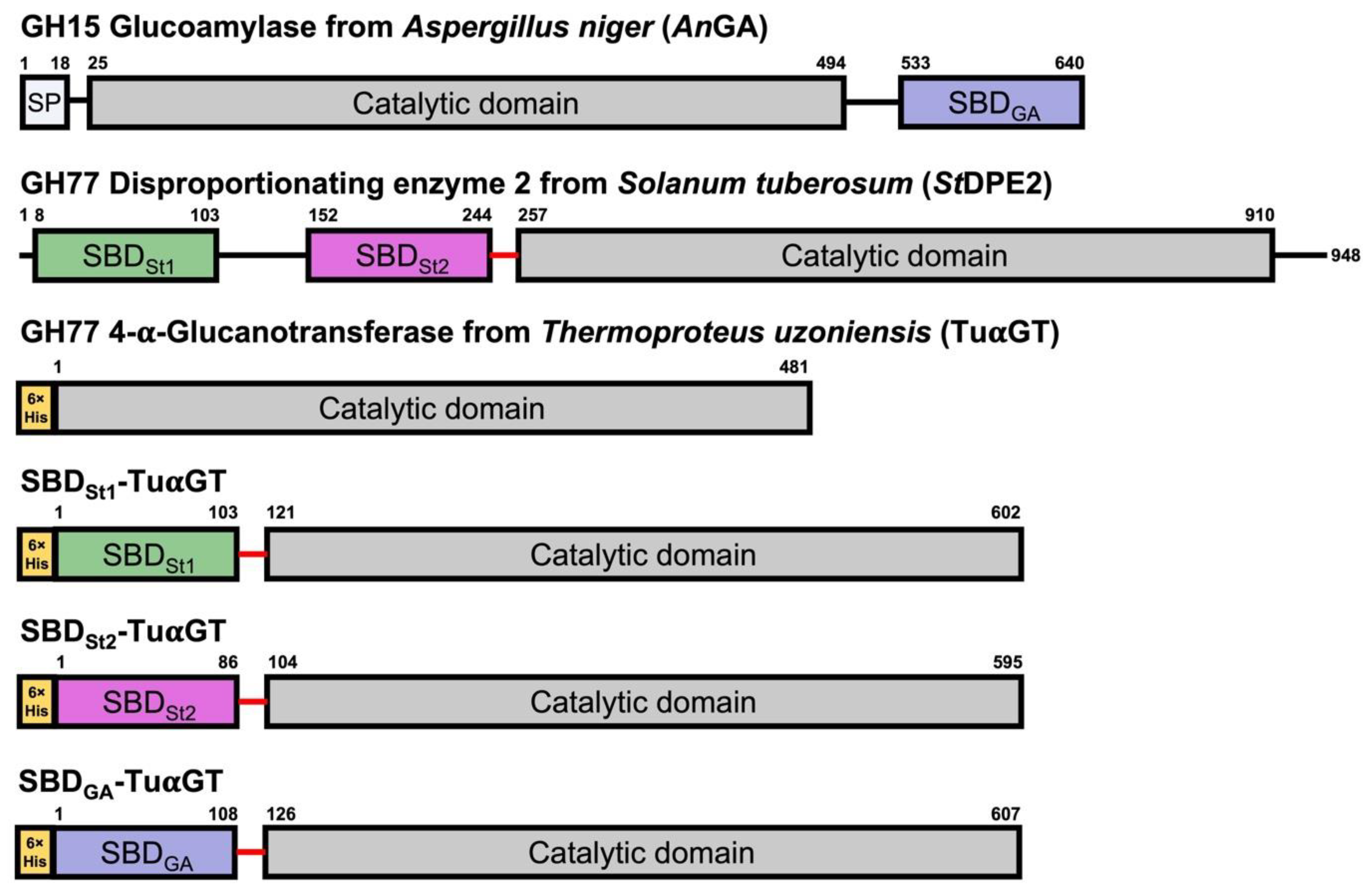
2.1. 4-α-Glucanotransferase SBD Fusions

Several 4-α-glucanotransferases have been reported to contain starch binding domains (SBDs) [5,13]. To improve starch affinity and modification for TuαGT, three different fusion proteins were constructed by attaching SBDs of the family CBM20 to the N-terminus of the enzyme. Two SBDs from Solanum tuberosum disproportionating enzyme 2 (StDPE2) [14] (SBDSt1, the N-terminal, and SBDSt2, the second in tandem), and one (SBDGA) from Aspergillus niger glucoamylase (AnGA) [15] were used (Figure 1). The fusions of the CD and SBDs were performed via an 18-residues linker (TTGESRFVVLSDGLMREM) that naturally connects the SBDSt1–SBDSt2 tandem with the CD in StDPE2 (Figure 1).

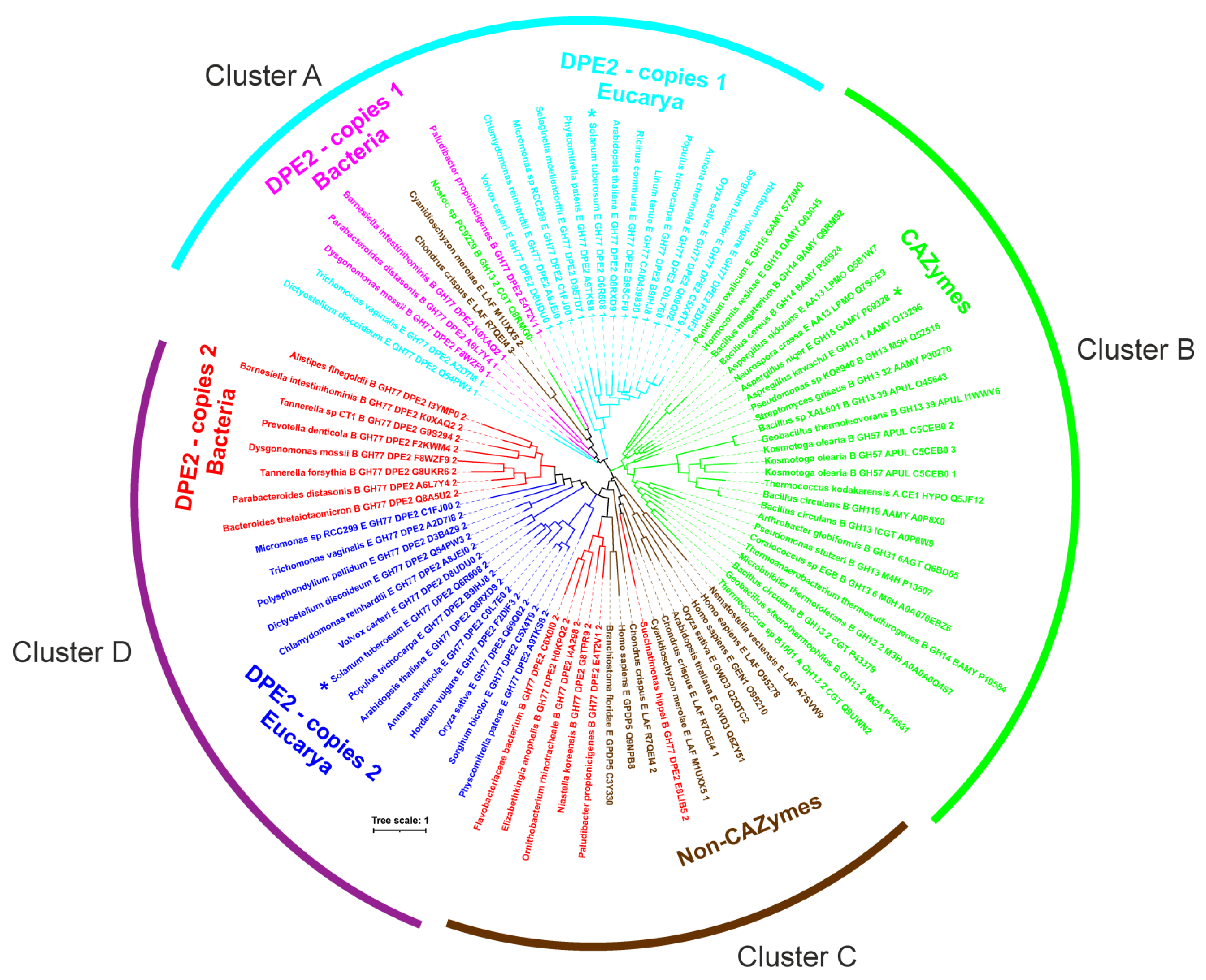
2.2. Bioinformatics Analysis

In order to put the three above-mentioned experimentally fused SBDSt1, SBDSt2, and SBDGA into the overall context of the CBM20 family, 65 different starch hydrolases and related enzymes were selected for in silico analysis (Table 1). The emphasis was mainly on GH77 DPE2s, both from Eukaryota (including the StDPE2) and Bacteria, known to contain two recognizable CBM20s [16]. The set to be analysed was completed by various well-known CBM20s from amylolytic enzymes classified into several CAZy families (including AnGA) as well as several non-CAZymes, such as phosphoglucan, water dikinase (GWD3), laforin, genethonin-1, etc. [5,16,17,18,19].
From the 65 selected enzymes, it was possible to sample 87 CBM20 sequences (see Table 1 for details). It is worth mentioning that although there was a stretch in almost each DPE2 sequence (regardless the bacterial or eukaryotic origin) for two CBM20 copies at the N-terminus, only those not lacking most of the known CBM20 functionally important binding site residues [8,9,12] were taken into the analysis. Interestingly—based on a detailed inspection of their amino acid sequences—the hypothetical DPE2s from Linum tenue (GenBank Acc. No.: CAI0439830.1) and Ricinus communis (UniProt Acc. No.: B9SCF0) obviously contain only one CBM20 copy (data not shown). It is of note that of the two potential starch binding sites of CBM20, only starch binding site one, being formed by Trp543, Lys578, and Trp590 (GH15 A. niger glucoamylase numbering [8]), is well conserved (Figure S1), whereas residues forming starch binding site two may vary [5], as evidenced by the structural complexes of CBM20s from GH15 A. niger glucoamylase with cyclodextrin (Tyr527, Tyr556 and Trp563) [8] and GH13_2 Bacillus circulans cyclodextrin glucanotransferase with maltose (Tyr633 and Trp636) [19]—having only the tryptophan (Trp563 vs Trp 636) conserved (Figure S1). Of the SBDSt1, SBDSt2, and SBDGA used in the present study, only SBDGA from GH15 A. niger glucoamylase, that possesses all the key residues involved in binding (Figure S1), was previously demonstrated to bind starch [8]. SBDSt1 and SBDSt2 each lack one of the conserved residues at starch binding site one—the SBDSt1 lysine (Lys578; A. niger GH15 CBM20 numbering) and the SBDSt2 tryptophan (Trp590)—and only the tryptophan (Trp563) of starch binding site two is conserved in both; however, SBDSt1 might have a stronger ability to bind since it has a tryptophan corresponding to Tyr527 at binding site two (Figure S1).
The evolutionary tree (Figure 2), constructed from the sequence alignment, illustrated several facts: (i) each of the two CBM20 copies from GH77 DPE2s forms its own cluster; (ii) all CBM20s from other CAZymes cluster together (including SBDGA of AnGA; cluster B) and separately from both groups covering the two CBM20 copies of GH77; (iii) the second CBM20 copy of GH77 DPE2s (including SBDSt2 of StDPE2; cluster D) exhibits a closer relatedness to CBM20s from non-CAZymes (such as GWD3, laforin, genethonin-1, etc.; cluster C) than to those from other CAZyme families (cluster B); and (iv) the clade of the first CBM20 copy of GH77 DPE2s (including SBDSt1 of StDPE2, cyan in Figure 2) covers also the second and the third CBM20 copies from laforins from Cyanidioschyzon merolae and Chondrus crispus, respectively, [18] (brown clade in cluster A, Figure 2) as well as the CBM20 from the four-domain GH13_2 cyclodextrin glucanotransferase from Nostoc sp. PC9229 [20] (green in cluster A, Figure 2). The results from the bioinformatics analysis thus indicate that the three CBM20s studied here, i.e., SBDSt1, SBDSt2, and SBDGA, are positioned in three different clusters of the evolutionary tree (Figure 2) and may confer the parental enzyme TuαGT distinctly different biochemical properties by the fusion.

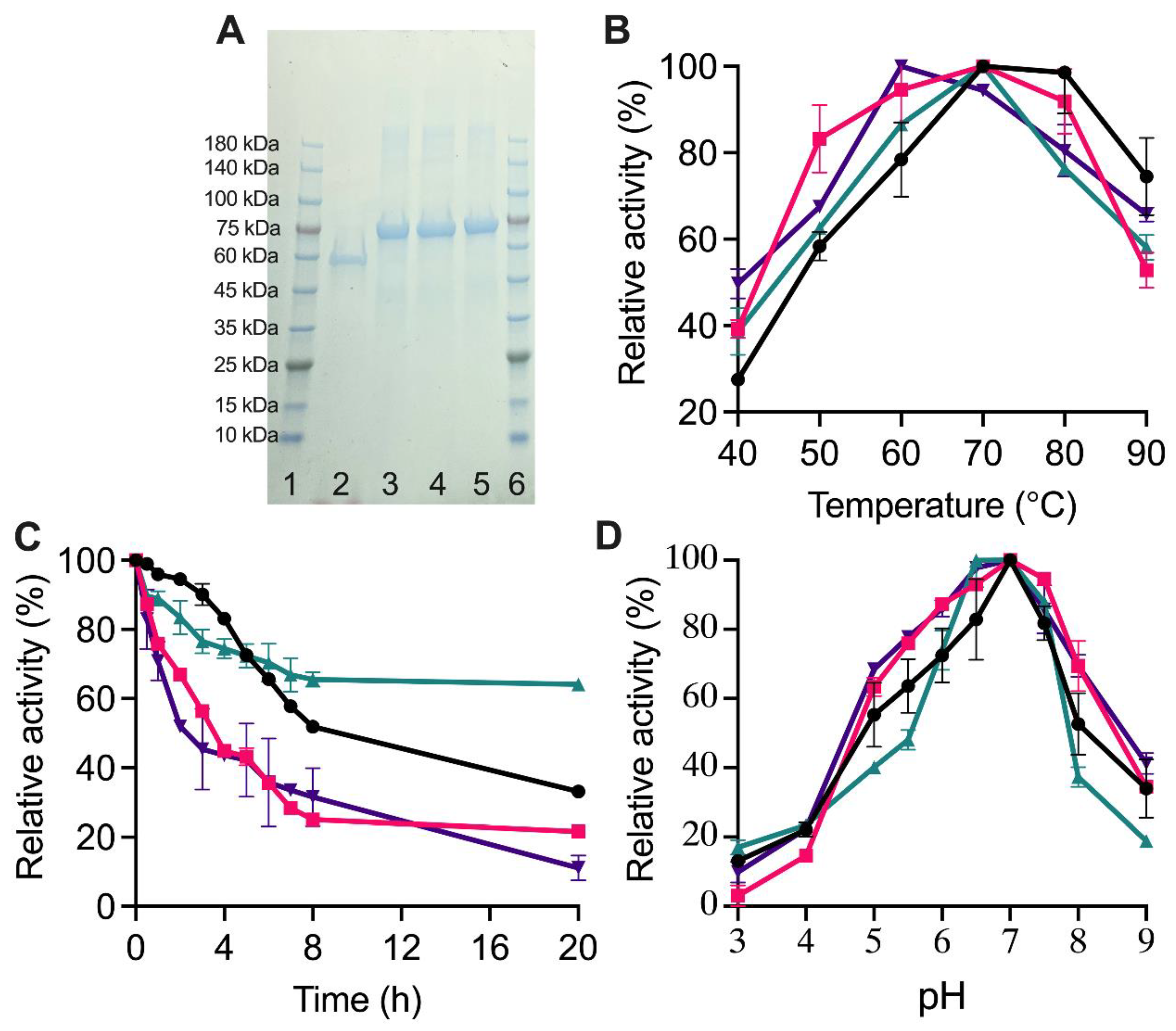
2.3. Biochemical Properties of TuαGT and SBD-TuαGT Fusions

The produced TuαGT, SBDSt1-TuαGT, SBDSt2-TuαGT, and SBDGA-TuαGT migrated in SDS-PAGE as single protein bands estimated to 56, 68, 67, and 69 kDa (Figure 3A), respectively, in agreement with the theoretical values (see Section 3.5). The optimal reaction temperature and pH for the maltotriose disproportionation activity were around 70 °C and 7.0 for the different forms of TuαGT (Figure 3B,D). However, SBDGA-TuαGT had a lower temperature optimum of 60 °C (Figure 3B). This is in good agreement with previously reported pH and temperature optima for the total activity on amylose and maltose of TuαGT at 6.0 and 75 °C [13]. TuαGT was nearly 100% active at 80 °C, indicating it is a thermophilic enzyme, which also showed significantly reduced activity at <60 °C. Notably, all three SBD-TuαGT fusions were relatively less active than TuαGT at >70 °C, but more active at <60 °C (Figure 3B). The improved affinity to starch of the SBD-fusions (see Section 2.4) may contribute to their relatively higher activity than the parent enzyme TuαGT at <60 °C, whereas the lower relative activity of the fusions at >70 °C may stem from their poorer thermostability as illustrated by the time progress for the loss of activity at 50 °C (Figure 3C). Notably, after 20 h at 50 °C, the parent TuαGT maintained ~35% activity. However, all SBD-TuαGT fusions lost more activity than TuαGT during the first 5 h at 50 °C and SBDSt1-TuαGT and SBDGA-TuαGT retained only about 20% activity after 8 h, whereas SBDSt2-TuαGT kept remarkably ~65% of its activity after 20 h (Figure 3C). Improved thermostability was previously found by N-terminal fusion of a CBM1 to β-mannanase from Aspergillus usamii YL-01-78 (reAuMan5A-CBM), having a temperature optimum at 75 °C compared with 70 °C for wild-type (reAuMan5A), indicating a stabilizing effect of the CBM1 on the CD [21]. In another study, Wang et al. [22] fused five different CBMs (of families CBM2, 3, 11, and 30) to the C-terminus of cis-epoxysuccinic acid hydrolase (CESH) and found a 5-times higher half-life for the CBM30-CESH than of wild-type CESH at 30 °C.

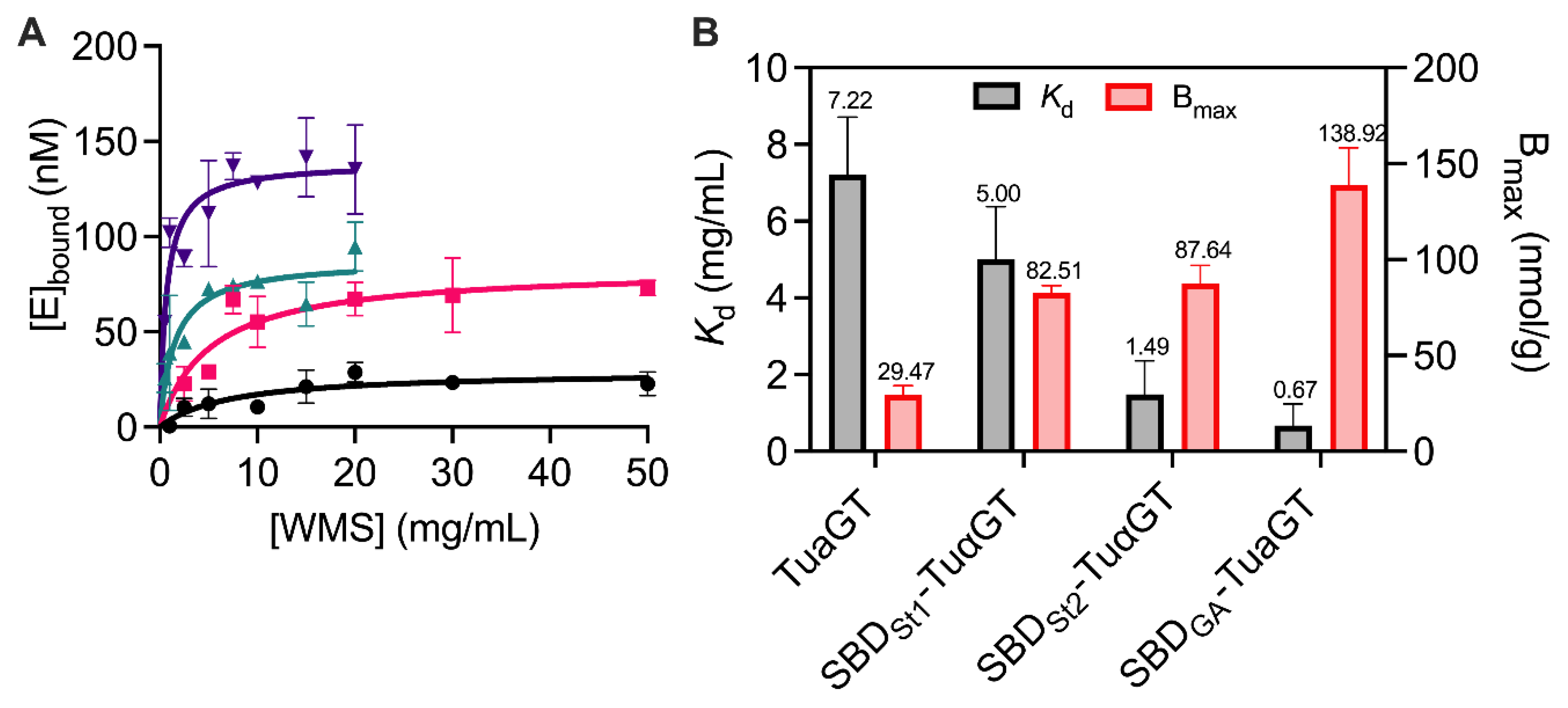
2.4. Adsorption and Enzyme Kinetic Parameters

The binding capacity to WMS granules was increased for all three SBD-TuαGT fusions, revealing that the SBD domains were functional and fulfilling the purpose (Figure 4). Overall, SBDGA-TuαGT had an almost 5 times higher binding capacity (Bmax, Figure 4) and 10 times stronger affinity (Kd = 0.7 mg/mL) than TuαGT (Kd = 7.2 mg/mL). While SBDSt1-TuαGT and SBDSt2-TuαGT both had an essentially 3 times higher binding capacity to WMS granules than TuαGT, their affinity was quite similar and 5-fold larger, respectively, than of TuαGT (Figure 4). This agrees with SBDSt1 lacking the lysine (Lys578, AnGA numbering) and SBDSt2 missing one of the two tryptophans (Trp590, AnGA numbering) at starch binding site one, respectively, compared with SBDGA (see Section 2.2; Figure S1). Notably, the positive effect of SBDSt2 on binding was larger than of SBDSt1 even though SBDSt2 misses a tryptophan at binding site one, indicating that other features of these SBDs contribute to their binding determinants for WMS granules. This may likely include differences at the larger and more flexible binding site two, which is claimed for SBDGA to be the tighter binding of the two sites [8,9]. Until now, there has been no report of different functions of the two SBDs arranged in tandem in StDPE2 or in other DPE2 enzymes.
The fusion of SBDs to TuαGT also influenced the enzymatic activity. Thus, the maltotriose disproportionation was reduced, SBDSt2-TuαGT and SBDGA-TuαGT having slightly lower Km than TuαGT, but 4-fold lower kcat, and yielding 3-fold lower catalytic efficiency (kcat/Km) for these two fusion enzymes. Notably, kcat/Km for SBDSt1-TuαGT was 15-times reduced compared with TuαGT, due to a doubled Km and an almost 9-fold lower kcat (Table 2). By contrast, using amylose as a substrate, the SBD-fusion improved activity and kinetic parameters somewhat (Table 2). Thus, the similar Km and higher kcat of SBDSt2-TuαGT more than doubled the catalytic efficiency compared with TuαGT, whereas the overall outcome for SBDSt1-TuαGT and SBDGA-TuαGT was essentially the same catalytic efficiency as of the parent enzyme. Overall, the kinetic analyses indicated that the SBD-fusion hampered the action of TuαGT on the oligosaccharide (maltotriose), but could improve it on the polysaccharide (amylose). Similarly, fusion of the SBDGA to barley α-amylase, albeit via the much longer natural linker from A. niger glucoamylase (AnGA), showed no adverse effect of the SBD on the active site integrity, as it did not change activity for soluble starch [23]. The improved catalytic efficiency for SBDSt2-TuαGT towards amylose may be caused by favourable polysaccharide binding to SBDSt2, increasing the local substrate concentration and perhaps also directing the substrate chain to the active site on the CD.

2.5. Hydrolysis and Cyclization Activities on Different Substrates

To gain insight into the modes of action of the SBD-TuαGT fusions on starch, the hydrolysis and cyclization activities were determined using different substrates (Table 3). SBDSt1-TuαGT had 1.3–1.7-fold higher hydrolytic activity on amylose and gelatinised starch and 1.5-fold higher cyclization activity on amylose than the TuαGT parent enzyme. Similarly, SBDSt2-TuαGT showed 1.5–1.7-fold increased hydrolysis of gelatinised starch, but more moderate 1.3-fold and 1.2-fold increased hydrolytic and cyclization activities, respectively, on amylose. As a glucanotransferase, it is not expected to show increased hydrolysis by SBD-fusion. However, from an industrial viewpoint, a small increase in hydrolytic activity can help to decrease the viscosity of gelatinised starch, which will also facilitate the TuαGT disproportionation reaction. Notably, for SBDGA-TuαGT containing an SBD that originates from the family GH15 of glucoamylases and not from the family GH77 of 4-α-glucanotransferases, to which TuαGT belongs, the hydrolysis and cyclization activities were both essentially the same as for the parent enzyme, except for a slight increase in hydrolysis of gelatinised waxy maize starch (WMS) (Table 3). We speculate that, perhaps, the domain architecture matters and the naturally N-terminally placed SBDs from the StDPE2 of the family GH77, which constitutes glycoside hydrolase clan H together with GH13 and GH70 [1], are able to provide support in the different GH77 4-α-glucanotransferase reactions as opposed to the naturally C-terminally placed SBDGA connected via a long O-glycosylated linker to the CD of glucoamylase of the family GH15 that acts in an exo-manner on non-reducing ends of malto-oligosaccharides and α-glucans catalysing release of glucose [24].

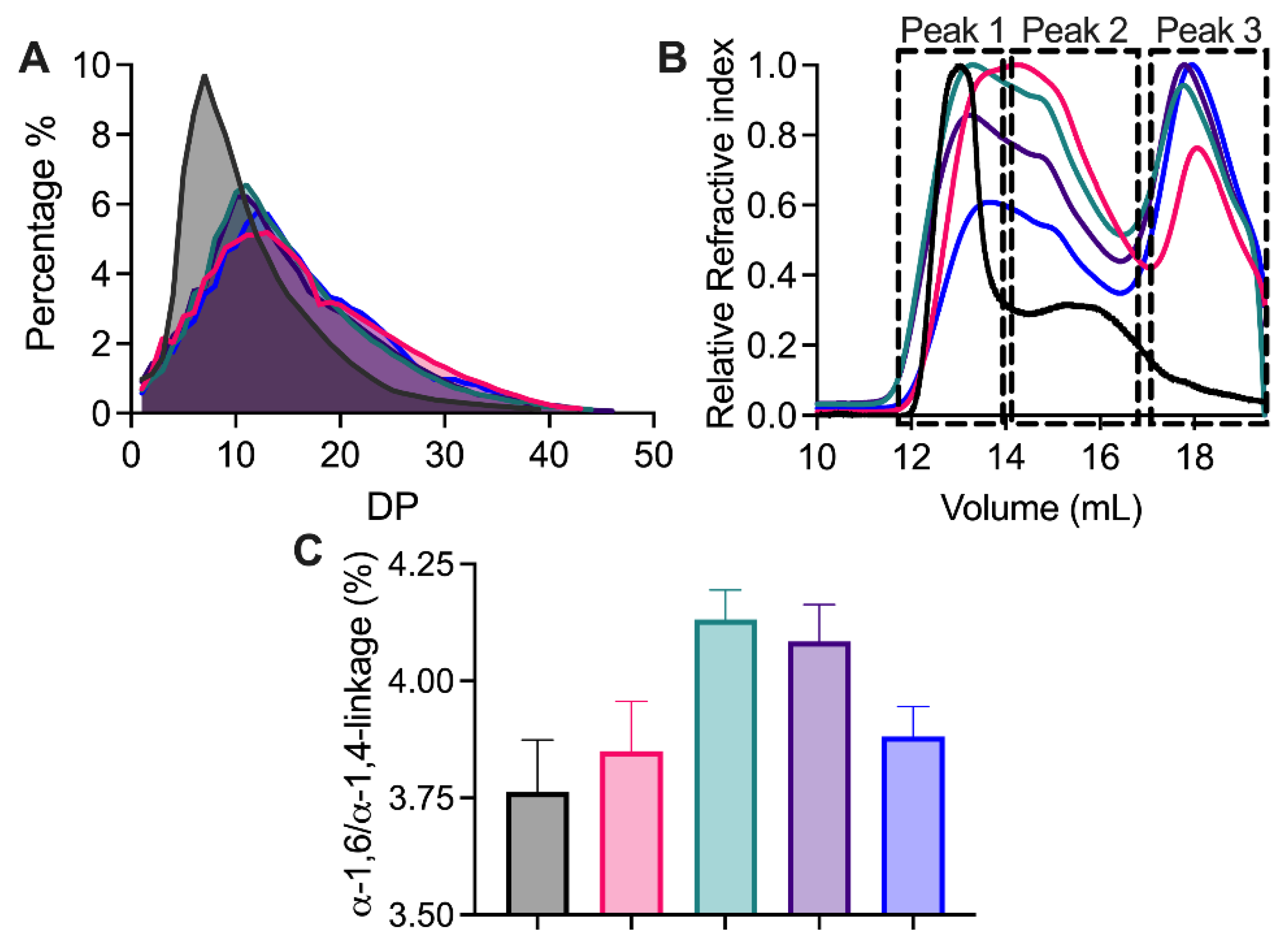
2.6. Structure Analysis of Modified NMS

The modification of maize starch both by TuαGT and the SBD-TuαGT fusions significantly affected its structural properties. Chain length distribution (CLD) of NMS and modified NMS (Figure 5A) and the percentage of A-chains as well as of B1-, B2-, and B3-chains (Table 4) showed that all NMS starches treated by TuαGT and its SBD-fusions, to different degrees, contained significantly fewer of the short A-chains and more of the longer B1-, B2-, and B3-chains. Still, only minor differences appeared for the CLD in starches modified by the TuαGT parent compared with SBD-TuαGT fusions (Figure 5A). Previous studies on tapioca starch similarly indicated that exterior chains of amylopectin were elongated by TuαGT [13].
The molecular weight distribution of NMS before and after enzyme treatment was analysed by SEC-MALLS-RI (Figure 5B). Before modification, typical amylopectin (peak 1) and amylose (peak 2) molecules were observed in NMS by SEC. However, after the enzyme modification, three peaks were observed, namely the peaks one and two as well as a distinct later eluting peak three of smaller polysaccharide chains. Furthermore, a later elution of peak one from all modified starch samples indicated that amylopectin has a reduced molecular weight and was less well resolved from peak two than found for unmodified NMS. The newly appearing prominent peak three of smaller molecules may contain large-ring cyclodextrins (LR-CDs) produced in cyclization reactions [25] as well as polysaccharide hydrolysis products.
To further understand the reaction of TuαGT and the SBD-TuαGT fusions, the α-1,6/α-1,4-linkage ratio that indicates the degree of branching, was determined for the modified starches by using 1H-NMR (Figure 5C). NMS modified by TuαGT and SBDGA-TuαGT showed a slight increase in the α-1,6/α-1,4-linkage ratio from 3.76 for unmodified to 3.84 and 3.88%, respectively, after modification, whereas treatment by SBDSt1-TuαGT and SBDSt2-TuαGT increased the ratio to 4.13 and 4.08%, respectively. As TuαGT can catalyze hydrolysis, disproportionation, cyclization, and coupling, which all involve α-1,4-linkages, the increase in the α-1,6/α-1,4-linkage ratio can reflect the level of hydrolysis, in which α-1,4 linkages are lost and not generated, in agreement with the two fusions with SBDSt1 and SBDSt2, i.e., the SBDs from StDPE2 belonging to the family GH77, showing an increased degree of hydrolysis of gelatinised NMS compared with TuαGT (Figure 5B; Table 3).

3. Material and Methods

3.1. Materials

Amylose (potato), maltotriose, and protease inhibitor cocktail tablets (cOmplete™, Mini, EDTA-free Protease Inhibitor Cocktail) were purchased from Sigma-Aldrich Co. Ltd. (St. Louis, MO, USA). Pullulanase M2 (from Bacillus licheniformis, 900 U/mL) and β-amylase (from barley, 600 U/mg) were purchased from Megazyme Co. Ltd. (Wicklow, Ireland). Waxy maize starch (WMS) was the kind gift of Cargill (USA) and normal maize starch (NMS) of Archer Daniels Midland (ADM, Decatur, IL, USA).

3.2. Bioinformatics Analysis of CBM20

In total, 87 CBM20 domains from 65 different amylolytic and related enzymes were collected (Table 1) based on previous studies focused on GH77 DPE2s and different starch-binding domain CBM families [5,16,17,18,19]. All sequences were retrieved from GenBank (https://www.ncbi.nlm.nih.gov/genbank/, accessed on 23 December 2022; [26]) and/or UniProt (https://www.uniprot.org/, accessed on 23 December 2022) [27]) sequence databases. For DPE2s selected from various bacteria and eukaryotes, the number of CBM20 copies and their borders in respective sequences were taken from UniProt [27] and complemented by data available from the literature [5,16,17,18]; questionable cases were also verified in the InterPro database (https://www.ebi.ac.uk/interpro/, accessed on 23 December 2022 [28]). Although each studied DPE2 could eventually contain two CBM20 copies in tandem at their N-terminus, putative CBM20 copies that lacked most of the functionally important binding site residues were not considered (Table 1). For CAZymes, the appropriate CAZy classification has been checked against the CAZy database (http://www.cazy.org/, accessed on 23 December 2022; [1]) and published data [5,16,17,18,19]. Sequences were aligned using the program Clustal Omega (https://www.ebi.ac.uk/Tools/msa/clustalo/, accessed on 23 December 2022; [29]) and the alignment was confirmed by comparison of three-dimensional structures of selected CBM20s: (i) two experimentally determined structures from Aspergillus niger GH15 glucoamylase [8,9] and Bacillus circulans GH13_2 cyclodextrin glucanotransferase [19] retrieved from Protein Data Bank (PDB; https://www.rcsb.org/, accessed on 23 December 2022; [30]) under their PDB codes 1AC0 and 1CXE, respectively; and (ii) the modelled structure of Solanum tuberosum GH77 DPE2 taken from the AlphaFold database (https://alphafold.ebi.ac.uk, accessed on 23 December 2022; [31]) via its UniProt accession No.: Q6R608. The corresponding CBM20 structures were superimposed using the program MultiProt (http://bioinfo3d.cs.tau.ac.il/MultiProt/, accessed on 23 December 2022; [32]). Since the structure superimpositions did not identify any significant discrepancies with the sequence alignment, the Clustal Omega program-produced output was used for calculating the maximum-likelihood evolutionary tree by the bootstrapping procedure with 1000 bootstrap trials [33], implemented in the MEGA-X package [34]. The calculated tree file was displayed with the program iTOL (https://itol.embl.de/, accessed on 23 December 2022; [35]).

3.3. Construction of TuαGT and SBD-TuαGT Fusions

4-α-Glucanotransferase from Thermoproteus uzoniensis (TuαGT, GenBank Accession WP_013679179.1) was produced recombinantly essentially as described [13]. Genes codon-optimised for Escherichia coli encoding full-length TuαGT connected N-terminally to the indicated SBD (SBDSt1, Uniprot Accession Q6R608_2 residues 3–112; SBDSt2, Uniprot Accession Q6R608_2 residues 147–259; SBDGA, Uniprot Accession P69328.1, residues 538–639) via an 18-residues linker (TTGESRFVVLSDGLMREM), that naturally connects the SBDSt1-SBDSt2 tandem with the CD in StDPE2 [14], were purchased and cloned into the expression vector pET-28a (+) using the restriction sites NheI and XhoI (GenScript, Leiden, The Netherlands) in frame with the N-terminal His-tag.

3.4. Production of TuαGT and SBD-TuαGT Fusions

TuαGT, SBDSt1-TuαGT, SBDSt2-TuαGT, and SBDGA-TuαGT encoding plasmids were transformed into E. coli BL21(DE3)* and screened on Lysogeny broth (LB) agar containing 50 µg/mL kanamycin for selection. Starter cultures (10 mL) made by inoculating LB medium (1% tryptone, 0.5% yeast extract, 0.5% NaCl, 50 µg/mL kanamycin) with a single colony and incubating (37 °C, 170 rpm, overnight) were used to inoculate 800 mL LB medium containing 10 mM glucose and 50 μg/mL kanamycin in shake flasks. Expression was induced at A600 = 0.6 by adding isopropyl-β-D-thiogalactopyranoside (IPTG) to 0.2 mM and incubated (18 °C, 160 rpm, 24 h). The cells were harvested (4000× g, 4 °C, 30 min) and stored at −20 °C until protein purification.

3.5. Purification of TuαGT and SBD-TuαGT Fusions

Cells (5 g) were thawed and resuspended in 20 mL HisTrap equilibration buffer (20 mM Hepes, 250 mM NaCl, 10% glycerol, pH 7.5), added 1 protease inhibitor cocktail tablet, lysed using a high-pressure homogenizer at 1 bar, added 2 μL Benzonase Nuclease (Sigma-Aldrich, St. Louis, MO, USA), and centrifuged (40,000× g, 4 °C, 30 min). The supernatant (~20 mL) was mixed with 2 mL HisPurTM nickel-nitrilotriacetic acid resin (Thermo Fisher Scientific, Waltham, MA, USA) pre-equilibrated with equilibration buffer and washed with 20 column volumes (CV) of equilibration buffer, added 10 mM imidazole. Bound protein was eluted by 10 CV of equilibration buffer, added 300 mM imidazole. Protein-containing fractions were pooled (10 mL) and further purified by gel filtration (Superdex 16/60 200 pre-equilibrated with 20 mM Hepes, 150 mM NaCl, 10% glycerol, pH 7.5) at a flow rate of 1 mL/min. Fractions containing disproportionation activity on maltotriose were pooled and buffer-exchanged to ion exchange chromatography equilibration buffer (20 mM Hepes, 10% glycerol, pH 7.5) using Amicon® Ultra-15 Centrifugal Filter Unit (Ultracel-30 regenerated cellulose membrane, 15 mL sample volume, Merck), concentrated to 2 mL using centrifugal filters (30 kDa MWCO; Amicon® Ultra), filtrated (0.45 μm), and loaded onto a Resource Q column (1 mL, Cytiva), pre-equilibrated with 15 CV equilibration buffer, and eluted by 50 CV of a linear gradient from 0 to 800 mM NaCl in equilibration buffer. Fractions presenting activity were verified by SDS-PAGE to contain TuαGT, SBDSt1-TuαGT, SBDSt2-TuαGT, and SBDGA-TuαGT with theoretical molecular weights calculated to 55,593, 68,272, 67,068, and 69,562 Da, respectively (https://web.expasy.org/protparam/, accessed on 23 December 2022). Protein concentrations were determined spectrophotometrically at 280 nm (Nanodrop Lite, Thermo Scientific, USA) using theoretical extinction coefficients (ε) for TuαGT, SBDSt1-TuαGT, SBDSt2-TuαGT and SBDGA-TuαGT of 141,750, 172,690, 160,200, 172,630 M−1cm−1, respectively (https://web.expasy.org/protparam/, accessed on 23 December 2022). Recombinant SBD-TuαGT fusion proteins and TuαGT wild type were obtained in yields of 0.05–0.1 and 2.5 mg, respectively, per 5 g E. coli cells from 0.8 L culture.

3.6. Enzyme Activity Assays

3.6.1. Total Activity

The total activity of TuαGT and the SBD-TuαGT fusions was determined by incubating amylose (2 mg/mL) in 900 μL assay buffer (50 mM Hepes, pH 7.0, 150 mM NaCl) with 100 μL enzyme (20 nM, final concentration) at 75 °C for 10 min [13]. The reaction was terminated by heating (99 °C, 15 min), and the amylose concentration was determined by mixing 20 μL heated sample with 200 μL iodine reagent (0.2% KI + 0.02% I2) for 1 min. The absorbance was measured at 620 nm (microplate reader, PowerWave XS, BIO-TEK) [36]. One unit of total activity was defined as the amount of enzyme degrading 0.5 mg/mL amylose per min under the above conditions.

3.6.2. Disproportionation

The disproportionation activity of TuαGT and SBD-TuαGT fusions was determined as reported [13] by incubating 1% (19.8 mM) maltotriose in 900 μL assay buffer (see Section 3.6.1) with 100 μL enzyme (10 nM, final concentration) at 75 °C for 1 h. The reaction was terminated (99 °C, 15 min) and the released glucose was quantified using the GOPOD assay (D-Glucose Assay Kit, Megazyme) with glucose (0–1000 μM) as standard [37]. One unit of disproportionation activity was defined as the amount of enzyme releasing 1 μmol/min glucose under the above conditions.

3.6.3. Hydrolysis

The hydrolytic activity of TuαGT and the SBD-TuαGT fusions was determined by incubating 2 mg/mL amylose in 900 μL assay buffer (see Section 3.6.1) with 100 μL enzyme (20 μM, final concentration) at 70 °C for 1 h [38]. Hydrolytic activity towards 25 mg/mL NMS (gelatinised at 99 °C, 30 min, 1100 rpm, and cooled to 70 °C before the assay) was determined by addition of enzyme (2 μM, final concentration) and incubated (70 °C, 1 h). The reaction was stopped by the PAHBAH reagent (1:1, v:v), heating (95 °C, 10 min) [39] and the absorbance was measured at 405 nm after cooling. One unit of activity was defined as the amount of enzyme releasing 1 μmol/min reducing sugar under the above conditions. Glucose (0–1000 μM) was used for the standard curve.

3.6.4. Cyclization

The cyclization activity of TuαGT and SBD-TuαGT fusions was determined by incubating 2 mg/mL amylose in 900 μL assay buffer (see Section 3.6.1) with 100 μL enzyme (20 μM, final concentration) at 70 °C for 1 h [40]. The reaction was terminated (99 °C, 15 min), and 0.24 U β-amylase was added and incubated at 40 °C for 10 h to degrade remaining amylose. The reaction was stopped by adding the PAHBAH reagent (1:1, v:v) and the absorbance was measured at 405 nm (as in Section 3.6.3). The amount of formed cycloamylose was determined by the difference of maltose released by β-amylase from untreated amylose and from amylose treated with TuαGT and SBD-TuαGT fusions. One unit of cyclization activity was defined as the amount of enzyme leading to release of 1 μmol less maltose per min under the above conditions using maltose (0–1000 μM) for the standard curve.

3.7. Effect of pH and Temperature on Activity

The pH optimum was determined at the optimum temperature 70 °C of TuαGT using the disproportionation activity assay (see Section 3.6.2) in universal buffer (20 mM MES, 20 mM Hepes, 150 mM NaCl, pH 4.0–9.0) [41]. The temperature optimum in the range of 50–90 °C was determined at the optimum pH 7.0 of TuαGT in the above buffer. To assess thermostability, TuαGT and SBD-TuαGT fusions (100 nM) were incubated at 50 °C and pH 7.0 (50 mM Hepes buffer, 150 mM NaCl) and the residual enzyme activity was measured during 8 h with 1 h intervals. The activity before incubation defined 100% stability.

3.8. Kinetic Parameters

Enzyme (10 nM, final concentration) was incubated (70 °C, 300 rpm) with maltotriose (1 mL; six concentrations, 0.5–7.5 μM) in assay buffer (see Section 3.6.1). Aliquots (100 μL) removed at 1, 2, 5, 10, 15 min were mixed with 20 μL 0.2 M NaOH (10 min), neutralized by 20 μL 0.2 M HCl, and the rate of glucose release was determined (see Section 3.6.2). Enzyme (10 nM, final concentration) was incubated (70 °C, 300 rpm) with amylose (1 mL; six concentrations, 0.1–2 mg/mL) in assay buffer (see Section 3.6.1). Aliquots (100 μL) removed at 1, 2, 5, 10, 15 min were mixed with DNS reagent (100 μL) and heated (99 °C, 5 min). After cooling, the absorbance was measured at 520 nm. Vmax, Km, and kcat were calculated by fitting the Michaelis–Menten equation using GraphPad Prism 6 (GraphPad Software Inc., San Diego, CA, USA).

3.9. Adsorption to Starch Granules

The binding capacity of TuαGT and SBD-TuαGT fusions on WMS granules at 25 °C was determined under the same conditions as used for the activity assay (see Section 3.6.1) by adding enzyme (200 nM, final concentration) to different WMS concentrations from 0.5 to 75 mg/mL [42]. After 10 min the mixtures were centrifuged (10,000× g, 5 min) and 100 μL supernatant was added to 100 μL 2.5-fold diluted protein assay dye reagent (Bio-Rad). The enzyme concentration was determined from the ratio of absorbance values at 590 over 450 nm using TuαGT and SBD-TuαGT (0–1.0 μM) as standards. The Langmuir isotherm (Equation (1)) is a commonly used model for analysis of molecular binding and was fitted to the results using GraphPad Prism 6 (GraphPad Software Inc.), where Kd is the dissociation constant, Γ is the bound protein concentration, and Bmax is the (apparent) saturation coverage.
Γ = B max · E free K d + E free

3.10. Preparation of Modified Maize Starch (MMS)

Enzymatic modification of NMS was performed essentially as reported [13]. Starch (6%, w/v) was suspended in activity assay buffer (see Section 3.6.1) and gelatinised (99 °C, 30 min, 1100 rpm). The modification was carried out by 1 μmol TuαGT or SBD-TuαGT fusions per 1 g starch at 70 °C for 8 h, and terminated by heating (99 °C, 30 min). The modified starch was precipitated by three volumes of ethanol overnight and isolated by centrifugation (4000× g, 10 min). The precipitated starch was kept overnight at −80 °C and freeze-dried for further analysis.

3.11. Molecular Weight Distribution

Size exclusion chromatography with multi-angle laser light scattering-refractive index detector (SEC-MALLS-RI) was used to analyse the molecular weight of starch samples [43]. Dry starch (5 mg/mL) was suspended in a mixture of DMSO and MilliQ water (9:1, v/v) and gelatinised on a boiling water bath (1 h, shaking every 10 min) until the solution was clear and free of floc. The gelatinised starch was incubated (30 °C, 250 rpm, 48 h) to disrupt remaining starch particles. The samples were re-boiled and filtrated through a 0.45 μm filter. Filtrate (100 µL) was injected on a tandem column (Ohpak SB-804 HQ, Ohpak SB-806 HQ) using 0.1 M NaNO3 (in 0.02% NaN3) as mobile phase at a flow rate of 0.6 mL/min with the column temperature set at 50 °C. Data obtained from the MALLS and RI detectors were analysed by ASTRA software version 5.3.4 (Wyatt Technologies).

3.12. Chain Length Distribution

High performance anion exchange chromatography with pulsed amperometric detection (HPAEC-PAD) was used to analyse the chain length distribution of NMS before and after enzyme modification. Starch (5 mg/mL, dry solid (w/v)) was suspended in 50 mM sodium acetate, pH 4.5, followed by gelatinisation (99 °C, 30 min). The gelatinised starch was debranched by incubation with 0.18 U pullulanase per 5 mg starch at 42 °C for 12 h and centrifuged (10,000× g, 10 min). The supernatant was analysed by HPAEC-PAD [44].

3.13. 1H-NMR

1D 1H NMR spectra of starch samples were acquired using a 600 MHz NMR spectrometer (Bruker Avance III, Bruker Biospin, Rheinstetten, Germany) [45]. Starch (5 mg/mL, dry solid (w/v)) was suspended in D2O, gelatinised (99 °C, 2 h), freeze-dried twice, dissolved in DMSO-d6 (90% DMSO-d6 in 10% D2O), and heated (99 °C, 30 min) before analysis. The percentage of glucan branch points of starch samples was estimated using the areas of signals representing anomeric protons (δ 5.35–5.45 α-1,4; δ 4.95–5.00 α-1,6).

4. Conclusions

In the present work, three phylogenetically diverse SBDs, two from StDPE2 and one from AnGA, fused one by one via an 18-residues linker to the N-terminus of the thermophilic 4-α-glucotransferase (TuαGT), conferred the TuαGT with altered distinct substrate binding and activity characteristics. The bioinformatics analysis shows the distant relationship between SBDSt1, SBDSt2, and SBDGA each found in well-separated clusters of the evolutionary tree and sharing this position with close homologues, i.e., copies one and two of GH77 DPE2s and SBDs from various CAZymes. Relative to the parent enzyme TuαGT, the SBDSt2-fusion had improved thermostability after 5 h of thermal treatment and also doubled the disproportionation activity on amylose. By contrast, all three SBD-fusions decreased the disproportionation activity using maltotriose as substrate. The SBDGA-fusion resulted in the highest binding affinity and binding capacity on starch granules, presumably reflecting the superior function of the two binding sites in this SBD containing all of the canonical aromatic residues. The structural analysis of starch before and after modification by TuαGT and the three SBD-fusion enzymes indicated that the fusion with SBDSt1 and SBDSt2 enhanced hydrolysis the most, along with their highest cyclization activity, and a slightly higher loss of the short A chains and gain of B chains, which is caused by the disproportionation reaction, compared with fusion by SBDGA. As is known for TuαGT, the starch products may represent nutritional values reminiscent of resistant starch dietary fibres. According to the separation in the evolutionary tree and the different functional improvements, we conclude that SBDSt1 and SBDSt2 contribute different effects by fusion with TuαGT and that they probably play different, albeit not yet identified, functional roles in the StDPE2. In the longer perspective, the obtained results disclose the potential for utilising insight into the wide diversity of SBDs for enzyme engineering and also to connect individual properties of the two “in tandem” SBDs with structure/function relationships of disproportionating enzymes in plants and bacteria.

Supplementary Materials

The following supporting information can be downloaded at: https://www.mdpi.com/article/10.3390/molecules28031320/s1, Figure S1: Sequence alignment of CBM20s with focus on GH77 DPE2s.

Author Contributions

M.S.M. and B.S. conceived the study and edited the manuscript; Y.W. (Yu Wang) designed and performed the experiments, collected data, and drafted the manuscript; Y.W. (Yazhen Wu) performed molecular weight and 1H-NMR analysis; S.J.C. collected CLD data; Š.J. did the bioinformatics; Y.B. edited the manuscript. All authors contributed to revision and editing of the manuscript. All authors have read and agreed to the published version of the manuscript.

Funding

This work was supported by a China Scholarship Council (CSC) grant #202006790033 (to YW), Technical University of Denmark, National Natural Science Foundation of China (No. 32072268), Fundamental Research Funds for the Central Universities (JUSRP2050205), a travel grant from Otto Mønsteds Fond (22-81-1681),and Slovak Grant Agency VEGA (No. 2/0146/21).

Institutional Review Board Statement

Not applicable.

Informed Consent Statement

Not applicable.

Data Availability Statement

All available data are included in the article.

Acknowledgments

Karina Jansen (Department of Biotechnology and Biomedicine, Technical University of Denmark, Denmark) is gratefully acknowledged for technical assistance. We are thankful to Cargill for providing waxy maize starch and to Archer Daniels Midland for providing normal maize starch.

Conflicts of Interest

The authors declare that they have no competing financial interest or personal relationship influencing the work reported in this paper.

Sample Availability

Samples of the compounds are available from the authors.

Abbreviations

AnGA: glucoamylase from Aspergillus niger; CBM, carbohydrate binding module; CD, catalytic domain; CLD, chain length distribution; CV, column volumes; LR-CD, large-ring cyclodextrin; NMS, normal maize starch; SBD, starch binding domain; StDPE2, disproportionating enzyme 2 from Solanum tuberosum; TuαGT, 4-α-glucanotransferase from Thermoproteus uzoniensis.

References

  1. Drula, E.; Garron, M.L.; Dogan, S.; Lombard, V.; Henrissat, B.; Terrapon, N. The carbohydrate-active enzyme database: Functions and literature. Nucleic Acids Res. 2022, 50, D571–D577. [Google Scholar] [CrossRef] [PubMed]
  2. Park, J.H.; Kim, H.J.; Kim, Y.H.; Cha, H.; Kim, Y.W.; Kim, T.J.; Kim, Y.R.; Park, K.H. The action mode of Thermus aquaticus YT-1 4-α-glucanotransferase and its chimeric enzymes introduced with starch-binding domain on amylose and amylopectin. Carbohydr. Polym. 2007, 67, 164–173. [Google Scholar] [CrossRef]
  3. Suksiri, P.; Ismail, A.; Sirirattanachatchawan, C.; Wangpaiboon, K.; Muangsin, N.; Tananuwong, K.; Krusong, K. Enhancement of large ring cyclodextrin production using pretreated starch by glycogen debranching enzyme from Corynebacterium glutamicum. Int. J. Biol. Macromol. 2021, 193, 81–87. [Google Scholar] [CrossRef] [PubMed]
  4. Li, X.; Wang, Y.; Wu, J.; Jin, Z.; Dijkhuizen, L.; Abou Hachem, M.; Bai, Y. Thermoproteus uzoniensis 4- α -glucanotransferase catalyzed production of a thermo-reversible potato starch gel with superior rheological properties and freeze-thaw stability. Food Hydrocoll. 2023, 134, 108026. [Google Scholar] [CrossRef]
  5. Janecek, S.; Marecek, F.; MacGregor, E.A.; Svensson, B. Starch-binding domains as CBM families—History, occurrence, structure, function and evolution. Biotechnol. Adv. 2019, 37, 107451. [Google Scholar] [CrossRef] [PubMed]
  6. Williamson, G.; Belshaw, N.J.; Williamson, M.P. O-glycosylation in Aspergillus glucoamylase. Conformation and role in binding. Biochem. J. 1992, 282, 423–428. [Google Scholar] [CrossRef] [Green Version]
  7. Paldi, T.; Levy, I.; Shoseyov, O. Glucoamylase starch-binding domain of Aspergillus niger B1: Molecular cloning and functional characterization. Biochem. J. 2003, 372, 905–910. [Google Scholar] [CrossRef]
  8. Sorimachi, K.; Le Gal-Coëffet, M.-F.; Williamson, G.; Archer, D.B.; Williamson, M.P. Solution structure of the granular starch binding domain of Aspergillus niger glucoamylase bound to β-cyclodextrin. Structure 1997, 5, 647–661. [Google Scholar] [CrossRef] [Green Version]
  9. Giardina, T.; Gunning, A.P.; Juge, N.; Faulds, C.B.; Furniss, C.S.M.; Svensson, B.; Morris, V.J.; Williamson, G. Both binding sites of the starch-binding domain of Aspergillus niger glucoamylase are essential for inducing a conformational change in amylose. J. Mol. Biol. 2001, 313, 1149–1159. [Google Scholar] [CrossRef]
  10. Southall, S.M.; Simpson, P.J.; Gilbert, H.J.; Williamson, G.; Williamson, M.P. The starch-binding domain from glucoamylase disrupts the structure of starch. FEBS Lett. 1999, 447, 58–60. [Google Scholar] [CrossRef]
  11. Armenta, S.; Moreno-Mendieta, S.; Sánchez-Cuapio, Z.; Sánchez, S.; Rodríguez-Sanoja, R. Advances in molecular engineering of carbohydrate-binding modules. Proteins 2017, 85, 1602–1617. [Google Scholar] [CrossRef] [PubMed]
  12. Penninga, D.; van der Veen, B.A.; Knegtel, R.M.A.; van Hijum, S.A.F.T.; Rozeboom, H.J.; Kalk, K.H.; Dijkstra, B.W.; Dijkhuizen, L. The raw starch binding domain of cyclodextrin glycosyltransferase from Bacillus circulans strain 251. J. Biol. Chem. 1996, 271, 32777–32784. [Google Scholar] [CrossRef] [PubMed] [Green Version]
  13. Wang, Y.; Li, X.; Ji, H.; Zheng, D.; Jin, Z.; Bai, Y.; Svensson, B. Thermophilic 4-α-glucanotransferase from Thermoproteus uzoniensis retards the long-term retrogradation but maintains the short-term gelation strength of tapioca starch. J. Agric. Food Chem. 2020, 68, 5658–5667. [Google Scholar] [CrossRef] [PubMed]
  14. Lloyd, J.R.; Blennow, A.; Burhenne, K.; Kossmann, J. Repression of a novel isoform of disproportionating enzyme (StDPE2) in potato leads to inhibition of starch degradation in leaves but not tubers stored at low temperature. Plant Physiol. 2004, 134, 1347–1354. [Google Scholar] [CrossRef] [Green Version]
  15. Svensson, B.; Pedersen, T.; Svendsen, I.; Sakai, T.; Ottesen, M. Characterization of two forms of glucoamylase from Aspergillus niger. Carlsb. Res. Commun. 1982, 47, 55–69. [Google Scholar] [CrossRef] [Green Version]
  16. Kuchtova, A.; Janecek, S. In silico analysis of family GH77 with focus on amylomaltases from borreliae and disproportionating enzymes DPE2 from plants and bacteria. Biochim. Biophys. Acta 2015, 1854, 1260–1268. [Google Scholar] [CrossRef]
  17. Janecek, S.; Svensson, B.; MacGregor, E.A. Structural and evolutionary aspects of two families of non-catalytic domains present in starch and glycogen binding proteins from microbes, plants and animals. Enzyme Microb. Technol. 2011, 49, 429–440. [Google Scholar] [CrossRef]
  18. Kuchtova, A.; Gentry, M.S.; Janecek, S. The unique evolution of the carbohydrate-binding module CBM20 in laforin. FEBS Lett. 2018, 592, 586–598. [Google Scholar] [CrossRef] [Green Version]
  19. Knegtel, R.M.A.; Strokopytov, B.; Penninga, D.; Faber, O.G.; Rozeboom, H.J.; Kalk, K.H.; Dijkhuizen, L.; Dijkstra, B.W. Crystallographic studies of the interaction of cyclodextrin glycosyltransferase from Bacillus circulans strain 251 with natural substrates and products. J. Biol. Chem. 1995, 270, 29256–29264. [Google Scholar] [CrossRef] [Green Version]
  20. Janecek, S.; Svensson, B.; MacGregor, E.A. Relation between domain evolution, specificity, and taxonomy of the α-amylase family members containing a C-terminal starch-binding domain. Eur. J. Biochem. 2003, 270, 635–645. [Google Scholar] [CrossRef]
  21. Tang, C.D.; Li, J.F.; Wei, X.H.; Min, R.; Gao, S.J.; Wang, J.Q.; Yin, X.; Wu, M.C. Fusing a carbohydrate-binding module into the Aspergillus usamii β-mannanase to improve its thermostability and cellulose-binding capacity by in silico design. PLoS ONE 2013, 8, e64766. [Google Scholar] [CrossRef] [PubMed]
  22. Wang, S.; Cui, G.Z.; Song, X.F.; Feng, Y.; Cui, Q. Efficiency and stability enhancement of cis-epoxysuccinic acid hydrolase by fusion with a carbohydrate binding module and immobilization onto cellulose. Appl. Biochem. Biotechnol. 2012, 168, 708–717. [Google Scholar] [CrossRef] [PubMed]
  23. Juge, N.; Nøhr, J.; Le Gal-Coëffet, M.F.; Kramhøft, B.; Furniss, C.S.M.; Planchot, V.; Archer, D.B.; Williamson, G.; Svensson, B. The activity of barley α-amylase on starch granules is enhanced by fusion of a starch binding domain from Aspergillus niger glucoamylase. Biochim. Biophys. Acta 2006, 1764, 275–284. [Google Scholar] [CrossRef] [PubMed]
  24. Sauer, J.; Sigurskjold, B.W.; Christensen, U.; Frandsen, T.P.; Mirgorodskaya, E.; Harrison, M.; Roepstorff, P.; Svensson, B. Glucoamylase: Structure/function relationships, and protein engineering. Biochim. Biophys. Acta 2000, 1543, 275–293. [Google Scholar] [CrossRef]
  25. Rho, S.J.; Mun, S.; Hong, J.S.; Kim, Y.L.; Do, H.V.; Kim, Y.W.; Han, S.I.; Kim, Y.R. Physicochemical interactions of cycloamylose with phenolic compounds. Carbohydr. Polym. 2017, 174, 980–989. [Google Scholar] [CrossRef]
  26. Sayers, E.W.; Cavanaugh, M.; Clark, K.; Pruitt, K.D.; Schoch, C.L.; Sherry, S.T.; Karsch-Mizrachi, I. GenBank. Nucleic Acids Res. 2021, 49, D92–D96. [Google Scholar] [CrossRef]
  27. UniProt Consortium. UniProt: The universal protein knowledgebase in 2021. Nucleic Acids Res. 2021, 49, D480–D489. [Google Scholar] [CrossRef]
  28. Blum, M.; Chang, H.-Y.; Chuguransky, S.; Grego, T.; Kandasaamy, S.; Mitchell, A.; Nuka, G.; Paysan-Lafosse, T.; Qureshi, M.; Raj, S.; et al. The InterPro protein families and domains database: 20 years on. Nucleic Acids Res. 2021, 49, D344–D354. [Google Scholar] [CrossRef]
  29. Sievers, F.; Wilm, A.; Dineen, D.; Gibson, T.J.; Karplus, K.; Li, W.; Lopez, R.; McWilliam, H.; Remmert, M.; Söding, J.; et al. Fast, scalable generation of high-quality protein multiple sequence alignments using Clustal Omega. Mol. Syst. Biol. 2011, 7, 539. [Google Scholar] [CrossRef]
  30. Burley, S.K.; Bhikadiya, C.; Bi, C.; Bittrich, S.; Chen, L.; Crichlow, G.V.; Christie, C.H.; Dalenberg, K.; Di Costanzo, L.; Duarte, J.M.; et al. RCSB Protein Data Bank: Powerful new tools for exploring 3D structures of biological macromolecules for basic and applied research and education in fundamental biology, biomedicine, biotechnology, bioengineering and energy sciences. Nucleic Acids Res. 2021, 49, D437–D451. [Google Scholar] [CrossRef]
  31. Varadi, M.; Anyango, S.; Deshpande, M.; Nair, S.; Natassia, C.; Yordanova, G.; Yuan, D.; Stroe, O.; Wood, G.; Laydon, A.; et al. AlphaFold Protein Structure Database: Massively expanding the structural coverage of protein-sequence space with high-accuracy models. Nucleic Acids Res. 2022, 50, D439–D444. [Google Scholar] [CrossRef] [PubMed]
  32. Shatsky, M.; Nussinov, R.; Wolfson, H.J. A method for simultaneous alignment of multiple protein structures. Proteins 2004, 56, 143–156. [Google Scholar] [CrossRef] [Green Version]
  33. Felsenstein, J. Confidence limits on phylogenies: An approach using the bootstrap. Evolution 1985, 39, 783–791. [Google Scholar] [CrossRef] [PubMed]
  34. Kumar, S.; Stecher, G.; Li, M.; Knyaz, C.; Tamura, K. MEGA X: Molecular evolutionary genetics analysis across computing platforms. Mol. Biol. Evol. 2018, 35, 1547–1549. [Google Scholar] [CrossRef] [PubMed]
  35. Letunic, I.; Bork, P. Interactive Tree Of Life (iTOL) v5: An online tool for phylogenetic tree display and annotation. Nucleic Acids Res. 2021, 49, W293–W296. [Google Scholar] [CrossRef]
  36. Bai, Y.; van der Kaaij, R.M.; Leemhuis, H.; Pijning, T.; van Leeuwen, S.S.; Jin, Z.; Dijkhuizen, L. Biochemical characterization of the Lactobacillus reuteri glycoside hydrolase family 70 GTFB type of 4,6-α-glucanotransferase enzymes that synthesize soluble dietary starch fibers. Appl. Environ. Microbiol. 2015, 81, 7223–7232. [Google Scholar] [CrossRef] [Green Version]
  37. Huggett, A.S.G.; Nixon, D.A. Enzymic determination of blood glucose. Biochem. J. 1957, 66, 12. [Google Scholar]
  38. Ballschmiter, M.; Futterer, O.; Liebl, W. Identification and characterization of a novel intracellular alkaline α-amylase from the hyperthermophilic bacterium Thermotoga maritima MSB8. Appl. Environ. Microbiol. 2006, 72, 2206–2211. [Google Scholar] [CrossRef] [Green Version]
  39. Schiano-di-Cola, C.; Røjel, N.; Jensen, K.; Kari, J.; Sørensen, T.H.; Borch, K.; Westh, P. Systematic deletions in the cellobiohydrolase (CBH) Cel7a from the fungus Trichoderma reesei reveal flexible loops critical for CBH activity. J. Biol. Chem. 2019, 294, 1807–1815. [Google Scholar] [CrossRef] [Green Version]
  40. Fujii, K.; Minagawa, H.; Terada, Y.; Takaha, T.; Kuriki, T.; Shimada, J.; Kaneko, H. Use of random and saturation mutageneses to improve the properties of Thermus aquaticus amylomaltase for efficient production of cycloamyloses. Appl. Environ. Microbiol. 2005, 71, 5823–5827. [Google Scholar] [CrossRef] [Green Version]
  41. Brooke, D.; Movahed, N.; Bothner, B. Universal buffers for use in biochemistry and biophysical experiments. AIMS Biophys. 2015, 2, 336–342. [Google Scholar] [CrossRef] [PubMed]
  42. Møller, M.S.; El Bouaballati, S.; Henrissat, B.; Svensson, B. Functional diversity of three tandem C-terminal carbohydrate-binding modules of a β-mannanase. J. Biol. Chem. 2021, 296, 100638. [Google Scholar] [CrossRef] [PubMed]
  43. Ji, H.; Bai, Y.; Liu, Y.; Wang, Y.; Zhan, X.; Long, J.; Chen, L.; Qiu, C.; Jin, Z. Deciphering external chain length and cyclodextrin production with starch catalyzed by cyclodextrin glycosyltransferase. Carbohydr. Polym. 2022, 284, 119156. [Google Scholar] [CrossRef] [PubMed]
  44. Christensen, S.J.; Madsen, M.S.; Zinck, S.S.; Hedberg, C.; Sørensen, O.B.; Svensson, B.; Meyer, A.S. Enzymatic potato starch modification and structure-function analysis of six diverse GH77 4-α-glucanotransferases. Int. J. Biol. Macromol. 2023, 224, 105–114. [Google Scholar] [CrossRef] [PubMed]
  45. Liu, J.; Bai, Y.; Ji, H.; Wang, Y.; Jin, Z.; Svensson, B. Controlling the fine structure of glycogen-like glucan by rational enzymatic synthesis. J. Agric. Food Chem. 2021, 69, 14951–14960. [Google Scholar] [CrossRef] [PubMed]
Figure 1. Domain architecture of amylolytic enzymes used in the present study. Aspergillus niger glucoamylase (AnGA), Solanum tuberosum disproportionating enzyme 2 (StDPE2), 4-α-glucanotransferase from Thermoproteus uzoniensis (TuαGT), and the three SBD-TuαGT fusions (SBDSt1-TuαGT, SBDSt2-TuαGT, and SBDGA-TuαGT) containing full length TuαGT and an SBD of family CBM20 connected to the N-terminus via an 18-residues linker (red: TTGESRFVVLSDGLMREM).
Molecules 28 01320 g001
Figure 2. Phylogenetic tree of CBM20s with focus on GH77 DPE2s. The tree is based on the alignment of entire CBM20 sequences (Figure S1). The labels of protein sources consist of the name of the organism, letter “A”, “B”, or “E” for the archaeal, bacterial, and eukaryotic origin, respectively, CAZy family affiliation (if any), enzyme abbreviated name (for details, see Table 1), and the UniProt accession number. If there are more CBM20 copies in a single protein, the copies in the order of their appearance in the sequence are also indicated by the relevant number “1”, “2”, and “3” (at the end of the protein label). The three CBM20 domains, two from GH77 Solanum tuberosum DPE2 and one from GH15 Aspergillus niger glucoamylase, studied in the present work, are marked by an asterisk.
Molecules 28 01320 g002
Figure 3. Biochemical characterization of TuαGT and SBD-TuαGT fusions. (A) SDS-PAGE of purified enzymes. Lanes 1 and 6: Marker, Lane 2: TuαGT (6.5 μg), Lane 3: SBDSt1-TuαGT (6.5 μg), Lane 4: SBDSt2-TuαGT (6.5 μg), Lane 5: SBDGA-TuαGT (6.5 μg); (B) Temperature dependence for maltotriose disproportionation; (C) Thermostability at 50 °C; (D) pH dependence for maltotriose disproportionation. TuαGT (black), SBDSt1-TuαGT (red), SBDSt2-TuαGT (green), and SBDGa-TuαGT (purple). Activity at pH or temperature optima was defined as 100% for the individual enzymes.
Molecules 28 01320 g003
Figure 4. Binding capacity of TuαGT and SBD-TuαGT fusions on waxy maize starch (WMS) granules. (A) Binding isotherms on WMS granules for TuαGT (black), SBDSt1-TuαGT (red), SBDSt2-TuαGT (green), and SBDGA-TuαGT (purple) at 25 °C and pH 7.0. Lines represent best fits of the Langmuir adsorption isotherm. (B) Dissociation constant (Kd) and (apparent) saturation coverage (Bmax) on WMS granules.
Molecules 28 01320 g004
Figure 5. Structural analysis of NMS modified by TuαGT and SBD-TuαGT fusions. (A) Chain length distribution; (B) Molecular weight distribution; (C) 1H-NMR analysis of α-1,6/α-1,4 linkage ratio. Before (black), after modification by TuαGT (red), SBDSt1-TuαGT (green), SBDSt2-TuαGT (purple), and SBDGA-TuαGT (blue).
Molecules 28 01320 g005
Table 1. The CBM20s originating from DPE2s, various other CAZymes, and related enzymes used in the present study a.
No.B/A/E bOrganismFamily cEnzyme dGenBank eUniProt eLength fCBM20_1 gCBM20_2 gCBM20_3 gInsert h
1EAnnona cherimolaGH77DPE2ACN50178.1C0L7E095310–119154–268 606–750
2EArabidopsis thalianaGH77DPE2AAL91204.1Q8RXD995513–122157–270 608–752
3EChlamydomonas reinhardtiiGH77DPE2EDO97689.1A8JEI09411–119155–271 631–775
4EDictyostelium discoideumGH77DPE2EAL65318.1Q54PW39071–102134–241 594–729
5EHordeum vulgareGH77DPE2BAJ94874.1F2DIF39311–108143–257 595–739
6ELinum tenueGH77DPE2CAI0439830.1---113710–119 499–643
7EMicromonas sp. RCC299GH77DPE2ACO70268.1C1FJ009751–114169–286 636–793
8EOryza sativaGH77DPE2BAD31425.1Q69Q029467–115150–264 602–746
9EPhyscomitrella patensGH77DPE2EDQ55980.1A9TKS8100614–123165–279 618–763
10EPolysphondylium pallidumGH77DPE2EFA84397.1D3B4Z91070 167–279 627–761
11EPopulus trichocarpaGH77DPE2EEF04969.1B9IHJ897510–119155–268 606–750
12ERicinus communisGH77DPE2EEF38704.1B9SCF090110–119 533–676
13ESelaginella moellendorffiiGH77DPE2EFJ19739.1D8S7D793015–128 600–740
14ESolanum tuberosumGH77DPE2AAR99599.1Q6R6089481–112147–259 597–741
15ESorghum bicolorGH77DPE2EER97686.1C5X4T99466–114149–263 601–745
16ETrichomonas vaginalisGH77DPE2EAY23705.1A2D7I89301–112142–249 594–704
17EVolvox carteriGH77DPE2EFJ42152.1D8UDU099551–178214–329 671–786
18BAlistipes finegoldiiGH77DPE2AFL78258.1I3YMP0867 115–225 556–691
19BBacteroides thetaiotaomicronGH77DPE2AAO77253.1Q8A5U2893 119–235 573–714
20BBarnesiella intestinihominisGH77DPE2EJZ64889.1K0XAQ28931–97123–239 577–718
21BDysgonomonas mossiiGH77DPE2EGK04046.1F8WZF98881–95119–231 571–712
22BElizabethkingia anophelisGH77DPE2EHM98897.1H0KPQ2885 119–225 572–711
23BFlavobacteriaceae bacteriumGH77DPE2ACU06866.1C6X0I0884 117–226 570–709
24BNiastella koreensisGH77DPE2AEV98902.1G8TPR9895 127–241 579–720
25BOrnithobacterium rhinotrachealeGH77DPE2AFL98082.1I4A298874 109–217 563–698
26BPaludibacter propionicigenesGH77DPE2ADQ79045.1E4T2V18971–101128–243 582–722
27BParabacteroides distasonisGH77DPE2ABR41798.1A6L7Y48951–98124–240 578–719
28BPrevotella denticolaGH77DPE2AEA21596.1F2KWM4897 126–233 581–722
29BSuccinatimonas hippeiGH77DPE2EFY07743.1E8LIB5879 112–223 562–703
30BTannerella forsythiaGH77DPE2AEW22695.1G8UKR6881 108–223 561–701
31BTannerella sp. CT1GH77DPE2EHL87887.1G9S294894 124–232 577–718
32EAspregillus kawachiiGH13_1AAMYBAA22993.1O13296640533–640
33BBacillus circulansGH13_2CGTCAA55023.1P43379713608–713
34BGeobacillus stearothermophilusGH13_2MGAAAA22233.1P19531719609–719
35BNostoc sp. PC9229GH13_2CGTAAM16154.1Q8RMG0642534–642
36BMicrobulbifer thermotoleransGH13_2M3HAID53183.1A0A0A0Q4S7761657–761
37AThermococcus sp. B1001GH13_2CGTBAA88217.1Q9UWN2739629–739
38BCoralococcus sp. EGBGH13_6M6HAII00648.1A0A076EBZ6522421–522
39BStreptomyces griseusGH13_32AAMYCAA40798.1P30270566465–566
40BGeobacillus thermoleovoransGH13_39APULAFI70750.1I1WWV616551252–1349
41BBacillus sp. XAL601GH13_39APULBAA05832.1Q4564320321330–1427
42BPseudomonas stutzeriGH13M4HAAA25707.1P13507548446–548
43BPseudomonas sp. KO-8940GH13M5HBAA01600.1Q52516614509–614
44BBacillus circulansGH13ICGTBAF37283.1A0P8W9995888–995
45BBacillus cereusGH14BAMYBAA75890.1P36924551444–551
46BBacillus megateriumGH14BAMYCAB61483.1Q9RM92545444–545
47BThermoanaerobacterium thermosulfurogenesGH14BAMYAAA23204.1P19584551448–551
48EAspergillus nigerGH15GAMYCAA25303.1P69328640533–640
49EHormoconis resinaeGH15GAMYCAA48243.1Q03045616501–608
50EPenicillium oxalicumGH15GAMYEPS30575.1S7ZIW0616508–616
51BArthrobacter globiformisGH316AGTBAD34980.1Q6BD65965859–965
52BKosmotoga_oleariaGH57APULACR80150.1C5CEB0135432–136155–258267–372
53BBacillus circulansGH119AAMYBAF37284.1A0P8X012901183–1290
54EAspergillus nidulansAA13LPMOCBF81866.1Q5B1W7385278–385
55ENeurospora crassaAA13LPMOEAA34371.2Q7SCE9385278–385
56AThermococcus kodakarensisCE1HYPOBAD84711.1Q5JF1244983–188
57EArabidopsis thaliana GWD3AAC26245.1Q6ZY51119666–166
58EOryza sativa GWD3ABA97816.2Q2QTC2120667–168
59EBranchiostoma floridae GPDP5EEN65442.1C3Y3306801–110
60EHomo sapiens GPDP5BAA92672.1Q9NPB86721–115
61EHomo sapiens GEN1AAC78827.1O95210358258–358
62EChondrus crispus LAFCDF36183.1R7QEI45491–100167–282285–387
63ECyanidioschyzon merolae LAFBAM83396.1M1UXX5532156–267268–374
64EHomo sapiens LAFAAG18377.1O952783311–124
65ENematostella vectensis LAFEDO32135.1A7SVW93241–125
a Sixty-five enzyme sources resulting in eighty-seven CBM20 domains were included in the present study: (i) 17 GH77 DPE2s from Eukarya (numbers 1–17)—30 CBM20 sequences; (ii) 14 GH77 DPE2s from Bacteria (numbers 18–31)—18 CBM20 sequences; (iii) 25 enzymes representing various other CAZymes (especially amylolytic enzymes; numbers 32–56)—27 CBM20 sequences; and 9 non-CAZymes recognised as possessing CBM20 (numbers 57–65)—12 CBM20 sequences. b Bacterial (B), archaeal (A), or eukaryotic (E) origin. c CAZy family/subfamily (if known). d The abbreviations of enzymes are as follows: DPE2, disproportionating enzyme 2; AAMY, α-amylase; CGT, cyclodextrin glucanotransferase; MGA, maltogenic amylase; M3H, maltotriohydrolase; M6H, maltohexaohydrolase; APUL, amylopullulanase; M4H, maltotetraohydrolase; M5H, maltopentaohydrolase; ICGT, isocyclomaltooligosaccharide glucanotransferase; BAMY, β-amylase; GAMY, glucoamylase; 6AGT, 6-α-glucanotransferase; LPMO, lytic polysaccharide monooxygenase; HYPO, hypothetical protein; GWD3, glucan, water dikinase 3; GPDP5, glycerophosphodiester phosphodiesterase-5; GEN1, genethonin-1; LAF, laforin. e The Accession Nos. from the GenBank and UniProt databases. f The length of the protein, i.e., the number of amino acid residues. g The individual CBM20 copies. h The insert in DPE2 sequences. The individual groups are distinguished from each other by different colors corresponding to representatives shown in Figure 2 and Figure S1.
Table 2. Activity and kinetic parameters of TuαGT and SBD-TuαGT fusions towards maltotriose and amylose at 70 °C and pH 7.0.
SubstrateParameterTuaGTSBDSt1-TuaGTSBDSt2-TuaGTSBDGA-TuaGT
MaltotrioseActivity (U/mg)27.5 ± 0.73.1 ± 0.510.3 ± 0.27.4 ± 0.4
Km (μM)1.5 ± 0.13.5 ± 0.21.1 ± 0.11.4 ± 0.1
kcat (s−1)0.04 ± 0.010.01 ± 0.00020.01 ± 0.0020.01 ± 0.0005
kcat/Km (μM−1∙s−1)0.03 ± 0.0040.002 ± 0.00030.01 ± 0.0010.01 ± 0.0004
AmyloseActivity (U/mg)1.3 ± 0.13.1 ± 0.22.5 ± 1.11.6 ± 0.9
Km (mg/mL)0.6 ± 0.041.9 ± 0.10.6 ± 0.10.8 ± 0.02
kcat (s−1)2.5 ± 0.37.0 ± 0.35.5 ± 0.43.3 ± 0.2
kcat/Km (mL∙[mg∙s]−1)3.9 ± 0.23.6 ± 0.038.6 ± 0.44.0 ± 0.2
Table 3. Hydrolysis and cyclization by TuαGT and SBD-TuαGT fusions acting on amylose and gelatinised maize starches at 70 °C and pH 7.0.
ActivitySubstrateTuaGTSBDSt1-TuaGTSBDSt2-TuaGTSBDGA-TuaGT
CyclizationAmylose3.2 ± 0.24.8 ± 0.23.9 ± 0.33.3 ± 0.1
HydrolysisAmylose0.3 ± 0.010.4 ± 0.010.4 ± 0.020.3 ± 0.01
WMS0.3 ± 0.020.5 ± 0.10.5 ± 0.10.4 ± 0.02
NMS0.2 ± 0.020.3 ± 0.030.3 ± 0.10.2 ± 0.1
Table 4. Percentage of different chains in normal maize starch (NMS) before and after modification by TuαGT and SBD-TuαGT fusions.
Type of Chain aNMSTuaGTSBDSt1-TuaGTSBDSt2-TuaGTSBDGA-TuaGT
A-chain67.2 ± 0.438.3 ± 0.735.8 ± 0.940.2 ± 2.041.6 ± 0.4
B1-chain28.0 ± 0.746.3 ± 2.052.0 ± 1.543.8 ± 3.045.1 ± 2.0
B2-chain4.4 ± 0.213.5 ± 0.810.2 ± 0.911.7 ± 0.911.8 ± 1.9
B3-chain0.6 ± 0.032.5 ± 0.22.5 ± 0.32.6 ± 0.61.9 ± 0.5
a A-chain: DP 1–12, B1-chain: DP 13–24, B2-chain: DP 25–36, and B3-chains: DP > 37.
Disclaimer/Publisher’s Note: The statements, opinions and data contained in all publications are solely those of the individual author(s) and contributor(s) and not of MDPI and/or the editor(s). MDPI and/or the editor(s) disclaim responsibility for any injury to people or property resulting from any ideas, methods, instructions or products referred to in the content.

